Embed Size (px)
Citation preview

ORIGINAL ARTICLE
Orthognathic surgery and dentofacialorthopedics in adult Class II Division 1treatment: Mandibular sagittal split osteotomyversus Herbst applianceSabine Ruf, DDS, Dr med dent habil,a and Hans Pancherz, DDS, Odont Dr, FCDSHK (Hon)b
Bern, Switzerland, and Giessen, Germany
The aim of this study was to assess to what extent adult Herbst treatment is an alternative to orthognathicsurgery by comparing the dentoskeletal treatment effects in 46 adult Class II Division 1 subjects treated witha combined orthodontic-orthognathic surgery approach (mandibular sagittal split osteotomy withoutgenioplasty) and 23 adult Class II Division 1 subjects treated with the Herbst appliance. Lateral headfilms inhabitual occlusion from before and after treatment (multibracket appliance treatment after surgery or Herbsttreatment) were analyzed. All surgery and Herbst subjects were treated successfully to Class I occlusalrelationships with normal overjet and overbite. In the surgery group, the improvement in sagittal occlusionwas achieved by skeletal more than dental changes; in the Herbst group, the opposite was the case. Skeletaland soft tissue facial profile convexity was reduced significantly in both groups, but the amount of profileconvexity reduction was larger in the surgery group. The success and predictability of Herbst treatment forocclusal correction was as high as for surgery. Thus, Herbst treatment can be considered an alternative toorthognathic surgery in borderline adult skeletal Class II malocclusions, especially when a great facialimprovement is not the main treatment goal. (Am J Orthod Dentofacial Orthop 2004;126:140-52)
In adult subjects having skeletal Class II malocclu-sions with mandibular deficiency, there tradition-ally are 2 possible treatment options. The first
option is camouflage orthodontics—extracting themaxillary premolars to allow retrusion of the maxillaryincisors to normalize the overjet and mask the under-lying skeletal problem. The second option is orthog-nathic surgery to reposition the mandible anteriorly.
In the orthodontic literature, there is little disagree-ment about which treatment option to choose for mildand severe Class II adults. Mild Class II problems aresolved by camouflage orthodontics and severe ones byorthognathic surgery. Disagreement arises, however, inborderline cases, which might be suitable to eithertreatment option.
Furthermore, clinical practice and research duringthe last few years have shown that the Herbst appliance
is effective in correcting adult Class II malocclu-sions.1-6 The Herbst appliance can stimulate condylargrowth and remodel the glenoid fossa in children andadults.3,5 This stimulatory effect on the temporoman-dibular-joint (TMJ) structures has been also provenhistologically in adult Rhesus monkeys treated with theHerbst appliance.7 Thus, the Herbst appliance might bean orthopedic tool for nonsurgical, nonextraction treat-ment in borderline Class II adults.
The aim of this study was to assess the extent ofHerbst treatment as an alternative to orthognathicsurgery by comparing the dentoskeletal and facialtreatment effects in Class II adults treated either withorthognathic surgery (mandibular sagittal split osteot-omy without genioplasty) or the Herbst appliance.
MATERIAL AND METHODS
The subjects were 46 adults (38 women, 8 men)treated by orthognathic surgery and 23 adults (19women, 4 men) treated by the Herbst approach. Allpatients had Class II Division 1 malocclusions, and allwere treated nonextraction. Tooth alignment before andafter surgery and after Herbst treatment was performedwith multibracket appliances. At the end of treatment,all surgery and Herbst subjects had Class I occlusionswith normal overjet and overbite.
aProfessor and head, Department of Orthodontics, University of Bern, Bern,Switzerland.bProfessor and head, Department of Orthodontics, University of Giessen,Giessen, Germany.Reprint requests to: Prof Dr Sabine Ruf, Klinik fur Kieferorthopadie,Universitat Bern, Freiburgstrasse 7, CH-3010 Bern, Switzerland e-mail,[email protected], November 2003; revised and accepted, February 2004.0889-5406/$30.00Copyright © 2004 by the American Association of Orthodontists.doi:10.1016/j.ajodo.2004.02.011
140
The mean pretreatment ages were 26 years (15.7-47.6 years) for the surgery subjects and 21.9 years(15.7-44.4 years) for the Herbst patients. Adulthood inthe Herbst subjects was defined by the pretreatmenthand-wrist radiographic skeletal maturity stages R-IJ (4subjects) or R-J (19 subjects) according to Hagg andTaranger.8 At the end of treatment, all Herbst subjectshad reached the stage R-J. Although no skeletal matu-rity data existed for the surgery subjects, all wereconsidered to have finished their growth.
The 46 surgery subjects were treated with mandib-ular advancement with a retromolar sagittal split osteot-omy without genioplasty; 23 were treated at the Or-thognathic Surgery Clinic in Malmo, Sweden, with amodification of the osteotomy according to Hunsuck9and Epker,10 and the other 23 were treated at theOrthognathic Surgery Clinic in Minden, Germany, witha modification according to Obwegeser11 and DalPont.12 The Herbst patients were all treated at theDepartment of Orthodontics, University of Giessen,Germany, with a casted splint Herbst appliance.13 Totaltreatment times averaged 1.7 years for the surgerysubjects and 1.8 years for the Herbst subjects.
Lateral headfilms in habitual occlusion from beforetreatment and after all treatment (after multibracketappliance treatment after surgery and Herbst, respec-tively) were analyzed. Tracings of the radiographs weremade, and linear and angular measurements were takento the nearest 0.5 mm and 0.5°, respectively. Nocorrection was made for linear enlargement (approxi-mately 8% in the median plane). To reduce the methoderror, all registrations of the 2 headfilms from eachsubject were done in the same session. Furthermore, allregistrations were done twice with an interval of at least2 weeks between the registrations. In the final evalua-tion, the mean value of the registrations was used.
In the surgery subjects, A-point was transferredfrom the first to the second radiograph after superim-posing the headfilms on the stable structures of theanterior cranial base.14 This procedure was consideredvalid because all subjects were nongrowing, the surgerywas limited to the mandible, and no marked dentalchanges were expected in the maxilla. In the Herbstsubjects, on the other hand, A-point was located oneach lateral headfilm, because dental maxillary changesinfluencing A-point are known to occur.
Cephalometric changes of sagittal and vertical jaw-base relationship, incisor relationship, facial height,facial profile convexity, and lip position were assessedby using standard variables not described in detail. Thecephalometric landmarks are shown in Figure 1.
The sagittal-occlusal analysis (SO analysis) ofPancherz15 was used to analyze the sagittal occlusal
changes during the observation period. For all re-cordings on the pretreatment and posttreatment ra-diographs, the occlusal line (OL) (defined by theincisal tip of the most protruded maxillary incisorand the distobuccal cusp of the first permanentmaxillary molar) and the occlusal line perpendicular(OLp) through sella from the first headfilm were usedas the reference grid. The grid was transferred fromthe pretreatment to the posttreatment radiographafter superimposing the radiographs on the stablebone structures of the anterior cranial base.14 The SOanalysis comprised the following linear variables(Fig 2):
1. Is/OLp minus Ii/OLp ! overjet2. Ms/OLp minus Mi/OLp ! molar relationship (pos-itive value indicates distal relationship; negativevalue indicates normal or mesial relationship)
3. A/OLp ! position of the maxillary jaw base4. Pg/OLp ! position of the mandibular jaw base5. Is/OLp ! position of the maxillary central incisor6. Ii/OLp ! position of the mandibular central incisor7. Ms/OLp, position of the maxillary permanent firstmolar
8. Mi/OLp ! position of the mandibular permanentfirst molar
Changes in the different measuring points in relation toOLp during treatment were calculated as after-minus-before differences (D) in landmark position. Variables3 and 4 describe skeletal changes, and variables 1, 2,and 5 through 8 represent a composite effect of skeletaland dental changes. Variables for dental changes in themaxilla and mandible were obtained by the followingcalculations (variables 9-12):
9. Is/OLp (D) minus A/OLp (D) ! changes in posi-tion of the maxillary incisor
10. Ii/OLp (D) minus Pg/OLp (D) ! changes inposition of the mandibular incisor
11. Ms/OLp (D) minus A/OLp (D) ! changes in theposition of the maxillary permanent first molar
12. Mi/OLp (D) minus Pg/OLp (D) ! changes in theposition of the mandibular permanent first molar
An important measure of success and predictabil-ity for a certain treatment approach is the consistencyof treatment changes. This consistency was calcu-lated as the percentage of subjects exhibiting acertain treatment change larger than or equal to 0.5°or 0.5 mm, respectively.
Statistical methods
For the different variables, the arithmetic mean(mean) and the standard deviation (SD) were calculated.
American Journal of Orthodontics and Dentofacial OrthopedicsVolume 126, Number 2
Ruf and Pancherz 141

Student t tests for unpaired samples were performed toassess possible differences between the 2 surgical ap-proaches (Swedish and German samples) as well asbetween the surgery and Herbst groups. Student t tests forpaired samples were performed to assess the significanceof treatment changes in the surgery and Herbst groups.The statistical significance was determined at the 0.1%,1%, and 5% levels of confidence. A level larger than 5%was considered statistically not significant.
The method error of the double registrations (trac-ings and measurements from before and after treatmentroentgenograms) of all subjects was calculated by usingthe formula of Dahlberg:16
ME ! ! "d22n
where d is the difference between 2 measurements of apair and n is the number of subjects. The maximummethod error for dental changes was 1.0 mm. Forskeletal and soft tissue changes, the method error didnot exceed 0.7 mm for linear variables, 1.0° for angularvariables, and 1.2 for index variables.
RESULTS
Because of the small number of men in the surgery (n! 8) and the Herbst (n! 4) groups as well as the identicalrelative frequency (17%) of men in the 2 groups, sex
differences were not considered. Thus, the male andfemales samples in each treatment group were pooled.
Because the comparison of the treatment effects ofthe 2 modifications of the retromolar sagittal splitosteotomy (Hunsuck/Epker and Obwegeser/Dal Pont)showed no statistically significant differences, the 2surgery samples were evaluated as 1 group.
The cephalometric records of the surgery andHerbst groups before and after treatment are shownin Tables I and II. With respect to the cephalometricstandard variables (Table I) from before treatment,the surgery group compared with the Herbst grouphad a larger Wits value (mean 2.2 mm; P " .01), alarger posterior facial height index (mean 5.5;P " .001), a smaller interjaw-base angle (mean 5.5;P " .01), and a larger soft tissue profile convexityincluding the nose (mean 4.9; P " .001). Withrespect to the variables of the SO analysis (Table II)from before treatment, no statistically significantdifferences between the surgery and the Herbstgroups were found.
Standard cephalometric treatment changes
The treatment changes in the surgery and Herbstgroups are shown in Table III. The changes insagittal maxillary position (SNA) were comparablein both groups. The surgery group had greatermandibular advancement (SNB, mean 1.3°,
Fig 1. Reference points and lines used in standardcephalometric analysis.
Fig 2. OL/OLp reference grid and measuring landmarksused in cephalometric analysis of sagittal occlusalchanges (SO analysis).
American Journal of Orthodontics and Dentofacial OrthopedicsAugust 2004
142 Ruf and Pancherz

P " .001; SNPg, mean 0.9°, P " .01) and conse-quently greater decreases in sagittal jaw-base rela-tionship angles (ANB, mean 1.7°, P " .001; SNPg,mean 1.3°, P " .001). The Wits appraisal showed a
larger reduction in the surgery than in the Herbstgroup (Wits mean, 3.0°, P " .001).
The amount of overbite reduction was comparablefor the surgery and Herbst subjects. The mandibular
Table I. Cephalometric standard records (mean, SD) before and after treatment in 46 adults treated withorthognathic surgery (mandibular sagittal split osteotomy) followed by multibracket appliance and 23 adultstreated with Herbst appliance followed by multibracket appliance
Variable
Surgery Herbst
Before After Before After
Mean SD Mean SD Mean SD Mean SD
Sagittal jaw SNA (°) 81.41 4.01 81.12 3.89 80.46 3.23 80.57 3.27relation SNB (°) 75.37 3.39 77.49 3.41 75.27 4.06 76.09 4.19
SNPg (°) 77.08 3.75 78.68 3.80 76.84 4.28 77.54 4.58ANB (°) 6.04 2.75 3.62 2.73 5.18 1.69 4.48 1.79ANPg (°) 4.33 3.46 2.44 3.19 3.62 2.30 3.02 2.45Wits (mm) 4.72 3.01 0.61 3.36 2.55 2.06 1.47 1.95
Vertical jaw ML/NSL (°) 30.08 7.82 33.41 7.86 34.12 8.61 33.43 8.97relation NL/NSL (°) 8.77 2.91 8.39 3.57 7.29 3.15 6.77 3.61
ML/NL (°) 21.31 7.34 25.02 7.78 26.83 7.91 26.72 7.72Incisor relation Overbite (mm) 4.23 2.84 2.16 0.94 4.43 1.83 1.95 0.68Facial height Spa-Gn # 100/N-Gn (index) 54.84 2.34 56.11 2.58 54.55 1.83 54.97 1.74
Spp-Go$ # 100/S-Go$ (index) 46.89 4.89 44.88 5.64 41.40 5.26 42.43 5.18Profile convexity NAPg (°) 170.87 7.33 175.32 6.79 172.08 5.21 173.17 5.42
NS/Sn/PgS (°) 158.12 6.71 163.57 6.71 159.68 6.25 162.82 6.79NS/No/PgS (°) 121.35 4.22 124.55 4.50 126.30 3.93 127.34 4.32
Lip position UL-E-line (mm) %2.55 2.84 %5.05 2.87 %3.11 2.28 %4.37 2.49LL-E-line (mm) %1.67 3.27 %2.79 3.38 %1.64 3.28 %1.90 3.02
Table II. SO analysis. Cephalometric records (mean, SD) before and after treatment in 46 adult Class II Division1 subjects treated with orthognathic surgery (mandibular sagittal split osteotomy) followed by multibracketappliance and 23 adult Class II Division 1 subjects treated with Herbst appliance followed by multibracketappliance
Variable (mm)
Surgery Herbst
Before After Before After
Mean SD Mean SD Mean SD Mean SD
1. OverjetIs/OLp-li/OLp
9.69 2.68 3.38 1.13 8.88 2.66 2.13 0.61
2. Molar relation*Ms/OLp-Mi/OLp
&1.77* 1.97 %3.23* 3.00 &1.53* 1.35 %2.58* 0.98
3. Maxillary baseA/OLp
78.89 4.80 78.89 4.80 78.52 3.99 78.91 3.77
4. Mandibular basePg/OLp
77.67 5.54 81.72 5.97 80.07 4.94 81.35 4.77
5. Maxillary incisorls/OLp
88.36 5.11 86.99 5.59 88.21 4.47 85.42 4.80
6. Mandibular incisorli/OLp
78.66 5.41 83.61 5.81 79.33 5.36 83.29 4.88
7. Maxillary molarMs/OLp
56.37 5.51 57.06 5.71 57.78 4.70 56.35 4.68
8. Mandibular molarMi/OLp
54.60 5.95 60.29 6.65 56.25 5.32 58.92 4.97
*Plus (&) implies Class II molar relationship; minus (%) implies Class I molar relationship.
American Journal of Orthodontics and Dentofacial OrthopedicsVolume 126, Number 2
Ruf and Pancherz 143

plane angle showed opposite changes in the 2 treatmentgroups. In the surgery group, the ML/NSL increased(mean 3.3°, P " .001), whereas a decrease (mean 0.7°,P " .05) was noted in the Herbst group. The interjaw-base angle (ML/NL) increased in the surgery group(mean 3.7°, P " .001) and decreased in the Herbstgroup (mean 0.1°, not significant). The inclination ofthe maxilla in relation to the anterior cranial base(NL/NSL) was unaffected by either surgery or Herbsttreatment.
Anterior facial height increased more in the surgerygroup than in the Herbst group (index mean 0.8, P ".001). Similar to the changes of the mandibular planeangle, the posterior facial height showed oppositechanges in the 2 groups. A reduction in posterior facialheight took place in the surgery group (index mean 2.0,P " .001), whereas an increase was seen in the Herbstgroup (index mean 1.0; P " .01).
The amount of profile convexity reduction waslarger in the surgery group than in the Herbst group.The largest group difference (mean 3.4°) was found forskeletal profile convexity (NAPg, mean 3.4°, P " .001)and the smallest for soft tissue profile convexity includ-ing the nose (NS/No/PgS, mean 2.2°, P " .01).
The positions of the upper and lower lips became
more retrusive in both treatment groups. A statisticallysignificant group difference was found only for theupper lip (UL-E-line), which became more retrusive(1.2 mm, P " .01) in the surgery group.
SO analysis treatment changes
The treatment changes in the surgery and Herbstgroups are shown in Table IV. The amounts of overjetreduction (surgery, 6.3 mm; Herbst, 6.7 mm), Class IImolar correction (surgery, 5.0 mm; Herbst, 4.1 mm),and mandibular molar mesialization (surgery, 1.6 mm,Herbst, 1.4 mm) were comparable in the 2 groups. Thesurgery group had greater (mean 2.8 mm, P " .001)mandibular advancement than the Herbst group. Incomparison with the surgery group, the Herbst groupshowed greater maxillary base forward development(mean 0.4 mm; P " .001), maxillary incisor retrusion(mean 1.8 mm, P " .01), and mandibular incisorprotrusion (mean 1.8 mm; P " .01). The maxillarymolars moved in opposite directions in the 2 groups. Amesial movement of the maxillary molars was found inthe surgery group (mean 0.7 mm, P " .05), and a distalmovement was seen in the Herbst group (mean 1.8 mm,P " .001).
The relationship between dental and skeletal
Table III. Standard cephalometrics. Comparison of treatment changes (mean, SD) in 46 adult Class II Division 1subjects treated with orthognathic surgery (mandibular sagittal split osteotomy) followed by multibracketappliance and 23 adult Class II Division 1 subjects treated with Herbst appliance followed by multibracketappliance
Variable
Treatment changes (after-before)
Surgery Herbst Surgery-Herbst
Mean SD t Mean SD t Mean t
Sagittal jaw SNA (°) %0.29 1.13 %1.72ns 0.11 0.64 0.82ns %0.40 %1.14nsrelation SNB (°) 2.12 1.31 10.98‡ 0.82 0.78 5.03‡ 1.30 4.33‡
SNPg (°) 1.60 1.31 8.25‡ 0.70 0.85 3.96‡ 0.90 2.90†ANB (°) %2.41 1.29 %12.71‡ %0.70 0.77 %4.33‡ %1.71 %5.70‡ANPg (°) %1.89 1.33 %9.59‡ %0.60 0.87 %3.28† %1.29 %4.03‡Wits (mm) %4.11 1.90 %14.64‡ %1.08 1.26 %4.12‡ %3.03 %6.73‡
Vertical jaw ML/NSL (°) 3.33 2.48 9.12‡ %0.69 1.27 %2.61* 4.02 7.05‡relation NL/NSL (°) %0.38 1.73 %1.47ns %0.52 1.45 %1.73ns 0.14 0.32ns
ML/NL (°) 3.71 2.63 9.54‡ %0.11 1.59 %0.33ns 3.00 4.91‡Incisor relation Overbite (mm) %2.06 2.56 %5.43‡ %2.48 1.94 %6.14‡ 0.42 0.68nsFacial height Spa-Gn # 100/N-Gn (index) 1.27 0.92 9.36‡ 0.42 0.73 2.75* 0.85 3.70‡
Spp-Go$ # 100/S-Go$ (index) %2.01 2.51 %5.43‡ 1.03 1.45 3.41† %3.05 %5.17‡Profile convexity NAPg (°) 4.45 2.80 10.78‡ 1.09 1.58 3.31† 3.36 5.17‡
NS/Sn/PgS (°) 5.45 3.37 10.97‡ 3.14 1.79 8.39‡ 2.31 2.96†NS/No/PgS (°) 3.20 2.67 8.12‡ 1.04 1.97 2.54* 2.16 3.32†
Lip position UL-E-line (mm) %2.49 1.74 %9.73‡ %1.26 1.07 %5.64‡ %1.23 %3.00†LL-E-line (mm) %1.12 2.33 %3.26* %0.26 1.10 %1.12ns %0.86 %1.62ns
ns implies P ' .05 (not significant).*implies P " .05.†implies P ".01.‡implies P " .001.
American Journal of Orthodontics and Dentofacial OrthopedicsAugust 2004
144 Ruf and Pancherz

changes contributing to Class II correction in theincisor and molar regions is shown in Figure 3. In theHerbst group, the improvement in sagittal occlusionwas achieved by dental more than skeletal changes; inthe surgery group, the opposite was the case. Theamount of skeletal changes contributing to overjet andmolar correction was larger in the surgery (63% and80%, respectively) than in the Herbst (13% and 22%,respectively) group.
Individual changes
The individual changes for 8 of the 25 analyzedcephalometric variables are given in Figure 4. Allvariables had substantial interindividual variation inboth the groups.
The maximum amounts of changes in individualsubjects of the 2 groups are given in Table V. Thelargest amount of overjet reduction was in a Herbstsubject (12.2 mm). For all other variables, the maxi-mum amount of individual changes was noted in thesurgery subjects. Even if the amount of changes dif-fered between the groups, the direction of changes wasthe same, except for the mandibular plane angle andposterior facial height.
The consistency of treatment reaction is given inTable V. Overjet and overbite were reduced consis-
tently in both the surgery (98% for both variables) andthe Herbst (100% and 96%, respectively) subjects;100% of the Herbst and 93% of the surgery subjectshad improved molar relationships. Mandibular progna-thism increased more consistently in the surgery (SNB! 91%) than in the Herbst (SNB ! 74%) subjects.Correspondingly, more patients with an ANB reductionwere seen in the surgery than in the Herbst (98% and74%, respectively) groups. The vertical jaw-base rela-tionship and the anterior and posterior facial heightswere more consistently affected by surgery (91% in-crease, 87% increase, and 78% decrease, respectively)than by Herbst treatment (56% decrease, 52% increase,and 65% increase, respectively). Skeletal profile con-vexity and soft tissue profile convexity including andexcluding the nose were reduced more consistently inthe surgery (91%, 96%, and 76%, respectively) than inthe Herbst (70%, 96%, and 83%, respectively) subjects.
The skeletofacial changes during treatment areshown for 2 surgery subjects (Figs 5 and 6) and 2Herbst subjects (Figs 7 and 8).
DISCUSSION
The subjects in this investigation can be consideredto be unselected. The Swedish and German samplesincluded all Class II Division 1 subjects (except those
Table IV. SO-Analysis. Comparison of treatment changes (mean, SD) in 46 adult Class II Division 1 subjectstreated with orthognathic surgery (mandibular sagittal split osteotomy) followed by multibracket appliance and 23adult Class II Division 1 subjects treated with Herbst appliance followed by multibracket appliance
Variable (mm)
Treatment changes (after-before)
Surgery Herbst Surgery-Herbst
Mean SD t Mean SD t Mean t
1. OverjetIs/OLp(D)-li/OLp(D)
%6.31 2.46 %17.40‡ %6.75 2.63 %12.30‡ 0.44 0.66ns
2. Molar relationMs/OLp(D)-Mi/OLp(D)
%5.00 3.13 %10.84‡ %4.11 1.45 %13.61‡ %0.89 %1.25ns
3. Maxillary baseA/OLp(D)
0 0 0ns 0.39 0.65 2.88† %0.39 %3.90‡
4. Mandibular basePg/OLp(D)
4.05 2.49 11.03‡ 1.28 1.25 4.91‡ 2.77 4.86‡
9. Maxillary incisorls/OLp (D)-A/OLp (D)
%1.36 2.21 4.17‡ %3.17 2.11 %7.21‡ 1.81 3.30†
10. Mandibular incisorli/OLp(D)-Pg/Olp(D)
0.90 2.36 2.56† 2.69 1.93 6.67‡ %1.79 %3.03†
11. Maxillary molarMs/OLp(D)-A/OLp(D)
0.69 1.99 2.35* %1.83 1.10 %7.95‡ 2.52 5.48‡
12. Mandibular molarMi/OLp(D)-Pg/OLp(D)
1.64 2.02 5.51‡ 1.39 1.14 5.85‡ 0.25 0.51ns
ns implies P ' 0.05 (not significant).*implies P " 0.05.†implies P " 0.01.‡implies P " 0.001.
American Journal of Orthodontics and Dentofacial OrthopedicsVolume 126, Number 2
Ruf and Pancherz 145

with severe open bite) treated during 10 years. TheHerbst sample comprised consecutive Class II Division1 adults treated with the Herbst appliance at theorthodontic department in Giessen.
There was a clear overrepresentation of women inboth samples. This agrees with earlier studies of adultorthodontic and orthognathic surgery patients.17-21 Thereason for this unequal sex distribution is unknown, butit might be associated with women’s greater interest inimproving their facial and dental appearance.22
For the pretreatment cephalometric parameters, thesurgery group had a significantly larger posterior facialheight and a smaller interjaw-base angle. Thus, consid-ering the vertical jaw-base relationship, the surgerysubjects had slightly better pretreatment conditions forClass II correction than did the Herbst subjects.23,24 Onthe other hand, the Wits appraisal and the soft tissueprofile convexity including the nose were significantlylarger in the surgery group. Therefore, from the sagittal
discrepancy point of view, the surgery group hadslightly more severe pretreatment conditions.
All subjects in both groups were treated success-fully to a Class I occlusal relationship. The amount ofoverjet reduction was greatest in the Herbst subjects.This was true when comparing group averages andlooking at the maximum individual overjet reduction.Proffit et al25 stated that orthodontic treatment waslikely to fail (even in adolescents when growth assistsClass II correction) if the overjet exceeds 10 mm. In ourHerbst sample, however, larger overjet reductions(maximum reduction, 12.2 mm) were found. Averageoverbite reduction also was larger in the Herbst than inthe surgery group, whereas Class II molar correctionwas, on average, slightly more pronounced in thesurgery group.
Even though Class II correction was very successfulin the Herbst patients, the mechanism behind it wasdifferent from that in the patients treated with orthog-
Fig 3. Mechanism of overjet and molar correction in 46 Class II Division 1 adults treated withorthognathic surgery (mandibular sagittal split osteotomy) followed by multibracket appliances and23 Class II Division 1 adults treated with Herbst appliances followed by multibracket appliances.
American Journal of Orthodontics and Dentofacial OrthopedicsAugust 2004
146 Ruf and Pancherz

nathic surgery. Both the standard cephalometric recordsand the SO analysis showed that the Class II malocclu-sions in the Herbst subjects were corrected by dentalmore than skeletal changes; in the surgery subjects, the
opposite was the case. This finding agrees with previ-ous studies comparing the treatment effects of ortho-dontics and orthognathic surgery in adults.17,21
The most profound difference between the surgery
Fig 4. Individual treatment changes of overjet, overbite, molar relation, SNB angle, ML/NSL angle,anterior facial height, posterior facial height, and profile convexity excluding nose in 46 Class IIDivision 1 adults treated with orthognathic surgery (mandibular sagittal split osteotomy) followed bymultibracket appliances and 23 Class II Division 1 adults treated with Herbst appliances followed bymultibracket appliances.
American Journal of Orthodontics and Dentofacial OrthopedicsVolume 126, Number 2
Ruf and Pancherz 147
and the Herbst subjects was the greater mandibular baseadvancement (SNB, SNPg, Pg/OLp), resulting in largerreductions of the ANB angle, the Wits appraisal, andthe skeletal and soft tissue profile convexities in thesurgery group. The greater upper lip retrusion in thesurgery group was most likely due to the larger man-dibular base advancement in those subjects. As a resultof the mandibular advancement, the reference line(esthetic line) automatically became more anteriorlypositioned, thus resulting in a relative lip retrusion.
Other marked differences between the 2 treatmentgroups were the direction in changes of the mandibularplane angle and posterior facial height. Although alarger increase in posterior facial height than in anterior
facial height (resulting in a reduction in the mandibularplane angle) was noted in the Herbst group, the oppo-site was true in the surgery group. Because the pretreat-ment mandibular plane angle of the surgery group wasnormal,26 the increase in the angle must be consideredunfavorable in Class II treatment. The angular increasein the surgery subjects was most probably due to boneremodeling in the gonion area. This remodeling hasbeen shown to continue long after surgery.20,27 Possiblecauses are an inadequate overlap between the 2 bonyfragments at the time of surgery,28,29 the partial detach-ment of the elevator muscles from the gonion area(operation according to Obwegeser/Dal Pont only) andtheir subsequent reattachment and adaptation,27 the
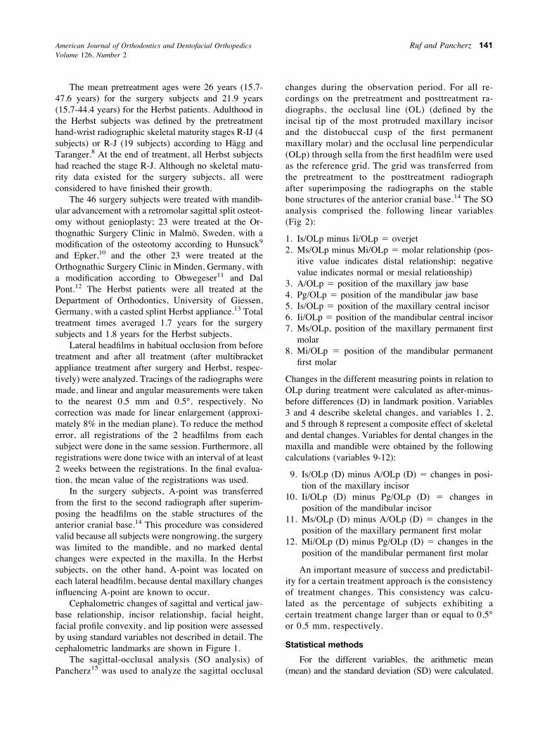
Table V. Maximum individual cephalometric treatment changes and consistency of treatment changes (%) in 46adult Class II Division 1 subjects treated with orthognathic surgery (mandibular sagittal split osteotomy) followedby multibracket appliance and 23 adult Class II Division 1 subjects treated with Herbst appliance followed bymultibracket appliance
Variable
Treatment changes (after-before)
Surgery Herbst
Maximum Consistency % Maximum Consistency %
Incisor relation Overjet (mm) %11.75 98 %12.25 100Overbite (mm) %9.50 98 %6.25 96
Molar relation* (mm) %16.00 93 %6.25 100Sagittal jaw relation SNB (°) 5.25 91 2.25 74
ANB (°) %5.25 98 %3.00 74Vertical jaw relation ML/NSL (°) 11.75 91 %2.75 56Facial height Spa-Gn # 100/N-Gn (index) 4.50 87 1.85 52
Spp-Go$ # 100/S-Go$ (index) %11.50 78 3.13 65Profile convexity NAPg (°) 12.00 91 4.75 70
NS/Sn/PgS (°) 12.50 96 7.50 96NS/No/PgS (°) 9.75 76 4.00 83
*minus (%) implies normalization
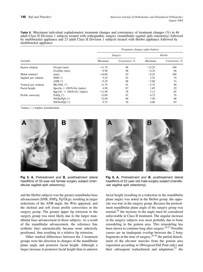
Fig 5. A, Pretreatment and B, posttreatment lateralheadfilms of 33-year-old female surgery subject (man-dibular sagittal split osteotomy).
Fig 6. A, Pretreatment and B, posttreatment lateralheadfilms of 22-year-old male surgery subject (mandib-ular sagittal split osteotomy).
American Journal of Orthodontics and Dentofacial OrthopedicsAugust 2004
148 Ruf and Pancherz
general postsurgical adaptive processes of all softtissues, tendons, and muscles that have been directly orindirectly affected by the surgical jaw displace-ment,27,30,31 and the possible condylar resorption thathas been reported rather frequently in orthognathicsurgery patients,17,27 especially in those with a pretreat-ment internal derangement of the TMJ.32
The direction of maxillary molar movements dif-fered between the 2 treatment groups. Although thedistal movement of the maxillary molar in the Herbstgroup was to be expected from the headgear effect ofthe appliance,33 the mesial movement of the maxillarymolars in the surgery group was unexpected because allsubjects were treated nonextraction. Even if decreasesin maxillary and mandibular arch lengths in adulthoodoccur,34 the amounts of change in the surgery subjectswere larger than those normally reported to occur over1 decade. A possible explanation for the mesial move-ment of the maxillary molars is a transverse maxillaryexpansion most likely performed in most of the patientsduring the presurgical orthodontic phase. The spacegained by this expansion might have been reciprocallyclosed, leading to the observed retrusion of the maxil-lary incisors (1.4 mm) and the mesial movement of themaxillary molars (0.7 mm).
The consistency in treatment reaction was larger forthe surgery group than for the Herbst group for vari-ables directly or indirectly affected by the amount ofmandibular advancement (SNB, ANB, skeletal profileconvexity). On the other hand, the reduction in softtissue profile convexity excluding the nose was thesame (96%) in both groups, whereas, for the reductionof the soft tissue profile convexity including the nose,the consistency was larger in the Herbst subjects.Furthermore, for the Class II corrective variables (over-jet, overbite, molar relationship), no marked group
differences were detected. Therefore, the predictabilityof the treatment outcome in terms of consistency ofchanges was, on average, comparable for both groups.This agrees with the findings of Tulloch et al,35 whoconcluded that the success rate of overjet reduction wasonly slightly higher for orthodontic than for surgicaltreatment irrespective of age and malocclusion severity.
Thus, the question arises which is the best treatmentmodality for a borderline Class II adult. Even when theknowledge from this study is added to what is knownfrom the literature, there seems to be no single, con-clusive answer to the question. Several factors must beconsidered in the treatment decision process: (1) thereason the patient is seeking treatment; (2) the effectsthat can be provided by Herbst treatment and ortho-gnathic surgery, respectively; and (3) the costs andrisks of the 2 treatment approaches.
There is agreement in the literature that the mainreasons for adults seeking treatment are dental andfacial esthetics as well as stomatognathic or functionalimprovement.36-38 Patients with severe skeletal Class IImalocclusions are more motivated to undergo ortho-dontic than surgical treatment.38 This is not surprisingbecause most people prefer the least invasive measureto solve their problems. Interestingly, the type oftreatment selected (surgery or orthodontics) dependsmainly on the subject’s self-perception of his or herfacial profile and is not associated with the degree ofdentoskeletal discrepancy. This means that the moredissatisfied the patients are with their facial esthetics,the more likely they are to choose surgery.39,40 How-ever, after treatment, surgical and orthodontic patientswere equally satisfied with their profile changes.17,20
In our study, larger reductions in profile convexitywere found in the surgery group than in the Herbstgroup. In contrast, Shell and Woods41 found that,
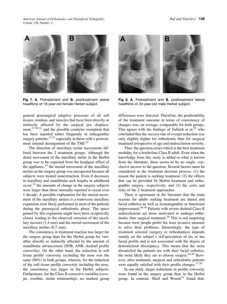
Fig 7. A, Pretreatment and B, posttreatment lateralheadfilms of 19-year-old female Herbst subject.
Fig 8. A, Pretreatment and B, posttreatment lateralheadfilms of 20-year-old male Herbst subject.
American Journal of Orthodontics and Dentofacial OrthopedicsVolume 126, Number 2
Ruf and Pancherz 149

regardless of whether Class II patients were treatedwith growth modification during adolescence or or-thognathic surgery during adulthood, facial estheticsimproved to a similar extent. Furthermore, the reduc-tion in facial profile convexity achieved by Herbsttreatment seems not to be age-dependent.4
Even if the occlusion can be corrected very suc-cessfully by adult Herbst treatment, chin projection andthus facial esthetics might be not be optimal aftertherapy. If, however, chin prominence is the mainproblem for the patient, advancement genioplasty of-fers a less costy, less risky alternative to enhance facialesthetics than a mandibular sagittal split osteotomy.42,43
Even though stomatognathic or functional improve-ment is the second most frequent reason for adults toseek treatment, little is known about the changes inmasticatory function after orthognathic surgery. ForClass III patients, scientific data show no significantimprovement in postoperative masticatory function.44For Class II patients, on the other hand, at least to ourknowledge, no such data exist.
Although there is controversy about the effect oforthognathic surgery on TMJ function, recent data45seem to support the view that patients with preexistingarticular disc displacements undergoing mandibularadvancement surgery are likely to have a significantworsening of the TMJ dysfunction problem postsur-gery. No comparable data exist for adult Herbst treat-ment. However, in a group of Herbst subjects thatincluded 8 of the present adults, TMJ function wasfound to improve during treatment.46
When looking at the costs of combined orthodon-tic-orthognathic surgery treatment, 60%-75% are due tothe surgical part.47-49 Therefore, a remarkable costreduction in adult Class II treatment can be achievedwith the Herbst appliance instead of orthognathic sur-gery.
The most common surgical risk of mandibularadvancement is neurosensory disturbances of the lowerlip that affect about 50% of the subjects.50 Addition-ally, nonunion or mal-union of the bony fragments, badsplits,51 and condylar resorption17,32 are frequent com-plications. Even if neurosensory disturbances of the lipoccur after genioplasty alone, the prevalence is signif-icantly lower than with mandibular sagittal split or acombination of sagittal split and genioplasty.52
A main complication in orthodontics is root resorp-tion. The amount of root resorption has been found tocorrelate with the amount of overjet reduction andhorizontal tooth movement.53 Furthermore, extensivepalatal root torque, which is likely to be applied duringorthodontic Class II treatment, has been shown to be apredisposing factor for root resorption of the mandib-
ular incisors.54 Thus, when comparing surgery anddentofacial orthopedics (Herbst appliance), the risksassociated with surgery are obviously much greater.Finally, the failure rate of surgical Class II treatment ishigher than that for a dentofacial orthopedic/orthodon-tic approach.35 Therefore, the important question intreatment planning is whether the greater improvementin facial esthetics accomplished by orthognathic sur-gery compared with dentofacial orthopedics with theHerbst appliance is worth the increased costs and risksof the surgical approach.
CONCLUSIONS
The Herbst appliance is a powerful tool for nonsur-gical, nonextraction treatment of adult Class II maloc-clusions. Thus, the treatment approach can be consid-ered as an alternative to orthognathic surgery inborderline skeletal Class II subjects. For Class IIcorrection, the success rate and predictability of Herbsttreatment is as high as for orthognathic surgery. If,however, the patient’s main wish is a greatly improvedfacial profile, orthognathic surgery is the better treat-ment alternative.
We thank the Orthognathic Surgery Department inMalmo, Sweden, and Drs Witschel and Wrede in BadOeynhausen, Germany, for providing access to theclinical records and the lateral headfilms of the surgerysubjects.
REFERENCES
1. Pancherz H. Dentofacial orthopedics or orthognathic surgery: isit a matter of age? Am J Orthod Dentofacial Orthop 2000;117:571-4.
2. Pancherz H, Ruf S. The Herbst appliance—research basedupdated clinical possibilities. World Orthod J 2000;1:17-31.
3. Ruf S, Pancherz H. Kiefergelenkwachstumsadaptation bei jun-gen Erwachsenen wahrend Behandlung mit der Herbst-Appara-tur. Eine prospektive magnetresonanztomographische undkephalometrische Studie. Inf Orthod Kieferorthop 1998;30:735-50.
4. Ruf S, Pancherz H. Dentoskeletal effects and facial profilechanges in young adults treated with the Herbst appliance. AngleOrthod 1999;69:239-46.
5. Ruf S, Pancherz H. Temporomandibular joint remodeling inadolescents and young adults during Herbst treatment: a prospec-tive longitudinal magnetic resonance imaging and cephalometricradiographic investigation. Am J Orthod Dentofacial Orthop1999;115:607-18.
6. Ruf S, Pancherz H. When is the ideal period for Herbsttherapy—early or late? Semin Orthod 2003;9:47-56.
7. McNamara JA, Peterson JE, Pancherz H. Histologic changesassociated with the Herbst appliance in adult rhesus monkeys(Macacca mulatta). Semin Orthod 2003;9:26-40.
8. Hagg U, Taranger J. Skeletal stages of the hand and wrist asindicators of the pubertal growth spurt. Acta Odontol Scand1980;38:187-200.
American Journal of Orthodontics and Dentofacial OrthopedicsAugust 2004
150 Ruf and Pancherz

9. Hunsuck EE. A modified intraoral sagittal splinting technique forcorrection of mandibular prognathism. J Oral Surg 1968;26:250-3.
10. Epker BN. Modifications in the sagittal osteotomy of the man-dible. J Oral Surg 1977;35:157-9.
11. Obwegeser H. The surgical correction of mandibular progna-thism and retrognathia with consideration of genioplasty. OralSurg 1957;10:677-89.
12. Dal Pont G. Retromolar osteotomy for the correction of progn-athism. J Oral Surg 1961;19:42-7.
13. Pancherz H. The Herbst appliance. Sevilla, Spain: EditorialAguiram; 1995.
14. Bjork A, Skieller V. Normal and abnormal growth of themandible. A synthesis of longitudinal cephalometric implantstudies over a period of 25 years. Eur J Orthod 1983;5:1-46.
15. Pancherz H. The mechanism of Class II correction in Herbstappliance treatment. A cephalometric investigation. Am J Orthod1982;82:104-13.
16. Dahlberg G. Statistical methods for medical and biologicalstudents. New York: Interscience Publications; 1940.
17. Cassidy DW, Herbosa EG, Rotskoff KS, Johnston LE. Acomparison of surgery and orthodontics in “borderline” adultswith Class II Division 1 malocclusions. Am J Orthod DentofacialOrthop 1993;104:455-70.
18. Gerzanic L, Jagsch R, Watzke IM. Psychologic implications oforthognathic surgery in patients with skeletal Class II or Class IIImalocclusion. Int J Adult Orthod Orthognath Surg 2002;17:75-81.
19. Lawrence TN, Ellis E, McNamara JA. The frequency anddistribution of skeletal and dental components in Class IIorthognathic surgery patients. J Oral Maxillofac Surg 1985;43:24-34.
20. Mihalik CA, Proffit WR, Phillips C. Long-term follow-up ofClass II adults treated with orthodontic camouflage: a compari-son with orthgnathic surgery outcomes. Am J Orthod DentofacialOrthop 2003;123:266-78.
21. Proffit WR, Phillips C, Douvartzidis N. A comparison ofoutcomes of orthodontic and surgical-orthodontic treatment ofClass II malocclusion in adults. Am J Orthod Dentofacial Orthop1992;101:556-65.
22. Hoppenreijs TJ, Hakman EC, van’t Hof MA, Stoelinga PJ,Tuinzing DB, Freihofer HP. Psychologic implications of surgicalorthodontic treatment in patients with anterior open bite. Int JAdult Orthod Orthognath Surg 1999;14:101-12.
23. Hirzel HG, Grewe JM. Activators: a practical approach. Am JOrthod 1974;66:557-70.
24. Skieller V, Bjork A, Linde Hansen T. Prediction of mandibulargrowth rotation evaluated from a longitudinal implant sample.Am J Orthod 1984;86:359-70.
25. Proffit WR, Phillips C, Tulloch JFC, Medland PH. Surgicalversus orthodontic correction of skeletal Class II malocclusion inadolescents: effects and indications. Int J Adult Orthod Orthog-nath Surg 1992;7:209-20.
26. Bathia SN, Leighton BC. A manual of facial growth. A computeranalysis of longitudinal growth data. Oxford: Oxford UniversityPress; 1993.
27. Schubert P, Bailey LTJ, White RP, Proffit WR. Long-termcephalometric changes in untreated adults compared to thosetreated with orthognathic surgery. Int J Adult Orthod OrthognathSurg 1999;14:91-9.
28. Kohn MW. Analysis of relapse after mandibular advancementsurgery. J Oral Surg 1978;9:676-84.
29. La Blanc JP, Turvey T, Epker BN, Hill C. Results following
simultaneous mobilization of the maxilla and mandible for thecorrection of dentofacial deformities. Oral Surg Oral Med OralPathol 1982;54:607-12.
30. Epker BN, Wessberg G. Mechanisms of early skeletal relapsefollowing surgical advancement of the mandible. Br J Oral Surg1982;20:175-82.
31. Turvey T, Phillips C, Laytown HS, Proffit WR. Simultaneoussuperior repositioning of the maxilla and mandibular advance-ment. A report on stability. Am J Orthod Dentofacial Orthop1988;94:372-83.
32. Schellhas KP, Piper MA, Bessette RW, Wilkes CH. Mandibularretrusion, temporomandibular joint derangement, and orthog-nathic surgery planning. Plast Reconstr Surg 1992;90:218-29.
33. Pancherz H, Anehus Pancherz M. The headgear effect of theHerbst appliance: a cephalometric long-term study. Am J OrthodDentofacial Orthop 1993;103:510-20.
34. Akgul AA, Toygar TU. Natural craniofacial changes in the thirddecade of life: a longitudinal study. Am J Orthod DentofacialOrthop 2002;122:512-22.
35. Tulloch JFC, Lenz BE, Phillips C. Surgical versus orthodonticcorrection for Class II patients: age and severity in treatmentplanning and treatment outcome. Semin Orthod 1999;5:231-40.
36. Flanary CM, Barnwell GM, Alexander JM. Patients’ perceptionsof orthognathic surgery. Am J Orthod 1985;88:137-45.
37. Mayo KH, Dryland-Vig KWL, Vig PS, Kowalski CJ. Attitudevariables of dentofacial deformity patients: demographic charac-teristics and associations. J Oral Maxillofac Surg 1991;49:594-602.
38. Wilmot JJ, Barber HD, Chou DG, Vig KWL. Associationsbetween severity of dentofacial deformity and motivation fororthodontic-orthognathic surgery treatment. Angle Orthod 1993;63:283-8.
39. Bell R, Kiyak HA, Joondeph DR, McNeill RW, Wallen TR.Perceptions of facial profile and their influence on the decision toundergo orthognathic surgery. Am J Orthod 1985;88:323-32.
40. Kiyak HA, McNeill RW, West RA, Hohl T, Heaton PJ. Person-ality characteristics as predictors and sequale of surgical andconventional orthodontics. Am J Orthod 1986;89:383-92.
41. Shell TL, Woods WG. Perception of facial esthetics: a compar-ison of similar Class II cases treated with attempted growthmodification or later orthognathic surgery. Angle Orthod 2003;73:365-73.
42. Brons R. Chin corrections. In: Brons R, editor. Facial harmony.Standards for orthognathic surgery and orthodontics. London:Quintessence; 1998. 145-58.
43. Proffit WR, Turvey T, Moriarty JD. Augmentation genioplasty asan adjunct to conservative orthodontic treatment. Am J Orthod1981;79:473-91.
44. Shiratsuchi Y, Kouno K, Tashiro H. Evaluation of masticatoryfunction following orthognathic surgical correction of mandibu-lar prognathism. J Cranio Max Fac Surg 1991;19:299-303.
45. Wolford LM, Reiche-Fischel O, Pushkar M. Changes in tem-poromandibular joint dysfunction after orthognathic surgery.J Oral Maxillofac Surg 2003;61:655-60.
46. Ruf S, Pancherz H. Does bite-jumping damage the TMJ? Aprospective longitudinal clinical and MRI study of Herbst pa-tients. Angle Orthod 2000;70:183-99.
47. Dolan P, White RP, Tulloch JFC. An analysis of hospital chargesfor orthognathic surgery. Int J Adult Orthod Orthognath Surg1987;1:9-14.
American Journal of Orthodontics and Dentofacial OrthopedicsVolume 126, Number 2
Ruf and Pancherz 151

48. Panula K, Keski-Nisula L, Keski-Nisula K, Oikarinen K, Keski-Nisula S. Costs of surgical-orthodontic treatment in communityhospital care: an analysis of the different phases of treatment. IntJ Adult Orthod Orthognath Surg 2002;17:297-306.
49. Thomas PM. Orthodontic camouflage versus orthognathic sur-gery in the treatment of mandibular deficiency. J Oral MaxillofacSurg 1995;53:579-87.
50. Kiyak HA, Bell R. Psychosocial considerations in surgery andorthodontics. In: Proffit WR, White RP, editors. Surgical-ortho-dontic treatment. Saint Louis: Mosby; 1990. 79-80.
51. Panula K, Oikarinen K, Finne K. Incidence of complications and
problems related to orthognathic surgery: a review of 655patients. J Oral Maxillofac Surg 2001;10:1128-37.
52. Gianni AB, Biglioli F, Bozzetti A, Brusati R. Neurosensoryalterations of the inferior alveolar and mental nerve after genio-plasty alone or associated with sagittal osteotomy of the man-dibular ramus. J Craniomaxillofac Surg 2002;30:295-303.
53. Sameshima GT, Sinclair PM. Predicting and preventing rootresorption. I. Diagnostic factors. Am J Orthod DentofacialOrthop 2001;119:505-10.
54. Kaley J, Phillips C. Factors related to root resorption in theorthodontic practice. Angle Orthod 1991;61:125-32.
American Journal of Orthodontics and Dentofacial OrthopedicsAugust 2004
152 Ruf and Pancherz