Embed Size (px)
Citation preview
Original Article
Percutaneous Occlusion of Left Atrial Appendage with the Amplatzer Cardiac Plug™ in Atrial FibrillationMárcio José Montenegro1, Edgard Freitas Quintella1, Aníbal Damonte2, Hugo de Castro Sabino1, Ricardo Zajdenverg1, Gustavo Pinaud Laufer1, Bernardo Amorim1, André Pereira Duque Estrada1, Cristian Paul Yugcha Armas1, Aline Sterque1
Instituto Estadual de Cardiologia Aloysio de Castro1, Rio de Janeiro, Brasil; Instituto Cardiovascular de Rosário2, Rosário, Argentina
AbstractBackground: Atrial fibrillation is associated with embolic strokes that often result in death or disability. Effective in reducing these events, anticoagulation has several limitations and has been widely underutilized. Over 90% of thrombi identified in patients with atrial fibrillation without valvular disease originate in the left atrial appendage, whose occlusion is investigated as an alternative to anticoagulation.
Objective: To determine the feasibility of percutaneous occlusion of the left atrial appendage in patients at high risk of embolic events and limitations to the use of anticoagulation.
Methods: We report our initial experience with Amplatzer Cardiac PlugTM (St. Jude Medical Inc., Saint Paul, USA) in patients with nonvalvular atrial fibrillation. We selected patients at high risk of thromboembolism, major bleeding, and contraindications to the use or major instability in response to oral anticoagulant. The procedures were performed percutaneously under general anesthesia and transesophageal echocardiography. The primary outcome was the presence of periprocedural complications and follow-up program included clinical and echocardiographic review within 30 days and by telephone contact after nine months.
Results: In five selected patients it was possible to occlude the left atrial appendage without periprocedural complications. There were no clinical events in follow-up.
Conclusion: Controlled clinical trials are needed before percutaneous closure of the left atrial appendage should be considered an alternative to anticoagulation in atrial fibrillation not associated with valvular disease. But the device has shown to be promissory in patients at high risk of embolism and restrictions on the use of anticoagulants. (Arq Bras Cardiol 2012;98(2):143-150)
Keywords: Atrial fibrillation; atrial appendage; stroke; prostheses and implants; thromboembolism.
Mailing Address: Márcio José Montenegro • Serviço de Hemodinâmica e Cardiologia Intervencionista. Instituto Estadual de Cardiologia Aloysio de Castro. Rua David Campista, 326 - 4º andar - Humaitá - 22.261-010 - Rio de Janeiro, RJ, Brasil E-mail: [email protected] Manuscript received August 16, 2011, revised mansucript received September 12, 2011, accepted September 12, 2011.
of stroke is as common in the paroxysmal AF as in persistent or permanent AF, being twice higher than the population in general7. It is estimated that approximately 20% of strokes are associated with AF2,3,8, and in this population, they often result in death or permanent9disability. Multiple conditions may be associated8 and different mechanisms can cause stroke in the presence of AF10. But it is believed that in most cases thromboembolic events occur, because thrombi in the left atrium have been widely documented in autopsies, surgeries and echocardiographic studies11-13 in which its detection is an independent predictor of ischemic attack. Although it has proven to be effective in reducing ischemic events in this context1,2,14, anticoagulation with vitamin K inhibitors has been widely underused1,2,15. Several factors contribute to this, but it stands out the narrow therapeutic window of these agents16 and the risk of bleeding complications16-18, particularly in older patients who would most benefit from this therapy15,18. The alternative agents under study are also associated to bleeding16,19. Since more than 90% of thrombi identified in patients with nonvalvular AF originate in the left atrial appendage (LAA)1,2,11,12, its occlusion emerged as an alternative to anticoagulation20-28. We report our initial experience in percutaneous occlusion of the LAA in patients with nonvalvular AF with the Amplatzer Cardiac PlugTM29.
IntroductionStroke (CVA) is one of the most feared complications of
cardiovascular diseases1,2. The Framingham Heart Study showed that the isolated presence of coronary artery disease (CAD), high blood pressure (hypertension) or heart failure (HF) increased the incidence of stroke adjusted for age at a rate of more than two, three or four times, respectively, while the presence of atrial fibrillation (AF) not associated with valvular diseases increased the risk fivefold. With age, the effects of the presence of CAD, hypertension and heart failure were attenuated, but the impact of AF were not3. Then considered an important risk factor for ischemic attack; AF is the most common sustained cardiac arrhythmia in clinical practice4,5, its incidence increases with age5 and its prevalence has been growing also6. The occurrence
143
Original Article
Arq Bras Cardiol 2012;98(2):143-150
Montenegro et alPercutaneous occlusion of left atrial appendage
MethodsIn November 2010, patients with nonvalvular AF,
permanent or long duration persistent, who use and present limitations to the use of oral anticoagulation (OAC), were selected to intervention. This is a retrospective account of this early experience.
Selection of patients
The selection criteria were:1. High risk of thromboembolic events, with a history
of ischemic attack, transient ischemic attacks, peripheral embolism, or CHADS2 score ≥ 21,2,8.
2. Objective evidence limiting the use of OAC, with a history of hemorrhagic stroke, major bleeding, great lability of the response to anticoagulant therapy, or high risk of bleeding with OAC. We considered major bleeding as bleeding episodes requiring hospitalization or transfusion30. Great lability of the therapeutic response to OAC was defined as less than 60% of the records of prothrombin time (PT)within the therapeutic range in the last year (INR ≤ 2.0 or ≥ 3.0)30, or when very high in more than one occasion (INR ≥ 4.0)31,32. The time interval of one year to review the records of PT, as well as the need for very high INR in more than one occasion were set arbitrarily. High risk of bleeding with OAC was defined as HAS-BLED score ≥ 32,30. Alcohol abuse was considered only when it influenced this score calculation and the risk of recurrent falls was disregarded, since its importance as a contraindication for OAC has been underestimated2.
3. Should be excluded from the study at initial assessment the patients presenting with a clinical situation that contraindicated intervention or echocardiographic evidence of intracardiac thrombus, valvular disease with risk of embolism, ulcerated atherosclerotic plaque in the aorta, or significant obstruction of the carotid or vertebral arteries. Inadequate anatomy was also a factor of exclusion, because the manufacturer recommends that the neck of the LAA has a minimum diameter of 12 mm and a maximum of 28 mm.
4. It was necessary the informed consent of patients and relatives involved.
DeviceThe AmplatzerTM Cardiac Plug (ACP - St. Jude Medical
Inc., Saint Paul, USA) was developed specifically for the LAA occlusion by percutaneous mean through transseptal puncture
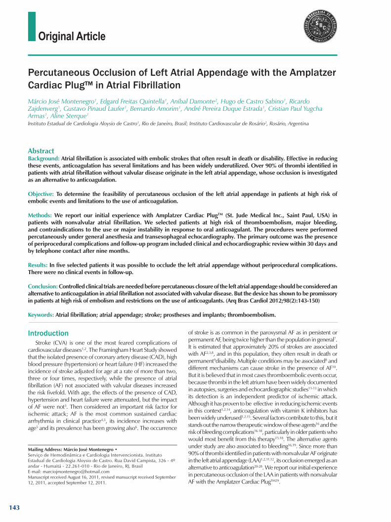
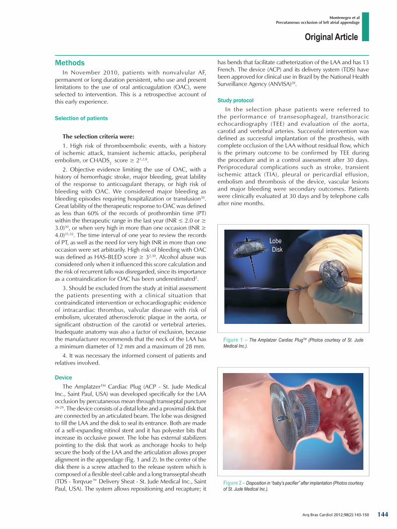
26-29. The device consists of a distal lobe and a proximal disk that are connected by an articulated beam. The lobe was designed to fill the LAA and the disk to seal its entrance. Both are made of a self-expanding nitinol stent and it has polyester bits that increase its occlusive power. The lobe has external stabilizers pointing to the disk that work as anchorage hooks to help secure the body of the LAA and the articulation allows proper alignment in the appendage (Fig. 1 and 2). In the center of the disk there is a screw attached to the release system which is composed of a flexible steel cable and a long transseptal sheath (TDS - Torqvue™ Delivery Sheat - St. Jude Medical Inc., Saint Paul, USA). The system allows repositioning and recapture; it
has bends that facilitate catheterization of the LAA and has 13 French. The device (ACP) and its delivery system (TDS) have been approved for clinical use in Brazil by the National Health Surveillance Agency (ANVISA)28.
Study protocolIn the selection phase patients were referred to
the performance of transesophageal, transthoracic echocardiography (TEE) and evaluation of the aorta, carotid and vertebral arteries. Successful intervention was defined as successful implantation of the prosthesis, with complete occlusion of the LAA without residual flow, which is the primary outcome to be confirmed by TEE during the procedure and in a control assessment after 30 days. Periprocedural complications such as stroke, transient ischemic attack (TIA), pleural or pericardial effusion, embolism and thrombosis of the device, vascular lesions and major bleeding were secondary outcomes. Patients were clinically evaluated at 30 days and by telephone calls after nine months.
Figure 2 – Disposition in “baby’s pacifier” after implantation (Photos courtesy of St. Jude Medical Inc.).
Figure 1 – The Amplatzer Cardiac PlugTM (Photos courtesy of St. Jude Medical Inc.).
144
Original Article
Arq Bras Cardiol 2012;98(2):143-150
Montenegro et alPercutaneous occlusion of left atrial appendage
ProcedurePercutaneous interventions were scheduled under general
anesthesia and intra-procedural TEE. We planned extubation in cathlab and anesthesia recovery in the Coronary Care Unit. The manufacturer has released an animation that illustrates the procedure. It can be accessed at (http://www.arquivosonline.com.br/2012/video/video.asp).
Results
Baseline CharacteristicsWe studied five patients with mean age of 72.8 (62-78)
years. Table 1 shows baseline clinical characteristics. All patients were in regular use of OAC and showed great lability in response to therapy. One patient had INR = 15.0 at the moment of hospitalization and another patient presented lower gastrointestinal bleeding by angiodysplasia of the colon during hospitalization. Two patients were using concomitant aspirin. Out of the four patients with a history of cerebral ischemia, three suffered during OAC. One patient had had a leg amputated in a probable embolic phenomenon. The three patients with a history of HF had had myocardial infarction and two of them presented with preserved ejection fraction. Two patients were already in a prolonged hospitalization due to HF. No patient was excluded from the study. Table 2 shows the profile of these
high-risk patients, either for embolic events (CHADS2 ≥ 2, CHA2DS2-VASC ≥ 2)8,33 as for bleeding with the OAC (HAS-BLED ≥ 3)30. But the HAS-BLED score of this group could have been a little better, if not for the fact that from the five patients with hypertension, four had systolic BP ≥ 160 mmHg.
Percutaneous InterventionThe OAC was discontinued two to five days before
the procedure, starting heparin administration (5000 IU SC 12/12h)34 and Dual Antiplatelet therapy (DAPT) with acetylsalicylic acid (ASA - 200 mg daily) and clopidogrel (initial dose of 300mg and maintenance of 75mg/day)27. At the beginning of the procedure, endocarditis prophylaxis was performed with injection of cefazolin (1 g IV 8/8h - 3 doses)35. Vascular access was performed by femoral puncture. The arterial access was intended to monitor blood pressure and to positioning of a Pigtail catheter in the aortic valve for anatomical reference. The transseptal puncture was performed by counter-lateral venous access, with echocardiographic monitoring and the usual techniques (Mullins sheath and Brockenbrough needle). Pericardial drainage and autotransfusion equipment were available at hands. After transseptal puncture, heparin was administered (5000 to 10000 IU IV), with no monitoring of activated coagulation time. A millimeter Pigtail catheter was placed in LAA being performed measurements of the diameters
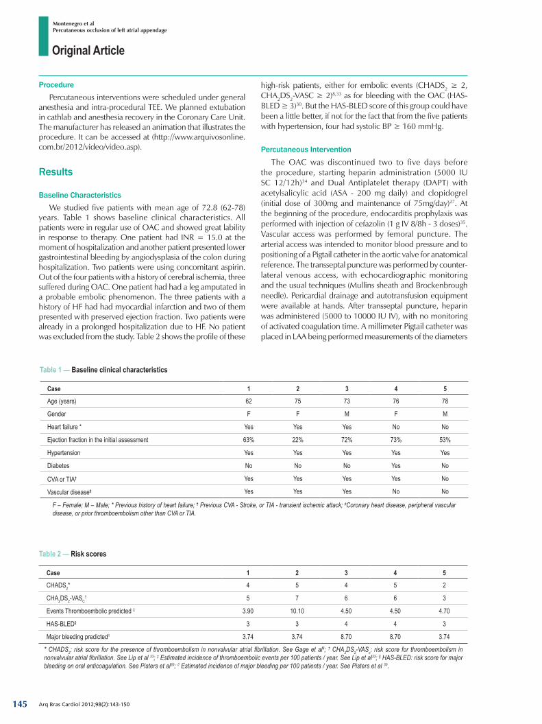
Table 1 — Baseline clinical characteristics
Case 1 2 3 4 5
Age (years) 62 75 73 76 78
Gender F F M F M
Heart failure * Yes Yes Yes No No
Ejection fraction in the initial assessment 63% 22% 72% 73% 53%
Hypertension Yes Yes Yes Yes Yes
Diabetes No No No Yes No
CVA or TIA† Yes Yes Yes Yes No
Vascular disease‡ Yes Yes Yes No No
F – Female; M – Male; * Previous history of heart failure; † Previous CVA - Stroke, or TIA - transient ischemic attack; ‡Coronary heart disease, peripheral vascular disease, or prior thromboembolism other than CVA or TIA.
Table 2 — Risk scores
Case 1 2 3 4 5
CHADS2* 4 5 4 5 2
CHA2DS2-VASC† 5 7 6 6 3
Events Thromboembolic predicted ‡ 3.90 10.10 4.50 4.50 4.70
HAS-BLED§ 3 3 4 4 3
Major bleeding predicted// 3.74 3.74 8.70 8.70 3.74
* CHADS2: risk score for the presence of thromboembolism in nonvalvular atrial fibrillation. See Gage et al8; † CHA2DS2-VASc: risk score for thromboembolism in nonvalvular atrial fibrillation. See Lip et al 33; ‡ Estimated incidence of thromboembolic events per 100 patients / year. See Lip et al33; § HAS-BLED: risk score for major bleeding on oral anticoagulation. See Pisters et al30; // Estimated incidence of major bleeding per 100 patients / year. See Pisters et al 30.
145
Original Article
Arq Bras Cardiol 2012;98(2):143-150
Montenegro et alPercutaneous occlusion of left atrial appendage
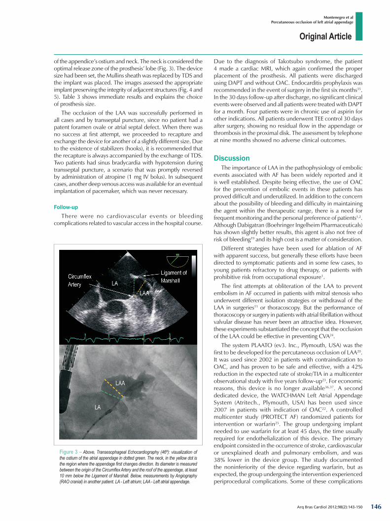
of the appendice’s ostium and neck. The neck is considered the optimal release zone of the prosthesis’ lobe (Fig. 3). The device size had been set, the Mullins sheath was replaced by TDS and the implant was placed. The images assessed the appropriate implant preserving the integrity of adjacent structures (Fig. 4 and 5). Table 3 shows immediate results and explains the choice of prosthesis size.
The occlusion of the LAA was successfully performed in all cases and by transseptal puncture, since no patient had a patent foramen ovale or atrial septal defect. When there was no success at first attempt, we proceeded to recapture and exchange the device for another of a slightly different size. Due to the existence of stabilizers (hooks), it is recommended that the recapture is always accompanied by the exchange of TDS. Two patients had sinus bradycardia with hypotension during transseptal puncture, a scenario that was promptly reversed by administration of atropine (1 mg IV bolus). In subsequent cases, another deep venous access was available for an eventual implantation of pacemaker, which was never necessary.
Follow-upThere were no cardiovascular events or bleeding
complications related to vascular access in the hospital course.
Due to the diagnosis of Takotsubo syndrome, the patient 4 made a cardiac MRI, which again confirmed the proper placement of the prosthesis. All patients were discharged using DAPT and without OAC. Endocarditis prophylaxis was recommended in the event of surgery in the first six months35. In the 30 days follow-up after discharge, no significant clinical events were observed and all patients were treated with DAPT for a month. Four patients were in chronic use of aspirin for other indications. All patients underwent TEE control 30 days after surgery, showing no residual flow in the appendage or thrombosis in the proximal disk. The assessment by telephone at nine months showed no adverse clinical outcomes.
DiscussionThe importance of LAA in the pathophysiology of embolic
events associated with AF has been widely reported and it is well established. Despite being effective, the use of OAC for the prevention of embolic events in these patients has proved difficult and underutilized. In addition to the concern about the possibility of bleeding and difficulty in maintaining the agent within the therapeutic range, there is a need for frequent monitoring and the personal preference of patients1,2. Although Dabigatran (Boehringer Ingelheim Pharmaceuticals) has shown slightly better results, this agent is also not free of risk of bleeding19 and its high cost is a matter of consideration.
Different strategies have been used for ablation of AF with apparent success, but generally these efforts have been directed to symptomatic patients and in some few cases, to young patients refractory to drug therapy, or patients with prohibitive risk from occupational exposure1.
The first attempts at obliteration of the LAA to prevent embolism in AF occurred in patients with mitral stenosis who underwent different isolation strategies or withdrawal of the LAA in surgeries11 or thoracoscopy. But the performance of thoracoscopy or surgery in patients with atrial fibrillation without valvular disease has never been an attractive idea. However, these experiments substantiated the concept that the occlusion of the LAA could be effective in preventing CVA24.
The system PLAATO (ev3. Inc., Plymouth, USA) was the first to be developed for the percutaneous occlusion of LAA20. It was used since 2002 in patients with contraindication to OAC, and has proven to be safe and effective, with a 42% reduction in the expected rate of stroke/TIA in a multicenter observational study with five years follow-up23. For economic reasons, this device is no longer available36,37. A second dedicated device, the WATCHMAN Left Atrial Appendage System (Atritech., Plymouth, USA) has been used since 2007 in patients with indication of OAC22. A controlled multicenter study (PROTECT AF) randomized patients for intervention or warfarin25. The group undergoing implant needed to use warfarin for at least 45 days, the time usually required for endothelialization of this device. The primary endpoint consisted in the occurrence of stroke, cardiovascular or unexplained death and pulmonary embolism, and was 38% lower in the device group. The study documented the noninferiority of the device regarding warfarin, but as expected, the group undergoing the intervention experienced periprocedural complications. Some of these complications
Figure 3 – Above, Transesophageal Echocardiography (46º): visualization of the ostium of the atrial appendage in dotted green. The neck, in the yellow dot is the region where the appendage first changes direction. Its diameter is measured between the origin of the Circumflex Artery and the roof of the appendage, at least 10 mm below the Ligament of Marshall. Below, measurements by Angiography (RAO cranial) in another patient. LA - Left atrium; LAA - Left atrial appendage.
146
Original Article
Arq Bras Cardiol 2012;98(2):143-150
Montenegro et alPercutaneous occlusion of left atrial appendage
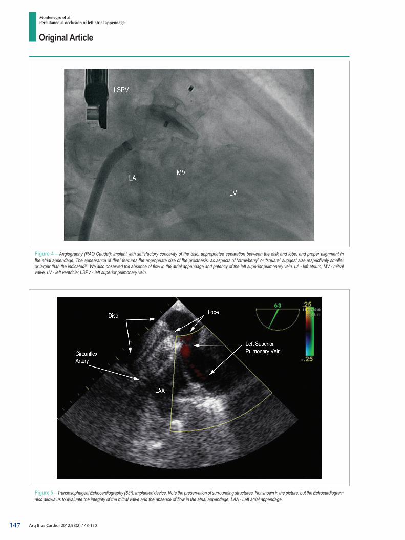
Figure 4 – Angiography (RAO Caudal): implant with satisfactory concavity of the disc, appropriated separation between the disk and lobe, and proper alignment in the atrial appendage. The appearance of “tire” features the appropriate size of the prosthesis, as aspects of “strawberry” or “square” suggest size respectively smaller or larger than the indicated28. We also observed the absence of flow in the atrial appendage and patency of the left superior pulmonary vein. LA - left atrium, MV - mitral valve, LV - left ventricle; LSPV - left superior pulmonary vein.
Figure 5 – Transesophageal Echocardiography (63º): Implanted device. Note the preservation of surrounding structures. Not shown in the picture, but the Echocardiogram also allows us to evaluate the integrity of the mitral valve and the absence of flow in the atrial appendage. LAA - Left atrial appendage.
147
Original Article
Arq Bras Cardiol 2012;98(2):143-150
Montenegro et alPercutaneous occlusion of left atrial appendage
led to changes in the device22 and others were related to the learning curve37. This device is not approved for clinical use in the United States, where the Food and Drug Administration (FDA) is awaiting the results of a continuous record of patients38. Part of these results was published, demonstrating the importance of the learning curve37.
Soon after the introduction of PLAATO, Meier et al21 reported percutaneous closure of the LAA with the use of AmplatzerTM devices traditionally used for closure of atrial septal defect. There were 16 cases, in which seven of them had a history of side effects or occupational exposure that prevents OAC usage. The other nine patients underwent implantation on their own volition. In 14 patients the procedure was performed under local anesthesia and in 11 patients there was no echocardiographic monitoring, being successful in all cases. Follow-up ranged from one to 12 months and there were no complications21. Despite this initial experiment, in a further series of 44 patients, three patients did not obtain the closure, one patient needed a new intervention and there were six events of embolization in the device35. Considering the fragility of the wall of the LAA, with approximately 1mm in thickness38, the manufacturer developed the ACP, a device designed for this intervention26-29.
In our initial experience, considering the proven effectiveness of the OAC, we were concerned on establishing inclusion criteria regards the risk of embolism and bleeding. The primary outcome of the LAA occlusion was performed in all cases and the device proved to be safe without periprocedural complications. Contrary to the experience of Jilaihawi and Kar39, though the echocardiographic measurements appeared relatively accurate in our study, we often oriented the choice of the prosthesis by angiographic measurements. But echocardiography guidance was valuable in monitoring the integrity of surrounding structures and the absence of flow in
the LAA, thereby ensuring the optimal implantation. We also believe that this method facilitates transseptal puncture and can expedite the detection of possible complications. The OAC was suspended days in advance to the intervention and device thrombosis was not found in the nine-month follow-up. Perhaps the use of DAPT could have been extended for up to three months40, but due to clinical and socioeconomic reasons it was not interesting in our population.
ConclusionsControlled clinical trials are needed before percutaneous
closure of the LAA becomes an alternative to OAC in patients with permanent or long-term persistent nonvalvular AF. But the device has shown to be promising in patients at high risk of embolism and limitations to the use of OAC. Although the importance of the learning curve cannot be underestimated35,37,38, the operators’ familiarity with the devices AmplatzerTM might be an advantage in the initial experience with this intervention28.
By the time of publication, there was no adverse endpoints on the 14 months follow-up.
Acknowledgements We would like to thank the cooperation of Dr. Claudio José
Gouvea Galhardo and the participation of the Nurse Wilson Pessanha, members of our clinical staff.
Potential Conflict of InterestDrs. Márcio José Montenegro and Aníbal Damonte provide
advice and supervision to St. Jude Medical Inc. cases and are paid for training sections
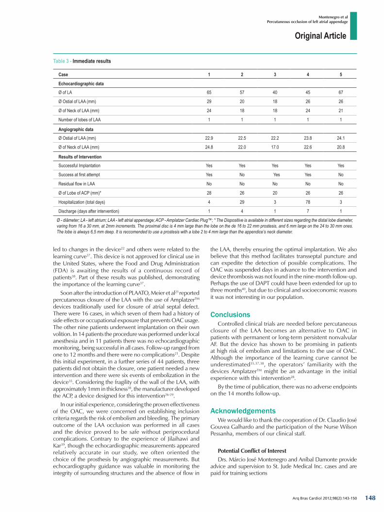
Table 3 - Immediate results
Case 1 2 3 4 5
Echocardiographic data
Ø of LA 65 57 40 45 67
Ø Ostial of LAA (mm) 29 20 18 26 26
Ø of Neck of LAA (mm) 24 18 18 24 21
Number of lobes of LAA 1 1 1 1 1
Angiographic data
Ø Ostial of LAA (mm) 22.9 22.5 22.2 23.8 24.1
Ø of Neck of LAA (mm) 24.8 22.0 17.0 22.6 20.8
Results of Intervention
Successful Implantation Yes Yes Yes Yes Yes
Success at first attempt Yes No Yes Yes No
Residual flow in LAA No No No No No
Ø of Lobe of ACP (mm)* 28 26 20 26 26
Hospitalization (total days) 4 29 3 78 3
Discharge (days after intervention) 1 4 1 7 1
Ø - diâmeter; LA - left atrium; LAA - left atrial appendage; ACP - Amplatzer Cardiac Plug™; * The Dispositive is available in different sizes regarding the distal lobe diameter, varing from 16 a 30 mm, at 2mm increments. The proximal disc is 4 mm large than the lobe on the de 16 to 22 mm prostesis, and 6 mm large on the 24 to 30 mm ones. The lobe is always 6,5 mm deep. It is reccomended to use a prostesis with a lobe 2 to 4 mm large than the appendice’s neck diameter.
148
Original Article
Arq Bras Cardiol 2012;98(2):143-150
Montenegro et alPercutaneous occlusion of left atrial appendage
References1. Zimerman LI, Fenelon G, Martinelli Filho M, Grupi C, Atié J, Lorga Filho A, et
al. / Sociedade Brasileira de Cardiologia. Diretrizes brasileiras de fibrilação atrial. Arq Bras Cardiol. 2009;92(6 supl 1):1-39.
2. European Society of Cardiology / European Heart Rhythm Association. ESC Guidelines for the management of atrial fibrillation. Eur Heart J. 2010;31(19):2369-429.
3. Wolf PA, Abbott RD, Kannel WB. Atrial fibrillation as an independent risk factor for stroke: the Framingham Heart Study. Stroke. 1991;22(8):983-8.
4. Lloyd-Jones DM, Wang TJ, Leip EP, Larson MG, Levy D, Vasan RS, et al. Lifetime risk for developing atrial fibrillation: the Framingham Heart Study. Circulation. 2004;110(9):1042-6.
5. Ruigomez A, Johansson S, Wallander MA, Rodriguez LA. Incidence of chronic atrial fibrillation in general practice and its treatment pattern. J Clin Epidemiol. 2002;55(4):358-63.
6. Miyasaka Y, Barnes ME, Gersh BJ, Cha SS, Bailey KR, Abhayaratna WP, et al. Secular trends in incidence of atrial fibrillation in Olmsted County, Minnesota, 1980 to 2000, and implications on the projections for future prevalence. Circulation. 2006;114(2):119-25.
7. Friberg L, Hammar N, Rosenqvist M. Stroke in paroxysmal atrial fibrillation: report from the Stockholm Cohort of Atrial Fibrilation. Eur Heart J. 2010;31(8):967-75.
8. Gage BF, Waterman AD, Shannon W, Boechler M, Rich MW, Radford MJ. Validation of clinical classification schemes for predicting stroke: results from the National Registry of Atrial Fibrillation. JAMA. 2001;285(22):2864-70.
9. Lamassa M, Di Carlo A, Pracucci G, Basile AM, Trefoloni G, Vanni P, et al. Characteristics, outcome, and care of stroke associated with atrial fibrillation in Europe: data from a multicenter multinational hospital-based registry (The European Community Stroke Project). Stroke. 2001;32(2):392-8.
10. Holmes DR Jr, Schwartz RS. Left atrial appendage occlusion eliminates the need for warfarin. Circulation. 2009;120(19):1919-26.
11. Blackshear JL, Odell JA. Appendage obliteration to reduce stroke in cardiac surgical patients with atrial fibrillation. Ann Thorac Surg. 1996;61(2):755-9.
12. Stoddard MF, Dawkins PR, Price CR, Ammash NM. Left atrial appendage thrombus is not uncommon in patients with acute atrial fibrillation and a recent embolic event: a transesophageal echocardiographic study. J Am Coll Cardiol. 1995;25(2):452-9.
13. Bernhardt P, Schmidt H, Hammerstingl C, Lüderitz B, Omran H. Patients with atrial fibrillation and dense spontaneous echo contrast at high risk: a prospective and serial follow-up over 12 months with transesophageal echocardiography and cerebral magnetic resonance imaging. J Am Coll Cardiol. 2005;45(11):1807-12.
14. Hart RG, Pearce LA, Aguilar MI. Meta-analysis: antithrombotic therapy to prevent stroke in patients who have nonvalvular atrial fibrillation. Ann Intern Med. 2007;146(12):857-67.
15. Bungard TJ, Ghali WA, Teo KK, McAlister FA, Tsuyuki RT. Why do patients with atrial fibrillation not receive warfarin? Arch Intern Med. 2000;160(1):41-6.
16. Connolly SJ, Eikelboom J, O’Donnell M, Pogue J, Yusuf S. Challenges of establishing new antithrombotic therapies in atrial fibrillation. Circulation. 2007;116(4):449-55.
17. Levine MN, Raskob G, Landefeld S, Kearon C. Hemorrhagic complications of anticoagulant treatment. Chest. 2001;119(Suppl I):108S-21S.
18. Hylek EM, Evans-Molina C, Shea C, Henault LE, Regan S. Major hemorrhage and tolerability of warfarin in the first year of therapy among elderly patients with atrial fibrillation. Circulation. 2007;115(21):2689-96.
19. Connolly SJ, Ezekowitz MD, Yusuf S, Eikelboom J, Oldgren J, Parekh A, et al. Dabigatran versus warfarin in patients with atrial fibrillation. N Engl J Med. 2009;361(12):1139-51.
20. Sievert H, Lesh MD, Trepels T, Omran H, Bartorelli A, Della Bella P, et al. Percutaneous left atrial appendage transcatheter occlusion to prevent stroke in high-risk patients with atrial fibrillation: early clinical experience. Circulation. 2002;105(16):1887-9.
21. Meier B, Palacios I, Windecker S, Rotter M, Cao Q, Keane D, et al. Transcatheter left atrial appendage occlusion with Amplatzer devices to obviate anticoagulation in patients with atrial fibrillation. Catheter Cardiovasc Interv. 2003;60(3):417-22.
22. Sick PB, Schuler G, Hauptmann KE, Grube E, Yakubov S, Turi ZG, et al. Initial worldwide experience with the WATCHMAN left atrial appendage system for stroke prevention in atrial fibrillation. J Am Coll Cardiol. 2007;49(13):1490-5.
23. Block PC, Burstein S, Casale PN, Kramer PH, Terstein P, Williams DO, et al. Percutaneous left atrial appendage occlusion for patients in atrial fibrillation suboptimal for Warfarin therapy. 5-Year results of the PLAATO (Percutaneous Left Atrial Appendage Transcatheter Occlusion) Study. JACC Cardiovasc Interv. 2009;2(7):594-600.
24. Ussia GP, Mulè M, Cammalleri V, Scarabelli M, Barbanti M, Immè S, et al. Percutaneous closure of left atrial appendage to prevent embolic events in high-risk patients with chronic atrial fibrillation. Catheter Cardiovasc Interv. 2009;74(2):217-22.
25. Holmes DR, Reddy VY, Turi ZG, Doshi SK, Sievert H, Buchbinder M, et al. Percutaneous closure of the left atrial appendage versus warfarin therapy for prevention of stroke in patients with atrial fibrillation: a randomized non-inferiority trial. Lancet. 2009;374(9689):534-42.
26. Rodés-Cabau J, Champagne J, Bernier M. Transcatheter closure of the left atrial appendage: initial experience with Amplatzer cardiac plug device. Catheter Cardiovasc Interv. 2010;76(2):186-92.
27. Park J, BethencourtA, Sievert H, Santoro G, Meier B, Walsh K, et al. Left atrial appendage closure with Amplatzer cardiac plug in atrial fibrillation: initial European experience. Catheter Cardiovasc Interv. 2011;77(5):700-6.
28. Armaganijan LV, Staico R, Pedra SF, Moreira DA, Braga SLN, Feres F, et al. Experiência inicial com o novo AmplatzerTM Cardiac Plug para oclusão percutânea do apêndice atrial esquerdo. Rev Bras Cardiol Invasiv. 2011;19(1):14-23.
29. Lam YY, Yip GW, Yu CM, Chan WW, Cheng BC, Yan BP, et al. Left atrial appendage closure with Amplatzer cardiac plug for stroke prevention in atrial fibrillation: initial Asia-Pacific experience. Catheter Cardiovasc Interv. 2011 May 3. [Epub ahead of print].
30. Pisters R, Lane DA, Nieuwlaat R, de Vos CB, Crijns HJ, Lip GY. A novel user-friendly score (HAS-BLED) to assess one-year risk of major bleeding in atrial fibrillation patients: the Euro Heart Survey. Chest. 2010;138(5):1093-100.
31. Hylek EM, Singer DE. Risk factors for intracranial hemorrhage in outpatients taking warfarin. Ann Intern Med. 1994;120(11):897-902.
32. Levine MN, Raskob G, Landefeld S, Kearon C. Hemorrhagic complications of anticoagulant treatment. Chest. 2001;119(1 Suppl):108S-21S.
Sources of Funding
There were no external funding sources for this study.
Study Association
This study is not associated with any post-graduation program.
149
Original Article
Arq Bras Cardiol 2012;98(2):143-150
Montenegro et alPercutaneous occlusion of left atrial appendage
33. Lip GY, Nieuwlaat R, Pisters R, Lane DA, Crijns HJ. Refining clinical risk stratification for predicting stroke and thromboembolism in atrial fibrillation using a novel risk factor-based approach: the Euro Heart Survey. Chest. 2010;137(2):263-72.
34. Douketis JD, Berger PB, Dunn AS, Jaffer AK, Spyropoulos AC, Becker RC, et al. The perioperative management of antithrombotic therapy: American College of Chest Physicians Evidence-Based Clinical Practice Guidelines (8th Edition). Chest. 2008;133(6 Suppl):299S-339S.
35. Cruz-Gonzalez I, Yan BP, Lam YY. Left atrial appendage exclusion: state of the art. Catheter Cardiovasc Interv. 2010;75(5):806-13.
36. Canadian Agency for Drugs and Technologies in Health. Health Technology Inquiry Service. Left atrial appendage occlusion: cost-effectiveness in a Canadian setting. Ottawa, Ont.; 2010. [Accessed on 2011 Aug 1]. Available from: http://books.scholarsportal.info/viewdoc.htm?
37. Reddy VY, Holmes D, Doshi SK, Neuzil P, Kar S. Safety of percutaneous left atrial appendage closure: results from the Watchman Left Atrial Appendage System for Embolic Protection in Patients With AF (PROTECT AF) clinical trial and the Continued Access Registry. Circulation. 2011;123(4):417-24.
38. Block PC. The LAA Occlusion Foxtrot: steps forward; steps back. Catheter Cardiovasc Interv. 2011;77(5):707-8.
39. Jilaihawi H, Kar S. Oclusão do apêndice atrial esquerdo: alternativa ao tratamento a longo prazo com varfarina em pacientes com fibrilação atrial. Rev Bras Cardiol Invasiv. 2011;19(1):9-10.
40. Cruz-Gonzalez I, Moreiras JM, García E. Thrombus formation after left atrial appendage exclusion using an amplatzer cardiac plug device. Catheter Cardiovasc Interv. 2011 Apr 26. [Epub ahead of print].
150