Embed Size (px)
Citation preview
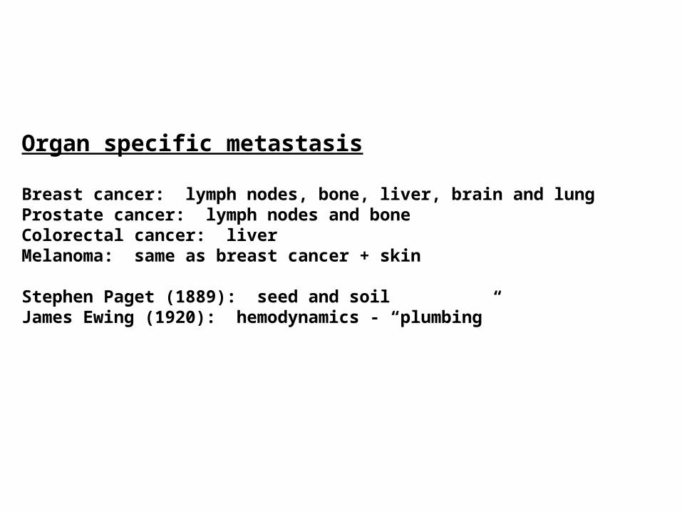
Organ specific metastasis
Breast cancer: lymph nodes, bone, liver, brain and lungProstate cancer: lymph nodes and boneColorectal cancer: liverMelanoma: same as breast cancer + skin
Stephen Paget (1889): seed and soilJames Ewing (1920): hemodynamics - “plumbing”
Possible Determinants of Site-Specific Metastasis
Hemodynamics (“Plumbing”)1. Blood flow patterns (How many cells are delivered to an organ?)2. Mechanics of cell arrest, growth and extravasation “Intravascular metastatic cancer cell homotypic aggregation at the sites of primary attachment to the endothelium.” (Glinsky and Quinn. Cancer Research. 63. July 2003).
Homing 1. Chemokines “Involvement of chemokine receptors in breast cancer metastasis.” (Mueller and Zlotnick. Nature. 410. March 2001). “Expression of CXC chemokine receptor-4 enhances the pulmonary metastatic potential of murine B16 melanoma cells.” (Murakami and Hwang. Cancer Research. 62. December 2002). “Multiple actions of the chemokine CXCL12 on epithelial tumor cells in human ovarian cancer.” (Scotton and Balkwill. Cancer Research 62. October 2002).
2. Selectins3. Vasculature specificity
Seed and Soil
1. Bone growth factors
“Metastasis to bone: causes, consequences and therapeutic opportunities.” (Mundy. Nature Reviews Cancer. 2. August 2002).
Possible Determinants of Site-Specific Metastasis
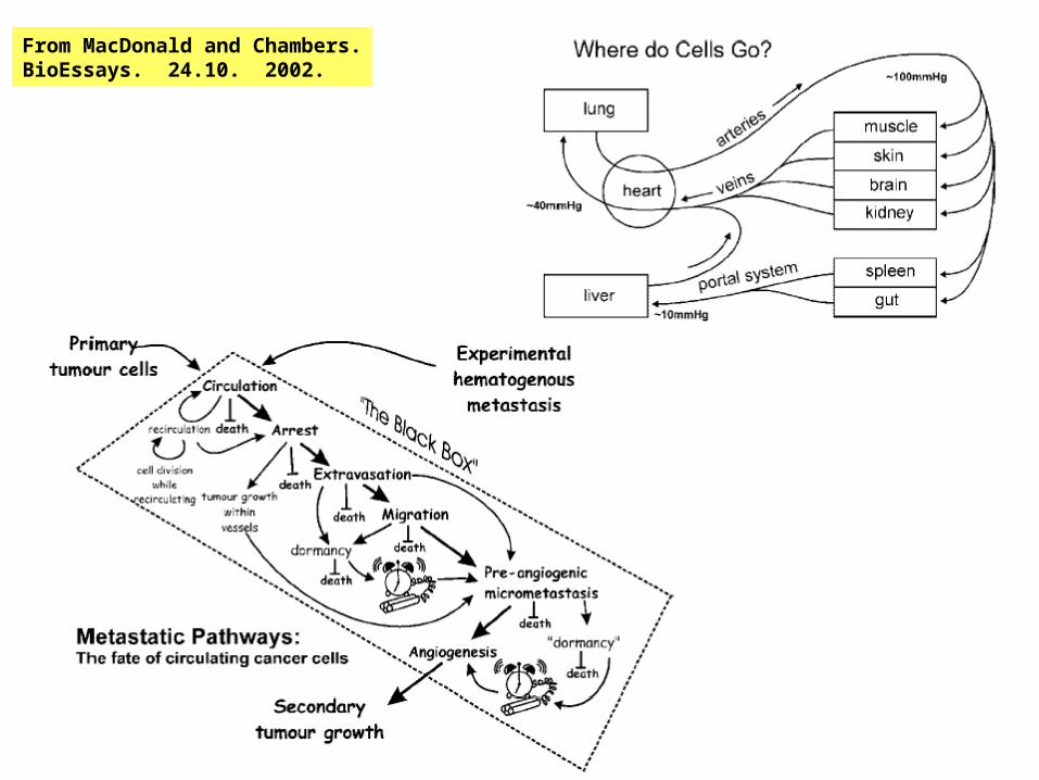
From MacDonald and Chambers.BioEssays. 24.10. 2002.
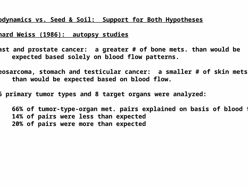
Hemodynamics vs. Seed & Soil: Support for Both Hypotheses
Leonard Weiss (1986): autopsy studies
Breast and prostate cancer: a greater # of bone mets. than would be expected based solely on blood flow patterns.
Osteosarcoma, stomach and testicular cancer: a smaller # of skin mets.than would be expected based on blood flow.
- 16 primary tumor types and 8 target organs were analyzed:
66% of tumor-type-organ met. pairs explained on basis of blood flow14% of pairs were less than expected 20% of pairs were more than expected
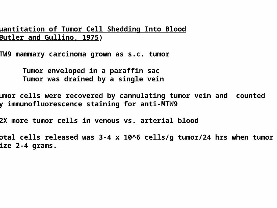
Quantitation of Tumor Cell Shedding Into Blood(Butler and Gullino, 1975)
MTW9 mammary carcinoma grown as s.c. tumor
Tumor enveloped in a paraffin sacTumor was drained by a single vein
Tumor cells were recovered by cannulating tumor vein and counted by immunofluorescence staining for anti-MTW9
12X more tumor cells in venous vs. arterial blood
Total cells released was 3-4 x 10^6 cells/g tumor/24 hrs when tumor wassize 2-4 grams.
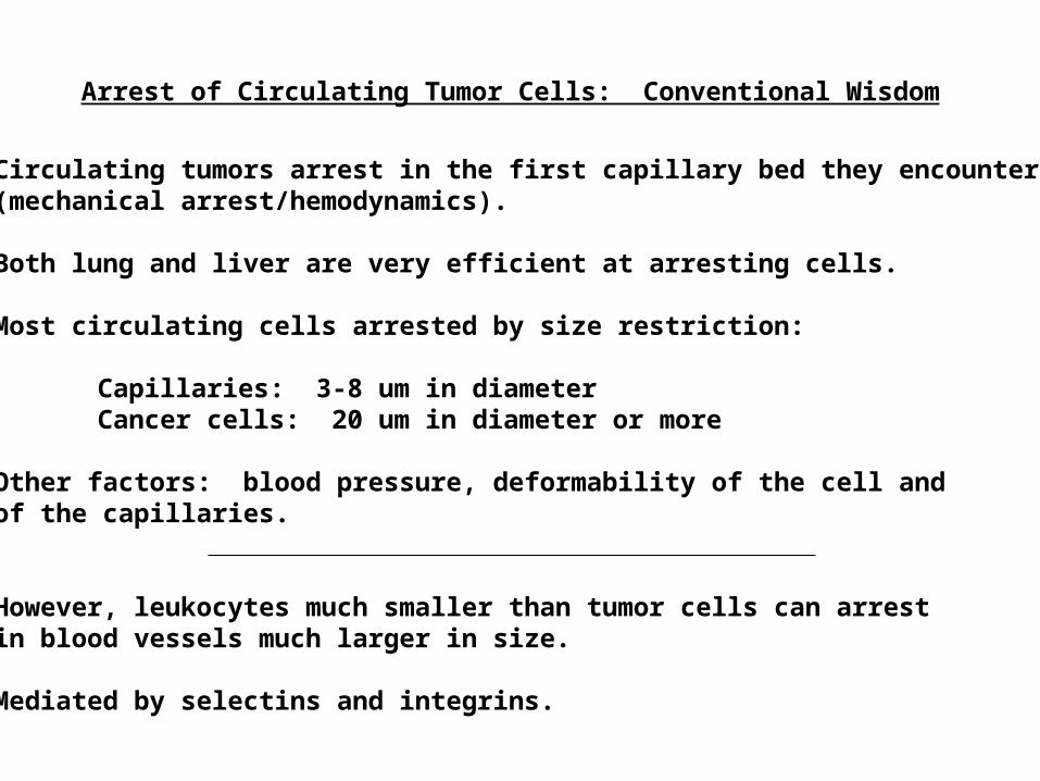
Circulating tumors arrest in the first capillary bed they encounter(mechanical arrest/hemodynamics).
Both lung and liver are very efficient at arresting cells.
Most circulating cells arrested by size restriction:
Capillaries: 3-8 um in diameterCancer cells: 20 um in diameter or more
Other factors: blood pressure, deformability of the cell andof the capillaries.
However, leukocytes much smaller than tumor cells can arrestin blood vessels much larger in size.
Mediated by selectins and integrins.
Arrest of Circulating Tumor Cells: Conventional Wisdom
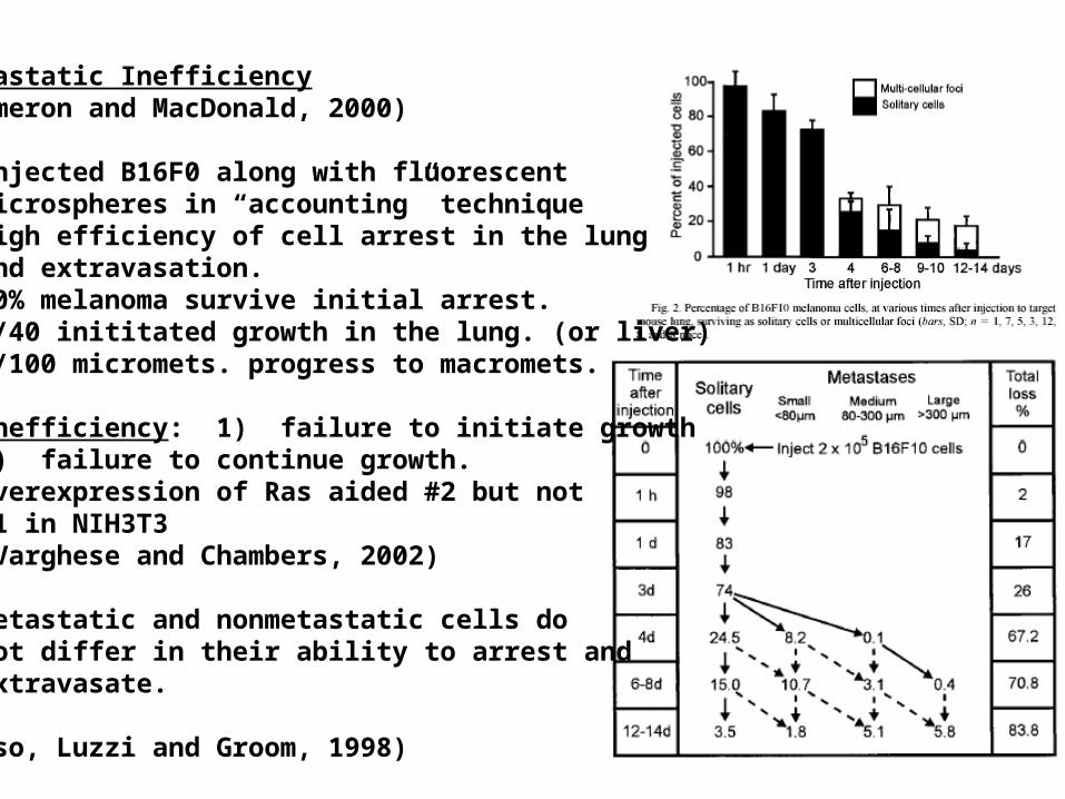
Metastatic Inefficiency(Cameron and MacDonald, 2000)
- Injected B16F0 along with fluorescent microspheres in “accounting” technique- High efficiency of cell arrest in the lung and extravasation.- 80% melanoma survive initial arrest.- 1/40 inititated growth in the lung. (or liver)- 1/100 micromets. progress to macromets.
- Inefficiency: 1) failure to initiate growth 2) failure to continue growth. Overexpression of Ras aided #2 but not #1 in NIH3T3 (Varghese and Chambers, 2002)
- Metastatic and nonmetastatic cells do not differ in their ability to arrest and extravasate.
(Also, Luzzi and Groom, 1998)
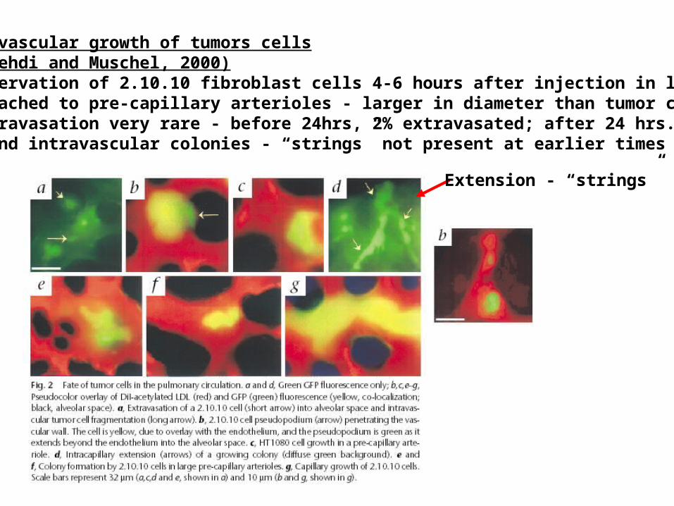
Intravascular growth of tumors cells(Al-Mehdi and Muschel, 2000)- Observation of 2.10.10 fibroblast cells 4-6 hours after injection in lung- Attached to pre-capillary arterioles - larger in diameter than tumor cell- Extravasation very rare - before 24hrs, 2% extravasated; after 24 hrs., 0%- Found intravascular colonies - “strings” not present at earlier times
Extension - “strings”
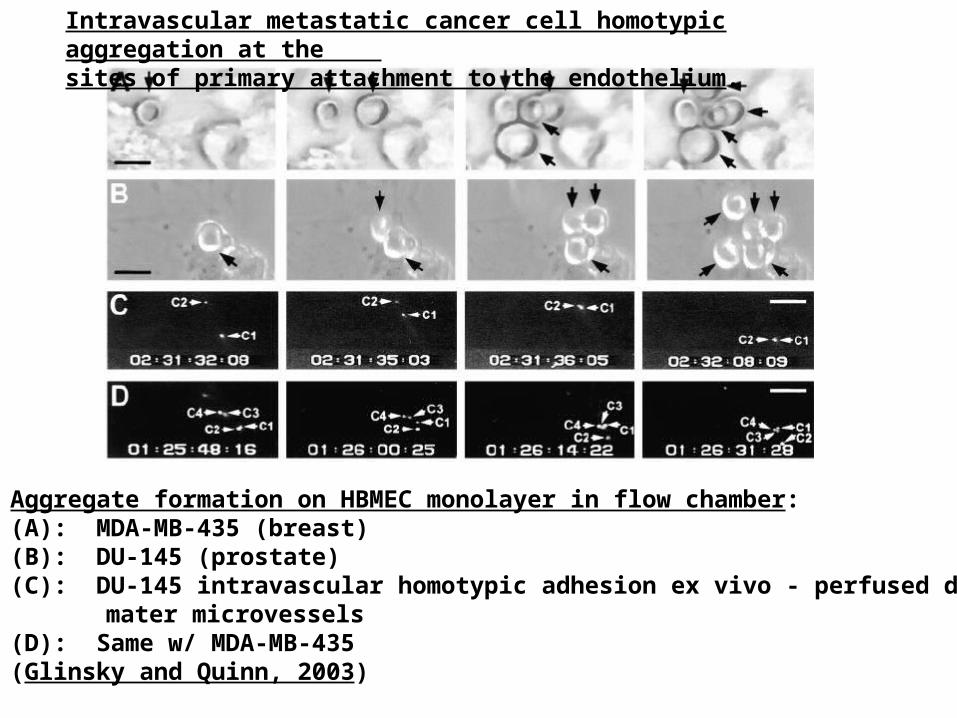
Aggregate formation on HBMEC monolayer in flow chamber:(A): MDA-MB-435 (breast)(B): DU-145 (prostate)(C): DU-145 intravascular homotypic adhesion ex vivo - perfused dura
mater microvessels(D): Same w/ MDA-MB-435(Glinsky and Quinn, 2003)
Intravascular metastatic cancer cell homotypic aggregation at the sites of primary attachment to the endothelium.
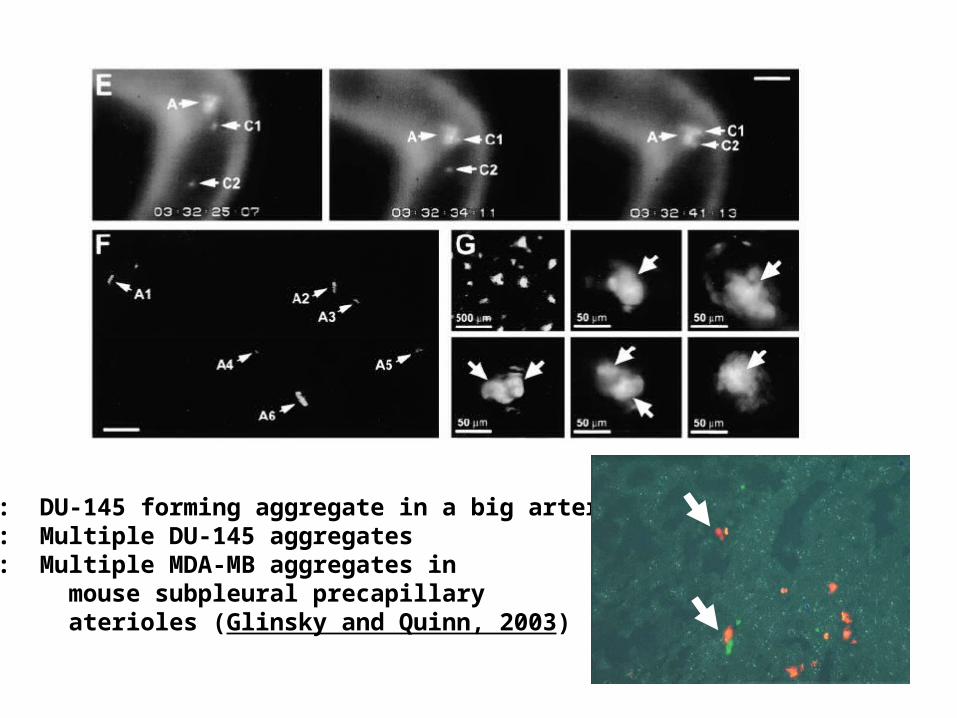
(E): DU-145 forming aggregate in a big arteriole(F): Multiple DU-145 aggregates(G): Multiple MDA-MB aggregates in mouse subpleural precapillary aterioles (Glinsky and Quinn, 2003)
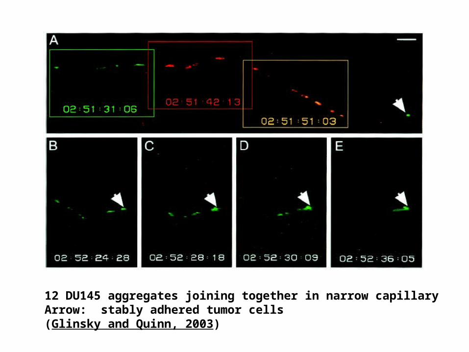
12 DU145 aggregates joining together in narrow capillaryArrow: stably adhered tumor cells(Glinsky and Quinn, 2003)
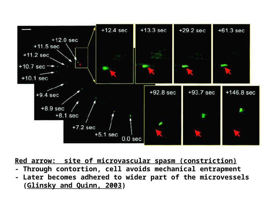
Red arrow: site of microvascular spasm (constriction)- Through contortion, cell avoids mechanical entrapment- Later becomes adhered to wider part of the microvessels (Glinsky and Quinn, 2003)
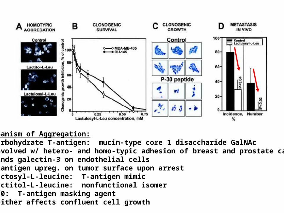
Mechanism of Aggregation:- Carbohydrate T-antigen: mucin-type core 1 disaccharide GalNAc- Involved w/ hetero- and homo-typic adhesion of breast and prostate canc.- Binds galectin-3 on endothelial cells- T-antigen upreg. on tumor surface upon arrest- Lactosyl-L-leucine: T-antigen mimic- Lactitol-L-leucine: nonfunctional isomer- P30: T-antigen masking agent- Neither affects confluent cell growth
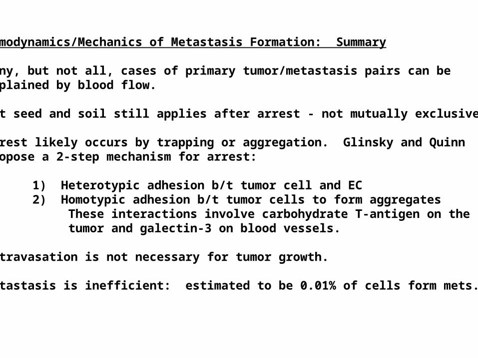
Hemodynamics/Mechanics of Metastasis Formation: Summary
Many, but not all, cases of primary tumor/metastasis pairs can be explained by blood flow.
But seed and soil still applies after arrest - not mutually exclusive.
Arrest likely occurs by trapping or aggregation. Glinsky and Quinnpropose a 2-step mechanism for arrest:
1) Heterotypic adhesion b/t tumor cell and EC2) Homotypic adhesion b/t tumor cells to form aggregates These interactions involve carbohydrate T-antigen on the tumor and galectin-3 on blood vessels.
Extravasation is not necessary for tumor growth.
Metastasis is inefficient: estimated to be 0.01% of cells form mets.
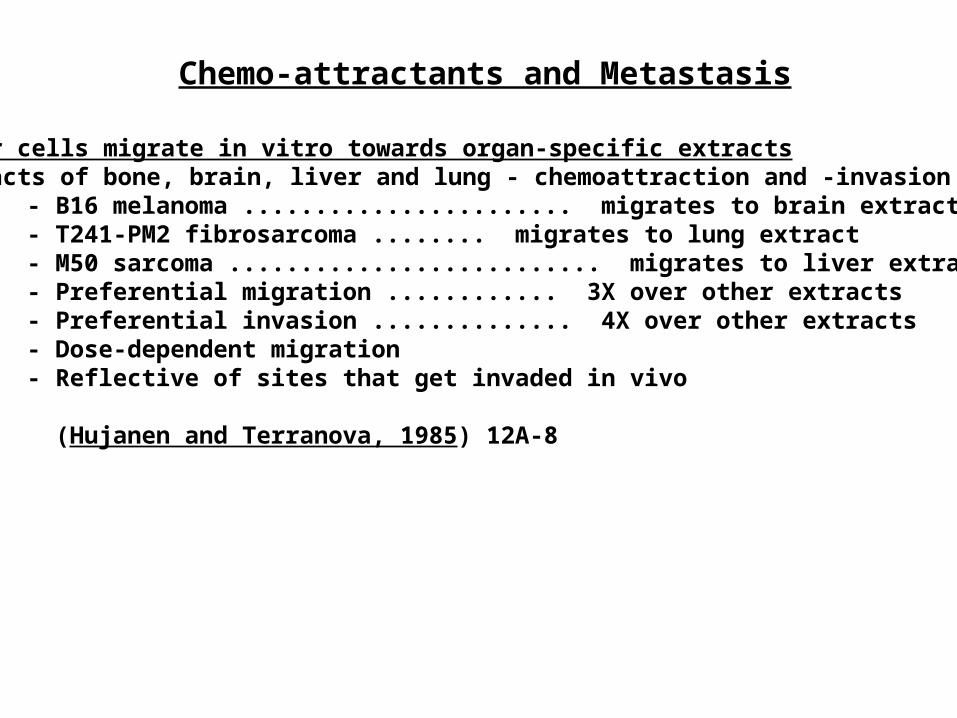
Chemo-attractants and Metastasis
Tumor cells migrate in vitro towards organ-specific extractsExtracts of bone, brain, liver and lung - chemoattraction and -invasion assays
- B16 melanoma ....................... migrates to brain extract- T241-PM2 fibrosarcoma ........ migrates to lung extract- M50 sarcoma .......................... migrates to liver extract- Preferential migration ............ 3X over other extracts- Preferential invasion .............. 4X over other extracts- Dose-dependent migration- Reflective of sites that get invaded in vivo
(Hujanen and Terranova, 1985) 12A-8
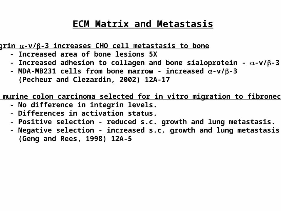
ECM Matrix and Metastasis
Integrin -v/-3 increases CHO cell metastasis to bone- Increased area of bone lesions 5X- Increased adhesion to collagen and bone sialoprotein - -v/-3 lig.- MDA-MB231 cells from bone marrow - increased -v/-3 (Pecheur and Clezardin, 2002) 12A-17
CT26 murine colon carcinoma selected for in vitro migration to fibronectin.- No difference in integrin levels.- Differences in activation status.- Positive selection - reduced s.c. growth and lung metastasis.- Negative selection - increased s.c. growth and lung metastasis. (Geng and Rees, 1998) 12A-5
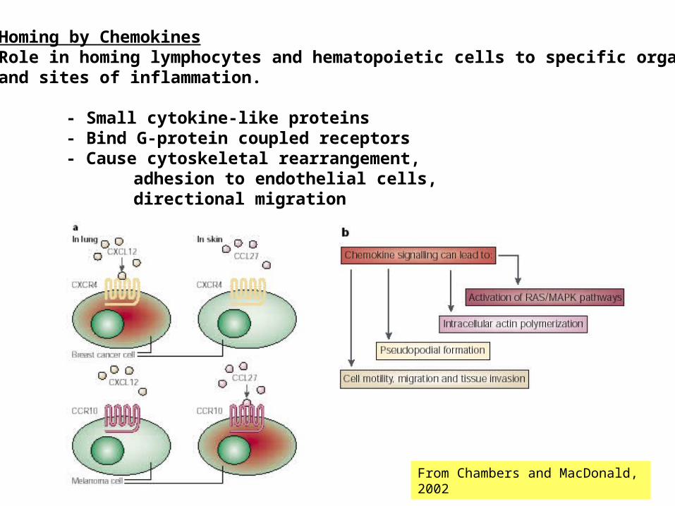
Homing by ChemokinesRole in homing lymphocytes and hematopoietic cells to specific organs and sites of inflammation.
- Small cytokine-like proteins- Bind G-protein coupled receptors- Cause cytoskeletal rearrangement,
adhesion to endothelial cells,directional migration
From Chambers and MacDonald, 2002

Normal Physiological Functions of Chemokines
- HSCs: Express chemokine receptor CXCR4. Ligand CXCL12 found in bone marrow - osteoblasts, stroma... KO chemokine/receptor: impaired stem cell migration f/ fetal liver to the bone marrow
- Dendritic Cells: Respond to and use ligands for CCR1/2/5/6/9 and CXCR4 to extravasate into stroma. Upon activation, they upregulate CCR7 - helps enter afferent lymphatics
- migrate to draining lymph nodes- entrance into T-zones of lymphoid tissues
- Other immune effects: - T-cell extravasation- CCL27/CCR10 and CCL17/CCR4 - skin homing- CCL25/CCR9 - transport of T cells into the gut
- CXCR12 (SDF-1) induces upreg. of cell adhesion receptors - integrinsVLA-4, VLA-5; LFA-1 - mediate HSC adhesion to BM-ECs andegress; also upreg. MMP-9.
- ELR+ chemokines are angiogenic; ELR- are anti-angiogenic.
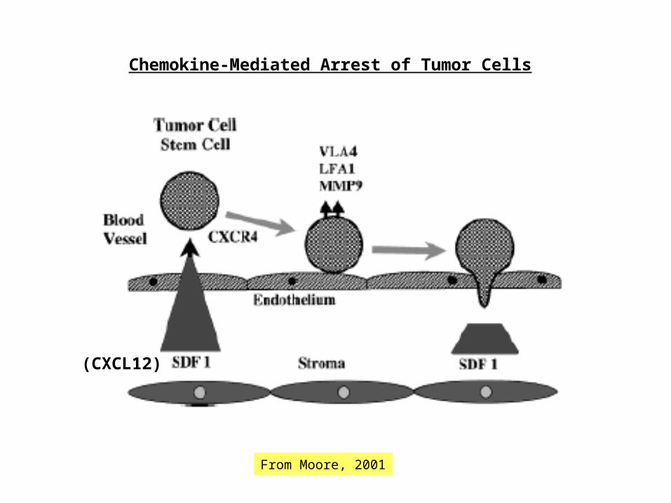
From Moore, 2001
(CXCL12)
Chemokine-Mediated Arrest of Tumor Cells
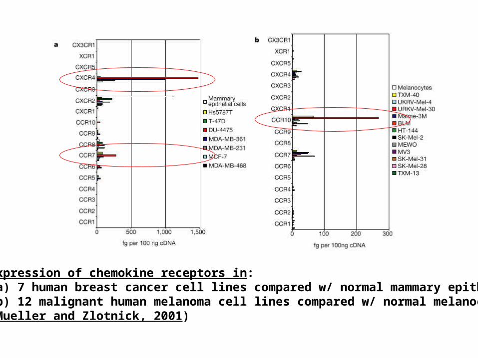
Expression of chemokine receptors in:(a) 7 human breast cancer cell lines compared w/ normal mammary epithelia(b) 12 malignant human melanoma cell lines compared w/ normal melanocytes(Mueller and Zlotnick, 2001)
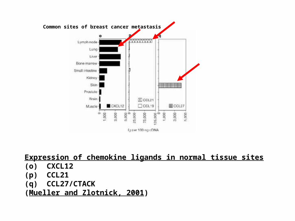
Expression of chemokine ligands in normal tissue sites(o) CXCL12(p) CCL21(q) CCL27/CTACK(Mueller and Zlotnick, 2001)
Common sites of breast cancer metastasis
Lung Liver
Bone Marrow Lymph Node Skin Muscle
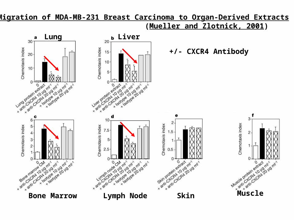
Migration of MDA-MB-231 Breast Carcinoma to Organ-Derived Extracts (Mueller and Zlotnick, 2001)
+/- CXCR4 Antibody
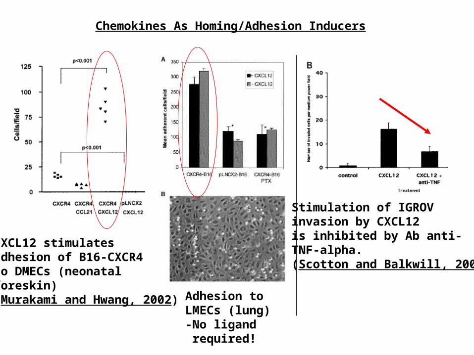
Chemokines As Homing/Adhesion Inducers
CXCL12 stimulates adhesion of B16-CXCR4to DMECs (neonatal foreskin) (Murakami and Hwang, 2002) Adhesion to
LMECs (lung)-No ligand required!
Stimulation of IGROVinvasion by CXCL12is inhibited by Ab anti-TNF-alpha.(Scotton and Balkwill, 2002)
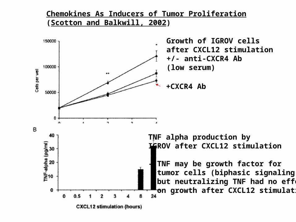
Chemokines As Inducers of Tumor Proliferation(Scotton and Balkwill, 2002)
Growth of IGROV cellsafter CXCL12 stimulation+/- anti-CXCR4 Ab(low serum)
+CXCR4 Ab
TNF alpha production byIGROV after CXCL12 stimulation
- TNF may be growth factor for tumor cells (biphasic signaling) - but neutralizing TNF had no effect on growth after CXCL12 stimulation
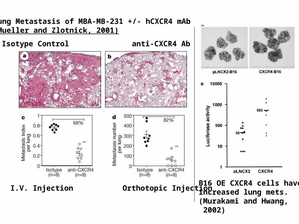
Lung Metastasis of MBA-MB-231 +/- hCXCR4 mAb(Mueller and Zlotnick, 2001)
I.V. Injection Orthotopic Injection
Isotype Control anti-CXCR4 Ab
B16 OE CXCR4 cells haveincreased lung mets.(Murakami and Hwang, 2002)
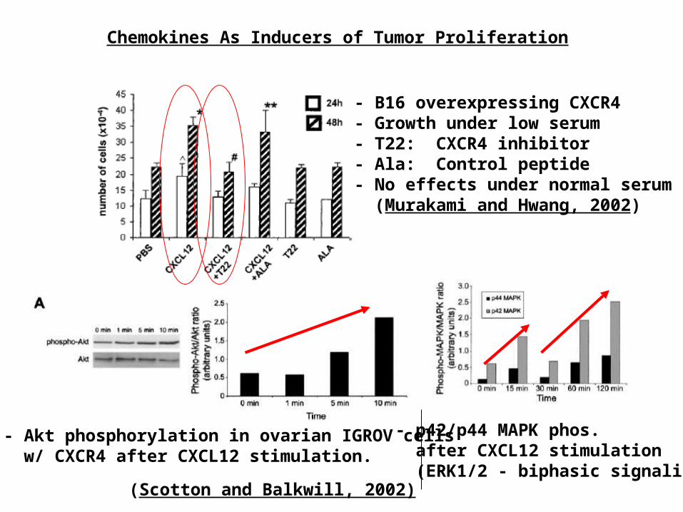
Chemokines As Inducers of Tumor Proliferation
- B16 overexpressing CXCR4- Growth under low serum- T22: CXCR4 inhibitor- Ala: Control peptide- No effects under normal serum (Murakami and Hwang, 2002)
- Akt phosphorylation in ovarian IGROV cells w/ CXCR4 after CXCL12 stimulation.
- p42/p44 MAPK phos. after CXCL12 stimulation (ERK1/2 - biphasic signaling)
(Scotton and Balkwill, 2002)
Chemokines and Metastasis: Summary
- MDA-MB-231 cells chemotactic to CXCL12/SDF-1 and CCL21/6Ckine.- Neutralizing CXCL12/CXCR4 interaction inhibits lung metastasis, 73-82%.- CCR10/CCL27 are overexpressed in melanoma, which metastasize to skin
May represent a skin-specific homing interactionAssociated with homing of memory T cells to skin
- Chemotaxis assays to organ-specific extracts could be inhibited by antibodies neutralizing chemokine interactions.
- The exact effect of chemokines is unclear and may involve many mechs.
Growth: signaling of CXCR4 to Akt/MAPK; indirectly via TNF-alpha; enhanced growth under low serum conditions
Adhesion: binding to endothelial cells (D/LMECs) in flow chamber
- Overall, overexpression of CXCR4 sufficient to increase lung mets.
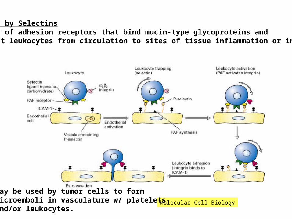
Homing by SelectinsFamily of adhesion receptors that bind mucin-type glycoproteins andrecruit leukocytes from circulation to sites of tissue inflammation or injury.
Molecular Cell Biology
May be used by tumor cells to formmicroemboli in vasculature w/ plateletsand/or leukocytes.
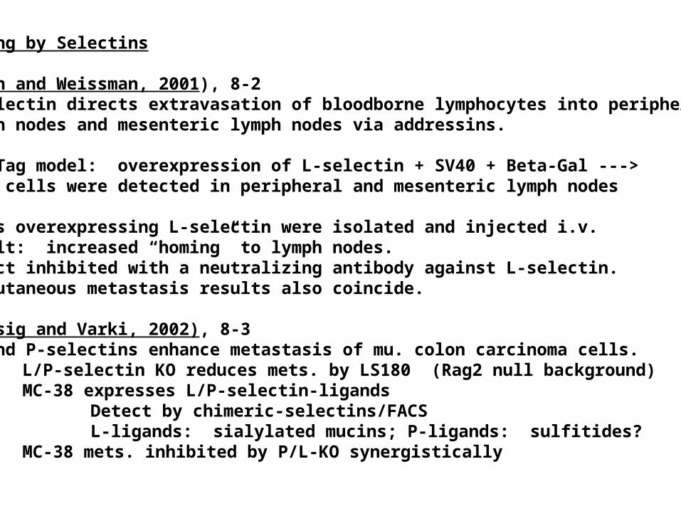
Homing by Selectins
(Qian and Weissman, 2001), 8-2L-selectin directs extravasation of bloodborne lymphocytes into peripherallymph nodes and mesenteric lymph nodes via addressins.
RIP-Tag model: overexpression of L-selectin + SV40 + Beta-Gal --->blue cells were detected in peripheral and mesenteric lymph nodes
Cells overexpressing L-selectin were isolated and injected i.v. Result: increased “homing” to lymph nodes.Effect inhibited with a neutralizing antibody against L-selectin.Subcutaneous metastasis results also coincide.
(Borsig and Varki, 2002), 8-3L- and P-selectins enhance metastasis of mu. colon carcinoma cells.
L/P-selectin KO reduces mets. by LS180 (Rag2 null background)MC-38 expresses L/P-selectin-ligands
Detect by chimeric-selectins/FACSL-ligands: sialylated mucins; P-ligands: sulfitides?
MC-38 mets. inhibited by P/L-KO synergistically
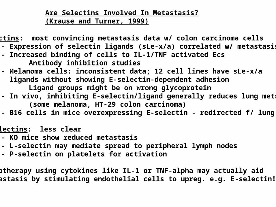
Are Selectins Involved In Metastasis?(Krause and Turner, 1999)
E-selectins: most convincing metastasis data w/ colon carcinoma cells- Expression of selectin ligands (sLe-x/a) correlated w/ metastasis- Increased binding of cells to IL-1/TNF activated Ecs
Antibody inhibition studies- Melanoma cells: inconsistent data; 12 cell lines have sLe-x/a ligands without showing E-selectin-dependent adhesion
Ligand groups might be on wrong glycoprotein- In vivo, inhibiting E-selectin/ligand generally reduces lung mets.
(some melanoma, HT-29 colon carcinoma)- B16 cells in mice overexpressing E-selectin - redirected f/ lung to liver
P/L-selectins: less clear- KO mice show reduced metastasis- L-selectin may mediate spread to peripheral lymph nodes- P-selectin on platelets for activation
- Chemotherapy using cytokines like IL-1 or TNF-alpha may actually aid metastasis by stimulating endothelial cells to upreg. e.g. E-selectin!
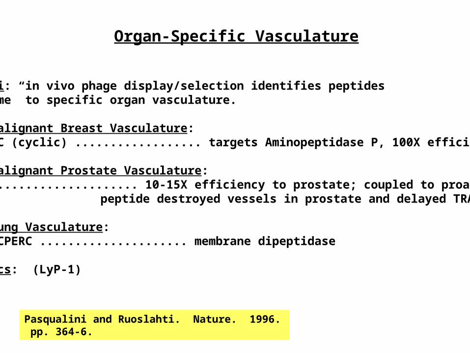
Organ-Specific Vasculature
Ruoslahti: in vivo phage display/selection identifies peptidesthat “home” to specific organ vasculature.
Normal/Malignant Breast Vasculature:CPGPEGAGC (cyclic) .................. targets Aminopeptidase P, 100X efficiency.
Normal/Malignant Prostate Vasculature:SMSIARL .................... 10-15X efficiency to prostate; coupled to proapoptotic
peptide destroyed vessels in prostate and delayed TRAMP
Normal Lung Vasculature:CGFECVRQCPERC ..................... membrane dipeptidase
Lymphatics: (LyP-1)
Pasqualini and Ruoslahti. Nature. 1996. pp. 364-6.
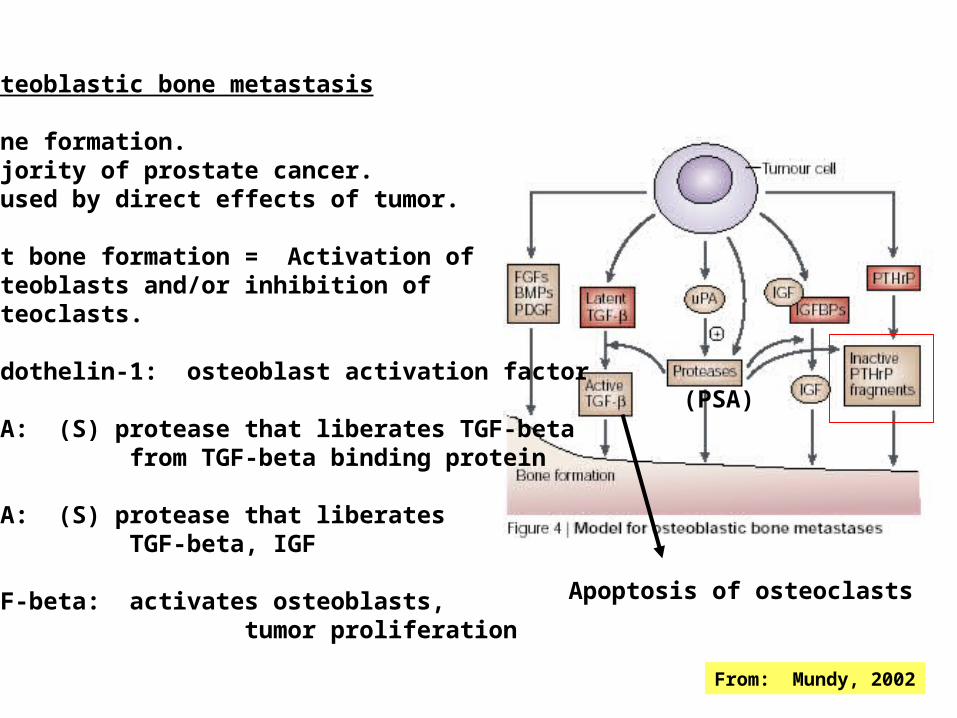
From: Mundy, 2002
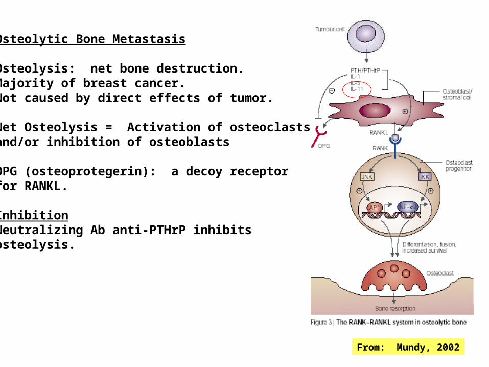
Osteolytic Bone Metastasis
Osteolysis: net bone destruction.Majority of breast cancer.Not caused by direct effects of tumor.
Net Osteolysis = Activation of osteoclastsand/or inhibition of osteoblasts
OPG (osteoprotegerin): a decoy receptorfor RANKL.
InhibitionNeutralizing Ab anti-PTHrP inhibitsosteolysis.
From: Mundy, 2002
Apoptosis of osteoclasts
(PSA)
Osteoblastic bone metastasis
Bone formation.Majority of prostate cancer.Caused by direct effects of tumor.
Net bone formation = Activation ofosteoblasts and/or inhibition ofosteoclasts.
Endothelin-1: osteoblast activation factor
uPA: (S) protease that liberates TGF-beta from TGF-beta binding protein
PSA: (S) protease that liberates TGF-beta, IGF
TGF-beta: activates osteoblasts, tumor proliferation

Vicious Cycle of Osteolytic Metastases
From: Mundy, 2002
Breast cancer cells that met. to bone with high efficiency secrete more PTHrP(Powell, 1991)
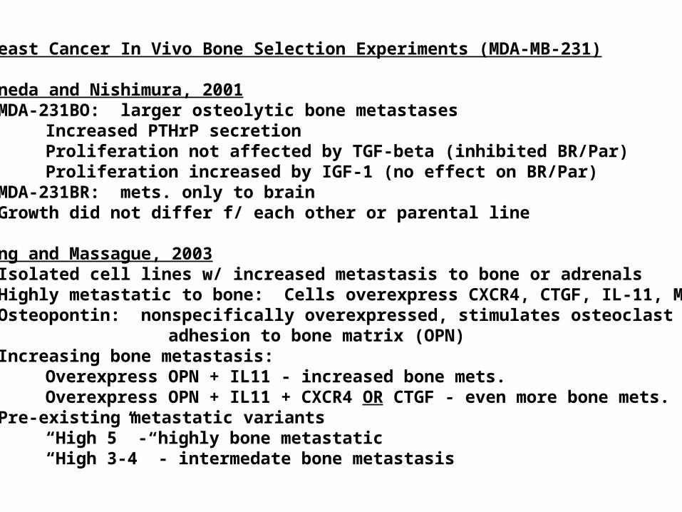
Breast Cancer In Vivo Bone Selection Experiments (MDA-MB-231)
Yoneda and Nishimura, 2001- MDA-231BO: larger osteolytic bone metastases
Increased PTHrP secretionProliferation not affected by TGF-beta (inhibited BR/Par)Proliferation increased by IGF-1 (no effect on BR/Par)
- MDA-231BR: mets. only to brain- Growth did not differ f/ each other or parental line
Kang and Massague, 2003- Isolated cell lines w/ increased metastasis to bone or adrenals- Highly metastatic to bone: Cells overexpress CXCR4, CTGF, IL-11, MMP1- Osteopontin: nonspecifically overexpressed, stimulates osteoclast
adhesion to bone matrix (OPN)- Increasing bone metastasis:
Overexpress OPN + IL11 - increased bone mets.Overexpress OPN + IL11 + CXCR4 OR CTGF - even more bone mets.
- Pre-existing metastatic variants“High 5” - highly bone metastatic“High 3-4” - intermedate bone metastasis