Embed Size (px)
Citation preview

OptotuneMicroscopy
Zurich, June 2018
Dr. David Leuenberger, Business Development Manager
Bernstrasse 388 | CH-8953 Dietikon | SwitzerlandPhone +41 58 856 3011 | www.optotune.com | [email protected]

2
Agenda
• Company presentation
• Why tunable lenses for microscopy?
• Tunable lens technology
• Integration of tunable lenses
• Application examples
• Conclusion

Optotune on a page
3
70 employees
25 sales partners in 30 countries
Founded 2008
Leader in tunable optics
HQ located in Zurich, Switzerland
Privately owned
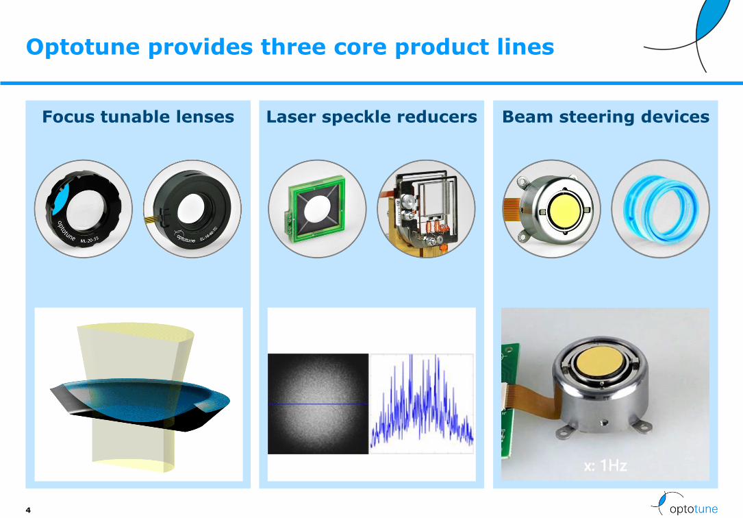
Focus tunable lenses
Optotune provides three core product lines
4
Laser speckle reducers Beam steering devices

Expansion of product portfolio over the years
5
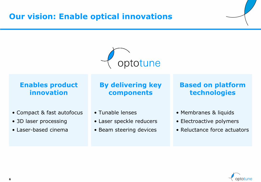
Our vision: Enable optical innovations
6
Enables product innovation
• Compact & fast autofocus
• 3D laser processing
• Laser-based cinema
By delivering key components
• Tunable lenses
• Laser speckle reducers
• Beam steering devices
Based on platform technologies
• Membranes & liquids
• Electroactive polymers
• Reluctance force actuators

Expertise in house from R&D to production
7

Optotune’s market focus
8
✓High-resolution, speckle-free projections
✓ Ultra-compact solution with no mechanics
✓ Low power consumption
✓ Axial focusing over several 100um within milliseconds
✓ Backward compatibility with several types of microscopes
✓ Speckle-free laser illumination
✓ Fast control of Z-axis
✓ Compact, reliable design with less mechanics
✓ Easy to integrate
✓ Compensation of visual defects
✓ Continuous adjustment in real-time
✓+/- 20 diopters spherical, +/- 10 diopters cylindrical
✓ Focus within milliseconds
✓Working distances from infinity to 50mm
✓Maximal flexibility
✓What is your application?
Laser projection Laser processingMachine vision
MicroscopyMedical Custom design

9
Agenda
• Company presentation
• Why tunable lenses for microscopy?
• Tunable lens technology
• Integration of tunable lenses
• Application examples
• Conclusion

Starting point
Today, most microscopes take 2D images, but …
…Life is 3-dimensional !!
10

Starting point
Modern biology wants
• Imaging of 3D cell cultures
• Imaging of whole embryos
• In-vivo imaging in living animals
Issue:
• Microscopes have a limited “depth of field” (DOF)
• The higher the lateral resolution, the smaller the DOF
Solution:
• 3D microscope
11

3D microscopy techniques
3
Wide-field microscopy
Light-sheet microscopy
Confocal microscopy
Two-photon microscopy

Need to scan along z-axis
Solutions:
➢ Motorized stages
Slow
bulky
➢ Piezo-stages
small travel
expensive
➢ Focus tunable lenses
Fast
Compact
accurate
13

USP for 3D microscopy
• Fast (> 100 Hz), compact and accurate 3D scanning
• >100x Faster than motorized solutions
• >3x cheaper than piezo stages
• Larger z-range than with piezo stages(up to 600 µm with 40x objective)
14

15
Agenda
• Company presentation
• Why tunable lenses for microscopy?
• Tunable lens technology
• Integration of tunable lenses
• Application examples
• Conclusion

16
How does it work?
Membrane
Fluid
Lensshaper
Videos available on www.optotune.com

-5
0
5
10
15
20
25
-300 -250 -200 -150 -100 -50 0 50 100 150 200 250 300
Op
tic
al
po
we
r [d
iop
ters
]
Current [mA]
EL-10-30-VIS-LD
EL-10-30-C-VIS-LD
EL-10-30-C-VIS-LD-MV (-150mm offset lens)
EL-10-42-OF (-150mm offset lens)
EL-16-40-TC-VIS-6D
17
The focal power (D = 1/f ) of Optotune’s lenses is controlled by current
Note: This curve varies from lens to lens. However, it is reproducible once calibrated
LP
MV
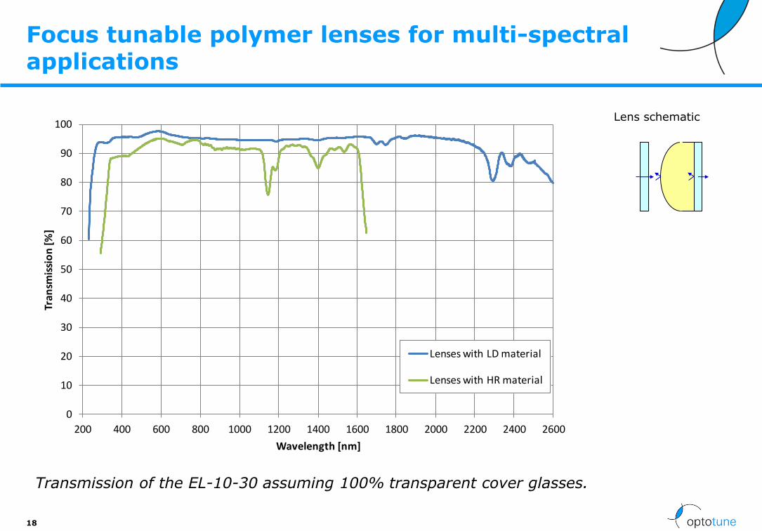
Focus tunable polymer lenses for multi-spectral applications
18
Lens schematic
0
10
20
30
40
50
60
70
80
90
100
200 400 600 800 1000 1200 1400 1600 1800 2000 2200 2400 2600
Tran
smis
sio
n [%
]
Wavelength [nm]
Lenses with LD material
Lenses with HR material
Transmission of the EL-10-30 assuming 100% transparent cover glasses.

Response time of ~10ms
19
Oscillation mode fast image stacking
Low-pass filtered:

Typical wavefront quality of the EL-10-30
20
Note: All measurements conducted at 80% of clear aperture; Wavelength is 525nm
• Precision optics quality if optical axis is vertical
• Wavefront error in horizontal axis dominated by a Y-coma, due to gravity
This can be reduced using stronger membranes
Gravity induced Y-coma
of about 0.2 λ
Gravity induced Y-coma
of about 0.4 λ
0.1 λLP
MV

Focal power mode for good reproducibility
• Why it is important:
- The focal power of our lenses drifts with temperature by about 0.06 diopters / °C (depends on lens model)
• Typical accuracy achieved: +/- 0.1dpt
21
Use focal power mode to set lens to 8
diopters
Lens calibration table and temperature read
by lens driver
Lens calibration curve stored on
lens internal memory
Lens characterization
f vs T vs I
Temperature compensated
control current to adjust lens to 8 diopters
“I need a lens with f=125mm (8 diopters)”
Temp sensor with EEPROM
f=125mm(8 diopters)

Optotune’s electrically focus tunable lenses
222222
EL-10-30-TC EL-10-30-C(i) EL-10-42-OF EL-16-40-TC-5D EL-16-40-TC-20D
Focal power range*
8 … 22 Dpt -1.5 … +3.5 Dpt -2 … +2 Dpt -2 … +3 Dpt -10 … +10 Dpt
Clear aperture 10mm 10mm 10mm 16mm 16mm
Outer diameter 30mm 30mm 42mm 40mm 40mm
Wavefront qualityRMS @525nm*****
<0.25 / 0.5 <0.15 / 0.25 <0.15 I: <0.15/ 0.5 II <0.25 / 0.5
I: <0.25 / 2.5 II: <0.5 / 2.5
Absolute focal power accuracy
<0.15 dpt < 0.1 dpt 0.008 dpt(temp. contr.)
< 0.1 dpt < 0.1 dpt
Built-in sensors None Temperature Temp./Optical feedback
Temp./Opticalfeedback
Temp./Opticalfeedback
Applications MVOCT
MVMicroscopy
Laser marking MV/MicroscopyOphthalmology
MV/MicroscopyOphthalmology
* Depends on selected optical fluid ** vertical / horizontal optical axis *** class I: high-mag, microscopyclass II: standard grade

Focus tunable polymer lenses are reliable
23
Test Test conditions Status
Mechanical cycling
40 million full-range cycles (0 to 300 mA rectangular, at 10 Hz) 5 billion sinusoidal cycles at resonant frequency
Passed
High temperature test
85±2°C; rel. hum. <6% for 168 hours, non-operational
Passed
Temperature cycling test
-40°C / +85°C for 30 min each, 3 min transition time, 100 cycles
Passed
Damp heat cycling test
25°C / 55°C at 90-100% relative humidity, 3 hour transition time, 24h per cycle (9h plus transition time each), 18 cycles
Passed
Shock test: 800g for 1ms duration, 5 pulses in each direction (30 pulses in total)
Passed
Solar radiation test:
1120 W per m2 (IEC 60068-2-5), 8 h irradiation & 16 h darkness, 10 cycles
Passed
24
Agenda
• Company presentation
• Why tunable lenses for microscopy?
• Tunable lens technology
• Integration of tunable lenses
• Application examples
• Conclusion

No Mag change Mag. change
Digital microscopy configurations
25
Obj(Inf)
Cam
TubeLens
ETL
Microscopy
• Large z-rangeObj
(Inf)
Cam
ETL
TubeLens
Relaylens
Relaylens
4f system
Intermediateimage plane

How to integrate the EL
Digital inspection microscope
Scientific microscope
26
OptotuneEL
Zoom lens
Tube lens
1. EL Pos.(after objective)
2. EL Pos.(filter cube)
3. EL Pos.(camera port)

Non-telecentric autofocus configuration: EL on top of objective
27
Zeiss Axioskophttp://labs.pbrc.edu/cellbiology/documents/AxioskopManual.pdf
• Zeiss Neofluar, 10x/0.3 Inf./0.17
• Zeiss LD Achroplan 20x/0.4 Korr Ph2 Inf./0-1.5
• Zeiss Plan-Neofluar 40x/0.75 Inf/0.17
Camera
Teledyne Dalsa Genie TS-C1920
Optotune Lens
EL-16-40-TC-VIS-20D, ANAA0380
Mounted with C-mount RMS adapters from Thorlabs
- RMSA6 - Adapter with External RMS Threads and Internal C-Mount Threads
- RMSA5 - Adapter with External C-Mount Threads and Internal RMS Threads
C-mount adapter
camera

Non-telecentric autofocus configuration: EL on top of objective
28
Mag -2 dpt 0 dpt +3 dpt
10x
20x
40x

Non-telecentric autofocus configuration: EL on top of objective
• The tunable lens was operated between -2dpt and +3 dpt (nominal tuning range)
• Compact autofocus solution without the need of mechanical translation
• However, in such a configuration, the field-of-view (FOV) and numerical aperture (NA) changes while focusing (non-telecentric behavior)
29
Z-range Mag change
10x 2.56mm(20D: 10.24)
7.5 %
20x 0.64mm(20D: 2.56 mm)
12.2%
40x 0.16mm(20D: 0.64mm)
23.7%

Image plane
without EL
Objective (Olympus 40x NA 0.8)*
Offset LensEL
* Japanese patent 8-292374
Non-telecentric autofocus configuration: optical layout
• With the tunable lens on top of the objective,the FOV and NA changes while focusing(non-telecentric behavior)
• The animation on the left shows this as an increasing distance between the blue (on-axis) and red (maximum FOV) foci
31

Objective (ideal lens)
with changing WD
Tube lens
Relay lens
Relay lens
CCD with
fixed image
EL at a conjugate
BFP of the objective
Relay system
Telecentric autofocus configuration: tunable lens EL with a relay system
• By inserting a relay system, composed of two lenses (a 4f-system), the back focal plane (BFP) of the objective can be reimaged to an accessible location
• When the EL is placed at that position, the system stays telecentric while focusing
32

This design principle can be found in this EL-lightsheet microscope (Fahrbach et al., Optics Express 2013)
Exemplary setup of a telecentric microscope with a tunable lens autofocus solution
33

• The displacement of the image plane is given by
where n is the refractive index of the immersion medium, Mdet is the magnification of the microscope objective, fr is the focal length of the relay lens and fETL,eff is the effective focal length of the Optotune lens (1/ fETL,eff≈ 1/ fETL +1/ fOL) and fOL is the focal length of a possible offset lens.
• To maintain the full NA of the detection lens, the ratio of the focal lengths of the relay lens fr and the tube lens fTL must not be larger than the ratio of the aperture of the ETL dETL and the diameter of the BFP of the detection lens dBFP, i.e.
Axial scan range
34
fTLzdet fr fr2DETL
dETLdBFP
Mdet
Intermediate image
dETL=16mm
Image circle:30mm

Axial scan range examples
35
Scenarios:
DETL = 1/fETL
dBFP<=10mm
Totallength
Microscopy adapter prototype (5D): 12um 40x 200mm 62mm 5D 31mm ~450mm
Image circle
12mm
MagRelay = fr / fr2
MagRelay
10x, long relay, 5D: 2000um 10x 200mm 200mm 5D 200mm 1100mm30mm1X
20X, long relay, 5D: 500um 20x 200mm 200mm 5D 200mm 1100mm30mm1X
Shorter relay lenses: 250um 20x 200mm 141mm 5D 141mm 864mm30mm1X
0.5X
10D lens: 500um 20x 200mm 141mm 10D 141mm 864mm30mm1X
1” sensor: 500um 20x 200mm 141mm 10D 70mm 744mm15mm0.5X
dETLdBFP
Intermediate image
dETL=16mm
Image circle:30mm
fTLzdet fr fr2DETLMdet
Shorter relay lenses: 1000um 10x 200mm 141mm 5D 141mm 864mm30mm1X
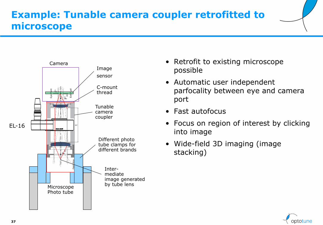
Example: Tunable camera coupler retrofitted to microscope
• Retrofit to existing microscope possible
• Automatic user independent parfocality between eye and camera port
• Fast autofocus
• Focus on region of interest by clicking into image
• Wide-field 3D imaging (image stacking)
37
EL-16
MicroscopePhoto tube
Different phototube clamps fordifferent brands
Tunablecameracoupler
C-mount thread
Image
sensor
Camera
Inter-mediateimage generatedby tube lens

Distance to intermediate image.
Nominal: 55.89mm
Total length 199mm
Distance to intermed image Nominal Nominal -4.0mm Nominal +4.0mm
Distance to intermed image [mm] 55.89 51.89 59.89
Image magnification 0.35 0.35 0.35
Image quality ok.Same over entire sensor.
Magnification stays the same when tuning EL-16
Total length further reduced with two positive meniscus lenses
Field curvature corrected
See Telecentric Microscopy Camera Adapter (real lenses - shorter front) v09 [tune check].zmx
See Telecentric Microscopy Camera Adapter (real lenses - shorter front) v09.zmx
Optical design with off-the-shelf catalog lenses for ½” sensor
Total thickness EL-16 incl. C-mount flange
Stop 3.6mm

Microscopy adapter without magnification change
44
Zeiss Axioskop
• Zeiss Neofluar, 10x/0.3 Inf./0.17
• Zeiss LD Achroplan 20x/0.4 Korr Ph2 Inf./0-1.5
• Zeiss Plan-Neofluar 40x/0.75 Inf/0.17
Camera
IDS UI-3580CP-C-HQ (1/2”, 5MP)
Optotune Lens
EL-16-40-TC-VIS-20D
Z-RangeMag EL-16-40-TC-VIS-5D EL-16-40-TC-VIS-20D
10x 262um 980um
20x 64um 254um
40x 12um 56um

Almost no magnification change
46
10x 20x 40x
10 dpt
0 dpt
-10 dpt
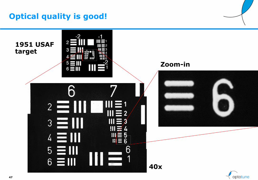
Optical quality is good!
47
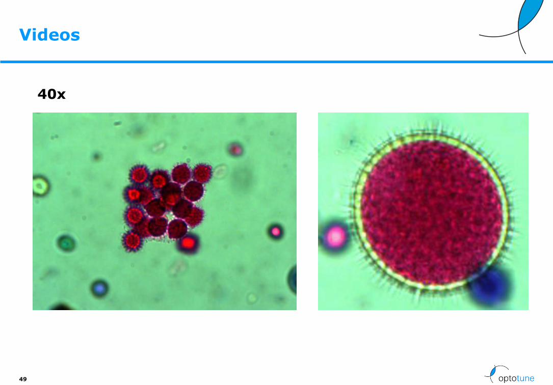
40x
Zoom-in
1951 USAF target

Stacking of pollen images
48
Images have been taken at 10x between -10dpt and 10dpt
-10 dpt
0 dpt
+10 dpt
Videos
49
40x

50
Agenda
• Company presentation
• Why tunable lenses for microscopy?
• Tunable lens technology
• Integration of tunable lenses
• Application examples
• Conclusion

18 publications using Optotune lenses for microscopy
51 http://www.optotune.com/technology/publications

Wide-field microscopy
52
Images courtesy of F. F. Voigt, Department of Neurophysiology, Brain Research Institute, University of Zurich
A B
ETL
ETL
Optical path of the Axiovert 35 microscope. The ETL/OL assembly can be placed at
the pupil without inserting an additional relay system. TL: Tube lens.

Wide-field microscopy
53
ETL-based focusing through a group of pollen grains.
Images courtesy of F. F. Voigt, Department of Neurophysiology, Brain Research Institute, University of Zurich
Video

Confocal microscopy
54
Max. intensity projection of a pollen corn
Video Video
Images courtesy of F. F. Voigt, Department of Neurophysiology, Brain Research Institute, University of Zurich

Confocal endomicroscopy
55
Focal power range
-127 mm to+44.3 mm
Axial scan range @ sample
700µm
Ref: J.M. Jabbour et al., BIOMEDICAL OPTICS EXPRESS 2014, 5, (2), pp. 645, 2014, “Optical axial scanning in confocal microscopy using an electrically tunable lens”
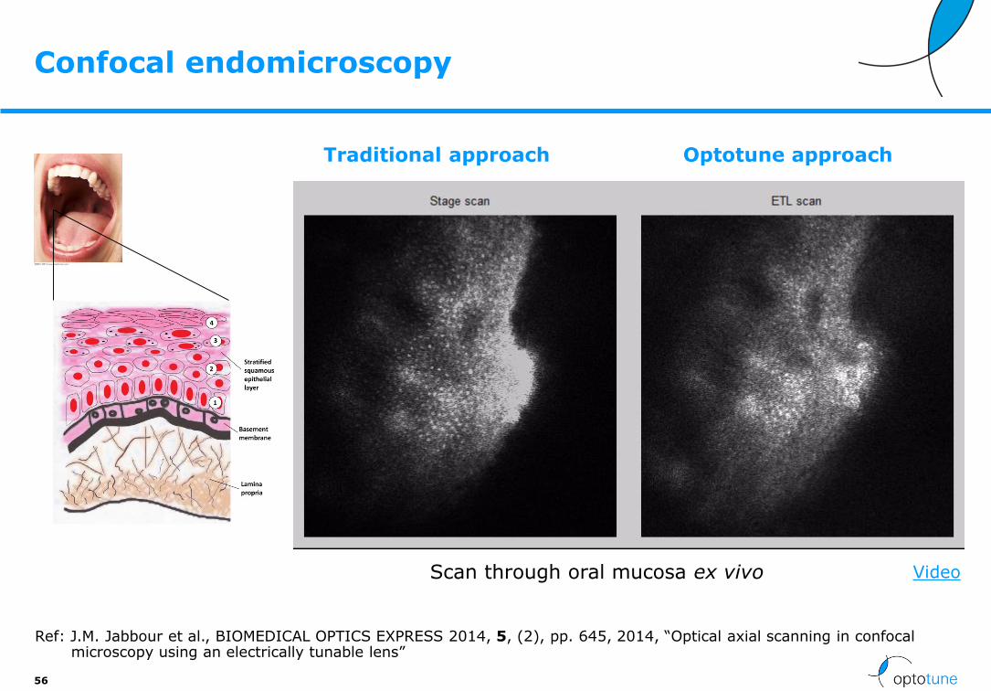
Confocal endomicroscopy
56
Ref: J.M. Jabbour et al., BIOMEDICAL OPTICS EXPRESS 2014, 5, (2), pp. 645, 2014, “Optical axial scanning in confocal microscopy using an electrically tunable lens”
Scan through oral mucosa ex vivo
Traditional approach Optotune approach
Video

Two-photon microscopy
57
B.F Grewe et al.,
Biomedical Express (2011),
2, (7), pp.2035
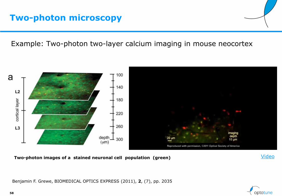
Two-photon microscopy
Example: Two-photon two-layer calcium imaging in mouse neocortex
58
Two-photon images of a stained neuronal cell population (green)
Benjamin F. Grewe, BIOMEDICAL OPTICS EXPRESS (2011), 2, (7), pp. 2035
Video

Two-photon in-vivo imaging of retinal microstructures
59
Adi Schejter, Proc. SPIE 8948, Multiphoton Microscopy in the Biomedical Sciences XIV, 894824 (February 28, 2014); doi:10.1117/12.2039375
Optical sectioning in mouse 2P fluorescence angiography. A. Two-photon images of the optic disc. The microscope objective lens and mouse were held in place, and each image was acquired at different ETL currents (10mA interval between successive images; each image is an average over 30 frames acquired at 1 fps). Arrowheads point to blood vessels visible in only a few images, but not in others. B. Images of blood vessels outside the optic disc, acquired at different scan zooms (average over 100 and 200 frames; different animal than A). The FOV of the lower image is marked by a white box. Scale bars = 50 µm.
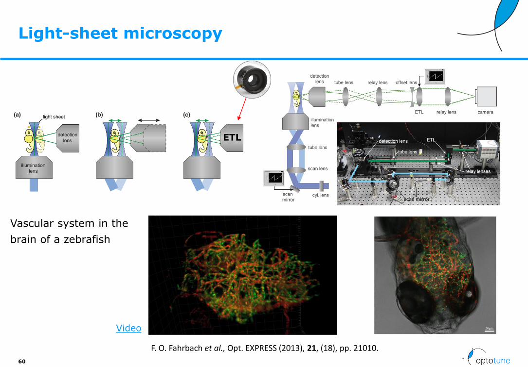
Light-sheet microscopy
60
Vascular system in the
brain of a zebrafish
ETL
F. O. Fahrbach et al., Opt. EXPRESS (2013), 21, (18), pp. 21010.
Video

Light-sheet microscopy
61
Large volume scan with an ETL through the heart of a zebrafish (10x magn.)
F. O. Fahrbach et al., Opt. EXPRESS (2013), 21, (18), pp. 21010.
Video
Courtesy of Florian Fahrbach, Michaela Mickoleit and Jan Huisken.
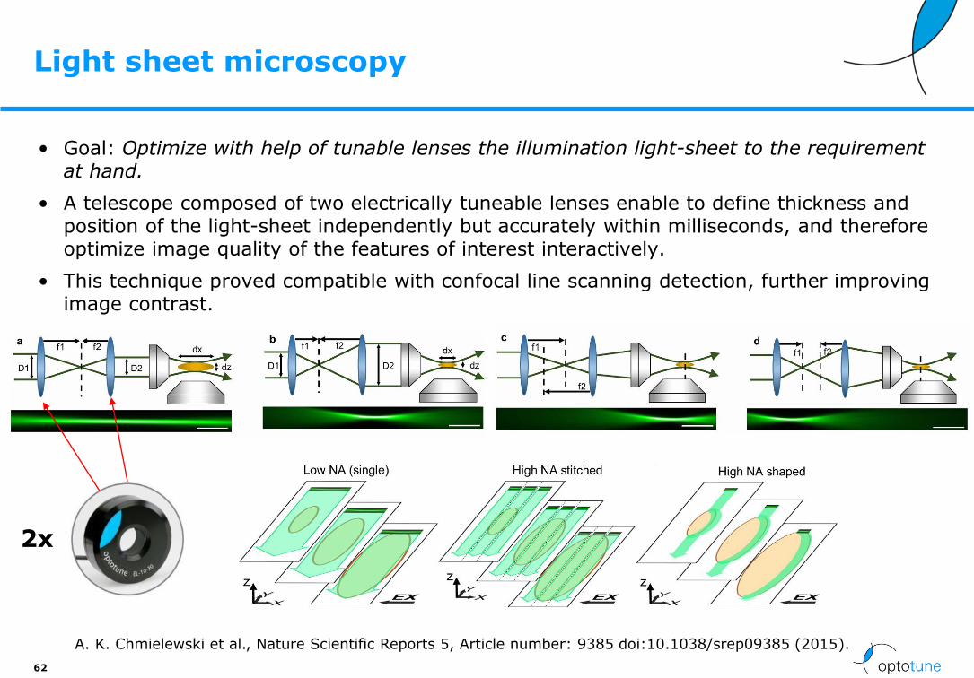
Light sheet microscopy
• Goal: Optimize with help of tunable lenses the illumination light-sheet to the requirement at hand.
• A telescope composed of two electrically tuneable lenses enable to define thickness and position of the light-sheet independently but accurately within milliseconds, and therefore optimize image quality of the features of interest interactively.
• This technique proved compatible with confocal line scanning detection, further improving image contrast.
62
A. K. Chmielewski et al., Nature Scientific Reports 5, Article number: 9385 doi:10.1038/srep09385 (2015).
2x

3D High- and super-resolution imaging using single-objective SPIM
• Single-objective selective-plane illumination microscopy (soSPIM) is achieved with micro-mirrored cavities combined with a laser beam–steering unit installed on a standard inverted microscope.
• Based on custom EL-C-10-30 focus-tunable lens (TL) from −80 mm to +1,000 mm.
63
Remi Galland, Nature Methods, published online 11 May 2015DOI:10.1038/NMETH.3402

Z-stacking with inverted microscope, 100x mag
64
http://scitation.aip.org/content/aip/journal/rsi/86/1/10.1063/1.4905330
microtubules in HeLa cells
Optotune
EL-10-30

A Rapid Image Acquisition Method for Focus Stacking in Microscopy
65
D. Clark, Microscopy Today / Volume 23 / Issue 04 / July 2015, pp 18-25Copyright
DOI: http://dx.doi.org/10.1017/S1551929515000577

Image Stacking example (Python + Helicon Focus)
66
Python
Script
Helicon focus
ImageStackC-mount camera
Empty C-mount tube,40-60mm long
Optotune lensEL-10-30-Ci-VIS-LD
25mm lens (reversed!)Edmund Optics 85358
Working distance: ~20mm
M22 to C-mount adapter
Hyperfocus
Image

Sanxo scope: Inspection at HD
67
• Inspection station with 10MP camera
• EL-10-30-Ci in front lens configuration with 25mm lens
• Driver integrated in machine vision software “Modular X”
• Features:
- Click to autofocus
- Focal sweep with 3D rendering

Microscope-integrated intraoperative OCT
• Optotune’s electrically tunable lens EL-10-30-NIR-LD allowed real-time adjustment of the OCT focal plane to maintain parfocality with the microscope view.
• Potential for iOCT-guided maneuvers and clinical decision-making in ophthalmic surgery
68
Y. K. Tao et al., BIOMEDICAL OPTICS EXPRESS (2014), 5, (6), pp. 1877.

Autofocus for high magnification with EL-10-30-C and Optem® 70XL by Qioptiq
Results:
69
C-mount camera1/2.5” 5MP sensor
1.5x mini tube lensP/N 29-90-28-000
Optotune lensEL-10-30-Ci-VIS-LD-MV
Optem 70XL zoom (0.75x-5.25x)P/N 399510-309
Coaxial lighting unit with lensP/N 296515-310
Working distance: ~90mm
Optem® is a registered trademark of Qioptiq, Inc
Magnification 1.1x 3.5x 7.9x
Z range 400mm 40mm 8mm
Z resolution 100µm 10µm 2µm
DOF (approx.) 1mm 0.3mm 0.1mm
HFOV 4.5mm 1.4mm 0.65mm
LED ring light (used instead)
• No vignetting
• Off-the-shelf components

Qioptiq Optem Fusion industrial microscope with EL-16-40-TC autofocus module
70
• The zoom is parfocal as the EL is placed BELOW the zoom
http://www.qioptiq.com/optem-fusion-lens, Optem® is a registered trademark of Qioptiq, Inc

Low cost AF microscope with fixed mag
71
C-mount camera
Empty C-mount tube,40-60mm long
Optotune lensEL-10-30-Ci-VIS-LD
25mm lens (reversed!)Edmund Optics 85358
Working distance: ~20mm
M22 to C-mount adapter
Tube length 40mm 60mm
Magnification 3X 4X
Z-range: ~3mm ~2mm
Resolution*: 3.7um 2.8um
Image circle 25mm 25mm
*Line width of group 7 element 4

Z-stepping solutions for microscopes and industry based on EL-10-30
72
www.phaseview.com

Edmund optics dynamic focus VZM with the EL-10-30-Ci-VIS-LD-MV integrated
• Very large focus range as EL is placed close to aperture stop
• The zoom is NOT parfocal, however, as the EL is placed above the zoom
73

Optotune’s LSR boosts image quality in super-resolution fluorescence microscope (STORM)
74
Ref: P. Georgiades et al., Journal of Microscopy (2016), http://onlinelibrary.wiley.com/doi/10.1111/jmi.12453/full
MRC5 cells stained with Alexa Fluor 647
LSR on
LSR off
LSR-3005-17S-VIS
Distribution of pixel greyscale values:
Setup:

All-optical microscope autofocus based on an ETL and a totally internally reflected IR laser
75
https://www.osapublishing.org/oe/abstract.cfm?uri=oe-26-3-2359

76
Agenda
• Company presentation
• Why tunable lenses for microscopy?
• Tunable lens technology
• Integration of tunable lenses
• Application examples
• Conclusion

Microscopy application overview
3D microscopy
• Wide-field microscopy
• Two-photon microscopy
• Confocal microscopy
• Light-sheet microscopy
2D microscopy
• Digital microscopy
• Adapter for video port
77
+ETL

Conclusion
• Trend towards 3D biomedical imaging
• Focus tunable polymer lenses are compatible with
- Wide-field microscopy
- Confocal microscopy
- Two-photon microscopy
- Light-sheet microscopy
• Tunable lenses:
- Fast
- Compact
- Large tuning range
- Vibration-free
- Broadband
78

Optotune – 1 slide
Optotune Switzerland AGBernstrasse 388CH-8953 DietikonSwitzerland
Phone: +41 58 856 3000 | Fax: +41 58 856 3001
www.optotune.com | [email protected]