Embed Size (px)
Citation preview

325
Salinity and its potential variations are among the mainfactors influencing reproduction, dispersal and recruitment oforganisms in marine, coastal and estuarine habitats (Anger,2003). Adaptation to constantly low or fluctuating salinityis, at least in part, achieved by cells specialized in ionicexchanges, the ionocytes. At low salinity, the ionocytescompensate the ion loss caused by osmotic gradients betweenthe hemolymph and the surrounding medium by active ionpumping (uptake of Na+ and Cl–). Along with apical microvilliand numerous mitochondria, basolateral infoldings of thecytoplasmic membrane are typical characteristics of ion-transporting cells (reviewed by Mantel and Farmer, 1983;Péqueux, 1995). Osmoregulation and the location of ion-transporting cells and tissues have been extensively studied, sothat a considerable amount of information is now available on
this topic in a great variety of decapod crustacean species andother aquatic invertebrates. In osmoregulating brachyurancrabs, numerous studies have pointed out that osmoregulatorystructures are mainly located in the posterior gills, whereasanterior gill lamellae generally possess thin respiratoryepithelia, which enable diffusive gas exchange (reviewed byMantel and Farmer, 1983; Gilles and Péqueux, 1985; Péqueuxand Gilles, 1988; Lucu, 1990; Taylor and Taylor, 1992;Péqueux, 1995).
In the process of ionic regulation, Na+/K+-ATPase is one ofthe most important enzymes (reviewed by Towle, 1981,1984a,b; Péqueux, 1995; Charmantier, 1998; Lucu and Towle,2003). By using ATP as a source of energy, it enables an activeion-exchange across epithelial membranes (Neufeld et al.,1980; De Renzis and Bornancin, 1984). Immunolocalization of
The Journal of Experimental Biology 207, 325-336Published by The Company of Biologists 2004doi:10.1242/jeb.00759
The ontogeny of osmoregulation, the development ofbranchial transporting epithelia and the expression of theenzyme Na+/K+-ATPase were studied in Carcinus maenas(L.) obtained from the North Sea, Germany. Laboratory-reared zoea larvae, megalopae and young crabs wereexposed to a wide range of salinities, and hemolymphosmolality was measured after 24·h exposure time (72·h injuveniles). Zoea I larvae slightly hyper-regulated in dilutemedia (10.2‰ and 17.0‰) and osmoconformed at >17‰.All later zoeal stages (II–IV) osmoconformed in salinitiesfrom 10.2‰ to 44.3‰. The megalopa hyper-regulatedat salinities from 10.2 to 25.5‰. Young crabs hyper-regulated at salinities from 5.3‰ to 25.5‰, showingan increase in their osmoregulatory capacity. Thedevelopment of transporting epithelia and theexpression of Na+/K+-ATPase were investigated bymeans of transmission electron microscopy andimmunofluorescence microscopy. In the zoea IV, only avery light fluorescence staining was observed in gill buds.Epithelial cells were rather undifferentiated, without
showing any features of ionocytes. Gills were present inthe megalopa, where Na+/K+-ATPase was located in basalfilaments of the posterior gills. In crab I juveniles andadults, Na+/K+-ATPase was noted in the three mostposterior pairs of gills, but lacking in anterior gills.Ionocytes could first be recognized in filaments ofmegalopal posterior gills, persisting through subsequentstages at the same location. Thus, the development of thegills and the expression of Na+/K+-ATPase are closelycorrelated with the ontogeny of osmoregulatory abilities.The morphological two-step metamorphosis of C. maenascan also be regarded as an osmo-physiologicalmetamorphosis, (i) from the osmoconforming zoeal stagesto the weakly regulating megalopa, and (ii) to theeffectively hyper-regulating juvenile and adult crabs.
Key words: osmoregulation, ontogeny, hemolymph osmolality,immunolocalization, Na+/K+-ATPase, gill, larva, ionocyte, Carcinusmaenas.
Summary
Introduction
Ontogeny of osmoregulatory structures and functions in the green crabCarcinus maenas (Crustacea, Decapoda)
Ude Cieluch1,*, Klaus Anger1, Fabien Aujoulat2, Friedrich Buchholz1,Mireille·Charmantier-Daures2 and Guy Charmantier2
1Biologische Anstalt Helgoland/Stiftung Alfred-Wegener-Institut für Polar- und Meeresforschung, Meeresstation,D-27498 Helgoland, Germanyand 2Equipe Adaptation Ecophysiologique et Ontogenèse, UMR 5000 GPIA,
Université Montpellier II, F-34095 Montpellier cedex 05, France*Author for correspondence (e-mail: [email protected])
Accepted 16 October 2003

326
Na+/K+-ATPase using monoclonal antibodies has recentlybeen used as a tool to identify transporting epithelia, e.g. in theterrestrial isopod Porcellio scaber(Ziegler, 1997), lobsterHomarus gammarus(Lignot et al., 1999; Lignot andCharmantier, 2001), and in crayfish Astacus leptodactylus(Barradas et al., 1999). By investigating the development,location and functionality of transporting epithelia, the precisecellular location of Na+/K+-ATPase is of special interest (Fliket al., 1994; Haond et al., 1998; Lignot et al., 1999; Lignot andCharmantier, 2001).
Several studies have been conducted on the ontogeny ofosmoregulation in various species (reviewed by Charmantier,1998). However, investigations on the ontogeny ofosmoregulating tissues and its potential variations throughoutdevelopment are still very limited (Hong, 1988; Bouaricha etal., 1994; Charmantier, 1998; Anger, 2001; Lignot andCharmantier, 2001). Among the few species in which theontogeny of ion-transporting epithelia have been investigatedby histological and/or electron microscopical studies areFarfantepenaeus aztecus(Talbot et al., 1972), Callianassajamaicense(Felder et al., 1986), Penaeus japonicus(Bouarichaet al., 1994) and Homarus gammarus(Lignot and Charmantier,2001). From these studies it appears that organs other than gillscan also play a major role in ion-transport and that the locationof epithelia involved in ion-exchange can change duringdevelopment (reviewed by Charmantier, 1998).
The adult green crab Carcinus maenas(L.) is aeuryhaline species that exhibits the ability of effectivehyperosmoregulation in habitats of low and/or fluctuatingsalinity (Theede, 1969; Siebers et al., 1982, 1985). In Europeanwaters, this ability has enabled the crab to cover a widegeographical area from the Baltic Sea to the Azores, living inhabitats where salinity ranges from 9‰ to 35‰ (Winkler etal., 1988). Its euryhalinity has also aided in it becoming aninvasive species in estuarine habitats of the east and westcoasts of the USA and Canada, as well as in West and SouthAfrica and Australia (Cohen et al., 1995; Grosholz and Ruiz,1995; Lafferty and Kuris, 1996).
The gills of adult C. maenashave received much attentionas the potential site of ionic exchange and much information,including the location and fine structure of ionocytes, is known(e.g. Compere et al., 1989; Taylor and Taylor, 1992; Lawsonet al., 1994; Hebel et al., 1999). In addition, anultracytochemical approach conducted in gills of C. maenasshowed that the presence of Na+/K+-ATPase is mainlyrestricted to basolateral infoldings of epithelial cells inposterior gill lamellae (Towle and Kays, 1986).
In contrast to the ability of adult C. maenasto live overextended periods in habitats with low salinity, thereproduction, embryogenesis and larval development of thisspecies require higher salt concentrations (Green, 1968; Kinne,1971; Nagaraj, 1993). A laboratory study on the tolerance ofC. maenaslarvae from the North Sea facing hypo-osmoticstress showed that a salinity of at least 25‰ is needed forsuccessful development (Anger et al., 1998). At reducedsalinities (≤20‰), significant decreases were found in the rates
of early zoeal survival, development, growth, respirationand assimilation (Anger et al., 1998). It is thus likely that theosmo-physiological pattern changes during the course ofdevelopment.
The present investigation was conducted (i) to study theontogeny of osmoregulation by direct measurements of thehemolymph osmolality, (ii) to locate and follow thedevelopment of osmoregulatory epithelia and the expression ofNa+/K+-ATPase using transmission electron microscopy(TEM) and immunofluorescence light microscopy (ILM), and(iii) to relate the ontogeny of osmoregulation to thedevelopment of transporting epithelia and to ecological traits.
Materials and methodsAnimals
Ovigerous females and juveniles of Carcinus maenasL.were collected from the rocky intertidal zone of the island ofHelgoland, North Sea, Germany. After transfer to theHelgoland Marine Station, females were kept individually in5·l plastic aquaria connected to an overflow system usingrunning seawater (salinity ≈32‰). Aquaria were maintained ina constant-temperature room at 15°C and subjected to a12·h:12·h light:dark cycle. Hatched larvae were collected insieves (200·µm mesh size) and individually reared throughmetamorphosis using glass vials (≈50·ml) at a constanttemperature of 18°C and under the same light:dark regime. Forthe osmoregulation experiment, larvae were reared at 32‰(941·mOsm·kg–1); the larvae used for immunofluorescencelight microscopy (ILM) and transmission electron microscopy(TEM) were reared at 25‰ (735·mOsm·kg–1). Water and food(freshly hatched Artemiasp. nauplii) were changed daily. Thedevelopmental stages tested in the osmoregulation experimentcomprised all zoeal stages (I–IV), the megalopa, the first andsecond crab instars (I and II), and larger juveniles collected inthe field (carapace width 14–18·mm). The following stageswere chosen for ILM and TEM: zoea IV, megalopa, first craband adults (body size 32–41·mm, acclimated to 25‰ salinityfor at least 2 weeks). Adult C. maenaswere fed thawed mussels(Mytilus edulis) every second day. For all experiments, animalsin the middle of an instar, i.e. in intermolt stage C (Drach,1939) were exclusively used. Prior to tissue sampling forhistology, adult crabs were anesthetized by immersion incooled water (≈3°C).
Preparation of media
Experimental media were obtained by diluting 1·µm-filteredsea water (32‰) with desalinated freshwater or by addingTropic Marin® salt (Wartenberg, Germany). Salinity wasexpressed as osmotic pressure (in mOsm·kg–1) and as saltcontent of the medium (in ‰); a value of 3.4‰ is equivalentto 100·mOsm·kg–1 (29.41·mOsm·kg–1≈1‰). The osmoticpressure of the media was measured with a micro-osmometerModel 3 MO plus (Advanced Instruments, Needham Heights,MA, USA), requiring 20·µl per sample. The following mediawere prepared, stored at 18°C and used in the osmoregulation
U. Cieluch and others

327Ontogeny of osmoregulation in C. maenas
experiment: 30·mOsm·kg–1 (1.0‰), 155·mOsm·kg–1 (5.3‰),300·mOsm·kg–1 (10.2‰), 500·mOsm·kg–1 (17.0‰),749·mOsm·kg–1 (25.5‰), 947·mOsm·kg–1 (32.2‰, referred toas seawater) and 1302·mOsm·kg–1 (44.3‰).
Osmoregulation
The experiment was carried out at a constant temperature of18°C, representative of typical summer conditions in the areaof origin of our material, the North Sea, and known to befavourable for both larval and adult C. maenas, both in thelaboratory (Dawirs, 1985; Anger et al., 1998) and in the field(Harms et al., 1994).
Larvae and juveniles were transferred directly to theexperimental media and exposed for 24·h (72·h in largejuveniles from the field) in covered Petri dishes. Followingtheir capture, large juvenile crabs from the field were kept inseawater (≈32‰) for 48·h at 18°C. The number of exposedanimals was kept to a minimum level of 9–11 individuals percondition. Dead animals were counted at the end of theexposure time to obtain survival rates. The survivingspecimens were superficially dried on filter paper and quicklyimmersed into mineral oil to prevent evaporation anddessication. Any remaining adherent water was removed usinga glass micropipette. A new micropipette was then inserted intothe heart for hemolymph sampling. For all experimental stages,hemolymph osmolality was measured with reference to themedium osmolality on a Kalber–Clifton nanoliter osmometer(Clifton Technical Physics, Hartford, CT, USA) requiringabout 30·nl. Results were expressed either as hemolymphosmolality or as osmoregulatory capacity. The latter is definedas the difference between the osmolality of the hemolymph andthat of the medium. Analysis of variance (ANOVA) andStudent’s t-tests were used for multiple and pairwise statisticalcomparisons of mean values, respectively, after appropriatechecks for normal distribution and equality of variance (Sokaland Rohlf, 1995).
Immunofluorescence light microscopy
After removal of the carapace, anterior and posterior gills ofadult C. maenaswere dissected from the inner body wall andfixed for 24·h in Bouin’s fixative. Zoeae, megalopae and crabI were fixed by direct immersion in the same fixative. Afterrinsing in 70% ethanol, samples were fully dehydrated ina graded ethanol series and embedded in Paraplast-extra(Sigma). Sections (4·µm) were cut on a Leitz Wetzlarmicrotome, collected on poly-L-lysine-coated slides and storedovernight at 38°C. Sections were then pre-incubated for 10·minin 0.01·mmol·l–1 Tween 20, 150·mmol·l–1 NaCl in 10·mmol·l–1
phosphate buffer, pH·7.3. To remove the free aldehyde groupsof the fixative, samples were treated for 5·min with50·mmol·l–1 NH4Cl in phosphate-buffered saline (PBS),pH·7.3. The sections were then washed in PBS and incubatedfor 10·min with a blocking solution (BS) containing 1% bovineserum albumin (BSA) and 0.1% gelatine in PBS. The primaryantibody (monoclonal antibody IgGα5, raised against the avianα-subunit of the Na+/K+-ATPase) was diluted in PBS to
20·µg·ml–1, placed in small droplets of 100·µl on the sectionsand incubated for 2·h at room temperature in a wet chamber.Control sections were incubated in BS without primaryantibody. To remove unbound antibodies, the sections werethen washed (3× 5·min) in BS and incubated for 1·h withsmall droplets (100·µl) of secondary antibody, fluorescein-isothiocyanate (FITC)-labeled goat anti-mouse IgG (JacksonImmunoresearch, West Baltimore, USA). After extensivewashes in BS (4× 5·min), the sections were covered with amounting medium and examined using a fluorescencemicroscope (Leitz Diaplan coupled to a Ploemopak 1-Lambdalamp) with an appropriate filter set (450–490·nm band-passexcitation filter) and a phase-contrast device.
Transmission electron microscopy
Anterior and posterior gills of adult crabs were cut into smallpieces and fixed for 1.5·h in 5% glutaraldehyde solutionbuffered at pH·7.4 with 0.1·mol·l–1 cacodylate buffer. Zoeae,megalopae and early crab stages were fixed for 1·h by directimmersion in the same fixative. For adjustment to the osmoticpressure of the hemolymph, NaCl was added to the fixative andbuffer to give a final osmolality of 735·mOsm·kg–1. Sampleswere then rinsed in buffer and postfixed for 1.5·h at roomtemperature in buffered 1% OsO4. After extensive washes inbuffer, the samples were fully dehydrated in graded acetoneand embedded in Spurr low viscosity medium. Semithinsections (1·µm) were prepared using glass knives with a LKBmicrotome and stained with Methylene Blue for lightmicroscopic observations. Ultrathin sections were obtainedusing a diamond knife, contrasted with uranyl acetate (Watson,1958) and lead citrate (Reynolds, 1963) and examined with atransmission electron microscope (EM 902, Zeiss, Germany)operated at 80·kV.
ResultsSalinity tolerance
Survival of the different stages during exposure for 24·h(72·h in juveniles) to the experimental media is givenin Table·1. Survival was 89–100% for all developmentalstages in media >749·mOsm·kg–1 (25.5‰). At 500 and300·mOsm·kg–1 (17.0 and 10.2‰, respectively), 80–100% ofyoung and juvenile crabs survived, but survival rates werelower in zoeae and megalopae (except for zoeae I) at500·mOsm·kg–1 (17.0‰). At 300·mOsm·kg–1 (10.2‰), zoealsurvival was low, with complete mortality in zoea IV. At asalinity of 155·mOsm·kg–1 (5.3‰), 40–100% of the juvenileand adult crabs survived, but all zoea larvae and megalopaedied, so that no data of larval osmoregulation could be obtainedfrom this treatment. Only juvenile crabs were exposed to thelowest salinity (30·mOsm·kg–1, 1.0‰), in which the mortalityrate reached 80–100%.
Osmoregulation
The developmental stages were exposed to a wide range ofsalinities. The experimental results are given as variations in

328
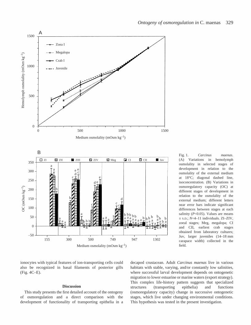
hemolymph osmolality and as osmoregulatory capacity inrelation to the osmolality of the experimental medium(Fig.·1A,B).
The pattern of osmoregulation changed during development.With the exception of the first zoea, no significant differenceswere observed between successive zoeal stages exposed to thesame salinities. Only zoeae I larvae were able of a slight hyper-regulation at 500·mOsm·kg–1 (17.0‰). All later zoeal stages(ZII–ZIV) osmoconformed over the entire tested salinity range,300–1302·mOsm·kg–1 (10.2–44.3‰). A significant change inthe pattern of osmoregulation was noted in the megalopae. Thisstage osmoconformed at high salinities (947·mOsm·kg–1 or32.2‰; 1302·mOsm·kg–1 or 44.3‰). At lower salinities(300–749·mOsm·kg–1 or 10.2–25.5‰), the megalopae showeda strong ability for hyper-regulation. Later developmentalstages (crabs instars I, II, larger juveniles) maintained theosmoregulatory pattern displayed by the megalopae, but withan increased osmoregulatory capacity in media from300 to 749·mOsm·kg–1 (10.2–25.5‰). For instance, at500·mOsm·kg–1 (17‰), the osmoregulatory capacity inmOsm·kg–1 was 33 in the zoea I, 1–5 in zoeal stages II–IV, 89in megalopa, and 188, 216 and 228, respectively, in crab I, crabII and larger juveniles. All juveniles hyper-regulated at a lowsalinity of 155·mOsm·kg–1 (5.3‰).
Immunolocalization of Na+/K+-ATPase
The method of fixation and Paraplast-embedding proceduresled to a good tissue preservation and a good antigenic response,as observed by phase-contrast microscopy (Figs·2B,D,F,3B,D,F) and fluorescent microscopy (Figs·2A,C,E, 3A,C,E).Control sections of posterior gills without the primary antibodyshowed no specific immunolabeling along the epithelial cellsof the gill filaments or along the gill shaft (Fig.·3E,F). A non-specific auto-fluorescence was observed along the surroundingcuticle of anterior and posterior gills (Fig.·3A,E).
In the zoea IV stage, gill buds were present withinthe branchial cavity. Only very weak traces ofimmunofluorescence staining were noted in gill buds(Fig.·2A,B). In the megalopa, the branchial cavity contained
slightly lamellated anterior and posterior gills. Anterior gillslacked immunofluorescence whereas posterior gills showedspecific binding of antibodies within the filaments and thecentral shaft of the gill (Fig.·2C,D). In the first crab instar,immunoreactivity was observed in the now well-formedfilaments, in the marginal vessels at the tip of each filamentand along the central shaft of the posterior gills (Fig.·2E).Immunofluorescence was mainly observed in the basalfilaments of the gills, whereas apical gill parts appeared freeof specific immunolabeling (Fig.·2E). In adults (Fig.·3A–D),no immunofluorescence was noted in the filaments, in themarginal vessels or along the gill shaft of anterior gills(Fig.·3A,B). A specific fluorescence was observed in theepithelial cells and pillar cells of proximal posterior gillfilaments (Fig.·3C,D). The marginal tips and the centralshaft of posterior gills showed no immunolabeling (notillustrated).
Ultrastructure of epithelial gill cells
Gills from adults
In the filaments of posterior gills, several principal cell typeswere recognized, including chief cells, pillar cells, nephrocytesand glycocytes (not illustrated). Ionocytes, or striated cells,which were mainly distributed towards the proximal part of gillfilaments, showed distinct features of ion-transporting cells.These include apical microvilli in close contact to the cuticleand numerous elongated mitochondria often in close contact tobasolateral infoldings of the cytoplasmic membrane (Fig.·4F).A basal membrane separates the epithelial cells fromhemolymphatic spaces (Fig.·4F).
Gills from larvae and juveniles
Epithelial cells in posterior gill buds of the zoea IV wererather undifferentiated. The cells possessed a central nucleussurrounded by a few mitochondria (Fig.·4A). In the megalopa,epithelial ionocytes were found in basal parts of posterior gillfilaments. The cytoplasmic membrane showed basolateralinfoldings and formed a microvillious border at the apical partof the epithelial cells (Fig.·4B). In the first crab stage,
U. Cieluch and others
Table·1. Percentage survival of Carcinus maenasat different developmental stages during 24·h exposure (72·h in later juveniles)to various salinities
Salinity [mOsm·kg–1 (‰)]
Stages 30 (1.0) 155 (5.3) 300 (10.2) 500 (17.0) 749 (25.5) 947 (32.2) 1302 (44.3)
ZI ND 034 1936 10036 10036 10019 10019
ZII ND 040 1240 7520 10018 10019 10017
ZIII 030 030 1730 3622 10015 10015 10015
ZIV 010 010 020 3730 10012 10012 10012
Megalopa ND 010 4416 6414 9010 9110 10011
CI 1010 8010 9010 10010 10010 10011 10010
CII 205 10011 10011 10011 10011 10010 10010
Juvenile 09 4010 8010 899 899 1009 1009
Subscript numbers are numbers of individuals at the start of the experiment; ND, not determined; ZI–ZIV, zoeal stages; CI and CII, crabstages.
329Ontogeny of osmoregulation in C. maenas
ionocytes with typical features of ion-transporting cells couldalso be recognized in basal filaments of posterior gills(Fig.·4C–E).
DiscussionThis study presents the first detailed account of the ontogeny
of osmoregulation and a direct comparison with thedevelopment of functionality of transporting epithelia in a
decapod crustacean. Adult Carcinus maenaslive in varioushabitats with stable, varying, and/or constantly low salinities,where successful larval development depends on ontogeneticmigration to lower estuarine or marine waters (export strategy).This complex life-history pattern suggests that specializedstructures (transporting epithelia) and functions(osmoregulatory capacity) change in successive ontogeneticstages, which live under changing environmental conditions.This hypothesis was tested in the present investigation.
1500
1000
500
350ZI
a
ZII ZIII ZI V Meg CI CII Juv
300
250
200
150
100
50
0
–50
A
B
0 500
155 300 500 749 947 1302
Medium osmolality (mOsm kg–1)
Medium osmolality (mOsm kg–1)
Zoea I
Megalopa
Crab I
Juvenile
Hem
olym
ph o
smola
lity
(mO
sm kg
–1)
OC
(m
Osm
kg–1
)
1000 15000
a
aaa
aa aa a
a aa a a a aaa a
aa a a
b
b
b b bb b bb bb b bb b bb
bb
cc
c cc
d
d
d
e e
ee
Fig.·1. Carcinus maenas.(A) Variations in hemolymphosmolality in selected stages ofdevelopment in relation to theosmolality of the external mediumat 18°C; diagonal dashed line,isoconcentration. (B) Variations inosmoregulatory capacity (OC) atdifferent stages of development inrelation to the osmolality of theexternal medium; different lettersnear error bars indicate significantdifferences between stages at eachsalinity (P<0.05). Values are means± S.D.; N=4–11 individuals. ZI–ZIV,zoeal stages; Meg, megalopa; CIand CII, earliest crab stagesobtained from laboratory cultures;Juv, larger juveniles (14–18·mmcarapace width) collected in thefield.

330
OsmoregulationThree alternative ontogenetic patterns can be recognized by
comparing the ontogeny of osmoregulation in decapodcrustaceans (Charmantier, 1998): (a) osmoregulation is weakand varies only little during the course of development; (b) thefirst postembryonic stage possesses the same osmoregulatorypattern as the adults; (c) the osmoregulatory pattern changesduring development, usually at or after metamorphosis, from
an osmoconforming to an osmoregulating response. Theshore crab C. maenasclearly belongs to the third category, inwhich the pattern of osmoregulation changes during thepostembryonic development. Adult C. maenasare euryhalinehyper-regulators in habitats with low and/or fluctuating salinity(Theede, 1969; Siebers et al., 1982, 1985; Winkler et al., 1988).In contrast to the euryhalinity in adults, successful larvaldevelopment through metamorphosis requires, at least in
U. Cieluch and others
A B
C D
E F
brst
ag
pg
gf
bc
brst
ag
pg
gf
bc
pg
ag
brst
pg
ag
brst
brst
gb
bc
cu
brst
gb
bc
cu
cu
Fig.·2. (A–F) Immunolocalization of the Na+/K+-ATPase in Carcinus maenas. (A,B) Branchial cavity of zoea IV (cuticle detached from thebranchiostegite epithelia). (C,D) Branchial cavity of megalopa. (E,F) Branchial cavity of crab I (cuticle detached from branchiostegiteepithelia). (A,C,E) Fluorescent micrographs. (B,D,F) Corresponding phase-contrast pictures. Ag, anterior gill; bc, branchial chamber; brst,branchiostegite; cu, cuticle; gb, gill bud; gf, gill filament; pg, posterior gill. Bars, 50·µm.

331Ontogeny of osmoregulation in C. maenas
marine populations, salinities of at least 20‰ or 25‰ (Nagaraj,1993; Anger et al., 1998). In our experiments, the zoeal stagesII–IV were stenohaline osmoconformers, while the zoea Iwas a weak hyper-osmoregulator in dilute medium (17‰).Remarkably, this ability to hyper-regulate in brackish waterwas already present in newly hatched zoea I, disappeared inthe subsequent zoeal stages and than reappeared in themegalopa. The ecological implications will be discussedbelow. A similar osmoregulatory pattern has also been notedin the larval development of the strongly hyper-regulatinggrapsoid crab Chasmagnathus granulata(Charmantier
et al., 2002). The authors suggested that a limitedhyperosmoregulatory capability of the freshly hatched zoea Ilarvae should allow for survival at low salinity after hatchingwithin the parental estuarine habitat, until the larvae aretransported to regions with higher salinities (for more detaileddiscussion of ecological implications of our findings, seebelow).
The later zoeal stages (II–IV) of C. maenaswere iso-osmoticover the entire range of tolerable salinities and can thus beregarded as true marine osmoconformers. The intolerance ofdilute medium was limited to ≈25‰, as increasing mortality
Fig.·3. (A–F) Immunolocalization of the Na+/K+-ATPase in gills from adult Carcinus maenas. (A,B) Section of anterior gill. (C,D) Gillfilaments of posterior gill. (E,F) Control section of posterior gill. (A,C,E) Fluorescent micrographs. (B,D,F) Corresponding phase-contrastpictures. Cu, cuticle; ep, epithelium; gf, gill filament; gs, gill shaft; hl, hemolymph lacuna; mv, marginal vessel; pc, pillar cell. Bars, 50·µm.
A B
FE
gf
gs
gf
gs
C
ep
cuhl
pc
cucu
mvgscu
hl
mvgscu
hl
D
ep
cuhl
pc

332 U. Cieluch and others
Fig.·4. (A–F) Transmission electron micrographs of Carcinus maenasepithelial gill cells in zoea IV(A), megalopa (B), juvenile crab I (C–E)and adult (F). (A) Two epithelial cells of gill buds with central nuclei surrounded by a few mitochondria. (B) Marginal tip of a posterior gillfilament in megalopal stage. The development of basolateral infoldings is visible around the hemolymph lacuna, and numerous vesicles arelocated within the cytoplasm of the epithelial cells. (C) Apical cell part of an ionocyte with mitochondria in close contact to apical microvilli,note endocuticle detached from epidermal layer. (D) High magnification of apical microvilli showing a band desmosome connection (arrow)between two neighbouring cells. (E) Basal cell part of an ionocyte with deep basolateral infoldings of the cytoplasmic membrane. Numerouselongated mitochondria are visible in close contact with basolateral infoldings. (F) Epithelial ionocyte of a posterior gill filament with apicalmicrovilli and numerous mitochondria in close contact with basolateral infoldings of the cytoplasmic membrane. Vesicles are noticeable atapical and basal cell poles. Bm, basal membrane; cu, cuticle; hl, hemolymph lacuna; mi, mitochondrium; mv, microvilli; nu, nucleus; ve,vesicle. Bars, 2·µm (A,B); 1·µm (C–F); 0.2·µm (D).

333Ontogeny of osmoregulation in C. maenas
levels occurred at lower salinities. This limited osmotic toleranceof the zoeal stages supports the findings by Anger et al. (1998),in which salinities below 25‰ led to decreasing early zoealsurvival, development, growth, respiration and assimilation.
A considerable shift in the osmoregulatory pattern occuredafter the first metamorphic molt, from the last zoeal stage (IV)to the megalopa. The megalopa was still osmoconforming insalinities ≥32‰, but was able to hyper-regulate in dilute mediadown to ≈10‰. Although its capability to hyper-regulate wasstill limited compared to the following instars (crabs I andII, later juveniles), the osmoregulatory pattern (hyper-isoregulation) of adult C. maenasis in principle alreadyestablished in the megalopa. The next metamorphic molt, fromthe megalopa to the first juvenile crab stage, showedconsiderably increased ability for hyper-regulation, allowingnow for a tolerance of salinities down to as low as ≈5‰. Theosmoregulatory capacity of the crab I did not differ greatlyfrom that in the following stage (crab II), and it increased onlyslightly in later juveniles. However, survival rates at salinitiesfrom 1.0‰ to 25.5‰ as well as hemolymph osmolality at5.3‰ observed in larger juveniles from the field were belowthose of laboratory-reared crab I and II. Other factors such astemperature, salinity, water and food quality, which can becontrolled and kept constant in the laboratory, are known toinfluence larval development and overall fitness (reviewed byAnger, 2001). Unknown natural variabilities in those factors inthe field might thus account for the slight but significantreduction in salinity tolerance and osmoregulatory capabilityobserved in later juvenile crabs.
As a preliminary conclusion, the establishment of the adultosmoregulatory pattern in C. maenasis accomplished throughtwo metamorphic steps: (1) the zoea–megalopa transition withthe appearance of limited hyper-regulation, and (2) themegalopa–crab transition with a further substantial increasein the osmoregulatory capacity and, in consequence, ahigher tolerance of lower salinities. A similar timing ofmetamorphosis-related changes in osmoregulatory patterns hasbeen reported for other brachyuran crabs such as the stronglyregulating grapsoids Armases miersii(Charmantier et al.,1998), Sesarma curacaoense(Anger and Charmantier, 2000),and Chasmagnathus granulata(Charmantier et al., 2002), or inthe ocypodid Uca subcylindrica(Rabalais and Cameron, 1985).An exception was found in the grapsoid Sesarma reticulatum,where the megalopa maintained the initial zoeal osmoregulatorypattern and the osmo-physiological shift only appeared after themegalopa–crab transition (Foskett, 1977). Sharing a pattern ofontogenetic changes in osmoregulation similar to those ofstrongly regulating grapsoids, but still limited by its osmotictolerance, this study confirms that C. maenasis a transitionalspecies between true marine osmoconformers like Cancerspp.(Charmantier and Charmantier-Daures, 1991), and verystrongly regulating, freshwater-invading species such asEriocheir sinensis(G. Charmantier, unpublished data).
Immunolocalization of Na+/K+-ATPase and gill ultrastructure
Osmoregulation is based on efficient ionic regulation
(mainly of Na+ and Cl–), accomplished by specializedtransporting epithelia where the enzyme Na+/K+-ATPase isabundantly located (Thuet et al., 1988; Lignot et al., 1999;Lignot and Charmantier, 2001; reviewed by Lucu and Towle,2003). The ontogeny of osmoregulation of C. maenasiscorrelated with the expression of Na+/K+-ATPase (presentstudy) and the development of gills (for detailed discription ofgill development in C. maenas, see Hong, 1988).
In the last zoeal stage (zoea IV), which is anosmoconformer, undifferentiated gill buds are formed withinthe branchial chamber. Na+/K+-ATPase was almost absentwithin these organs, suggesting that these simple branchialextensions are not yet involved in effective ionic exchange.This suggestion is supported by the ultrastructure of epithelialcells found within these organs (see Fig.·4A). They lack typicalfeatures of ionocytes such as apical microvilli and basolateralinfoldings. Gill morphology begins to differentiate aftermetamorphosis to the megalopa, which has limited ability tohyper-osmoregulate. The arthobranchs and pleurobranchsarising from the thoracal appendages then becomedifferentiated into a central gill shaft and partially lamellatedfilaments. Epithelial cells with typical ion-tansporting featurescan now be found within the gill filaments. This morphologicaland ultrastructural change coincides with a possibleinvolvement of gills in osmoregulation, also supported by thepresence of Na+/K+-ATPase in epithelia of the gill shaft andof the filaments of the posterior two pleurobranchs. Thepresence of Na+/K+-ATPase can be related to an involvementof the epithelial cells in ionic exchange (Lignot et al., 1999;Lignot and Charmantier, 2001). Different immunoreactivitybetween anterior and posterior gills was observed in themegalopa and in the following juvenile stages. In the crab I, astrong hyper-osmoregulator, the three posterior gills (1arthobranch and 2 pleurobranchs) are well developed withinthe branchial chamber, and Na+/K+-ATPase is distributedmainly in the basal filaments. Ionocytes found in the filamentsare similar to those observed in adults, including typicalfeatures such as a microvillous border and numerousmitochondria in close contact to basolateral infoldings of thecytoplasmic membrane. Thus, most posterior gills are involvedin osmoregulation, whereas the anterior gills with thinepithelial cells and lack of Na+/K+-ATPase seem to attainrespiratory functions. These findings agree with previousstudies conducted on the crabs Callinectes sapidusandCarcinus maenas(Towle and Kays, 1986). Our study supportsalso the observation that, in adult C. maenas, Na+/K+-ATPaseis mainly restricted to basolateral infoldings of thick epithelialgill cells in posterior gills (Towle and Kays, 1986).
In other decapod crustaceans species, organs likebranchiostegites and epipodites play, at least at certain pointsduring development, an important role in ionic exchange. Forinstance, an immunohistochemical approach in juvenileHomarus gammarusshowed that Na+/K+-ATPase is mainlyrestricted to epithelia of the epipodite and the inner epitheliumof the branchiostegite (Lignot et al., 1999). Following theontogeny of osmoregulatory functions in H. gammarus, the

334
presence of Na+/K+-ATPase in epipodites has been alreadyestablished in embryos, and the branchiostegite appears as anadditional osmoregulatory organ after metamorphosis (Haondet al., 1998; Flik and Haond, 2000; Lignot and Charmantier,2001). In C. maenas, no shift in location or function of ion-transporting epithelia was observed in this study. Thetemporary and low hyper-osmoregulatory ability that we reportin zoea I might originate from the temporary occurrence ofionocytes along the branchiostegites, but this remains to bestudied. The posterior gills appear as the dominant organsinvolved in the process of ionic regulation. An increase inNa+/K+-ATPase after abrupt transfer to low salinity has beenobserved in anterior gills of C. maenas(Lucu and Flik, 1999),but the present study confirms the major role of posterior gillsof C. maenas in the process of ionic exchange (Towle andKays, 1986; Goodman and Cavey, 1990; Taylor and Taylor,1992; Lawson et al., 1994; Hebel et al., 1999). Thedevelopment of gills and the expression of Na+/K+-ATPase cantherefore be considered as one of the main processes enablingthe effective hyper-osmoregulatory abilities of C. maenas.
Ecological implications
In the natural environments of the shore crab C. maenas, lowand/or fluctuating salinities are common, but osmotic stress isinitiated or compensated by effective hyper-regulation ofinternal ionic concentration (Theede, 1969; Siebers et al., 1982,1985). This mechanism was observed, although only weaklydeveloped, as early as in the zoea I stage, which hatches inthe same habitat where adults live. In contrast, later zoealstages of C. maenasfrom the North Sea are true marineosmoconformers, which suffer osmotic stress when they areexposed to constantly low or varying salinities (Anger et al.,1998). Behavioural mechanisms such as tide-related release oflarvae and endogenous vertical migration rhythms are knownfrom estuarine crab populations living adjacent to the sea(Queiroga et al., 1994; Zeng and Naylor, 1996a,b,c). Thesemechanisms provide a rapid off-shore export of larvae toregions with higher salt concentrations (Anger, 2001). In thecase of a retention in areas with lower salinity, zoea larvaemust face hypo-osmotic stress. During a short-term exposureto such conditions, mechanisms of isosmotic intracellularregulation may allow for survival, as observed in the lobsterHomarus gammarus(Haond et al., 1999).
Typical of a brachyuran crab, the morphologicalmetamorphosis of C. maenasis accomplished over two molts(Rice and Ingle, 1975). After the first metamorphic molt, themegalopa resembles an intermediate stage between theplanktonic zoeae and the benthic crabs. Towards the end of thisinstar, the megalopa settles and molts to the first juvenile crabinstar (Crothers, 1967). The megalopa can be also regarded asan intermediate stage in terms of osmoregulation, differingfrom the zoeae by its hyper-osmoregulatory ability in salinities<25‰, yet limited in its osmotic tolerance and ion-regulatingcapacity compared to the subsequent juvenile crab instars.However, the osmoregulatory ability in the megalopa allowsfor a reinvasion of areas with low salt concentrations. The
second shift, with another substantial increase in the hyper-regulating ability and, consequently, an enhanced tolerance oflow salinities, takes place at the transition from the megalopato the first crab stage, in which the morphological, anatomicaland osmo-physiological metamorphosis of C. maenascan beconsidered as complete. The young crab is able to cope withlow and/or fluctuating salinities, which extends its habitat toareas with brackish water conditions, e.g. estuaries (Siebers etal., 1982, 1985).
Populations of C. maenasthat live in coastal areas andestuaries of the North Sea are influenced by tidally fluctuatingsalinities, while their counterparts living in the Baltic Sea areexposed to rather constant conditions of low osmotic pressure.It is still unknown whether the population of C. maenasin thewestern Baltic Sea is capable of reproduction. Although freshlyhatched zoeae of C. maenasare seasonally abundant in thesurface plankton at average salinities of 15‰ in the Kiel Fjord,Baltic Sea (Kändler, 1961), advanced developmental stageshave not so far been observed. Comparative studies haveshown that adult C. maenasfrom the Baltic Sea have a highercapacity of hyper-regulation than crabs from the North Sea,and a cross-wise adaptation to higher or lower salinities wasonly partially reversible (Theede, 1969). Anger et al. (1998)suggested that there might be genetic differences between thesepopulations. Although physiological variations have beenstudied in adult crabs from geographically separatedpopulations (Theede, 1969) and in different colour morphs(McGaw and Naylor, 1992a,b), no information is availableabout the larval response of crabs from the Baltic Sea tosalinity variations. A comparative study on the ontogeny ofosmoregulation and of reproductive traits in C. maenasfromthe North Sea and the Baltic Sea might thus provide valuableinformation on the processes required for a succesfulestablishment of decapod crustacean species in habitats withconstantly low salinity.
The authors would like to thank Jessica Wintz and UweNettelmann for their extensive help in rearing crabs andlarvae, and Kerstin Beyer for her technical assistance inelectron microscopy. U.C. was financially supported by theUniversity of Hamburg by a Sokrates/Erasmus-grant toperform research in Montpellier. The monoclonal antibodywas purchased from the DSHB, University of Iowa, USA.
ReferencesAnger, K. (2001). The biology of decapod crustacean larvae. In Crustacean
Issues, vol.14 (ed. R. Vonk), pp. 263-318. Abingdon, Exton (PA): Lisse.Tokyo: A. A. Balkema.
Anger, K. (2003). Salinity as a key parameter in the larval biology of decapodcrustaceans.Inv. Repr. Dev.43, 29-45.
Anger, K. and Charmantier, G. (2000). Ontogeny of osmoregulation andsalinity tolerance in a mangrove crab, Sesarma curacaoense(Decapoda,Grapsidae).J. Exp. Mar. Biol. Ecol.251, 265-274.
Anger, K., Spivak, E. and Luppi, T. (1998). Effects of reduced salinities ondevelopment and bioenergetics of early larval shore crab, Carcinus maenas.J. Exp. Mar. Biol. Ecol.220, 287-304.
Barradas, C., Dunel-Erb, S., Lignon, J. and Pequeux, A. (1999).Superimposed morphofunctional study of ion regulation and respiration in
U. Cieluch and others
335Ontogeny of osmoregulation in C. maenas
single gill filaments of the crayfish Astacus Leptodactylus. J. Crust. Biol.19, 14-25.
Bouaricha, N., Charmantier-Daures, M., Thuet, P., Trilles, J. P. andCharmantier, G. (1994). Ontogeny of osmoregulatory structures in theshrimp Penaeus japonicus(Crustacea, Decapoda).Biol. Bull. 186, 29-40.
Charmantier, G. (1998). Ontogeny of osmoregulation in crustaceans: areview. Inv. Repr. Dev.33, 177-190.
Charmantier, G. and Charmantier-Daures, M. (1991). Ontogeny ofosmoregulation and salinity tolerance in Cancer irroratus; elements ofcomparison with C. borealis(Crustacea, Decapoda).Biol. Bull. 180, 125-134.
Charmantier, G., Charmantier-Daures, M. and Anger, K. (1998).Ontogeny of osmoregulation in the grapsid crab Amarses miersii(Crustacea:Decapoda).Mar. Ecol. Prog. Ser.164, 285-292.
Charmantier, G., Giménez, L., Charmantier-Daures, M. and Anger, K.(2002). Ontogeny of osmoregulation, physiological plasticity and larvalexport strategy in the grapsid crab Chasmagnathus granulata(Crustacea,Decapoda).Mar. Ecol. Prog. Ser.229, 185-194.
Cohen, A. D., Carlton, J. T. and Fountain, M. C.(1995). Introduction,dispersal and potential impacts of the green crab Carcinus maenasin SanFrancisco Bay.California Mar. Biol.122, 225-237.
Compere, P., Wanson, S., Pequeux, A., Gilles, R. and Goffinet, G.(1989).Ultrastructural changes in the gill epithelium of the green crab Carcinusmaenas in relation to the external medium.Tissue Cell21, 299-318.
Crothers, J. H. (1967). The biology of the shore crab Carcinus maenas(L.).I. The background-anatomy, growth and life history.Field Studies2, 407-434.
Dawirs, R. R. (1985). Temperature and larval development of Carcinusmaenas (Decapoda) in the laboratory; predictions of larval dynamics in thesea.Mar. Ecol. Prog. Ser.24, 297-302.
De Renzis, G. and Bornancin, M.(1984). Ion Transport and Gill ATPase(ed. W. S. Hoar and D. J. Randall), pp. 65-104. Orlando: Academic Press.
Drach, P.(1939). Mue et cycle d’intermue chez les Crustacés Décapodes.Ann.Inst. Oceanogr. Monaco19, 103-391.
Felder, J., Felder, D. and Hand, S.(1986). Ontogeny of osmoregulation inthe estuarine ghost shrimp Callianassa jamaicensevar. louisianensisSchmitt (Decapoda, Thalassinidae).J. Exp. Mar. Biol. Ecol.99, 91-105.
Flik, G. and Haond, C. (2000). Na+ and Ca2+ pumps in the gills, epipoditesand branchiostegites of the European lobster Homarus gammarus: effectsof dilute sea water.J. Exp. Biol.203, 213-220.
Flik, G., Verbost, P. M., Atsma, W. and Lucu, C.(1994). Calcium transportin gill plasma membranes of the crab Carcinus maenas: evidence for carriersdriven by ATP and a Na+ gradient.J. Exp. Biol.195, 109-122.
Foskett, J.(1977). Osmoregulation in the larvae and adults of the grapsid crabSesarma reticulatum. Biol. Bull. 153, 505-562.
Gilles, R. and Péqueux, A. J. R.(1985). Ion transport in crustacean gills:physiological and ultrastructural approaches. In Transport Processes, Iono-and Osmoregulation (ed. R. Gilles and M. Gilles-Baillien), pp. 136-158.Berlin, Heidelberg, New York, Tokyo: Springer-Verlag.
Goodman, S. H. and Cavey, M. J.(1990). Organization of a phyllobranchiategill from the green crab Carcinus maenas(Crustacea, Decapoda).CellTissue Res.260, 495-505.
Green, J. (1968). The Biology of Estuarine Animals, pp. 401 pp. London:Sigwick and Jackson.
Grosholz, E. D. and Ruiz, G. M.(1995). Spread and potential impact of therecently introduced European green crab, Carcinus maenas, in centralCalifornia.Mar. Biol. 122, 239-247.
Haond, C., Bonnal, L., Sandeaux, R., Charmantier, G. and Trilles, J.-P.(1999). Ontogeny of intracellular isosmotic regulation in the Europeanlobster Homarus gammarus. Physiol. Biochem. Zool.72, 534-544.
Haond, C., Flik, G. and Charmantier, G. (1998). Confocal laser scanningand electron microscopical studies on osmoregulatory epithelia in thebranchial cavity of the lobster Homarus gammarus. J. Exp. Biol.201, 1817-1833.
Harms, J., Meyer-Harms, B., Dawirs, R. R. and Anger, K.(1994).Growth and physiology of Carcinus maenas(Decapoda, Portunidae)larvae in the field and in laboratory experiments.Mar. Ecol. Prog. Ser.108, 107-118.
Hebel, D. K., Jones, M. B., Moate, R. M. and Depledge, M. H.(1999).Differing sensitivities of respiratory and osmoregulatory gill tissue ofCacinus maenas (Crustacea: Decapoda) to water-borne copper.Mar. Biol.133, 675-681.
Hong, S. Y.(1988). Development of epipods and gills in some pagurids andbrachyurans.J. Nat. Hist.22, 1005-1040.
Kändler, R. (1961). Über das Vorkommen von Fischbrut, Decapodenlarvenund Medusen in der Kieler Förde.Kieler Meeresforsch.17, 48-64.
Kinne, O. (1971). Salinity: 4.3. Animals; 4.31 Invertebrates. In AComprehensive, Integrated Treatise on Life in Oceans and Coastal Waters,vol. 1 (ed. O. Kinne), pp. 683-1244. London: Wiley and Sons.
Lafferty, K. D. and Kuris, A. M. (1996). Biological control of marine pests.Ecology77, 1989-2000.
Lawson, S. L., Jones, M. B. and Moate, R. M.(1994). Structural variabilityand distribution of cells in a posterior gill of Carcinus maenas(Decapoda:Brachyura).J. Mar. Biol. Assn. UK74, 771-785.
Lignot, J. H., Charmantier-Daures, M. and Charmantier, G. (1999).Immunolocalization of Na+/K+-ATPase in the organs of the branchial cavityof the european lobster Homarus gammarus(Crustacea: Decapoda).CellTissue Res.296, 417-426.
Lignot, J.-H. and Charmantier, G. (2001). Immunolocalization of Na+,K+-ATPase in the branchial cavity during the early development of theEuropean lobster Homarus gammarus(Crustacea, Decapoda).J. Histochem.Cytochem.49, 1013-1023.
Lucu, C. (1990). Ionic regulatory mechanisms in crustacean gill epithelia.Comp. Biochem. Physiol. 97A, 297-306.
Lucu, C. and Flik, G. (1999). Na+-K+-ATPase and Na+/Ca2+ exchangeactivities in gills of hyperregulating Carcinus maenas. Am. J. Physiol.276,R490-R499.
Lucu, C. and Towle, D. W. (2003). Na++K+-ATPase in gills of aquaticcrustacea.Comp. Biochem. Physiol. 135A, 195-214.
Mantel, L. H. and Farmer, L. L. (1983). Osmotic and Ionic Regulation, pp.53-161. New York, London: Academic Press, Inc.
McGaw, I. J. and Naylor, E. (1992a). The effect of shelter on salinitypreference behavior of the shore crab Carcinus maenas. Mar. Behav.Physiol.21, 145-152.
McGaw, I. J. and Naylor, E. (1992b). Salinity preferences of the shore crabCarciuns maenasin relation to coloration during intermolt and to prioracclimation.J. Exp. Mar. Biol. Ecol.155, 145-159.
Nagaraj, M. (1993). Combined effects of temperature and salinity on the zoealdevelopment of the green crab, Carcinus maenas(Linnaeus, 1758)(Decapoda, Portunidae).Sci. Mar.57, 1-8.
Neufeld, G. J., Holliday, C. W. and Pritchard, J. B. (1980). Salinityadaptation of gill Na+/K+-ATPase in the blue crab, Callinectes sapidus. J.Exp. Zool.211, 215-224.
Péqueux, A.(1995). Osmotic regulation in crustaceans.J. Crust. Biol.15, 1-60.
Péqueux, A. and Gilles, R.(1988). NaCl Transport in Gills and RelatedStructures, part I: Invertebrates(ed. R. Gregor), pp. 2-47. Berlin: Springer-Verlag.
Queiroga, H., Costlow, J. D. and Moreira, M. H.(1994). Larval abundancepatterns of Carcinus maenas(Decapoda, Brachyura) in Canal de Mira (Riade Aveiro, Portugal).Mar. Ecol. Prog. Ser.111, 63-72.
Rabalais, N. N. and Cameron, J. N.(1985). The effects of factors importantin semi-arid environments on the early development of Uca subcylindrica.Biol. Bull. 168, 135-146.
Reynolds, E. S.(1963). The use of lead citrate at high pH as an electron-opaque stain in electron microscopy.J. Cell Biol.35, 313-323.
Rice, A. L. and Ingle, R. W. (1975). The larval development of Carcinusmaenas(L.) and C. mediterraneusCzerniavsky (Crustacea, Brachyura,Portunidae) reared in the laboratory.Bull. Br. Mus. Nat. Hist. (Zool.)28,103-119.
Siebers, D., Leweck, K., Markus, H. and Winkler, A. (1982). Sodiumregulation in the shore crab Carcinus maenasas related to ambient salinity.Mar. Biol. 69, 37-43.
Siebers, D., Winkler, A., Lucu, C., Thedens, G. and Weichart, D.(1985).Na-K-ATPase generates an active transport potential in the gills of thehyperregulating shore crab Carcinus maenas. Mar. Biol. 87, 185-192.
Sokal, R. R. and Rohlf, F. J.(1995). Biometry: The Principles and Practiceof Statistics in Biological Research.San Francisco: W. H. Freeman andCo.
Talbot, P., Clark, W. H. and Lawrence, A. L. (1972). Ultrastructuralobservations of muscle insertion and modified branchiostegite epidermis inlarval brown shrimp, Penaeus aztecus. Tissue Cell4, 613-628.
Taylor, H. H. and Taylor, E. W. (1992). Gills and Lungs: The Exchange ofGases and Ions(ed. F. W. Harrison and A. G. Humes), pp. 203-293. NewYork: Wiley-Liss, Inc.
Theede, H.(1969). Einige neue Aspekte bei der Osmoregulation von Carcinusmaenas. Mar. Biol. 2, 114-120.
Thuet, P., Charmantier-Daures, M. and Charmantier, G.(1988). Relation

336
entre osmorégulation et activités d’ATPase Na+-K+ et d’anhydrase chezlarves et postlarves de Homarus gammarus(L.) (Crustacea: Decapoda).J.Exp. Mar. Biol. Ecol.115, 249-261.
Towle, D. W. (1984a). Membrane-bound ATPase in arthropod ion-transporting tissues.Am. Zool.24, 177-185.
Towle, D. W. (1984b). Regulatory Functions of Na++K+-ATPase in Marineand Estuarine Animals(ed. A. Péqueux and L. Bolis), pp. 157-170. Berlin,Heidelberg, New York, Tokyo: Springer Verlag.
Towle, D. W. (1981). Role of Na++K+- ATPase in ionic regulation by marineand estuarine animals.Mar. Biol. Lett.2, 107-121.
Towle, D. W. and Kays, W. T.(1986). Basolateral localization of Na+/K+-ATPase in gill epithelium of two osmoregulating crabs, Callinectes sapidusand Carcinus maenas. J. Exp. Zool.239, 311-318.
Watson, M. L. (1958). Staining of tissue sections with heavy metals.J.Biophys. Biochem. Cytol.4, 475-478.
Winkler, A. , Siebers, D. and Becker, W.(1988). Osmotic and ionic
regulation in shore crabs Carcinus maenasinhabiting a tidal estuary.Helgoländer Meeresunters42, 99-111.
Zeng, C. and Naylor, E. (1996a). Endogenous tidal rhythms of verticalmigration in field collected zoea-1 larvae of the shore crab Carcinusmaenas: implications for ebb tide offshore dispersal.Mar. Ecol. Prog. Ser.132, 71-82.
Zeng, C. and Naylor, E.(1996b). Heritability of circatidal vertical migrationrhythms in zoea larvae of the shore crab Carcinus maenas(L.). J. Exp. Mar.Biol. Ecol.202, 239-257.
Zeng, C. and Naylor, E. (1996c). Occurence in coastal waters andendogenous swimming rhythms of late megalopa of the shore crab Cracinusmaenas: implications for onshore recruitment.Mar. Ecol. Prog. Ser.136,69-79.
Ziegler, A. (1997). Immunocytochemical localization of Na+,K+-ATPase inthe calcium-transporting sternal epithelium of the terrestrial isopodPorcellio scaberL. (Crustacea).J. Histochem. Cytochem.45, 437-446.
U. Cieluch and others