Embed Size (px)
Citation preview
POLLEN ONTOGENY IN EPHEDRA AMERICANA (GNETALES)
Allison S. Doores,* Jeffrey M. Osborn,1,* and Gamal El-Ghazaly2,y
*Division of Science, Truman State University, Kirksville, Missouri 63501-4221, U.S.A.; andyPalynological Laboratory, Swedish Museum of Natural History,
SE-104 05 Stockholm, Sweden
Several investigations have focused on mature pollen of Ephedra; however, little is known about pollenontogeny. This article is the first to describe the complete pollen developmental sequence in the genus. Com-bined light, scanning electron, and transmission electron microscopy were used to document all major devel-opmental stages in Ephedra americana. Microspore mother cells are characterized by an unevenly thickenedcallose envelope. Both tetrahedral and tetragonal tetrads occur, but the majority of tetrads exhibit tetrahedralgeometry. Pollen wall development is initiated in regions that will become characteristic plicae. The majority ofexine deposition, including formation of the tectum, infratectum, foot layer, and endexine, occurs throughoutthe tetrad stage, although endexine deposition continues into the free microspore stage. The intine forms duringthe late free microspore stage. Two types of infratectal elements occur. Granular infratectal elements are thepredominant type and develop throughout ontogeny, whereas columellar elements form in the early tetrad stagebut subsequently become indiscernible. Mature grains are elliptic, have a series of longitudinal plicae, and areinaperturate, yet the exine is very thin in the furrow regions between plicae. Pollen grains with straight andundulated furrows co-occur in the same pollen sac, with the straight morphology dominating. Because severalkey characters are revealed only during an investigation of the full ontogenetic sequence, this study emphasizesthe importance of integrating pollen developmental characters into phylogenetic analyses.
Keywords: pollen ontogeny, pollen development, Ephedra americana, Gnetales, Coniferales, basal angiosperms,anthophyte.
Introduction
Ephedra is a member of the gymnospermous order Gne-tales, a rare clade also including Welwitschia and Gnetum.The Gnetales are generally regarded as monophyletic, withEphedra basal to the derived Gnetum and Welwitschia (Arberand Parkin 1908; Doyle 1996; Friedman 1996; Price 1996;Bowe et al. 2000; Ickert-Bond et al. 2003; Rydin et al. 2004).Although the generic relationships within Gnetales are gener-ally well recognized, their phylogenetic position among seedplants has received considerable attention in recent years.Many morphologically based phylogenetic studies have placedGnetales as the closest living relatives to angiosperms (e.g.,Arber and Parkin 1908; Crane 1985; Doyle and Donoghue1992), and along with the fossil groups Bennettitales andPentoxylales, Gnetales and angiosperms have comprised the‘‘anthophyte clade’’ (e.g., Crane 1985; Doyle and Donoghue1992; Osborn 2000). A series of molecular studies, however,have questioned this phylogenetic interpretation and have in-dicated that Gnetales are more closely allied with other gym-nosperms, specifically, that they are nested within conifers
(Samigullin et al. 1999; Bowe et al. 2000; Donoghue andDoyle 2000). Because these molecular data have also beendisputed by other molecular studies (e.g., Rydin et al. 2002),developmental and paleobotanical investigations have becomeincreasingly important in reconciling the phylogenetic differ-ences between morphological and molecular data.Despite the phylogenetic importance of the group, limited
information is available about the geologic history of Gnetales.The majority of data about gnetalean evolution through geo-logic time come from palynological evidence. The record ofdispersed, ‘‘polyplicate’’ pollen resembling that of Ephedraand Welwitschia, at the light-microscope level, extends fromthe Lower Permian to recent sediments (see Osborn et al. 1993and references therein; Crane 1996; Osborn 2000; Dilcheret al. 2005). Polyplicate pollen is also produced by severalangiosperm groups (e.g., Arales, Laurales, and Zingiberales);however, the pollen walls of these polyplicate pollen types arestructurally and chemically different from those of Gnetales(Osborn et al. 1993; Hesse et al. 2000). A limited number ofGnetalean megafossils have been described; examples includeMasculostrobus clathratus (Ash 1972), Drewria potomacensis(Crane and Upchurch 1987), Piroconites kuesperti (vanKonijnenburg-van Cittert 1992), Craytonia cotyledon (Rydinet al. 2003), and Welwitschiella austroamericana (Dilcheret al. 2005). Fertile reproductive shoots and seeds containingcharacteristics of both Gnetales and conifers have also been as-signed to the morphospecies Ephedra archaeorhytidosperma(Wang 2004). Recently, early Cretaceous seeds and in situ
1 Author for correspondence; current address: School of Science,The College of New Jersey, Ewing, New Jersey 08628, U.S.A.; [email protected].
2 Deceased.
Manuscript received November 2006; revised manuscript received March
2007.
985
Int. J. Plant Sci. 168(7):985–997. 2007.! 2007 by The University of Chicago. All rights reserved.1058-5893/2007/16807-0002$15.00
pollen grains have been assigned to the genera Ephedra andEphedrispermum (Rydin et al. 2006).Ephedra is the largest of the three extant genera of Gne-
tales. The genus comprises ca. 50 species, which occur pri-marily in xeric habitats in both the Old and New World.Pollen morphology has been used as a source of importanttaxonomic characters. Previous studies on mature pollen ofEphedra show that the pollen grains are elliptic to elongateand range from 20 to 80 mm in length and from 16 to 50 mmin width. Grains are characterized by a series of longitudinalribs, or plicae, that typically have a psilate ornamentation.Plicae number ranges from four to 19 (Steeves and Barghoorn1959). The polyplicate pollen of Ephedra is typically consid-ered inaperturate. However, the exine is considerably thinnerwithin the plicae furrows (El-Ghazaly and Rowley 1997;Osborn 2000), and the entire exine is completely discardedbefore pollen tube germination (El-Ghazaly et al. 1998). Speciesof Ephedra are principally distinguished from one anotherpalynologically by the number of plicae, plica height and de-gree of slope, and furrow morphology. The furrow may beeither straight or undulated/branched. Pollen with both typesof furrow morphologies has also been shown to occur withina single species and within a single microsporangium (El-Ghazalyand Rowley 1997; Ickert-Bond et al. 2003).Although several studies have focused on the morphology
of mature pollen in Ephedra, no published studies have ex-amined the ultrastructural aspects of pollen ontogeny in thegenus. Investigations of pollen development can potentiallygenerate many important characters for assessing phyloge-netic relationships that cannot be observed when only maturegrains are studied (Blackmore and Barnes 1991; Kreunen andOsborn 1999; Taylor and Osborn 2006).The primary objective of this article is to comprehensively
document pollen wall development in Ephedra americanaHum. & Bonpl. ex Willd. Key ontogenetic characters are alsocompared with those of both saccate and nonsaccate pollen ofconifers, as well as with basal-angiosperm pollen, in an effortto help clarify the phylogenetic position of Gnetales.
Material and Methods
Sixty-five pollen cones of Ephedra americana were col-lected from the greenhouse of the Botany Department atStockholm University in Stockholm, Sweden. Pollen sacswere dissected from the compound strobili, and the sacswere fixed in Karnovsky’s fixative plus 1% sucrose (bufferedin 0.2 M sodium phosphate buffer; pH 7.2) for 24 h andbuffer washed four times. Pollen sacs were postfixed in buff-ered 1% osmium tetroxide for 3 h, buffer washed four times,and dehydrated in a graded ethanol series.The pollen sacs were infiltrated and embedded in Spurr ep-
oxy resin and thick- and thin-sectioned on a Leica UltracutT Ultramicrotome using a diamond knife. Thick sections(850 nm) were collected on microscope slides, stained withRichardson’s stain (methylene blue and Azure II) and imagedwith bright-field illumination using an Olympus BHS com-pound light microscope. Thin sections (70 nm) were collectedon 13 2-mm slot grids and dried onto Formvar support films.Grids were stained with uranyl acetate and lead citrate and
examined/imaged using a JEOL JEM-100SX transmissionelectron microscope at 80 kV.Additional specimens were dehydrated in a graded ethanol
series, critical-point dried, and macerated using a syringe nee-dle. These specimens were mounted onto aluminum stubs,sputter-coated with a layer of gold, and examined/imagedusing a JEOL JSM-6400 scanning electron microscope at20 kV.
Results
Pollen cones in Ephedra americana are compound; thecone axis bears several pairs of bracts, which separate the mi-crosporangiate structures. Each microsporangiate shoot hasan axis bearing a pair of bracteoles that enclose a microspo-rophyll with several pollen sacs.Pollen sacs within the same cone generally show consistent
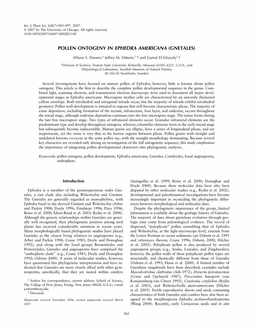
ontogenetic timing of the pollen grains; however, a limitedamount of variation occurs among pollen sacs. Pollen grainswithin a single pollen sac show consistent and synchronousdevelopment. Pollen ontogeny is presented below as it occursin four major stages, the microspore mother cell, tetrad, freemicrospore, and mature pollen grain stages. Once the firsttectal elements are laid down, each developing plica is char-acterized by crest and margin regions, with a furrow regionbetween adjacent plicae. These characters are illustrated infigure 1 as they appear in the nearly mature pollen wall, toserve as structural landmarks for comparison with ontoge-netic data.
Microspore Mother Cell Stage
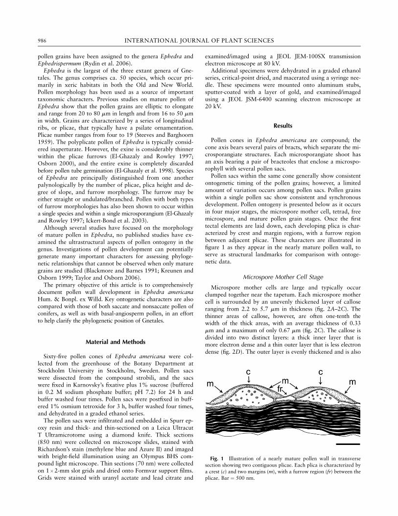
Microspore mother cells are large and typically occurclumped together near the tapetum. Each microspore mothercell is surrounded by an unevenly thickened layer of calloseranging from 2.2 to 5.7 mm in thickness (fig. 2A–2C). Thethinner areas of callose, however, are often one-tenth thewidth of the thick areas, with an average thickness of 0.33mm and a maximum of only 0.67 mm (fig. 2C). The callose isdivided into two distinct layers: a thick inner layer that ismore electron dense and a thin outer layer that is less electrondense (fig. 2D). The outer layer is evenly thickened and is also
Fig. 1 Illustration of a nearly mature pollen wall in transversesection showing two contiguous plicae. Each plica is characterized bya crest (c) and two margins (m), with a furrow region (fr) between theplicae. Bar ¼ 500 nm.
986 INTERNATIONAL JOURNAL OF PLANT SCIENCES
covered with an electron-dense microspore mother cell coat.Variation in thickness of the inner layer is responsible for theuneven thickening of the callose as a whole. Numerous spheri-cal channels/infiltrations occur in the inner callose layer, andthese channels/infiltrations consist of a central electron-densecore surrounded by a less electron-dense layer (fig. 2D).When developing microspores are in close proximity to
one another, callosic connections may be established betweentwo or more microspores. These connections result in an in-terruption of the outer callose wall of each microspore, withthe inner layer of the callose wall extending between adjacentmicrospores (fig. 2E–2G). As the callose thickens, these inter-microspore connections remain intact and widen (fig. 2G).The tapetum is robust at the microspore mother cell stage.
Individual cells comprising the tapetum can be distinguished
from one another because of their distinct nuclei (fig. 2A).The tapetum extends 80–100 mm into the pollen sac.
Tetrad Stage
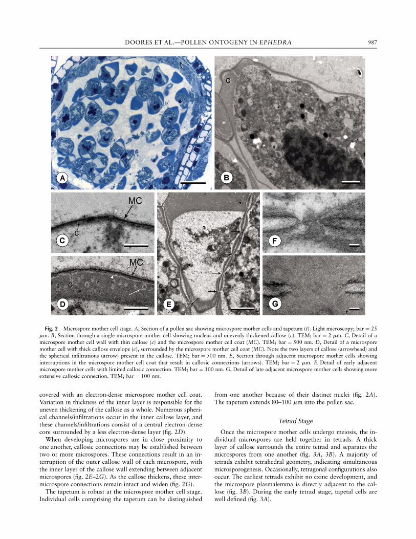
Once the microspore mother cells undergo meiosis, the in-dividual microspores are held together in tetrads. A thicklayer of callose surrounds the entire tetrad and separates themicrospores from one another (fig. 3A, 3B). A majority oftetrads exhibit tetrahedral geometry, indicating simultaneousmicrosporogenesis. Occasionally, tetragonal configurations alsooccur. The earliest tetrads exhibit no exine development, andthe microspore plasmalemma is directly adjacent to the cal-lose (fig. 3B). During the early tetrad stage, tapetal cells arewell defined (fig. 3A).
Fig. 2 Microspore mother cell stage. A, Section of a pollen sac showing microspore mother cells and tapetum (t). Light microscopy; bar ¼ 25mm. B, Section through a single microspore mother cell showing nucleus and unevenly thickened callose (c). TEM; bar ¼ 2 mm. C, Detail of amicrospore mother cell wall with thin callose (c) and the microspore mother cell coat (MC). TEM; bar ¼ 500 nm. D, Detail of a microsporemother cell with thick callose envelope (c), surrounded by the microspore mother cell coat (MC). Note the two layers of callose (arrowhead) andthe spherical infiltrations (arrow) present in the callose. TEM; bar ¼ 500 nm. E, Section through adjacent microspore mother cells showinginterruptions in the microspore mother cell coat that result in callosic connections (arrows). TEM; bar ¼ 2 mm. F, Detail of early adjacentmicrospore mother cells with limited callosic connection. TEM; bar ¼ 100 nm. G, Detail of late adjacent microspore mother cells showing moreextensive callosic connection. TEM; bar ¼ 100 nm.
987DOORES ET AL.—POLLEN ONTOGENY IN EPHEDRA
As development continues, the plasmalemma pulls awayfrom the callose at the crest regions of the future plicae, andan electron-translucent primexine develops (fig. 3C–3E). Athin, electron-dense layer forms adjacent to the callose, creat-ing the first elements of the tectum (fig. 3F). The tectum sepa-rates unevenly from the plasmalemma to form plicae beforeadditional pollen wall elements begin to develop. Because of
the uneven separation and uneven thickening of the tectum,the crest and margin regions of the plicae can be distin-guished in this early tetrad stage (fig. 3E). Tectal thickeningoccurs initially only at the margins of the plicae (fig. 3F, 3G).The tectum in the crest region of each plica thickens later indevelopment, after the margins are well established (fig. 4C).At this stage, no infratectal elements are detectable, and the
Fig. 3 Early tetrad stage. A, Section of a single pollen sac showing both tetragonal (arrow) and tetrahedral (arrowhead) types. Lightmicroscopy; bar ¼ 25 mm. B, Section of a single tetrahedral tetrad showing more evenly thickened callose (c). Note that there is no detectablemicrospore wall development. TEM; bar ¼ 2 mm. C, Section of a single microspore showing developing primexine in regions where theplasmalemma has pulled back from the callose (arrows). TEM; bar ¼ 2 mm. D, Detail of two adjacent microspore walls showing theplasmalemma pulling away from the callose (arrow). TEM; bar ¼ 500 nm. E, Detail showing plasmalemma separating from the callose such thatthe future crest, margin, and furrow regions can be seen. TEM; bar ¼ 100 nm. F, Detail showing developing microspore wall. Note that the firsttectal elements (T) form along the plicae margins. TEM; bar ¼ 100 nm.G, Detail showing developing microspore wall. Note that the tectum (T) isthicker along the margins and thinner at the crest and that the initial elements of the foot layer (F) have formed. TEM; bar ¼ 100 nm.
988 INTERNATIONAL JOURNAL OF PLANT SCIENCES
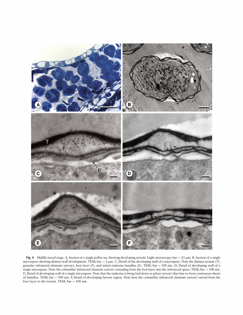
Fig. 4 Middle tetrad stage. A, Section of a single pollen sac showing developing tetrads. Light microscopy; bar ¼ 25 mm. B, Section of a singlemicrospore showing distinct wall development. TEM; bar ¼ 2 mm. C, Detail of the developing wall of a microspore. Note the distinct tectum (T),granular infratectal elements (arrow), foot layer (F), and initial endexine lamellae (E). TEM; bar ¼ 100 nm. D, Detail of developing wall of asingle microspore. Note the columellar infratectal elements (arrow) extending from the foot layer into the infratectal space. TEM; bar ¼ 100 nm.E, Detail of developing wall of a single microspore. Note that the endexine is being laid down as plates (arrow) that fuse to form continuous sheetsof lamellae. TEM; bar ¼ 100 nm. F, Detail of developing furrow region. Note how the columellar infratectal elements (arrow) extend from thefoot layer to the tectum. TEM; bar ¼ 100 nm.
overall thickness of the exine is between 250 and 450 nm. Athin foot layer develops next (fig. 3G), directly before the ini-tiation of the endexine.At the middle tetrad stage (fig. 4), endexine lamellae are
initially laid down in discontinuous fragments that later fuseto form continuous sheets (fig. 4E). Granulelike units existinitially between adjacent endexine lamellations (fig. 4C, 4E).As the number of endexine lamellae reaches four to six, the to-tal thickness of the exine ranges from 1.0 to 1.8 mm. Just abovethe endexine, the foot layer thickens evenly throughout thisstage (fig. 4C). Small, granular infratectal elements begin to de-velop in the middle tetrad stage (fig. 4C–4E). In addition,columellar infratectal elements form that span from the footlayer to the tectum. Although these columellar elements aremore abundant near furrows (fig. 4F), they are initially presentthroughout the infratectum, including under the crests (fig. 4D).At the late tetrad stage (fig. 5), the endexine increases in
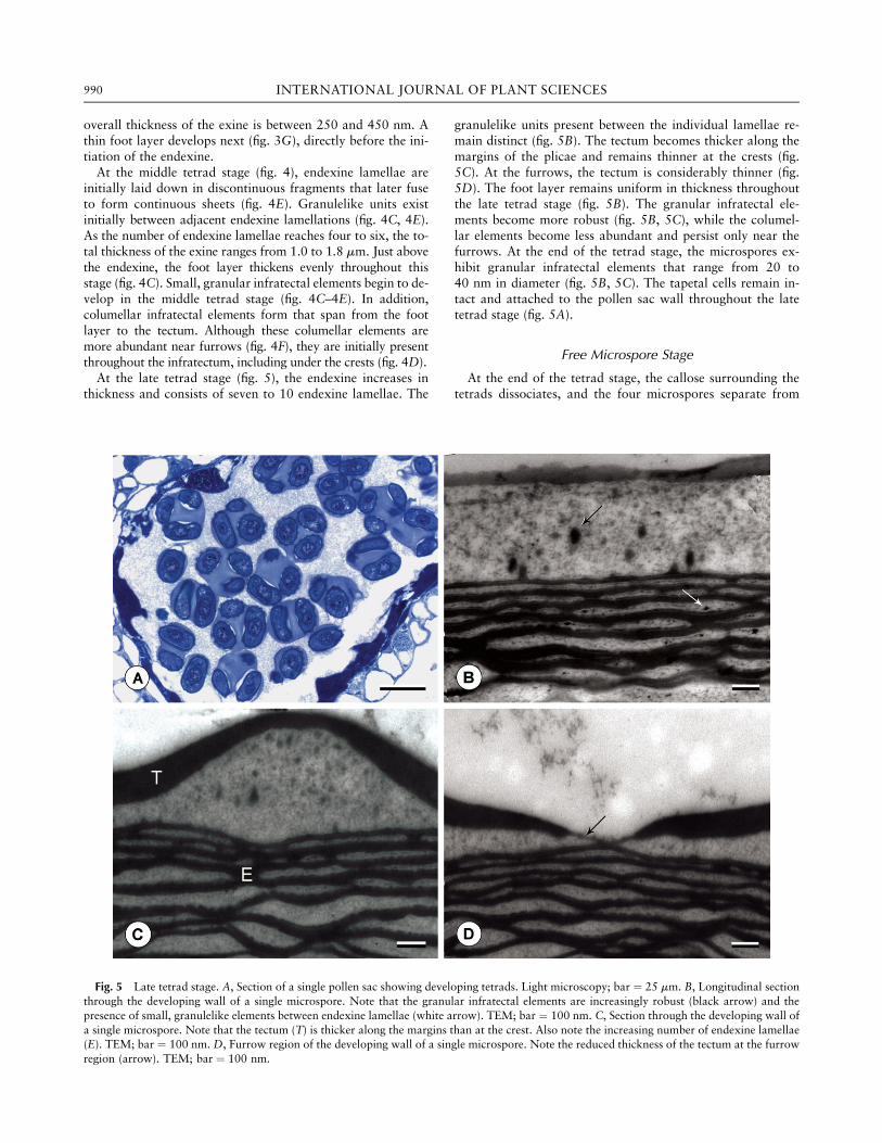
thickness and consists of seven to 10 endexine lamellae. The
granulelike units present between the individual lamellae re-main distinct (fig. 5B). The tectum becomes thicker along themargins of the plicae and remains thinner at the crests (fig.5C). At the furrows, the tectum is considerably thinner (fig.5D). The foot layer remains uniform in thickness throughoutthe late tetrad stage (fig. 5B). The granular infratectal ele-ments become more robust (fig. 5B, 5C), while the columel-lar elements become less abundant and persist only near thefurrows. At the end of the tetrad stage, the microspores ex-hibit granular infratectal elements that range from 20 to40 nm in diameter (fig. 5B, 5C). The tapetal cells remain in-tact and attached to the pollen sac wall throughout the latetetrad stage (fig. 5A).
Free Microspore Stage
At the end of the tetrad stage, the callose surrounding thetetrads dissociates, and the four microspores separate from
Fig. 5 Late tetrad stage. A, Section of a single pollen sac showing developing tetrads. Light microscopy; bar ¼ 25 mm. B, Longitudinal sectionthrough the developing wall of a single microspore. Note that the granular infratectal elements are increasingly robust (black arrow) and thepresence of small, granulelike elements between endexine lamellae (white arrow). TEM; bar ¼ 100 nm. C, Section through the developing wall ofa single microspore. Note that the tectum (T) is thicker along the margins than at the crest. Also note the increasing number of endexine lamellae(E). TEM; bar ¼ 100 nm. D, Furrow region of the developing wall of a single microspore. Note the reduced thickness of the tectum at the furrowregion (arrow). TEM; bar ¼ 100 nm.
990 INTERNATIONAL JOURNAL OF PLANT SCIENCES
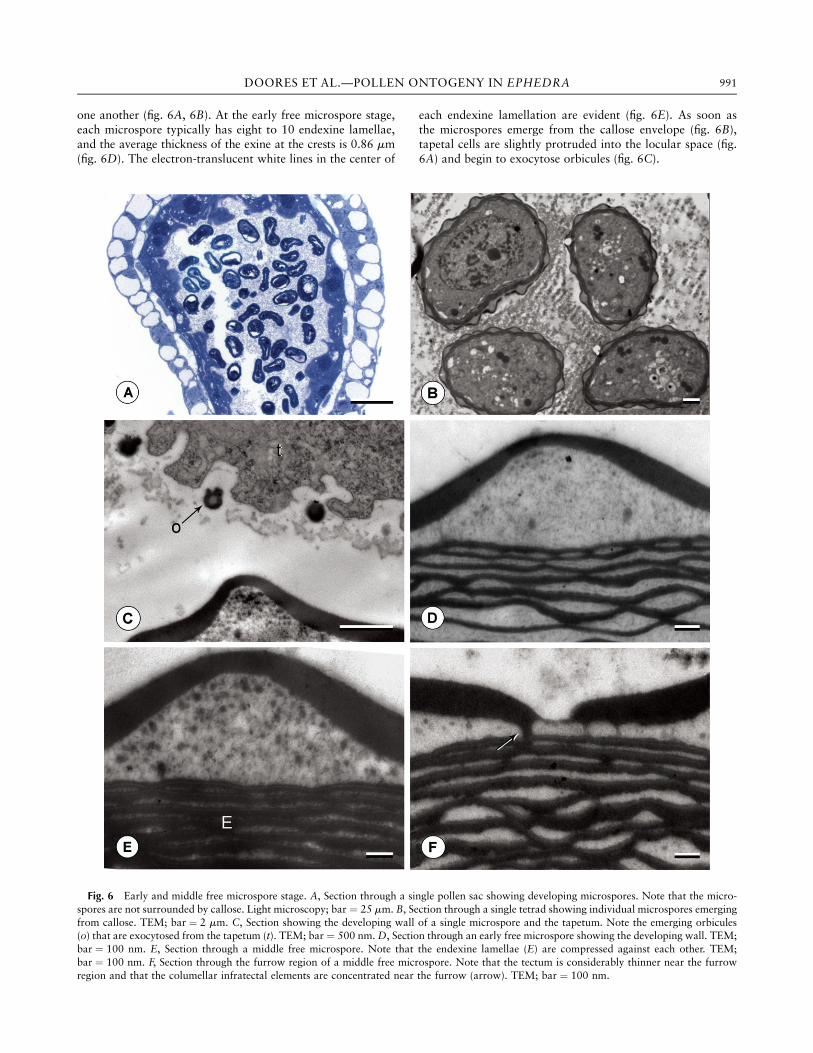
one another (fig. 6A, 6B). At the early free microspore stage,each microspore typically has eight to 10 endexine lamellae,and the average thickness of the exine at the crests is 0.86 mm(fig. 6D). The electron-translucent white lines in the center of
each endexine lamellation are evident (fig. 6E). As soon asthe microspores emerge from the callose envelope (fig. 6B),tapetal cells are slightly protruded into the locular space (fig.6A) and begin to exocytose orbicules (fig. 6C).
Fig. 6 Early and middle free microspore stage. A, Section through a single pollen sac showing developing microspores. Note that the micro-spores are not surrounded by callose. Light microscopy; bar ¼ 25 mm. B, Section through a single tetrad showing individual microspores emergingfrom callose. TEM; bar ¼ 2 mm. C, Section showing the developing wall of a single microspore and the tapetum. Note the emerging orbicules(o) that are exocytosed from the tapetum (t). TEM; bar ¼ 500 nm.D, Section through an early free microspore showing the developing wall. TEM;bar ¼ 100 nm. E, Section through a middle free microspore. Note that the endexine lamellae (E) are compressed against each other. TEM;bar ¼ 100 nm. F, Section through the furrow region of a middle free microspore. Note that the tectum is considerably thinner near the furrowregion and that the columellar infratectal elements are concentrated near the furrow (arrow). TEM; bar ¼ 100 nm.
991DOORES ET AL.—POLLEN ONTOGENY IN EPHEDRA
During the middle free microspore stage, the number ofendexine lamellae increases to 10–12, and the lamellae be-come more compressed against each other; thus, the granule-like units previously seen between lamellations can no longerbe distinguished (fig. 6E). The granular infratectal elementsbecome much more robust and begin to fill the majority of thespace in the infratectum. Granules range in diameter from 50to 80 nm (fig. 6E). In contrast, as the tectum concurrently in-creases in thickness, the columellar infratectal elements becomeeven more reduced in number during the free microspore stageand are only rarely distinguishable at the furrows (fig. 6F).In late free microspores (fig. 7), overall exine thickness
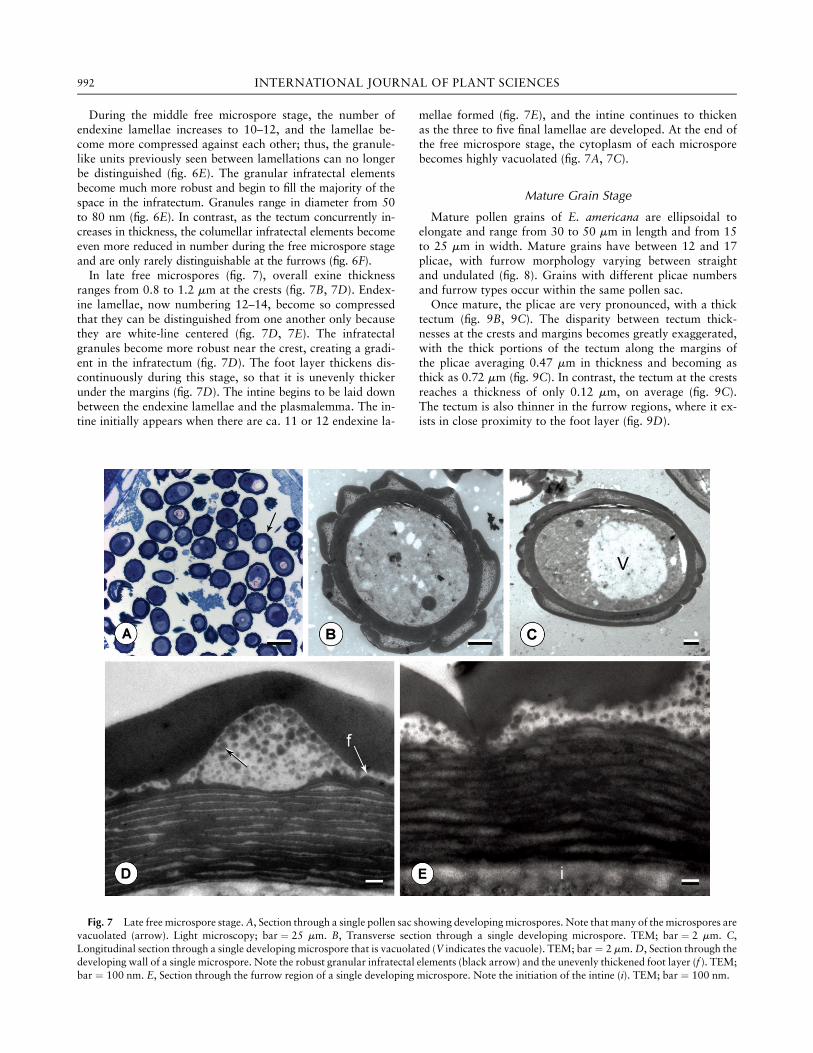
ranges from 0.8 to 1.2 mm at the crests (fig. 7B, 7D). Endex-ine lamellae, now numbering 12–14, become so compressedthat they can be distinguished from one another only becausethey are white-line centered (fig. 7D, 7E). The infratectalgranules become more robust near the crest, creating a gradi-ent in the infratectum (fig. 7D). The foot layer thickens dis-continuously during this stage, so that it is unevenly thickerunder the margins (fig. 7D). The intine begins to be laid downbetween the endexine lamellae and the plasmalemma. The in-tine initially appears when there are ca. 11 or 12 endexine la-
mellae formed (fig. 7E), and the intine continues to thickenas the three to five final lamellae are developed. At the end ofthe free microspore stage, the cytoplasm of each microsporebecomes highly vacuolated (fig. 7A, 7C).
Mature Grain Stage
Mature pollen grains of E. americana are ellipsoidal toelongate and range from 30 to 50 mm in length and from 15to 25 mm in width. Mature grains have between 12 and 17plicae, with furrow morphology varying between straightand undulated (fig. 8). Grains with different plicae numbersand furrow types occur within the same pollen sac.Once mature, the plicae are very pronounced, with a thick
tectum (fig. 9B, 9C). The disparity between tectum thick-nesses at the crests and margins becomes greatly exaggerated,with the thick portions of the tectum along the margins ofthe plicae averaging 0.47 mm in thickness and becoming asthick as 0.72 mm (fig. 9C). In contrast, the tectum at the crestsreaches a thickness of only 0.12 mm, on average (fig. 9C).The tectum is also thinner in the furrow regions, where it ex-ists in close proximity to the foot layer (fig. 9D).
Fig. 7 Late free microspore stage.A, Section through a single pollen sac showing developingmicrospores. Note that many of themicrospores arevacuolated (arrow). Light microscopy; bar ¼ 25 mm. B, Transverse section through a single developing microspore. TEM; bar ¼ 2 mm. C,Longitudinal section through a single developing microspore that is vacuolated (V indicates the vacuole). TEM; bar ¼ 2 mm.D, Section through thedeveloping wall of a single microspore. Note the robust granular infratectal elements (black arrow) and the unevenly thickened foot layer (f ). TEM;bar ¼ 100 nm. E, Section through the furrow region of a single developing microspore. Note the initiation of the intine (i). TEM; bar ¼ 100 nm.
992 INTERNATIONAL JOURNAL OF PLANT SCIENCES
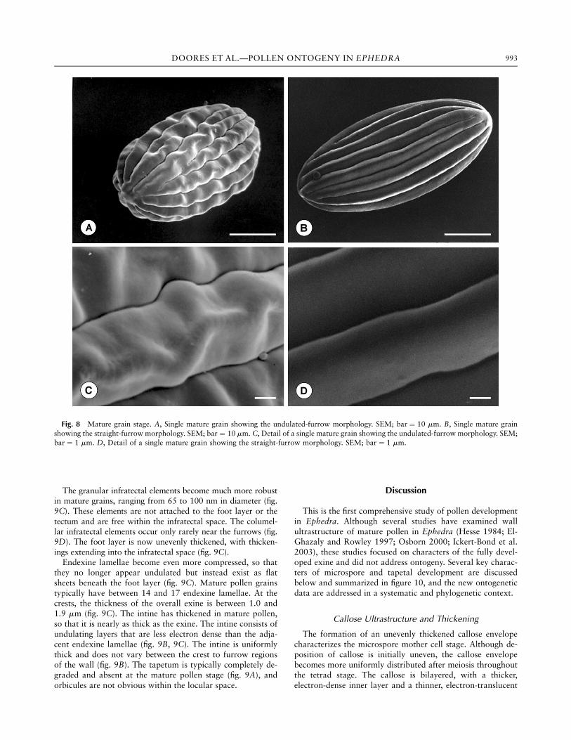
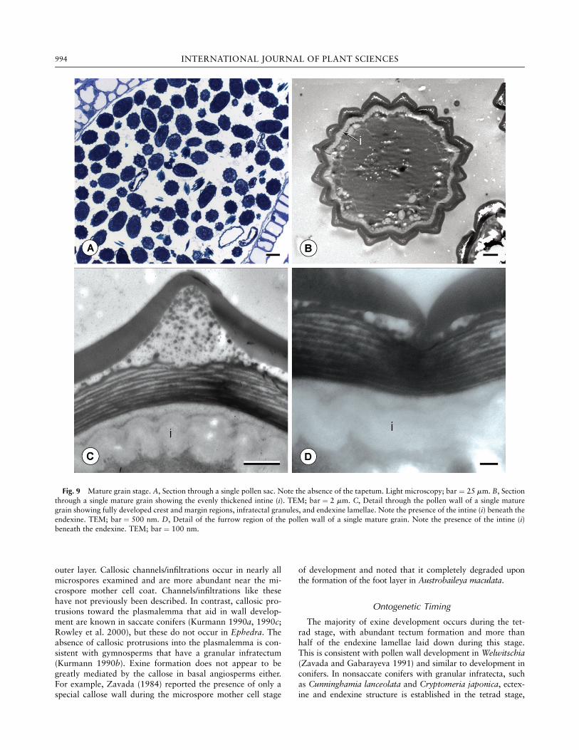
The granular infratectal elements become much more robustin mature grains, ranging from 65 to 100 nm in diameter (fig.9C). These elements are not attached to the foot layer or thetectum and are free within the infratectal space. The columel-lar infratectal elements occur only rarely near the furrows (fig.9D). The foot layer is now unevenly thickened, with thicken-ings extending into the infratectal space (fig. 9C).Endexine lamellae become even more compressed, so that
they no longer appear undulated but instead exist as flatsheets beneath the foot layer (fig. 9C). Mature pollen grainstypically have between 14 and 17 endexine lamellae. At thecrests, the thickness of the overall exine is between 1.0 and1.9 mm (fig. 9C). The intine has thickened in mature pollen,so that it is nearly as thick as the exine. The intine consists ofundulating layers that are less electron dense than the adja-cent endexine lamellae (fig. 9B, 9C). The intine is uniformlythick and does not vary between the crest to furrow regionsof the wall (fig. 9B). The tapetum is typically completely de-graded and absent at the mature pollen stage (fig. 9A), andorbicules are not obvious within the locular space.
Discussion
This is the first comprehensive study of pollen developmentin Ephedra. Although several studies have examined wallultrastructure of mature pollen in Ephedra (Hesse 1984; El-Ghazaly and Rowley 1997; Osborn 2000; Ickert-Bond et al.2003), these studies focused on characters of the fully devel-oped exine and did not address ontogeny. Several key charac-ters of microspore and tapetal development are discussedbelow and summarized in figure 10, and the new ontogeneticdata are addressed in a systematic and phylogenetic context.
Callose Ultrastructure and Thickening
The formation of an unevenly thickened callose envelopecharacterizes the microspore mother cell stage. Although de-position of callose is initially uneven, the callose envelopebecomes more uniformly distributed after meiosis throughoutthe tetrad stage. The callose is bilayered, with a thicker,electron-dense inner layer and a thinner, electron-translucent
Fig. 8 Mature grain stage. A, Single mature grain showing the undulated-furrow morphology. SEM; bar ¼ 10 mm. B, Single mature grainshowing the straight-furrow morphology. SEM; bar ¼ 10 mm. C, Detail of a single mature grain showing the undulated-furrow morphology. SEM;bar ¼ 1 mm. D, Detail of a single mature grain showing the straight-furrow morphology. SEM; bar ¼ 1 mm.
993DOORES ET AL.—POLLEN ONTOGENY IN EPHEDRA
outer layer. Callosic channels/infiltrations occur in nearly allmicrospores examined and are more abundant near the mi-crospore mother cell coat. Channels/infiltrations like thesehave not previously been described. In contrast, callosic pro-trusions toward the plasmalemma that aid in wall develop-ment are known in saccate conifers (Kurmann 1990a, 1990c;Rowley et al. 2000), but these do not occur in Ephedra. Theabsence of callosic protrusions into the plasmalemma is con-sistent with gymnosperms that have a granular infratectum(Kurmann 1990b). Exine formation does not appear to begreatly mediated by the callose in basal angiosperms either.For example, Zavada (1984) reported the presence of only aspecial callose wall during the microspore mother cell stage
of development and noted that it completely degraded uponthe formation of the foot layer in Austrobaileya maculata.
Ontogenetic Timing
The majority of exine development occurs during the tet-rad stage, with abundant tectum formation and more thanhalf of the endexine lamellae laid down during this stage.This is consistent with pollen wall development in Welwitschia(Zavada and Gabarayeva 1991) and similar to development inconifers. In nonsaccate conifers with granular infratecta, suchas Cunninghamia lanceolata and Cryptomeria japonica, ectex-ine and endexine structure is established in the tetrad stage,
Fig. 9 Mature grain stage. A, Section through a single pollen sac. Note the absence of the tapetum. Light microscopy; bar ¼ 25 mm. B, Sectionthrough a single mature grain showing the evenly thickened intine (i). TEM; bar ¼ 2 mm. C, Detail through the pollen wall of a single maturegrain showing fully developed crest and margin regions, infratectal granules, and endexine lamellae. Note the presence of the intine (i) beneath theendexine. TEM; bar ¼ 500 nm. D, Detail of the furrow region of the pollen wall of a single mature grain. Note the presence of the intine (i)beneath the endexine. TEM; bar ¼ 100 nm.
994 INTERNATIONAL JOURNAL OF PLANT SCIENCES
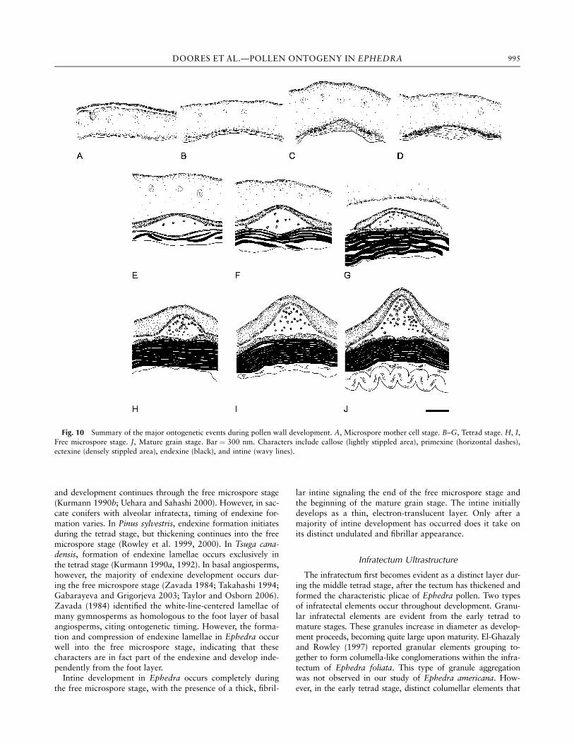
and development continues through the free microspore stage(Kurmann 1990b; Uehara and Sahashi 2000). However, in sac-cate conifers with alveolar infratecta, timing of endexine for-mation varies. In Pinus sylvestris, endexine formation initiatesduring the tetrad stage, but thickening continues into the freemicrospore stage (Rowley et al. 1999, 2000). In Tsuga cana-densis, formation of endexine lamellae occurs exclusively inthe tetrad stage (Kurmann 1990a, 1992). In basal angiosperms,however, the majority of endexine development occurs dur-ing the free microspore stage (Zavada 1984; Takahashi 1994;Gabarayeva and Grigorjeva 2003; Taylor and Osborn 2006).Zavada (1984) identified the white-line-centered lamellae ofmany gymnosperms as homologous to the foot layer of basalangiosperms, citing ontogenetic timing. However, the forma-tion and compression of endexine lamellae in Ephedra occurwell into the free microspore stage, indicating that thesecharacters are in fact part of the endexine and develop inde-pendently from the foot layer.Intine development in Ephedra occurs completely during
the free microspore stage, with the presence of a thick, fibril-
lar intine signaling the end of the free microspore stage andthe beginning of the mature grain stage. The intine initiallydevelops as a thin, electron-translucent layer. Only after amajority of intine development has occurred does it take onits distinct undulated and fibrillar appearance.
Infratectum Ultrastructure
The infratectum first becomes evident as a distinct layer dur-ing the middle tetrad stage, after the tectum has thickened andformed the characteristic plicae of Ephedra pollen. Two typesof infratectal elements occur throughout development. Granu-lar infratectal elements are evident from the early tetrad tomature stages. These granules increase in diameter as develop-ment proceeds, becoming quite large upon maturity. El-Ghazalyand Rowley (1997) reported granular elements grouping to-gether to form columella-like conglomerations within the infra-tectum of Ephedra foliata. This type of granule aggregationwas not observed in our study of Ephedra americana. How-ever, in the early tetrad stage, distinct columellar elements that
Fig. 10 Summary of the major ontogenetic events during pollen wall development. A, Microspore mother cell stage. B–G, Tetrad stage. H, I,Free microspore stage. J, Mature grain stage. Bar ¼ 300 nm. Characters include callose (lightly stippled area), primexine (horizontal dashes),ectexine (densely stippled area), endexine (black), and intine (wavy lines).
995DOORES ET AL.—POLLEN ONTOGENY IN EPHEDRA
span from the foot layer to the tectum are present throughoutthe infratectum. These elements develop separately from thegranular elements, and, rather than increasing in width andbecoming more robust as the grains mature, they become lessfrequent and almost indiscernible throughout exine ontogeny.At maturity, the columellae are only rarely discernible near fur-rows. These columellar elements may serve as a remnant fromthe separation of the foot layer and the tectum, which occursduring the early tetrad stage, or they may aid in foot layer andtectum development. Columellar elements like these have notbeen reported previously in Ephedra.Overall, the granular infratectum present at maturity is
more similar to that of nonsaccate conifer pollen grains (e.g.,Cunninghamia). Columellar infratectal elements have beenreported in several basal angiosperms, including Illiciumreligiosum (Takahashi 1994), A. maculata (Zavada 1984),Schisandra chinensis (Gabarayeva and Grigorjeva 2003), Ca-bomba caroliniana (Osborn et al. 1991), Brasenia schreberi(Taylor and Osborn 2006), and other genera of Nymphaeales(Osborn et al. 2004). Amborella trichopoda is reported tohave a meandering tectum that extends to the foot layer butis described as lacking true columellae (Hesse 2001).
Overall Exine Thickness
The exine first begins to develop in the earliest tetrad stageand continues its development until the grains are mature. Thethickness of the exine continues to increase from initial pri-mexine formation in the earliest tetrad stage until several end-exine lamellae are laid down in the middle tetrad stage. Duringthe late tetrad stage and the early free microspore stage, the end-exine lamellae become compressed. Because of this, the overallthickness of the exine is actually less in the free microspore stagethan in the middle tetrad stage. However, overall exine thick-ness is greatest when endexine deposition is complete, despitethe compression of the lamellae throughout the ontogeneticsequence. This is due primarily to considerable thickening of thetectum and expansion of the infratectal space in the crest re-gions, as well as modest thickening of the foot layer.
Pollen Shape and Surface Ornamentation
The shape and surface morphology of E. americana pollenare typical of ephedroid pollen, in that E. americana grainsare elliptic to elongate in shape, with distinctive plicae and fur-rows. Furrow morphology has historically been used as a defin-ing feature in Ephedra (Steeves and Barghoorn 1959; Kedves1987); however, this study has documented variation within E.americana. Pollen grains with straight and undulated furrowsco-occur in the same pollen sac. The straight morphology domi-nates, occurring ca. 90% of the time, with undulating furrowspresent only occasionally. Because varying furrow morphologieshave now been described within the same pollen sac in severalspecies (Ickert-Bond et al. 2003; this study), this character mustbe reexamined as a taxonomically diagnostic tool in Ephedra.
Tapetal Ontogeny
Although an array of tapetal types have been described inangiosperms (Pacini et al. 1985; Furness and Rudall 2001), ta-petal ontogeny is more consistent among gymnosperms. How-ever, less information is available regarding tapetum biologyin gymnosperms. In E. americana, the tapetum is distinguish-able as a distinct cell layer from the beginning of the micro-spore mother cell stage. It initially exists as a thick, robust celllayer attached to the middle layers of the pollen sac wall. Asthe microspores mature, the tapetum begins to disintegrate,and it is completely absent or remains only as a remnant celllayer by the time the grains mature. Because it never becomesseparated from the pollen sac wall, the tapetum of Ephedra isof the secretory type, like that of most gymnosperms (Paciniet al. 1985). Orbicules, or Ubisch bodies, are typically pro-duced by a secretory tapetum. Orbicules are present in E.americana; they become evident in the late tetrad stage andpersist into the free microspore stage. Orbicules first emergefrom the tapetum as evaginations that become distinct spheresand migrate toward the developing microspores.
Conclusion
Ephedra, along with Welwitschia and Gnetum, comprisesthe gymnospermous order Gnetales. Because the systematicand phylogenetic position of the Gnetales has been controversial,both morphological and molecular studies are needed to clarifyphylogeny. This study reexamined morphological and ultrastruc-tural characters of the mature pollen wall and described anddocumented pollen developmental characters in Ephedra forthe first time. Several key characters, such as endexine develop-mental timing, ephemeral columellar infratectal elements, andcallosic channels/infiltrations, are revealed only during an exami-nation of the full ontogenetic sequence. Some characters, suchas tripartite endexine lamellae, endexine developmental timing,and a secretory tapetum, align Ephedra with conifers and gym-nosperms. However, other characters indicate a possible linkbetween Gnetales and basal angiosperms. These include thebrief presence of columellar infratectal elements. This study em-phasizes the importance of including developmental pollencharacters in systematic studies and the need for integrating on-togenetic data, along with other morphological and molecularcharacters, in determining phylogenetic relationships.
Acknowledgments
We thank Elisabeth Grafstrom (Swedish Museum of Natu-ral History) and Mackenzie L. Taylor and Tara C. Theimann(Truman State University) for help with plant collection andspecimen preparation, as well as Bjorn Walles (StockholmUniversity, Sweden) for access to plant material. This studywas supported in part by the J. William Fulbright SeniorScholar Program, the National Science Foundation (grantsIBN-0212521, MRI-0216391), and Truman State University(Undergraduate Student Research Grant).
996 INTERNATIONAL JOURNAL OF PLANT SCIENCES
Literature Cited
Arber EAN, J Parkin 1908 The relationship of the angiosperms to theGnetales. Ann Bot 22:489–515.
Ash SR 1972 Late Triassic plants from the Chinle Formation innortheastern Arizona. Paleontology 15:598–618.
Blackmore S, SH Barnes 1991 Pollen and spores: pattern of diversi-fication. Clarendon, New York.
Bowe LM, G Coat, CW dePamphilis 2000 Phylogeny of seed plantsbased on all three genomic compartments: extant gymnosperms aremonophyletic and Gnetales’ closest relatives are conifers. Proc NatlAcad Sci USA 97:4092–4097.
Crane PR 1985 Phylogenetic analysis of seed plants and the origin ofangiosperms. Ann Mo Bot Gard 72:716–793.
——— 1996 The fossil history of the Gnetales. Int J Plant Sci157(suppl):S50–S57.
Crane PR, GR Upchurch 1987 Drewria potomacensis gen. et sp.nov., an early Cretaceous member of Gnetales from the PotomacGroup of Virginia. Am J Bot 74:1722–1736.
Dilcher DL, ME Bernardes-De-Oliveira, D Pons, TA Lot 2005Welwitschiaceae from the lower Cretaceous of northeastern Brazil.Am J Bot 92:1294–1310.
Donoghue MJ, JA Doyle 2000 Seed plant phylogeny: demise of theanthophyte hypothesis? Curr Biol 10:R106–R109.
Doyle JA 1996 Seed plant phylogenies and the relationships of theGnetales. Int J Plant Sci 157(suppl):S3–S39.
Doyle JA, Donoghue MJ 1992 Fossils and seed plant phylogenyreanalyzed. Brittonia 44:89–106.
El-Ghazaly G, JR Rowley 1997 Pollen wall of Ephedra foliata.Palynology 21:7–18.
El-Ghazaly G, JR Rowley, M Hesse 1998 Polarity, aperture conditionand germination in pollen grains of Ephedra (Gnetales). Plant SystEvol 213:217–231.
Friedman WE 1996 Introduction to biology and evolution of theGnetales. Int J Plant Sci 157(suppl):S1–S2.
Furness CA, PJ Rudall 2001 The tapetum in basal angiosperms: earlydiversity. Int J Plant Sci 162:375–392.
Gabarayeva N, V Grigorjeva 2003 Comparative study of the pollenwall development in Illicium floridanum (Illiciaceae) and Schisan-dra chinensis (Schisandraceae). Taiwania 48:147–167.
Hesse M 1984 Pollenkitt is lacking in Gnetatae: Ephedra andWelwitschia; further proof for its restriction to the angiosperms.Plant Syst Evol 144:9–16.
——— 2001 Pollen characters of Amborella trichopoda (Amborella-ceae): a reinvestigation. Int J Plant Sci 162:201–208.
Hesse M, M Weber, H Halbritter 2000 A comparative study of poly-plicate pollen types in Arales, Laurales, Zingiberales, Gnetales. Pages227–239 in MM Harley, CM Morton, S Blackmore, eds. Pollen andspores: morphology and biology. Royal Botanic Gardens, Kew.
Ickert-Bond SM, JJ Skvarla, WF Chissoe 2003 Pollen dimorphisms inEphedra L. (Ephedraceae). Rev Palaeobot Palynol 124:325–334.
Kedves M 1987 LM and EM studies on pollen grains of recentWelwitschia mirabilis Hook, and Ephedra species. Acta Bot Hung33:81–103.
Kreunen SS, JM Osborn 1999 Pollen and anther development inNelumbo. Am J Bot 86:1662–1676.
Kurmann MH 1990a Development of the pollen wall in Tsugacanadensis (Pinaceae). Nord J Bot 10:63–78.
——— 1990b Exine formation in Cunninghamia lanceolata (Tax-odiaceae). Rev Palaeobot Palynol 64:175–179.
——— 1990c Exine ontogeny in conifers. Pages 157–172 in SBlackmore, RB Knox, eds. Microspores: evolution and ontogeny.Academic Press, London.
——— 1992 Exine stratification in extant gymnosperms: a review ofpublished transmission electron micrographs. Kew Bull 47:25–39.
Osborn JM 2000 Pollen morphology and ultrastructure of gymno-spermous anthophytes. Pages 163–185 inMMHarley, CMMorton,S Blackmore, eds. Pollen and spores: morphology and biology.Royal Botanic Gardens, Kew.
Osborn JM, JA Schwartz, BL Gutman, NA Melrose, AM Ingraham,ML Taylor, JN Strandquist, et al 2004 Pollen ontogeny in theNymphaeales. Polen 14:20–21.
Osborn JM, TN Taylor, MR de Lima 1993 The ultrastructure offossil ephedroid pollen with gnetalean affinities from the LowerCretaceous of Brazil. Rev Palaeobot Palynol 77:171–184.
Osborn JM, TN Taylor, EL Schneider 1991 Pollen morphology andultrastructure of the Cabombaceae: correlations with pollinationbiology. Am J Bot 78:1367–1378.
Pacini E, GG Franchi, M Hesse 1985 The tapetum: its form, func-tion, and possible phylogeny in Embryophyta. Plant Syst Evol 149:155–185.
Price RA 1996 Systematics of the Gnetales: a review of morpholog-ical and molecular evidence. Int J Plant Sci 157(suppl):S40–S49.
Rowley JR, JJ Skvarla, B Walles 1999 Microsporogenesis in Pinussylvestris. VII. Exine expansion and tapetal development. Taiwania44:325–344.
——— 2000 Microsporogenesis in Pinus sylvestris VI. Exine andtapetal development during the tetrad stage. Nord J Bot 20:67–87.
Rydin C, M Kallersjo, EM Friis 2002 Seed plant relationships andthe systematic position of Gnetales based on nuclear and chloro-plast DNA: conflicting data, rooting problems and the monophylyof conifers. Int J Plant Sci 163:197–214.
Rydin C, B Mohr, EM Friis 2003 Craytonia cotyledon gen. et sp.nov., a unique Cretaceous seedling related to Welwitschia. Biol Lett270:29–32.
Rydin C, KR Pedersen, PR Crane, EM Friis 2006 Former diversity ofEphedra (Gnetales): evidence from early Cretaceous seeds fromPortugal and North America. Ann Bot 98:123–140.
Rydin C, KR Pedersen, EM Friis 2004 On the evolutionary history ofEphedra: Cretaceous fossils and extant molecules. Proc Natl AcadSci USA 101:16571–16576.
Samigullin, TK, WF Martin, AV Troitsky, AS Antonov 1999 Molec-ular data from the chloroplast rpoC1 gene suggest a deep anddistinct dichotomy of contemporary spermatophytes in two mono-phyla: gymnosperms (including Gnetales) and angiosperms. J MolEvol 49:310–315.
Steeves MW, ES Barghoorn 1959 The pollen of Ephedra. J ArnoldArbor Harv Univ 40:221–255.
Takahashi M 1994 Exine development in Illicium religiosum Sieb. etZucc. (Illiciaceae). Grana 33:309–312.
Taylor ML, JM Osborn 2006 Pollen ontogeny in Brasenia (Cabom-baceae, Nymphaeales). Am J Bot 93:334–356.
Uehara K, Sahashi N 2000 Pollen wall development in Cryptomeriajaponica (Taxodiaceae). Grana 39:267–274.
van Konijnenburg-van Cittert H 1992 An enigmatic Liassic micro-sporophyll yielding Ephedripites pollen. Rev Palaeobot Palynol 71:239–254.
Wang Z 2004 A new Permian gnetalean cone as fossil evidence forsupporting current molecular phylogeny. Ann Bot 94:281–288.
Zavada MS 1984 Pollen wall development of Austrobaileya mac-ulata. Bot Gaz 145:11–21.
Zavada MS, N Gabarayeva 1991 Comparative pollen wall develop-ment of Welwitschia mirabilis and selected primitive angiosperms.Bull Torrey Bot Club 118:292–302.
997DOORES ET AL.—POLLEN ONTOGENY IN EPHEDRA