Embed Size (px)
Citation preview
Objectives
• Label the layers, chambers, valves & major blood vessels on a diagram of the heart.
•Differentiate between what happens in the heart during each phase
•List the three major types of blood vessels and the action of each type.
•Compare the three main blood cells by describing the function of each type.
•Describe at least five diseases of the circulatory system.
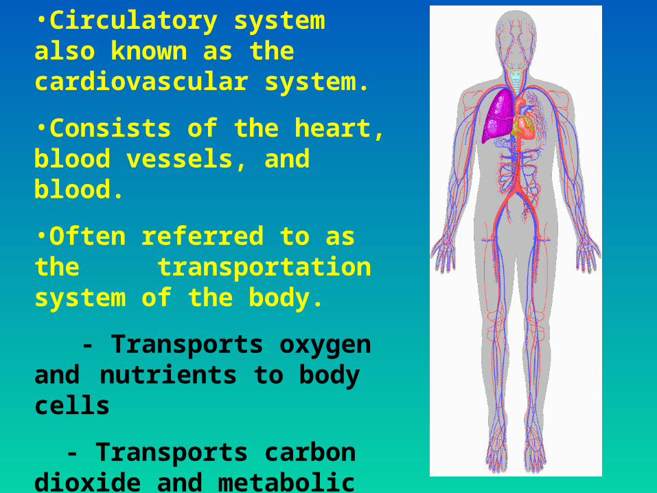
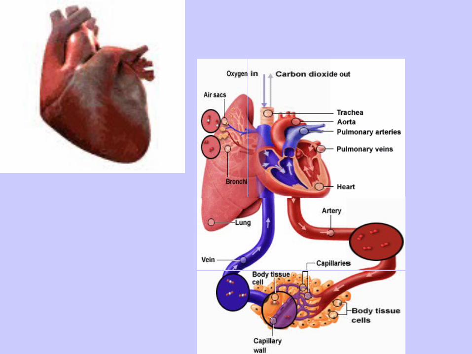
•Circulatory system also known as the cardiovascular system.
•Consists of the heart, blood vessels, and blood.
•Often referred to as the transportation system of the body.
- Transports oxygen and nutrients to body cells
- Transports carbon dioxide and metabolic materials away from body cells.
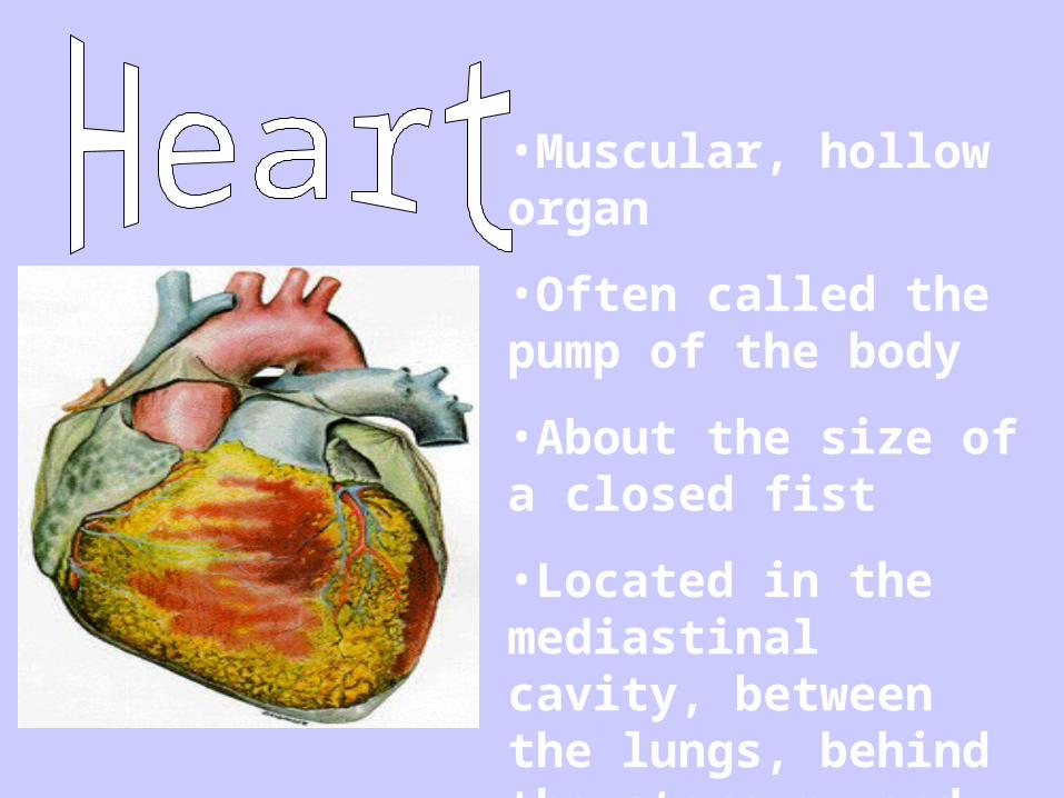
•Muscular, hollow organ
•Often called the pump of the body
•About the size of a closed fist
•Located in the mediastinal cavity, between the lungs, behind the sternum, and above the diaphragm.
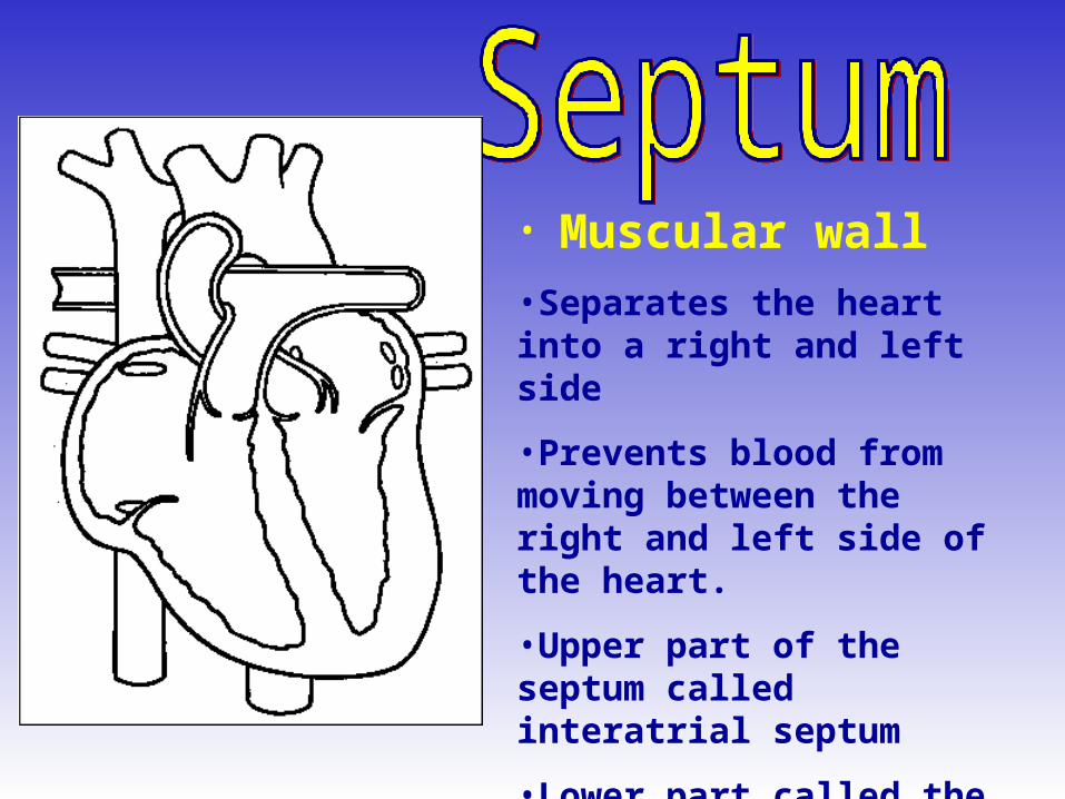
• Muscular wall•Separates the heart into a right and left side
•Prevents blood from moving between the right and left side of the heart.
•Upper part of the septum called interatrial septum
•Lower part called the interventricular septum.
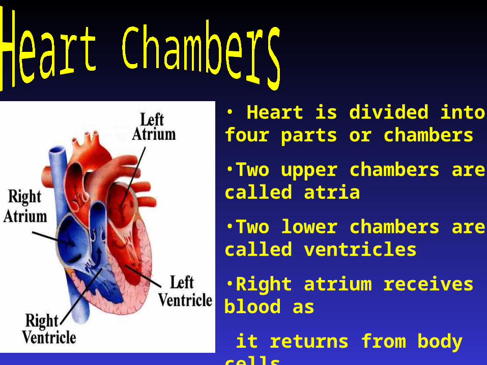
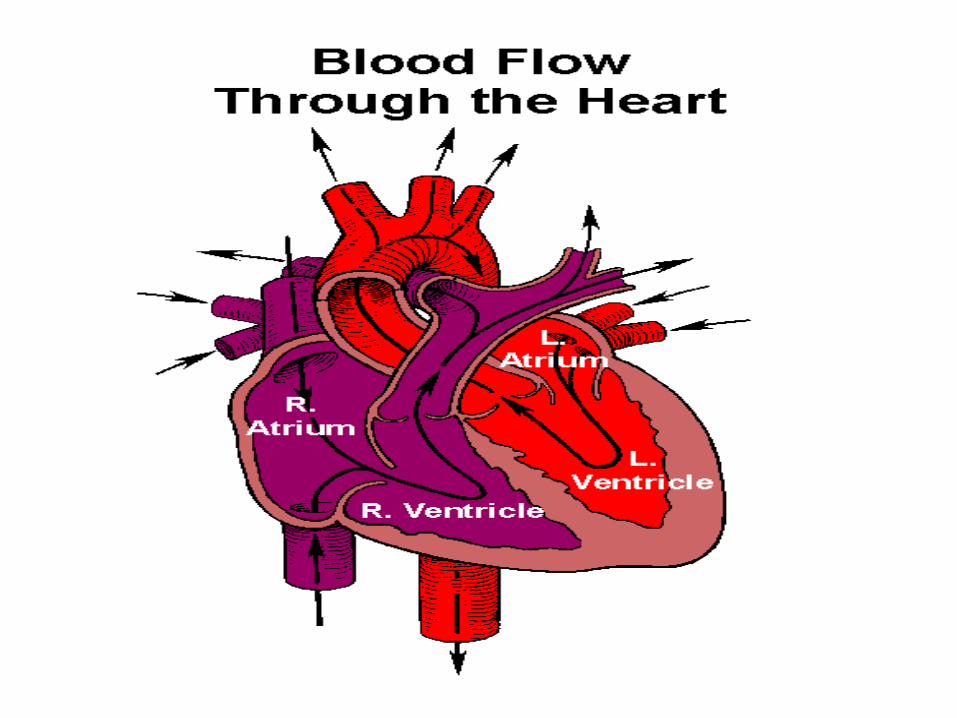
• Heart is divided into four parts or chambers
•Two upper chambers are called atria
•Two lower chambers are called ventricles
•Right atrium receives blood as
it returns from body cells
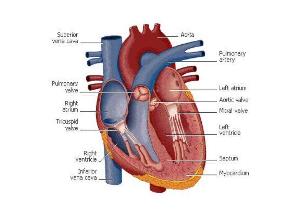
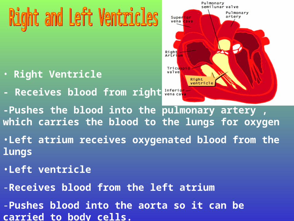
• Right Ventricle
- Receives blood from right atrium
-Pushes the blood into the pulmonary artery , which carries the blood to the lungs for oxygen
•Left atrium receives oxygenated blood from the lungs
•Left ventricle
-Receives blood from the left atrium
-Pushes blood into the aorta so it can be carried to body cells.
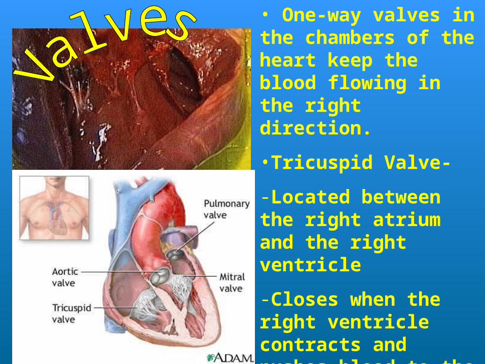
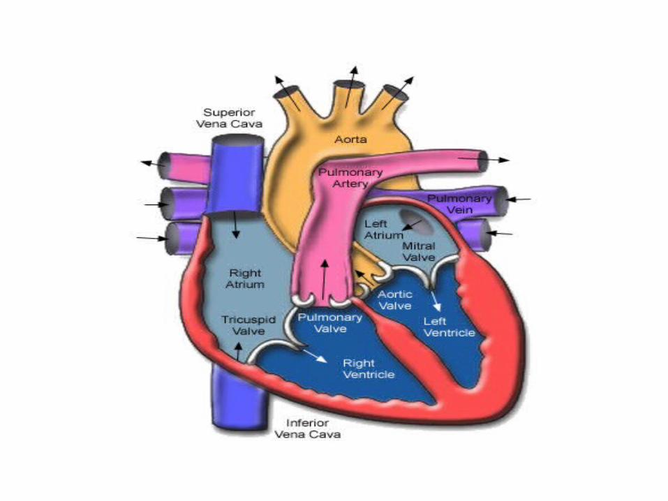
• One-way valves in the chambers of the heart keep the blood flowing in the right direction.
•Tricuspid Valve-
-Located between the right atrium and the right ventricle
-Closes when the right ventricle contracts and pushes blood to the lungs
-Prevents blood from flowing back into the right atrium
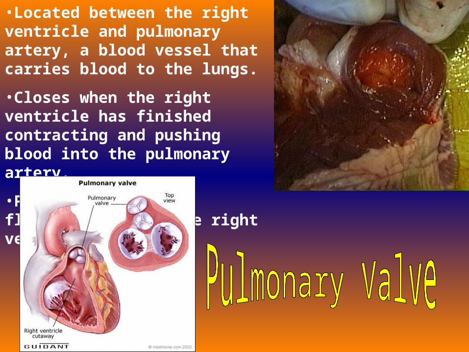
•Located between the right ventricle and pulmonary artery, a blood vessel that carries blood to the lungs.
•Closes when the right ventricle has finished contracting and pushing blood into the pulmonary artery.
•Prevents blood from flowing back into the right ventricle.
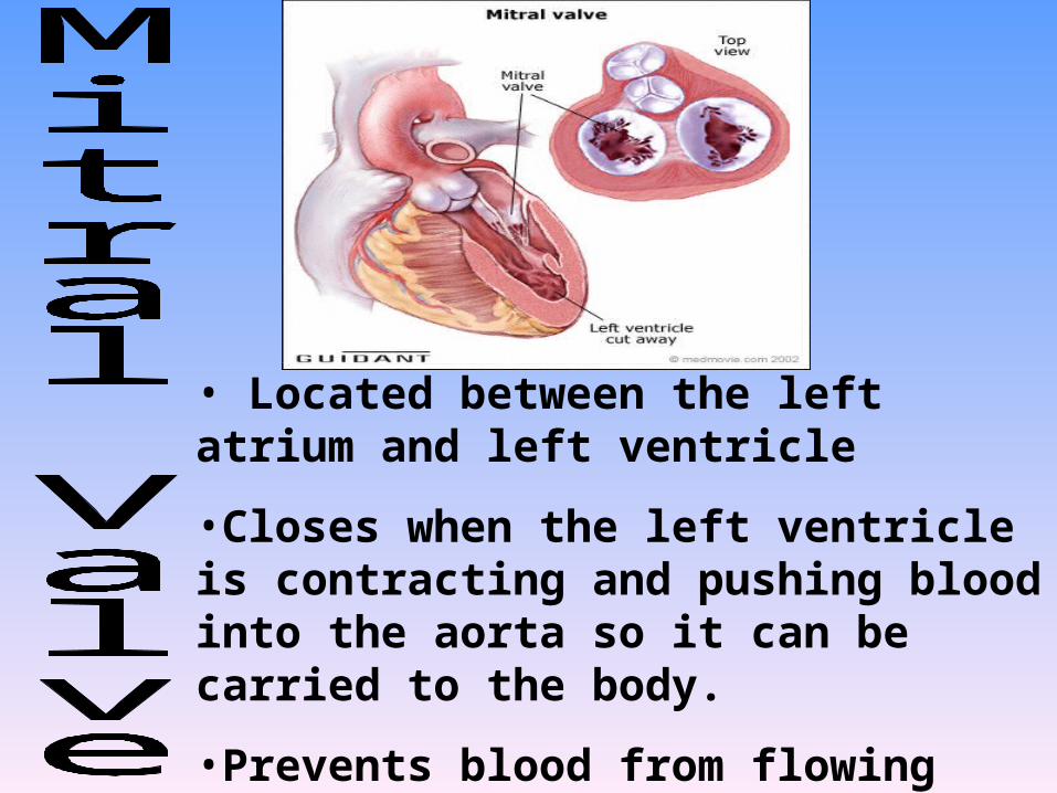
• Located between the left atrium and left ventricle
•Closes when the left ventricle is contracting and pushing blood into the aorta so it can be carried to the body.
•Prevents blood from flowing back into left atrium.
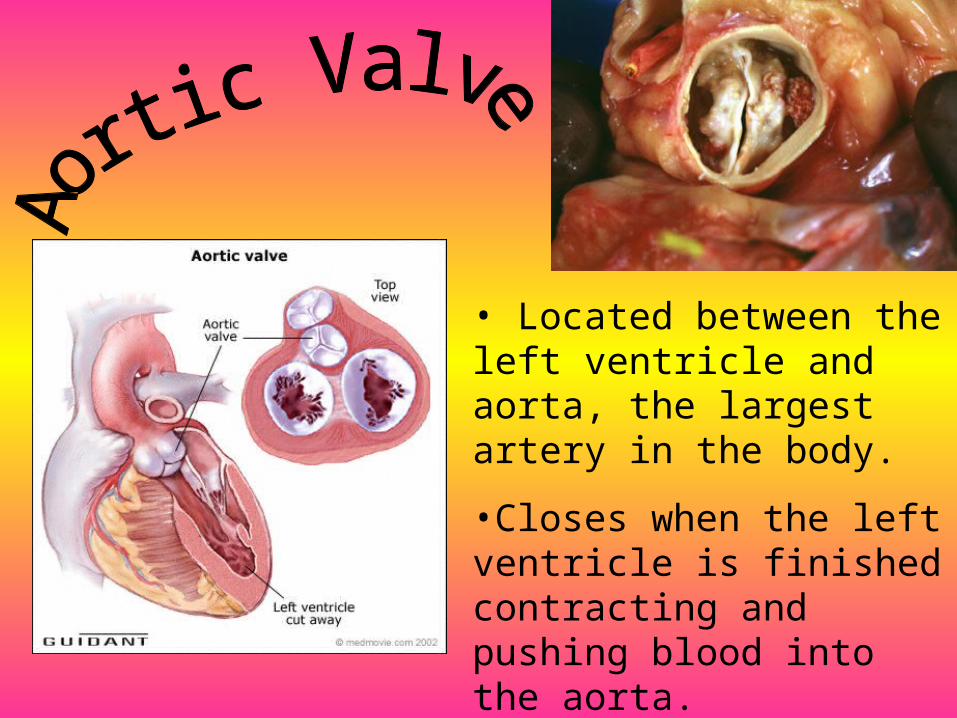
• Located between the left ventricle and aorta, the largest artery in the body.
•Closes when the left ventricle is finished contracting and pushing blood into the aorta.
•Prevents blood from flowing back into the left ventricle.
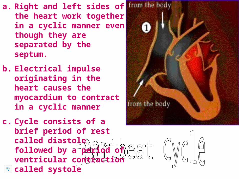
a. Right and left sides of the heart work together in a cyclic manner even though they are separated by the septum.
b. Electrical impulse originating in the heart causes the myocardium to contract in a cyclic manner
c. Cycle consists of a brief period of rest called diastole followed by a period of ventricular contraction called systole
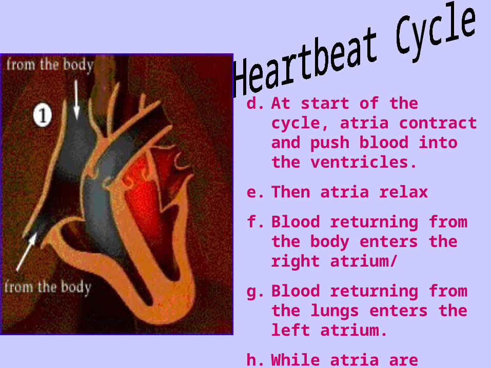
d. At start of the cycle, atria contract and push blood into the ventricles.
e. Then atria relax
f. Blood returning from the body enters the right atrium/
g. Blood returning from the lungs enters the left atrium.
h. While atria are filling, systole begins and the ventricles contract.
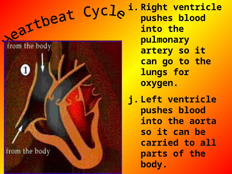
i. Right ventricle pushes blood into the pulmonary artery so it can go to the lungs for oxygen.
j. Left ventricle pushes blood into the aorta so it can be carried to all parts of the body.
k. Blood in the right side of the heart is low in oxygen and high in carbon dioxide.
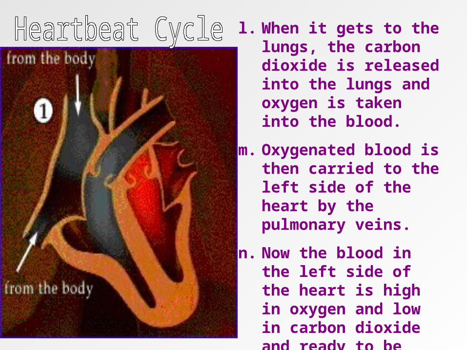
l. When it gets to the lungs, the carbon dioxide is released into the lungs and oxygen is taken into the blood.
m. Oxygenated blood is then carried to the left side of the heart by the pulmonary veins.
n. Now the blood in the left side of the heart is high in oxygen and low in carbon dioxide and ready to be carried to body cells.
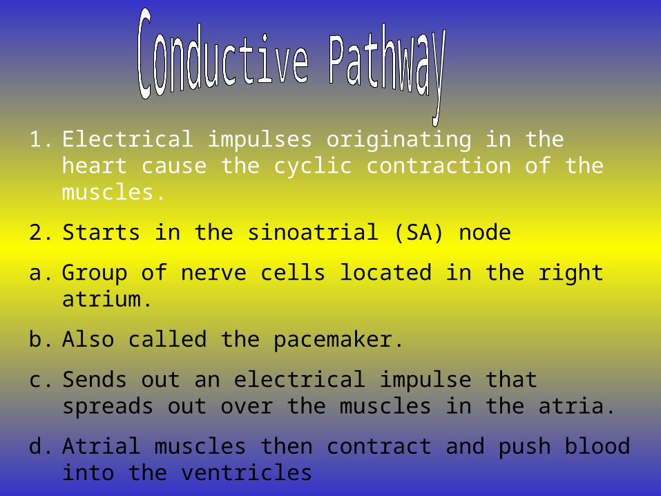
1. Electrical impulses originating in the heart cause the cyclic contraction of the muscles.
2. Starts in the sinoatrial (SA) node
a. Group of nerve cells located in the right atrium.
b. Also called the pacemaker.
c. Sends out an electrical impulse that spreads out over the muscles in the atria.
d. Atrial muscles then contract and push blood into the ventricles
e. After electrical impulse passes through the atria, it reaches the atrioventricular (AV) node.
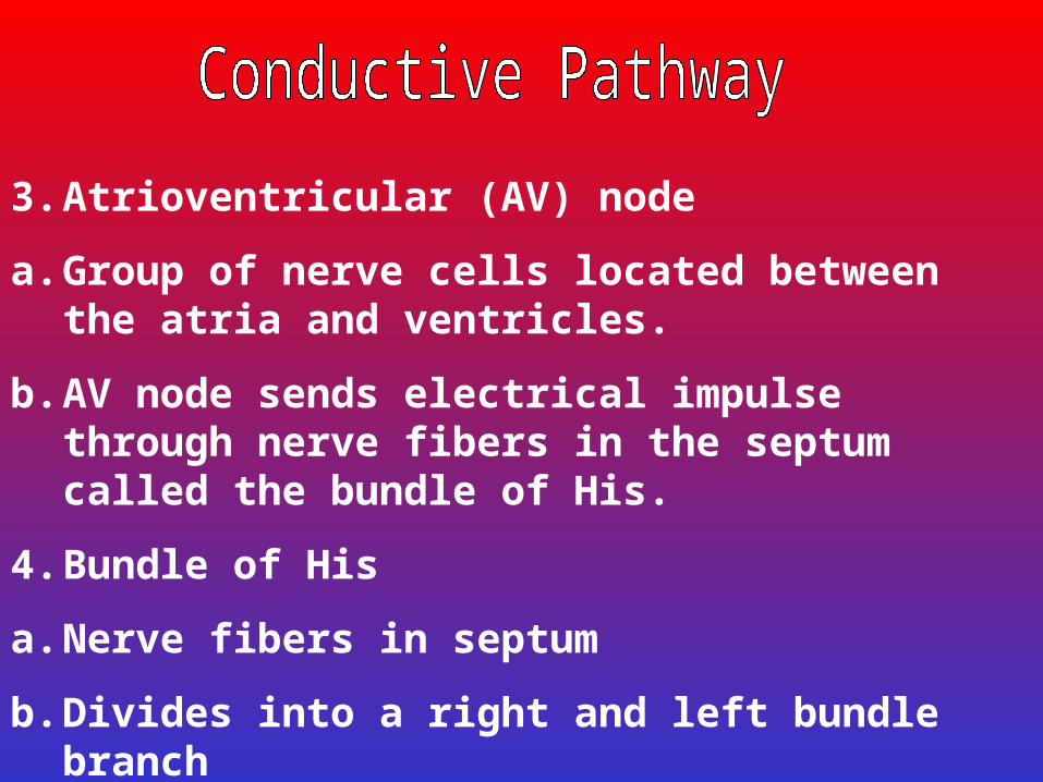
3. Atrioventricular (AV) node
a. Group of nerve cells located between the atria and ventricles.
b. AV node sends electrical impulse through nerve fibers in the septum called the bundle of His.
4. Bundle of His
a. Nerve fibers in septum
b. Divides into a right and left bundle branch
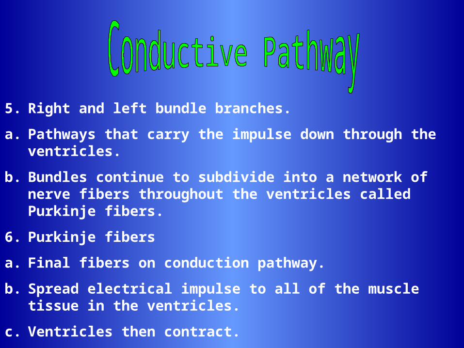
5. Right and left bundle branches.
a. Pathways that carry the impulse down through the ventricles.
b. Bundles continue to subdivide into a network of nerve fibers throughout the ventricles called Purkinje fibers.
6. Purkinje fibers
a. Final fibers on conduction pathway.
b. Spread electrical impulse to all of the muscle tissue in the ventricles.
c. Ventricles then contract.
7. Electrical conduction pattern occurs approximately every 0.8 seconds.
8. Movement of the electrical impulse can be recorded on an electrocardiogram (ECG or EKG) and used to detect abnormal activity or disease.
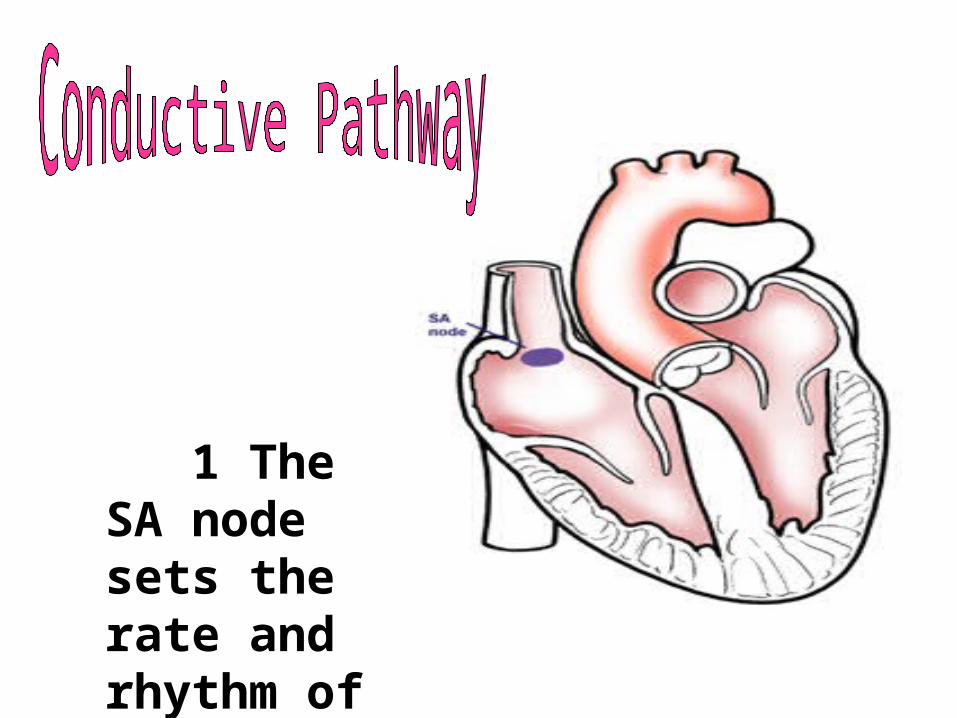
1 The SA node sets the rate and rhythm of your heartbeat
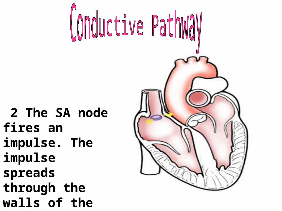
2 The SA node fires an impulse. The impulse spreads through the walls of the right and left atria, causing them to contract. This forces blood into the ventricles.
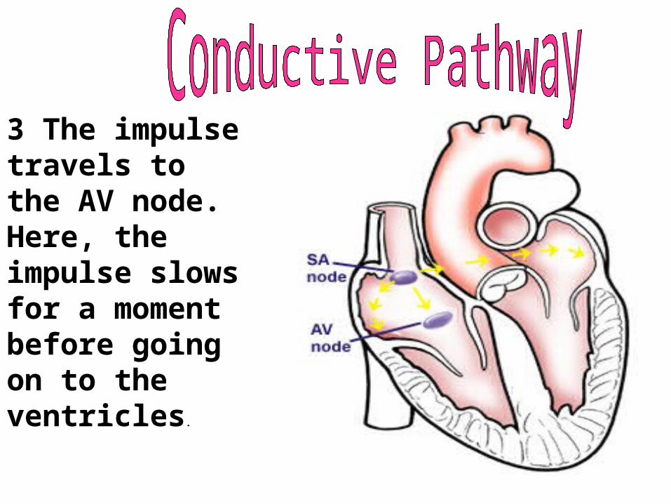
3 The impulse travels to the AV node. Here, the impulse slows for a moment before going on to the ventricles.
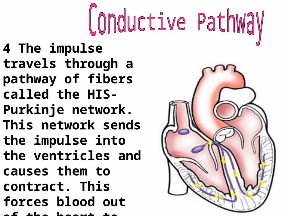
4 The impulse travels through a pathway of fibers called the HIS-Purkinje network. This network sends the impulse into the ventricles and causes them to contract. This forces blood out of the heart to the lungs and body.
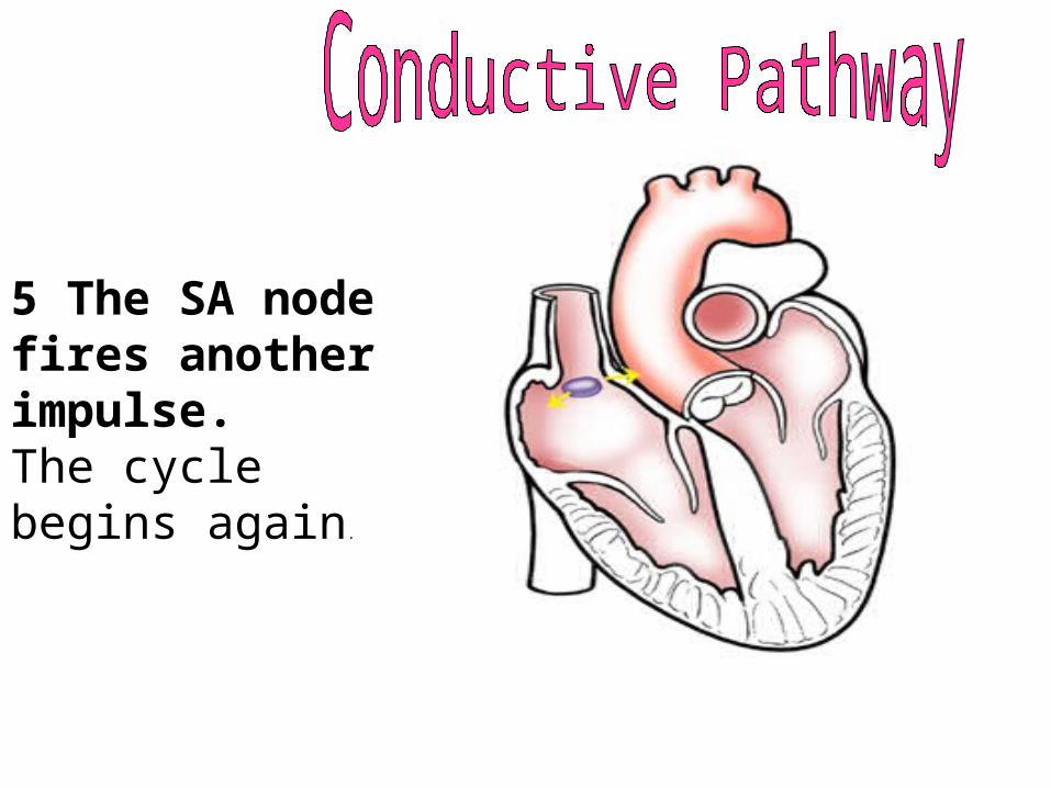
5 The SA node fires another impulse. The cycle begins again.
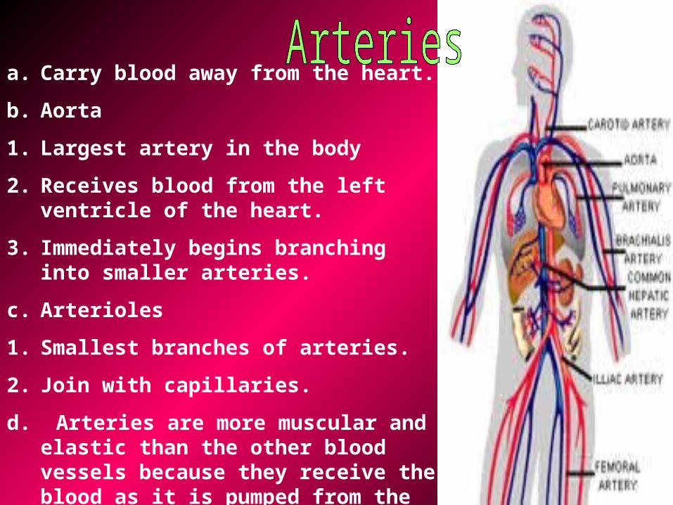
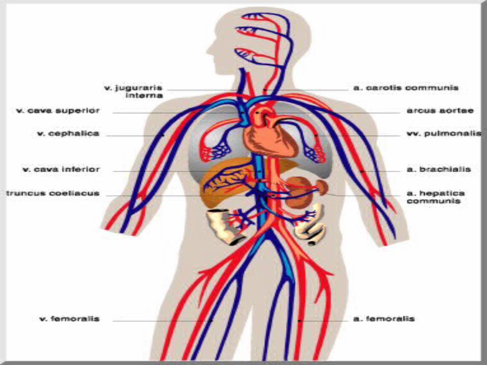
a. Carry blood away from the heart.
b. Aorta
1. Largest artery in the body
2. Receives blood from the left ventricle of the heart.
3. Immediately begins branching into smaller arteries.
c. Arterioles
1. Smallest branches of arteries.
2. Join with capillaries.
d. Arteries are more muscular and elastic than the other blood vessels because they receive the blood as it is pumped from the heart.
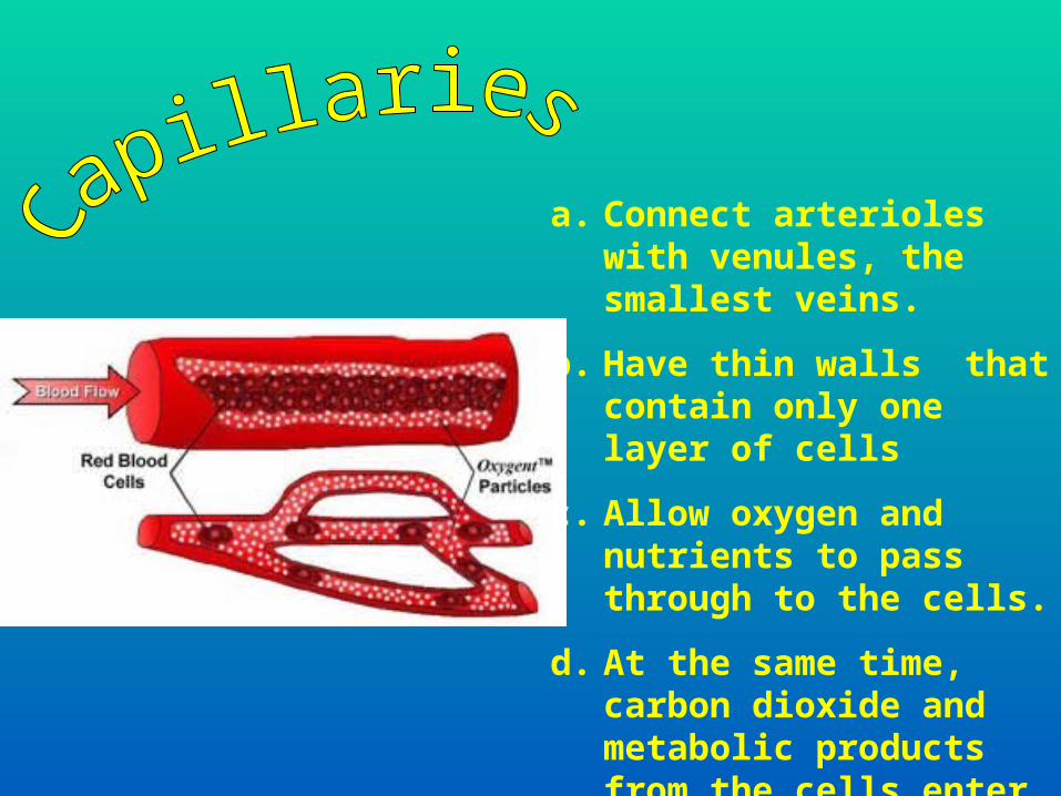
a. Connect arterioles with venules, the smallest veins.
b. Have thin walls that contain only one layer of cells
c. Allow oxygen and nutrients to pass through to the cells.
d. At the same time, carbon dioxide and metabolic products from the cells enter the capillaries.
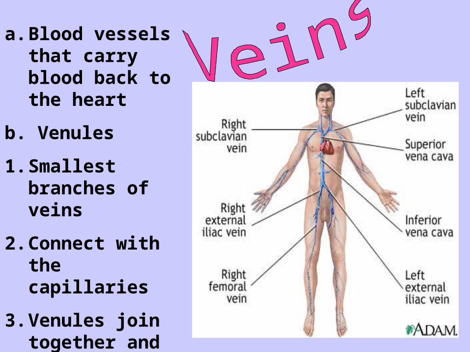
a. Blood vessels that carry blood back to the heart
b. Venules
1. Smallest branches of veins
2. Connect with the capillaries
3. Venules join together and become larger to form veins
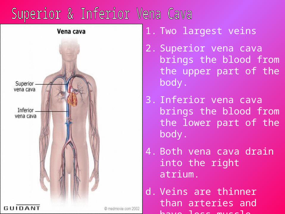
1. Two largest veins
2. Superior vena cava brings the blood from the upper part of the body.
3. Inferior vena cava brings the blood from the lower part of the body.
4. Both vena cava drain into the right atrium.
d. Veins are thinner than arteries and have less muscle tissue.
e. Most contain valves that keep the blood from flowing in a backward direction.
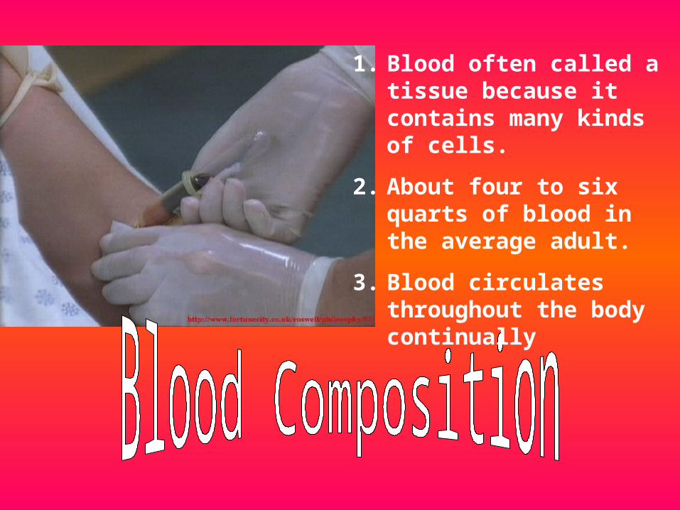
1. Blood often called a tissue because it contains many kinds of cells.
2. About four to six quarts of blood in the average adult.
3. Blood circulates throughout the body continually
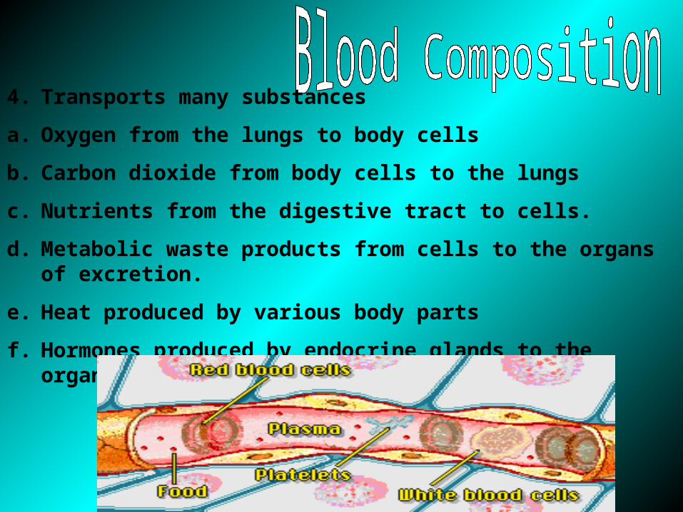
4. Transports many substances
a. Oxygen from the lungs to body cells
b. Carbon dioxide from body cells to the lungs
c. Nutrients from the digestive tract to cells.
d. Metabolic waste products from cells to the organs of excretion.
e. Heat produced by various body parts
f. Hormones produced by endocrine glands to the organs in the body.
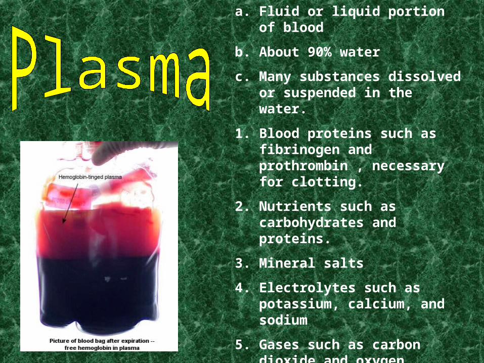
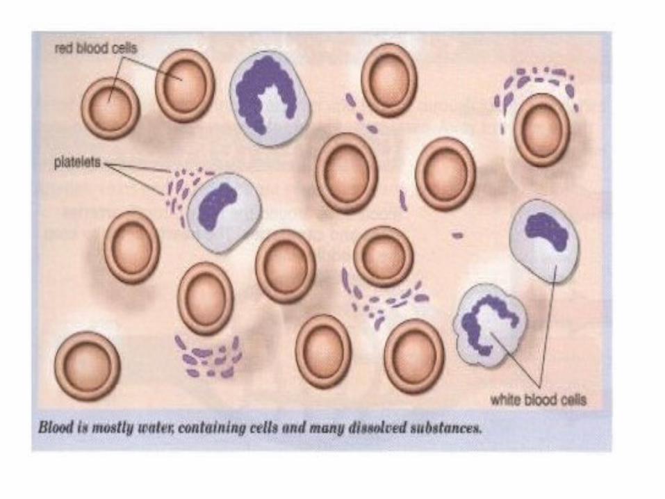
a. Fluid or liquid portion of blood
b. About 90% water
c. Many substances dissolved or suspended in the water.
1. Blood proteins such as fibrinogen and prothrombin , necessary for clotting.
2. Nutrients such as carbohydrates and proteins.
3. Mineral salts
4. Electrolytes such as potassium, calcium, and sodium
5. Gases such as carbon dioxide and oxygen
6. Metabolic and waste products
7. Hormones
8. Enzymes

a. Solid elements of blood.
b. Three main kinds of blood cells or corpuscles: erythrocytes, leukocytes, and thrombocytes
c. Erythrocytes or red blood cells.
1. Produced in the red bone marrow at a rate of about one million per minute.
2. Live about 120 days before being broken down by the liver and spleen.
3. Four and a half to five and a half million erythrocytes per cubic millimeter of blood ( approximately one drop of blood), or 25 trillion in body
4. Mature form circulating in the blood does not have a nucleus and is shaped like a disc with a thinner central area.
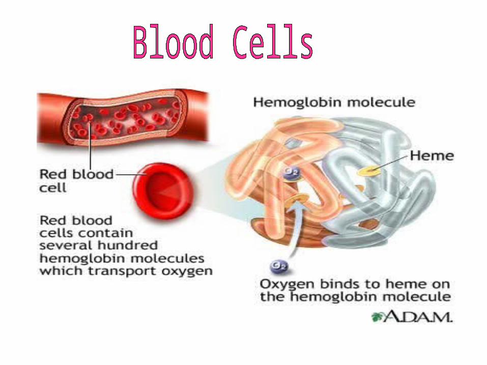
5. Contain a complex protein called hemoglobin
aa. Composed of protein molecule called globin and an iron compound called heme
ab. Carries both oxygen and carbon dioxide
ac. When hemoglobin carries oxygen and carbon dioxide.
ad. If blood contains a lot of oxygen, it is bright red.
ae. When there is less oxygen and more carbon dioxide, it is a much darker red.
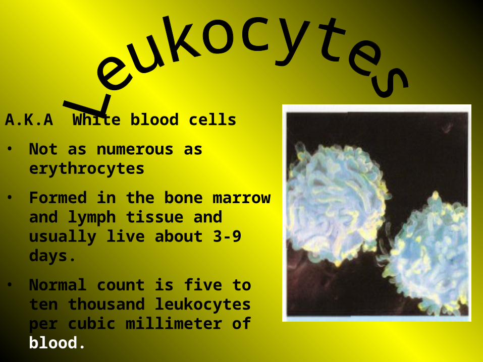
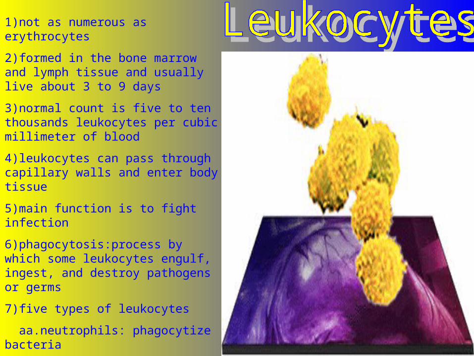
A.K.A White blood cells
• Not as numerous as erythrocytes
• Formed in the bone marrow and lymph tissue and usually live about 3-9 days.
• Normal count is five to ten thousand leukocytes per cubic millimeter of blood.
• Leukocytes can pass through capillary walls and enter body tissue.
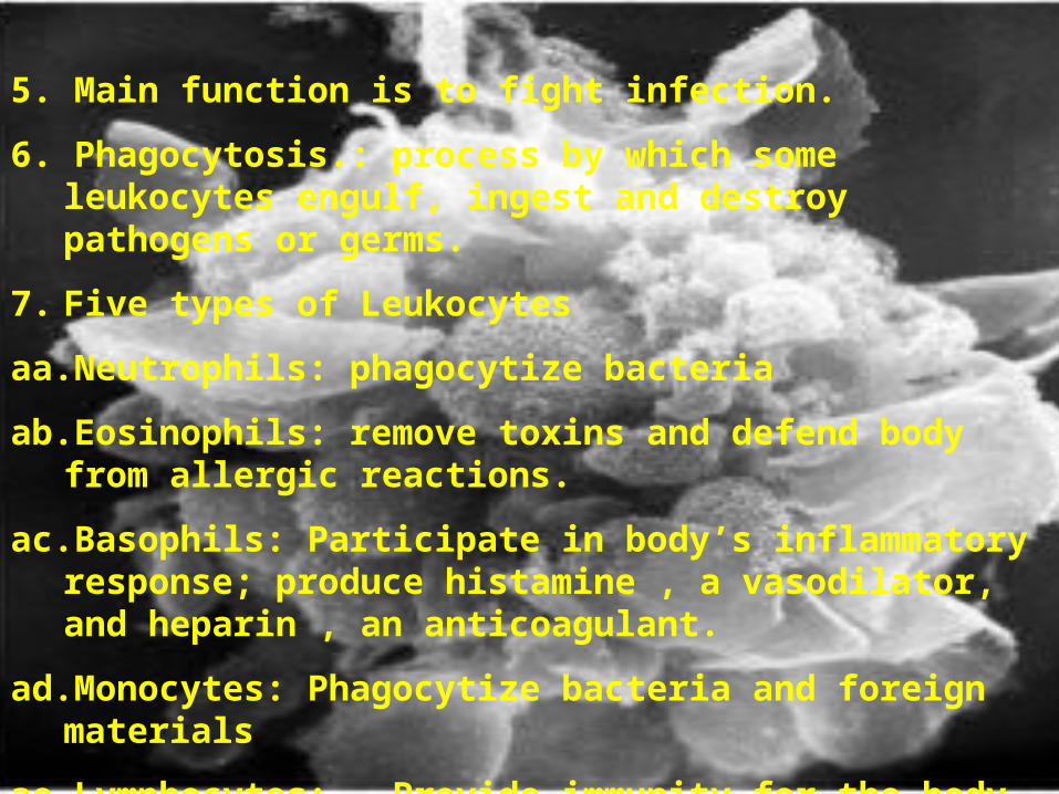
5. Main function is to fight infection.
6. Phagocytosis.: process by which some leukocytes engulf, ingest and destroy pathogens or germs.
7. Five types of Leukocytes
aa. Neutrophils: phagocytize bacteria
ab. Eosinophils: remove toxins and defend body from allergic reactions.
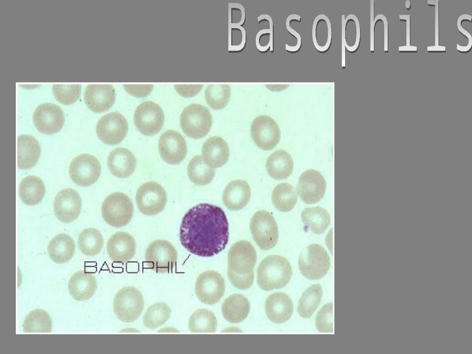
ac. Basophils: Participate in body’s inflammatory response; produce histamine , a vasodilator, and heparin , an anticoagulant.
ad. Monocytes: Phagocytize bacteria and foreign materials
ae. Lymphocytes: Provide immunity for the body by developing antibodies; protect against formation of cancer cells.
1)not as numerous as erythrocytes
2)formed in the bone marrow and lymph tissue and usually live about 3 to 9 days
3)normal count is five to ten thousands leukocytes per cubic millimeter of blood
4)leukocytes can pass through capillary walls and enter body tissue
5)main function is to fight infection
6)phagocytosis:process by which some leukocytes engulf, ingest, and destroy pathogens or germs
7)five types of leukocytes
aa.neutrophils: phagocytize bacteria
eosinophils
basophils
monocytes
lymphocytes
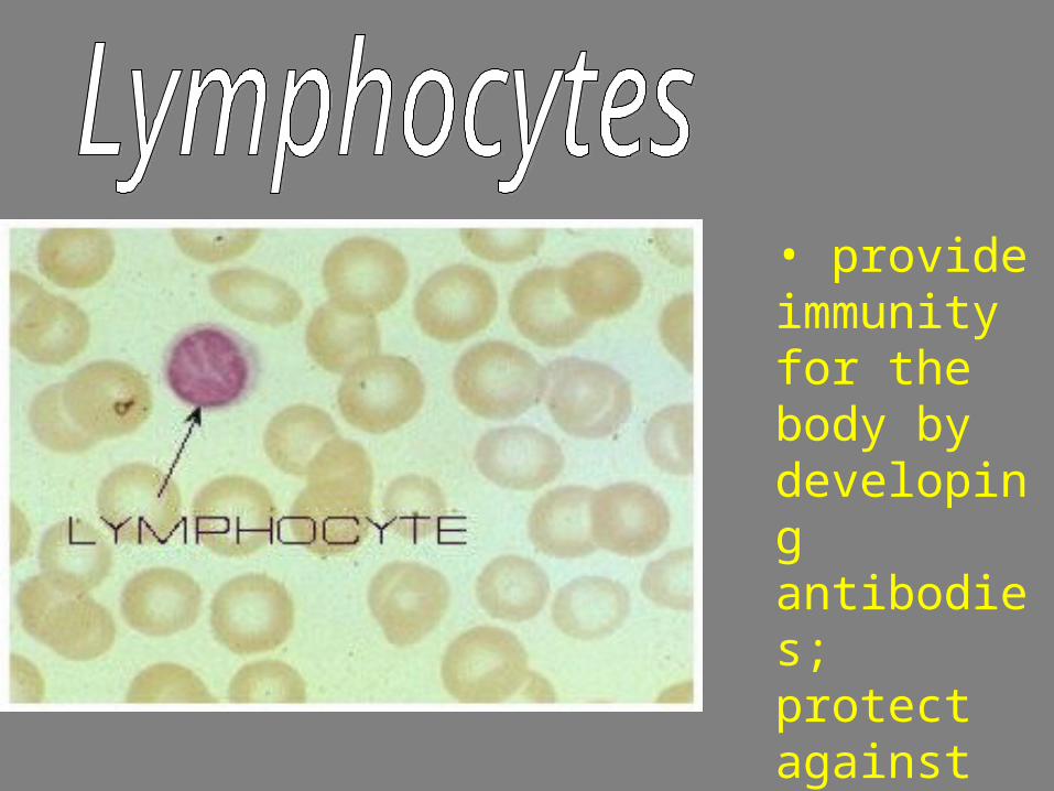
• provide immunity for the body by developing antibodies; protect against the formation of cancer cells.
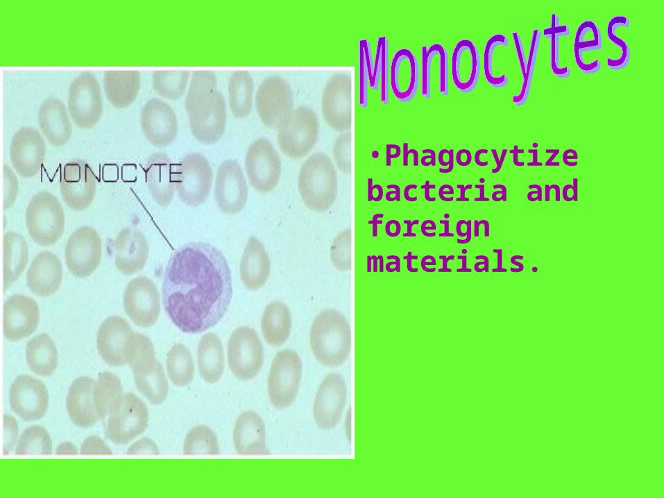
•Phagocytize bacteria and foreign materials.
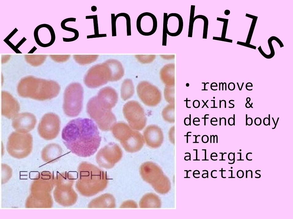
• remove toxins & defend body from allergic reactions

•phagocytize bacteria
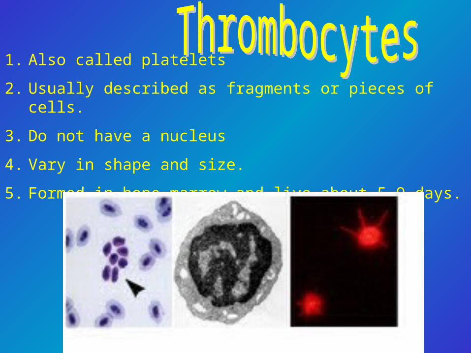
1. Also called platelets
2. Usually described as fragments or pieces of cells.
3. Do not have a nucleus
4. Vary in shape and size.
5. Formed in bone marrow and live about 5-9 days.

6. Important for the clotting process which stops bleeding.
aa. When a blood vessel is cut, thrombocytes collect at the site to form a sticky plug.
ab. They secrete a chemical, serotonin, which causes the blood vessel to spasm and narrow, decreasing the flow of blood.
ac. Also release an enzyme, thromboplastin, which acts with calcium and other substances in the plasma to form thrombin.
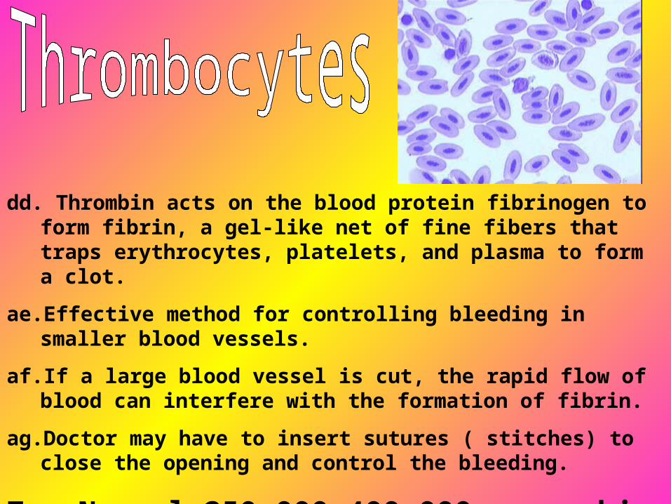
dd. Thrombin acts on the blood protein fibrinogen to form fibrin, a gel-like net of fine fibers that traps erythrocytes, platelets, and plasma to form a clot.
ae. Effective method for controlling bleeding in smaller blood vessels.
af. If a large blood vessel is cut, the rapid flow of blood can interfere with the formation of fibrin.
ag. Doctor may have to insert sutures ( stitches) to close the opening and control the bleeding.
7. Normal-250,000-400,000 per cubic mm of blood.
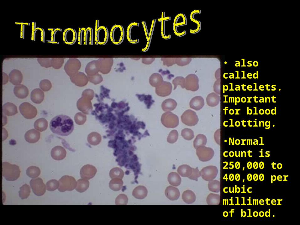
• also called platelets. Important for blood clotting.
•Normal count is 250,000 to 400,000 per cubic millimeter of blood.
Diseases and abnormal conditions
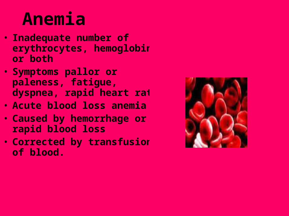
Anemia• Inadequate number of
erythrocytes, hemoglobin or both• Symptoms pallor or paleness,
fatigue, dyspnea, rapid heart rate
• Acute blood loss anemia• Caused by hemorrhage or rapid
blood loss• Corrected by transfusions of
blood.
• Iron Deficiency anemia
•Caused by inadequate amount of iron to form hemoglobin in erythrocytes
•Treatment: iron supplements and increased iron intake from green leafy vegetables and other foods.
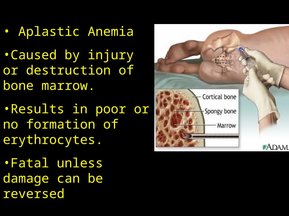
• Aplastic Anemia
•Caused by injury or destruction of bone marrow.
•Results in poor or no formation of erythrocytes.
•Fatal unless damage can be reversed
• Pernicious anemia
•Caused by lack of intrinsic factor, which results in poor absorption of vitamin B12
•Results in formation of inadequate and abnormally large erythrocytes.
•Treatment: replacing intrinsic factor and administering b 12 injections.
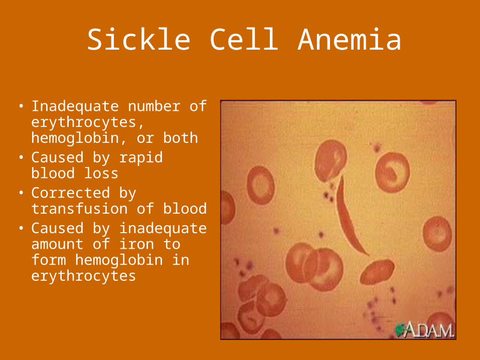
Sickle Cell Anemia
• Inadequate number of erythrocytes, hemoglobin, or both
• Caused by rapid blood loss
• Corrected by transfusion of blood
• Caused by inadequate amount of iron to form hemoglobin in erythrocytes
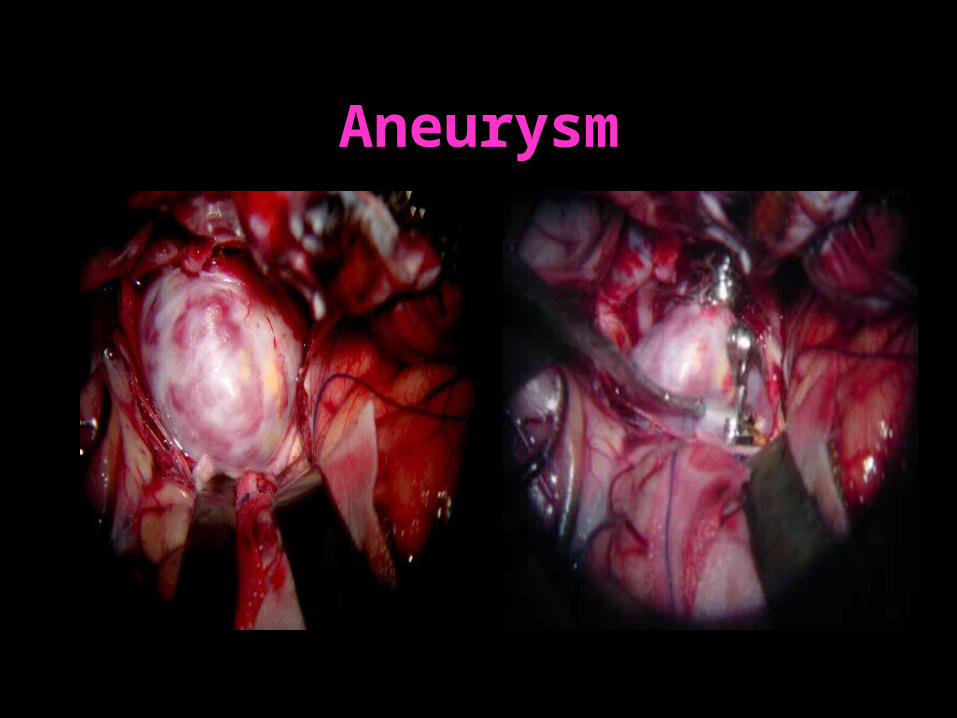
Aneurysm• Ballooning out or saclike formation on the wall
of the artery.
• A weakness in the wall of the artery through which blood can break through causing hemorrhage into surrounding spaces or tissues
•Some cause pain and pressure, some have no symptoms
•If aneurysm ruptures it will cause hemorrhage, can cause death
Aneurysm
• •
Arteriosclerosis
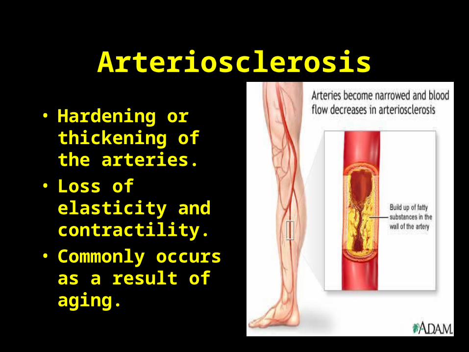
• Hardening or thickening of the arteries.
• Loss of elasticity and contractility.
• Commonly occurs as a result of aging.
•
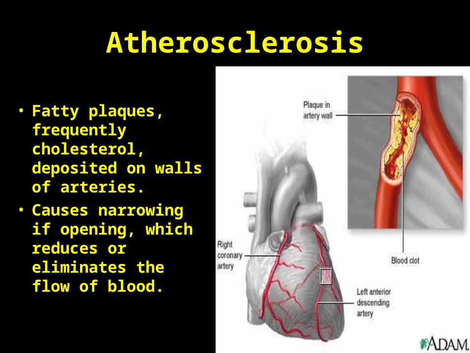
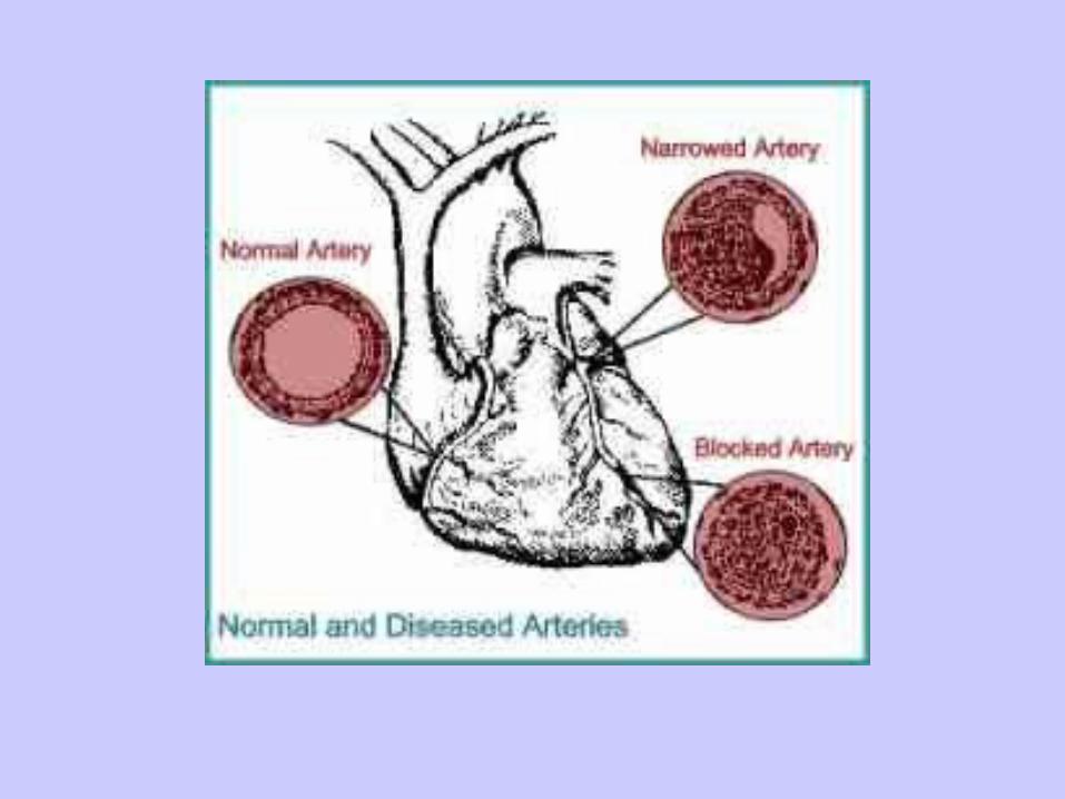
Atherosclerosis
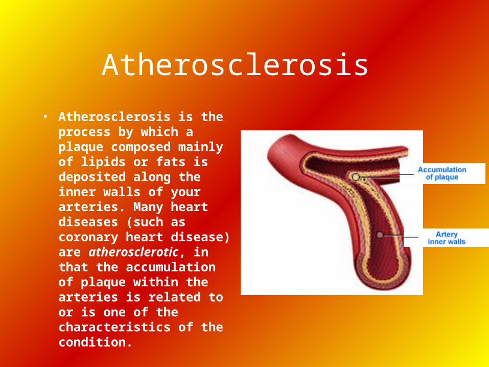
• Atherosclerosis is the process by which a plaque composed mainly of lipids or fats is deposited along the inner walls of your arteries. Many heart diseases (such as coronary heart disease) are atherosclerotic, in that the accumulation of plaque within the arteries is related to or is one of the characteristics of the condition.
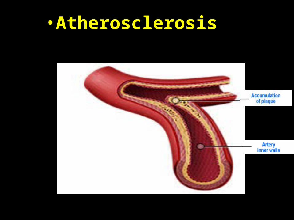
Atherosclerosis
• Fatty plaques, frequently cholesterol, deposited on walls of arteries.
• Causes narrowing if opening, which reduces or eliminates the flow of blood.
•
•Atherosclerosis
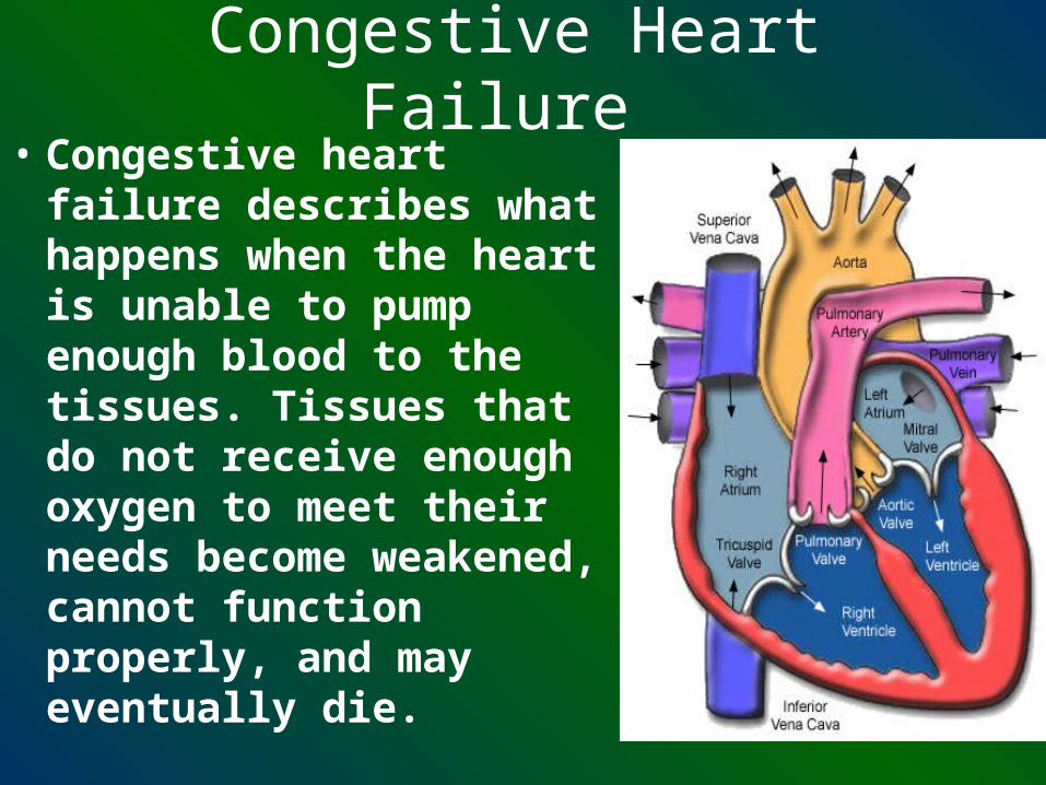
Congestive Heart Failure • Congestive heart failure
describes what happens when the heart is unable to pump enough blood to the tissues. Tissues that do not receive enough oxygen to meet their needs become weakened, cannot function properly, and may eventually die.
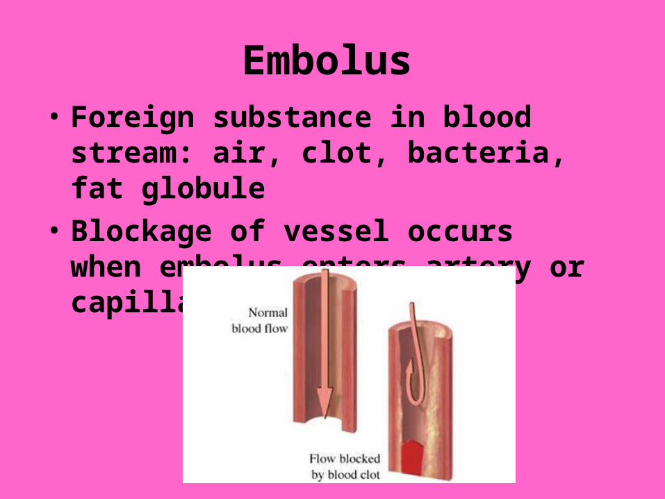
Embolus• Foreign substance in blood stream: air,
clot, bacteria, fat globule
• Blockage of vessel occurs when embolus enters artery or capillary.
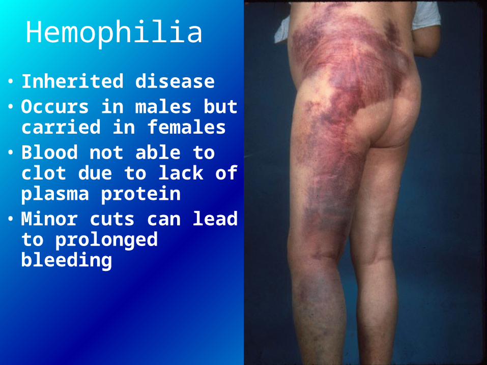
Hemophilia
• Inherited disease • Occurs in males but
carried in females• Blood not able to clot
due to lack of plasma protein
• Minor cuts can lead to prolonged bleeding
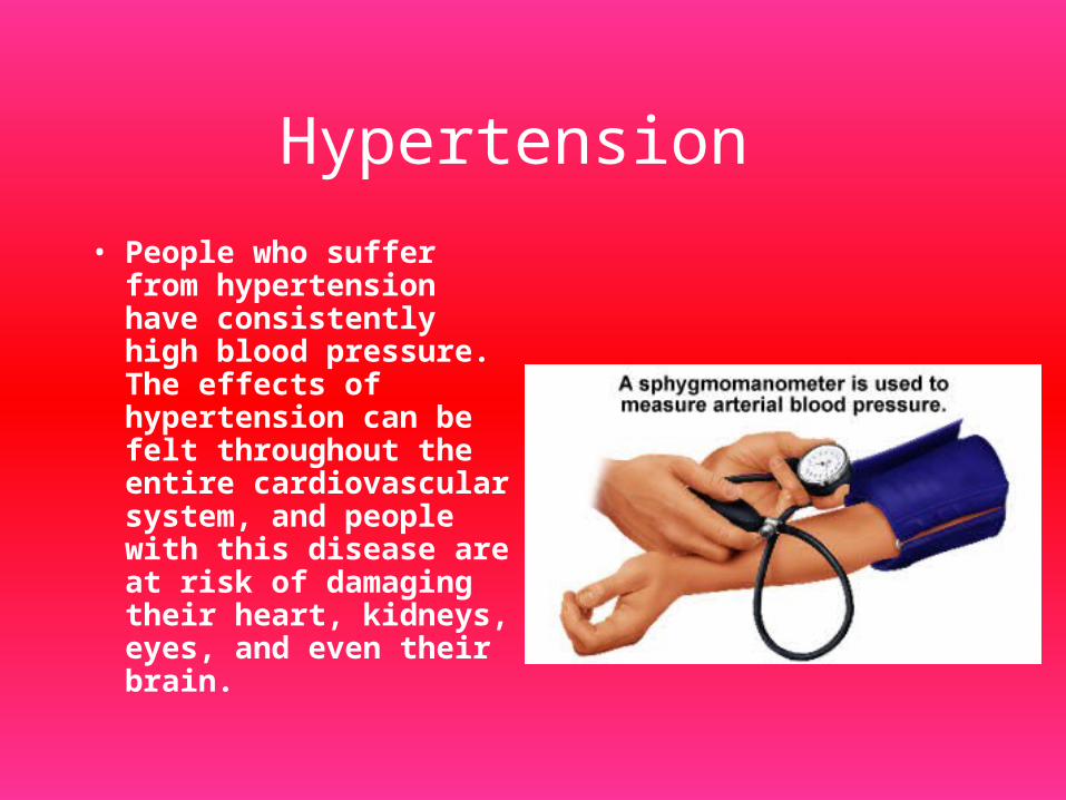
Hypertension
• People who suffer from hypertension have consistently high blood pressure. The effects of hypertension can be felt throughout the entire cardiovascular system, and people with this disease are at risk of damaging their heart, kidneys, eyes, and even their brain.
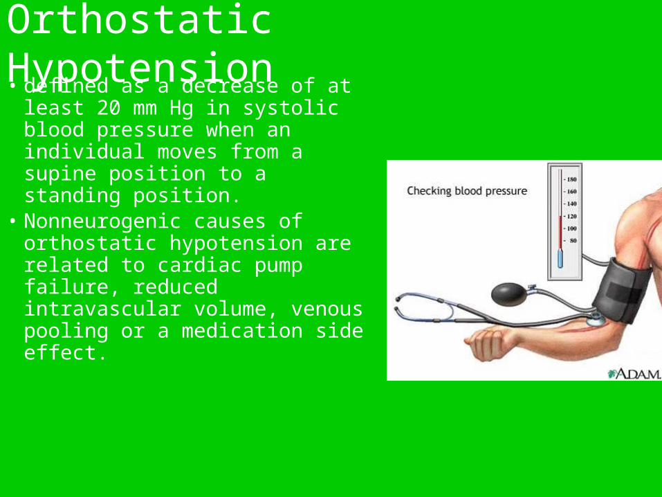
Orthostatic Hypotension • defined as a decrease of at least
20 mm Hg in systolic blood pressure when an individual moves from a supine position to a standing position.
• Nonneurogenic causes of orthostatic hypotension are related to cardiac pump failure, reduced intravascular volume, venous pooling or a medication side effect.
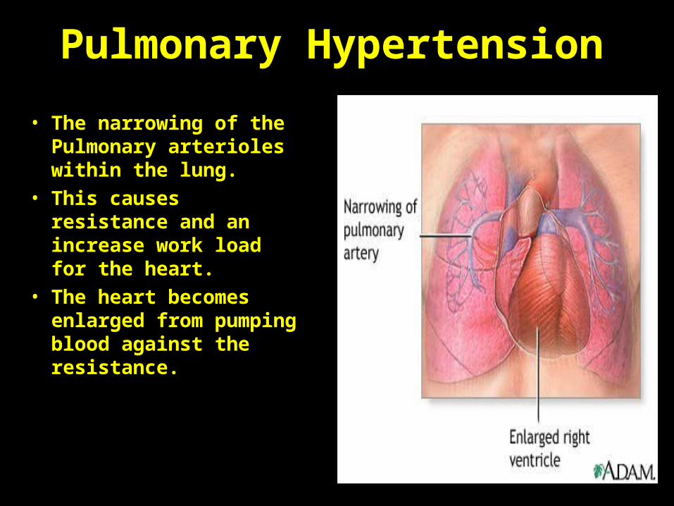
Pulmonary Hypertension
• The narrowing of the Pulmonary arterioles within the lung.
• This causes resistance and an increase work load for the heart.
• The heart becomes enlarged from pumping blood against the resistance.
•

• symptoms result from the body not producing enough healthy blood cells.
• Healthy bone marrow makes stem cells that grow into the three types of blood cells: red blood cells, white blood cells, and platelets.
• An AML patient's bone marrow makes too many blast cells (immature white blood cells).
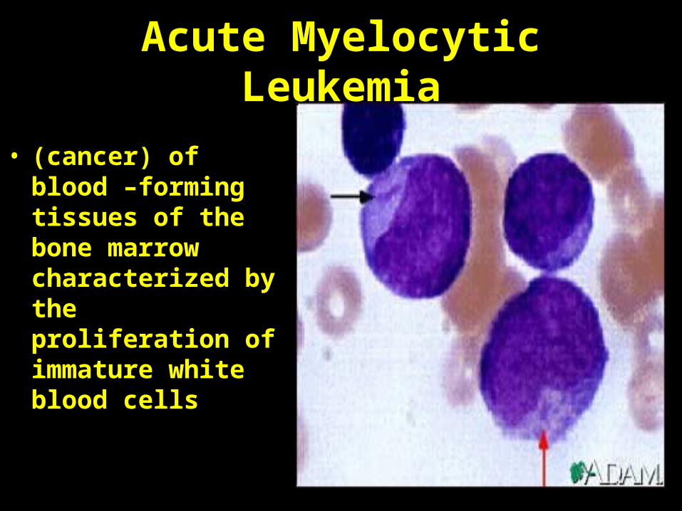
Acute Myelocytic Leukemia
• (cancer) of blood –forming tissues of the bone marrow characterized by the proliferation of immature white blood cells
Arrhythmias
• Disturbance in electrical conduction resulting in abnormal heart rhythms
• Can be mild or life threatening- PAC (premature atrial contraction) or ventricular fibrillation
• Treatment- defibrillator or pacemaker
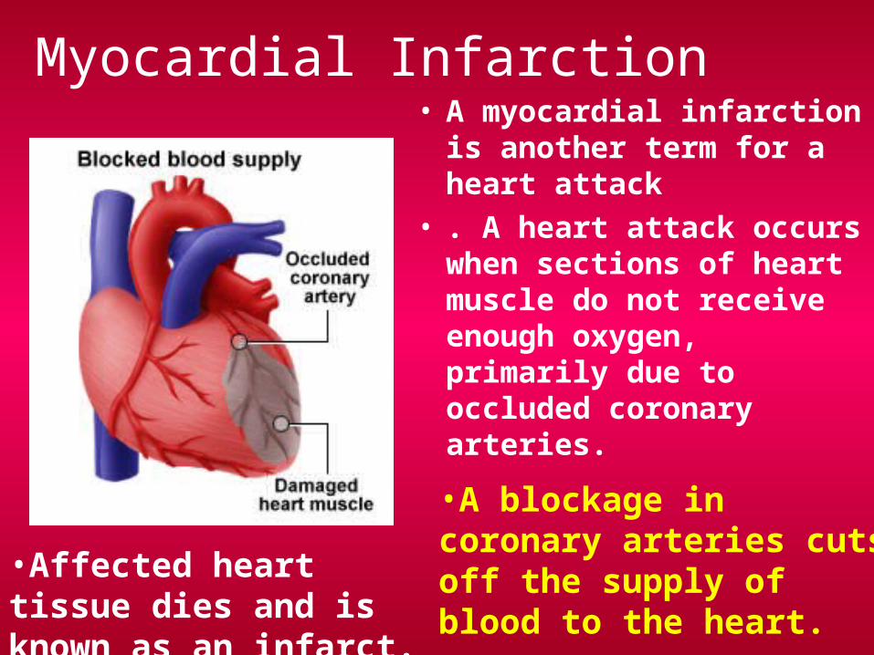
Myocardial Infarction• A myocardial infarction is
another term for a heart attack
• . A heart attack occurs when sections of heart muscle do not receive enough oxygen, primarily due to occluded coronary arteries.
•Affected heart tissue dies and is known as an infarct.
•A blockage in coronary arteries cuts off the supply of blood to the heart.
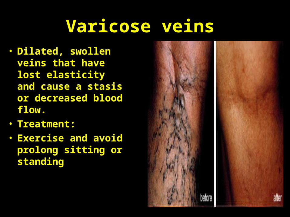
Varicose veins• Dilated, swollen veins
that have lost elasticity and cause a stasis or decreased blood flow.
• Treatment:• Exercise and avoid
prolong sitting or standing
•
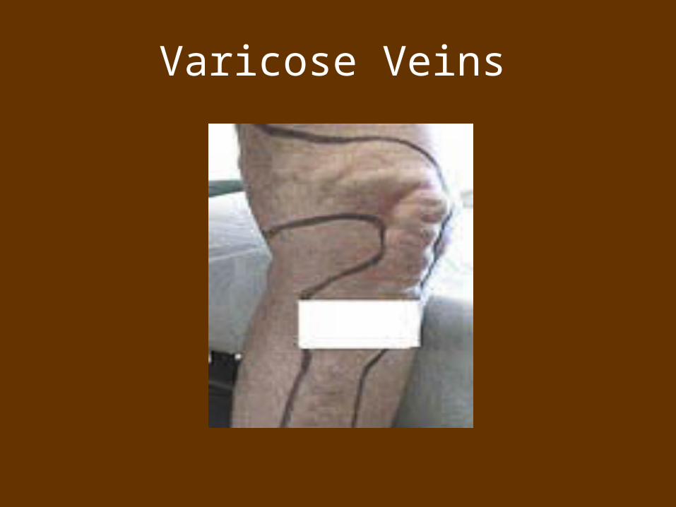
Varicose Veins
Coronary Heart Disease• When plaque or blood fragments are deposited
along the inside wall of the coronary arteries (vessels that supply the heart muscle with blood), these vessels may become occluded. Coronary occlusion, which also may result from atherosclerosis or coronary artery muscle spasm, refers to the complete obstruction of the blood vessel that prevents blood flow to the cells of the heart. When the coronary arteries become occluded, the result is the development of coronary heart disease, also known as coronary artery disease or ischemic heart disease.
Dementia
• a progressive brain dysfunction, leads to a gradually increasing restriction of daily activities. The most well-known type of dementia is Alzheimer's disease. Dementia not only affects patients, but also those surrounding them, as most patients require care in the long-term.
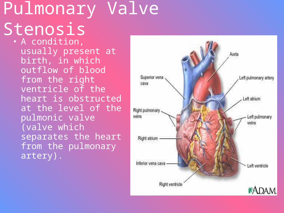
Pulmonary Valve Stenosis• A condition, usually
present at birth, in which outflow of blood from the right ventricle of the heart is obstructed at the level of the pulmonic valve (valve which separates the heart from the pulmonary artery).
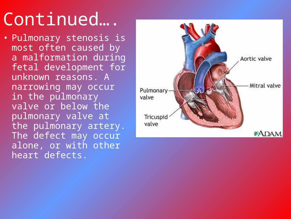
Continued….• Pulmonary stenosis is
most often caused by a malformation during fetal development for unknown reasons. A narrowing may occur in the pulmonary valve or below the pulmonary valve at the pulmonary artery. The defect may occur alone, or with other heart defects.
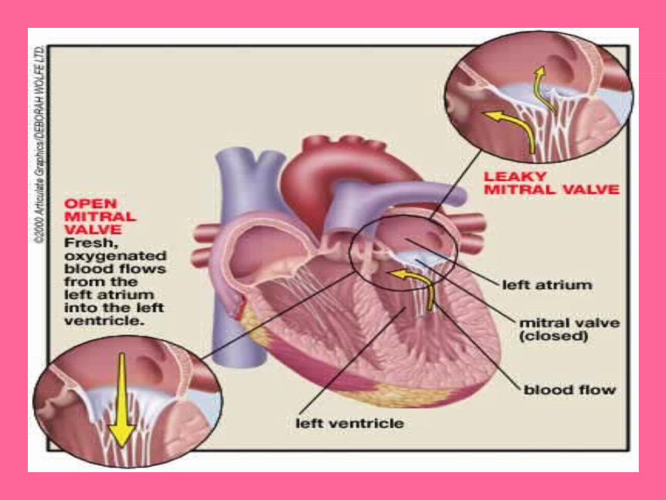
Mitral valve prolapse (MVP)
• Common condition in which cusps of mitral valve prolapse into left atrium during systole
• Symptoms- nonanginal chest pain, palpitations, dyspnea, fatigue- murmur at apex holosystolically
• Treatment- if mitral regurgitation is present- antibiotic prophylaxis indicated during surgical and dental procedures