Embed Size (px)
Citation preview

• Circuits
• Chambers
• Valves(one-way-flow)
• Myocardiocytes
The Heart

Volumes? Pressures?
heart –> lungs –> heart
heart –> body –> heart
Artery = Vein =

Trace a RBC!
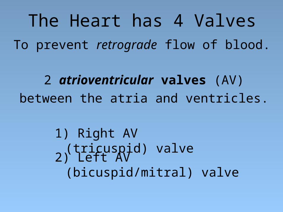
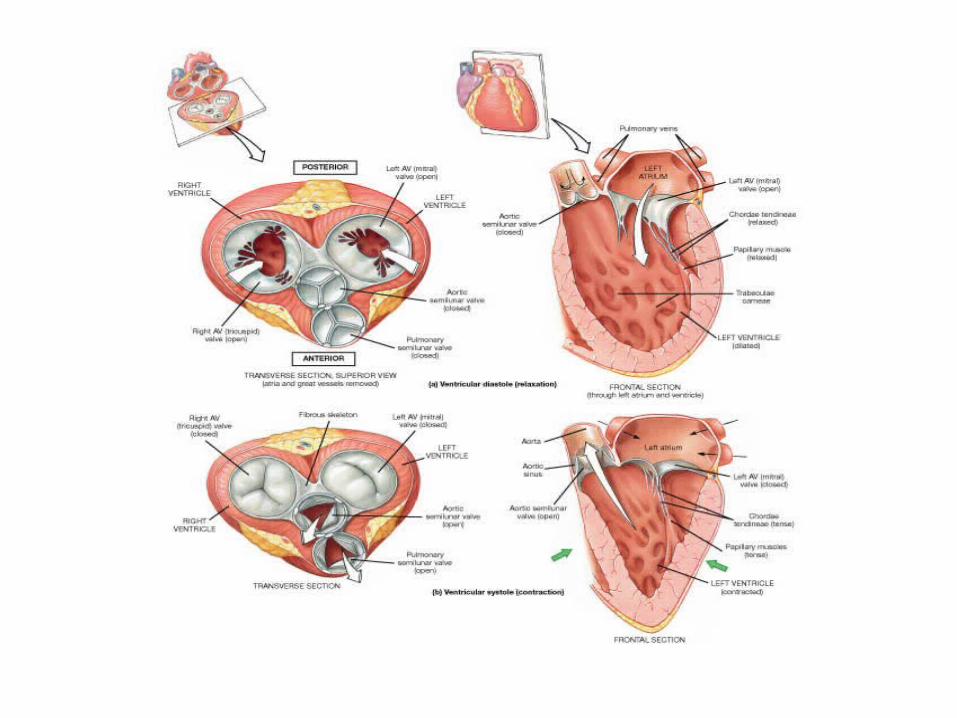
The Heart has 4 ValvesTo prevent retrograde flow of blood.
2 atrioventricular valves (AV)
between the atria and ventricles.
1) Right AV (tricuspid) valve
2) Left AV (bicuspid/mitral) valve

2 semilunar valvesbetween a ventricle and artery.
1) Aortic semilunar valve
2) Pulmonary semilunar valve
Two heart sounds: “Lub” and “Dup”
2. Closure of Semilunar valves = “Dup”
1. Closure of AV valves = “Lub”
http://www.openheartsurgery.com/heart_murmur.html

Normal Heart Valves Problems Opening:
Stenosis – narrowing of valve, when a valve doesn't open completely.
Turbulence = noise = murmur.
Problems Closing: Prolapse –overlapping or when valve doesn't close tightly. Also termed valvular insufficiency (regurgitation) Retrograde flow = noise = murmur.
Disorders of Heart Valves
Problem Heart Valves
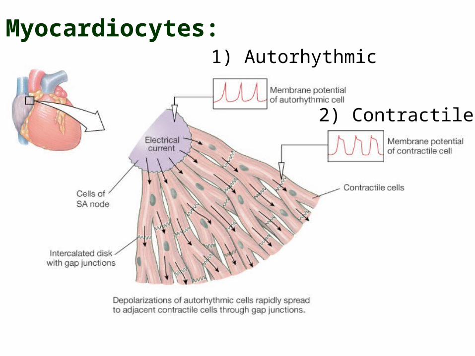
Myocardiocytes:1) Autorhythmic
2) Contractile


Action Potentials for Autorhythmic Myocardiocytes

stimulus
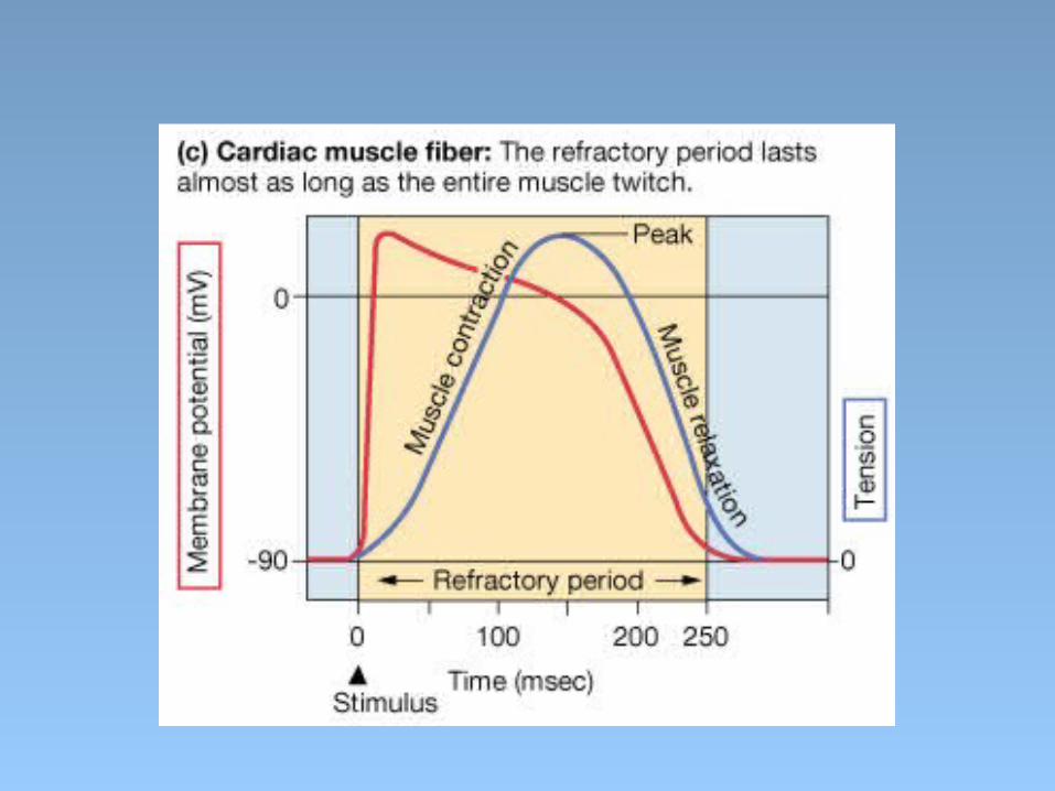
Action Potentials for Contractile Myocardiocytes

Myocardiocytes: Calcium induced Calcium release
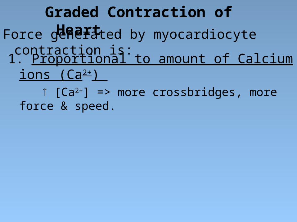
Graded Contraction of HeartForce generated by myocardiocyte contraction is:
1. Proportional to amount of Calcium ions (Ca2+) [Ca2+] => more crossbridges, more force & speed.

Graded Contraction of Heart
Force generated by myocardiocyte contraction is:
2. Modulated by Autonomic N.S.
=> Sym HR and Force
=> Para HR
1. Proportional to amount of Calcium ions (Ca2+) [Ca2+] => more crossbridges, more force & speed.

Sympathetic – speeds heart rate by Ca2+ influx.
Parasympathetic – slows rate by K+ efflux, Ca2+ influx.

Graded Contraction of Heart
Force generated by myocardiocyte contraction is:
2. Modulated by Autonomic N.S.
=> Sym HR and Force
=> Para HR
1. Proportional to amount of Calcium ions (Ca2+) [Ca2+] => more crossbridges, more force & speed.
3. Stretch-Length-Tension Relationship
stretch, => Ca2+ entering => contraction force
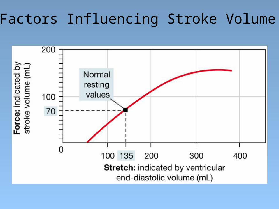
Factors Influencing Stroke Volume

The Cardiac Cycle
http://www.youtube.com/watch?v=rguztY8aqpk

1. Late Diastole: “Heart at rest” all chambers relaxed
filling with blood (passive filling ~ 80% full).
2. Atrial Systole: atria contract, adds the last 20% of
blood to ventricles (top off ventricles)
Occurs after P-wave on EKG
The Cardiac Cycle: Mechanical Events of the
Heart
End Diastolic Volume (EDV) = Maximum ventricular volume*

3. Ventricular Systole (part 1):
Ventricular contraction begins - Pressure (P).
Closure of AV valves = 1st heart sound ("lub")
Sealed Compartment – all valves are closed.
Isovolumetric ventricular contraction:
=> pressure builds as volume stays the same.

4. Ventricular Systole (part 2):
Ejection phase: P pushes open semilunar valves,
blood forced out into artery leaving ventricle.
Pulmonary Semilunar => 25 mmHg (minimum pressure)
Aortic Semilunar => 80 mmHg (minimum pressure)
End Systolic Volume (ESV) = volume remaining in heart after ejection (~½)*.
Stroke Volume = EDV - ESV (ml/beat)

5. Ventricular Diastole:
Relaxation of ventricles, artery back flow slams
semilunar valves shut = 2nd heart sound ("dup").
The AV valves then open, refilling starts – back to start of cycle.
Sealed Compartment again – all valves are closed.
Isovolumetric ventricular relaxation:
=> pressure as volume stays the same.
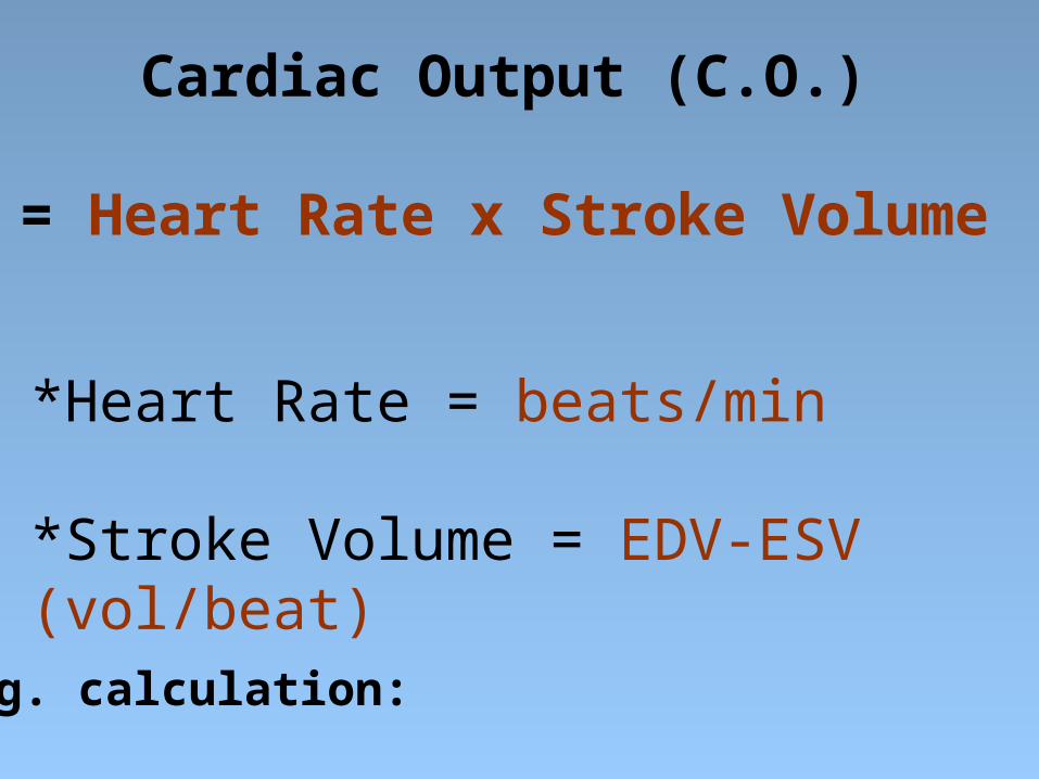
Cardiac Output (C.O.)
= Heart Rate x Stroke Volume
*Heart Rate = beats/min
*Stroke Volume = EDV-ESV (vol/beat)
e.g. calculation:

Electrical Conduction System
Sino Atrial (SA) Node
Atrial Ventricular (AV) Node
AV Bundle (of His)
L and R Bundle Branches
Purkinje Fibers

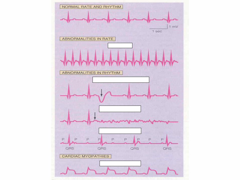
Trace of an ElectroCardioGram (ECG)

The ECG
P wave:
PR interval:
QRS complex:
T wave:
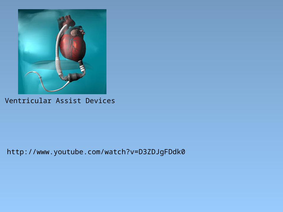
Ventricular Assist Devices
http://www.youtube.com/watch?v=D3ZDJgFDdk0

![(ISO 70I5)TI)m7a Globe valves. Control Valves – flange · Emergency control function Globe valves 2-way 3-Weg Tmax = 120°C 1) Closed circuits DN [mm] k vs [m3/h] s [kPa] max [kPa]](https://img.dokumen.tips/doc/110x75/5fb45bf985a0ec4e6c260fdc/iso-70i5tim7a-globe-valves-control-valves-a-flange-emergency-control-function.jpg)