Embed Size (px)
Citation preview

1
Joo Ha Hwang, MD, PhD
Department of Medicine, Division of Gastroenterology
Department of Radiology
Applied Physics Laboratory, Center for Industrial and Medical Ultrasound
University of Washington, Seattle, USA
Current Status of HIFU Therapy for
Treatment of Benign and Malignant Tumors
of the Abdomen, Pelvis and Bone
Objectives
Overview of current HIFU clinical systems
Overview of current clinical applications
• Uterine fibroids
• Pancreatic tumors
• Liver tumors
• Renal cell carcinoma
• Bone metastases
• Breast cancer
• Thyroid/parathyroid tumors
Discuss future clinical applications
YDME, Beijing
Shanghi A&S
HIAFU, Chonqing
Clinical HIFU SystemsUS-guided
Thereclion, France
Ultrasound targeting
Ultrasound-guided HIFU
devices do not provide
monitoring of lesion
development (other than
hyperecho from boiling)
Methods for estimating in situ intensity exist
Methods for monitoring
HIFU therapy are in
development
• Thermometry
• ARFI
• Elastography

2
InSightec, Isreal
Clinical HIFU SystemsMR-guided
Philips, US/France
Temperature mappingProton Resonance Frequency shift
- =Zoom x4
Phase Imaging
Before Heating After Heating Difference
0BT
T
E
γ = 2π ∙ 42,56 MHz/T Gyromagnetic Ratio
α = 0.0101 ppm/°C Water Frequency Shift
TE ~ 20ms Echo Time
B0 = 1.5 T Magnetic Field
Courtesy of Philips
Real Time FeedbackReliable necrosis volume
Thermal map & Dose map
Real time visualization
+ Feedback
T > 57°C* or Dose >240 EM
Stop heating
Reliable necrosis volume
Thermal map Dose map
Threshold
Non-perfused volumeCourtesy of Phiilips
Reliable necrosis volume
No a-priori knowledge needed
Simple and robust
* Applies to the border of the cell. Temperatures at the
center are higher, especially for larger cells.
Indications: Non-invasive ablation of solid
benign or malignant tumors
Requirement:
• Acoustic window – critical
Non-oncologic applications
• Uterine fibroids
Clinical Applications for HIFU
Ablation
3
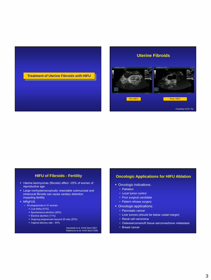
Treatment of Uterine Fibroids with HIFU
Pre-HIFU Post- HIFU
Courtesy of Dr. Hu
Uterine Fibroids
Uterine leiomyomas (fibroids) affect ~25% of women of
reproductive age
Large nonhysteroscopically resectable submucosal and
intramural fibroids can cause cavitary distortion
impacting fertility
MRgFUS
• 54 pregnancies in 51 women
Live births (41%)
Spontaneous abortion (28%)
Elective abortion (11%)
Ongoing pregnancies beyond 20 wks (20%)
Vaginal delivery rate – 64%
Hanstede et al. Fertil Steril 2007
Rabinovici et al. Fertil Steril 2008
HIFU of Fibroids - Fertility
Oncologic indications:
• Palliation
• Local tumor control
• Poor surgical candidate
• Patient refuses surgery
Oncologic applications:
• Pancreatic cancer
• Liver tumors (should be below costal margin)
• Renal cell carcinoma
• Osteosarcoma/soft tissue sarcomas/bone metastasis
• Breast cancer
Oncologic Applications for HIFU Ablation

4
HIFU Ablation for Palliation of
Advanced Pancreatic Cancer
4th leading cause of cancer deaths in the US
>42,000 new cases – >35,000 deaths in 2010
80-90% are “unresectable” when diagnosed
Poor outcomes
• Median survival without therapy: 3 months
• Median survival with therapy: 6-12 months
Palliation of symptoms is important
• Pain relief
Pancreatic Cancer
Treatment of Pancreatic Cancer
Pre-HIFU
6 months post- HIFU
Pancreatic Cancer

5
HIFU Ablation of
Liver Tumors
Acoustic window is a problem
Treatment through the ribs has been reported
Treatment after rib resection
Randomized study (Wu et al.)• TACE vs. HIFU+TACE (50 patients)
• HIFU treatment 2-4 weeks following TACE
outcome TACE TACE+HIFU p-value
Median survival 4.0 months 11.3 months 0.004
6-month survival 13.2% 80.5% 0.002
1-year survival 0% 42.9% <0.001
HIFU Treatment of Liver Tumors
Pre-HIFU 3 months post- HIFU
Courtesy of Dr. Hu
Liver Tumors
HIFU Ablation of
Renal Cell Carcinoma

6
Renal Cell Carcinoma
Small renal tumors are
being more frequently
identified
Surgical resection has
significant morbidity
Many pts are poor surgical
candidates
Other less invasive
procedures are needed
Marberger et al. BJU 2005
HIFU Ablation of
Bone Metastasis
Bone is a common site for metastatic disease
• Prostate CA and Breast CA
Pain from bone metastasis is the most common cause of
cancer pain
Current treatment options:
• Analgesics
• Chemotherapy
• Biphosphonates
• Local therapies:
Radiation (no relief in 20-30%)
Surgery
RFA
Bone Metastasis Feasibility Study
• Treated bone mets from primary tumor
type: renal, colorectal, lung, breast,
prostate and other cancers
• Treated lesion locations: iliac bone,
ischium bone, sacrum, femur, scapula,
humerus, clavicle
• Treated lesion type: both osteolytic and
osteoblastic
Patient population:
36 treatments in 31 patients were
conducted, targeting 32 metastatic
lesions
Patient tumor characteristics:
Liberman B et al. Pain palliation in patients with bone metastases using MR-guided focused ultrasound surgery: a multicenter
study. Ann Surg Oncol. 2009 Jan;16(1):140-6. Epub 2008 Nov 11.

7
Catane et al. Ann Oncol 2007
Bone Metastasis – Palliation of Pain
HIFU Ablation of
Breast Cancer
Courtesy of Breastopia Namba Medical Center,
Miyazaki, Japan
Furusawa H, Namba K, Thomasen S, Akiyama F, Bendet A, Tanaka C, Yasuda Y, Nakahara H. Magnetic Resonance-Guided Focused Ultrasound
Surgery of Breast Cancer: Reliability and Effectiveness, J Am Coll Surg, 2006, 203(1):54-63
Breast Cancer – Phase II Study Results.
• Evaluation of Safety and Efficacy
• Total 195 patients treated, 30 were treated
with contrast enhanced planning images
T1w contrast enhanced subtracted images
Hyperemia
at edge of
treated
region
Treatment boundary
Necrosis boundaryAblated invasive cancer
Ablated possible cancer
Post-treatment pathologic results
Mean necrosis of ablated tumor = 96.9 ± 4%
•15 patients had 100% necrosis
•3 patients had < 95% necrosis
HIFU Ablation of
Thyroid/Parathyroid Tumors

8
Courtesy of Theraclion
Clinical studies Gross Specimen
Treated nodule
Left lobe
Courtesy of Theraclion
HIFU: Future
Expanded indications for therapy
Enhanced tumor-specific immunity
Drug delivery
• Targeted delivery (Heat activation)
• Enhancement of vascular permeability
• Enhanced penetration
Improved treatment monitoring
• Thermometry
• Elastography
• Radiation Force Imaging
Device approvals
The Future of HIFU

9
Summary
Overview of current clinical systems
Overview of current clinical applications
• Uterine fibroids
• Pancreatic tumors
• Liver tumors
• Renal cell carcinoma
• Bone metastases
• Breast cancer
• Thyroid/parathyroid tumors
Discussed future clinical applications
Thank You