Embed Size (px)
Citation preview
Carol Rees Parrish, M.S., R.D., Series Editor
Percutaneous endoscopy gastrostomy (PEG) tubes are a valuable tool for providing long-term enteral nutrition or gastric decompression; certain circumstances that complicate PEG placement warrant novel approaches and merit review and discussion. Ascites and portal hypertension with varices have been associated with poorer outcomes. Bleeding is one of the most common serious complications affecting approximately 2.5% of all procedures. This article will review what evidence exists in these high risk scenarios and attempt to provide more clarity when considering these challenging clinical circumstances.
28 PRACTICAL GASTROENTEROLOGY • MAY 2012
INFLAMMATORY BOWEL DISEASE: A PRACTICAL APPROACH, SERIES #73
Iris Vance, University Virginia School of MedicineNeeral Shah, MD, Assistant Professor, Division of Gastroenterology and Hepatology
NUTRITION ISSUES IN GASTROENTEROLOGY, SERIES #105
High Risk Percutaneous Endoscopic Gastrostomy Tubes: Issues to Consider
Iris Vance Neeral Shah
INTRODUCTION
Since the first Percutaneous Endoscopic Gastrostomy tube was placed in 1979 (1), they have become an invaluable tool for providing
long-term enteral nutrition (EN) and are commonly used in patients with dysphagia following stroke, disabling motor neuron diseases such as multiple sclerosis and amyotrophic lateral sclerosis, and in those with head and neck cancer.They are also used for patients with prolonged mechanical intubation, as well as gastric decompression in those with severe gastroparesis, malignant bowel obstruction, or chronic intestinal obstruction as in the case of peritoneal carcinomatosis (2). There is less available guidance, however, when considering relative contraindications to PEG placement including patients with cirrhosis, ascites, varices and elevated bleeding risk. Recently hypoalbuminemia
has been found by multiple authors to portend a poor prognosis in PEG placement (3,4, 5,6,7,8). This review will endeavor to provide more clarity when considering these challenging clinical circumstances.
Ascites & Gastric VaricesThe presence of ascites is frequently viewed as a relative, if not absolute, contraindication to PEG placement. Ascites adds technical difficulties and the risk for potential complications (see Table 1). PEGs are placed endoscopically after finding the optimum location by transillumination across the abdominal wall. The presence of ascites may increase the difficulty in finding a safe window for PEG placement. Fluid in the peritoneal cavity can prevent proper apposition of the gastric body to the abdominal wall to ensure the healing of a proper PEG tract. Ascites that accumulates can also drain through the PEG tract and increase the risk of infection. Particularly in the setting of the hypocomplementemia in cirrhosis, the decreased
NUTRITION ISSUES IN GASTROENTEROLOGY, SERIES #105
High Risk Percutaneous Endoscopic Gastrostomy Tubes
PRACTICAL GASTROENTEROLOGY • MAY 2012 29
NUTRITION ISSUES IN GASTROENTEROLOGY, SERIES #105
opsonization and the open communication from the skin flora to subcutaneous tissue and ascitic fluid could further increase the risk of bacterial peritonitis. Portal hypertension with varices is frequently cited with ascites in discussing relative contraindications to PEG placement; the concern is for puncture of a variceal vessel during the procedure. In addition, portal hypertension can lead to many small collateral vessels
subcutaneously that also elevate bleeding risk.Baltz et al recently published a single center case
series of 26 cirrhotics who underwent PEG placement, 17 (65%) of whom had ascites (9). Ten patients (38.5%) died within 30 days of the procedure, with one additional death at 90 days. Of those who died within the first 30 days, 9 (90%) had ascites, which led the authors to suggest the possibility that the presence of ascites may be an independent risk factor. In total, 9/17 (52.9%) of the patients with ascites in the study died within the first 30 days. This certainly merits further study, though ascites may also have been present more frequently in patients with more advanced disease. Only 2 of the deaths were attributable to the procedure; 1 patient died after an aspiration event during the procedure and the other 5 days following the procedure with newly diagnosed peritonitis. The other causes of death were sepsis caused by pneumonia, pancreatitis with an epidural abscess, splenic rupture after a motor vehicle accident, and a myocardial infarction (9).
Although PEG placement is a higher risk procedure in cirrhotic patients, these patients are also at a markedly higher risk of malnutrition, which has been shown to significantly worsen their prognosis. In examining data from over 114,000 hospital admissions for cirrhosis and
Table 1. Difficulties of PEG Placement with Ascites
Apposition of the stomach and abdominal wall more challenging
Impaired tract formation and healing
Reaccumulation and catheter dislodgement
Leakage of ascites
Increased risk of infection
Increased risk of bleeding
Table 2. Innovations Facilitating PEG in Patients with Ascites
Reference Study Type Intervention
Kynci et al Case report Albumin, mannitol and furosemideTopical nitropaste to decrease portal pressureUltrasound guided paracentesis on the day of PEG placement
Höroldt et al Case report Abdominal ultrasound during PEG placement to identify an area without varices
Pothuri et al Case series of 94, including 59 with ascites
Pre-PEG paracentesis
Wejda et al Case series of 4 3 suture triangle gastropexy during PEG placement (2)or several days after placement for new onset ascites (2)
(continued on page 34)
34 PRACTICAL GASTROENTEROLOGY • MAY 2012
NUTRITION ISSUES IN GASTROENTEROLOGY, SERIES #105
High Risk Percutaneous Endoscopic Gastrostomy Tubes
portal hypertension in the Nationwide Inpatient Sample, Sam and Nguyen showed that mortality increases in patients with cirrhosis and portal hypertension who have diagnosed protein-calorie malnutrition with an adjusted mortality of 1.76 (10). In addition, protein-calorie malnutrition was associated with longer hospital stays (8.7 vs. 5.7 days) and 46% higher hospital costs (10).
Patients listed for liver transplantation or being considered for listing are of particular concern. Though the finding has not been universal, studies have found preoperative malnutrition has negatively affected transplant outcomes. ESPEN guidelines emphasize the importance of preoperative nutrition support with enteral nutrition (EN) if necessary and early postoperative EN (11). Selberg and colleagues showed a statistically and clinically significant divergence in survival at one and five years following liver transplant based on measures of preoperative malnutrition (12).
Estimates of the prevalence of malnutrition in patients with end-stage liver disease range from 65-100%, so index of suspicion should be high and early aggressive nutritional intervention is warranted, particularly in patients under consideration for liver transplantation (13). If intensive counseling and oral supplementation fail to provide adequate nutritional support, tube feeding may occasionally be necessary, with small bore nasogastric tubes being a significantly safer option than PEG tubes.
Technical ApproachesRecent case reports have described technical innovations that can facilitate PEG placement in patients with advanced liver disease, ascites, and gastric varices (see Table 2). Kynci and colleagues described a successful and uncomplicated PEG placement in a patient with esophageal and gastric varices and ascites (14). The patient received albumin, mannitol and furosemide in addition to his previously prescribed spironolactone and then underwent ultrasound-guided paracentesis of the remaining ascites on the day of the procedure. Topical nitropaste was also administered on the day of the procedure in an effort to reduce portal pressure and ultrasound was used prior to the procedure to mark the location of gastric varices (14). In a different case of a patient with widespread gastro-splenic venous collaterals throughout the upper abdomen and surrounding the stomach, Höroldt et al reported
the use of real-time abdominal ultrasound to identify an area without varices (15). Pothuri et al reported a series of 94 patients with ovarian carcinoma who underwent palliative PEG placement for malignant bowel obstruction. Of these, 59 patients had ascites, 25 requiring pre-PEG paracentesis; 8 of these developed leakage and 1 developed peritonitis, which resolved with antibiotics (16). The good outcomes in this larger series involving malignant ascites may not be applicable to patients with cirrhotic ascites as the low complement levels and opsonization defects accompanying cirrhosis increase the susceptibility to infection. In addition, ascites reaccumulation may be more difficult to address in patients with underlying cirrhosis as diuretic use is often limited by baseline renal insufficiency and concern for precipitating or worsening hepatorenal syndrome. In a small series of 4 patients that included patients with decompensated cirrhosis, Wejda et al reported success with a 3 suture triangle gastropexy in PEG placement in 2 patients (17). The other 2 patients developed new-onset ascites with leakage after PEG placement and then underwent triangle gastropexy 2 and 4 days after the original PEG, which resolved the leakage.
Radiologically Placed GastrostomyOne alternative to PEG placement is radiologically
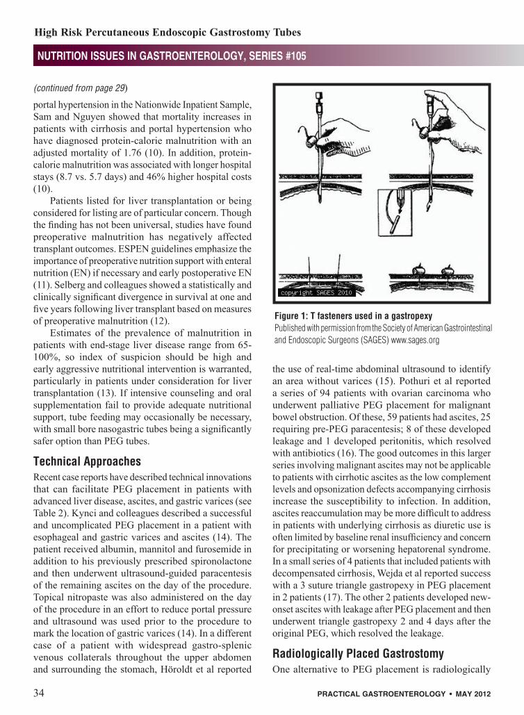
Figure 1: T fasteners used in a gastropexyPublished with permission from the Society of American Gastrointestinal and Endoscopic Surgeons (SAGES) www.sages.org
(continued from page 29)

NUTRITION ISSUES IN GASTROENTEROLOGY, SERIES #105
High Risk Percutaneous Endoscopic Gastrostomy Tubes
PRACTICAL GASTROENTEROLOGY • MAY 2012 35
guided placement of a gastrostomy tube. Lee et al described safe radiologic placement of gastrostomy tubes in 9 patients with ascites after pre-procedural paracentesis (18). Ryan et al then reported a 97.8% success rate in 45 patients with malignant ascites and small bowel obstruction who underwent radiologically guided gastrostomy or gastrojejunostomy placement, and advocated gastropexy (see Figure 1) to decrease leakage as well as sonography every 2-5 days for the first four weeks to detect reaccumulation of ascites.
The mean follow-up was 11 weeks, and of the 45 patients, one died following massive hemorrhage for a 30 day mortality of 2.2%; other major complications included gastropexy breakdown and peritonitis due to massive ascites reaccumulation in one patient, and peritonitis due leakage of gastric contents a week after the procedure in another (19). Ho and colleagues have also reported success with pre-procedural paracentesis in facilitating radiologically guided G tube placement, and advocate insufflation of the stomach with CO2 prior to puncture as well as gastropexy. In their series of 400 patients an unspecified number of whom had ascites, there was one procedure-related mortality secondary to peritonitis from peri-catheter leakage; other complications included 3 other cases of peritonitis, 1 liver abscess following inadvertent liver puncture and
1 transcolonic gastrostomy placement with no adverse sequelae (20).
Percutaneous Transesophageal GastrostomyOishi and colleagues pioneered the percutaneous transesophageal gastrostomy (PTEG) (See Figure 2), in 1994 in Japan (21). The procedure uses ultrasound and fluoroscopy to insert a Dobhoff through the neck into the esophagus and down into the stomach and is a useful alternative when PEG placement is contraindicated. In brief, a guidewire is inserted through the nose into the esophagus, and the rupture free balloon (RFB) catheter is inserted over the guidewire and then inflated. External ultrasound is used to visualize the RFB, which is pressed between the left carotid artery and thyroid until they are separated, at which time the RFB is punctured. A guidewire is passed through the needle into the RFB and used to push it into the stomach, with a dilator and then tube inserted over the guidewire. Positioning is verified by fluoroscopy (21). Oishi et al reported success using this technique for 13 patients with massive ascites with no serious complications (21). Udomsawaengsup et al also reported using PTEG without serious complications for long-term decompression in a series of patients with contraindications to PEG placement, 15 of 17 of whom had malignant gastrointestinal obstruction,
Figure 2: PTEG A. The guidewire is inserted through the nose and the attached RFB balloon is inflated in the esophagus,
with subsequent ultrasound guided puncture of the RFB. B. Schematic of view by ultrasound used to insert PTEG: 1. RFB enlarges esophagus to facilitate puncture
2. Cervical vertebra 3. Trachea 4. Thyroid 5. Carotid artery 6. Jugular vein C. Feeding tube enters at lower neck and terminates in stomach. The PTEG access site is concealed by a
standard men’s dress shirt

High Risk Percutaneous Endoscopic Gastrostomy Tubes
NUTRITION ISSUES IN GASTROENTEROLOGY, SERIES #105
36 PRACTICAL GASTROENTEROLOGY • MAY 2012
carcinomatosis and massive ascites. (22). PTEG is a promising innovation that circumvents some of the limitations of PEG tubes, but its utility will remain limited until there is more widespread experience with the technique.
Severe Malnutrition Multiple recent analyses of morbidity and mortality after PEG placement have found that hypoalbuminemia is an independent predictor of higher mortality and increased complications (3-8). Albumin is known to be problematic as a marker of nutritional status and is neither sensitive nor specific for malnutrition; in one series of 102 patients, 44% of well-nourished patients were noted to have hypoalbuminemia and 11.2% of malnourished patients had normal serum albumin levels (23). Albumin is also a negative acute phase reactant; in inflammatory states, the albumin level decreases due to decreased hepatic synthesis, TNF-alpha induced increased capillary permeability and albumin extravasation into the extravascular space and dilution from subsequent fluid resuscitation (24). The poorer outcomes noted in the recent flurry of studies in patients with low albumin may be attributable to
the inflammatory response in acutely ill patients, or to the underlying conditions producing this inflammatory state and not merely malnutrition as some authors have suggested. Elevations in more direct markers of inflammation also adversely affect outcomes in PEG placement; in a series of 168 patients Figueiredo et al found that elevated CRP is a predictor of mortality within the first thirty days (25).
However, more direct measures of poor nutrition have also been associated with increased mortality following PEG placement. Zopf and colleagues used a BMI < 20 as one of the three most important variables predictive of 30 day mortality after PEG insertion in modeling based on analysis of 787 patients. The authors mentioned that the risk of mortality continued to rise as BMI decreased beyond that point, but did not provide specific data (26). In a study of 112 patients who underwent PEG placement, receiving no enteral nutrition for at least 7 days was also shown to be an independent predictor of 30 day mortality (3). There is certainly a sound theoretical basis to expect poorer outcomes with both malnutrition and hypoalbuminemia. Wound dehiscence is more common in malnourished
Table 3. Alternatives to PEG in Patients with Ascites
Reference Study Type Intervention
Lee et al Case series of 9 Radiologic gastrostomy with pre-procedural paracentesis
Ryan et al Case series of 45 Radiologic gastrostomy with gastropexy and ultrasound every 2-5 days for 4 weeks to monitor ascites reaccumulation
Ho et al Case series of 400, unspecified number with ascites
Radiologic gastrostomy with pre-procedural paracentesis, stomach insufflation and gastropexy
Oishi et al Case series of 13 PTEG (percutaneous transesophageal gastrostomy) using external ultrasound of the neck and fluoroscopy to verify placement
Udomsawaengsup et al
Case series of 17 PTEG as Oishi et al describe
(continued on page 38)
38 PRACTICAL GASTROENTEROLOGY • MAY 2012
NUTRITION ISSUES IN GASTROENTEROLOGY, SERIES #105
High Risk Percutaneous Endoscopic Gastrostomy Tubes
patients and in patients with an albumin < 2.0 g/dL (27). Inadequate enteral nutrition is also well known to increase vulnerability to infection. Without enteral stimulation, there is decreased signaling to naïve T and B cells that would direct them to Peyer’s patches with subsequent atrophy within a day as well as decreased cytokine signaling for IgA production and secretion (28).
The studies reviewed suggest that outcomes of PEG placement are markedly worse in severely malnourished patients and in acutely ill patients. In addressing the latter, a rational approach is suggested by Abuksis and colleagues, who proposed waiting 30 days after hospital discharge for PEG placement (29). They aimed to show that a 30 day period after hospital discharge with nasogastric tube feeding improved nutritional status and resolved any acute inflammatory state prior to PEG placement. They examined 61 consecutive patients in whom PEG was placed as soon as possible after requested followed by 67 consecutive patients in whom PEG placement was delayed 30 days after hospital discharge. In the group with a waiting period, mortality 30 days after PEG placement was 87.5% lower (3% versus 22%). In addition, there was a 40% lower 30 day mortality rate after the request for PEG in patients in whom PEG was delayed compared to patients in whom PEG was placed as soon as possible (29). This finding implies PEG placement during hospitalization may directly increase mortality independent of the underlying medical illnesses, which were similar in both groups, and merits further study. While further research is indicated, in patients with severe malnutrition and
a non-urgent indication for PEG placement, it would be reasonable to attempt to optimize nutritional status prior to the procedure. Similarly, in the hospitalized patient lacking any urgent indication for PEG, it may be prudent to defer PEG placement for a set period until the patient is stable and recovered.
Antiplatelet and Antithrombotic TherapyThe management of patients on antiplatelet agents and antithrombotics who undergo PEG placement is an active area of research. PEG placement is considered a high risk procedure in the 2009 guidelines of the American Society for Gastrointestinal Endoscopy with an overall bleeding complication rate of approximately 2.5% (30). The most recent guidelines stratify patients into high and low thrombotic risk. Low risk patients include those with atrial fibrillation without valvular disease, bioprosthetic valves and mechanical aortic valves, while higher-risk patients include those with valvular heart disease, mitral mechanical valves, any mechanical valve with a history of a previous thromboembolic event and coronary stents (30). For aspirin and non-steroidal anti-inflammatory drugs (NSAIDs), the recommendation is to continue therapy in high risk patients and to consider continuing therapy in low risk patients. For clopidogrel, the recommendation is to discontinue therapy in low risk patients and to consider discontinuing therapy in higher risk patients with continuation of aspirin or initiation of aspirin during the period of time without clopidogrel. For warfarin, discontinuation of therapy with consideration of unfractionated heparin bridge therapy (UFH) is recommended, with the timing dependent on underlying illnesses (30). Cessation of
Table 4. Medications and Risk of Bleeding after PEG
Drug ASGE Guidelines Richter et al
Aspirin and NSAIDs Continue therapy in patients with high risk of thromboembolism; consider continuation in low risk
No increase in bleeding risk
Clopidogrel Discontinue therapy in low risk patients; consider discontinuation in high risk patients
No increase in bleeding risk
SSRIs Not addressed Increased bleeding risk
(continued from page 36)
NUTRITION ISSUES IN GASTROENTEROLOGY, SERIES #105
High Risk Percutaneous Endoscopic Gastrostomy Tubes
PRACTICAL GASTROENTEROLOGY • MAY 2012 39
warfarin therapy 3-5 days prior to the procedure and resumption within 24 hours is recommended in the lowest risk patients, predominantly those with uncomplicated atrial fibrillation. There are three additional categories stratified largely by history of thromboembolic events and the type and location of mechanical valves. The suggested management of the highest risk patients is to:
• Hold warfarin
• Begin UFH when INR is less than 2.0
• Stop UFH 4-6 hours prior to the procedure
• Restart UFH immediately after the procedure
• Restart warfarin on the evening of the procedure with discontinuation of UFH, only once INR is therapeutic (30).
A more recent large retrospective review of 990 patients who underwent PEG placement over ten years may ultimately help to refine these guidelines (31). In this study, Richter et al found that there was no association between use of peri-procedural aspirin at any dose or clopidogrel and bleeding risk following PEG placement. However, the study did find that administration of serotonin reuptake inhibitors in the 24 hours prior to PEG placement was associated with a four-fold increase in risk for post-procedural bleeding. Platelets are unable to synthesize serotonin, which plays an important role in promoting platelet aggregation; inhibition of serotonin uptake may therefore impair platelet function (31). Further study is needed to determine whether it is necessary to discontinue serotonin reuptake inhibitors prior to high risk endoscopic procedures and to define an appropriate interval for discontinuation of therapy in the peri-procedural period.
It is important for clinicians to follow developments in this quickly evolving area in order to optimize the balance between prevention of serious thromboembolic events and reduction of peri-procedural risks. Current evidence would support continuation of aspirin and NSAIDs during PEG placement. There is conflicting evidence regarding the management of patients on clopidogrel with a recent study suggesting no adverse outcomes with continuation of therapy (31). However, the standard of care remains to discontinue clopidogrel in accordance with the American
Society for Gastrointestinal Endoscopy’s most recent recommendations (30). It remains prudent to discontinue warfarin therapy prior to PEG placement and consider bridge therapy with unfractionated heparin or low molecular weight heparin.
CONCLUSIONPEG tubes are a valuable therapeutic option for providing long-term enteral nutrition or gastric decompression and are associated with exceedingly low rates of morbidity and mortality in the general population. However, ascites, varices, and portal hypertension can complicate PEG placement or warrant novel approaches. Alternatives to PEG placement in patients with ascites include: radiologic gastrostomy and PTEG. Hypoalbuminemia, elevated CRP and low BMI have all been shown to predict poorer outcomes and increased mortality after PEG placement.Finally, bleeding is one of the most common complications of PEGs, and affects approximately 2.5% of all procedures (30). Current ASGE guidelines would support continuation of aspirin and NSAIDs, but advise the discontinuation of clopidogrel, although a recent large study showed no adverse outcomes with continuation of clopidogrel. It also identified a new association between SSRI use and elevated bleeding risk. The proper peri-procedural management of patients on clopidogrel and SSRIs awaits clarification from further studies.n
References
1. Ponsky JL: Percutaneous Endoscopy Gastrostomy: Origin and Evolution. Nutrition 1998; 14:9: 736-738.
2. Delegge MH, Nutrition in Gastrointestinal Disease. In: Feldman M, Friedman LS, Brandt LJ ed. Feldman: Sleisenger and Fordtran’s Gastrointestinal and Liver Disease, 9th edition. Saunders Elsevier, Philadelphia, PA, 2010; 277-97.
3. Janes SEJ, Price CSG, Khan S: Percutaneous endoscopic gas-trostomy: 30-day mortality trends and risk factor. J Postgrad Med 2005; 51: 23-29.
4. Nair S, Hertan H, Pitchumoni CS: Hypoalbuminemia Is a Poor Predictor of Survival After Percutaneous Endoscopic Gastrostomy in Elderly Patients with Dementia. Am J Gastroenterol 2000; 95: 133-136.
5. Higaki F, Yokota O, Ohishi M: Factors Predictive of Survival After Percutaneous Endoscopic Gastrostomy in the Elderly: Is Dementia Really a Risk Factor? Am J Gastroenterol 2008; 103: 1011-1016.
6. Blomberg J, Lagergren P, Martin L, et al: Albumin and C-reactive protein levels predict short-term mortality after per-cutaneous endoscopic gastrostomy in a prospective cohort study. Gastrointestinal Endoscopy 2011; 73: 29-36.
7. Johnston D, Tham CK, Mason M: Death after PEG: results of the National Confidential Enquiry into Patient Outcome and Death. Gastrointestinal Endoscopy 2008; 68: 223-227.
8. Tominaga N, Shimoda R, Iwakiri R, et al: Low serum albumin level is a risk factor for patients with percutaneous endoscopic gastrostomy. Internal Medicine 2010; 49: 2283-2288.

40 PRACTICAL GASTROENTEROLOGY • MAY 2012
NUTRITION ISSUES IN GASTROENTEROLOGY, SERIES #105
High Risk Percutaneous Endoscopic Gastrostomy Tubes
9. Baltz JG, Argo CK, Al-Osaimi AM: Mortality after percutane-ous endoscopic gastrostomy in patients with cirrhosis: a case series. Gastrointestinal Endoscopy 2010; 72:5: 1072-1075.
10. Sam J, Nguyen GC. Protein-calorie malnutrition as a prognostic indicator of mortality among patients hospitalized with cirrhosis and portal hypertension. Liver International 2009; 29: 1396-1402
11. Weimann A, Braga M, Harsanyi L, et al: ESPEN Guidelines on Enteral Nutrition: Surgery Including Organ Transplantation. Clinical Nutrition 2006; 25: 224-244.
12. Selberg O, Böttcher J, Tusch G, et al: Identification of High- and Low- Risk Patients Before Liver Transplantation: A Prospective Cohort Study of Nutritional and Metabolic Parameters in 150 Patients. Hepatology 1997; 25: 652-657.
13. Ferreira LG, Anastacio LR, Correia MI. The impact of nutrition on cirrhotic patients awaiting liver transplantation. Curr Opin Clin Nutr Metab Care 2010; 13: 554-561
14. Kynci JA, Chodash HB, Tsang T-K: PEG in a patient with asci-tes and varices. Gastrointestinal Endoscopy 1995; 42: 100-101.
15. Höroldt BS, Lee FKT, Gleeson D, et al: Ultrasound guidance in the placement of a percutaneous endoscopic gastrostomy (PEG): An adjuvant technique in patients with abdominal wall varices? Digestive and Liver Disease 2005; 37: 709-712.
16. Pothuri B, Montemarano M, Gerardi M, et al: Percutaneous endoscopic gastrostomy tube placement in patients with malig-nant bowel obstruction due to ovarian carcinoma. Gynecologic Oncology 2005; 96: 330-334.
17. Wejda BUJ, Deppe H, Huchzermeyer H, et al: PEG placemen in patients with ascites: a new approach. Gastrointestinal Endoscopy 2005; 61: 178-179.
18. Lee MJ, Saini S, Brink JA, Morrison MC, Hahn PF, Mueller PR. Malignant small bowel obstruction and ascites: not a con-traindication to percutaneous gastrostomy. Clin Radiol 1991; 44: 332-334.
19. Ryan JM, Hahn PF, Mueller PR. Performing radiologic gastros-tomy or gastrojejunostomy in patients with malignant ascites. AJR Am J Roentgenol 1998;171: 1003– 1006.
20. Ho SGF, Marchnikov LO, Legiehn GM, et al: Radiological Percutaneous Gastrostomy. Clinical Radiology 2001; 56: 902-910.
21. Oishi H, Shindo H, Shirotani N, et al: A nonsurgical technique to create an esophagostomy for difficult cases of percutaneous endoscopic gastrostomy. Surg Endosc 2003; 17: 1224-1227.
22. Udomsawaengsup S, Brethauer S, Kroh M, et al: Percutaneous transesophageal gastrostomy (PTEG): a safe and effective tech-nique for gastrointestinal decompression in malignant obstruc-tion and massive ascites. Surg Endosc 2008; 22: 2314-2318.
23. Forse RA, Shizgal HM: Serum albumin and nutritional status. J Parenter Enteral Nutr. 1980; 4: 450-454
24. Fuhrman MP, Charney P, Mueller CM: Hepatic Proteins and Nutrition Assessment. J Am Diet Assoc 2004; 104: 1258-1264.
25. Figueiredo FAF, da Costa MC, Pelosi AD, et al: Predicting outcomes and complications of percutaneous endoscopic gas-trostomy. Endoscopy 2007; 39: 333-338.
26. Zopf Y, Maiss J, Konturek P: Predictive Factors of Mortality After PEG Insertion: Guidance for Clinical Practice. J Parenter Enteral Nutr 2011; 35: 50-55.
27. Ethridge RT, Leong M, Phillips LG, Wound Healing. In: Townsend CM, Beachamp RD, Evers BM ed. Townsend: Sabiston Textbook of Surgery, 18th edition. Saunders Elsevier, Philadelphia, PA, 2008: 191-216.
28. Kudsk KA: Beneficial Effects of Enteral Feeding. Gastrointest Endoscopy Clin N Am 2007; 17: 647-662
29. Abuksis G, Mor M, Plaut S, et al: Outcome of percutaneous endoscopic gastrostomy (PEG): comparison of two policies in a 4-year experience. Clinical Nutrition 2004; 23: 341-346.
30. Anderson MS, Ben-Menachem T, Gan SI, et al: Management of antithrombotic agents for endoscopic procedures. Gastrointestinal Endoscopy 2009; 70: 1060-1070.
31. Richter JA, Patrie JT, Richter RP, et al: Bleeding after percu-taneous endoscopic gastrostomy is linked to serotonin reup-take inhibitors, not aspirin or clopidogrel. Gastrointestinal Endoscopy 2011; 74: 22-34.
Our
36 th Year
REPRINTS
Special rates are available forquantities of 100 or more.
For further details email us at:[email protected]
PRACTICALGASTROENTEROLOGY