Embed Size (px)
Citation preview

Proc. Natl. Acad. Sci. USAVol. 94, pp. 6983–6988, June 1997Neurobiology
Novel expression mechanism for synaptic potentiation: Alignmentof presynaptic release site and postsynaptic receptor
[long-term potentiationydentate gyrusy(6)-a-amino-3-hydroxy-5-methylisoxazole-4-propionic acid receptoryN-methyl-D-aspartic acidreceptorysynaptic model]
XIAPING XIE*†‡, JIM-SHIH LIAW*†, MICHEL BAUDRY†§, AND THEODORE W. BERGER*†
*Department of Biomedical Engineering, §Department of Biological Sciences, and †Program in Neuroscience, University of Southern California,Los Angeles, CA 90089
Communicated by Richard F. Thompson, University of Southern California, Los Angeles, CA, April 14, 1997 (received for reviewNovember 6, 1996)
ABSTRACT A combination of experimental and modelingapproaches was used to study cellular–molecular mechanismsunderlying the expression of short-term potentiation (STP)and long-term potentiation (LTP) of glutamatergic synaptictransmission in the hippocampal slice. Electrophysiologicalrecordings from dentate granule cells revealed that high-frequency stimulation of perforant path afferents induced arobust STP and LTP of both (6)-a-amino-3-hydroxy-5-methylisoxazole-4-propionic acid (AMPA) and N-methyl-D-aspartic acid (NMDA) receptor-mediated synaptic responses.However, the decay time constant for STP of the AMPAreceptor-mediated excitatory postsynaptic potential was ap-proximately 6 min, whereas the decay time constant for STPof the NMDA receptor-mediated excitatory postsynaptic po-tential was only 1 min. In addition, focal application ofagonists during the expression of STP revealed that themagnitude of conductance change elicited by NMDA applica-tion was significantly enhanced, whereas the magnitude ofconductance change elicited by application of AMPA re-mained constant. These findings are most consistent with apostsynaptic mechanism of STP and LTP. Different putativemechanisms were evaluated formally using a computationalmodel that included diffusion of glutamate within the synapticcleft, different kinetic properties of AMPA and NMDA recep-torychannels, and geometric relations between presynapticrelease sites and postsynaptic receptorychannels. Simulationresults revealed that the only hypothesis consistent withexperimental data is that STP and LTP ref lect a relocation ofAMPA receptorychannels in the postsynaptic membrane suchthat they become more closely ‘‘aligned’’ with presynapticrelease sites. The same mechanism cannot account for STP orLTP of NMDA receptor-mediated responses; instead, poten-tiation of the NMDA receptor subtype is most consistent withan increase in receptor sensitivity or number.
Long-term potentiation (LTP) is a widely studied form ofuse-dependent synaptic plasticity expressed robustly by gluta-matergic synapses of the hippocampus. The initial stage ofLTP expression, typically identified as short-term potentiation(STP), is characterized by a rapid decay in the magnitude ofpotentiation to an asymptotic, steady-state level. Althoughthere is a convergence of evidence concerning the cellularymolecular mechanisms mediating the induction of N-methyl-D-aspartic acid (NMDA) receptor-dependent STP and LTP(1), there remains substantial debate as to whether the ex-pression of potentiation reflects change in presynaptic releasemechanisms or postsynaptic receptor-channel function. Be-cause of the synaptic coexistence of (6)-a-amino-3-hydroxy-
5-methylisoxazole-4-propionic acid (AMPA) and NMDA glu-tamatergic receptor subtypes (2), substantial differences in themagnitude of LTP expressed by AMPA and NMDA receptorswould favor a mechanism that is postsynaptic in origin. Severalstudies have reported a more substantial induction of AMPAreceptor-mediated LTP compared with NMDA receptor-mediated LTP (3–7), though in the presence of low concen-trations of extracellular magnesium (8) or upon depolarizationof the postsynaptic neuron (9), procedures that relieve block-ade of the NMDA receptorychannel, equivalent magnitudeincreases in AMPA and NMDA receptor-mediated LTP con-sistently are observed. The possibility of an exclusively AMPAreceptor-mediated postsynaptic expression of LTP also hasbeen weakened by persistent difficulties in detecting an in-creased responsivity of AMPA receptors to exogenously ap-plied glutamate or receptor agonist (10–13). In the studiesreported here, we have investigated the potential differentialexpression of STP by AMPA and NMDA receptors and foundthat the decay time course of STP is markedly different forAMPA and NMDA receptor-mediated EPSPs. Furthermore,during both STP and LTP, we found evidence of a differentialresponsivity of AMPA and NMDA receptors to focal appli-cation of their respective agonists. With these results stronglysupportive of a postsynaptic expression mechanism of STP andLTP, a computational model incorporating parameters for thesynaptic space, the kinetic properties of AMPA and NMDAreceptorychannels, and the relative locations of the two re-ceptorychannel subtypes was used to investigate several morespecific hypotheses. Results suggest a unique expression mech-anism not previously proposed, namely, receptorychannelrelocalization in the postsynaptic membrane. Our findingsdemonstrate that this hypothesis is sufficient to explain bothSTP and LTP of the AMPA receptorychannel, though not theNMDA receptorychannel, and moreover, can explain exper-imental observations that cannot be accounted for by previ-ously hypothesized mechanisms.
METHODS
Standard techniques (14) were used to prepare transversehippocampal slices from male New Zealand White rabbits andto evoke and record extracellular EPSPs and whole cellexcitatory postsynaptic currents (EPSC) from dentate gyrus.Perfusing medium included reduced Mg21 (0.1 mM) andpicrotoxin (50 mM) to enhance NMDA receptor-gated con-
The publication costs of this article were defrayed in part by page chargepayment. This article must therefore be hereby marked ‘‘advertisement’’ inaccordance with 18 U.S.C. §1734 solely to indicate this fact.
© 1997 by The National Academy of Sciences 0027-8424y97y946983-6$2.00y0
Abbreviations: AMPA, (6)-a-amino-3-hydroxy-5-methylisoxazole-4-propionic acid; NMDA, N-methyl-D-aspartic acid; LTP, long-termpotentiation; STP, short-term potentiation; EPSP, excitatory postsyn-aptic potential; CNQX, 6-cyano-7-nitroquinoxaline-2,3-dione;[Mg21]o, extracellular concentration of magnesium; EPSC, excitatorypostsynaptic current.‡To whom correspondence should be addressed. e-mail: [email protected].
6983
ductance (15, 16). 6-Cyano-7-nitroquinoxaline-2,3-dione(CNQX) (10 mM, Tocris Neuramin, Bristol, U.K.), an AMPAreceptor antagonist (17), was added as indicated. In someexperiments, the AMPA receptor agonist, AMPA (500 mM),or the NMDA receptor agonist, NMDA (500 mM), wasmicroejected into the middle 1y3 dendritic region of thegranule cell (single pulse, 5 psi, 100 msec in duration, pH 7.3)via a multibarrel pipette. Whole cell EPSCs were recordedfrom dentate granule cell soma. The pipettes were filled with120 mM cesium gluconate, 5 mM KCl, 2 mM MgSO4, 10 mMHepes, 0.1 mM CaCl2, 1 mM 1,2-bis(2-aminophenoxy)ethane-N,N,N9,N9-tetraacetate, and 3 mM MgATP. The membranepotential of the recorded cells (n 5 20) was held at 280 mVexcept during high-frequency stimulations at which the mem-brane potential was reduced to between 240 and 220 mV.
The mathematical model of a glutamatergic synapse in-cludes the representations of a synaptic cleft as a discretizedtwo-dimensional array where the neurotransmitter releasesites are located at the uppermost row and the kinetic modelsof AMPA and NMDA receptor channels. The diffusion ofneurotransmitter (glutamate) in the cleft is described by thefollowing equation:
Nx,y,t
t5 DN¹2Nx,y,t, [1]
where Nx,y,t denotes the concentration of neurotransmittermolecules at the point (x, y) at time t and DN 5 7.6 3 1026
cm2ys (18). Eq. 1 was solved numerically with appropriateboundary conditions (19).
The EPSCs were computed from kinetic models of AMPAand NMDA receptorychannels. The kinetic model of theAMPA receptorychannel was adopted from Ambros-Ingersonand Lynch (20), while that of the NMDA receptorychannelfollowed the model developed by Lester and Jahr (21). TheAMPA model includes one glutamate binding site whereas theNMDA model assumes two binding sites.
RESULTS
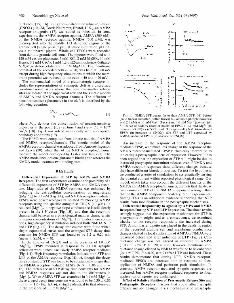
Differential Expression of STP by AMPA and NMDAReceptors. The first experiments examined the possibility of adifferential expression of STP by AMPA and NMDA recep-tors. Magnitude of the NMDA response was enhanced byreducing the extracellular concentration of magnesium([Mg21]o) to 0.1 mM (Fig. 1A), and NMDA receptor-mediatedEPSPs were pharmacologically isolated by blocking AMPAreceptors using the specific antagonist CNQX (10 mM). Inreduced [Mg21]o, a negative slope conductance is still clearlypresent in the I–V curve (Fig. 1B), and thus the receptorychannel still behaves in a physiological manner characteristicof higher concentrations of [Mg21]o (15). Under these condi-tions, high-frequency stimulation induced both NMDA STPand LTP (Fig. 1C). The decay time courses were fitted with asingle exponential curve, and the averaged STP decay timeconstant for NMDA STP was found to be 1.2 6 0.20 min(mean 6 SEM, n 5 8).
In the absence of CNQX and in the presence of 1.0 mM[Mg21]o, EPSPs recorded in response to 0.1 Hz synapticactivation were almost exclusively AMPA receptor-mediated(Fig. 1A). High-frequency stimulation induced both STP andLTP of the AMPA response (Fig. 1D, E), though the decaytime constant of STP was found to be substantially longer thanfor NMDA receptor-mediated EPSPs (6.39 6 0.86 min, n 512). The difference in STP decay time constants for AMPAand NMDA responses was not due to the differences in[Mg21]o. When AMPA STP was examined in 0.1 mM [Mg21]o,the averaged decay time constant was found to be 6.30 6 0.86min (n 5 11) (Fig. 1D, F), virtually identical to that observedin the presence of 1.0 mM [Mg21]o.
An increase in the response of the AMPA receptor-mediated EPSP, with much less change in the response of theNMDA receptor-mediated EPSP is classically interpreted asindicating a postsynaptic locus of expression. However, it hasbeen argued that the expression of STP still might be due toincreased presynaptic transmitter release, even if NMDA andAMPA receptor responses show different changes becausethey have different kinetic properties. To test the hypothesis,we conducted a series of simulations by systematically varyingthe quantal content within reported physiological range. Ourmodel, which takes into account the different kinetics of theNMDA and AMPA receptorychannels, predicts that the decaytime course of STP of the NMDA component is longer thanthat of the AMPA component, contrary to our experimentalfinding. This is an additional evidence supporting that STPresults from modifications in the postsynaptic mechanisms.
Differential Responsivity to Agonist by AMPA and NMDAReceptors During STP and LTP Expression. The above resultsstrongly suggest that the expression mechanism for STP ispostsynaptic in origin, and as a consequence, we examinedwhether or not receptor responsivity was increased duringSTP. A multibarrel pipette was placed in the dendritic regionof the recorded granule cell and membrane conductancechanges elicited by focal application of AMPA or NMDA weremeasured before and after induction of LTP (Fig. 2). Con-ductance change was not altered in response to AMPA(20.7 6 3.5%; P . 0.20, n 5 8); however, membrane con-ductance change elicited by NMDA was found to be enhanced(24.1 6 7.2%; P , 0.02, n 5 7) after LTP was induced. Theseresults demonstrate that during LTP, NMDA receptor-mediated EPSCs are increased both in response to focalapplication of NMDA and perforant path stimulation. Incontrast, AMPA receptor-mediated synaptic responses areincreased, but AMPA receptor-mediated responses to focalapplication of agonist are unchanged.
Effect of Relative Position of Presynaptic Release Site andPostsynaptic Receptors. Factors that could affect synapticefficacy include changes in (i) mechanisms of presynaptic
FIG. 1. NMDA STP decays faster than AMPA STP. (A) Before(solid traces) and after (dotted traces) D-2-amino-5-phosphonovalericacid (50 mM) at 0.1 mM Mg21 (Upper) and 1.0 mM Mg21 (Lower). (B)I–V curve of NMDA receptor-mediated EPSC at 0.1 mM Mg21 (inpresence of CNQX). (C) STP and LTP expressed by NMDA-mediatedEPSPs (in presence of CNQX). (D) STP and LTP expressed byAMPA-mediated EPSPs (in absence of CNQX).
6984 Neurobiology: Xie et al. Proc. Natl. Acad. Sci. USA 94 (1997)

glutamate release, (ii) number of postsynaptic receptors, (iii)kinetic properties of postsynaptic receptors, and (iv) thegeometry of the synaptic junction. Because alteration ofpresynaptic release mechanisms would be expected to affectAMPA and NMDA receptor-mediated responses equivalently,the observed differences in expression of STP and LTP by thetwo receptor subtypes strongly suggest that the mechanismsare postsynaptic in origin. The fact that potentiation of theAMPA response is not observed during focal application ofagonist further suggests that the mechanism of AMPA STPand LTP involves changes other than an increase in receptorresponsivity or number. Because the contribution of changesin synaptic geometry is more difficult to evaluate, we con-ducted a series of computer simulations to investigate thepossible consequences of changes in synaptic morphology.
Initial simulation studies revealed that the extreme narrow-ness of the synaptic cleft (20 nm) leads to a highly localizeddistribution of glutamate molecules along the postsynapticmembrane (Fig. 3A). As a consequence, we tested the hypoth-esis that the relative positions of the presynaptic release sitesand postsynaptic receptorychannels will be a significant factorin determining the magnitude and time course of the EPSCs.Specifically, we simulated AMPA and NMDA receptor-mediated EPSCs for different lateral displacements of releasesites and receptorychannels. Results showed that the AMPAreceptor-mediated EPSC is 42% larger and the NMDA re-ceptor-mediated EPSC is 17% larger in magnitude when therelease sites and receptors are in complete alignment (a zerolateral displacement) compared with the EPSCs simulated fora 40-nm displacement (Fig. 3 B and C). Furthermore, whenrelease sites and receptors are in complete alignment, AMPAreceptor-mediated EPSCs are characterized by a faster rate ofonset and decay, in agreement with experimental findings(Figs. 2 and 3). The differential sensitivity of AMPA andNMDA receptors to the degree of ‘‘alignment’’ with presyn-aptic release sites can be explained by their different affinitiesto glutamate (30 mM for AMPA receptorychannel and 0.9 mMfor NMDA receptorychannel in our model; also see refs.20–22). Simulation results showed that due to NMDA recep-tors’ higher affinity, more than 98% of NMDA receptors arebound when a 40-nm displacement is assumed. As a conse-quence, changes in the postsynaptic location of the NMDA
receptor have little effect on the probability of binding withglutamate in changing the magnitude of the EPSC. In contrast,because only 23% of the AMPA receptors are liganded whena 40-nm displacement is assumed, lateral movement thatincreases alignment with the presynaptic release site will resultin a higher proportion of bound receptors.
When glutamate is delivered to the synapse by means ofperfusion, such as during focal application of agonists, itsdistribution would be more uniform and the influence ofreceptor relocation should be reduced for both AMPA andNMDA receptor-mediated EPSCs. This condition was simu-lated by assuming that glutamate diffuses into the synaptic cleftfrom its perimeter: virtually no difference was seen in eitherAMPA or NMDA receptor-mediated EPSCs. To test theplausibility that changes in receptorychannel kinetics accountfor STP and LTP of AMPA receptor-mediated responses,opening and closing rate were increased in magnitude asproposed by Ambros-Ingerson and Lynch (20). Simulationresults showed that the AMPA receptor-mediated response tofocal application of agonist increases by 31%. These resultsstrongly support the hypothesis that potentiation of AMPAreceptor-mediated EPSCs reflect a relocation of the receptorychannel in the postsynaptic membrane so as to become morealigned with presynaptic release sites. The same mechanismcannot account for STP or LTP of the NMDA receptor-mediated EPSP.
DISCUSSION
Different Expression Mechanisms Underlie Potentiation ofAMPA and NMDA Receptors. The major findings of thepresent study are that STP of the NMDA receptor exhibits amuch shorter decay time course than that for the AMPAreceptor. In addition, during the expression of both STP andLTP, only the postsynaptic response to microejection ofNMDA is enhanced; the response to microejection of AMPAis unchanged. Thus, there are different mechanisms of expres-sion for STP and LTP of AMPA and NMDA receptorychannels. Assuming colocalization of the two glutamatergicreceptor subtypes, these data also strongly argue that, for bothAMPA and NMDA receptors, the expression of STP and LTPinvolve mechanisms that are postsynaptic in origin. Only the
FIG. 2. (A) Membrane conductance in response to microapplication of NMDA, but not AMPA, is increased during STP. (Upper) The uppertwo are responses induced by pressure ejection of NMDA at the moments indicated by downward arrowheads, (500 mM, 100 msec in duration);the lower two are responses induced by AMPA (500 mM, 100 msec in duration); before (Left) and after (Right) induction of LTP. (B) STP wasexpressed by whole cell EPSCs in both experimental groups. (Upper) The upper two EPSCs are obtained from the same hippocampal slice as theone shown in A for focal application of NMDA; the lower two are from that for focal application of AMPA before (Dotted) and during (Solid)STP.
Neurobiology: Xie et al. Proc. Natl. Acad. Sci. USA 94 (1997) 6985

extremely rapid decay rate for NMDA STP suggests thepossibility of a presynaptic mechanism, i.e., that STP of theNMDA receptorychannel reflects primarily PTP. This hypoth-esis has been dealt with elsewhere (23). Results of our simu-lation studies showed that either sensitization of the NMDAreceptor or an increase in number of receptors is sufficient toaccount for the increased postsynaptic responsivity to agonist,and thus, the LTP expressed by the NMDA receptor. Thefailure of others to detect a similar change in NMDA receptorresponsivity with focal application of glutamate (10–13) ismost likely due to the use of high concentrations of Mg21 inthe perfusing media, suppressing NMDA channel opening.
Enhancement of synaptically evoked AMPA responses inthe absence of increased responsivity to agonist further sug-gests that the expression mechanisms of AMPA STP and LTPinvolve changes other than an increase in number or sensitivityof the receptor. Because receptors and other membraneproteins can diffuse laterally in the plane of the cytoplasmicmembrane (24, 25), the effects of changes in position ofpostsynaptic AMPA and NMDA receptors were investigatedusing computer simulations of a glutamatergic synapse. Re-sults revealed that the relative positions of the presynapticrelease site and the postsynaptic receptor can have a profoundeffect on the magnitude and time course of the EPSCs.Detailed studies found that properties of AMPA receptor-mediated STP and LTP were most consistent with the hypoth-esis that, in response to high-frequency stimulation, the re-ceptorychannel becomes relocalized in the postsynaptic mem-
brane so as to be more closely aligned with the presynapticrelease site.
Postsynaptic AMPA Receptor Realignment Model. Thus, wepropose the following model for the mechanism of expressionof NMDA receptor-dependent STP and LTP of AMPAreceptor-mediated synaptic responses. NMDA receptor acti-vation is known to be critical for glutamatergic synapse stabi-lization during development (26), and thus, we assume that aclose alignment of NMDA receptors and presynaptic releasesites emerges from the process of synapse formation. Forsynapses in the unpotentiated state, we further assume thatAMPA receptors have a wider spatial distribution, with manybeyond the range of effective concentration of released neu-rotransmitter. Ca21 influx through the NMDA receptorychannel activates several calcium-dependent processes, amongthem, the calcium-dependent protease calpain (27). Calpain-mediated degradation of cytoskeletal proteins has been shownto play an important role in the redistribution of cell surfacereceptors (28), and we postulate that it allows the lateraldiffusion of AMPA receptors in synaptic membranes, and theirmigration toward the region of recently activated NMDAreceptorychannels. This initial clustering of AMPA receptorsin membrane locations more closely aligned with presynapticrelease sites is the basis for STP.
As the mechanisms that promote receptor aggregationdecay with time, AMPA receptors diffuse away from mem-brane locations proximal to the initially activated NMDAreceptors (and thus to locations more displaced from therelease site), and there is a corresponding time-dependentdecay in the magnitude of the potentiated AMPA receptor-mediated response. Activation of other calcium-dependentprocesses and second messenger systems, including thoserelated to adhesion molecules (29, 30), have been shown to berequired for the stabilization of STP into LTP. If AMPAreceptors are ‘‘anchored’’ in their new position before relax-ation of STP is complete, then the newly stabilized locationsrelative to the release sites leads to LTP. Without activation ofthe anchoring mechanisms, however, recently aggregated re-ceptors will diffuse back to their original positions (or equiv-alently distant positions), with the resulting synaptic potenti-ation expressed only as STP. Thus, the magnitude of LTP isdetermined by the decay time course of the clustering processand the onset time course of the anchoring process.
The proposed model is consistent with previous conceptu-alizations of two parallel and independent processes for STPand LTP (31–33), as well as numerous studies indicating thatinhibition of second messenger pathways effectively blocksstabilization of LTP while having minimal effect on themagnitude of STP. In particular, the proposed model isconsistent with our recent observation that STP can be in-duced repetitively in the presence of saturating levels of LTP(23). Saturation of LTP is presumed to represent the maximumdensity of receptors that can be anchored in the synapticregion. If the processes underlying clustering (increased re-ceptor mobility and lateral displacement) are independent ofthe mechanisms underlying anchoring, then neither the mag-nitude of STP nor its repetitive induction should be altered bysaturation of LTP.
A variant of this model is that new AMPA receptors areinserted in close proximity to NMDA receptors, and thus, atmembrane positions in closer alignment with presynapticrelease sites. To account for the differential effects of exoge-nous AMPA and NMDA found in the present studies, how-ever, it also must be postulated that the total number of AMPAreceptors remains constant, i.e., that some receptors distantfrom the NMDA receptors are internalized. Recent studieshave shown that the C-terminal domain of various AMPAreceptor subunits is truncated after NMDA receptor stimula-tion and the resulting activation of the calcium-dependentprotease calpain (34, 35). Furthermore, several sources of
FIG. 3. The effect of redistribution of receptors. (A) Simulation ofneurotransmitter release from a vesicle into the synaptic cleft. Becausethe cleft is extremely narrow, the concentration of glutamate at thepostsynaptic membrane is highly localized. (B) Simulated AMPAresponse (Upper) and experimental whole cell EPSCs (Lower). Theyexhibit a similar profile (note the normalized results). In simulation,the AMPA-mediated current is 43% greater when the receptor isperfectly aligned with the release site than when they are 40 nm away(Left). The two traces are normalized to show that the time course ofthe larger response (solid trace) is faster (Right). (C) A smaller changeis seen in the NMDA-mediated EPSC (Left) with virtually no changein its time course (Right).
6986 Neurobiology: Xie et al. Proc. Natl. Acad. Sci. USA 94 (1997)

evidence indicate the existence of a cytoplasmic pool ofAMPA receptors that could be inserted in postsynaptic mem-branes in a calcium-dependent manner (36). Though a changein channel kinetic properties, such as an increase in theopening and closing rates (20), has been shown to reproducethe EPSP waveforms characteristic of LTP expression, such amechanism cannot explain why no increase is observed withfocal application of AMPA or glutamate in the present or inthe majority of past studies (10–13, 37).
Predictions of the Postsynaptic AMPA Realignment Model.There are a number of results that can be predicted orexplained by our model. First, increased responses to micro-ejection of AMPA may occur during later phases of LTP. Asshown in Fig. 4, receptor aggregation may cause an overallreceptor redistribution on the membrane surface. This redis-tribution may serve as a signal for more receptors to besupplied to the membrane. As a result, the absolute number ofAMPA receptors could gradually increase over time. Davies etal. (38) observed such a progressive increase in AMPA re-sponsivity, which started about 30 min after the induction ofLTP with the peak magnitude appearing approximately 90 minlater.
Second, if AMPA receptors are poorly aligned relative topresynaptic site, the synapse can be functionally silent. Oursimulation results showed that the amplitude of EPSP pro-duced by an AMPA receptorychannel displaced by 100 nmfrom the presynaptic release site is 32% of that generated bya receptorychannel aligned with the release site, whereas areceptorychannel displaced by 200 nm will generate an EPSPonly 6% in amplitude, making it almost undetectable. This
prediction is consistent with reports that LTP arises fromrecruitment of inactive synapses or latent receptors (39–43).More recently, Liao et al. (44) and Isaac et al. (45) haveconfirmed that a high proportion of synapses in hippocampalarea CA1 exhibits NMDA receptor-mediated transmissiononly, making these synapses effectively nonfunctional at nor-mal resting potentials. These ‘‘silent’’ synapses acquire AMPAreceptor-mediated response characteristics after LTP induc-tion.
Third, our results show that the dynamics of neurotransmit-ter distribution in the synapse varies as a function of distancefrom the release site, namely, the greater the distance from therelease site, the slower the variation in concentration ofneurotransmitter. Because the model predicts that synapticpotentiation reflects the migration of AMPA receptors topositions closer to the release site, the rise and decay times ofAMPA EPSCs should be more rapid during the expression ofLTP, as found in the present and previous studies (46). Moregenerally, the location of AMPA receptorychannels in thepostsynaptic membrane should range from optimal alignmentwith the presynaptic release site to beyond the range ofeffective concentration of released neurotransmitter, i.e., func-tionally ‘‘silent.’’ Within this spectrum, the rise-fall time andpeak amplitude of the AMPA EPSC waveform should corre-late positively with proximity to the presynaptic release site.
Finally, the model predicts a nonuniform distribution ofAMPA and NMDA receptors in the synapse. It is expectedthat NMDA receptors are at the most focal point opposite agiven release site, with an ‘‘annulus’’ of more peripherallylocated AMPA receptors. The degree of nonuniformity shouldbe more pronounced for potentiated synapses.
We thank M. Yeckel for his generous advice and technical help. Thiswork was supported by the Office of Naval Research, NationalInstitute of Mental Health, National Center for Research Resources,the Human Frontiers Science Organization, and a National ResearchService Award fellowship to J.-S.L.
1. Bliss, T. V. P. & Collingridge, G. L. (1993) Nature (London) 361,31–39.
2. Bekkers, J. M. & Stevens, C. F. (1989) Nature (London) 341,230–233.
3. Kauer, J. A., Malenka, R. C. & Nicoll, R. A. (1988) Neuron 1,911–917.
4. Muller, D., Joly, M. & Lynch, G. (1988) Science 242, 1694–1697.5. Muller, D. & Lynch, G. (1988) Proc. Natl. Acad. Sci. USA 85,
9346–9350.6. Perkel, D. J. & Nicoll, R. A. (1993) J. Physiol. 471, 481–500.7. Asztely, F., Wigstrom, H. & Gustafsson, B. (1992) Eur. J. Neu-
rosci. 4, 681–690.8. Xie, X., Berger, T. W. & Barrionuevo, G. (1992) Soc. Neurosci.
Abstr. 18, 628.5.9. O’Connor, J. J., Rowan, M. J. & Anwyl, R. (1995) J. Neurosci. 15,
2013–2020.10. Lynch, G., Gribkoff, V. & Deadwyler, S. A. (1976) Nature
(London) 263, 151–153.11. Murali, M. P. & Sastry, B. R. (1985) Eur. J. Pharmacol. 114,
335–341.12. Taube, J. S. & Schwartzkroin, P. A. (1988) J. Neurosci. 8, 1632–
1644.13. Malgaroli, A. & Tsien, R. W. (1992) Nature (London) 357,
134–139.14. Xie, X., Berger, T. W. & Barrionuevo, G. (1992) J. Neurophysiol.
67, 1009–1013.15. Nowak, L., Bregestovski, P. & Ascher, P. (1984) Nature (London)
307, 462–465.16. Mayer, M. L. & Westbrook, G. L. (1987) J. Physiol. 394, 501–527.17. Drejer, J. & Honore, T. (1988) Neurosci. Lett. 87, 104–108.18. Longsworth, L. G. (1953) J. Am. Chem. Soc. 75, 5705–5709.19. Carnahan, B., Luther, H. A. & Wilkes, J. O. (1969) Applied
Numerical Methods (Wiley, New York).20. Ambros-Ingerson, J. & Lynch, G. (1993) Proc. Natl. Acad. Sci.
USA 90, 7903–7907.21. Lester, R. A. J. & Jahr, C. E. (1992) J. Neurosci. 12, 635–643.
FIG. 4. Schematic representation of our STP-LTP model. Rest,patches of presynaptic and postsynaptic membrane in a nonpotentiatedstate. HFS, presynaptic LTP-inducing pulses arrive that cause NMDAreceptorychannel activation and Ca21 influx, which, in turn, trigger thereceptor attraction mechanism. STP, as a result, AMPA receptors areattracted toward the activated NMDA receptorychannel, which isaligned previously with the presynaptic release zone. LTP, dependingon the effectiveness of the anchoring mechanism, all or part AMPAreceptors will diffuse again. An electrophysiological observer then willinterpret it as only STP or STP plus LTP correspondingly. Note thatdue to diffusion, AMPA receptors used to be farther located may enterthis domain and can be used for further potentiation of synapticstrength.
Neurobiology: Xie et al. Proc. Natl. Acad. Sci. USA 94 (1997) 6987

22. Patneau, D. K. & Mayer, M. L. (1990) J. Neurosci. 10, 2385–2399.23. Xie, X., Barrionuevo, G. & Berger, T. W. (1996) Learn. Mem. 3,
115–123.24. Froehner, S. C. (1993) Annu. Rev. Neurosci. 16, 347–368.25. Alberts, B., Bray, D., Lewis, J., Raff, M., Roberts, K. & Watson,
J. D. (1994) Molecular Biology of the Cell (Garland, New York),3rd Ed., pp. 475–506.
26. Debski, E. A., Cline, H. T. & Constantine-Paton, M. (1990)J. Neurobiol. 21, 18–32.
27. Vanderklish, P., Saido, T. C., Gall, C. & Lynch, G. (1995) Mol.Brain Res. 32, 25–35.
28. Croall, D. E. & Demartino, G. N. (1991) Physiol. Rev. 71, 813–847.
29. Luthi, A., Laurent, J. P., Figurov, A., Muller, D. & Schachner, M.(1994) Nature (London) 372, 777–779.
30. Xiao, P., Bahr, B. A., Staubli, U., Vanderklish, P. & Lynch, G.(1991) Neuroreport 2, 461–464.
31. Gustafsson, B., Asztely, F., Hanse, E. & Wigstrom, H. (1989) Eur.J. Neurosci. 1, 382–394.
32. Nicoll, R. A. & Malenka, R. C. (1995) Nature (London) 377,115–118.
33. Arai, A., Larson, J. & Lynch, G. (1990) Brain Res. 511, 353–357.
34. Bi, X., Chang, V., Tocco, G. & Baudry, M. (1996) Brain Res. 726,98–108.
35. Bi, X., Tocco, G. & Baudry, M. (1994) Neuroreport 6, 61–64.36. Standley, S., Bi, X. & Baudry, M. (1996) in Long-Term Potenti-
ation 3, eds. Baudry, M. & Davis, J. L. (MIT Press, Cambridge,MA), pp. 17–40.
37. Poolos, N. P. & Kocsis, J. D. (1990) Brain Res. 524, 342–346.38. Davies, S. N., Lester, R. A. J., Reymann, K. G. & Collingridge,
G. L. (1989) Nature (London) 338, 500–503.39. Edwards, F. (1991) Nature (London) 350, 271–272.40. Liao, D., Jones, A. & Malinow, R. (1992) Neuron 9, 1089–1097.41. Manabe, T., Renner, P. & Nicoll, R. A. (1992) Nature (London)
355, 50–55.42. Kullmann, D. M. (1994) Neuron 12, 1111–1120.43. Andrew, R. D. & Macvicar, B. A. (1994) Neurosci. 62, 371–383.44. Liao, D., Hessler, N. A. & Malinow, R. (1995) Nature (London)
375, 400–404.45. Isaac, J. T. R., Nicoll, R. A. & Malenka, R. C. (1995) Neuron 15,
427–434.46. Ambros-Ingerson, J., Xiao, P., Larson, J. & Lynch, G. (1993)
Brain Res. 620, 237–244.
6988 Neurobiology: Xie et al. Proc. Natl. Acad. Sci. USA 94 (1997)