Embed Size (px)
Citation preview
Cellular/Molecular
Estradiol Acutely Potentiates Hippocampal ExcitatorySynaptic Transmission through a Presynaptic Mechanism
Tereza Smejkalova and Catherine S. WoolleyDepartment of Neurobiology and Physiology, Northwestern University, Evanston, Illinois 60208
Although recent evidence suggests that the hippocampus is a source of 17�-estradiol (E2), the physiological role of this neurosteroid E2,as distinct from ovarian E2, is unknown. One likely function of neurosteroid E2 is to acutely potentiate excitatory synaptic transmission,but the mechanism of this effect is not well understood. Using whole-cell voltage-clamp recording of synaptically evoked EPSCs in adultrat hippocampal slices, we show that, in contrast to the conclusions of previous studies, E2 potentiates excitatory transmission througha presynaptic mechanism. We find that E2 acutely potentiates EPSCs by increasing the probability of glutamate release specifically atinputs with low initial release probability. This effect is mediated by estrogen receptor � (ER�) acting as a monomer, whereas ER� is notrequired. We further show that the E2-induced increase in glutamate release is attributable primarily to increased individual vesiclerelease probability and is associated with higher average cleft glutamate concentration. These two findings together argue strongly thatE2 promotes multivesicular release, which has not been shown before in the adult hippocampus. The rapid time course of acute EPSCpotentiation and its concentration dependence suggest that locally synthesized neurosteroid E2 may activate this effect in vivo.
IntroductionRecent studies support the idea that the hippocampus, a knowntarget of the steroid hormone 17�-estradiol (E2), is also a site ofE2 synthesis. The possibility that the brain produces its own ste-roids, neurosteroids, was first proposed almost 30 years ago (Cor-pechot et al., 1981). More recently, all enzymes needed tosynthesize E2 from cholesterol, as well as significant levels ofintermediate metabolites and E2 itself, have been demonstratedin the adult rat hippocampus (Kimoto et al., 2001; Hojo et al.,2004, 2009). This raises the question, what is the physiologicalrole of neurosteroid E2 as distinct from ovarian E2?
One likely answer lies in the ability of E2 to acutely potentiateexcitatory synaptic transmission. Within minutes of applicationto hippocampal slices, E2 increases field EPSP slope (Teyler et al.,1980; Sharrow et al., 2002; Kim et al., 2006; Kramar et al., 2009)and potentiates intracellularly recorded EPSPs and EPSCs in CA1(Wong and Moss, 1992; Foy et al., 1999; Rudick and Woolley,2003). E2 also acutely increases CA1 neurons’ responses to glu-tamate receptor agonists applied to slices (Wong and Moss, 1992)or dissociated cells (Gu and Moss, 1996, 1998). Several observa-tions suggest that brain-derived E2, not ovarian E2, is the endog-enous steroid that activates these effects in vivo. First, althoughacute potentiation can occur with E2 concentrations matchingpeak circulating levels (�100 pM), it is more robust with higherconcentrations, and potentiation of agonist-evoked responses re-quires �10 nM E2. Second, acute potentiation occurs in slices
from male as well as female hippocampus (Teyler et al., 1980;Kramar et al., 2009). Finally, the ability of E2 to act within min-utes is much more rapid than any fluctuations in circulating E2.
The mechanism(s) of acute potentiation of synaptic transmis-sion by E2 are not well understood. Intracellular recording stud-ies have shown that only some CA1 cells are E2 responsive,suggesting that E2 action is cell specific. Furthermore, the studiesusing exogenous agonist application led to the conclusion that E2enhances postsynaptic sensitivity to glutamate. It is not clear,however, whether conclusions based on nonsynaptic stimulationapply to E2 effects on synaptic transmission. Therefore, we stud-ied acute potentiation of synaptic responses by E2 using whole-cell voltage-clamp recording of synaptically evoked EPSCs inadult rat hippocampal slices. Consistent with previous reports,we observed a response to E2 in only a subset of experiments.Interestingly, however, the E2-responsive subset shared the char-acteristic of relatively high initial paired-pulse ratio (PPR), andE2 decreased PPR in parallel with synaptic potentiation, sugges-tive of presynaptic mechanisms. Indeed, we found that E2 acutelypotentiates excitatory synaptic transmission in an input-specificmanner, through an increase in the probability of glutamate re-lease specifically at inputs with low initial release probability. Thispotentiation depends on activation of estrogen receptor � (ER�)and not ER�. Further investigation of mechanisms involved inthe E2-induced increase in glutamate release revealed that E2increases individual vesicle release probability as well as averagecleft glutamate concentration, strongly suggesting that E2 pro-motes multivesicular release.
Materials and MethodsAnimals. All animal procedures were performed in accordance with theNational Institutes of Health Guide for the Care and Use of LaboratoryAnimals and were approved by the Northwestern University Animal Careand Use Committee. Adult female Sprague Dawley rats (50 – 60 d old;
Received Aug. 9, 2010; revised Sept. 21, 2010; accepted Sept. 23, 2010.This work was supported by National Institutes of Health Grants NS037324, MH067564, and RR015497. We thank
Indira Raman for helpful discussions.Correspondence should be addressed to Dr. Catherine S. Woolley, Department of Neurobiology and Physiology,
2205 Tech Drive, Northwestern University, Evanston, IL 60208. E-mail: [email protected]:10.1523/JNEUROSCI.4161-10.2010
Copyright © 2010 the authors 0270-6474/10/3016137-12$15.00/0
The Journal of Neuroscience, December 1, 2010 • 30(48):16137–16148 • 16137
Harlan) were ovariectomized under ketamine (85 mg/kg, i.p; BionichePharma) and xylazine (13 mg/kg, i.p.; Lloyd Laboratories) anesthesiausing aseptic surgical procedures. Either 3 or 7 d after surgery, each ratwas given two injections (subcutaneously) of 10 �g of 17�-estradiolbenzoate in 100 �l of sesame oil, 24 h apart, and slices were prepared 2 dafter the second injection. This estradiol pretreatment produces serumlevels of �30 – 40 pg/ml (corresponding to peak proestrus levels) (Smithet al., 1975) at the time the animal is killed (Woolley and McEwen, 1993),and has been shown to increase CA1 responsiveness to acute effects ofestradiol on excitatory synaptic transmission (Wong and Moss, 1992).
Chemicals. Stock solutions of the following drugs were prepared inDMSO: (�)-bicuculline, cyclothiazide (CTZ), propylpyrazole triol(PPT), diarylpropionitrile (DPN), ICI 182,870, 2,3-dioxo-6-nitro-1,2,3,4-tetrahydrobenzo[f]quinoxaline-7-sulfonamide (NBQX), G-1(all from Tocris Bioscience), and 17�- and 17�-estradiol (Sigma-Aldrich). Stock solutions of �-D-glutamylglycine (�DGG) and (RS)-3-(2-carboxypiperazin-4-yl)-propyl-1-phosphonic acid [(RS)-CPP] (bothfrom Tocris Bioscience) were prepared in ddH2O. Stock solutions werediluted in artificial CSF (ACSF) on the day of recording to the finalconcentrations indicated. Control ACSF contained an equivalent con-centration of DMSO (�0.1%). Other chemicals were either from FisherScientific (NaCl, NaHCO3, dextrose, KCl, and NaH2PO4) or fromSigma-Aldrich (sesame oil, CaCl2, MgCl2, K-gluconate, HEPES, Na2-creatine phosphate, MgATP, NaGTP, biocytin, and QX314).
Slice preparation and electrophysiology. Rats were deeply anesthetizedwith sodium pentobarbital (80 mg/kg, i.p.; Virbac Animal Health) andperfused transcardially with ice-cold oxygenated ACSF containing thefollowing (in mM): 125 NaCl, 25 NaHCO3, 25 dextrose, 2.5 KCl, 1.25NaH2PO4, 1 MgCl2, and 2 CaCl2, 315 Osm, pH 7.4. Transverse slices ofdorsal hippocampus (300 �m) were cut into an ice-cold bath of oxygen-ated ACSF using a Leica VT1000S oscillating tissue slicer. Slices wereallowed to recover submerged in oxygenated ACSF at 35°C for 30 minand were then kept at room temperature until recording. During record-ing, slices were perfused with warm (32–35°C) or room temperature(20 –22°C, for high-frequency train experiments and �DGG/NBQX ex-periments) oxygenated ACSF at a rate of �2 ml/min in a recordingchamber mounted on a Zeiss Axioskop. Somatic whole-cell voltage-clamp recordings were obtained from visually identified CA1 pyramidalcells using patch electrodes (3–5 M�) filled with intracellular solutioncontaining the following (in mM): 115 K-gluconate, 20 KCl, 10 HEPES,10 Na2-creatine phosphate, 2 MgATP, 0.3 NaGTP, 0.1% biocytin, and1–5 QX314, 280 Osm, pH 7.3. One or two glass bipolar stimulatingelectrodes filled with ACSF were placed in the stratum radiatum �300�m apart. At the beginning of every double-stimulation experiment, itwas confirmed that no paired-pulse facilitation [interpulse interval (IPI),100 ms] occurred when the first pulse was delivered at one stimulationsite and the second pulse was delivered at the other stimulation site,indicating no overlap of stimulated inputs. During an experiment,paired-pulse stimulation (IPI, 100 or 50 ms) was delivered every 15 s; indouble-stimulation experiments, stimulation was delivered every 15 s,alternating between the two stimulation sites; in high-frequency trainexperiments, 2 min of paired-pulse stimulation (delivered every 15 s)alternated with 3 min of train stimulation (100-pulse, 20 Hz trains deliv-ered every 45 s). In experiments using both 50 and 100 ms IPI, there wereno effects of IPI on either baseline PPR or the E2 effect on PPR. Seriesresistance was monitored using 5 mV, 10 ms voltage steps and rangedbetween 5 and 25 M� between experiments. Only experiments withstable series resistance and holding current were included in the analysis,and series resistance did not differ between E2-responsive and E2-nonresponsive experiments. Synaptically evoked AMPA-receptor-mediated EPSCs were recorded at a holding potential of �70 mV in thepresence of the GABAA and NMDA receptor blockers, (�)-bicuculline(10 �M) and (RS)-CPP (10 �M), respectively. Data were acquired with anAxopatch 200B amplifier and pClamp 9.0 software (Molecular Devices),filtered at 2 kHz, and digitized at 20 kHz using a Digidata 1322A dataacquisition system (Molecular Devices).
After a stable 10 –15 min baseline recording in control ACSF, differentdrugs were bath applied as follows: E2 (100 pM to 100 nM) for 10 –15 min,in most cases followed by a 10 –30 min washout period; CTZ (100 �M),
PPT (100 or 200 nM), DPN (100 or 500 nM), ICI 182,780 (100 nM), or G-1(100 nM) alone for 12 min, followed by coapplication with E2 (100 nM)for 12 min, followed by E2 (100 nM) alone (see Fig. 4); �DGG (1 mM) for10 min either in the absence or presence of E2 (100 nM) (see Fig. 6);NBQX (200 nM) for 15 min either in the absence or in the presence of E2(100 nM) (see Fig. 6). Effects of different drugs were evaluated by com-paring data from the last 5 min of recording in each condition or from a5 min period after the maximal effect of a drug was reached (in �DGGand NBQX experiments). Because effects of E2 were not readily revers-ible, E2 and ER agonists were applied to each slice only once.
In experiments using high-frequency stimulus trains, a 15–20 minbaseline recording was followed by a 20 min recording in the presence ofE2 (100 nM). Effects of E2 were evaluated by comparing data from the last10 min of E2 application to data from the last 10 min of baseline toinclude a sufficient number of train sweeps. The analysis of trains wasbased on Schneggenburger et al. (1999) and Wesseling and Lo (2002).Baseline EPSC trains showed early facilitation followed by gradual de-pression, and reached a steady state after �70 pulses, consistent withprevious reports (Wesseling and Lo, 2002). The cumulative EPSC chargeduring the last second of the train (sum of pulses 81–100) was used toestimate the rate of steady-state release attributable to vesicle replenish-ment, assumed to be constant throughout the train. The relative size ofthe readily-releasable vesicle pool (RRP) was estimated from the cumu-lative EPSC charge recorded before the steady state was reached (sum ofpulses 1– 80) after subtracting the EPSC charge caused by vesicle replen-ishment during the train (four times the sum of pulses 81–100). Theproportion of the total RRP released by the first stimulus was used toestimate the relative individual vesicle release probability, Pves.
Data were analyzed off-line using Igor Pro (Wavemetrics) software,and statistical significance was evaluated with Student’s t tests or ANOVAfollowed by Bonferroni post hoc tests, as indicated. A p value of �0.05was considered significant.
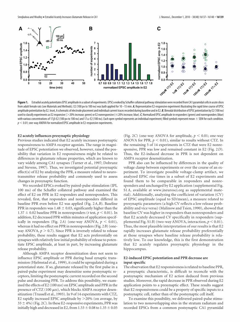
Results17�-Estradiol acutely potentiates EPSC amplitude in a subsetof experimentsWe investigated the ability of E2 to rapidly potentiate excitatorysynaptic transmission in adult female rat hippocampus. We stim-ulated the Schaffer collateral pathway in acute slices and madewhole-cell voltage-clamp recordings of synaptically evokedEPSCs from CA1 pyramidal cells. In a subset of experiments, bathapplication of E2 (100 pM to 100 nM) potentiated EPSC ampli-tude within minutes (Fig. 1A). This effect was not readily revers-ible, possibly because of slow wash out of E2 from the slice orinitiation of a persistent effect that outlasts the presence of E2. Wequantified EPSC potentiation by normalizing average EPSC am-plitude recorded during the last 5 min in E2 to baseline EPSCsduring the last 5 min before E2 application. This showed that themagnitude of E2-induced potentiation ranged from 0.76 to 1.76,with a clearly bimodal distribution among experiments (Fig. 1B).Based on this distribution, we subsequently divided all experi-ments into two groups: those showing �20% potentiation wereclassified as E2 responsive and the rest as E2 nonresponsive.
Using the �20% potentiation criterion, we found that boththe frequency of responders and the extent of potentiation inresponders increased with increasing concentrations of E2. At100 pM, E2 potentiated EPSCs in 6 of 14 (43%) experiments, by26 � 2%; 1 nM E2 potentiated EPSCs in 9 of 22 (41%) experi-ments, by 29 � 3%; 10 nM E2 potentiated EPSCs in 8 of 17 (47%)experiments, by 33 � 4%; and 100 nM E2 potentiated EPSCs in 18of 28 (64%) experiments, by 42 � 3% (Fig. 1C). The effect of E2was stereospecific, as no potentiation was observed with 100 nM
17�-estradiol (n � 5).
16138 • J. Neurosci., December 1, 2010 • 30(48):16137–16148 Smejkalova and Woolley • Estradiol Acutely Increases Glutamate Release in CA1
E2 acutely influences presynaptic physiologyPrevious studies indicated that E2 acutely increases postsynapticresponsiveness to AMPA receptor agonists. The range in magni-tude of EPSC potentiation we observed, however, raised the pos-sibility that variation in E2 responsiveness might be related todifferences in glutamate release properties, which are known tovary widely among CA1 synapses (Turner et al., 1997; Dobrunzand Stevens, 1997). Thus, we investigated potential presynapticeffect(s) of E2 by analyzing the PPR, a measure related to neuro-transmitter release probability and commonly used to assesschanges in presynaptic function.
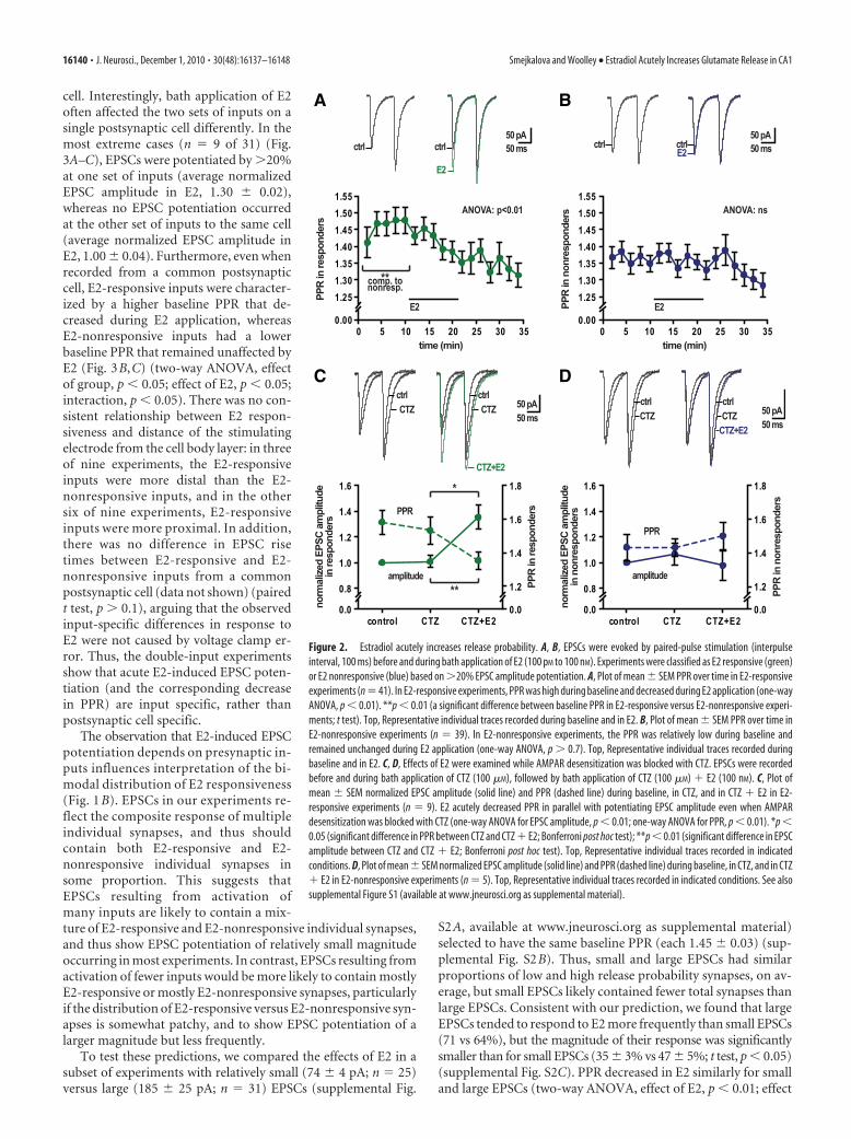
We recorded EPSCs evoked by paired-pulse stimulation (IPI,100 ms) of the Schaffer collateral pathway and examined theeffect of E2 on PPR in E2 responders and nonresponders. Thisrevealed, first, that responders and nonresponders differed inbaseline PPR even before E2 was applied (Fig. 2A,B). BaselinePPR in responders was 1.48 � 0.03, significantly higher than the1.37 � 0.02 baseline PPR in nonresponders (t test, p � 0.01). Inaddition, E2 decreased PPR within minutes of application specif-ically in responders (Fig. 2A) (one-way ANOVA, p � 0.01),whereas it had no effect on PPR in nonresponders (Fig. 2B) (one-way ANOVA, p � 0.7). Since PPR is inversely related to releaseprobability, these results suggest that E2 acts preferentially onsynapses with relatively low initial probability of release to poten-tiate EPSC amplitude, at least in part, by increasing glutamaterelease probability.
Although AMPA receptor desensitization does not seem toinfluence EPSC amplitude or PPR during basal synaptic trans-mission (Hjelmstad et al., 1999), it could be upregulated during apotentiated state. If so, glutamate released by the first pulse in apaired-pulse experiment may desensitize some postsynaptic re-ceptors, limiting the postsynaptic current recorded on the secondpulse and decreasing PPR. To address this possibility, we exam-ined the effects of E2 (100 nM) on EPSC amplitude and PPR in thepresence of CTZ (100 �M), which blocks AMPA receptor desen-sitization (Trussell et al., 1993). In 9 of 14 experiments with CTZ,E2 rapidly increased EPSC amplitude by �20% (on average, by33 � 4%) (Fig. 2C). In these E2-responsive experiments, PPR wasinitially high and decreased in E2, from 1.53 � 0.08 to 1.35 � 0.05
(Fig. 2C) (one-way ANOVA for amplitude, p � 0.01; one-wayANOVA for PPR, p � 0.01), similar to results without CTZ. Inthe remaining 5 of 14 experiments in CTZ that were E2 nonre-sponsive, PPR was low and remained constant in E2 (Fig. 2D).Thus, the E2-induced decrease in PPR is not dependent onAMPA receptor desensitization.
PPR also can be influenced by differences in the quality ofvoltage clamp between experiments or over the course of an ex-periment. To investigate possible voltage-clamp artifact, weanalyzed EPSC rise times in a subset of E2 experiments andfound them to be comparable in responders and nonre-sponders and unchanged by E2 application (supplemental Fig.S1 A, available at www.jneurosci.org as supplemental mate-rial). Additionally, analyzing the coefficient of variation (CV)of EPSC amplitude (equal to SD/mean), a measure related topresynaptic parameters (a high CV reflects a low release prob-ability and vice versa) (Malinow and Tsien, 1990), showed thatbaseline CV was higher in responders than nonresponders andthat E2 acutely decreased CV specifically in responders (sup-plemental Fig. S1 B) (two-way ANOVA, interaction, p � 0.01).Thus, the most plausible interpretation of our results is that E2rapidly increases glutamate release probability preferentiallyat those synapses where baseline release probability is rela-tively low. To our knowledge, this is the first demonstrationthat E2 acutely regulates presynaptic physiology in thehippocampus.
E2-induced EPSC potentiation and PPR decrease areinput specificThe observation that E2 responsiveness is related to baseline PPR,a presynaptic characteristic, is difficult to reconcile with thepostsynaptic mechanism of E2 action deduced from previousstudies. Moreover, the rapid decrease in PPR observed during E2application points to a presynaptic effect. These results suggestthat E2 responsiveness could be a property of specific inputs to apostsynaptic cell, rather than of the postsynaptic cell itself.
To examine this possibility, we delivered paired-pulse stimu-lation to two nonoverlapping sites in the stratum radiatum andrecorded EPSCs from a common postsynaptic CA1 pyramidal
A B C
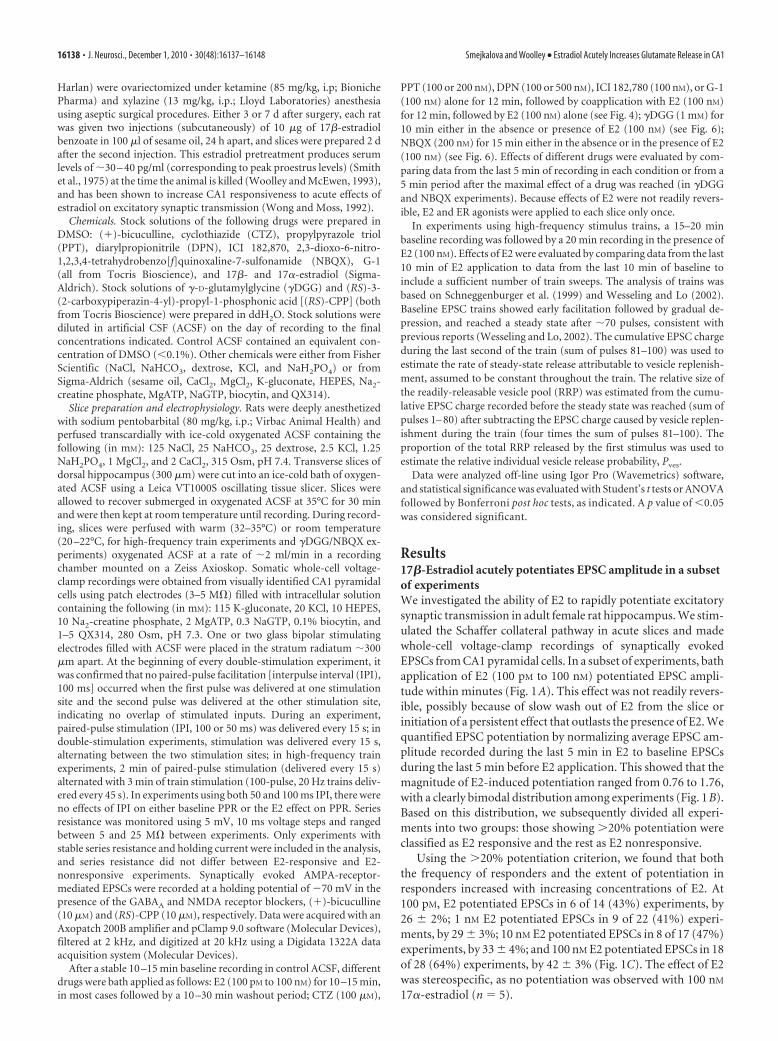
Figure 1. Estradiol acutely potentiates EPSC amplitude in a subset of experiments. EPSCs evoked by Schaffer collateral pathway stimulation were recorded from CA1 pyramidal cells in acute slicesfrom adult female rats (see Materials and Methods). E2 (100 pM to 100 nM) was bath applied for 10 –15 min. A, Representative E2-responsive experiment illustrating the rapid time course of EPSCamplitude potentiation by E2. Inset, A schematic of electrode placement and individual current traces recorded during baseline and in E2. B, Bimodal distribution of EPSC potentiation by E2 (100 nM)used to classify experiments as E2 responsive (�20% increase; green) or E2 nonresponsive (�20% increase; blue). C, Normalized EPSC amplitude in responders (green) and nonresponders (blue)with various concentrations of 17�-E2 (100 pM to 100 nM) and 17�-E2 (100 nM). Each open symbol represents an individual experiment; filled symbols represent mean � SEM for each condition.p � 0.01; one-way ANOVA for normalized EPSC amplitude in E2-responsive experiments.
Smejkalova and Woolley • Estradiol Acutely Increases Glutamate Release in CA1 J. Neurosci., December 1, 2010 • 30(48):16137–16148 • 16139
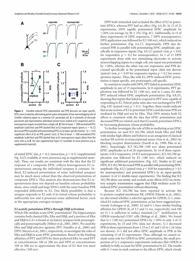
cell. Interestingly, bath application of E2often affected the two sets of inputs on asingle postsynaptic cell differently. In themost extreme cases (n � 9 of 31) (Fig.3A–C), EPSCs were potentiated by �20%at one set of inputs (average normalizedEPSC amplitude in E2, 1.30 � 0.02),whereas no EPSC potentiation occurredat the other set of inputs to the same cell(average normalized EPSC amplitude inE2, 1.00 � 0.04). Furthermore, even whenrecorded from a common postsynapticcell, E2-responsive inputs were character-ized by a higher baseline PPR that de-creased during E2 application, whereasE2-nonresponsive inputs had a lowerbaseline PPR that remained unaffected byE2 (Fig. 3B,C) (two-way ANOVA, effectof group, p � 0.05; effect of E2, p � 0.05;interaction, p � 0.05). There was no con-sistent relationship between E2 respon-siveness and distance of the stimulatingelectrode from the cell body layer: in threeof nine experiments, the E2-responsiveinputs were more distal than the E2-nonresponsive inputs, and in the othersix of nine experiments, E2-responsiveinputs were more proximal. In addition,there was no difference in EPSC risetimes between E2-responsive and E2-nonresponsive inputs from a commonpostsynaptic cell (data not shown) (pairedt test, p � 0.1), arguing that the observedinput-specific differences in response toE2 were not caused by voltage clamp er-ror. Thus, the double-input experimentsshow that acute E2-induced EPSC poten-tiation (and the corresponding decreasein PPR) are input specific, rather thanpostsynaptic cell specific.
The observation that E2-induced EPSCpotentiation depends on presynaptic in-puts influences interpretation of the bi-modal distribution of E2 responsiveness(Fig. 1 B). EPSCs in our experiments re-flect the composite response of multipleindividual synapses, and thus shouldcontain both E2-responsive and E2-nonresponsive individual synapses insome proportion. This suggests thatEPSCs resulting from activation ofmany inputs are likely to contain a mix-ture of E2-responsive and E2-nonresponsive individual synapses,and thus show EPSC potentiation of relatively small magnitudeoccurring in most experiments. In contrast, EPSCs resulting fromactivation of fewer inputs would be more likely to contain mostlyE2-responsive or mostly E2-nonresponsive synapses, particularlyif the distribution of E2-responsive versus E2-nonresponsive syn-apses is somewhat patchy, and to show EPSC potentiation of alarger magnitude but less frequently.
To test these predictions, we compared the effects of E2 in asubset of experiments with relatively small (74 � 4 pA; n � 25)versus large (185 � 25 pA; n � 31) EPSCs (supplemental Fig.
S2A, available at www.jneurosci.org as supplemental material)selected to have the same baseline PPR (each 1.45 � 0.03) (sup-plemental Fig. S2B). Thus, small and large EPSCs had similarproportions of low and high release probability synapses, on av-erage, but small EPSCs likely contained fewer total synapses thanlarge EPSCs. Consistent with our prediction, we found that largeEPSCs tended to respond to E2 more frequently than small EPSCs(71 vs 64%), but the magnitude of their response was significantlysmaller than for small EPSCs (35 � 3% vs 47 � 5%; t test, p � 0.05)(supplemental Fig. S2C). PPR decreased in E2 similarly for smalland large EPSCs (two-way ANOVA, effect of E2, p � 0.01; effect
A B
C D
Figure 2. Estradiol acutely increases release probability. A, B, EPSCs were evoked by paired-pulse stimulation (interpulseinterval, 100 ms) before and during bath application of E2 (100 pM to 100 nM). Experiments were classified as E2 responsive (green)or E2 nonresponsive (blue) based on �20% EPSC amplitude potentiation. A, Plot of mean � SEM PPR over time in E2-responsiveexperiments (n � 41). In E2-responsive experiments, PPR was high during baseline and decreased during E2 application (one-wayANOVA, p � 0.01). **p � 0.01 (a significant difference between baseline PPR in E2-responsive versus E2-nonresponsive experi-ments; t test). Top, Representative individual traces recorded during baseline and in E2. B, Plot of mean � SEM PPR over time inE2-nonresponsive experiments (n � 39). In E2-nonresponsive experiments, the PPR was relatively low during baseline andremained unchanged during E2 application (one-way ANOVA, p � 0.7). Top, Representative individual traces recorded duringbaseline and in E2. C, D, Effects of E2 were examined while AMPAR desensitization was blocked with CTZ. EPSCs were recordedbefore and during bath application of CTZ (100 �M), followed by bath application of CTZ (100 �M) � E2 (100 nM). C, Plot ofmean � SEM normalized EPSC amplitude (solid line) and PPR (dashed line) during baseline, in CTZ, and in CTZ � E2 in E2-responsive experiments (n � 9). E2 acutely decreased PPR in parallel with potentiating EPSC amplitude even when AMPARdesensitization was blocked with CTZ (one-way ANOVA for EPSC amplitude, p � 0.01; one-way ANOVA for PPR, p � 0.01). *p �0.05 (significant difference in PPR between CTZ and CTZ � E2; Bonferroni post hoc test); **p � 0.01 (significant difference in EPSCamplitude between CTZ and CTZ � E2; Bonferroni post hoc test). Top, Representative individual traces recorded in indicatedconditions. D, Plot of mean � SEM normalized EPSC amplitude (solid line) and PPR (dashed line) during baseline, in CTZ, and in CTZ� E2 in E2-nonresponsive experiments (n � 5). Top, Representative individual traces recorded in indicated conditions. See alsosupplemental Figure S1 (available at www.jneurosci.org as supplemental material).
16140 • J. Neurosci., December 1, 2010 • 30(48):16137–16148 Smejkalova and Woolley • Estradiol Acutely Increases Glutamate Release in CA1
of initial EPSC size, p � 0.2; interaction, p � 0.3) (supplementalFig. S2D, available at www.jneurosci.org as supplemental mate-rial). Thus, our results are consistent with the idea that the E2response of a composite EPSC reflects heterogeneous E2 re-sponsiveness among the individual synapses it contains. In-deed, E2-induced potentiation of some individual synapsesmay be much more robust than the observed potentiation ofcomposite EPSCs. This analysis also demonstrates that E2 re-sponsiveness does not depend on baseline release probabilityalone, since small and large EPSCs with the same baseline PPRresponded differently to E2. One likely possibility is that asynapse responds to E2 only if its initial release probability issufficiently low and it possesses some additional factor, suchas the appropriate estrogen receptor.
E2 acutely potentiates EPSCs through ER� activationWhich ERs mediate acute EPSC potentiation? The hippocampuscontains both classical ERs, ER� and ER�, and a portion of ER�and ER� in CA1 is found at excitatory synapses both presynapti-cally and postsynaptically (Milner et al., 2001, 2005). We used theER� and ER� selective agonists, PPT (Stauffer et al., 2000) andDPN (Meyers et al., 2001), respectively, to investigate the roles ofER� and ER� in acute EPSC potentiation. Based on the bindingaffinities of PPT and DPN for their preferred ERs, we used PPTat concentrations 100 or 200 nM and DPN at concentrations100 or 500 nM to approximate the dose of E2 that was mosteffective (100 nM).
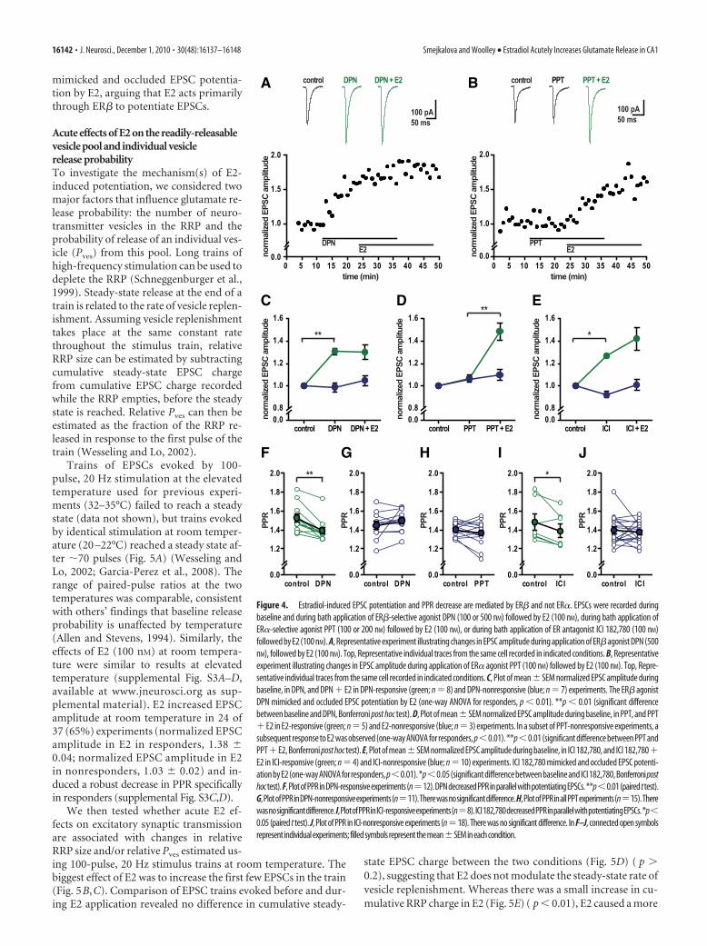
DPN both mimicked and occluded the effect of E2 to poten-tiate EPSCs, whereas PPT had no effect (Fig. 4A,B). In 12 of 23experiments, DPN rapidly potentiated EPSC amplitude by�20% (on average, by 30 � 2%) (Fig. 4C). Additionally, in 15 ofthese experiments (8 DPN responsive, 7 DPN nonresponsive),DPN application was followed by E2 (100 nM), which induced nofurther potentiation (Fig. 4A,C). Importantly, DPN also de-creased PPR in parallel with potentiating EPSC amplitude, spe-cifically in responsive inputs (Fig. 4F,G) (paired t test, p � 0.01for responders; p � 0.2 for nonresponders). In 5 of 11 DPNexperiments done with two stimulating electrodes to activatenonoverlapping inputs to a single cell, one input was potentiatedby �20%, whereas the other was not responsive, and PPR de-creased specifically at the potentiated input (data not shown)(paired t test, p � 0.05 for responsive inputs; p � 0.2 for nonre-sponsive inputs). Thus, like with E2, DPN-induced EPSC poten-tiation is input specific, not postsynaptic cell specific.
In contrast to results with DPN, PPT failed to potentiate EPSCamplitude in any of 15 experiments. In 8 experiments, PPT ap-plication was followed by E2 (100 nM), and in 5 cases, E2 afterPPT induced robust EPSC amplitude potentiation (Fig. 4B,D),showing that inputs that failed to respond to PPT were capable ofresponding to E2. Paired-pulse ratio also was unchanged by PPT(Fig. 4H) (paired t test, p � 0.1). Together, these results indicatethat acute actions of E2 to potentiate EPSCs and decrease PPR aremediated by ER� and not by ER�. That DPN mimics both E2effects is consistent with the idea that EPSC potentiation anddecreased PPR are related, such that E2 acutely potentiates EPSCsby increasing glutamate release probability.
To further investigate the role of ERs in mediating acute EPSCpotentiation, we used ICI 182,780, which binds ER� and ER�with similar high affinity and behaves as an antagonist of classicalnuclear ER activity in transcriptional assays (Sun et al., 2002) byblocking receptor dimerization (Fawell et al., 1990; Pike et al.,2001). Surprisingly, ICI 182,780 (100 nM) alone potentiatedEPSC amplitudes in 8 of 26 experiments. In 14 of these experi-ments (4 ICI responsive, 10 ICI nonresponsive), ICI 182,780 ap-plication was followed by E2 (100 nM), which induced nosignificant additional potentiation (Fig. 4E). Similar to E2 andDPN, ICI 182,780 decreased PPR in parallel with potentiating EPSCamplitude (Fig. 4I,J) (paired t test, p � 0.05 for responders; p � 0.3for nonresponders) and potentiated EPSCs in an input-specificmanner (4 of 12 double-input experiments). The finding that ICI182,780 alone can mimic and occlude acute effects of E2 on excita-tory synaptic transmission suggests that ER� mediates acute E2-induced EPSC potentiation without dimerizing.
Because ICI 182,780 has been reported to activate theG-protein-coupled membrane ER, GPR30 (Filardo et al., 2002),we also tested whether a selective agonist for GPR30, G-1, mim-icked E2-induced EPSC potentiation, as has been suggested pre-viously (Lebesgue et al., 2009). E2 and G-1 have similar bindingaffinities for GPR30 (Ki of 5.7 and 11 nM, respectively) and 100nM G-1 is sufficient to induce maximal Ca 2� mobilization inGPR30-transfected COS7 cells (Bologa et al., 2006). We foundthat G-1 (100 nM) potentiated EPSCs by �20% in only 2 of 15experiments, by 25 and 33% (data not shown). G-1 decreasedPPR in these experiments from 1.53 to 1.47 and 1.42 to 1.24 (datanot shown). G-1 did not affect EPSC amplitude or PPR in theremaining 13 of 15 experiments. Thus, although we cannot ex-clude some role for GPR30 in EPSC potentiation, the small pro-portion of G-1-responsive experiments indicates that GPR30 isunlikely to fully account for EPSC potentiation by E2. The resultswith G-1 were in stark contrast to the effects DPN, which closely
A
B C
Figure 3. Estradiol-induced EPSC potentiation and PPR decrease are input specific.EPSCs were evoked by alternating paired-pulse stimulation of two nonoverlapping sets ofSchaffer collateral inputs to a common CA1 pyramidal cell. A, A schematic of electrodeplacement and representative individual current traces evoked at E2-responsive and E2-nonresponsive inputs recorded from a single cell. B, Plot of mean � SEM normalized EPSCamplitude (solid line) and PPR (dashed line) at E2-responsive inputs (green; n � 9); E2decreased PPR in parallel with potentiating EPSCs in an input-specific manner. *p � 0.05(significant effect of E2 on PPR; paired t test). C, Plot of mean � SEM normalized EPSCamplitude (solid line) and PPR (dashed line) at E2-nonresponsive inputs (blue) from thesame cells as in B. See also supplemental Figure S2 (available at www.jneurosci.org assupplemental material).
Smejkalova and Woolley • Estradiol Acutely Increases Glutamate Release in CA1 J. Neurosci., December 1, 2010 • 30(48):16137–16148 • 16141
mimicked and occluded EPSC potentia-tion by E2, arguing that E2 acts primarilythrough ER� to potentiate EPSCs.
Acute effects of E2 on the readily-releasablevesicle pool and individual vesiclerelease probabilityTo investigate the mechanism(s) of E2-induced potentiation, we considered twomajor factors that influence glutamate re-lease probability: the number of neuro-transmitter vesicles in the RRP and theprobability of release of an individual ves-icle (Pves) from this pool. Long trains ofhigh-frequency stimulation can be used todeplete the RRP (Schneggenburger et al.,1999). Steady-state release at the end of atrain is related to the rate of vesicle replen-ishment. Assuming vesicle replenishmenttakes place at the same constant ratethroughout the stimulus train, relativeRRP size can be estimated by subtractingcumulative steady-state EPSC chargefrom cumulative EPSC charge recordedwhile the RRP empties, before the steadystate is reached. Relative Pves can then beestimated as the fraction of the RRP re-leased in response to the first pulse of thetrain (Wesseling and Lo, 2002).
Trains of EPSCs evoked by 100-pulse, 20 Hz stimulation at the elevatedtemperature used for previous experi-ments (32–35°C) failed to reach a steadystate (data not shown), but trains evokedby identical stimulation at room temper-ature (20 –22°C) reached a steady state af-ter �70 pulses (Fig. 5A) (Wesseling andLo, 2002; Garcia-Perez et al., 2008). Therange of paired-pulse ratios at the twotemperatures was comparable, consistentwith others’ findings that baseline releaseprobability is unaffected by temperature(Allen and Stevens, 1994). Similarly, theeffects of E2 (100 nM) at room tempera-ture were similar to results at elevatedtemperature (supplemental Fig. S3A–D,available at www.jneurosci.org as sup-plemental material). E2 increased EPSCamplitude at room temperature in 24 of37 (65%) experiments (normalized EPSCamplitude in E2 in responders, 1.38 �0.04; normalized EPSC amplitude in E2in nonresponders, 1.03 � 0.02) and in-duced a robust decrease in PPR specificallyin responders (supplemental Fig. S3C,D).
We then tested whether acute E2 ef-fects on excitatory synaptic transmissionare associated with changes in relativeRRP size and/or relative Pves estimated us-ing 100-pulse, 20 Hz stimulus trains at room temperature. Thebiggest effect of E2 was to increase the first few EPSCs in the train(Fig. 5B,C). Comparison of EPSC trains evoked before and dur-ing E2 application revealed no difference in cumulative steady-
state EPSC charge between the two conditions (Fig. 5D) ( p �0.2), suggesting that E2 does not modulate the steady-state rate ofvesicle replenishment. Whereas there was a small increase in cu-mulative RRP charge in E2 (Fig. 5E) ( p � 0.01), E2 caused a more
A
C
F G H I J
D E
B
Figure 4. Estradiol-induced EPSC potentiation and PPR decrease are mediated by ER� and not ER�. EPSCs were recorded duringbaseline and during bath application of ER�-selective agonist DPN (100 or 500 nM) followed by E2 (100 nM), during bath application ofER�-selective agonist PPT (100 or 200 nM) followed by E2 (100 nM), or during bath application of ER antagonist ICI 182,780 (100 nM)followed by E2 (100 nM). A, Representative experiment illustrating changes in EPSC amplitude during application of ER�agonist DPN (500nM), followed by E2 (100 nM). Top, Representative individual traces from the same cell recorded in indicated conditions. B, Representativeexperiment illustrating changes in EPSC amplitude during application of ER� agonist PPT (100 nM) followed by E2 (100 nM). Top, Repre-sentative individual traces from the same cell recorded in indicated conditions. C, Plot of mean�SEM normalized EPSC amplitude duringbaseline, in DPN, and DPN � E2 in DPN-responsive (green; n � 8) and DPN-nonresponsive (blue; n � 7) experiments. The ER� agonistDPN mimicked and occluded EPSC potentiation by E2 (one-way ANOVA for responders, p � 0.01). **p � 0.01 (significant differencebetween baseline and DPN, Bonferroni post hoc test). D, Plot of mean�SEM normalized EPSC amplitude during baseline, in PPT, and PPT�E2 in E2-responsive (green; n�5) and E2-nonresponsive (blue; n�3) experiments. In a subset of PPT-nonresponsive experiments, asubsequent response to E2 was observed (one-way ANOVA for responders, p�0.01). **p�0.01 (significant difference between PPT andPPT�E2, Bonferroni post hoc test). E, Plot of mean�SEM normalized EPSC amplitude during baseline, in ICI 182,780, and ICI 182,780�E2 in ICI-responsive (green; n � 4) and ICI-nonresponsive (blue; n � 10) experiments. ICI 182,780 mimicked and occluded EPSC potenti-ationbyE2(one-wayANOVAforresponders, p�0.01).*p�0.05(significantdifferencebetweenbaselineandICI182,780,Bonferroniposthoctest).F,PlotofPPRinDPN-responsiveexperiments(n�12).DPNdecreasedPPRinparallelwithpotentiatingEPSCs.**p�0.01(pairedttest).G,PlotofPPRinDPN-nonresponsiveexperiments(n�11).Therewasnosignificantdifference.H,PlotofPPRinallPPTexperiments(n�15).Therewasnosignificantdifference.I,PlotofPPRinICI-responsiveexperiments(n�8).ICI182,780decreasedPPRinparallelwithpotentiatingEPSCs.*p�0.05 (paired t test). J, Plot of PPR in ICI-nonresponsive experiments (n�18). There was no significant difference. In F–J, connected open symbolsrepresent individualexperiments; filledsymbolsrepresentthemean�SEMineachcondition.
16142 • J. Neurosci., December 1, 2010 • 30(48):16137–16148 Smejkalova and Woolley • Estradiol Acutely Increases Glutamate Release in CA1
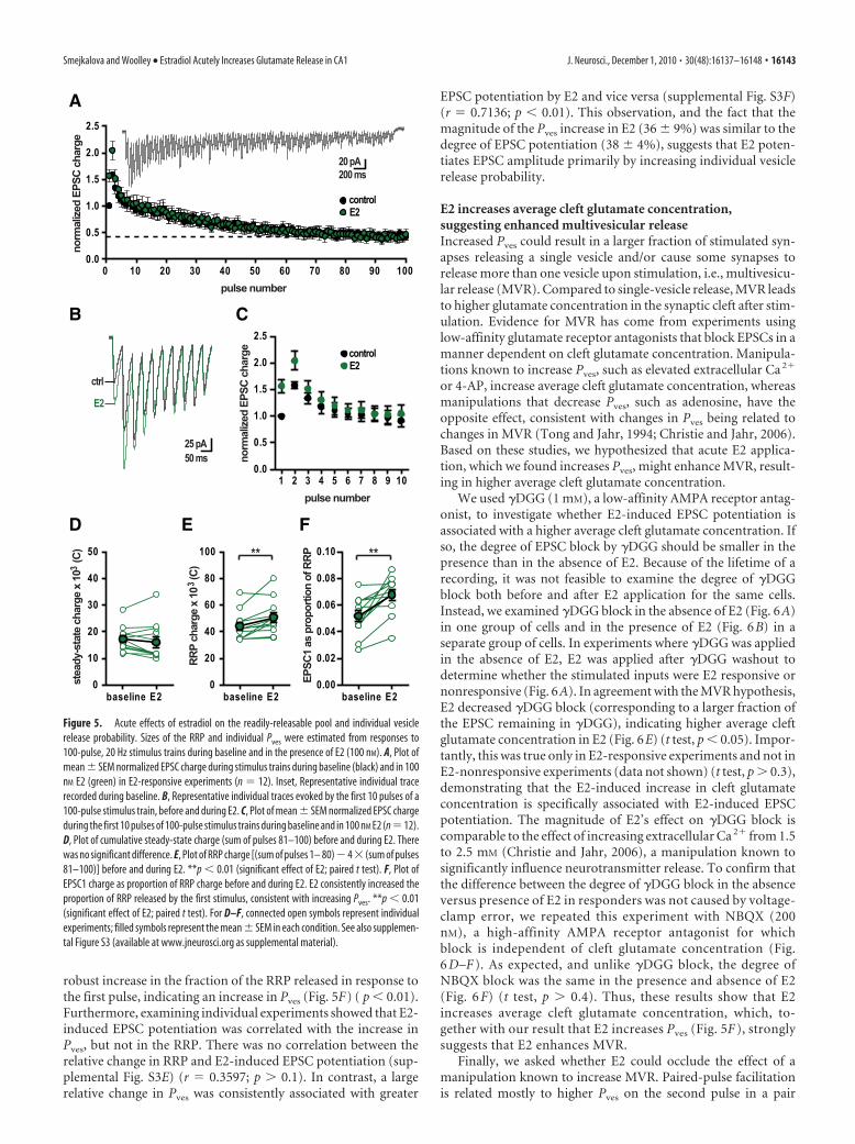
robust increase in the fraction of the RRP released in response tothe first pulse, indicating an increase in Pves (Fig. 5F) ( p � 0.01).Furthermore, examining individual experiments showed that E2-induced EPSC potentiation was correlated with the increase inPves, but not in the RRP. There was no correlation between therelative change in RRP and E2-induced EPSC potentiation (sup-plemental Fig. S3E) (r � 0.3597; p � 0.1). In contrast, a largerelative change in Pves was consistently associated with greater
EPSC potentiation by E2 and vice versa (supplemental Fig. S3F)(r � 0.7136; p � 0.01). This observation, and the fact that themagnitude of the Pves increase in E2 (36 � 9%) was similar to thedegree of EPSC potentiation (38 � 4%), suggests that E2 poten-tiates EPSC amplitude primarily by increasing individual vesiclerelease probability.
E2 increases average cleft glutamate concentration,suggesting enhanced multivesicular releaseIncreased Pves could result in a larger fraction of stimulated syn-apses releasing a single vesicle and/or cause some synapses torelease more than one vesicle upon stimulation, i.e., multivesicu-lar release (MVR). Compared to single-vesicle release, MVR leadsto higher glutamate concentration in the synaptic cleft after stim-ulation. Evidence for MVR has come from experiments usinglow-affinity glutamate receptor antagonists that block EPSCs in amanner dependent on cleft glutamate concentration. Manipula-tions known to increase Pves, such as elevated extracellular Ca 2�
or 4-AP, increase average cleft glutamate concentration, whereasmanipulations that decrease Pves, such as adenosine, have theopposite effect, consistent with changes in Pves being related tochanges in MVR (Tong and Jahr, 1994; Christie and Jahr, 2006).Based on these studies, we hypothesized that acute E2 applica-tion, which we found increases Pves, might enhance MVR, result-ing in higher average cleft glutamate concentration.
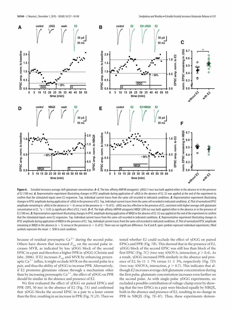
We used �DGG (1 mM), a low-affinity AMPA receptor antag-onist, to investigate whether E2-induced EPSC potentiation isassociated with a higher average cleft glutamate concentration. Ifso, the degree of EPSC block by �DGG should be smaller in thepresence than in the absence of E2. Because of the lifetime of arecording, it was not feasible to examine the degree of �DGGblock both before and after E2 application for the same cells.Instead, we examined �DGG block in the absence of E2 (Fig. 6A)in one group of cells and in the presence of E2 (Fig. 6B) in aseparate group of cells. In experiments where �DGG was appliedin the absence of E2, E2 was applied after �DGG washout todetermine whether the stimulated inputs were E2 responsive ornonresponsive (Fig. 6A). In agreement with the MVR hypothesis,E2 decreased �DGG block (corresponding to a larger fraction ofthe EPSC remaining in �DGG), indicating higher average cleftglutamate concentration in E2 (Fig. 6E) (t test, p � 0.05). Impor-tantly, this was true only in E2-responsive experiments and not inE2-nonresponsive experiments (data not shown) (t test, p � 0.3),demonstrating that the E2-induced increase in cleft glutamateconcentration is specifically associated with E2-induced EPSCpotentiation. The magnitude of E2’s effect on �DGG block iscomparable to the effect of increasing extracellular Ca 2� from 1.5to 2.5 mM (Christie and Jahr, 2006), a manipulation known tosignificantly influence neurotransmitter release. To confirm thatthe difference between the degree of �DGG block in the absenceversus presence of E2 in responders was not caused by voltage-clamp error, we repeated this experiment with NBQX (200nM), a high-affinity AMPA receptor antagonist for whichblock is independent of cleft glutamate concentration (Fig.6 D–F ). As expected, and unlike �DGG block, the degree ofNBQX block was the same in the presence and absence of E2(Fig. 6 F) (t test, p � 0.4). Thus, these results show that E2increases average cleft glutamate concentration, which, to-gether with our result that E2 increases Pves (Fig. 5F ), stronglysuggests that E2 enhances MVR.
Finally, we asked whether E2 could occlude the effect of amanipulation known to increase MVR. Paired-pulse facilitationis related mostly to higher Pves on the second pulse in a pair
A
B
D E F
C
Figure 5. Acute effects of estradiol on the readily-releasable pool and individual vesiclerelease probability. Sizes of the RRP and individual Pves were estimated from responses to100-pulse, 20 Hz stimulus trains during baseline and in the presence of E2 (100 nM). A, Plot ofmean � SEM normalized EPSC charge during stimulus trains during baseline (black) and in 100nM E2 (green) in E2-responsive experiments (n � 12). Inset, Representative individual tracerecorded during baseline. B, Representative individual traces evoked by the first 10 pulses of a100-pulse stimulus train, before and during E2. C, Plot of mean � SEM normalized EPSC chargeduring the first 10 pulses of 100-pulse stimulus trains during baseline and in 100 nM E2 (n�12).D, Plot of cumulative steady-state charge (sum of pulses 81–100) before and during E2. Therewas no significant difference. E, Plot of RRP charge [(sum of pulses 1– 80)�4 (sum of pulses81–100)] before and during E2. **p � 0.01 (significant effect of E2; paired t test). F, Plot ofEPSC1 charge as proportion of RRP charge before and during E2. E2 consistently increased theproportion of RRP released by the first stimulus, consistent with increasing Pves. **p � 0.01(significant effect of E2; paired t test). For D–F, connected open symbols represent individualexperiments; filled symbols represent the mean � SEM in each condition. See also supplemen-tal Figure S3 (available at www.jneurosci.org as supplemental material).
Smejkalova and Woolley • Estradiol Acutely Increases Glutamate Release in CA1 J. Neurosci., December 1, 2010 • 30(48):16137–16148 • 16143
because of residual presynaptic Ca 2� during the second pulse.Others have shown that increased Pves on the second pulse in-creases MVR, as indicated by less �DGG block of the secondEPSC in a pair and therefore a higher PPR in �DGG (Christie andJahr, 2006). If E2 increases Pves and MVR by enhancing presyn-aptic Ca 2� influx, it might occlude MVR on the second pulse in apair, and thus the ability of �DGG to increase PPR. Alternatively,if E2 promotes glutamate release through a mechanism otherthan by increasing presynaptic Ca 2�, the effect of �DGG on PPRshould be similar in the absence and presence of E2.
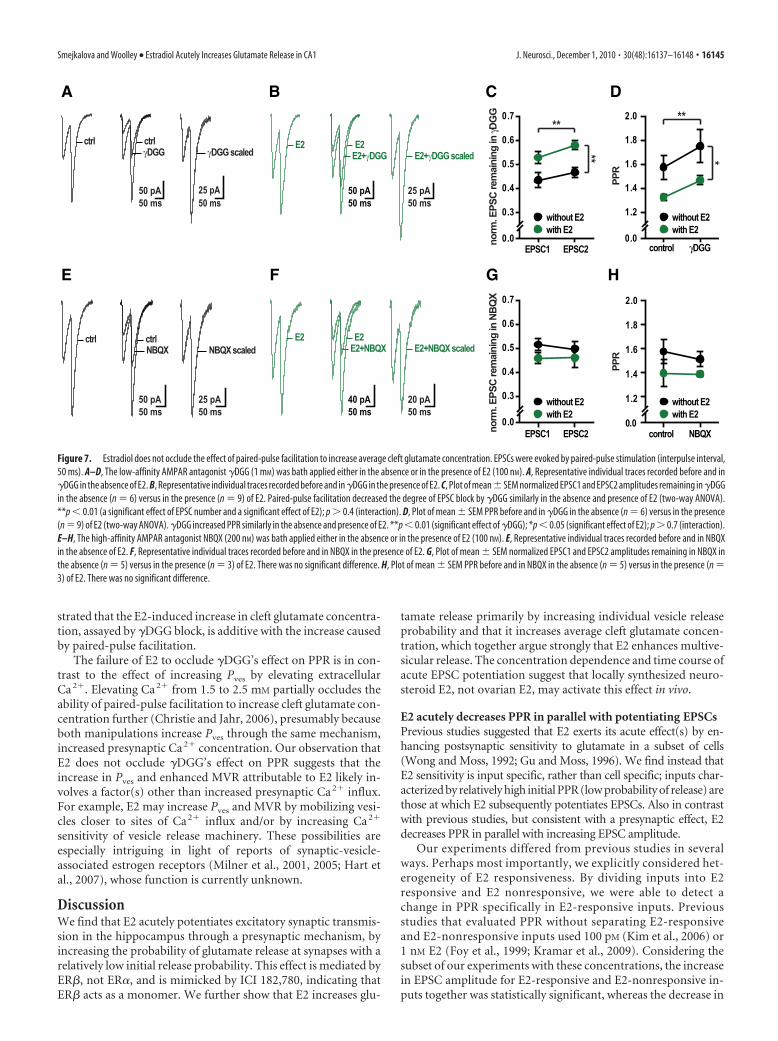
We first evaluated the effect of �DGG on paired EPSCs andPPR (IPI, 50 ms) in the absence of E2 (Fig. 7A) and confirmedthat �DGG blocks the second EPSC in a pair to a lesser extentthan the first, resulting in an increase in PPR (Fig. 7C,D). Then we
tested whether E2 could occlude the effect of �DGG on pairedEPSCs and PPR (Fig. 7B). This showed that in the presence of E2,�DGG block of the second EPSC was still less than block of thefirst EPSC (Fig. 7C) (two-way ANOVA, interaction, p � 0.4). Asa result, �DGG increased PPR similarly in the absence and pres-ence of E2, by 11 � 7% versus 11 � 3%, respectively (Fig. 7D)(two-way ANOVA, interaction, p � 0.7). This indicates that al-though E2 increases average cleft glutamate concentration duringthe first pulse, glutamate concentration increases even further onthe second pulse. As with single-pulse �DGG experiments, weexcluded a possible contribution of voltage-clamp error by show-ing that the two EPSCs in a pair were blocked equally by NBQX,both in the absence and presence of E2, resulting in no change inPPR in NBQX (Fig. 7E–H). Thus, these experiments demon-
A
D E
F
B
C
Figure 6. Estradiol increases average cleft glutamate concentration. A–C, The low-affinity AMPAR antagonist �DGG (1 mM) was bath applied either in the absence or in the presenceof E2 (100 nM). A, Representative experiment illustrating changes in EPSC amplitude during application of �DGG in the absence of E2. E2 was applied at the end of the experiment toconfirm that the stimulated inputs were E2 responsive. Top, Individual current traces from the same cell recorded in indicated conditions. B, Representative experiment illustratingchanges in EPSC amplitude during application of �DGG in the presence of E2. Top, Individual current traces from the same cell recorded in indicated conditions. C, Plot of normalized EPSCamplitude remaining in �DGG in the absence (n � 6) versus in the presence (n � 9) of E2. �DGG was less effective in the presence of E2, consistent with higher average cleft glutamateconcentration in E2. *p � 0.05 (a significant effect of E2; t test). D–F, The high-affinity AMPAR antagonist NBQX (200 nM) was bath applied either in the absence or in the presence ofE2 (100 nM). D, Representative experiment illustrating changes in EPSC amplitude during application of NBQX in the absence of E2. E2 was applied at the end of the experiment to confirmthat the stimulated inputs were E2 responsive. Top, Individual current traces from the same cell recorded in indicated conditions. E, Representative experiment illustrating changes inEPSC amplitude during application of NBQX in the presence of E2. Top, Individual current traces from the same cell recorded in indicated conditions. F, Plot of normalized EPSC amplituderemaining in NBQX in the absence (n � 5) versus in the presence (n � 3) of E2. There was no significant difference. For C and F, open symbols represent individual experiments; filledsymbols represent the mean � SEM in each condition.
16144 • J. Neurosci., December 1, 2010 • 30(48):16137–16148 Smejkalova and Woolley • Estradiol Acutely Increases Glutamate Release in CA1
strated that the E2-induced increase in cleft glutamate concentra-tion, assayed by �DGG block, is additive with the increase causedby paired-pulse facilitation.
The failure of E2 to occlude �DGG’s effect on PPR is in con-trast to the effect of increasing Pves by elevating extracellularCa 2�. Elevating Ca 2� from 1.5 to 2.5 mM partially occludes theability of paired-pulse facilitation to increase cleft glutamate con-centration further (Christie and Jahr, 2006), presumably becauseboth manipulations increase Pves through the same mechanism,increased presynaptic Ca 2� concentration. Our observation thatE2 does not occlude �DGG’s effect on PPR suggests that theincrease in Pves and enhanced MVR attributable to E2 likely in-volves a factor(s) other than increased presynaptic Ca 2� influx.For example, E2 may increase Pves and MVR by mobilizing vesi-cles closer to sites of Ca 2� influx and/or by increasing Ca 2�
sensitivity of vesicle release machinery. These possibilities areespecially intriguing in light of reports of synaptic-vesicle-associated estrogen receptors (Milner et al., 2001, 2005; Hart etal., 2007), whose function is currently unknown.
DiscussionWe find that E2 acutely potentiates excitatory synaptic transmis-sion in the hippocampus through a presynaptic mechanism, byincreasing the probability of glutamate release at synapses with arelatively low initial release probability. This effect is mediated byER�, not ER�, and is mimicked by ICI 182,780, indicating thatER� acts as a monomer. We further show that E2 increases glu-
tamate release primarily by increasing individual vesicle releaseprobability and that it increases average cleft glutamate concen-tration, which together argue strongly that E2 enhances multive-sicular release. The concentration dependence and time course ofacute EPSC potentiation suggest that locally synthesized neuro-steroid E2, not ovarian E2, may activate this effect in vivo.
E2 acutely decreases PPR in parallel with potentiating EPSCsPrevious studies suggested that E2 exerts its acute effect(s) by en-hancing postsynaptic sensitivity to glutamate in a subset of cells(Wong and Moss, 1992; Gu and Moss, 1996). We find instead thatE2 sensitivity is input specific, rather than cell specific; inputs char-acterized by relatively high initial PPR (low probability of release) arethose at which E2 subsequently potentiates EPSCs. Also in contrastwith previous studies, but consistent with a presynaptic effect, E2decreases PPR in parallel with increasing EPSC amplitude.
Our experiments differed from previous studies in severalways. Perhaps most importantly, we explicitly considered het-erogeneity of E2 responsiveness. By dividing inputs into E2responsive and E2 nonresponsive, we were able to detect achange in PPR specifically in E2-responsive inputs. Previousstudies that evaluated PPR without separating E2-responsiveand E2-nonresponsive inputs used 100 pM (Kim et al., 2006) or1 nM E2 (Foy et al., 1999; Kramar et al., 2009). Considering thesubset of our experiments with these concentrations, the increasein EPSC amplitude for E2-responsive and E2-nonresponsive in-puts together was statistically significant, whereas the decrease in
A
E F G H
B C D
Figure 7. Estradiol does not occlude the effect of paired-pulse facilitation to increase average cleft glutamate concentration. EPSCs were evoked by paired-pulse stimulation (interpulse interval,50 ms). A–D, The low-affinity AMPAR antagonist �DGG (1 mM) was bath applied either in the absence or in the presence of E2 (100 nM). A, Representative individual traces recorded before and in�DGG in the absence of E2. B, Representative individual traces recorded before and in �DGG in the presence of E2. C, Plot of mean�SEM normalized EPSC1 and EPSC2 amplitudes remaining in �DGGin the absence (n � 6) versus in the presence (n � 9) of E2. Paired-pulse facilitation decreased the degree of EPSC block by �DGG similarly in the absence and presence of E2 (two-way ANOVA).**p � 0.01 (a significant effect of EPSC number and a significant effect of E2); p � 0.4 (interaction). D, Plot of mean � SEM PPR before and in �DGG in the absence (n � 6) versus in the presence(n � 9) of E2 (two-way ANOVA). �DGG increased PPR similarly in the absence and presence of E2. **p � 0.01 (significant effect of �DGG); *p � 0.05 (significant effect of E2); p � 0.7 (interaction).E–H, The high-affinity AMPAR antagonist NBQX (200 nM) was bath applied either in the absence or in the presence of E2 (100 nM). E, Representative individual traces recorded before and in NBQXin the absence of E2. F, Representative individual traces recorded before and in NBQX in the presence of E2. G, Plot of mean � SEM normalized EPSC1 and EPSC2 amplitudes remaining in NBQX inthe absence (n � 5) versus in the presence (n � 3) of E2. There was no significant difference. H, Plot of mean � SEM PPR before and in NBQX in the absence (n � 5) versus in the presence (n �3) of E2. There was no significant difference.
Smejkalova and Woolley • Estradiol Acutely Increases Glutamate Release in CA1 J. Neurosci., December 1, 2010 • 30(48):16137–16148 • 16145
PPR was not. Additionally, particularly for previous studies withdissociated cells (Gu and Moss, 1996), it is possible that agonist ap-plication activated primarily extrasynaptic receptors, which may notaccurately reflect properties of synaptic transmission. Finally, al-though our results strongly support a presynaptic mechanism forsynaptic potentiation by E2, they do not exclude the possibility thatE2 also increases postsynaptic sensitivity to glutamate.
E2 enhances glutamate release primarily by increasing Pves
We used high-frequency stimulus trains to investigate the mech-anism(s) by which E2 enhances glutamate release. We found asmall increase in cumulative release during a train, indicatingincreased RRP size, and a stronger effect on the fraction of theRRP released by the first pulse, indicating increased Pves. Al-though it is possible that E2 increases both the RRP and Pves inparallel, a more parsimonious explanation is that increased Pves
accounts for both observations.Although the RRP has been functionally defined as cumula-
tive release during various depletion protocols, including stimu-lus trains (Stevens and Tsujimoto, 1995; Rosenmund andStevens, 1996; Wesseling and Lo, 2002), release during a train isinfluenced by factors in addition to how many vesicles are avail-able for release at the beginning of stimulation. For example, acommon assumption in estimating the RRP from trains is thatthe rate of vesicle replenishment during the train is constant asestimated from steady-state charge (Schneggenburger et al.,1999). However, there is evidence that the rate of replenishmentincreases in a Ca 2�-dependent manner during the first 10 –20pulses, reaching an elevated level that persists for the rest of thetrain (Stevens and Wesseling, 1998; Wesseling and Lo, 2002). Achange in the time course of such activity-dependent upregula-tion of vesicle replenishment could result in more total vesiclesreleased during the train and a larger RRP estimate. Second, theidentity/properties of vesicles constituting RRP may vary underdifferent conditions. For example, under conditions of increasedPves, additional, “reluctant” vesicles may be released during atrain, resulting in a larger RRP estimate (Moulder and Menner-ick, 2005). These considerations suggest that E2 could increaseboth the RRP estimate and Pves by a single mechanism. For ex-ample, E2 could elevate intracellular Ca 2�, mobilize vesiclescloser to Ca 2� sources, or upregulate one of the biochemicalsteps in the release process, any of which could result in higherPves, faster vesicle replenishment, and/or release of additional,“reluctant” vesicles.
E2 promotes multivesicular releaseThe observation that E2 consistently and robustly increased Pves
suggested that E2 might promote MVR. The possibility and phys-iological significance of MVR at hippocampal synapses has beenquestioned based on minimal stimulation experiments (Stevensand Wang, 1995) and estimates of glutamate receptor saturationafter release of a single vesicle (Clements et al., 1992). However,other studies suggest that MVR does occur, particularly underconditions of elevated Pves (Tong and Jahr, 1994; Oertner et al.,2002; Christie and Jahr, 2006), and argue that glutamate recep-tors are not saturated by a single vesicle (Liu et al., 1999; McAl-lister and Stevens, 2000). Although evidence for MVR in theimmature hippocampus is growing, no studies to date have re-ported MVR in the adult.
We used the low-affinity AMPAR antagonist �DGG to com-pare relative cleft glutamate concentration in the presence versusabsence of E2. We found that �DGG blocked EPSCs less effec-tively in E2, indicating that E2 increases average cleft glutamate
concentration. Although there are several mechanisms by whichE2 could increase cleft glutamate concentration, MVR is the mostlikely. One alternative is that E2 downregulates astrocytic gluta-mate uptake, resulting in increased spillover. This is unlikely,however, because glutamate uptake is extremely efficient (Dia-mond and Jahr, 2000), such that even when it is pharmacologi-cally reduced, the degree of �DGG block of EPSCs remainsunaffected (Christie and Jahr, 2006). E2 also could increase cleftglutamate by causing faster or more complete vesicle emptying.This too is unlikely, as it should result in faster EPSC rise times inE2 (Choi et al., 2000), which we did not observe. Thus, the bestexplanation for our findings that E2 increases both Pves and av-erage cleft glutamate concentration is that E2 increases MVR. Toour knowledge, this is the first demonstration of MVR in adulthippocampus. Furthermore, our observation that the increase incleft glutamate attributable to E2 fails to occlude the increaseattributable to paired-pulse facilitation suggests that E2 may en-hance MVR by a mechanism other than elevating presynapticCa 2�. This finding argues for the possibility that E2 mobilizesvesicles toward sites of Ca 2� influx and/or regulates vesicle re-lease machinery.
Rapid E2 signalingIn neurons, E2 acutely increases Ca 2� influx through L-typeCa 2� channels and activates protein kinases including Src,Erk1/2, CaMKII, and protein kinase A (Gu and Moss, 1996; Lee etal., 2004; Wu et al., 2005). Most of these effects are mediated byclassical ERs acting outside the nucleus (Wade and Dorsa 2003;Wu et al., 2005; Zhao and Brinton 2007), but neither the initialsteps of extranuclear ER activation, nor the consequences ofrapid E2 signaling are well understood.
We found that E2-induced EPSC potentiation is mimicked byboth an ER� agonist and ICI 182,780. ICI compounds interferewith ER dimerization (Fawell et al., 1990; Pike et al., 2001), andconsequently block classical nuclear ER activity (Sun et al., 2002).In contrast, rapid E2 signaling is less consistently blocked by ICIcompounds (Singh et al., 1999) and may even be activated bythem (Zhao et al., 2006). Our results are consistent with theselatter reports, and suggest that ER� acts as a monomer to activateacute EPSC potentiation.
Interestingly, targets of E2-activated kinases include synapticvesicle proteins critical for vesicle mobilization and Ca 2�-dependent release (Hilfiker et al., 1999; Chi et al., 2003; Kushneret al., 2005; Menegon et al., 2006; Onofri et al., 2007), suggestingthat E2 could enhance release by regulating synaptic vesicle pro-teins. If E2 initiates a cascade of molecular events leading to en-hanced vesicular release, such as vesicle protein phosphorylation,this may explain our observation that EPSC potentiation per-sisted after E2 washout. Since EM immunocytochemical studieshave found ER� in presynaptic boutons in CA1 (Milner et al.,2005), exploring the possibility that E2 acts via vesicle-associatedERs to regulate synaptic vesicle proteins will be an important areaof future research.
A possible role for neurosteroid E2Although EPSC potentiation occurred with 100 pM E2, the effectwas more robust at higher than circulating E2 concentrations.Additionally, the time course of E2-induced EPSC potentiation isfaster than fluctuations in circulating E2 in vivo. These issues raisethe question of whether acute E2-induced EPSC potentiationoccurs physiologically. Importantly, recent studies show that thehippocampus can generate neurosteroid E2, resulting in highlocal E2 concentrations that can change rapidly (Hojo et al., 2004,
16146 • J. Neurosci., December 1, 2010 • 30(48):16137–16148 Smejkalova and Woolley • Estradiol Acutely Increases Glutamate Release in CA1
2009). Additionally, EM immunocytochemistry shows that aro-matase, the rate-limiting enzyme in E2 synthesis, is present bothpresynaptically and postsynaptically at a subset of synapses inhippocampal CA1 (Hojo et al., 2004). Together, these findingssuggest that neurosteroid E2 could activate EPSC potentiation invivo. Indeed, the concentration dependence of EPSC potentiationlikely protects hippocampal synaptic transmission from modulationby the relatively slow and low amplitude fluctuations in circulatingE2 from the ovaries. Thus, neurosteroid E2 may interact with pre-synaptic ER� to activate a distinct suite of cellular/molecular eventsthat increase glutamate release resulting in rapid, input-specific po-tentiation of hippocampal synaptic transmission.
ReferencesAllen C, Stevens CF (1994) An evaluation of causes for unreliability of syn-
aptic transmission. Proc Natl Acad Sci U S A 91:10380 –10383.Bologa CG, Revankar CM, Young SM, Edwards BS, Arterburn JB, Kiselyov
AS, Parker MA, Tkachenko SE, Savchuck NP, Sklar LA, Oprea TI,Prossnitz ER (2006) Virtual and biomolecular screening converge on aselective agonist for GPR30. Nat Chem Biol 2:207–212.
Chi P, Greengard P, Ryan TA (2003) Synaptic vesicle mobilization is regu-lated by distinct synapsin I phosphorylation pathways at different fre-quencies. Neuron 38:69 –78.
Choi S, Klingauf J, Tsien RW (2000) Postfusional regulation of cleft gluta-mate concentration during LTP at “silent synapses.” Nat Neurosci3:330 –336.
Christie JM, Jahr CE (2006) Multivesicular release at Schaffer collateral-CA1 hippocampal synapses. J Neurosci 26:210 –216.
Clements JD, Lester RA, Tong G, Jahr CE, Westbrook GL (1992) The timecourse of glutamate in the synaptic cleft. Science 258:1498 –1501.
Corpechot C, Robel P, Axelson M, Sjovall J, Baulieu E-E (1981) Character-ization and measurement of dehydroepiandrosterone sulfate in rat brain.Proc Natl Acad Sci U S A 78:4704 – 4707.
Diamond JS, Jahr CE (2000) Synaptically released glutamate does not over-whelm transporters on hippocampal astrocytes during high-frequencystimulation. J. Neurophysiol 83:2835–2843.
Dobrunz LE, Stevens CF (1997) Heterogeneity of release probability, facili-tation, and depletion at central synapses. Neuron 18:995–1008.
Fawell SE, White R, Hoare S, Sydenham M, Page M, Parker MG (1990)Inhibition of estrogen receptor-DNA binding by the “pure” antiestrogenICI 164,384 appears to be mediated by impaired receptor dimerization.Proc Natl Acad Sci U S A 87:6883– 6887.
Filardo EJ, Quinn JA, Frackelton AR Jr, Bland KI (2002) Estrogen action viathe G protein-coupled receptor, GPR30: Stimulation of adenyl cyclaseand cAMP-mediated attenuation of the epidermal growth factorreceptor-to-MAPK signaling axis. Mol Endocrinol 16:70 – 84.
Foy MR, Xu J, Xie X, Brinton RD, Thompson RF, Berger TW (1999) 17�-estradiol enhances NMDA receptor-mediated EPSPs and long-term po-tentiation. J Neurophysiol 81:925–929.
Garcia-Perez E, Lo DC, Wesseling JF (2008) Kinetic isolation of a slowlyrecovering component of short-term depression during exhaustive use atexcitatory hippocampal synapses. J Neurophysiol 100:781–795.
Gu Q, Moss RL (1996) 17�-estradiol potentiates kainate-induced currentsvia activation of the cAMP cascade. J Neurosci 16:3620 –3629.
Gu Q, Moss RL (1998) Novel mechanism for non-genomic action of 17�-oestradiol on kainate-induced currents in isolated rat CA1 hippocampalneurones. J Physiol 506:745–754.
Hart SA, Snyder MA, Smejkalova T, Woolley CS (2007) Estrogen mobilizesa subset of estrogen receptor-�-immunoreactive vesicles in inhibitorypresynaptic boutons in hippocampal CA1. J Neurosci 27:2102–2111.
Hilfiker S, Pieribone VA, Nordstedt C, Greengard P, Czernik AJ (1999) Reg-ulation of synaptotagmin I phosphorylation by multiple protein kinases.J Neurochem 73:921–932.
Hjelmstad GO, Isaac JT, Nicoll RA, Malenka RC (1999) Lack of AMPAreceptor desensitization during basal synaptic transmission in the hip-pocampal slice. J Neurophysiol 81:3096 –3099.
Hojo Y, Hattori T, Enami T, Furukawa A, Suzuki K, Ishii H, Mukai H,Morrison JH, Janssen WGM, Kominami S, Harada N, Kimoto T, KawatoS (2004) Adult male rat hippocampus synthesizes estradiol from preg-nenolone by cytochromes P45017� and P450 aromatase localized in neu-rons. Proc Natl Acad Sci U S A 101:865– 870.
Hojo Y, Higo S, Ishii H, Ooishi Y, Mukai H, Murakami G, Kominami T,Kimoto T, Honma S, Poirier D, Kawato S (2009) Comparison betweenhippocampus-synthesized and circulation-derived sex steroids in the hip-pocampus. Endocrinology 150:5106–5112.
Kim MT, Soussou W, Gholmieh G, Ahuja A, Tanguay A, Berger TW, BrintonRD (2006) 17�-estradiol potentiates field excitatory postsynaptic po-tentials within each subfield of the hippocampus with greatest potentia-tion of the associational/commissural afferents of CA3. Neuroscience141:391– 406.
Kimoto T, Tsurugizawa T, Ohta Y, Makino J, Tamura H, Hojo Y, Takata N,Kawato S (2001) Neurosteroid synthesis by cytochrome P450-containingsystems localized in the rat brain hippocampal neurons: N-methyl-D-aspartate and calcium-dependent synthesis. Endocrinology 142:3578–3589.
Kramar EA, Chen LY, Brandon NJ, Rex CS, Liu F, Gall CM, Lynch G (2009)Cytoskeletal changes underlie estrogen’s acute effects on synaptic trans-mission and plasticity. J Neurosci 29:12982–12993.
Kushner SA, Elgersma Y, Murphy GG, Jaarsma D, van Woerden GM, HojjatiMR, Cui Y, LeBoutillier JC, Marrone DF, Choi ES, De Zeeuw CI, Petit TL,Pozzo-Miller L, Silva AJ (2005) Modulation of presynaptic plasticityand learning by the h-ras/extracellular signal-regulated kinase/synapsin Isignaling pathway. J Neurosci 25:9721–9734.
Lebesgue D, Chevaleyre V, Zukin RS, Etgen AM (2009) Estradiol rescuesneurons from global ischemia-induced cell death: multiple cellular path-ways of neuroprotection. Steroids 74:555–561.
Lee SJ, Campomanes CR, Sikat PT, Greenfield AT, Allen PB, McEwen BS(2004) Estrogen induces phosphorylation of cyclic AMP response elementbinding (pCREB) in primary hippocampal cells in a time-dependent man-ner. Neuroscience 124:549–560.
Liu G, Choi S, Tsien RW (1999) Variability of neurotransmitter concentra-tion and nonsaturation of postsynaptic AMPA receptors at synapses inhippocampal cultures and slices. Neuron 22:395– 409.
Malinow R, Tsien RW (1990) Presynaptic enhancement shown by whole-cell recordings of long-term potentiation in hippocampal slices. Nature346:177–180.
McAllister AK, Stevens CF (2000) Nonsaturation of AMPA and NMDA re-ceptors at hippocampal synapses. Proc Natl Acad Sci U S A97:6173– 6178.
Menegon A, Bonanomi D, Albertinazzi C, Lotti F, Ferrari G, Kao H-T,Benfenati F, Baldelli P, Valtorta F (2006) Protein kinase A-mediatedsynapsin I phosphorylation is a central modulator of Ca 2�-dependentsynaptic activity. J Neurosci 26:11670 –11681.
Meyers MJ, Sun J, Carlson KE, Marriner GA, Katzenellenbogen BS, Katzenel-lenbogen JA (2001) Estrogen receptor-� potency-selective ligands:structure-activity relationship studies of diarylpropionitriles and theiracetylene and polar analogues. J Med Chem 44:4230 – 4251.
Milner TA, McEwen BS, Hayashi S, Li CJ, Reagan LP, Alves SE (2001) Ul-trastructural evidence that hippocampal alpha estrogen receptors are lo-cated at extranuclear sites. J Comp Neurol 429:355–371.
Milner TA, Ayoola K, Drake CT, Herrick SP, Tabori NE, McEwen BS, WarrierS, Alves SE (2005) Ultrastructural localization of estrogen receptor �immunoreactivity in the rat hippocampal formation. J Comp Neurol491:81–95.
Moulder KL, Mennerick S (2005) Reluctant vesicles contribute to the totalreadily releasable pool in glutamatergic hippocampal neurons. J Neurosci25:3842–3850.
Oertner TG, Sabatini BL, Nimchinsky EA, Svoboda K (2002) Facilitation atsingle synapses probed with optical quantal analysis. Nat Neurosci5:657– 664.
Onofri F, Messa M, Matafora V, Bonanno G, Corradi A, Bachi A, Valtorta F,Benfenati F (2007) Synapsin phosphorylation by src tyrosine kinase en-hances src activity in synaptic vesicles. J Biol Chem 282:15754 –15767.
Pike ACW, Brzozowski AM, Walton J, Hubbard RE, Thorsell A-G, Li Y-L,Gustafsson J-A, Carlquist M (2001) Structural insights into the mode ofaction of a pure antiestrogen. Structure 9:145–153.
Rosenmund C, Stevens CF (1996) Definition of the readily releasable poolof vesicles at hippocampal synapses. Neuron 16:1197–1207.
Rudick CN, Woolley CS (2003) Selective estrogen receptor modulators reg-ulate phasic activation of hippocampal CA1 pyramidal cells by estrogen.Endocrinology 144:179 –187.
Schneggenburger R, Meyer AC, Neher E (1999) Released fraction and totalsize of a pool of immediately available transmitter quanta and a calyxsynapse. Neuron 23:399 – 409.
Smejkalova and Woolley • Estradiol Acutely Increases Glutamate Release in CA1 J. Neurosci., December 1, 2010 • 30(48):16137–16148 • 16147
Sharrow KM, Kumar A, Foster TC (2002) Calcineurin as a potential con-tributor in estradiol regulation of hippocampal synaptic function. Neu-roscience 113:89 –97.
Singh M, Setalo G, Guan X, Warren M, Toran-Allerand CD (1999)Estrogen-induced activation of mitogen-activated protein kinase in cere-bral cortical explants: convergence of estrogen and neurotrophin signal-ing pathways. J Neurosci 19:1179 –1188.
Smith MS, Freeman ME, Neill JD (1975) The control of progesterone secre-tion during the estrous cycle and early pseudopregnancy in the rat: pro-lactin, gonadotropin and steroid levels associated with rescue of thecorpus luteum of pseudopregnancy. Endocrinology 96:219 –226.
Stauffer SR, Coletta CJ, Tadesco R, Nishiguchi G, Carlson K, Sun J, Katzenel-lenbogen BS, Katzenellenbogen JA (2000) Pyrazole ligands: structure-affinity/activity relationships and estrogen receptor-�-selective agonists.J Med Chem 43:4934 – 4947.
Stevens CF, Tsujimoto T (1995) Estimates for the pool size of releasablequanta at a singe central synapse and for the time required to refill thepool. Proc Natl Acad Sci U S A 92:846 – 849.
Stevens CF, Wang Y (1995) Facilitation and depression at single central syn-apses. Neuron 14:795– 802.
Stevens CF, Wesseling JF (1998) Activity-dependent modulation of the rateat which synaptic vesicles become available to undergo exocytosis. Neu-ron 21:415– 424.
Sun J, Huang YR, Harrington WR, Sheng S, Katzenellenbogen JA, Katzenel-lenbogen BS (2002) Antagonists selective for estrogen receptor �. Endo-crinology 143:941–947.
Teyler TJ, Vardaris RM, Lewis D, Rawitch AB (1980) Gonadal steroids: effectson excitability of hippocampal pyramidal cells. Science 209:1017–1019.
Tong G, Jahr CE (1994) Multivesicular release from excitatory synapses ofcultured hippocampal neurons. Neuron 12:51–59.
Trussell LO, Zhang S, Raman IM (1993) Desensitization of AMPA receptorsupon multiquantal neurotransmitter release. Neuron 10:1185–1196.
Turner DA, Chen Y, Isaac JT, West M, Wheal HV (1997) Excitatory synapticsite heterogeneity during paired pulse plasticity in CA1 pyramidal cells inrat hippocampus in vitro. J Physiol 500:441– 461.
Wade CB, Dorsa DM (2003) Estrogen activation of cyclic adenosine 5-monophosphate response element-mediated transcription requires theextracellularly regulated kinase/mitogen-activated protein kinase path-way. Endocrinology 144:832– 838.
Wesseling JF, Lo DC (2002) Limit on the role of activity in controlling therelease-ready supply of synaptic vesicles. J Neurosci 22:9708 –9720.
Wong M, Moss RL (1992) Long-term and short-term electrophysiologicaleffects of estrogen on the synaptic properties of hippocampal CA1 neu-rons. J Neurosci 12:3217–3225.
Woolley CS, McEwen BS (1993) Roles of estradiol and progesterone in reg-ulation of hippocampal dendritic spine density during the estrous cycle inthe rat. J Comp Neurol 336:293–306.
Wu T-W, Wang JM, Chen S, Brinton RD (2005) 17�-estradiol inducedCa 2� influx via L-type calcium channels activates the src/erk/cyclic-AMPresponse element binding protein signal pathway and bcl-2 expression inrat hippocampal neurons: a potential initiation mechanism for estrogen-induced neuroprotection. Neuroscience 135:59 –72.
Zhao L, Brinton RD (2007) Estrogen receptor � and � differentially regulateintracellular Ca 2� dynamics leading to ERK phosphorylation and estro-gen neuroprotection in hippocampal neurons. Brain Res 1172:48 –59.
Zhao L, O’Neill K, Brinton RD (2006) Estrogenic agonist activity of ICI182,780 (Faslodex) in hippocampal neurons: implications for basic sci-ence understanding of estrogen signaling and development of estrogenmodulators with a dual therapeutic profile. J Pharmacol Exp Ther 319:1124 –1132.
16148 • J. Neurosci., December 1, 2010 • 30(48):16137–16148 Smejkalova and Woolley • Estradiol Acutely Increases Glutamate Release in CA1