Embed Size (px)
Citation preview
Print | Close Window
Note: Large images and tables on this page may necessitate printing in landscape mode.
| Pediatric Emergency Medicine > Section XV: Ophthalmologic Emergencies >
CHAPTER 92. EYE TRAUMA
HIGH-YIELD FACTSA history of exposure to power tools or metal striking metal should raise the suspicion of an intraocular foreign body.
Children with sickle cell disease or coagulopathy are more likely to suffer complications associated with a hyphema.
Visual acuity is the vital sign of the eye and should be documented in every child with an ocular injury or visual complaint.
Be concerned about the appearance of a corneal abrasion in the absence of known trauma as this may represent herpetic dendrites rather thancorneal abrasions.
Obtain an ophthalmology consultation in the emergency department on all patients with hyphemas, suspected globe rupture, or significant visualimpairment.
A caustic injury to the eye (acid or alkali) is one of the few situations in which treatment must occur prior to examination and visual acuity testing.Copious irrigation with normal saline takes precedence over all but lifesaving interventions and should begin at the time and site of exposure.
EYE TRAUMA: INTRODUCTIONOcular trauma is the leading cause of noncongenital blindness in individuals younger than 20 years. Every year in the United Statesapproximately 840 000 children injure an eye with an estimated cost of more than $88 million. Motor vehicle crashes account for the majority ofhospitalized eye injuries. Recreational and sports injuries are more prevalent in the pediatric population than they are in adults. In addition,ocular trauma may occur as a consequence of child abuse. In preschool children, most injuries are due to falls, motor vehicle collisions, andaccidental blows to the eye. In adolescents and teenagers, ocular trauma occurs twice as often in male patients.1 This is also the age group inwhich sports-related injuries become an important factor, especially baseball, ice hockey, racquet sports, soccer, archery, and fishing injuries.2
Fireworks and gun-related eye injuries have decreased in incidence in the last 10 years but remain significant causes of pediatric eye injury.3,4
HISTORYObtain a full history and note any preexisting eye abnormality and whether or not the child normally wears glasses or contacts. A history ofexposure to power tools or metal striking metal should raise the suspicion of an intraocular foreign body. Inquire as to whether the child ishaving double vision. If so, determine whether the diplopia is monocular or binocular. Monocular double vision implies a problem with the lens orretina, whereas double vision that occurs only with both eyes open is associated with periorbital fractures, extraocular muscle injury, or palsy.
Past medical history is also important. Children with sickle cell disease or coagulopathy are more likely to suffer complications associated with ahyphema. Children with osteogenisis imperfecta have fragile sclera and are more prone to open globe injuries from trauma.5
PHYSICAL EXAMINATIONThe physical examination of the eye should be performed early in the emergency department (ED) course, after any life-threatening injurieshave been excluded or addressed. Progressive lid edema can prevent an adequate examination of the eye.
Visual AcuityVisual acuity is the vital sign of the eye and should be documented in every verbal and conscious child with an ocular injury or visual complaint.In trauma, the best predictor of ultimate visual outcome is initial visual acuity. Assessment should be done before intervention, except in thecase of major trauma or caustic exposure. Topical anesthetic given before testing visual acuity decreases pain and blepharospasm and assists indiagnosis (Fig. 92–1).
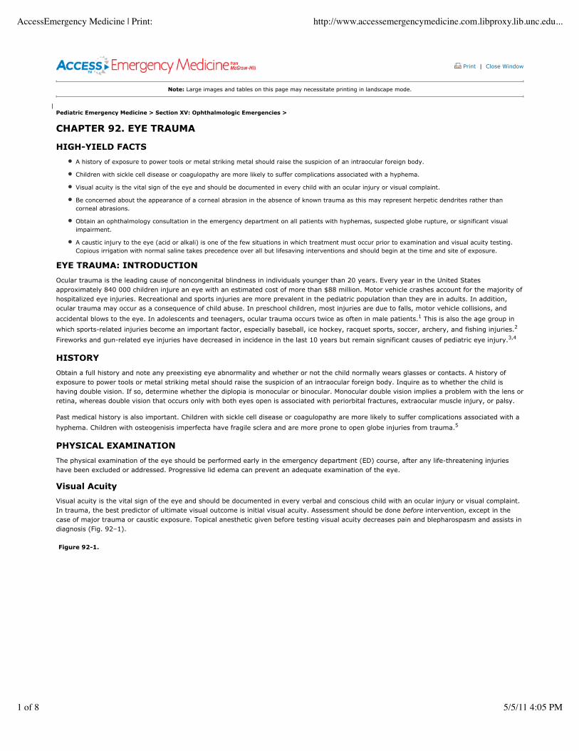
Figure 92-1.
AccessEmergency Medicine | Print: http://www.accessemergencymedicine.com.libproxy.lib.unc.edu...
1 of 8 5/5/11 4:05 PM
Diagnostic algorithm for ocular trama.
If the child wears glasses, measure acuity with the glasses on. If the glasses have been lost or damaged, correct refractive error by having thechild look through a pinhole in a piece of paper. Lack of correction with pinhole testing suggests significant pathology.
Evaluate preliterate children with an Allen or "E" chart and move a toy or a light to test the young child's ability to track with each eye. If thechild is unable to read an eye chart, ask him or her to finger count at 6 ft; if that fails, due to visual loss, assess for light perception.
Test visual fields in older children in the usual fashion. Younger children will glance toward a toy brought into the field of view. Assess forsymmetrical ocular motion and symptoms of diplopia.
AdnexaeTo avoid missing subtle signs of trauma, begin the examination with the lids and periorbital structures and work centrally in a focused fashion.Examine the lids for swelling or penetrating injury and inspect for ecchymosis. Retract swollen lids with a finger, being careful not to putpressure on the affected eye. Lid retractors or bent paper clips can be used in the case of massive lid edema. Periorbital soft tissue air indicatesfracture into a sinus or nasal antrum and is most commonly seen with a blowout fracture. Examine the eye for normal lacrimal drainage. Lidlacerations involving the medial third of the eyelid often involve the cannilicular system and injured ducts may require stenting by anophthalmologist. Epiphora, tears spilling over the lid margins, may be secondary to injury of the canalicular system.
Conjunctiva and ScleraExamine the conjunctiva for injection and presence of ciliary flush around the iris (perilimbal injection), which may indicate iritis. Check forchemosis (edema of the conjunctiva), which can be seen in globe rupture. Look at the sclera carefully for any disruption or penetration. Thelocation of any subconjunctival hemorrhage should be documented.
Pupils, Iris, Lens, and Anterior Chamber
AccessEmergency Medicine | Print: http://www.accessemergencymedicine.com.libproxy.lib.unc.edu...
2 of 8 5/5/11 4:05 PM
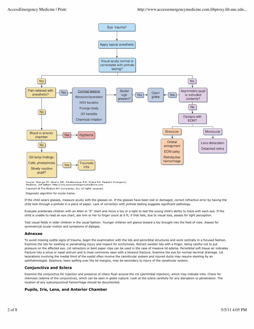
Evaluate the pupils for any asymmetry or irregularity. A pointing pupil indicates iris detachment and possible extrusion (Fig. 92–2). Congenitalanisocoria may be detected by obtaining history from the parents or by checking a photograph of the child to see if the asymmetry waspreexisting. Pupillary dilatation may occur with direct blows to the eye (posttraumatic mydriasis), with anticholinergic medication, or with a thirdnerve palsy. A dilated pupil in a conscious patient is not due to a herniation syndrome. Pilocarpine drops will constrict a pupil that is dilatedsecondary to a third nerve lesion but will have no effect on pharmacologic mydriasis. The emergency physician should also never overlook thepossibility of a glass eye.
Figure 92-2.
Ruptured globe with extravasation of ocular contents. (With permission from Khaw PT, Shah P, Elkington AR. Injury to the eye. BMJ. 2004;328:36–38.)
The lens should be transparent and the margins should not be visible. Examine the anterior chamber for abnormal shallowness or depth. Tospecifically assess optic nerve function, perform the swinging flashlight test. Swing the flashlight from eye to eye. A pupil that initially dilateswhen illuminated by light (even if it later constricts) has a sensory (afferent) defect (Marcus Gunn pupil).
Fundoscopic Examination and Intraocular PressureThe vitreous should be clear, allowing visualization of the retina. However, visualization of the retina does not completely exclude retinaldetachment, as many cases are anterior and thus undetectable with a direct ophthalmoscope. Do not measure intraocular pressure if globerupture or penetrating injury is apparent, as this may herniate ocular contents. Normal intraocular pressure is between 15 and 20 mm Hg.
EQUIPMENTThe basic equipment for a standard pediatric eye examination includes the following:
Visual acuity charts (Snellen and Allen "E" chart)
Pen light
Ophthalmoscope
Wood's lamp
Slit lamp
Ocular spud
Fluorescein strips
Shiotz tonometer or Tono-Pen
Morgan lens
Metal eye shields
MEDICATIONSTopical anesthetics, such as 0.5% tetracaine or proparacaine, have an onset of action within 1 minute and typically last for 15 to 20 minutes.Topical anesthetics should never be prescribed for home use, as prolonged lack of sensation and loss of normal protective reflexes may lead tocorneal damage. Cycloplegics such as homatropine (2%–5%) and cyclopentolate hydrochloride (1%–2%) dilate the eye and decrease pain byovercoming ciliary spasm. Always document the use of such medications to avoid later confusion regarding the etiology of an unreactive pupil.Avoid atropine due to its extremely long duration of action.
Steroids are useful in some inflammatory conditions but may lead to glaucoma, cataract formation, and acceleration of fungal and herpeticinfections, resulting in visual loss. Never use ocular steroids without first consulting an ophthalmologist. Evaluate the need for tetanusprophylaxis in all patients with ocular injuries. To administer medications to young or uncooperative children, lay them down in a dark room,
AccessEmergency Medicine | Print: http://www.accessemergencymedicine.com.libproxy.lib.unc.edu...
3 of 8 5/5/11 4:05 PM
secure the head, and place the drop in the medial canthus. The child will open the eyes and the medication will reach the conjunctiva.
SPECIFIC INJURIES
Lid LacerationsMinor lacerations, superficial to the tarsal plate, that do not involve the lid margins and with no suggestion of injury to deeper structures may berepaired by the emergency physician. Other lacerations require specialty repair.
Consider the possibility of globe penetration or injury to deeper structures whenever an eyelid is lacerated. Because eyelids have nosubcutaneous fat, the appearance of fat in a wound suggests underlying globe injury. Significant lacerations involving the medial third of theupper or lower lids may involve the lacrimal system and should be referred to an ophthalmologist for repair. One way to detect injury of thelacrimal system is to instill fluorescein in the eye and then use a Wood's lamp to check the wound for fluorescence.
Injuries to the lid margins are also problematic as they may result in deformity of the lids and abnormal lid movement if repair is not precise. Ifthe levator palpebrae muscle is injured and not repaired, posttraumatic ptosis will result. Trauma involving the tarsal plate (which only exists inthe upper lid as a dense band of fibrous tissue) indicates a complex laceration. Plastic surgery or ophthalmologic consultation should be obtainedfor repair of any of these lacerations described above. For isolated eyelid lacerations, delayed surgical repair of 12 to 36 hours will not affect theoutcome of closure and may allow a better repair due to decreased swelling.6
Subconjunctival HemorrhageThe most important aspect of the evaluation of subconjunctival hemorrhage is to rule out other, more serious injury. With significant blunttrauma, a subconjunctival hemorrhage may hide a scleral rupture or provide evidence of periorbital fracture. Clues to severe globe injury includedecreased visual acuity, severe pain, photophobia, and extension of hemorrhage beyond the limbus. Evaluate for the presence of Seidel's signindicating open globe injury with fluorescein (see later). Consider the possibility of a periorbital fracture in patients with traumaticsubconjunctival hemorrhage. Lateral hemorrhages in particular are frequently associated with zygomatic fractures (tripod fractures).
In the more common instances when the etiology involves minor trauma with no evidence of severe globe injury, coughing or sneezing spells, orspontaneous subconjunctival hemorrhage, parents should be reassured, told that spontaneous resolution will occur over 2 weeks, and informedof the dramatic color changes that may occur.
Corneal AbrasionChildren with corneal abrasions complain of a foreign body sensation, pain, and photophobia and present with marked blepharospasm and a redeye. Infants may present with only crying. Use tetracaine or proparacaine prior to examination to decrease blepharospasm and pain.
A few drops of fluorescein placed in the conjunctival sac followed by examination under a Wood's lamp or the cobalt blue light of a slit lamp willresult in marked fluorescence of corneal abrasions. Make sure that no foreign body is present and that there is no evidence of penetration of thesclera. Multiple vertical striations (ice-rink sign) usually indicate a retained foreign body under the upper lid. Careful examination with lideversion should reveal the offending agent.
Be concerned about the appearance of a corneal abrasion in the absence of known trauma. This may represent herpetic dendrites rather thancorneal abrasions. If this finding is present, test to see whether the corneal reflex is equally brisk in each eye by gently touching the cornea witha wisp of cotton (either before topical anesthesia or after the anesthesia has worn off). Herpetic lesions may decrease the corneal reflex in theinvolved eye. Patients may have concurrent oral or genital herpes. In the case of herpes keratitis, ophthalmologic consultation is required.
Treat corneal abrasions with ophthalmic antibiotics to prevent secondary infection. The multiple layers of the cornea normally provide a barrieragainst infection from common organisms such as Pseudomonas sp. and Staphylococcus aureus, and injury to this barrier may lead tosecondary infection. One drop of antibiotic solution applied every 4 to 6 hours for 4 days is sufficient. Ointment in young children and infantscan be given at the same frequency by applying a 1-cm length inside the lower eyelid. Several drops of a mydriatic agent such as cyclopentolate1% or homatropine 5% applied two to three times a day for 1 to 2 days will decrease ciliary spasm and provide comfort.
Eye patching is not recommended for mild to moderate corneal abrasions. It has not been shown to speed healing or decrease pain, and it mayincrease infection risk if prophylactic antibiotics are not given due to the patch.7,8 Corneal abrasions associated with Bell's palsy or other causesof incomplete lid closure should be patched to protect the eye from further injury. Any patch should be rechecked and removed in 24 hours.Some centers suggest that all abrasions should be rechecked in 24 to 48 hours to assess healing. Others tell patients to return if they are stillsymptomatic after 48 hours. Symptoms and redness should gradually decrease each day. If not, secondary infection, retained foreign body, orcorneal erosion should be suspected.
Traumatic IritisPatients with posttraumatic iritis usually present 1 to 2 days after blunt trauma to the eye, complaining of photophobia, pain, and tearing. Theyoften have marked blepharospasm and perilimbal injection (ciliary flush). Test for pain on accommodation by having the patient first look acrossthe room at a distant object and then quickly focus on the examiner's finger held several inches away. If near gaze causes pain, there is a highprobability of iritis. The pupil may be large or small. Posttraumatic miosis develops secondary to spasm of the pupillary sphincter muscle,whereas posttraumatic mydriasis results when sphincter fibers are ruptured. Slit lamp examination will usually reveal cells in the anteriorchamber, the hallmark of iritis.
Treat with a long-acting topical cycloplegic, such as 5% homatropine, four times a day for 1 week, oral anti-inflammatory medication, and darksunglasses to decrease pain. Symptoms may persist for up to 1 week. Although ocular steroids decrease inflammation, prescribe them only after
AccessEmergency Medicine | Print: http://www.accessemergencymedicine.com.libproxy.lib.unc.edu...
4 of 8 5/5/11 4:05 PM
consultation with the ophthalmologist who will see the patient in follow-up.
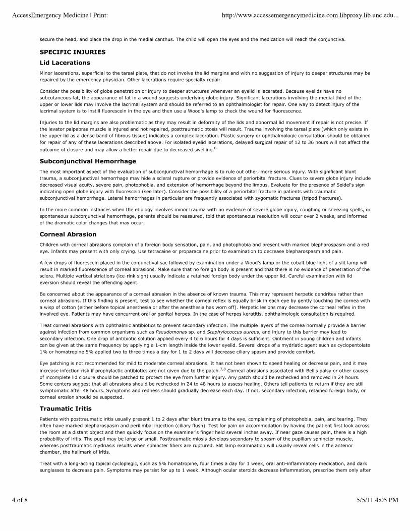
HyphemaA hyphema is defined by blood in the anterior chamber (see Fig. 92–3). Hyphemas are almost always secondary to blunt trauma. The blood maylayer out or may present initially as a diffuse red haze that takes hours to settle. Hyphemas are described by the percentage of the anteriorchamber that is filled with blood. A 100% hyphema, known as an "eight ball," may cause complete loss of light perception. Hyphemas are easilyoverlooked in cases of massive lid edema, and for this reason, early lid retraction is necessary for the diagnosis. Older children with hyphemasusually complain of pain, whereas young children may present with somnolence. However, decreased mental status should not be attributed tothe hyphema until intracranial injury has been ruled out. Other injuries associated with hyphema include lens dislocation, vitreous hemorrhage,and retinal damage. A large untreated hyphema may result in permanent corneal staining with subsequent loss of visual acuity anddeprivational amblyopia. Often it is not the initial hyphema that causes serious morbidity but the rebleed that can occur in several days as theclot breaks down. Rebleeding occurs in up to 16% to 25% of patients and may obstruct the aqueous outflow system, causing increasedintraocular pressure. Patients with rebleeding have a significantly worse prognosis. Because hemoglobinopathies, particularly sickle cell disease,sickle cell trait, and sickle thalassemia, predispose to rebleeding and other complications, it is critical to determine whether any of theseconditions exist in the child with hyphema.
Figure 92-3.
Hyphema. (With permission from Khaw PT, Shah P, Elkington AR. Injury to the eye. BMJ. 2004;328: 36–38.)
Initial treatment involves bed rest with the head elevated at 30 degrees. Shield the involved eye, taking care not to touch or apply pressure tothe eye. Obtain an ophthalmology consultation in the ED on all patients with hyphemas.
The subsequent treatment of hyphemas is controversial. Traditionally, all patients have been admitted and placed on strict bed rest. However,older children and reliable adults are now being treated with bed rest at home if they have only a microhyphema (circulation of red blood cellsonly, with no layering) and if daily reexamination for 5 days is possible. Some specialists utilize antifibrinolytics such as aminocaproic acid toreduce rebleeding.9 The decision as to use of mydriatics, ocular steroids, osmotic agents, or acetazolamide should be left to the ophthalmologist.Acetazolamide or osmotic agents are contraindicated in patients with sickle cell disease due to increased risk of bleeding. Patients should avoidaspirin or other platelet-active medications, as these increase the risk of rebleeding.
Lens InjuryBlunt ocular trauma may result in lens subluxation or dislocation. Subluxation may cause monocular diplopia, whereas dislocation results inprofoundly blurred vision. The lens may sublux either posteriorly or anteriorly, resulting in a deep or shallow anterior chamber and a visible lensmargin. Iridodonesis is a shimmering or shaking of the iris provoked by rapidly changing gaze and is associated with posterior dislocation.
Acute blunt trauma to the eye can cause a cataract if the capsule of the lens is disrupted. The lens subsequently absorbs fluid, taking on acloudy appearance. Lens injuries should be referred to an ophthalmologist.
Retinal InjuryRetinal trauma often occurs in conjunction with other eye injuries. Older children with retinal injury complain of light flashes or a "curtain" overthe visual field. Central vision will be spared if the macula remains unaffected.
Fundoscopy may reveal a variety of hemorrhage patterns or may be normal with direct fundoscopy. Preretinal hemorrhages are boat shaped,with a horizontal "deck," whereas superficial hemorrhages are flame shaped. Deep hemorrhages are round, with a purple-gray color. The shakenbaby syndrome causes linear retinal hemorrhages and associated exudates. Any suspicion of retinal detachment or injury requiresophthalmology consultation.
AccessEmergency Medicine | Print: http://www.accessemergencymedicine.com.libproxy.lib.unc.edu...
5 of 8 5/5/11 4:05 PM
Retrobulbar HemorrhageBleeding behind the globe may result in deficits in extraocular motion and lead to proptosis or bulging of the eye. Subsequent compromise ofthe optic nerve produces an afferent pupillary defect (Marcus Gunn pupil). In cases of severe proptosis, progressively worsening vision, andpotential optic nerve injury surgical decompression may be necessary. In rare instances, a lid release procedure (lateral canthotomy) may berequired in the ED.
Conjunctival and Scleral LacerationsPerform a slit lamp examination on all children with conjunctival lacerations to assess for deeper scleral violation. Scleral rupture from blunttrauma often occurs at the insertions of the intraocular muscles or at the limbus. Clues to the presence of scleral disruption include decreasedvisual acuity, an abnormal anterior chamber, low intraocular pressure (<6), and a positive Seidel's test. To perform the Seidel's test for sclerallaceration, place fluorescein on the cornea and observe the suspicious area under the cobalt blue light of the slit lamp. A swirling dilution offluorescein secondary to leaking aqueous denotes scleral disruption.10 If a scleral laceration is seen, tonometry is contraindicated. The additionalpressure against the eye from the tonometer can extrude the iris.
Treat small conjunctival lacerations with topical antibiotic drops alone. Sutures are not usually necessary. If the patient has deeper injury (i.e.,scleral laceration), place an eye shield on the child, administer intravenous antibiotics, provide adequate sedation, and obtain an ophthalmologyconsultation.
Corneal and Scleral Foreign BodiesPatients with a superficial foreign body are in pain. Their eye is usually red and tearing, and verbal children will complain of "something in myeye." Corneal abrasions, however, will present in a similar mode.
Perform a thorough examination in all patients by doubly everting the lids after anesthetizing the cornea. This may be done with a cotton swabplaced on the middle of the upper lid and folding the lid upward over the swab. Look carefully for a foreign body on the inner surface of the lids.Attempt removal with a cotton applicator soaked in topical anesthetic. In older cooperative children, remove more tenacious foreign bodiesunder slit lamp guidance using an eye spud or 25-gauge needle on a tuberculin syringe. A foreign body sensation may persist even afterremoval due to an underlying corneal abrasion. Iron-containing foreign bodies may leave rust rings that may result in photophobia anddecreased visual acuity. Some emergency physicians refer these to a specialist, whereas others use an ophthalmic burr to remove superficialrings.
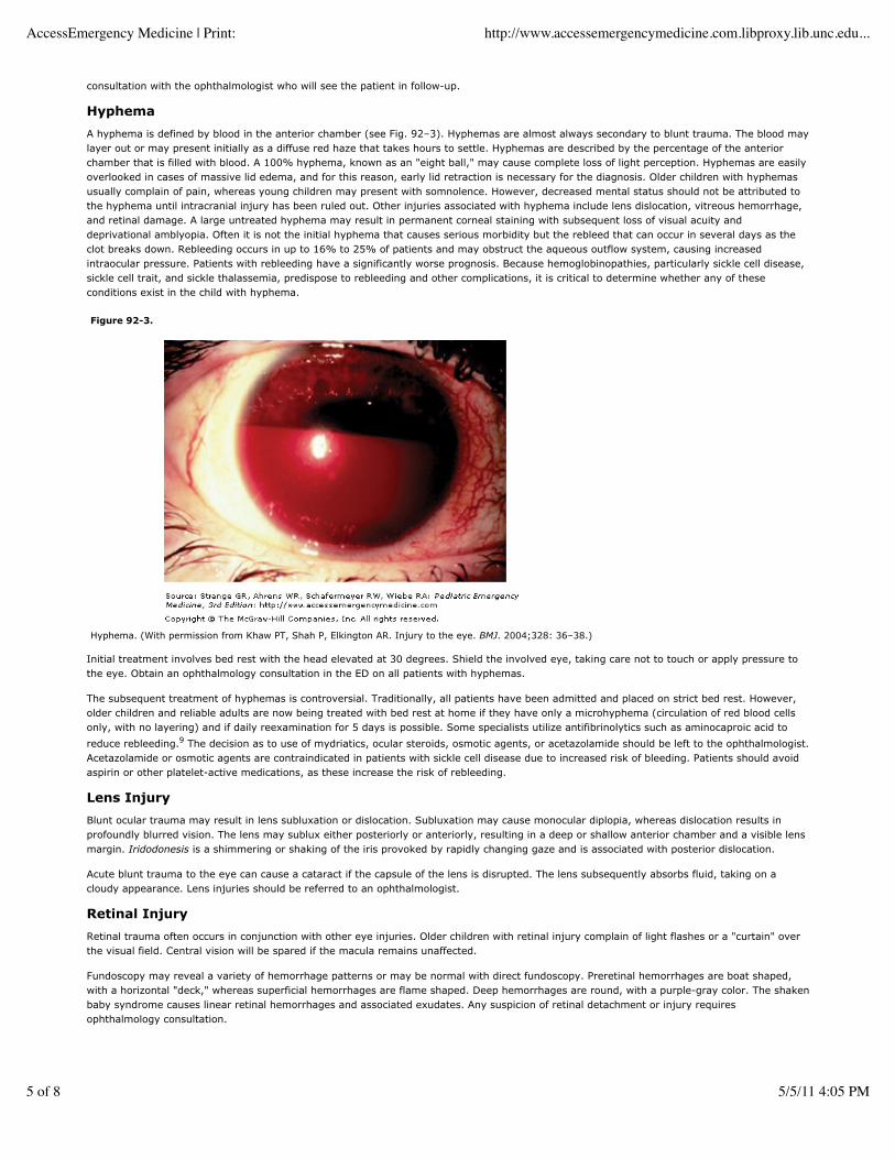
After removing a foreign body, check for additional foreign bodies and abrasions. Instill both a mydriatic agent and a topical antibiotic andrecheck the child in 24 hours. Prescribe appropriate analgesia including narcotic agents if appropriate. If a foreign body penetrates the cornealstroma or the sclera (Fig. 92–4), consult an ophthalmologist for evaluation in the ED.
Figure 92-4.
Intraocular foreign body. Note foreign body at arrow. Also note perforation of globe with asymmetric pupil and leaking vitreous. (With permission fromKhaw PT, Shah P, Elkington AR. Injury to the eye. BMJ. 2004;328: 36–38.)
Intraocular Foreign BodyIntraocular foreign bodies are vision-threatening injuries that may be easily overlooked. The key to identification of an intraocular foreign bodyis to consider it. Ask specific questions about risk factors. Exposure to power tools and metal striking metal, such as a hammer on a nail,predisposes to occult intraocular foreign body.
Certain foreign bodies place the patient at greater risk than others. Iron (siderosis) and copper (chalcosis) are particularly toxic to the eye,whereas glass and plastic are less inflammatory. Organic foreign bodies pose a high risk for intraocular infection.
AccessEmergency Medicine | Print: http://www.accessemergencymedicine.com.libproxy.lib.unc.edu...
6 of 8 5/5/11 4:05 PM

Children with intraocular foreign bodies may have decreased visual acuity, pupillary distortion, and relatively little pain. A foreign bodypenetration through the cornea may damage the iris, causing a teardrop-shaped pupil that will "point" to the perforation site.
A number of imaging modalities can detect intraocular foreign bodies. Although larger metallic objects may be seen by routine radiography, acomputed tomography scan of the orbit provides greater resolution if the foreign body is small. Ocular ultrasound is highly sensitive for bothmetallic and nonmetallic penetrations. Magnetic resonance imaging (MRI) accurately detects organic, plastic, and glass particles but may causefurther injury if mistakenly used in the case of metal objects. In such cases, the MRI magnet may move the foreign body, causing greaterdamage.
Cover the involved eye with a metal eye shield and keep the child at rest. Administer broad-spectrum intravenous antibiotics, such asvancomycin and ceftazidime.11
Topical antibiotics are not indicated with penetrating globe injuries and, in particular, antibiotic ointments should be avoided as they can produceintraocular granulomas and obscure examination.
Should a child with penetrating globe injury require emergency intubation, some authorities suggest a nondepolarizing blocker, such asrocuronium, in place of succinylcholine. Succinylcholine and ketamine may increase intraocular pressure and could theoretically extrudeintraocular contents. However, if a difficult airway is anticipated, then succinylcholine may be the agent of choice.6
Foreign Bodies In SituForeign bodies that protrude from the eye, such as a nail or wire, must be left in place and removed in the operating room. Cover the affectedeye with a cup to prevent further manipulation of the eye and patch the unaffected eye closed to prevent extraoccular movement.
Chemical Injuries to the EyeA caustic injury to the eye (acid or alkali) is one of the few situations in which treatment must occur prior to examination and visual acuitytesting. Copious irrigation with normal saline takes precedence over all but lifesaving interventions and should begin at the time and site ofexposure. Extent of caustic injury is dependent on the quantity, the pH, and the duration of the exposure (i.e., time to irrigation). Alkali injuriesresult in the most serious damage to the eye by causing liquefaction necrosis with saponification of ocular tissues and deep penetration. Acidsproduce a coagulation necrosis resulting in a protein barrier that blocks further penetration. Complications of caustic injuries include blindness,perforation, corneal neovascularization, secondary glaucoma, cataract formation, and retinal damage.
Begin irrigation in the prehospital setting immediately after injury. Upon arrival at the ED, the child may require sedation with a rapidly actingintramuscular or intravenous agent to allow irrigation of the eye, but topical anesthesia alone may be adequate. If particulate matter remains inthe eye, pour liter bottles of irrigant into the eye while retracting the lids to wash the particles out. Perform double lid eversion to expose thefornices and irrigate or swab out any caustic particulate matter. When irrigating the eyes, a Morgan lens (a contact lens connected tointravenous tubing) is useful. An alternative to the Morgan lens is a nasal oxygen cannula placed on the bridge of the nose, which can beconnected to intravenous bags of normal saline. This allows bilateral irrigation of the eyes through the nasal prongs.
Never attempt to neutralize acids with alkalis, or vice versa, because the resultant heat release will further damage the eye. Lavage the eyes forat least 20 minutes, ideally with 2 L of normal saline. Irrigation may be beneficial for up to 24 hours after alkali exposure. In cases of seriousalkali exposure, continuously irrigate the eyes until stopped by the ophthalmologist. Use litmus paper to check the pH in the conjunctival sacafter 20 minutes of irrigation, and continue irrigation until the pH is between 7.4 and 7.6. If irrigation is stopped, recheck the pH after 10minutes to ensure a stable level.
Hydrofluoric acid exposure is a unique situation and may require irrigation with a magnesium oxide solution. Consult a poison center for thelatest recommendations.
Ultraviolet KeratitisUltraviolet keratitis occurs when children are exposed to prolonged glare from snow, water, or white sand or when they stare at an eclipse.Unprotected vision of an eclipse can also cause severe retinal damage and blindness. Older children who watch a welder's torch or use tanningbooths without special glasses may also suffer this injury. Photophobia and eye pain usually occur 8 to 12 hours after exposure and, for thisreason, patients with ultraviolet keratitis generally present at night. They exhibit both scleral and perilimbal injection accompanied by tearingand blepharospasm. Slit lamp examination using fluorescein shows thousands of punctate, shallow lesions on the cornea (keratitis), which looksas if it had been sandblasted. Treat with cycloplegia and oral analgesia. Ultraviolet keratitis is usually bilateral and generally heals in 24 to 48hours.
Thermal BurnsBecause of reflex blinking, lids are more often damaged from thermal injury than is the globe. Eyelashes and eyebrows are often burned.Evaluate for corneal injury with the slit lamp in the usual fashion with and without fluorescein staining, and apply topical antibiotics to burnedlids. Third-degree burns to the eye and periorbital tissues require admission.
CONCLUSIONSThe emergency physician is often the first and only physician to evaluate ocular trauma. Proper identification depends primarily on theconsideration of the potential for injury, and on the performance of a thorough, systematic examination. Unfortunate sequelae can often beavoided through timely identification and appropriate specialty consultation. Any eye injury regardless of acuity should be considered anopportunity for physicians to inform parents and patients about protective eyewear and risk reduction behavior.
AccessEmergency Medicine | Print: http://www.accessemergencymedicine.com.libproxy.lib.unc.edu...
7 of 8 5/5/11 4:05 PM
Copyright © The McGraw-Hill Companies. All rights reserved.Privacy Notice. Any use is subject to the Terms of Use and Notice.
REFERENCES1. Brophy M, Sinclair S, Hosteler SG, Xiang H. Pediatric eye injury-related hospitalizations in the United States. Pediatrics. 2006;117:e1263–e1271.
2. McGwin G Jr, Owsley C. Incidence of emergency department-treated eye injury in the Unitied States. Arch Ophthalmol. 2005;123:662–666.[PMID: 15883286]
3. McGwin G Jr, Xie A, Owsley C. Gun related eye injury in the United States, 1993–2002. Ophthalmic Epidemiol. 2006;13:15–21.[PMID:16510342]
4. Witsaman RJ, Comstock RD, Smith GA. Pediatric fireworks related injuries in the United States: 1990–2003. Pediatrics. 2006;118:293–303.
5. Pirouzian A, O'Halloran H, Scher C, et al. Traumatic and spontaneous scleral rupture and uveal prolapse in osteogenisis imperfecta. J PediatrOphthalmol Strabismus. 2007;44:315–317.[PMID: 17913179]
6. Salvin J. Systematic approach to pediatric ocular trauma. Curr Opin Ophthal. 2007;18:366–372.[PMID: 17700228]
7. Burnette DD. Opthalmology. In: Marx JA, ed. Rosen's Emergency Medicine: Concepts and Clinical Practice. Philadelphia,PA: CV Mosby;2006:70.
8. Turner AR. Patching for corneal abrasion. Cochrane Database Syst Rev. 2006;(2):CD004764.
9. Rocha KM, Martins EN, Melo LA Jr, et al. Outpatient management of traumatic hyphema in children: prospective evaluation. J AAPOS.2004;8:357–361.[PMID: 15314597]
10. Pokhrel PK, Loftus SA. Ocular emergencies. Am Fam Physician. 2007;76:829–836.[PMID: 17910297]
11. Waheed NK, Young LH. Intraocular foreign body related endophthalmitis. Int Opthalmol Clin. 47:165–171.
AccessEmergency Medicine | Print: http://www.accessemergencymedicine.com.libproxy.lib.unc.edu...
8 of 8 5/5/11 4:05 PM