Embed Size (px)
Citation preview

18
Non-Herpetic Acute Limbic Encephalitis: A New Subgroup of Limbic Encephalitis?
Hiroshi Shoji1, Noriyuki Kimura2, Toshihide Kumamoto2, Takashi Ichiyama3 and Yukitoshi Takahashi4
1Division of Neurology, St. Mary Hospital, Fukuoka 830-8543, 2Department of Internal Medicine III, Faculty of Medicine,
Oita University, Oita, 3Department of Pediatrics, Yamaguchi University Graduate
School of Medicine, Yamaguchi, 4National Epilepsy Center, Shizuoka Institute of Epilepsy
and Neurological Disorders, Shizuoka Japan
1. Introduction
Non-herpetic acute limbic encephalitis (NHALE) was found at 1994 during the survey of herpes simplex encephalitis (HSE) in the Kyushu district, Japan. NHALE is characterized by a lack of evidence of the herpes simplex virus (HSV) genome or HSV antibody, non-paraneoplastic limbic encephalitis, and magnetic resonance imaging (MRI) abnormalities in bilateral medial temporal lobes such as the hippocampi and amygdalae (Kusuhara et al., 1994; Kaji et al., 1996; Shoji et al., 2004). Etiologies of NHALE consist of various causes, including viral origins, autoimmune disorders, and several anti-neural antibodies. Since Urgent Conference on Non-Herpetic Limbic Encephalitis, at Ichikawa City, Japan, November 2002, many cases have been accumulated as viral related acute limbic encephalitis, autoantibody-mediated acute limbic encephalitis, paraneoplastic limbic encephalitis or encephalopathy (Yuasa et al, 2003). Cerebrospinal fluid (CSF) shows a mild pleocytosis with increase of pressure, mild increase of protein, and sometimes, a lack of the pleocytosis. The CSF level of interferon-γ (IFN-γ) is unchanged with an increase of interleukin (IL)-6 (Asaoka et al., 2004; Ichiyama et al., 2008a). Among them, NHALE patient group with the onset symptoms of abnormal behavior and incoherence, and the detection of anti-glutamate receptor (GluRε2 NR2B) antibodies is gaining attention (Nemoto et al., 2005; Hayashi et al. 2005; Takahashi et al., 2007). This NHALE type is indicated to form a new subgroup of acute limbic encephalitis or encephalopathy. GluRε2 is a subunit of the N-methyl-D-asparate (NMDA) glutamate receptor. NHALE overlaps clinically to anti-NMDA receptor (NR1+2A) encephalitis, but the NMDA encephalitis is usually associated with ovarian teratoma (Dalmau et al., 2007; Iizuka et al., 2008). In this review, two NHALE patients with positive anti-GluRε2 antibody are briefly
described, and the pathogenesis of NHALE, clinical features, differential diagnosis,
prognosis, and sequelae are discussed.
www.intechopen.com

Pathogenesis of Encephalitis
268
Fig. 1. Possible relationships of NHALE and related disorders. NHALE=non-herpetic acute limbic encephalitis, NMDAR=N-methyl-D-asparate receptor VGKC =voltage-gated potassium channel, LE=limbic encephalitis
2. Case presentation
Here, we present two representative patients with NHALE who were evaluated at A rehabilitation hospital 3 months to 2 years after the onset (Noto et al., 2008; Shoji et al., 2009). Patient 1: A 50 year-old man was restless and became talkative with a fever and palpitation
in the end of July 200x. Several days later, he was admitted with collapsed state to a nearby
hospital. On admission, he showed a fever of 38.4℃, coma state, and generalized myoclonus
without nuchal stiffness, then paroxysmal atrial fibrillation, status epileptics, and needed an
artificial respirator for 3 months. The CSF exhibited no abnormalities in the cell count,
protein and glucose contents, and polymerase chain reaction (PCR) for HSV and human
herpesvirus (HHV)-6 was negative. The serum antibodies were negative for herpesvirus
groups (HSV, HHV-6, varicella-zoster virus and cytomegalovirus). The MRI revealed
hyperintensity lesions bilaterally in the hippocampi, amygdalae and claustrum. His CSF at
the 5th illness day was positive for anti-GluRε2 IgG and IgM antibodies. Acyclovir (ACV)
administration, thereafter methylprednisolone pulse therapy was performed with sodium
valproate and clonazepam. Amnestic syndrome and partial seizure remained. Three months
after the onset he was transferred to A rehabilitation hospital which was near to his home
town. Neuropsychological tests such as the Wechsler adult intelligence scale-revised
(Japanese edition, WAIS-R), Wechsler memory scale-revised (Japanese edition, WMS-R),
and functional independence measure (FIM) were conducted to assess the sequelae for 2
years after the onset. WMS-R showed severe impairment of recent memory with normal
total WAIS IQ and FIM points. The MRI at the recovery stage showed bilateral atrophic
changes in the hyppocampi and amygdalae with hyperintensity lesions in the rectus gyri
(Fig 2a). Thereafter, he moved to an another rehabilitation hospital. At 2 years after the
onset, moderate amnestic syndrome and focal seizure remained, and he is still unable to
return to his previous job.
Patient 2: A 36 year-old man had diarrhea for several days, and he then developed a fever of 40℃ for one week, but he did not stop his job of rescue party. Convulsive seizures and
delirium appeared, and he was admitted to a nearby hospital. His consciousness level was stupor state without nuchal stiffness and pathologic reflexes. The CSF contained 6 mononuclear cells per mm3 and protein content of 46 mg/dl, and HSV PCR was negative in
www.intechopen.com
Non-Herpetic Acute Limbic Encephalitis: A New Subgroup of Limbic Encephalitis?
269
the CSF. Serum enzyme immunosorbent assay (EIA) tests for herpesvirus groups, rubella, measles and mumps were all negative. His CSF showed positive for anti-GluRε2 IgG and IgM antibodies. Electroencephalogram (EEG) revealed periodic lateralized epileptiform discharges (PLEDs). The MRI exhibited hyperintensity lesions in the bilaterally medial temporal lobes including hippocampi and amygdalae. Patient 2 was diagnosed as having NHALE with positive anti-GluRε2 antibodies. Anticonvulsant drugs and ACV were administered, but he developed status epileptics, and put on a respirator, and methylpredonisolone pulse and immunoglobulin therapies were conducted. Three months after the onset, he was transferred to A rehabilitation hospital. He showed a fever of 37ºC, attention impairment, nystagmus, myoclonus at the left face and upper extremity, and weakness and atrophy of the both legs as a disuse syndrome. MRI revealed bilaterally atrophy of the hippocampi and amygdalae with hyperintensity lesions (Fig.2b). WAIS-R showed 69 of total IQ, and 44/126 at FIM. Intensive rehabilitation was performed, and 6 months later he was discharged with improvement of scores of WAIS-R, WMS-R and FIM. At 2 years after the onset, mild dysarthria, attention impairment, and gait disturbance have remained, and he is now trying to return to his previous job.
Fig. 2. a,b: MRIs of Patients 1 & 2 with NHALE at the recovery stage; a: an axial fluid attenuated inversion recovery (FLAIR) MRI of Patient 1 reveals hyperintensity lesions in the rectus gyri, and mild dilatation of the inferior horn. b: an axial FLAIR MRI of Patient 2 shows moderate dilatation of the inferior horn.
3. Pathogenesis
In the CSF cytokines of NHALE patients, the IFN-γ value is not elevated with an increase of
IL-6 (Asaoka et al., 2004; Ichiyama et al., 2008a; Fig.3). CSF IFN-γ levels were elevated in the
central nervous system (CNS) disorders due to direct viral invasion, such as viral meningitis
and HSE (Matsubara et al., 2000; Ichiyama et al., 2005; Ichiyama et al., 2008a), but not in
immune-mediated CNS disorders, such as acute disseminated encephalomyelitis, influenza-
associated encephalopathy, and acute encephalopathy following prolonged febrile seizures,
and hemolytic uremic syndrome with encephalopathy (Ichiyama et al., 2002; Ichiyama et al.,
2004; Ichiyama et al., 2008b; Shiraishi et al., 2008). IFN-γ, which is produced by NK cells,
www.intechopen.com

Pathogenesis of Encephalitis
270
CD8+ and Th1 type CD4+ T lymphocytes, plays an important role in host defense against
viral infection, and inhibits viral replication (Samuel., 1991). Therefore, IFN-γ elevating in
the CSF may exert an inhibitory effect against viruses invading the CNS. With respect to
CSF IFN-γ levels, we suggest that the main pathogenesis of NHALE is not caused by the
direct invasion of virus into the CNS.
Fig. 3. The CSF concentrations of IFN-γ, IL-6, IL-10, and sTNFR1 in patients with NHALE, HSE, and controls. Horizontal lines indicate geometric means. Shaded areas indicate values below the detection limits. IFN-γ=interferon-γ, IL=interleukin, sTNFR1=soluble tumor necrosis factor receptor-1
In a major subgroup of NHALE patients presenting psychiatric symptoms such as abnormal behavior or incoherence, a subunit of NMDA glutamate receptor antibody (GluRε2 NR2B) was detected in the sera and CSF. In Japan, many similar cases have been accumulated (Nemoto et al., 2005; Hayashi et al., 2005; Noto et al., 2008). Patients 1 & 2 here described are regarded as anti-GluRε2 positive NHALE. Mochizuki et al. (2006) reported an autopsy case of NHALE with positive Anti-GluRε2 NR2B antibody, and a few autopsy cases followed (Okamoto et al., 2008; Maki et al., 2008). Thus, this NHALE type should be regarded as belonging to a new subgroup of acute limbic encephalitis. Anti-NMDA type GluR
antibodies categorized into NMDA glutamate receptor complex antibodies {GluR1 (NR1) +
GluR1 (NR2A) or GluR2 (NR2B)} and antibodies to each subunits composing of the complex antibodies. Antibodies to GluRε2 (NR2B) subunit are antibodies to a whole
www.intechopen.com

Non-Herpetic Acute Limbic Encephalitis: A New Subgroup of Limbic Encephalitis?
271
molecules of GluRε2 (NR2B) subunit (Takahashi et al., 2008, Fig.4). Takahashi et al. (2008) reported 60% positive rate of Anti-GluRε2 antibodies in the serum of NHALE patients from the acute to convalescent stage, whereas these Anti-GluRε2 antibodies in the CSF of NHALE patients were detected in 50% at the acute stage, and 40% at the recovery stage; anti-GluRε2 antibodies is indicated to invade from serum to CNS with injury of the blood-brain barrier, and may involve the pathogenesis of acute limbic encephalitis or encephalopathy (Takahashi et al., 2007; 2010). Ichiyama et al. (2009) reported that high matrix metalloproteinase-9/tissue inhibitor of metalloproteinase 1 (MMP-9/TIMP-1) ratio which indicates to injure the blood-brain barrier continues from the acute to convalescent stages (Fig. 5).
Fig. 4. Antibodies to NMDA-type GluR complex and each domain of GluR subunits. Antibodies to NMDA-type GluR complex (antibodies to NMDAR) were examined using
HEK cells transfected by expression plasmid of GluR1 (NR1) + GluR1 (NR2A) or GluR2 (NR2B) (Dalmau et al., 2007). Antibodies against whole molecules of GluRε2 were examined by immunoblot (Takahashi Y et al., 2003). Antibodies against each domain of GluRε2
(NR2B) and GluR1 (NR1) were examined by ELISA with synthesized peptides. NMDA=N-methyl-D-asparate, GluR= glutamate receptor
On the other hand, the anti-GluR ε2 antibodies are also detected in Rasmussen encephalitis, acute encephalitis with refractory repetitive partial seizures (AERRPS), mild encephalopathy with a reversible splenial lesion (MERS), or HSE (Takahashi et al., 2003; 2005; Sakuma et al., 2009; Kai et al., 2009). Anti-GluRε2 antibody encephalitis may form a wider spectrum of encephalitis and encephalopathy. The antibody reacts cross to anti-NMDA-type GluR complex antibodies, and the clinical significance and specificity to NHALE of anti-GluRε2 antibodies is not established. In near future, the specificity, or further pathological mechanism of GluRε2 NR2B for NHALE should be investigated.
www.intechopen.com

Pathogenesis of Encephalitis
272
Fig. 5. Serum concentrations of MMP-9 and TIMP-1 and the ratio of MMP-9/TIMP-1 in patients with NHALE and in controls. The high ratio of MMP-9/TIMP-1 continues from the acute to convalescent stages. A: Patients with NHALE in the acute stage, B: patients with NHALE in the convalescent stage, and C: controls. The horizontal lines indicate median values. MMP-9=matrix metalloproteinase-9 , TIMP-1=tissue inhibitor of metalloproteinase 1
4. Clinical characteristics
4.1 Clinical findings Incidence rate is 4.7 persons per million person-year. Mean onset ages of NHALE show 44.8 years in male and 31.6 years in female, respectively (Wada-Isoe et al., 2008). In response to the limbic system disorder, various symptoms such as changeable disorientation, memory impairment, catalepsy, schizophrenic-like delusions, anger, hallucinations, convulsive seizures, autonomic seizures, pulse rate or respiratory abnormalities, and hyponatremia can appear (Yuasa, 2003). Our Patients 1 & 2 showed restlessness, palpitation and delirious state as initial symptoms. In the acute stage of NHALE, schizophrenic-like symptoms consisting of abnormal behavior, incoherence, delusions, and hallucination are frequently observed, and then convulsive seizures, status epileptics, and autonomic seizures are follows. Meningeal irritation signs appear mildly. Takahashi et al. (2008) analyzed 53 cases of non-paraneoplastic non-herpetic acute limbic encephalitis compared with 16 negative anti-GluRε2 antibody cases; abnormal behavior, delusions, and hallucination are more frequently observed including autonomic seizures, pyramidal and extrapyramidal signs, and cerebellar symptoms. Status epileptics appeared at 56.5% of the adult cases, and the other complexes partial seizures without convulsive seizure are also described. Prognostic outcome is comparative favorable, and several limited autopsy cases have been reported (Mochizuki et al., 2006). As the complications of NHALE, hypertrophic pachymeningitis or Vogt-Koyanagi-Harada disease has been reported (Usui et al., 2007; Masuda et al., 2009).
www.intechopen.com

Non-Herpetic Acute Limbic Encephalitis: A New Subgroup of Limbic Encephalitis?
273
4.2 Laboratory findings PCR and antibody tests including sensitive enzyme-linked immunosorbent assay for herpesvirus groups result in all negative. However, in the serum tests of the adults or elderly patients we may sometimes encounter carrier having HSV positive antibody (Shoji et al., 2004). CSF shows a mild pleocytosis with increase of pressure, mild increase of protein, and sometimes, a lack of the pleocytosis. The CSF level of IFN-γ is unchanged with an increase of IL-6 (Asaoka et al., 2004; Ichiyama et al., 2008a). Ichiyama et al. (2008a) reported the comparison of CSF cytokines between NHALE and HSE; increases of IFN-γ, IL-6, IL-10, and sTNFR were observed in HSE, whereas only increase of IL-6 was found in NHALE. Brain MRI exhibits often bilateral abnormalities in the limbic areas; abnormal signals distribute in the bilateral hippocampi, amygdalae, rectus, cingulated gyri, and insula. Diffusion weighted images (DWI) is most sensitive to detect the lesions, and the hyperintensity lesions decreased by apparent diffusion coefficient (ADC) (Fig.6). Brain CT is difficult to detect the medial temporal lesions. Also, it should be evaluated that the hyperintensity lesions are apt to appear by DWI as similar lesions after status epilepticus. At the acute stage the inferior horn narrowing is often observed by MRI or CT probably reflecting the brain edema, and at the recovery stage the inferior horn is dilated suggesting the brain atrophy. 99mTc-ECD using easy z-score imaging system (eZIS) analysis single photon emission CT (SPECT) may reveal wider hypo- or hyper-perfusion lesions than the corresponding MRI lesions in the limbic areas (Fig. 7). Moreover, the hyperintensity lesions on DWI may show relatively increased regional cerebral blood flow (rCBF) on eZIS analysis. EEG reveals whole moderate slowness, and PLEDs or periodic synchronous discharges (PSD) are observed at approximately 20-30% for the acute stage.
Fig. 6. MRIs of patient with NHALE at the acute stage. Diffusion-weighted MR images showed hyperintensity lesions in the bilateral medial temporal lobes, cingulate gyri, and insulas. NHALE=non-herpetic acute limbic encephalitis
www.intechopen.com
Pathogenesis of Encephalitis
274
4.3 Treatment There is no standard therapy for NHALE. Most treatment approaches have employed some
form of nonspecific immunosuppressant therapy similar to that used for other
encephalitis/encephalopathy and autoimmune diseases, including corticosteroid,
intravenous immunoglobulin (IVIg), plasmapheresis, or a combination of these therapies.
High-dose intravenous steroids are widely accepted as first-line therapy. Most of the data
describing treatment for NHALE are derived from case reports and small series. To date,
there have been no randomized, controlled trials for the treatment of NHALE. On the other
hand, ACV therapy is usually conducted at the acute stage due to difficulty to exclude HSE.
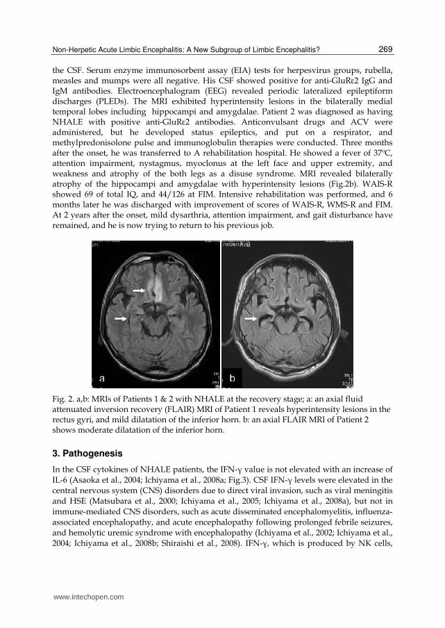
Fig. 7. eZIS images in (axial view) patients with NHALE at acute stage.
The eZIS analysis detected a significantly increased rCBF in the bilateral medial temporal lobes, left insula, and left putamen, and a significantly decreased rCBF in the bilateral frontal lobes. The color images represent the statistical significance (Z-score) of the increase (red) or decrease (blue) in rCBF. eZIS =easy z-score imaging system, NHALE=non-herpetic acute limbic encephalitis, rCBF=regional cerebral blood flow
4.4 Differential diagnosis Differential diagnosis with HSE is important in the choice of anti-viral drug starting or
immunological therapy. HSE presents a fever, meningeal irritation signs, consciousness
impairment, convulsive seizures, and memory impairment, while NHALE shows the onset
symptoms with abnormal behavior or talk, and meningeal signs appear mildly.
Neuroimaging of HSE often involves predominantly unilateral temporal lobe, but NHALE
tends to localize bilateral medial temporal lobes. The CSF findings of NHALE are apt to
reveal mild changes. Differentiation between NHALE and NMDAR encephalitis at the
initial stage may be difficult, because 25% of NMDAR encephalitis exhibits localized limbic
encephalitis form (Iizuka et al., 2008). Therefore, particularly in juvenile female patients with
acute encephalitis, ovarian teratoma should be checked rapidly (Kamei S., 2004, 2008). On
the other hand, anti-voltage-gated potassium channel (VGKC) antibody limbic encephalitis
www.intechopen.com

Non-Herpetic Acute Limbic Encephalitis: A New Subgroup of Limbic Encephalitis?
275
shows often encephalopathy type with hyponatremia, and overlaps Morvan syndrome with
neuromyotonia. Hashimoto encephalopathy should be noticed to present limbic
encephalopathy type, although this encephalopathy usually shows whole encephalitis type.
5. Sequelae
As sequelae, memory impairment, and personality or emotional changes have been most frequently described. Regards with the sequelae of our Patients 1 & 2 at 2 years after the onset, Patient 1 shows prominent impairment for recent memory with intact intelligence and immediate memory. Another Patient 2 presents memory and intelligence impairments, involuntary movement, and paresis in both legs as disuse syndrome. They are still unable to return their previous jobs. Next, we analyzed the sequelae evaluated in questionnaires from 19 cases of NHALE and 13 cases of anti- NMDAR encephalitis, which was registered for an encephalitis research group of Japanese Ministry of Health & Welfare (Shoji et al., 2010). The Barthel score contains activity of daily living (ADL 0-20), epilepsy (0-4), psychosis (0-2), intelligence (0-4), memory impairment (0-2), and motor function (0-3) were assessed at 3 months and 1 year after the onset. Average ages of the NHALE and anti-NMDAR encephalitis groups were mean ± SD=37.5 ± 17.5 and 29.4 ± 3.2 years, respectively. Male and female ratio was all women in the NMDAR encephalitis. The statistical significance was evaluated using the Wilcoxon test. P values less than 0.10 were considered significant, because of the small case study for both groups.
Fig. 8. The comparison of sequelae between NHALE and anti-NMDAR encephalitis (average/full score%). NHALE1= non-herpetic acute limbic encephalitis; 3 months after the onset, NHALE2=1 year after the onset, NMDAR1=anti- N-methyl-D-asparate receptor encephalitis; 3 months after the onset, NMDAR2=1 year after the onset, ADL=activity of daily living.
0.00%
20.00%
40.00%
60.00%
80.00%
100.00%
120.00%
ADL
epilepsy
psychosis
intelligence
memory
motor
P=0.055 P=0.068
www.intechopen.com

Pathogenesis of Encephalitis
276
In the comparison between NHALE and NMDAR groups at 3 months after onset, in NHALE group memory impairment most frequently involved, while in NMDAR group ADL, intelligence and memory impairments severely disturbed (Fig. 8, p=0.055 by Wilcoxon test). These results may suggest that NHALE has a localized limbic lesion with comparative good prognosis, and NMDAR may represent whole brain encephalitis. However, the comparison between 3 months and 1 year after onset in both groups showed significant improvement in NMDAR than NHALE group (p=0.068 by Wilcoxon test). Prognostic outcome of NHALE is relatively favorable compared with HSE. However, many patients with NHALE are unable to return to their jobs because of memory impairments, and personality or emotional changes. Further collaborative investigation for the sequelae of NHALE and related disorders are expected.
6. Conclusion
Etiologies of NHALE consist of various causes, including viral origins, collagen disorders, and several anti-neural antibodies. Among them, anti- GluRε2 NR2B antibodies were detected. This NHALE type is indicated to form a new subgroup of acute limbic encephalitis or encephalopathy. The NHALE type may be characterized by the onset of abnormal behavior or incoherence, positive anti-GluRε2 NR2B antibodies, a lack of evidence of the HSV genome or HSV antibody, non-paraneoplastic limbic encephalitis, and MRI abnormalities predominantly in bilateral limbic systems, including hippocampi and amygdalae. CSF shows a mild pleocytosis, and sometimes, a lack of the pleocytosis. The CSF level of IFN-γ is not elevated with an increase of IL-6. The CSF cytokines’ profile suggests that the main pathogenesis of NHALE is not caused by the direct invasion of virus into the CNS. Prognostic outcome is relatively favorable, and differential diagnosis with HSE is important in the choice of anti-viral drug starting or immunological therapy. Furthermore, the specificity and pathological role of GluRε2 NR2B antibodies for NHALE should be investigated.
7. References
Asaoka, K.; Shoji, H., Nishizaka, S., Ayabe, M., Abe, T., Ohhori, N., Ichiyama, T. & Eizuru, Y. (2004). Non-herpetic acute limbic encephalitis. Intern Med 43. 42-48.
Dalmau, J.; Turzen. E., Wu, H.Y., Masjuan, J., Voloschin, A., Baehring, J.M., Shimazaki, H., Koide, R., King,D., Mason, W., Sansing, L.H., Dichter, M.A., Rosenfeld, M.R. & , D.R. (2007). Paraneoplastic anti-N-methyl-D-asparate receptor encephalitis associated with ovarian teratoma. Ann Neurol 61. 25-36.
Hayashi, Y.; Matsuyama, Z., Takahashi, Y., Wakida, K., Hashizume, T., Kimura, A., Hozumi, I., Murase, M. & Inuzuka, T.う2005え. A case of non-herpetic acute encephalitis
with autoantibodies for ionotropic glutamate receptor delta2 and epsilon2. Rinshou Shinkeigaku 45. 657-662.
Ichiyama, T.; Shoji, H., Kato, M., Sawaishi, Y., Ozawa, H., Matsubara, T. & Furukawa, S. (2002) . Cerebrospinal fluid levels of cytokines and soluble tumor necrosis factor receptor in acute disseminated encephalomyelitis. Eur J Pediatr 161. 133-137.
www.intechopen.com

Non-Herpetic Acute Limbic Encephalitis: A New Subgroup of Limbic Encephalitis?
277
Ichiyama, T.; Morishima, T., Isumi, H., Matsufuji, H., Matubara, T. & Furukawa, S. (2004). Analysis of cytokine levels and NF-κB activation in peripheral blood mononuclear cells in influenza virus-associated encephalopathy. Cytokine 27. 31-37.
Ichiyama, T.; Maeba, S., Suenaga, N., Saito, K., Matsubara, T. & Furukawa, S. (2005). Analysis of cytokine levels in cerebrospinal fluid in mumps meningitis: comparison with echovirus type 30 meningitis. Cytokine 30. 243-247.
Ichiyama, T.; Shoji, H., Takahashi, Y., Matsushige, T., Kajimoto, M., Inuzuka, T. & Furukawa, S. (2008a). Cerebrospinal fluid levels of cytokines in non-herpetic acute limbic encephalitis: comparison with herpes simplex encephalitis. Cytokine 44.149-153.
Ichiyama, T.; Suenaga, N., Kajimoto, M., Tohyama, J., Isumi, H., Kubota, M. & Mori, M. & Furukawa, S. (2008b). Serum and CSF levels of cytokines in acute encephalopathy following prolonged febrile seizures. Brain Dev 30. 47-52.
Ichiyama, T.; Takahashi, Y., Matsushige, T., Kajimoto, M., Fukunaga, S. & Furukawa, S. (2009). Serum matrix metalloproteinase-9 and tissue inhibitor of metalloproteinase-1 levels in non-herpetic acute limbic encephalitis. J Neurol 256. 1846-1850.
Iizuka, T.; Sakai, F., Ide, T., Monzen, T., Yoshii, S., Iigaya, M., Suzuki, K., Lynch, D.R., Suzuki, N., Hata, T. & Dalmau, J. (2008). Anti-NMDA receptor encephalitis in Japan:long-term outcome without tumor removal. Neurology 70. 504-511.
Iizuka, T. (2008). Pathophysiology of anti-NMDAR antibody positive limbic encephalitis. Clin Neurosci 26. 516-522.
Kai, T.; Wada-Isoe, K., Nakashima, K. & Takahashi, Y. (2009). Clinically mild encephalitis/encephalopathy with a reversible splenial lesion(MERS) with anti-glutamate receptor antibody. A case report. Shinkeinaika 71. 397-401.
Kaji, M.; Kusuhara, T., Ayabe, M., Hino, H., Shoji, H. & Nagao, T. (1996). Survey of herpes simplex virus infections of the central nervous system, including acute disseminated encephalomyelitis, in the Kyushu and Okinawa regions of Japan. Mult Scler 2. 83-87.
Kamei, S. (2004). Acute juvenile female non-herpetic encephalitis. Adv neurol Sci 48. 827-836. Kamei, S.; Kuzuhara, S., Ishihara, M., Morita, A., Taira, N., Togo, M., Matsui, M., Ogawa, M.,
Hisanaga, K., Mizutani, T. & Kuno, S. (2009). Nationwide survey of acute juvenile female non-herpetic encephalitis in Japan: relationship to anti-N-methyl-D-aspartate receptor encephalitis. Intern Med 48. 673-679.
Kusuhara, T.; Shoji, H., Kaji, M., Ayabe, M. & Hino, H. (1994). Non-herpetic acute limbic encephalitis. Rinshou Shinkeigaku 34. 1083-1088.
Maki, T.; Kokubo, Y., Nishida, S., Suzuki, H. & Kuzuhara, S. (2008). An autopsy case with non-herpetic acute limbic encephalitis (NHALE). Neuropathlogy 28. 521-525.
Masuda, T., Kimura, N., Ishibashi, M., Ito, M., Takahashi, Y. & Kumamoto, T. (2009). A case of Vogt-Koyanagi-Harada disease associated with non-herpetic acute limbic encephalitis with autoantibodies against glutamate receptor epsilon2 in the cerebrospinal fluid. Rinshou Shinkeigaku 49. 483-487.
Matsubara, T.; Matsuoka, T., Katayama, K., Yoshitomi, T., Nishikawa, M., Ichiyama, T. & Furukawa, S. (2000) Mononuclear cells and cytokines in the cerebrospinal fluid of echovirus 30 meningitis patients. Scand J Infect Dis 32. 471-474.
Mochizuki, Y.; Mizutani, T., Isozaki, E., Ohtake, T. & Takahashi, Y. (2006). Acute limbic encephalitis: a new entity? Neurosci Lett 394. 5-8.
www.intechopen.com

Pathogenesis of Encephalitis
278
Nemoto, H.; Takahahsi, Y. & Yuasa, T. (2005). Autoantibody mediated acute reversible Limbic encephalitisうAMED-ARLE). Neuroinfection 1. 44-46.
Noto, Y.; Mori, S., Kawakami, O., Yamada, K. & Takahashi, Y. (2008). A patient with non-herpetic limbic encephalitis followed-up using MRI. Shinkeinaika 68. 378-382.
Okamoto, K.; Yamazaki, T., Banno, H., Sobue, G., Yoshida, M. & Takatama, M. (2008). Neuropathological studies of patients with possible non-herpetic acute limbic encephalitis and so-called acute juvenile female non-herpetic encephalitis. Intern Med 47. 231-236.
Sakuma, H.; (2009). Acute encephalitis with refractory, repetitive partial seizures. Brain Dev 31. 510-514.
Samuel, C.E.; (1991). Antiviral actions of interferon. Interferon-regulated cellular proteins and their surprisingly selective antiviral activities. Virology 183. 1-11.
Shiraishi, M.; Ichiyama, T., Matsushige, T., Iwaki, T., Iyoda, K., Fukuda, K., Makata, H., Matsubara, T. & Furukawa, S. (2008) Soluble tumor necrosis factor receptor 1 and tissue inhibitor of metalloproteinase-1 in hemolytic uremic syndrome with encephalopathy. J Neuroimmunol 196. 147-152.
Shoji, H.; Asaoka, K., Ayabe, M., Ichiyama, T. & Sakai, K. (2004). Non-herpetic acute limbic encephalitis: a new subgroup of limbic encephalitis? Intern Med 43. 348.
Shoji, H.; Hazama, K., Tanaka, R., Koike, F., Nakahara, K., Utsunomia, H., Tabata, E., Eriguchi, M. & Takahashi, Y. (2009). A study of sequelae in non-herpetic acute limbic encephalitis. Bulletin of the Fukuoka School of Rehabilitation Sciences & Fukuoka School of Nursing International University of Health & Welfare 6. 7-12.
Shoji, H.; Tamekazu., T., Kaneko, M., Muraoka, N., Koike, F., Tabata, E. & Takahashi, Y. (2010). A study of sequelae in non-herpetic acute limbic encephalitis and related disorders. Bulletin of the Fukuoka School of Rehabilitation Sciences & Fukuoka School of Nursing International University of Health & Welfare 6. 7-12.
Takahashi, Y., Mori, H., Mishima, M., Watanabe, M., Fujiwara, T., Shimomura, J., Aiba, H., Miyajima, T., Saito Y, Nezu A, Nishida H, Imai K, Sakaguchi, N. & Kondo N. (2003). Autoantibodies to NMDA receptor in patients with chronic forms of epilepsia partialis continua. Neurology 61.891-896.
Takahashi Y., Mori, H., Mishima, M., Watanabe, M., Kondo, N., Watanabe, M., Kondo, N., Shimomura, J., Kubota, Y., Matsuda, K., Fukushima, K., Shiroma, N., Akasaka, N., Nishida, H., Imamura, A., Watanabe, H., Sugiyama, N., Ikezawa, M. & Fujiwara, T. (2005):Autoantibodies and cell-mediated autoimmunity to NMDA-type GluR ε2 in patients with Rasmussen's encephalitis and chronic progressive epilepsia partialis continua. Epilepsia 46 (Suppl 5). 152-158.
Takahashi, Y., Yamasaki, E., Kubota, Y., Nishimura, S., Tsunogae, H., Ikeda, H., Takahashi, H., Mine, J., Otani, S. & Fujiwara, T. (2007). Autoantibodies against glutamate receptors in patients with encephalitis. Rinshou Shinkeigaku 47. 848-851.
Takahashi, Y., Mogami, Y., Takayama, R., Ikeda, H. & Imai, K. (2010). Antibodies to glutamate receptorin limbic encephalitis. Brain Nerve 62. 827-837.
Usui, T., Nishizawa, E. & Tei, H. (2007). Nonherpetic acute limbic encephalitis with hypertrophic pachymeningitis. A case report. Shinkeinaika 66. 464-468.
Wada-Isoe, K., Kusumi, M., Kai, T., Awaki, E., Shimoda, M.,Yano, H., Suzuki, K., Nakayasu, H., Oota, K., Kowa, H. & Nakashima, K. (2008). Epidemiological study of acute encephalitis in Tottori Prefecture, Japan. Eur J Neurol 15. 1075-1079.
www.intechopen.com

Pathogenesis of EncephalitisEdited by Dr. Daisuke Hayasaka
ISBN 978-953-307-741-3Hard cover, 344 pagesPublisher InTechPublished online 09, December, 2011Published in print edition December, 2011
InTech EuropeUniversity Campus STeP Ri Slavka Krautzeka 83/A 51000 Rijeka, Croatia Phone: +385 (51) 770 447 Fax: +385 (51) 686 166www.intechopen.com
InTech ChinaUnit 405, Office Block, Hotel Equatorial Shanghai No.65, Yan An Road (West), Shanghai, 200040, China
Phone: +86-21-62489820 Fax: +86-21-62489821
Many infectious agents, such as viruses, bacteria, and parasites, can cause inflammation of the centralnervous system (CNS). Encephalitis is an inflammation of the brain parenchyma, which may result in a moreadvanced and serious disease meningoencephalitis. To establish accurate diagnosis and develop effectivevaccines and drugs to overcome this disease, it is important to understand and elucidate the mechanism of itspathogenesis. This book, which is divided into four sections, provides comprehensive commentaries onencephalitis. The first section (6 chapters) covers diagnosis and clinical symptoms of encephalitis with someneurological disorders. The second section (5 chapters) reviews some virus infections with the outlines ofinflammatory and chemokine responses. The third section (7 chapters) deals with the non-viral causativeagents of encephalitis. The last section (4 chapters) discusses the experimental model of encephalitis. Thedifferent chapters of this book provide valuable and important information not only to the researchers, but alsoto the physician and health care workers.
How to referenceIn order to correctly reference this scholarly work, feel free to copy and paste the following:
Hiroshi Shoji, Noriyuki Kimura, Toshihide Kumamoto, Takashi Ichiyama and Yukitoshi Takahashi (2011). Non-Herpetic Acute Limbic Encephalitis: A New Subgroup of Limbic Encephalitis?, Pathogenesis of Encephalitis, Dr.Daisuke Hayasaka (Ed.), ISBN: 978-953-307-741-3, InTech, Available from:http://www.intechopen.com/books/pathogenesis-of-encephalitis/non-herpetic-acute-limbic-encephalitis-a-new-subgroup-of-limbic-encephalitis-

© 2011 The Author(s). Licensee IntechOpen. This is an open access articledistributed under the terms of the Creative Commons Attribution 3.0License, which permits unrestricted use, distribution, and reproduction inany medium, provided the original work is properly cited.