Embed Size (px)
Citation preview

REVIEW ARTICLE
Autoimmune Encephalitis: Pathophysiology and ImagingReview of an Overlooked Diagnosis
X B.P. Kelley, X S.C. Patel, X H.L. Marin, X J.J. Corrigan, X P.D. Mitsias, and X B. Griffith
ABSTRACTSUMMARY: Autoimmune encephalitis is a relatively new category of immune-mediated disease involving the central nervous system thatdemonstrates a widely variable spectrum of clinical presentations, ranging from the relatively mild or insidious onset of cognitiveimpairment to more complex forms of encephalopathy with refractory seizure. Due to its diverse clinical features, which can mimic avariety of other pathologic processes, autoimmune encephalitis presents a diagnostic challenge to clinicians. Imaging findings in patientswith these disorders can also be quite variable, but recognizing characteristic findings within limbic structures suggestive of autoimmuneencephalitis can be a key step in alerting clinicians to the potential diagnosis and ensuring a prompt and appropriate clinical work-up. In thisarticle, we review antibody-mediated encephalitis and its various subtypes with a specific emphasis on the role of neuroimaging in thediagnostic work-up.
ABBREVIATIONS: NMDA � N-methyl D-aspartate; NMDAr � N-methyl D-aspartate receptor; VGKC � voltage-gated potassium channel
Autoimmune encephalitis is an important cause of new-onset
altered mental status, the scope of which has only recently
begun to be recognized in the medical literature.1-3 Despite this
increased recognition, it has yet to become an established diag-
nostic consideration outside of large tertiary referral centers.1-5
The term “autoimmune encephalitis” generally refers to a family
of closely related disease processes that share overlapping clinical
features and neuroimaging findings but are ultimately differenti-
ated by the specific antibody subtypes driving the underlying im-
mune-mediated attack on different CNS structures.6-8 This anti-
body-mediated attack on neuronal structures results in a localized
inflammatory response. Thus, the clinical and imaging manifes-
tations are dictated by the specific location of the underlying im-
mune response within the nervous system, which leads to sub-
stantial variability. While limbic dysfunction is the single most
consistent finding in autoimmune encephalitis, varying degrees of
involvement are seen within the neocortex, striatum, hindbrain,
spine, and peripheral nervous system based on the unique anti-
body profile.3,9-12 In addition, certain antibody subtypes consis-
tently lack imaging manifestations, while others characteristically
demonstrate prominent “extralimbic” involvement.3,7,13-15
Although it was initially thought to be relatively rare, there is
growing consensus that autoimmune encephalitis is responsible
for a subset of altered mental status previously considered idio-
pathic.3-5 Despite its growing recognition as a rare cause of altered
mental status, autoimmune encephalitis remains a diagnosis of
exclusion with more common causes often identified during the
standard diagnostic evaluation.16,17 However, more complex pre-
sentations of altered mental status may display atypical features
without a clear etiology identified after routine testing.16,17 In
these situations, recognition of potential cases of autoimmune
encephalitis by the radiologist can be the first step to optimizing
clinical outcomes through ensuring that a prompt and appropri-
ate clinical work-up is performed, including the use of specialized
serum/CSF antibody panels, with the ultimate goal of establishing
an effective treatment regimen before the onset of devastating
complications.3,5,8,18
The purpose of this article is to discuss the subset of immune-
mediated CNS conditions with features of autoimmune enceph-
alitis (ie, antibody-mediated inflammation of the brain), provide
a framework for radiologists to understand the relevant neuroim-
munology, review the major antibody subtypes, and describe the
constellation of clinical and imaging features that are most sug-
gestive of this diagnosis.
From the Departments of Neuroradiology (B.P.K., S.C.P., H.L.M., J.J.C., B.G.), andNeurology (P.D.M.), Henry Ford Hospital, Detroit, Michigan.
Paper previously presented, in part, at: Annual Meeting of the American Society ofNeuroradiology and the Foundation of the ASNR Symposium, May 23–26, 2016;Washington, DC.
Please address correspondence to Brendan P. Kelley, MD, MSc, Department ofRadiology, Henry Ford Hospital, 2799 W Grand Blvd, Detroit, MI 48202; e-mail:[email protected]; @brendanpkelley
Indicates open access to non-subscribers at www.ajnr.org
Indicates article with supplemental on-line table.
http://dx.doi.org/10.3174/ajnr.A5086
1070 Kelley Jun 2017 www.ajnr.org

Pathophysiology: Models for Disease ClassificationAntibody-mediated CNS disorders can be classified into 2 broad
categories, paraneoplastic or nonparaneoplastic, based on the
presence or absence of an underlying malignancy, respective-
ly.18-20 Paraneoplastic syndromes affecting the CNS are generally
thought to develop in cancer when antigens shared by tumor cells
and native nonneoplastic neuronal cells result in an antibody-
mediated attack on previously immune-privileged neuronal
structures.2,6-8,19 Initially thought to occur in �1% of patients
with cancer, more recent data suggest that the true incidence is
likely much higher.3-6 Paraneoplastic syndromes are most often
seen in small-cell lung cancer but can also be seen in a variety of
other cancers as well, such as neuroblastoma, germ cell tumor of
the testis, breast cancer, Hodgkin lymphoma, thymoma, and im-
mature ovarian teratomas.19-22
Regardless of the etiology and antibody profile, there is a clear
predilection in autoimmune encephalitis for antigens within the
limbic system (Figs 1 and 2).3,10,23,24 Paraneoplastic limbic en-
cephalitis, a specific paraneoplastic syndrome affecting the tem-
poral lobe and limbic structures, was first described by the British
neuropathologists Corsellis et al25 in 1968 after identifying
postinflammatory changes in the mesial temporal lobes of pa-
tients with progressive memory loss after being diagnosed with
lung cancer. Kohler et al26 later correlated these inflammatory
changes with T2-weighted hyperintense signal changes on MR
imaging of the brain. These characteristic neuroimaging findings
were later validated by a larger study of 50 patients with paraneo-
plastic limbic encephalitis across differ-ent antibody profiles that found that 39of 50 patients (79%) had similar T2-FLAIR hyperintense signal changes intheir temporal lobes and limbic struc-tures.19 This study, conducted byGultekin et al19 in 2000, proposed thefirst diagnostic criteria for paraneoplas-tic limbic encephalitis, which includedthe following: 1) short-term memoryloss, seizures, or psychiatric symptoms;2) �4 years between symptom onset andcancer diagnosis; 3) exclusion of metas-tases, infection, metabolic, or othercauses; and 4) one of the following: in-flammatory CSF findings, temporal lobeT2 or FLAIR hyperintensity on MR im-aging, or electroencephalogram abnor-mality in the temporal lobes.19 Tuzunand Dalmau27 subsequently modifiedthese criteria in 2007 to account for thegrowing subset of nonparaneoplasticforms of autoimmune encephalitis,which also demonstrated prominentlimbic involvement.
In addition to the “paraneoplastic-versus-nonparaneoplastic” categoriza-tion, antibody-mediated encephalitidescan also be characterized as either groupI or group II according to the location of
their neuronal antigens (On-line Table),
with group I antibodies targeting intracellular antigens and group
II antibodies targeting antigens on the cell surface.1,2,6,7,9,27 This
distinction is clinically relevant because it has implications for
treatment response, association with an underlying malignancy,
and overall long-term prognosis.9,20,22,28
Group I Antibodies: Autoimmune Encephalitis withIntracellular AntigensGroup I antibodies target intracellular neuronal antigens, are more
closely associated with an underlying malignancy, and use the same
cytotoxic T-cell mechanisms when targeting the intracellular neuro-
nal antigens and onconeuronal antigens as part of the immune re-
sponse to cancer.1,7,9,29 Group I antibodies are also associated with
poor clinical outcomes, characterized by a decreased response to im-
munomodulatory therapy and an increased prevalence of “irrevers-
ible” neuronal damage, and often have the additional burden of an
underlying malignancy.9,19,21 Compared with group II antibodies,
group I antibodies are less specific clinical markers of disease for
autoimmune encephalitis and can also be seen in patients with cancer
without paraneoplastic syndromes.29,30
Anti-HuAnti-Hu (anti-neuronal nuclear antibody 1) encephalitis is the
most common paraneoplastic form of autoimmune encephalitis,
has a relatively poor prognosis compared with other subtypes, and
is associated with small-cell lung cancer in most cases (75%).19,31
Anti-Hu syndrome is a distinct clinical phenotype described in
FIG 1. Anti-Hu encephalitis. A 68-year-old man with chronic obstructive pulmonary diseasepresented with gradually worsening memory deficits and confusion, with subclinical seizures. MRimaging of the brain demonstrates T2-FLAIR hyperintensity and mild expansion in the right medialtemporal lobe (A), right insular cortex (not shown), and left dorsal thalamus (not shown), withoutrestricted diffusion (not shown) or postcontrast enhancement (not shown). FDG-PET of the braindemonstrates a hypermetabolic focus within the right medial temporal lobe lesion (B). PET of thebody demonstrates a hypermetabolic focus in the left lung (E), consistent with biopsy-provedsmall-cell lung cancer. The patient was in remission following treatment with intravenous immu-noglobulin infusions, oral steroids, and chemotherapy, but he presented approximately 2.5 yearslater with worsening memory decline. MR imaging at that time (C and D) shows new T2-FLAIRhyperintensity in the left medial temporal lobe (white arrow) with volume loss within the rightmedial temporal lobe (white arrowhead). An old right occipital lobe infarct is also incidentallynoted.
AJNR Am J Neuroradiol 38:1070 –78 Jun 2017 www.ajnr.org 1071

patients with cancer expressing anti-Hu antibodies and has
features of paraneoplastic encephalomyelitis, paraneoplastic sub-
acute sensory neuropathy, and paraneoplastic cerebellar degener-
ation.19,31,32 While anti-Hu encephalitis is not as closely associ-
ated with seizures as some of the other major subtypes of
autoimmune encephalitis, a subset of patients with anti-Hu en-
cephalitis can present with epilepsia partialis continua, a specific
seizure disorder characterized by extended focal motor epileptic
seizures prominently involving the face and distal extremities that
recur every few seconds/minutes.32,33 MR imaging findings cor-
relate with clinical features and typically include T2-FLAIR hy-
perintense lesions in the medial temporal lobes with variable in-
volvement of the cerebellum and brain stem (Figs 1 and
2).3,19,21,23
Anti-Ma/TaAnti-Ma (Ma1/Ma2/Ma3) encephalitis has a better prognosis
than anti-Hu and is strongly associated with testicular tumors in
young men and small-cell lung cancer or breast cancer in older
patients.7,9,34,35 The association with testicular tumors in young
men is so strong that some authors have advocated empiric orchi-
ectomy in refractory cases of severe anti-Ma encephalitis for pre-
sumed microscopic neoplastic testicular tumors if certain diag-
nostic criteria are met and no other etiology is found.36 According
to a review of 38 patients with anti-Ma encephalitis, most patients
(62%) presented with neurologic symptoms before the identifi-
cation of their malignancy, which included any combination of
limbic, diencephalic, or brain stem dysfunction.13 Notably, only a
minority of patients (26%) had classic
symptoms of limbic encephalitis, and
most patients with brain stem involve-
ment had ophthalmoplegia (92%).13
Abnormal findings on brain MR imag-
ing were common (74%) and often in-
volved classic T2-FLAIR hyperintense
lesions in the medial temporal lobes
with variable involvement of the thala-
mus and brain stem.13 Although not
classic, nodular postcontrast enhance-
ment that can mimic tumor or infection
has also been described.13,35,36
Anti-CV2Anti-CV2 (collapsin response mediator
protein 5) encephalitis is a unique sub-
type associated with small-cell lung can-
cer and malignant thymoma that has
prominent T2-FLAIR hyperintense le-
sions in the striatum and clinically re-
sembles choreiform movement disor-
ders.3,37 MR imaging features are also
atypical compared with other types of
autoimmune encephalitis in that there is
less prominent involvement of the me-
dial temporal lobe.3,37 Most important,
there is typically no restricted diffusion
or T2-FLAIR hyperintense lesions in the
striatum, which can help differentiate
this condition from prion diseases like Creutzfeldt-Jakob dis-
ease.3,16 When one considers this relatively rare diagnosis, it is
important to first rule out more common toxometabolic dis-
orders such as hyperammonemia, carbon monoxide poison-
ing, and hypoglycemia.
Anti-Glutamic Acid DecarboxylaseGlutamic acid decarboxylase (GAD) is an intracellular enzyme
that catalyzes the synthesis of �-aminobutyric acid, the major in-
hibitory neurotransmitter in the CNS. Anti-glutamic acid decar-
boxylase antibodies are unique because they are a group I anti-
body not typically associated with malignancy and are also
associated with other nonneoplastic autoimmune conditions
such as type 1 diabetes mellitus.9,38 The anti-glutamic acid decar-
boxylase antibody subtype can cause a form of autoimmune en-
cephalitis with classic temporal lobe lesions on MR imaging with
the expected clinical findings of limbic encephalitis plus addi-
tional features of stiff person syndrome with early and prominent
development of seizures (Fig 3).9,38
Additional Type I Antibody SubtypesAmphiphysin antibodies are most often seen in breast cancer and
small-cell lung cancer with associated clinical features of stiff person
syndrome, myelopathy, myoclonus, and encephalomyelitis.7,39 Ri
(anti-neuronal nuclear antibody 2) antibodies are also most often
seen in breast cancer and small-cell lung cancer, with features of brain
stem encephalitis and opsoclonus-myoclonus syndrome.7,39 Yo (pa-
FIG 2. Graves ophthalmopathy with anti-Hu encephalitis. A 63-year-old woman with severeencephalopathy and diffuse enlargement of the extraocular muscles developed fatal autonomicdysfunction. MR imaging of the brain demonstrates prominent T2-FLAIR abnormalities in themesial temporal lobes (A), right thalamus (B), right � left insular cortex (B), and posterior righttemporal lobe (B), without enhancement (C) and with T2 shinethrough but no restricted diffusionon DWI (D) and the corresponding ADC map (E). There is also diffuse symmetric enlargement ofthe extraocular muscles, resulting in exophthalmos (F).
1072 Kelley Jun 2017 www.ajnr.org

rietal cell autoantibodies 1) antibodies are most often seen in ovarian
cancer and breast cancer, with characteristic features of paraneoplas-
tic cerebellar degeneration, but they can also demonstrate features of
autoimmune encephalitis.7,39,40
Group II Antibodies: Autoimmune Encephalitis withCell-Surface AntigensGroup II antibodies target cell-surface neuronal antigens, are less
likely to be associated with an underlying malignancy, and use
more “restricted” humoral immune mechanisms of neurotoxicity
that typically respond better to early immunomodulatory ther-
apy.9,20,41 Group II antibodies also represent a more specific clin-
ical marker of disease for antibody-mediated encephalitis, with
reduction in serum antibody titers following treatment directly
associated with improved neurologic outcomes.41,42 Group II an-
tibodies often target synaptic proteins and can result in the down-
regulation of receptors that leads to altered synaptic transmission
associated with epileptiform activity.9,11,15 Patients with non-
neoplastic forms of autoimmune encephalitis associated with
group II antibodies may have an underlying systemic autoim-
mune disorder or can develop symptoms following a viral infec-
tion or vaccination, but in many cases, no clear etiology is identi-
fied.1,4,11,43 The current list of group II antibodies will likely
continue to grow on the basis of the number of case reports in the
medical literature of “suspected autoimmune encephalitis” or
“steroid-responsive limbic encephalitis,” in which a specific anti-
body or malignancy is not identified but the diagnosis is strongly
suggested by a combination of characteristic clinical features, typ-
ical neuroimaging findings, good empiric treatment response,
and no convincing alternative diagnosis.43-45
N-Methyl D-Aspartate ReceptorN-methyl D-aspartate receptor (NMDAr) encephalitis is one of
the most common and best characterized subtypes of autoim-
mune encephalitis classically seen in young women and children
with autoimmunity not associated with cancer (Fig 4).20,28,46 This
subtype is mediated by immunoglobulin G antibodies against the
GluN1 subunit of the neuronal NMDAr, with inflammatory neu-
ronal dysfunction that is thought to be initially reversible but
potentially progresses to permanent neuronal destruction if un-
treated, due to prolonged inflammation and N-methyl D-aspar-
tate (NMDA)-mediated glutamate excitotoxicity.9,47,48 NMDAr
encephalitis has a well-characterized progression of features char-
acterized by an initial viral-like prodrome (fever, malaise, head-
aches, and anorexia), followed by psychiatric symptoms (anxiety,
depression, schizophrenia, and psychosis), which progress to in-
clude temporal lobe dysfunction (amnesia and seizures) and
ultimately culminate in severe neurologic deficits, including
autonomic dysfunction, dystonia/dyskinesia, and profound en-
cephalopathy.3,20,49,50 There are many cautionary reports in the
medical literature of young women with NMDAr encephalitis and
FIG 3. Anti-glutamic acid decarboxylase encephalitis. A 61-year-oldwoman presented with headaches, mild confusion, and nystagmuswithout development of psychosis, severe encephalopathy, or sei-zures. MR imaging of the brain demonstrates T2-FLAIR hyperintensityin the right � left hippocampus (A and B), right � left insular cortex(B), and bilateral cingulate gyrus (C and D) without restricted diffusion(not shown), hemorrhage (not shown), or postcontrast enhancement(not shown).
FIG 4. Anti-N-methyl D-aspartate receptor encephalitis. A 32-year-old woman presented with headaches, vertigo, and psychosis withsubsequent development of encephalopathy and seizures. MR imag-ing of the brain performed after the onset of seizures 2 weeks afterinitial presentation demonstrates T2-FLAIR hyperintensity in the leftinferior temporal lobe (A), left � right insular cortex (B and C), andleft � right cingulate gyrus (B–D), without restricted diffusion (notshown), hemorrhage (not shown), or postcontrast enhancement (notshown).
AJNR Am J Neuroradiol 38:1070 –78 Jun 2017 www.ajnr.org 1073

no significant medical history who present with initial psychiatric
symptoms that prompt admission to a psychiatric facility but later
require transfer to the intensive care unit after development
of the more severe neurologic deficits associated with this
condition.18,20,48,51
With early diagnosis and treatment, patients with NMDAr en-
cephalitis have a relatively good prognosis and can experience a re-
turn to their baseline functional status with complete resolution of
neuroimaging abnormalities on follow-up examinations.20,41,49 Re-
lapsing forms of nonparaneoplastic NMDAr encephalitis have been
reported, and long-term prophylaxis with steroid-sparing agents like
rituximab may be required in a subset of cases.42,48,50 A minority of
cases of NMDAr encephalitis can be associated with an underlying
malignancy, especially in older patients.20,52,53 According to 1 study,
45% of adult women with NMDAr encephalitis had an underlying
ovarian teratoma but only 9% of young girls had this finding.53 In
women older than 45 years of age, this same study found that 23% of
women had an ovarian carcinoma instead of a teratoma.53 This find-
ing highlights the need to screen all patients with autoimmune en-
cephalitis for an underlying malignancy, regardless of the antibody
profile, and even to consider the possibility of a contralateral or con-
current tumor with a poor response to treatment despite removal of
a tumor.27,49 NMDAr encephalitis is an especially important diagno-
sis to consider in young patients with limbic encephalitis because the
California Encephalitis Project found that the number of young pa-
tients in the study with NMDAr encephalitis was greater than those
with any single viral etiology.54 Anti-NMDAr antibodies have even
been found in patients with herpes simplex virus encephalitis55 and
Rasmussen encephalitis,56 which can further complicate the diag-
nostic work-up.
One unique feature of the NMDAr encephalitis subtype is that
it is unlikely to have associated neuroimaging abnormalities on
initial presentation (89%) or follow-up MR imaging of the brain
(79%).14 The lack of neuroimaging findings in NMDAr enceph-
alitis is consistent across the medical literature, with another study
reporting that most patients with NMDAr encephalitis (66%) had
normal brain MR imaging findings, and the remaining 44% had
wide variation in the distribution and degree of T2-FLAIR hyper-
intense signal changes throughout the brain.53 Recognizing this
established progression of specific symptoms and a lack of neuro-
imaging findings is essential to prospectively consider the diagno-
sis in the appropriate clinical setting, particularly when patients
demonstrate characteristic electroencephalogram findings.57
When brain MR imaging abnormalities are present, these T2-
FLAIR hyperintense lesions can typically demonstrate mild tran-
sient cortical enhancement without restricted diffusion or hem-
orrhage (Fig 4).3,14,53 When brain MR imaging findings are
absent but the clinical findings suggest the possibility of an auto-
FIG 5. Anti-voltage-gated calcium channel encephalitis. A 39-year-old woman presented with left-sided weakness and left visual field deficitswith subsequent development of encephalopathy and seizures. Initial MR imaging of the brain (A–D) demonstrates multifocal T2-FLAIRhyperintense lesions in the right parieto-occipital region (A), with associated pial/sulcal enhancement (B) and mild cortical restricted diffusionand T2 shinethrough within the subcortical white matter on DWI (C) and the corresponding ADC map (D). Follow-up MR imaging of the brainperformed 34 days later (E–H) demonstrates decreased T2-FLAIR hyperintensity (E) with cortical laminar necrosis and petechial hemorrhage (F)at the original lesion, with progressive development on subsequent examinations of similar cortical lesions in the contralateral frontal, parietal,and occipital lobes (E–H).
1074 Kelley Jun 2017 www.ajnr.org
immune encephalitis, brain FDG-PET imaging may be indicated,
especially early in the disease process if clinical suspicion for au-
toimmune encephalitis is high, because it appears to be a more
sensitive imaging technique for detecting temporal lobe abnor-
malities with normal brain MR imaging findings.29,40,44,58
Voltage-Gated Potassium ChannelVoltage-gated potassium channel (VGKC) encephalitis is one of
the most common group 2 subtypes of autoimmune encephalitis,
which can demonstrate classic features of limbic encephalitis but
is primarily defined by the early and prominent development of
medically intractable epilepsy.24 The near-universal development
of seizures in patients with VGKC encephalitis is partially ex-
plained by the high concentration of potassium channels in the
limbic structures, and the epileptogenic potential of these anti-
bodies is further supported by the observation that up to 6% of
patients with a long-standing history of epilepsy were found to
have circulating VGKC autoantibodies.59 It remains unclear
whether VGKC antibodies directly contribute to neuronal dys-
function independent of seizure activity, but there is growing con-
sensus that a genetic predisposition to VGKC autoimmunity is
probably an independent risk factor for the development of tem-
poral lobe epilepsy.9,59
According to a recent review of 42 patients with VGKC en-
cephalitis, most (69%) demonstrated MR imaging findings classic
for autoimmune encephalitis in the acute setting (T2-FLAIR hy-
perintense lesions in 1 or both medial temporal lobes) and had an
increased propensity to develop chronic findings of mesial tem-
poral sclerosis on follow-up imaging
(48%).24 A subset of patients with me-
dial temporal lobe lesions demonstrated
additional findings of restricted diffu-
sion and postcontrast enhancement
(21%) that was highly associated with
the development of mesial temporal
sclerosis (66%).24 Another important
finding was that “extralimbic” involve-
ment in VGKC encephalitis was exceed-
ingly rare (5%).24 The number of spe-
cific antibodies within the spectrum of
VGKC encephalitis continues to grow,
with distinction now being made for an-
tibodies to particular antigens like leu-
cine-rich glioma-inactivated 1, contac-
tin-associated protein-like 2, and
dipeptidyl-peptidase-like protein-6,
which represent distinct subtypes of au-
toimmune encephalitis because these
antibodies bind not to the Kv1 neuronal
antigens of the VGKC but to other jux-
taparanodal proteins with a different
clinical profile.6,60-63
Voltage-Gated Calcium ChannelVoltage-gated calcium channel (VGCC)
encephalitis is a relatively rare subtype
described in women and young chil-
dren, which is associated with the classic
clinical progression of symptoms described in group II antibodies
(viral prodrome 3 neuropsychiatric symptoms 3 limbic dys-
function3 seizures) and can have prominent “migratory” extra-
limbic involvement with gyriform postcontrast enhancement and
cortical laminar necrosis (Figs 5 and 6).64,65
�-Aminobutyric Acid Receptor�-aminobutyric acid encephalitis (GABAr) has 2 subtypes that
both have clinical features similar to those of VGKC encephalitis
but are less common and have a better overall prognosis.3,15,43,66
The 2 �-aminobutyric acid receptor subtypes have different clin-
ical profiles and are characterized by antibodies to either the
�-aminobutyric acid A-receptor or B-receptor subunits.3,15,43,66
Patients with antibodies to the �-aminobutyric acid B-receptor
present with classic features of limbic encephalitis defined by early
and frequent seizures with the development of T2-FLAIR hyper-
intense signal changes in 1 or both temporal lobes.3,43,66 Patients
with �-aminobutyric acid B-receptor antibodies have a higher
association with cancer than most other group II antibodies and
are more often seen with small-cell lung cancer or pulmonary
neuroendocrine tumors.3,43,66 The development of autoimmune
encephalitis in these patients usually precedes the diagnosis of
cancer but responds well to immunosuppression and removal of
the underlying tumor.66 Patients with �-aminobutyric acid A-re-
ceptors also have a good prognosis with adequate treatment, are
not associated with cancer, and are unique because in addition to
classic MR imaging findings, these patients often demonstrate
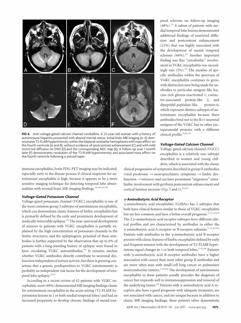
FIG 6. Anti-voltage-gated calcium channel cerebellitis. A 23-year-old woman with a history ofautoimmune hepatitis presented with altered mental status. Initial brain MR imaging (A–E) dem-onstrates T2-FLAIR hyperintensity within the bilateral cerebellar hemispheres with mass effect onthe fourth ventricle (A and B), without evidence of postcontrast enhancement (C) and with mildrestricted diffusion on DWI (D) and the corresponding ADC map (E). A follow-up scan 1 monthlater (F) demonstrates resolution of the T2-FLAIR hyperintensity and associated mass effect onthe fourth ventricle following a steroid taper.
AJNR Am J Neuroradiol 38:1070 –78 Jun 2017 www.ajnr.org 1075
extensive T2-FLAIR hyperintense lesions outside of the limbic
system.3,15
Alpha-Amino-3-Hydroxy-5-Methyl-4-IsoxazolepropionicAcid ReceptorAlpha-amino-3-hydroxy-5-methyl-4-isoxazolepropionic acid re-
ceptor (AMPAr) encephalitis is an uncommon subtype character-
ized by the subacute onset of purely psychiatric symptoms with
T2-FLAIR hyperintensities isolated to the hippocampus. This
subtype has a higher association with cancer than other cell-sur-
face antibody subtypes and is most often seen in women with
lung, breast, or thymic tumors.3,67
Additional Type II Antibody SubtypesAnti-glutamate receptor 3 (GluR3) antibodies have been associ-
ated with Rasmussen encephalitis.7 Anti-metabotropic glutamate
receptor 1 (mGluR1) antibodies have been described in patients
with lymphoma with cerebellar ataxia.7 Anti-metabotropic gluta-
mate receptor 5 (mGluR5) antibodies have been linked to limbic
encephalitis associated with Hodgkin lymphoma (Ophelia syn-
drome).7 Anti-D2 dopamine receptor antibodies represent a rare
subtype associated with basal ganglia encephalitis.8 Anti-glyoxy-
late reductase 1 (GlyR1) antibodies can be seen in 3 related groups
distinguished by having dominant clinical features of stiff leg syn-
drome, stiff person syndrome, or progressive encephalomyelitis
with rigidity and myoclonus.6
Systemic Autoimmunity with EncephalopathyNeuropsychiatric manifestations of systemic autoimmune
conditions such as systemic lupus erythematosus can occur
and may be mediated by an antibody profile that includes an-
tiphospholipid antibodies and anti-glutamate receptor anti-
bodies.68 Catastrophic antiphospholipid antibody syndrome is
a condition that can present with strokelike symptoms and
multifocal petechial hemorrhages throughout the brain, which
are best seen on susceptibility-weighted MR imaging se-
quences.68 Patients with thyroid dysfunction and antithyroid
antibodies in conditions like Graves disease or Hashimoto thy-
roiditis can develop encephalopathy associated with their au-
toimmune thyroid disease that has a characteristic “migratory
pattern” of neuroimaging findings with cortical T2-FLAIR le-
sions in different regions of the brain on sequential MR imag-
ing examinations (Fig 7).11,69 Hashimoto encephalopathy, in
particular, is closely associated with autoimmune encephalitis,
given its propensity for the combination of encephalopathy,
psychiatric symptoms, and seizures.69,70 MR imaging findings
in Hashimoto encephalopathy often have more prominent fea-
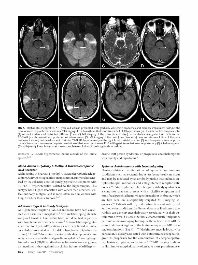
FIG 7. Hashimoto encephalitis. A 41-year-old woman presented with gradually worsening headaches and memory impairment without thedevelopment of psychosis or seizures. MR imaging of the brain (time, 0) demonstrates T2-FLAIR hyperintensity in the inferior left temporal lobe(A) without evidence of restricted diffusion (B and C). MR imaging of the brain (time, 21 days) demonstrates enlargement of the lesion onT2-FLAIR (not shown) without postcontrast enhancement (D). MR imaging of the brain (time, 3 months) demonstrates resolution of the priorlesion (not shown) but development of similar T2-FLAIR hyperintensity in the right frontoparietal junction (E). A subsequent scan at approxi-mately 5 months shows near-complete resolution of that lesion with a new T2-FLAIR hyperintense lesion more posteriorly (F). A follow-up scan(G and H) nearly 1 year from onset shows complete resolution of the imaging abnormalities.
1076 Kelley Jun 2017 www.ajnr.org

tures of leukoencephalopathy (patchy and confluent T2-
FLAIR hyperintense lesions in the subcortical, periventricular,
and deep white matter).2,43,70
CONCLUSIONSAutoimmune encephalitis is an important diagnostic consider-
ation in patients presenting with new onset of altered mental sta-
tus of unclear etiology. It includes a myriad of clinical conditions
that have a common pathophysiology (ie, antibodies directed
against CNS structures). The 2 distinct groups (group I, intracel-
lular directed antibodies, and group II, cell-surface directed anti-
bodies) have overlapping clinical and imaging features. Neuroim-
aging findings will most often involve the limbic structures, but
involvement of the striatum, diencephalon, or rhombencephalon
can be seen. A subset of patients with autoimmune encephalitis
will have no neuroimaging findings despite profound neuropsy-
chiatric dysfunction, but serum antibody testing can still ulti-
mately lead to the diagnosis of autoimmune encephalitis. While
there is no single diagnostic feature that can make this diagnosis in
isolation, recognizing a certain constellation of findings during
the work-up of complex and atypical cases of new-onset altered
mental status is crucial to confirm the diagnosis with serologic
testing and initiate treatment in a timely fashion.
ACKNOWLEDGMENTSWe are grateful to our clinical colleagues in the Department of
Neurology within the Henry Ford Health System for their role in
diagnosing and treating patients with autoimmune encephalitis at
our institution. We would specifically like to acknowledge Dr
Daniel Newman, Director of the Henry Ford Hoenselaar ALS
Clinic, for his enthusiastic clinical support and expertise. We
would also like to thank Dr Chunhai Hao, Chief of Neuropathol-
ogy in the Department of Pathology and Laboratory Medicine at
Henry Ford Hospital, for his work characterizing the histopathol-
ogy of brain biopsy specimens in cases of suspected autoimmune
encephalitis at our institution.
REFERENCES1. Dalmau J, Rosenfeld MR. Autoimmune encephalitis update. Neuro
Oncol 2014;16:771–78 CrossRef Medline2. Caselli RJ, Drazkowski JF, Wingerchuk DM. Autoimmune encepha-
lopathy. Mayo Clin Proc 2010;85:878 – 80 CrossRef Medline3. da Rocha AJ, Nunes RH, Maia AC Jr, et al. Recognizing autoim-
mune-mediated encephalitis in the differential diagnosis of limbicdisorders. AJNR Am J Neuroradiol 2015;36:2196 –205 CrossRefMedline
4. Glaser CA, Gilliam S, Schnurr D, et al; California Encephalitis Project,1998 –2000. In search of encephalitis etiologies: diagnostic chal-lenges in the California Encephalitis Project, 1998 –2000. Clin InfectDis 2003;36:731– 42 CrossRef Medline
5. Armangue T, Leypoldt F, Dalmau J. Autoimmune encephalitis asdifferential diagnosis of infectious encephalitis. Curr Opin Neurol2014;27:361– 68 CrossRef Medline
6. Vincent A. Autoimmune channelopathies: new antibody-mediateddisorders of the central nervous system. F1000 Biol Rep 2009;1:61CrossRef Medline
7. Graus F, Saiz A, Dalmau J. Antibodies and neuronal autoimmunedisorders of the CNS. J Neurol 2010;257:509 –17 CrossRef Medline
8. Ramanathan S, Mohammad SS, Brilot F, et al. Autoimmuneencephalitis: recent updates and emerging challenges. J Clin Neuro-sci 2014;21:722–30 CrossRef Medline
9. Bien CG, Vincent A, Barnett MH, et al. Immunopathology of au-toantibody-associated encephalitides: clues for pathogenesis. Brain2012;135:1622–38 CrossRef Medline
10. Demaerel P, Van Dessel W, Van Paesschen W, et al. Autoimmune-mediated encephalitis. Neuroradiology 2011;53:837–51 CrossRefMedline
11. Moscato EH, Jain A, Peng X, et al. Mechanisms underlying autoim-mune synaptic encephalitis leading to disorders of memory, behav-ior and cognition: insights from molecular, cellular and synapticstudies. Eur J Neurosci 2010;32:298 –309 CrossRef Medline
12. Oyanguren B, Sanchez V, Gonzalez FJ, et al. Limbic encephalitis: aclinical-radiological comparison between herpetic and autoim-mune etiologies. Eur J Neurol 2013;20:1566 –70 CrossRef Medline
13. Dalmau J, Graus F, Villarejo A, et al. Clinical analysis of anti-Ma2-associated encephalitis. Brain 2004;127:1831– 44 CrossRef Medline
14. Irani SR, Bera K, Waters P, et al. N-methyl-D-aspartate antibodyencephalitis: temporal progression of clinical and paraclinical ob-servations in a predominantly non-paraneoplastic disorder of bothsexes. Brain 2010;133:1655– 67 CrossRef Medline
15. Petit-Pedrol M, Armangue T, Peng X, et al. Encephalitis with refrac-tory seizures, status epilepticus, and antibodies to the GABAAreceptor: a case series, characterisation of the antigen, and analysisof the effects of antibodies. Lancet Neurol 2014;13:276 – 86 CrossRefMedline
16. Degnan AJ, Levy LM. Neuroimaging of rapidly progressive demen-tias, part 1: neurodegenerative etiologies. AJNR Am J Neuroradiol2014;35:418 –23 CrossRef Medline
17. Degnan AJ, Levy LM. Neuroimaging of rapidly progressive demen-tias, part 2: prion, inflammatory, neoplastic, and other etiologies.AJNR Am J Neuroradiol 2014;35:424 –31 CrossRef Medline
18. Mittal MK, Rabinstein AA, Hocker SE, et al. Autoimmune encepha-litis in the ICU: analysis of phenotypes, serologic findings, and out-comes. Neurocrit Care 2016;24:240 –50 CrossRef Medline
19. Gultekin SH, Rosenfeld MR, Voltz R, et al. Paraneoplastic limbicencephalitis: neurological symptoms, immunological findings andtumour association in 50 patients. Brain 2000;123(pt 7):1481–94CrossRef Medline
20. Dalmau J, Lancaster E, Martinez-Hernandez E, et al. Clinical experi-ence and laboratory investigations in patients with anti-NMDARencephalitis. Lancet Neurol 2011;10:63–74 CrossRef Medline
21. Rees JH. Paraneoplastic syndromes: when to suspect, how to con-firm, and how to manage. J Neurol Neurosurg Psychiatry 2004;75(suppl 2):ii43–50 Medline
22. de Beukelaar JW, Sillevis Smitt PA. Managing paraneoplastic neuro-logical disorders. Oncologist 2006;11:292–305 CrossRef Medline
23. Urbach H, Soeder BM, Jeub M, et al. Serial MRI of limbic encepha-litis. Neuroradiology 2006;48:380 – 86 CrossRef Medline
24. Kotsenas AL, Watson RE, Pittock SJ, et al. MRI findings in autoim-mune voltage-gated potassium channel complex encephalitis withseizures: one potential etiology for mesial temporal sclerosis. AJNRAm J Neuroradiol 2014;35:84 – 89 CrossRef Medline
25. Corsellis JA, Goldberg GJ, Norton AR. “Limbic encephalitis” and itsassociation with carcinoma. Brain 1968;91:481–96 CrossRef Medline
26. Kohler J, Hufschmidt A, Hermle L, et al. Limbic encephalitis: twocases. J Neuroimmunol 1988;20:177–78 CrossRef Medline
27. Tuzun E, Dalmau J. Limbic encephalitis and variants: classification,diagnosis and treatment. Neurologist 2007;13:261–71 CrossRef Medline
28. Suleiman J, Brilot F, Lang B, et al. Autoimmune epilepsy in children:case series and proposed guidelines for identification. Epilepsia2013;54:1036 – 45 CrossRef Medline
29. Dalmau J, Bataller L. Clinical and immunological diversity of limbicencephalitis: a model for paraneoplastic neurologic disorders. He-matol Oncol Clin North Am 2006;20:1319 –35 CrossRef Medline
30. Pittock SJ, Kryzer TJ, Lennon VA. Paraneoplastic antibodies coexistand predict cancer, not neurological syndrome. Ann Neurol 2004;56:715–19 CrossRef Medline
31. Graus F, Keime-Guibert F, Rene R, et al. Anti-Hu-associated para-
AJNR Am J Neuroradiol 38:1070 –78 Jun 2017 www.ajnr.org 1077

neoplastic encephalomyelitis: analysis of 200 patients. Brain 2001;124:1138 – 48 CrossRef Medline
32. Rudzinski LA, Pittock SJ, McKeon A, et al. Extratemporal EEG andMRI findings in ANNA-1 (anti-Hu) encephalitis. Epilepsy Res 2011;95:255– 62 CrossRef Medline
33. Jacobs DA, Fung KM, Cook NM, et al. Complex partial status epi-lepticus associated with anti-Hu paraneoplastic syndrome. J NeurolSci 2003;213:77– 82 CrossRef Medline
34. Rosenfeld MR, Eichen JG, Wade DF, et al. Molecular and clinicaldiversity in paraneoplastic immunity to Ma proteins. Ann Neurol2001;50:339 – 48 CrossRef Medline
35. Hoffmann LA, Jarius S, Pellkofer HL, et al. Anti-Ma and anti-Taassociated paraneoplastic neurological syndromes: 22 newly diag-nosed patients and review of previous cases. J Neurol Neurosurg Psy-chiatry 2008;79:767–73 CrossRef Medline
36. Mathew RM, Vandenberghe R, Garcia-Merino A, et al. Orchiectomyfor suspected microscopic tumor in patients with anti-Ma2-associ-ated encephalitis. Neurology 2007;68:900 – 05 CrossRef Medline
37. Crespo-Burillo JA, Hernando-Quintana N, Ruiz-Palomino P, et al.Chorea secondary to striatal encephalitis due to anti-CV2/CRMP5antibodies: case description and review of the literature. Neurologia(Barcelona, Spain) 2015;30:451–53 CrossRef Medline
38. Malter MP, Helmstaedter C, Urbach H, et al. Antibodies to glutamicacid decarboxylase define a form of limbic encephalitis. Ann Neurol2010;67:470 –78 CrossRef Medline
39. Ko MW, Dalmau J, Galetta SL. Neuro-ophthalmologic manifesta-tions of paraneoplastic syndromes. J Neuroophthalmol 2008;28:58 – 68 CrossRef Medline
40. Younes-Mhenni S, Janier MF, Cinotti L, et al. FDG-PET improvestumour detection in patients with paraneoplastic neurological syn-dromes. Brain 2004;127:2331–38 CrossRef Medline
41. Nunez-Enamorado N, Camacho-Salas A, Belda-Hofheinz S, et al.Fast and spectacular clinical response to plasmapheresis in a paedi-atric case of anti-NMDA encephalitis [in Spanish]. Rev Neurol 2012;54:420 –24 Medline
42. Gresa-Arribas N, Titulaer MJ, Torrents A, et al. Antibody titresat diagnosis and during follow-up of anti-NMDA receptorencephalitis: a retrospective study. Lancet Neurol 2014;13:167–77CrossRef Medline
43. Saraya A, Mahavihakanont A, Shuangshoti S, et al. Autoimmunecauses of encephalitis syndrome in Thailand: prospective study of103 patients. BMC Neurol 2013;13:150 CrossRef Medline
44. Ances BM, Vitaliani R, Taylor RA, et al. Treatment-responsive lim-bic encephalitis identified by neuropil antibodies: MRI and PETcorrelates. Brain 2005;128:1764 –77 CrossRef Medline
45. von Rhein B, Wagner J, Widman G, et al. Suspected antibody nega-tive autoimmune limbic encephalitis: outcome of immunotherapy.Acta Neurol Scand 2017;135:134 – 41 Medline
46. Matoq AA, Rappoport AS, Yang Y, et al. Anti-NMDA-receptor anti-body encephalitis in infants. Epilepsy Behav Case Rep 2015;4:99 –101CrossRef Medline
47. Bravo-Oro A, Acosta-Yebra D, Grimaldo-Zapata IP, et al. Reversiblecortical atrophy secondary to anti-NMDA receptor antibody en-cephalitis [in Spanish]. Rev Neurol 2015;60:447–52 Medline
48. Kayser MS, Titulaer MJ, Gresa-Arribas N, et al. Frequency and char-acteristics of isolated psychiatric episodes in anti-N-methyl-d-aspartate receptor encephalitis. JAMA Neurol 2013;70:1133–39CrossRef Medline
49. Irani SR, Vincent A. NMDA receptor antibody encephalitis. CurrNeurol Neurosci Rep 2011;11:298 –304 CrossRef Medline
50. Gabilondo I, Saiz A, Galan L, et al. Analysis of relapses in anti-NMDAR encephalitis. Neurology 2011;77:996 –99 CrossRef Medline
51. Titulaer MJ, McCracken L, Gabilondo I, et al. Treatment and prog-nostic factors for long-term outcome in patients with anti-NMDAreceptor encephalitis: an observational cohort study. Lancet Neurol2013;12:157– 65 CrossRef Medline
52. Tuzun E, Zhou L, Baehring JM, et al. Evidence for antibody-medi-ated pathogenesis in anti-NMDAR encephalitis associated with
ovarian teratoma. Acta Neuropathol 2009;118:737– 43 CrossRefMedline
53. Florance NR, Davis RL, Lam C, et al. Anti-N-methyl-D-aspartatereceptor (NMDAR) encephalitis in children and adolescents. AnnNeurol 2009;66:11–18 CrossRef Medline
54. Gable MS, Sheriff H, Dalmau J, et al. The frequency of autoimmuneN-methyl-D-aspartate receptor encephalitis surpasses that of indi-vidual viral etiologies in young individuals enrolled in the Califor-nia Encephalitis Project. Clin Infect Dis 2012;54:899 –904 CrossRefMedline
55. Pruss H, Finke C, Holtje M, et al. N-methyl-D-aspartate receptorantibodies in herpes simplex encephalitis. Ann Neurol 2012;72:902–11 CrossRef Medline
56. Takahashi Y, Mori H, Mishina M, et al. Autoantibodies and cell-mediated autoimmunity to NMDA-type GluRepsilon2 in patientswith Rasmussen’s encephalitis and chronic progressive epilepsiapartialis continua. Epilepsia 2005;46(suppl 5):152–58 Medline
57. Gitiaux C, Simonnet H, Eisermann M, et al. Early electro-clinicalfeatures may contribute to diagnosis of the anti-NMDA receptorencephalitis in children. Clin Neurophysiol 2013;124:2354 – 61CrossRef Medline
58. Baumgartner A, Rauer S, Mader I, et al. Cerebral FDG-PET and MRIfindings in autoimmune limbic encephalitis: correlation with auto-antibody types. J Neurol 2013;260:2744 –53 CrossRef Medline
59. Majoie HJ, de Baets M, Renier W, et al. Antibodies to voltage-gatedpotassium and calcium channels in epilepsy. Epilepsy Res 2006;71:135– 41 Medline
60. Boronat A, Gelfand JM, Gresa-Arribas N, et al. Encephalitis and an-tibodies to dipeptidyl-peptidase-like protein-6, a subunit of Kv4.2potassium channels. Ann Neurol 2013;73:120 –28 CrossRef Medline
61. Lai M, Huijbers MG, Lancaster E, et al. Investigation of LGI1 as theantigen in limbic encephalitis previously attributed to potassiumchannels: a case series. Lancet Neurol 2010;9:776 – 85 CrossRefMedline
62. Irani SR, Alexander S, Waters P, et al. Antibodies to Kv1 potassiumchannel-complex proteins leucine-rich, glioma inactivated 1 pro-tein and contactin-associated protein-2 in limbic encephalitis,Morvan’s syndrome and acquired neuromyotonia. Brain 2010;133:2734 – 48 CrossRef Medline
63. Lancaster E, Huijbers MG, Bar V, et al. Investigations of caspr2, anautoantigen of encephalitis and neuromyotonia. Ann Neurol 2011;69:303–11 CrossRef Medline
64. Finkel L, Koh S. N-type calcium channel antibody-mediated auto-immune encephalitis: an unlikely cause of a common presentation.Epilepsy Behav Case Rep 2013;1:92–96 CrossRef Medline
65. Kaira K, Okamura T, Takahashi H, et al. Small-cell lung cancer withvoltage-gated calcium channel antibody-positive paraneoplasticlimbic encephalitis: a case report. J Med Case Rep 2014;8:119CrossRef Medline
66. Lancaster E, Lai M, Peng X, et al. Antibodies to the GABA(B) recep-tor in limbic encephalitis with seizures: case series and characteri-sation of the antigen. Lancet Neurol 2010;9:67–76 CrossRef Medline
67. Joubert B, Kerschen P, Zekeridou A, et al. Clinical spectrum of en-cephalitis associated with antibodies against the �-amino-3-hy-droxy-5-methyl-4-isoxazolepropionic acid receptor: case seriesand review of the literature. JAMA Neurol 2015;72:1163– 69CrossRef Medline
68. Gerosa M, Poletti B, Pregnolato F, et al. Antiglutamate receptor an-tibodies and cognitive impairment in primary antiphospholipidsyndrome and systemic lupus erythematosus. Front Immunol 2016;7:5 CrossRef Medline
69. Tamagno G, Celik Y, Simo R, et al. Encephalopathy associated withautoimmune thyroid disease in patients with Graves’ disease: clin-ical manifestations, follow-up, and outcomes. BMC Neurol 2010;10:27 CrossRef Medline
70. Rosenbloom MH, Smith S, Akdal G, et al. Immunologically medi-ated dementias. Curr Neurol Neurosci Rep 2009;9:359 – 67 CrossRefMedline
1078 Kelley Jun 2017 www.ajnr.org