Embed Size (px)
Citation preview

111
NOG mice
Introductionand
applications
August 3, 2009Central Institute for Experimental Animals

222
NOG mice
1. History
2. General characteristics
3. Basic characteristics3-1. Body weight3-2. Life span and tumors3-3. Organ weight and histology3-4. Irradiation sensitivity3-5. No B cell leakiness3-6. Clinical chemistry and hematology3-7. Immunological characteristics3-8. Microbiological agents affecting NOG mice
4. Humanized NOG after HSC transfer
5. Applications5-1. Infectious disease model
5-1-1. HIV-1 infection5-1-2. ATL infection5-1-3. EBV infection
5-2. Cancer model5-2-1. Liver metastasis5-2-2. Multiple myeloma5-2-3. Acute myeloid leukemia
5-3. Human tissue or organ model5-3-1. Model with human ovary5-3-2. Model with human liver5-3-3. Model with human endometrium
5-4. Other model5-4-1. GVHD model5-4-2. Efficacy test model for thrombopoietic drugs5-4-3. Tumorigenicity test model for human cell transplantation
6. Collaborative Studies at CIEA
7. Reference list
Edited by Ito M and Suemizu H

333
1. History
A schematic diagram of the development of NOG and the related immunodeficient mice is shown.NOG mice were established based on NOD/Shi-scid mice, one of the SCID congenic strains developedby the Central Institute for Experimental Animals (CIEA). NOD/Shi-scid IL2rgnull (NOG) mice wereestablished by introducing the IL2rgnull gene of IL-2Rgnull mice that were produced by Dr. Sugamura’sgroup in 1996 on NOD/Shi-scid mice by a 10th generation backcross mating in 2000. Our experimentalstudies with human hematopoietic stem cells transferred to NOG mice demonstrated that they wereextremely efficient for humanized mice. The formal names for the NOD-scid IL2rgnull mice areNOD.Cg-PrkdcscidIl2rgtm1Sug. In addition to the development of NOG mice, another type of immunodeficient mice with Rag2null
genes replacing the scid mutation has been developed because these inactive genes cause the samephenotypic T and B cell deficiency in the mice. The NOG mice that we developed were mainly used in Japan for various research studies onxenotransplantation, including human hematopoiesis. BALB/cA and C57BL/6J- Rag2null IL2rγnull micewere sent to the United States and later on to Europe, with the successful development of humanizedmice that used irradiated newborn BALB/c- Rag2null IL2rγnull mice, as reported by Dr. Traggiai et al. in2003.
Strain nomenclature: CB-17-scid, C.BkaIghb-Prkdcscid/IcrJic; NOD-scid, NOD.CB17- Prkdcscid/ShiJic;BALB/cA-Rag2null, C.129S1-Rag2tm1Fwa/AJic; C57/B6J-Rag2null, B6.129S1-Rag2tm1Fwa/JJic; BALB/cA-IL2rγnull, C.129S1-Il2rgtm1Sug/ShiJic; C57/B6J- IL2rγnulll, B6.129S1- Il2rgtm1Sug/Jic; NOG (NOD/Shi-scidIL2rγnull); NOD.Cg-Prkdcscid Il2rgtm1Sug/ShiJic, BALB/cA-Rag2null IL2rgnul, C.Cg-Rag2tm1Fwa
Il2rgtm1Sug/AJic; C57B6J-Rag2null IL2rgnul, B6.Cg-Rag2tm1Fwa Il2rgtm1Sug/JJic; NOD/Shi-Rag2null IL2rgnul,NOD.Cg-Rag2tm1Fwa Il2rgtm1Sug/ShiJic(Curr Top Microbiol Immunol Vol. 324, (2008) p56 , “NOD/Shi-scid IL2rgamma(null) (NOG) mice moreappropriate for humanized mouse models.”, Ito, M., Kobayashi, K. & Nakahata, T., Fig. 1. With kindpermission of Springer Science+Business Media)

4
1. T and B cell deficient2. NK cell deficient3. Reduced macrophage and dendritic cell function4. Complement activity deficient5. No incidence of lymphoma6. Sensitive to irradiation7. Long life span8. Sensitive to microbiological pathogens 9. High engraftment for xenotransplants
3. Basic characteristics
3-1. Body weight
2. General characteristics of NOG mice
The body weight of 10 males and females of three strains of mice was measuredevery week until the age of 20 weeks .
4

555
Thymic lymphoma that often occurred in NOD-scid mice was rarely observed in NOG mice. Arecent publication by Kato C et al. (Lab. Animals, 2009) reported that lymphoma occurred in only 0.7% (16 of 2406) of NOG mice. Therefore NOG mice have a long life span and survive at least one and ahalf years under strict SPF conditions.
(x 4)
NOG
NOD/SCID NOD
NOG
NOD/SCID NOD
(x 40)
Histology of thymus from newborn mice
3-2. Life span and tumors
3-3. Organ weight and histology
Age (weeks) Sex No. of
miceBW(g)
Brain(mg)
Thyroidgland (mg)
Mandibulargland (mg)
Lung(mg)
Thymus(mg)
12 Male 10 24.5± 1.53
450± 32
1.4± 0.9
182± 23
141± 7
4.7± 4.3
Female 10 21.3± 0.53
479± 27
1.2± 0.8
107± 11
148± 10
2.3± 1.2
20 Male 10 28.1± 1.15
478± 25
1.7± 0.7
213± 20
180± 18
2.5± 1.9
Female 10 23.2± 1.2
504± 32
2.0± 0.9
111± 11
165± 11
2.2± 0.9
Age (weeks) Sex Heart
(mg)Liver
(g)Spleen(mg)
Kidney(mg)
Adrenalgland (mg)
Testis(mg)
Ovary(mg)
12 Male 108± 12
1.25± 0.12
20.7± 2.3
384± 25
5.8± 1.2
175± 11
Female 95± 8
0.99± 0.07
23.7± 4.0
279± 13
7.1± 0.5
15.2± 2.1
20 Male 115.9± 38
1.44± 0.17
28.3± 8.9
449± 24
6.9±1.3
192± 12
Female 111± 10
1.11± 0.12
28.4± 10
322± 29
8.4± 1.1
17.7± 2.7

666
Five mice in each group were irradiated with 2 to 4 Gy with using an X-ray device (MBR-1505R,Hitachi Medical Co., Tokyo) at age of 8 weeks.
3-4. Irradiation sensitivity
3-5. No B cell leakiness
Survival rates of NOG mice after irradiation
IgG+M antibody levels in the sera from aged NOG (7-10 months old) and NOD-scid (6-7months old) mice were measured by ELISA. In C.B-17- scid (6-9 months old) mice, serumIgG+M+A antibody levels measured in 1989 were used in this figure.

7
3-6. Clinical chemistry and hematology
AST ALT ALP Ca TG UN Crea TP T-ChoIU/L IU/L IU/L mg/dL mg/dL mg/dL mg/dL g/dL mg/dL
Male N=8 49.38 25.00 133.25 7.49 37.13 25.24 0.32 3.98 61.13±12.24 ±13.79 ±15.6 ±0.44 ±11.90 ±1.53 ±0.02 ±0.16 ±6.13
Female N= 10 67.30 23.90 212.30 7.28 30.40 22.21 0.30 3.65 45.50±12.37 ±5.4 ±18.14 ±0.29 ±17.04 ±2.52 ±0.04 ±0.14 ±5.19
WBC RBC HGB HCT MCV MCH MCHC PLTx10 /μl x10 /μl g/dl % fl pg % x10 /μl
Male N=8 6.50 766.25 12.31 39.34 51.34 16.04 31.31 106.73±1.93 ±28.62 ±0.45 ±1.73 ±0.58 ±0.16 ±0.36 ±5.92
Female N= 10 11.80 773.00 12.72 39.12 50.73 16.47 32.47 83.85±3.79 ±45.12 ±0.59 ±2.26 ±0.39 ±0.26 ±0.50 ±8.89
Blood was collected from retro-orbital venous plexus of mice at 12 weeks of age under slight anesthesiawith Isofurane. Differential diagnosis of blood cells was performed with an automatic blood cell counter(XT-2000i, Sysmex, Osaka).
a. Clinical chemistry
b. Hematology
c. Mouse hematopoietic cells in spleen and bone marrow
% P
ositi
ve%
Pos
itive
Bone marrow (BM) and spleen (APL)were removed from 12-week-old NOGmice. Single cell suspensions prepared inthe usual manner were stained with FITC-or PE-labeled anti-mouse CD45+, CD3+,CD4+, CD8+, CD19+, B220+, CD11b+,CD11c+, Gr-1+ and analyzed withFACSCanto (BD Sciences, CA)
888
NOD/Shi-scid NOD/Shi-scid(+ α-asialo GM-1)
NOD/SCID/β2mnull NOG
NOD/Shi-scid(Non-stained control)
1.7% 25.2% 9.3%
30.1% 2.8%
DX5
Figure 1. NK cell Activity of NOG mice The NK cell activity was determined by a cytotoxicity test using NK sensitive YAC-1 cells as a target cell. Micewere intraperitoneally inoculated with 100 mg of polyinosinic-polycytidylic acid (poly I:C, SIGMA Chemical Co., St.Louis, MO) to stimulate NK cell activity for 48 hrs before assay. Spleen cells were separated from 4 mice of eachstrain of mice, pooled and co-cultured with 51Cr-labeled YAC-1 cells as target cells for 4 hrs at 37C in 5% CO2. Thesupernatants harvested were assayed on a gamma counter. The present specific 51Cr release was calculated using thefollowing formula, where X is the mean experimental release from triplicate wells. Total release (T) was determinedfrom wells with 51Cr labeled YAC-1 cells and 1H HCl, and spontaneous release (S) was determined from wells with51Cr –labeled YAC-1 cells and medium: % specific release = [(X-S)/(T-S)] x 100.(This research was originally published in Blood. Ito, M. et al. NOD/SCID/gamma(c)(null) mouse: an excellentrecipient mouse model for engraftment of human cells. Blood. 2002;100: 3175-3182. © the American Society ofHematology. )
Figure 2. NK cells in NOG mice. Spleen cells from mice were stained with streptavidin-FITC and biotin-labeled anti-pan NK cell antibody. No NKcells were observed in spleen cells from NOG mice and NOD/Shi-scid treated with asialo GM1 antibody to eliminateNK cells.(This research was originally published in Blood. Ito, M. et al. NOD/SCID/gamma(c)(null) mouse: an excellentrecipient mouse model for engraftment of human cells. Blood. 2002; 100: 3175-3182 © the American Society ofHematology. )
3-7. Immunological characteristics
a. Defect of NK cells and NK activity

999
Serum dilution
% 5
1C
r re
lease
c. Defect of hemolytic complement activity in serum
Figure 4. Complement-dependent hemolytic activity Complement-dependent hemolytic activity in sera was determined by measurement of 51Cr released in the supernatantsafter 30 min incubation of mouse sera and 51Cr labeled SRBC/anti SRBC antibody conjugates. Spontaneous release (S) wasdetermined from wells with 51Cr SRBC-Ab conjugate in media. Total release (T) was determined from wells with 51CrSRBC-Ab conjugates and 100 ml 2% SDS. Percent specific release = [(X-S)/(T-S)] x 100.(This research was originally published in Blood. Ito, M. et al. NOD/SCID/gamma(c)(null) mouse: an excellent recipientmouse model for engraftment of human cells. Blood. 2002; 100: 3175-3182. © the American Society of Hematology. )
Figure 3. IL-1 production from bone marrow cells IL-1 production from bone marrow cells stimulated with IFN-γ and LPS was determined. Bone marrow cells were culturedwith 500 U/ml human rM-CSF, with and without 10 U/ml rat rIFN- γ and cultured for 4 days at 37 in 5% CO2. After 4 days,the medium was replaced with fresh medium alone or with medium containing Escherichia coli LPS at 10 mg/ml. After anadditional 24 hr incubation period, the culture supernatants were harvested and assayed for IL-1a levels using ELISA kits.(This research was originally published in Blood. Ito, M. et al. NOD/SCID/gamma(c)(null) mouse: an excellent recipientmouse model for engraftment of human cells. Blood. 2002; 100: 3175-3182. © the American Society of Hematology. )
b. Reduced IL-1 production from macrophages

101010
0.0
5.0
10.0
15.0
20.0
25.0
IFN-g
NOD/Shi-scid
NOD/Shi-scid (+AGM)
CD11c+ depletedNOD/Shi-scid (+AGM)NOD/SCID/b2m null
NOD/SCID/gc null
**
**
**
*
0.0
2.5
5.0
7.5
10.0
12.5
15.0
IL-6
**
****
Figure 5. In vitro cytokine production of L.monocytogenes-stimulated spleen cells from 3strains of mice Spleen cells were separated after injection of 1 ml of 1mg/ml collagenase D solution into the spleen. CD11c+
cells were depleted from spleen cells from NOD/Shi-scidmice treated with anti-asialo GM1 antiserum, using anti-CD11c antibody labeled magnetic beads, by a magneticcell sorter (MACS). The cell suspension was co-culturedwith 107 of heat-killed L. monocytogenes for 8 hrs at37oC. The IFN-γ and IL-6 levels in the supernatants weredetermined using ELISA kits. Asterisk indicates asignificant difference (* vs **: P<0.01).(This research was originally published in Blood. Ito, M.et al. NOD/SCID/gamma(c)(null) mouse: an excellentrecipient mouse model for engraftment of human cells.Blood. 2002; 100: 3175-3182. © the American Society ofHematology. )
d. No production of IFNγ from spleen cells of NOG mice
Figure 6. Expression of the genes associated with immunological responses in NOD, NOD/Shi-scidand NOG mice At 48 hrs after intraperitoneal infection of 1 x 10⁷ Listeria monocytogenes, spleens were removed and the RNA wasextracted. Gene expression was examined by microarray (Toyobo Inc.). The yellow areas express the intensity of geneexpression in the NOD mice. The blue bars express higher intensity, and the grey bars express lower intensity whencompared with those of the NOD/Shi mice.(Curr Top Microbiol Immunol Vol. 324, (2008) p59, “NOD/Shi-scid IL2rgamma(null) (NOG) mice more appropriatefor humanized mouse models.”, Ito, M., Kobayashi, K. & Nakahata, T., Fig. 2. With kind permission of SpringerScience+Business Media) e.

111111
NOG mice may have higher sensitivity against opportunistic pathogens because they are moreimmunodeficient than conventional immunodeficient mice, i. e., nude and SCID mice. Therefore,NOG mice must be maintained in a facility under strict SPF conditions. NOG mice may also be easilyaffected by stress, therefore control of environmental factors in sites is required to assurereproducibility of the results of animal experiments. Therefore, we recommend to maintenance ofNOG mice in a special room not together with other stains of mice. The room should be changed evertyear.
Figure 1. Histopathological diagnosis of bacteremia in NOG mice infected with Pseudomonasaeruginosa. Interstitial pneumontis and suppurative nephritis were found in the lungs and kidneys of affected mice.A. Interstitial pneumonitis (H&E, x 200). B. Suppurative nephritis (H&E, x200). P. aeruginosa was isolated fromblood and lesions in the mice.(Curr Top Microbiol Immunol Vol. 324, (2008) p17, “Basic concept of development and practical application ofanimal models for human diseases. ”, Nomura, T., Tamaoki, N., Takakura, A. & Suemizu, H. , Fig. 7. With kindpermission of Springer Science+Business Media)
A B
Figure 2. Results of necropsy of diarrheal NOG mouse that died with unknown causes. Severediarrhea, bile congestion, duodenitis, and intestinal hypertrophy were observed in the mouse.(Curr Top Microbiol Immunol Vol. 324, (2008) p19, “Basic concept of development and practical application ofanimal models for human diseases. ”, Nomura, T., Tamaoki, N., Takakura, A. & Suemizu, H. , Fig. 8. With kindpermission of Springer Science+Business Media)
3-8. Microbiological agents affecting NOG mice

121212
4. Humanized NOG after HSC transfer
4-1. Human cells developed in humanized NOG mice after transfer of human cordblood CD34+ hematopoietic stem cells (HSCs).
8-12 week old NOG miceWith irradiation of 2.5 Gy
Intravenous transfer of 1 -0.5 x 10⁵ hCD34+ cells
Newborn NOG mice with irradiation of 1 Gy
Intrahepatic orintravenous transfer of1-0.1 x 10⁵ hCD34+ cells
Humanized NOG
8 - 20 weeks after cell transfer
General protocol of human HSC transfer
Generally, human cord blood derived CD34+ cells were used as a source of hematopoietic stem cells.CD34+ cells from bone marrow and peripheral blood can be also used although their reconstitutioncapacity is relatively lower than those from cord blood.
Umbilical cord blood (CB) cells are collected during normal full-term deliveries after obtaininginformed consent. Mononuclear cells (MNC) are separated by Ficoll-Hypaque density gradientcentrifugation after depletion of phagocytes with Silica (Immuno Biological Laboratories, Fujioka,Japan). MNC separated from CB are suspended at 4 x 10⁷ cells/ml in phosphate buffered saline (PBS)containing 2% bovine serum albumin (BSA), 0.6% citrate, and 100 IU/ml of penicillin andstreptomycin. The MNC suspension is incubated at 4oC for 30 min with Dynabeads M-450 CD34(Dynal AS, Oslo, Norway) with a bead-to-cell ratio of 1:1. The beads with attached cells are collectedusing a Magnetic Particle Concentrator (MPC, Dynal) and incubated with DETACHaBEAD CD34(Dynal) at 37oC for 15 min to release the cells which are collected by MPC. The purity is evaluatedby flow cytometric analysis. Approximately 95% of the cells were CD34 positive. Commerciallyavailable CB CD34+ cells can be also used for reconstitution of human cells.
Purification of CD34+ cells from human cord blood

131313
Transplantation protocol of human HSC into NOG mice
Mice:For adult mice, 10 -12 week-old NOG mice are usually used. For newborn mice, mice at 1-2days after birth are used.
Irradiation:Irradiation with 2 - 2.5 Gy of adult mice and with 1 Gy of newborns is performed under SPFconditions one day before cell transfer. Mice weighting less than 18 g will sometimes die at thisdose of irradiation.
Cell preparation1. When CD34+ cells (e.g. 1 x 10⁶/tube) are frozen, thaw the cells in a 37°C water bath.2. Transfer cells into a 50 mL conical tube and add 1 mL of PBS containing 2% fetal
bovine serum (2%FBS-PBS) drop by drop, shaking the tube slowly.3. After further addition of 18 mL of 2% FBS-PBS, centrifuge at 1,200 rpm for 5 min at
room temperature.4. Resuspend the cells in 10 mL of 2% FBS-PBS and re-centrifuge.5. Resuspend in 2 mL of PBS.6. Using 15 µL of the suspension, count the cells and determine viability after adding 15
µL of 0.25% trypan-blue solution. Add 3 mL of PBS to adjust to a total of 5 mL.7. Transfer 1mL of the cell suspension into each 1.5 mL cryotube.8. Place the tubes in a 50 mL conical tube and take it to the animal facility.9. After dipping the tube in an antiseptic solution, take it into the animal room.
Cell transfer For adult mice
1. Inject 0.25 mL (1 - 0.5 x 10⁴) of the cell suspension into the mice via the tail vein with a1 mL syringe with a 27 G needle or a microinjector syringe with a 29 G needle underslight anesthesia with isoflurane.
For newborn mice1. Slightly anesthetize the newborn mice with isoflurane.2. Hold the newborn mice by the left hand with the mouse head facing the right.3. Inject 0.1 mL (1-5 x 10⁴ ) of the cell suspension via the face vein with a microinjector
syringe with a 29 G needle.4. Return the newborn mice to the mother gently, being careful not to apply any force.

141414
4-2. High engraftment rate of human cells in NOGmice
Table 1. Limiting dilution assay in NOG mice.
CD34+ cell dose No. of micetransplanted
No. of mice successfullyengrafted*
1,000 5 5
200 3 2
100 6 3
* Successful engraftment was defined as the presence of at least0.1% human CD45+ cells in bone marrow by flow cytometry.
At 8 - 20 weeks after intravenous transplantation of 5 x 10⁴ human cord blood derived CD34+ cells into NOG andNOD-scid mice, human CD45+ hematopoietic cells were detected in peripheral blood of mice by flow cytometry.A high engraftment rate of human cells was observed in NOG mice.
In order to evaluate the frequency of CB CD34+ cells capable of engrafting in NOG mice, these mice weretransplanted with CB CD34+ cells in a limiting dose. All recipient mice transplanted with 1 x 10³ CB CD34+ cellsshowed successful engraftment (>0.1%). Further, as few as 100 cells could be engrafted in 2 of 6 mice and theirmulti-lineage differentiation.
(This research was originally published in Blood. Ito, M. et al.NOD/SCID/gamma(c)(null) mouse: an excellent recipient mouse model forengraftment of human cells. Blood. 2002; 100: 3175-3182. © the AmericanSociety of Hematology. )

151515
4-3. Multi-lineage cell differentiation from HSCs in NOG mice
Figure 1. The rate of human CD45+ cellsin bone marrow of transplanted mice andthe high efficacy of multi-lineage celldifferentiation At 11 weeks after CD34+ cell transplantation,bone marrow was removed fromNOD/SCID/gc
null and NOD/Shi-scid mice treatedwith anti-asialo GM1 antibody two days beforetransplantation, and subjected to flow cytometry.NOD/SCID/gc
null showed significantly higherpercentage of CD45+ cells. Multi-lineage cellshave been differentiated from CD34+ cells withhigh efficacy (* vs **: P<0.01).(This research was originally published in Blood.Ito, M. et al. NOD/SCID/gamma(c)(null) mouse:an excellent recipient mouse model forengraftment of human cells. Blood. 2002; 100:3175-3182. © the American Society ofHematology. )
1. Ito, M., H. Hiramatsu, K. Kobayashi, K. Suzue, M. Kawahata, K. Hioki, Y. Ueyama, Y.Koyanagi, K. Sugamura, K. Tsuji, T. Heike, and T. Nakahata. 2002. NOD/SCID/gamma(c)(null)mouse: an excellent recipient mouse model for engraftment of human cells. Blood 100:3175-3182.
2. Yahata, T., K. Ando, Y. Nakamura, Y. Ueyama, K. Shimamura, N. Tamaoki, S. Kato, and T.Hotta. 2002. Functional human T lymphocyte development from cord blood CD34+ cells innonobese diabetic/Shi-scid, IL-2 receptor gamma null mice. J Immunol 169:204-209.
3. Saito, Y., Y. Kametani, K. Hozumi, N. Mochida, K. Ando, M. Ito, T. Nomura, Y. Tokuda, H.Makuuchi, T. Tajima, and S. Habu. 2002. The in vivo development of human T cells fromCD34(+) cells in the murine thymic environment. Int Immunol 14:1113-1124.
4. Hiramatsu, H., R. Nishikomori, T. Heike, M. Ito, K. Kobayashi, K. Katamura, and T. Nakahata.2003. Complete reconstitution of human lymphocytes from cord blood CD34+ cells using theNOD/SCID/gammacnull mice model. Blood 102:873-880.
5. Watanabe, Y., T. Takahashi, A. Okajima, M. Shiokawa, N. Ishii, I. Katano, R. Ito, M. Ito, M.Minegishi, N. Minegishi, S. Tsuchiya, and K. Sugamura. 2009. The analysis of the functions ofhuman B and T cells in humanized NOD/shi-scid/{gamma}cnull (NOG) mice (hu-HSC NOGmice). Int Immunol. 21:843-858

161616
4-3. Multi-lineage cell differentiation from HSCs in NOG mice
The above figure shows human cells in mouse peripheral blood of 20 weeks after intravenous humanHSC cell transfer. Human T and B cells were developed in NOG mice. CD4+ and CD8+ T cells werealso differentiated in NOG mice.
CD8
CD4
CD19
CD3
In development of human cells from HSCs in NOG mice, B cells develop of 4-8 weeks after transferprior to T cells. T cells can be detected of 8-12 weeks after transfer and later become dominant in theperiphery including peripheral blood and spleen.
A 2 months B
31.5%
29.3%
6.7%
81.9%
10.3%
67.7%
13.1%
52.3%
8.4%
26.1%
15.5%
43.9%
69.1%
18.8%
Human CD45
Mou
se C
D45
3.6%
14.9%
22.2%
5.1%
15.0%
3.3%
41.7%
0.7%
17.6%
0.9%
Human CD3
Hum
an C
D19
blood spleen
C 7 months4 months
blood
blood
bloodblood
blood spleen spleen
spleen spleen spleen
B cells develop prior to T cells.
Development of CD4+ and CD8+ T cells
(This research was originally published in Blood. Watanabe, S. et al. Hematopoietic stem cell-engraftedNOD/SCID/IL2Rgamma null mice develop human lymphoid systems and induce long-lasting HIV-1 infection withspecific humoral immune responses. Blood. 2007; 109: 212-218. © the American Society of Hematology. )

171717
Development of human platelets in peripheral blood and megakaryocytes in bone marrow ofNOG mice
M
HH H
MMM
M
M
HH
H
A. Bone marrow
About 1.5%
Human platelets
B. Peripheral blood
A: Human (h) CD42b+ human megakaryocytes(H),hCD42b- mouse megakaryocytes (M)
B: hCD41+ human platelets Courtesy of Dr. Y. Miyakawa, Keio Univ.
Development of human monocytes/macrophages and dendritic cells
0.6%
61.5%
1.1%
63.6%
9.0%
72.3%
blood spleen BMA. Human CD14+ monocytes
B. Human CD68+ macrophages in various organs
CD68
Lung
CD68
Lymph node
Human CD45
Hum
an C
D14
(This research was originally published in Blood. Watanabe, S. et al. Hematopoietic stem cell-engraftedNOD/SCID/IL2Rgamma null mice develop human lymphoid systems and induce long-lasting HIV-1 infection withspecific humoral immune responses. Blood. 2007; 109: 212-218. © the American Society of Hematology. )

181818
Development of human mast cells
The transplantation of primitive human cells into sublethally irradiated immunedeficient mice is the well-established in vivo system for the investigation of human hematopoietic stem cell function. Although mast cellsare the progeny of hematopoietic stem cells, human mast cell development in mice that underwent humanhematopoietic stem cell transplantation has not been reported. Here we report on human mast cell developmentafter xenotransplantation of human hematopoietic stem cells into nonobese diabetic severe combinedimmunodefici (NOD/SCID)/ c null (NOG) mice with severe combined immunodeficiency and interleukin 2 (IL-2) receptor -chain allelic mutation. Supported by the murine environment, human mast cell clusters developed inmouse dermis, but they required more time than other forms of human cell reconstitution. In lung and gastrictract, mucosal- type mast cells containing tryptase but lacking chymase located on gastric mucosa and in alveoli,whereas connective tissue-type mast cells containing both tryptase and chymase located on gastric submucosaand around major airways, as ent in the human body. Mast cell development was also observed in lymphnodes,spleen, and peritoneal cavity but not in the peripheral blood. Xenotransplantation of human hematopoieticstem cells into NOG mice can be expected to result in a highly effective model for the investigation of humanmast cell development and function in vivo. (Blood. 2004;103: 860-867)(This research was originally published in Blood. Kambe N, Hiramatsu H, Shimonaka M, et al. Development of both humanconnective tissue-type and mucosal-type mast cells in mice from hematopoietic stem cells with identical distribution pattern tohuman body. Blood. 2004; 103: 860-867. ©the American Society of Hematology. )
Figure 1. Representative flow cytometric analysis of peripheral blood from NOG mice after HSCtransplantation. Four weeks after the transplantation, less than 2% of the cells were human CD45, in which CD33 myeloidcells were predominant, and CD19 B cells and CD56 NK cells were also present. Twelve weeks after the transplantation, morethan 40% cells were replaced by human CD45 cells, among which abundant human CD3 T cells were identified.(This research was originally published in Blood. Kambe N, Hiramatsu H, Shimonaka M, et al. Development of both humanconnective tissue-type and mucosal-type mast cells in mice from hematopoietic stem cells with identical distribution pattern tohuman body. Blood. 2004; 103: 860-867. ©the American Society of Hematology. )

191919
Figure 2. Human mast cell development in the mouse skin. (A) RT-PCR analysis for tryptase and chymase mRNAexpression. The skin of NOG mice 12 weeks after the transplantation of human CD34 cells expressed human mast cell-specifictryptase and chymase mRNA.CB-hCMC indicates cord blood-derived human culturedmast cells. (B-C) Acetone-fixed frozensections of NOG mouse skin 12 weeks (B) and 20 weeks (C) after thetransplantation of human cord blood CD34 cells werestained for human CD45 (red fluorescent with Cy3), mast cells (yellowish green with FITC-avidin), and nuclei (blue withHoechst 33342). Arrows indicate human CD45 mast cells, which are stained orange. Magnification, x 200. (D-F) Human MCspecific chymase cells in themouse skin. Acetone-fixed frozen sections of NOG mouse skin 24 weeks after the transplantationwere stained with antihuman chymase mAb. Human chymase cells proliferated focally in the upper dermis (e), represented bythe bar bellows, whereas in other lesions on the same samples nonstained granulated cells were located in the upper dermis (f).Magnification, x 12.5 (D) and x 200 (E-F). (G) The number of chymase human mast cellsand nonstained granulated mousemast cells in NOG mouse skin 24 weeks after the transplantation. The number of human and mouse mast cells supported bymouse dermis was almost identical. Bar graphs display mean ± SD values from 5 different preparation.(This research was originally published in Blood. Kambe N, Hiramatsu H, Shimonaka M, et al. Development of both humanconnective tissue-type and mucosal-type mast cells in mice from hematopoietic stem cells with identical distribution pattern tohuman body. Blood. 2004; 103: 860-867. ©the American Society of Hematology. )
Figure 3. Human mast cell development in the mouse lung and gastric stomach. (A) RT-PCR analysis for tryptaseand chymase mRNA expression. The lung and gastric stomach of NOG mice after the transplantation of human CD34 cellsexpressed human mast cell-specific tryptase and chymase mRNA. CB-hCMC indicates cord blood-derived human cultured mastcells. (B-C) Histologic findings for lung (B) and gastric stomach (C). Very small numbers of formalin resistant toluidine blue cellsappeared in the lung 20 weeks after the transplantation and, in sequential sections, were almost identical to tryptase cells (arrows).In gastric stomach, formalinresistant toluidine blue cells were identified in both the mucosa and submucosa. In the acetone fixedfrozen thin sections stained with antichymase mAb, chymase cells (white arrowheads) were located only in submucosal lesions.Magnification, x 200 (toluidine blue and tryptase) and x 100 (chymase).(This research was originally published in Blood. Kambe N, Hiramatsu H, Shimonaka M, et al. Development of both humanconnective tissue-type and mucosal-type mast cells in mice from hematopoietic stem cells with identical distribution pattern tohuman body. Blood. 2004; 103: 860-867. ©the American Society of Hematology. )
202020
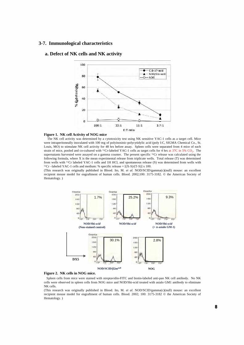
Reconstitution of human lymphoid-like structure in NOG spleen
Bone marrowHLA staining
HEHuman lymphoid follicle like structures (White arrow) were observed in spleen of NOG miceat 18 weeks after human HSC transfer
HLA
Spleen
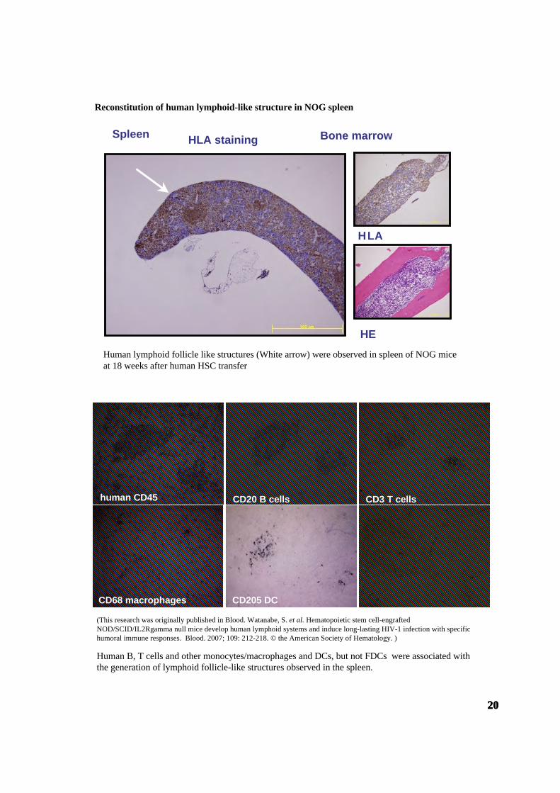
Human B, T cells and other monocytes/macrophages and DCs, but not FDCs were associated withthe generation of lymphoid follicle-like structures observed in the spleen.
FDC-M1 murine FDC
human CD45 CD20 B cells CD3 T cells
CD205 DCCD68 macrophages
(This research was originally published in Blood. Watanabe, S. et al. Hematopoietic stem cell-engraftedNOD/SCID/IL2Rgamma null mice develop human lymphoid systems and induce long-lasting HIV-1 infection with specifichumoral immune responses. Blood. 2007; 109: 212-218. © the American Society of Hematology. )

212121
Human T cells in NOG mice of 4 months after transplantation C
D8
Thymus Spleen Mesenteric LN
CD4(CD3-gate)
CD45RA(CD3-gate)
CD
45R
O
66.4%
19.7%
28.5% 27%
67%
A C
D
B
68.4%
0
5
10
15
20
1 2 3 4 5.1 5.2 5.3 7.1 7.2 8 9 11 12 13.113.213.614 16 17 18 20 21.322 23
%
Vβ
Experiment 1Experiment 2Experiment 3
At 4 to 6 months after transplantation of 2×10⁴ to 5×10⁴ CB CD34+ cells, spleen cells were taken andthe TCR Vß repertoire was analyzed by flow cytometry using a panel of 24 different antibodies. Theresults of three independent experiments are shown.
TCR Vß repertoire analysis of human T cells in spleen.
Courtesy of Dr. K. Sugamura Tohoku University.
(This research was originally published in Blood. Hiramatsu, H., R. Nishikomori, T. Heike, M. Ito, K. Kobayashi, K. Katamura,and T. Nakahata. Complete reconstitution of human lymphocytes from cord blood CD34+ cells using the NOD/SCID/gammacnullmice model. Blood. 2003; 102: 873-880. © the American Society of Hematology. )

222222
Antibody production in NOG mice transferred human HSCs
NOG mice transferred human HSCz can produce IgM and IgG antibodies. However, antigen-specific IgG but notIgM antibodies cannot be produced.
Courtesy of Dr. S. Habu, Tokai University
Activation of human T cells in the spleen of humanized NOG mouse by CD3 stimulation
++++++
IgG4IgG3IgG2IgG1
Non-specific antibodies
Ant
ibod
y co
nc. (
ng/m
l)
WeeksBALB/cA
NOG/hCD34+
Mouse No.
Anti-DNP-KLH antibodies
Dotted lines; human PBMC control
100 101 102 103 104CD154 APC
M130.0
100 101 102 103 104CD25 APC
M154.8
100 101 102 103 104CD69 APC
M168.8
100 101 102 103 104CD154 APC
M123.9
100 101 102 103 104CD25 APC
M142.0
100 101 102 103 104CD69 APC
M159.5
NOG/SPLhCD4T
hPBMChCD4T
CD154 CD25 CD69
coun
t
coun
t
coun
t

232323
Summary of human hematopoietic cells differentiated from human cord bloodderived CD34+ cells in NOG mice
HSC
CLP (Commonlymphoidprogenitor)
CMP (Commonmyeloidprogenitor)
Progenitor cells Mature blood cells
MEP(Megakaryocyte/rythroidprogenitor)
GMP(Granulocyte/ monocyteprogenitor)
Erythrocytes ±
Megakaryocytes (platellet) +
Macrophages +
Monocytes +
Granulocytes (Neutrophils/Eosinophils/Basophils) ±
Mast cells +
B lymphocytes ++
T lymphocytes ++
NK cells +
DCs +

242424
Clonal analysis of short-term and long-term human HSCs
Figure 1. Differentiation ability and self-renewal capacity of individual SRCclones(A) Study design for clonal analysis of STRC andLTRC activity. 34: CD34+ stem/progenitor cells.M: CD33+ myeloid lineage cells. B: CD19+ B-lymphoid lineage cells. T: CD3+ (spleen) orCD4/CD8 DP (thymus) T-lymphoid lineage cells.(B) Relative frequencies of each clone type. Grayareas in each bar represent the clones detected inCD34+ cells, and black areas represent the clonesnot detected in CD34+ cells. A total of 116 cloneswere analyzed (Table S1). Mean ± SD of 4independent experiments are shown. *, P < 0.01.(Reprinted from Stem Cells, 26 (2008) p3232,Yahata T, et al. “Quiescent human hematopoieticstem cells in the bone marrow niches organizehierarchical structure of hematopoiesis.”AlphaMed Press)
Hematopoiesisis a dynamic and strictly regulated process or chestrated by self-renewing hematopoieticstemcells (HSCs) and the supporting microenvironment.However, the exact mechanisms by whichindividual human HSCs sustain hematopoieti chomeostasis remain to be clarified.To understand how thelong-term repopulating cell (LTRC) activity of individual human HSCs and the hematopoietic hierarchyare maintained in the bone marrow (BM) microenvironment, we traced the repopulating dynamics ofindividual human HSC clones using vira lintegration site analysis.Our study presents several lines ofevidence regarding the in vivo dynamics of human hematopoiesis. First, human LTRCs exist edinar arepopulation of CD34 CD38 cells that localized to the stem cell niches and maintained their stem cellactivities while being in a quiescent state. Second, clonally distinctL TRCs controlled hematopoietichomeostasis and created a stem cell pool hierarchy by a symmetric self-renewal division that producedlineage-restricted short-term repopulating cells and long-lasting LTRCs. Third, we demonstrated thatquiescentLTRCclones expanded remarkably to reconstitute the hematopoiesis of the secondary recipient.Finally, we further demonstrated that human mesenchymal stem cells differentiated into key componentsof the niche and maintained LTRC activity by closely interacting with quiescent human LTRCs, resultingin more LTRCs. Taken together, this study provides a novel insight into repopulation dynamics, turnover, hierarchical structure, and the cell cycle status of human HSCs in the recipient BMmicroenvironment. STEM CELLS 2008;26:3228-3236(Reprinted from Stem Cells, 26 (2008) p3232, Yahata T, et al. “Quiescent human hematopoietic stem cells in the bone marrowniches organize hierarchical structure of hematopoiesis.” AlphaMed Press)

252525
Figure 3. Quiescent human LTRCs interacted with niche components in the endosteal region(A) Two human CD34+ cells, a PCNA-negative (arrowhead) and a PCNA-positive (double arrows), are adjacent to each otherand are attached to the endosteum. A large CD34+PCNA-positive cell is found away from endosteum (arrow). (B) A PCNA-negative CD34+ cell (arrowhead) interacts with CD31-expressing murine endothelial cells (arrow) in the endosteal region. (C)Majority of human cells are positive for CD38. A CD38negPCNA-negative EYFP-transduced human cell is attached to theendosteum (arrowhead). (D and E) EGFP-marked HMRCs differentiate into fibroblastic reticular cells that associate withCD31+ vascular cells. PCNA-negative quiescent CD34+ cells (arrowheads) interact with human reticular cells (arrow). (F)HMRCs in the vascular niche express SDF-1 (arrows) and interact with a CD34+ cell (arrowhead). All bars in the figurerepresent10mm.(Reprinted from Stem Cells, 26 (2008) p3233, Yahata T, et al. “Quiescent human hematopoietic stem cells in the bone marrowniches organize hierarchical structure of hematopoiesis.” AlphaMed Press)
Figure 2. Differentiation ability and self-renewal capacity of individual SRCclones(A) Study design for clonal analysis of STRC andLTRC activity. 34: CD34+ stem/progenitor cells.M: CD33+ myeloid lineage cells. B: CD19+ B-lymphoid lineage cells. T: CD3+ (spleen) orCD4/CD8 DP (thymus) T-lymphoid lineage cells.(B) Relative frequencies of each clone type. Grayareas in each bar represent the clones detected inCD34+ cells, and black areas represent the clonesnot detected in CD34+ cells. A total of 116 cloneswere analyzed (Table S1). Mean ± SD of 4independent experiments are shown. *, P < 0.01.(Reprinted from Stem Cells, 26 (2008) p3232,Yahata T, et al. “Quiescent human hematopoieticstem cells in the bone marrow niches organizehierarchical structure of hematopoiesis.”AlphaMed Press)

262626
5. Applications 5-1. Infectious disease model
5-1-1. HIV-1 infection
Figure 1. Surface expression of HIV-1coreceptors on CD4 cells in variousorgans of mice 4 months aftertransplantation. A representative FACS profile of humanCXCR4 and CCR5 on CD4 cells shows theexistence of CXCR4 CD4 and CCR5 CD4 cellsin blood (A), spleen (B), and BM (D), but noCCR5 CD4 cells in the thymus (C). BM resultsshow that many CD4 cells are neither CD3 Tcells nor CD14 monocytes. A gate was set onthe human CD45 population.(This research was originally published inBlood. Watanabe S, Terashima K, Ohta S, et al.Hematopoietic stem cell-engraftedNOD/SCID/IL2Rgamma null mice develophuman lymphoid systems and induce long-lasting HIV-1 infection with specific humoralimmune responses. Blood. 2007;109:215. ©theAmerican Society of Hematology. )
Table 1. Comparison of viral RNA copies in Plasma and HIV-DNA copies in the spleen, BM, and thymusfrom hNOG mice receiving low- and high-dose viral inoculations
Seven mice inoculated with a low infection dose of HIV-1JRCSF (200 TCID50) or HIV-1JRCSF (180 TCID50), and 7 mice receivinga high dose of HIV-1JRCSF (65000 TCID50) or HIV-1JRCSF (20000 TCID50) were listed. ND indicates not done.(This research was originally published in Blood. Watanabe S, Terashima K, Ohta S, et al. Hematopoietic stem cell-engraftedNOD/SCID/IL2Rgamma null mice develop human lymphoid systems and induce long-lasting HIV-1 infection with specifichumoral immune responses. Blood. 2007;109:216. ©the American Society of Hematology. )
A
CD
8
5.7%
blood B
18.8%
spleen
12.6%
C thymus
3.7%
D BM
CD4
4.1%
66.1%
CD14
5.9%
0.1%
5.0%
0.7%
CD35.8%
8.9%
13.7%
1.0%
1.9%
10.5%
2.5%
9.7%
1.1%
4.5%
8.2%
8.2%
78.0%
6.9%
39.9%
45.0%
0.4%
91.1%
25.7%
28.7%
0.7%
22.3%
5.2%
3.7%
CXC
R4
CC
R5
CD
19
CXC
R4
CC
R5
CD14
1.Watanabe, S., S. Ohta, M. Yajima, K. Terashima, M. Ito, H. Mugishima, S. Fujiwara, K. Shimizu,M. Honda, N. Shimizu, and N. Yamamoto. 2007. Humanized NOD/SCID/IL2R{gamma}null MiceTransplanted with Hematopoietic Stem Cells under non-Myeloablative Condition Show ProlongedLifespans and Allow Detailed Analysis of HIV-1 Pathogenesis. J Virol.2.
2. Watanabe, S., K. Terashima, S. Ohta, S. Horibata, M. Yajima, Y. Shiozawa, M. Z. Dewan, Z. Yu,M. Ito, T. Morio, N. Shimizu, M. Honda, and N. Yamamoto. 2007. Hematopoietic stem cell-engrafted NOD/SCID/IL2Rgamma null mice develop human lymphoid systems and induce long-lasting HIV-1 infection with specific humoral immune responses. Blood 109:212-218.

272727
Figure 3. Detection of anti-HIV-1 antibodies from the plasma of HIV-1-infected mice. An ELISA assay was conducted by using plasma from 14 mice inoculated with either HIV-1JRCSF or HIV-1MNp, and from2 uninfected control mice. Representatives (n . 8) of the 14 HIV-1-inoculated mice, and the 2 uninfected mice, are shown in thepanels. Measurements of specific human antibodies for HIV-1IIIB gp120 (A), HIV-1MN gp120 (B), and HIV-1IIIB p24antigens (C) were shown. Results are expressed as the means from triplicate assays in 3 different experiments.(This research was originally published in Blood. Watanabe S, Terashima K, Ohta S, et al. Hematopoietic stem cell-engraftedNOD/SCID/IL2Rgamma null mice develop human lymphoid systems and induce long-lasting HIV-1 infection with specifichumoral immune responses. Blood. 2007;109:216-217. ©the American Society of Hematology. )
Figure 2. The numbers of RNA viral copies in plasma, CD4 /CD8 T-cell ratios in the spleen, and p24detection in the immunohistochemistry of HIV/SHIVinfected mice. (A) Viral copy numbers of 8 mice inoculated with a high infectious dose of HIV-1JRCSF (65 000 TCID50) and killed on days 33and 43 after inoculation. (B) Viral copy numbers of 8 mice inoculated with a high infectious dose of SHIV-C2/1 (50 000TCID50) and killed on days 18 and 42 after inoculation. Note that all the mice showed high levels of viremia that lasted morethan 40 days after inoculation. (C) CD4/CD8 cell ratios in the spleens of 16 infected mice and 9 uninfected control mice.Control mice were not inoculated with HIV/SHIV and were killed on days 105 to 166 after stem cell transplantation. There wasno significant rapid loss of CD4 cells in HIV-1JRCSF-infected mice, while a decline of the CD4/CD8 ratio was detected inSHIV-C2/1-infected mice on day 42 after infection compared with uninfected control mice (*P <.05). The short bars indicatethe means of each group. (D) P24. cells are clearly observed in thespleen, LNs, and lungs. Arrow indicates p24 positive formacrophage-like cells. Original magnification, x 100.
B HIV-1MN gp120
0.2
0.4
0.6
0.8
0 1/20 1/60 1/180
serum dilution
A
O.D
. 405
nm
0.5
1.0
1.5
2.0
2.5
3.0
1/20 1/60 1/1800
HIV-1IIIB gp120 C HIV-1IIIB p24
1/20 1/60 1/1800
0.4
0.8
1.2
1.61.0
uninfected control #1 uninfected control #2#136-2 HIV-1JRCSF-infected #136-3 HIV-1JRCSF-infected
#112-4 HIV-1MNp-infected #113-4 HIV-1MNp-infected#141-1 HIV-1JRCSF-infected #161-3 HIV-1JRCSF-infected
#157-3 HIV-1MNp-infected #157-4 HIV-1MNp-infected
100101102
103104105106
day43day33
Cop
y nu
mbe
rs /
ml
day18 day42
HIV-1JRCSFA B SHIV-C2/1
C
D
spleen
LN
lung
HIV-1JRCSF SHIV-C2/1
CD
4/C
D8
ratio
0
0.5
1.0
1.52.0
2.5
3.0
control day18 day42day43day33
*
282828
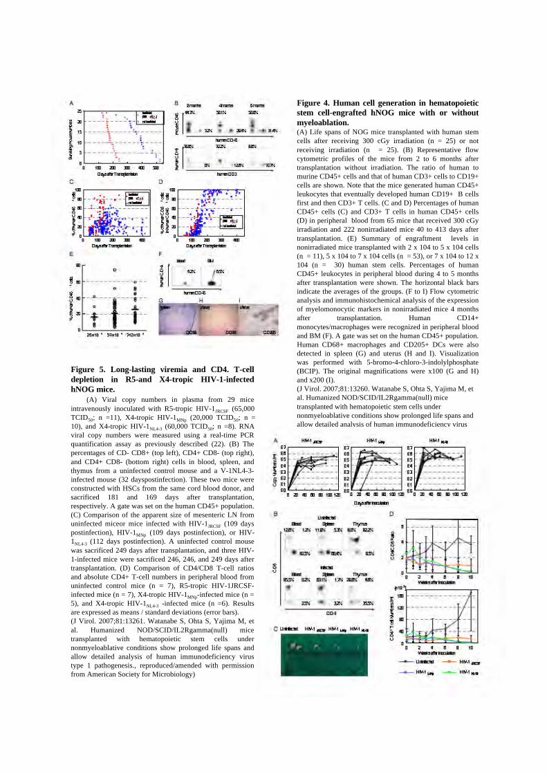
Figure 4. Human cell generation in hematopoieticstem cell-engrafted hNOG mice with or withoutmyeloablation.(A) Life spans of NOG mice transplanted with human stemcells after receiving 300 cGy irradiation (n = 25) or notreceiving irradiation (n = 25). (B) Representative flowcytometric profiles of the mice from 2 to 6 months aftertransplantation without irradiation. The ratio of human tomurine CD45+ cells and that of human CD3+ cells to CD19+cells are shown. Note that the mice generated human CD45+leukocytes that eventually developed human CD19+ B cellsfirst and then CD3+ T cells. (C and D) Percentages of humanCD45+ cells (C) and CD3+ T cells in human CD45+ cells(D) in peripheral blood from 65 mice that received 300 cGyirradiation and 222 nonirradiated mice 40 to 413 days aftertransplantation. (E) Summary of engraftment levels innonirradiated mice transplanted with 2 x 10⁴ to 5 x 10⁴ cells(n = 11), 5 x 10⁴ to 7 x 10⁴ cells (n = 53), or 7 x 10⁴ to 12 x104 (n = 30) human stem cells. Percentages of humanCD45+ leukocytes in peripheral blood during 4 to 5 monthsafter transplantation were shown. The horizontal black barsindicate the averages of the groups. (F to I) Flow cytometricanalysis and immunohistochemical analysis of the expressionof myelomonocytic markers in nonirradiated mice 4 monthsafter transplantation. Human CD14+monocytes/macrophages were recognized in peripheral bloodand BM (F). A gate was set on the human CD45+ population.Human CD68+ macrophages and CD205+ DCs were alsodetected in spleen (G) and uterus (H and I). Visualizationwas performed with 5-bromo-4-chloro-3-indolylphosphate(BCIP). The original magnifications were x100 (G and H)and x200 (I).(J Virol. 2007;81:13260. Watanabe S, Ohta S, Yajima M, etal. Humanized NOD/SCID/IL2Rgamma(null) micetransplanted with hematopoietic stem cells undernonmyeloablative conditions show prolonged life spans andallow detailed analysis of human immunodeficiency virustype 1 pathogenesis., reproduced/amended with permissionfrom American Society for Microbiology)
Figure 5. Long-lasting viremia and CD4. T-celldepletion in R5-and X4-tropic HIV-1-infectedhNOG mice. (A) Viral copy numbers in plasma from 29 miceintravenously inoculated with R5-tropic HIV-1JRCSF (65,000TCID50; n =11), X4-tropic HIV-1MNp (20,000 TCID50; n =10), and X4-tropic HIV-1NL4-3 (60,000 TCID50; n =8). RNAviral copy numbers were measured using a real-time PCRquantification assay as previously described (22). (B) Thepercentages of CD- CD8+ (top left), CD4+ CD8- (top right),and CD4+ CD8- (bottom right) cells in blood, spleen, andthymus from a uninfected control mouse and a V-1NL4-3-infected mouse (32 dayspostinfection). These two mice wereconstructed with HSCs from the same cord blood donor, andsacrificed 181 and 169 days after transplantation,respectively. A gate was set on the human CD45+ population.(C) Comparison of the apparent size of mesenteric LN fromuninfected miceor mice infected with HIV-1JRCSF (109 dayspostinfection), HIV-1MNp (109 days postinfection), or HIV-1NL4-3 (112 days postinfection). A uninfected control mousewas sacrificed 249 days after transplantation, and three HIV-1-infected mice were sacrificed 246, 246, and 249 days aftertransplantation. (D) Comparison of CD4/CD8 T-cell ratiosand absolute CD4+ T-cell numbers in peripheral blood fromuninfected control mice (n = 7), R5-tropic HIV-1JRCSF-infected mice (n = 7), X4-tropic HIV-1MNp-infected mice (n =5), and X4-tropic HIV-1NL4-3 -infected mice (n =6). Resultsare expressed as means / standard deviations (error bars).(J Virol. 2007;81:13261. Watanabe S, Ohta S, Yajima M, etal. Humanized NOD/SCID/IL2Rgamma(null) micetransplanted with hematopoietic stem cells undernonmyeloablative conditions show prolonged life spans andallow detailed analysis of human immunodeficiency virustype 1 pathogenesis., reproduced/amended with permissionfrom American Society for Microbiology)

292929
5-1-2. ATL infection We established a novel experimental model for human T-cell leukemia virus type 1 (HTLV-1)-induced tumor using NOD-SCID/ cnull (NOG) mice. This model is very useful for investigating themechanism of tumorigenesis and malignant cell growth of adult T-cell leukemia (ATL)/lymphoma,which still remains unclear. Nine HTLV-1-infected cell lines were inoculated subcutaneously in thepostauricular region of NOG mice. As early as 2 to 3 weeks after inoculation, seven cell linesproduced a visible tumor while two transformed cell lines failed to do so. Five of seven lines produceda progressively growing large tumor with leukemic infiltration of the cells in various organs thateventually killed the animals. Leukemic cell lines formed soft tumors, whereas some transformed celllines developed into hemorrhagic hard tumors in NOG mice. One of the leukemic cell lines, ED-40515( ), was unable to produce visible tumors in NOD-SCID mice with a common -chain after 2weeks. In vivo NF- B DNA binding activity of the ED-40515( ) cell line was higher and the NF- Bcomponents were changed compared to cells in vitro. Bay 11-7082, a specific and effective NF- Binhibitor, prevented tumor growth at the sites of the primary region and leukemic infiltration invarious organs of NOG mice. This in vivo model of ATL could provide a novel system for use inclarifying the mechanism of growth of HTLV-1-infected cells as well as for the development of newdrugs against ATL.(J Virol. 2003;77:5286. Dewan MZ, Terashima K, Taruishi M, et al. Rapid tumor formation of human T-cellleukemia virus type 1-infected cell lines in novel NOD-SCID/gammac(null) mice: suppression by an inhibitoragainst NF-kappaB., reproduced/amended with permission from American Society for Microbiology)
Figure 1. Tumor growth and infiltration in NOGmice. (A) Photographs of normal NOG mice and thoseinoculated with ED-40515(-) cells subcutaneously in thepostauricular region after 3 weeks. H&E staining of tumortissue of an ED-40515(-) injected mouse (B) and a sectionof the tumor-bearing liver of an SLB-1-inoculated mouse(C). In vivo expression of CD4 and CD25 are revealed byimmunohistochemistry. Immunohistochemical stainingusing anti-CD4 (D) and anti-CD25 (E) was conducted ontumor tissues from mice 2 weeks after inoculation of theED-40515(-) cell line.(J Virol. 2003;77:5289. Dewan MZ, Terashima K, TaruishiM, et al. Rapid tumor formation of human T-cell leukemiavirus type 1-infected cell lines in novel NOD-SCID/gammac(null) mice: suppression by an inhibitoragainst NF-kappaB., reproduced/amended with permissionfrom American Society for Microbiology)
Figure 2. Comparison of ED-40515(-) cell growth
in NOG and NOD-SCID mice. To evaluate the in vivogrowth pattern of tumor cells in SCID mice, we inoculatedthe ED-40515(-) cell line (107) in both NOD-SCID andNOG mice. (A) Tumor cells obtained from mice on day 8and 15 were counted by the trypan blue method. Open andblack bars represent the number of cells in individual NOD-SCID ($) and NOG (#) mice, respectively. (B) Mean resultsstandard error (error bars) from three mice of individualstrains on day 8 and 15 (the squares and triangles representNOG and NOD-SCID mice, respectively)(J Virol. 2003;77:5290. Dewan MZ, Terashima K, TaruishiM, et al. Rapid tumor formation of human T-cell leukemiavirus type 1-infected cell lines in novel NOD-SCID/gammac(null) mice: suppression by an inhibitoragainst NF-kappaB., reproduced/amended with permissionfrom American Society for Microbiology).

303030
5-1-3. EBV infection
The functional human immune system, including T, B, and natural killer lymphocytes, is reconstitutedin NOD/Shi-scid/IL-2R null(NOG)mice that receive hematopoietic stem cell transplants. Here, weshow that these humanizedmice can recapitulate key aspects of Epstein-Barr virus (EBV) infection inhumans. Inoculation with 1 x 10³ TD50 (50% transforming dose) of EBV caused B celllymphoproliferative disorder, with histopathological findings and latent EBV gene expressionremarkably similar to that in immunocompromised patients. Inoculationwith a low dose of virus ( 1 x101 TD50), in contrast, resulted in apparently asymptomatic persistent infection. Levels of activatedCD8 T cells increased dramatically in the peripheral blood of infected mice, and enzyme-linkedimmunospot assay and flow cytometry demonstrated an EBV-specific T cell response.ImmunoglobulinMantibody specific to the EBV-encoded protein BFRF3 was detected in serum frominfected mice. The NOG mouse is the most comprehensive small-animal model of EBV infectiondescribed to date and should facilitate studies of the pathogenesis, prevention, and treatment of EBVinfection.Yajima M, Imadome K, Nakagawa A, et al. A new humanized mouse model of Epstein-Barr virusinfection that reproduces persistent infection, lymphoproliferative disorder, and cell-mediated andhumoral immune responses. J Infect Dis. 2008;198:673-682.© 2008 by University of Chicago Press
Figure1. Peripheral blood Epstein Barr virus (EBV) DNA load and body weight in humanized NOG (hNOG)mice infected with EBV. A, Infection at 3 a high dose of virus. Six mice were inoculated intravenously with 1 x 10³ TD 50 ofEBV. Peripheral blood EBV DNA load (upperpanels) and body weight 50 (lowerpanels) were then determined weekly. Eachsymbol in the graphs represents an individual mouse. Interruption of records indicates the death or killing of a mouse. B, Infectionat lower doses. Peripheral blood EBV DNA load (upperpanel) and body weight (lowerpanel) of 2 mice inoculated with low doses ofEBV( blackcircle,1 x 101 TD50; whitecircle, 1 x 101 TD50) are shown. © 2008 by University of Chicago Press

313131
Figure 2. Pathological analyses of Epstein-Barr virus (EBV)–infected humanized NOG (hNOG) mice. A,Photograph of an EBV-infected mouse showing tumors in the cervical area. B, Photographs of spleens, liver, lymph node, and kidneyfrom EBV-infected mice with lymphoproliferative disorder. The upper left panel shows the spleen from an uninfected mouse. C,Photomicrographs of hematoxylin-eosin–stained tissues from mice with lymphoproliferative disorder. The arrow indicates a Reed-Sternberg–like cell, and the arrowheads indicate Hodgkin-like cells. Original magnifications, 1000 for spleen, 400 for lymph node,and 200 for liver, lung, kidney, and adrenal gland. D, Immunohistochemical staining for lymphocyte surface markers (CD3, CD20,CD23, and Mum1) and EBV-encoded proteins (latent membrane protein [LMP] 1 and Epstein-Barr nuclear antigen [EBNA] 2), aswell as in situ hybridization for EBV-encoded small RNA (EBER), in a lymph node from a mouse with lymphoproliferative disorder.The bottom right panel represents double staining for EBER and CD20. Original magnifications, x200 for all except EBER/CD20,which is x400. E and F, Reverse-transcription polymerase chain reaction detection of latent-cycle (E) and lytic-cycle (F) EBV geneexpression in tumors from EBV-infected hNOG mice. Spleen tumors from 3 different mice were examined for the expression ofEBNA1, EBNA2, LMP1, LMP2A, LMP2B, EBER1, BZLF1, BMRF1, and BLLF1. RNA samples from a lymphoblastoid celll ine(LCL) (E) and anti-IgG treated Akata cells (F) were used as positive controls, and an RNA sample from EBV-negative Akata cells(EandF) was used as a negative control. Assays were done with (+) or without(-) reversetranscriptase (RT) in panel F. Expression ofGAPDH was examined as a reference. G, Double staining of EBER and CD20 in the liver of an hNOG mouse that was persistentlyinfected with EBV without developing lymphoproliferative disorder. EBER is stained navy in the nucleus, and CD20 is stainedbrown in the membrane. Original magnification, x1000. © 2008 by University of Chicago Press

323232
Figure 3. Surface marker expression by peripheral blood T cells in Epstein-Barr virus (EBV)–infectedhumanized NOG (hNOG) mice. A, Changes in the percentages of CD3 T cells and CD19 B cells among human CD45leukocytes (upper panels) and in the percentages of CD8 cells and CD4 cells among CD3 cells (lower panels) after infectionwith EBV. Results obtained from 3 EBV-infected mice and an uninfected mice are shown. White symbols indicate thepercentage of CD19 cells (upper panels) or CD4 cells (lower panels); black symbols indicate the percentage of CD3 cells(upper panels) or CD8 cells (lower panels). A vertical arrow in the graph area shows the time point at which EBV DNA wasfirst detected in peripheral blood. B, Changes in the no. of CD8 T cells in the peripheral blood of EBV-infected hNOG mice.White symbols indicate uninfected mice, and black symbols indicate infected mice. Note that cell no. is plotted in a logarithmicscale. C, Viral dose– dependent T cell responses in hNOG mice inoculated with serially diluted EBV. Ten-fold serial dilutionsof an EBV sample starting from 1 103 TD50 per inoculate were injected intravenously into NOG mice that had undergonetransplantation with the same lot of human hematopoietic stem cells (HSCs). Changes in the percentages of CD3 T cells amonghuman CD45 leukocytes (upper panel) and in the percentages of CD8 cells among CD3 cells (lower panel) after inoculationwith EBV are shown. The viral dose for each mouse is shown in the key. D, Comparison of surface marker expression betweenEBV-infected mice and control mice. Two mice that underwent transplantation with the same lot of human HSCs were eitherinoculated with EBV or left uninfected; 10 weeks after inoculation, mononuclear cells obtained from peripheral blood or spleenwere gated for the expression of human CD3 and then examined for the expression of CD8 and CD4 (top panels), CD45RO andCD45RA (second from top), HLA-DR and CD4 (second from bottom), and HLA-DR and CD8 (bottom).
© 2008 by University of Chicago Press

333333
5-2. Cancer model5-2-1. Liver metastasis I
We developed a reliable new model system for assaying liver metastasis using NOD/SCID/γcnull
(NOG) mice. Seven human pancreatic cancer cell lines were examined for their ability to form diversemetastatic foci in the livers of NOD/SCID and NOG mice. Capan-2 and PL45 showed no metastasiswhen seeded at up to 105 cells in both strains, and no BxPC-3 metastasis was observed in NOD/SCIDmice. The NOD/SCID mice model could detect liver metastasis only in the AsPC-1 cell line wheninoculated with more than 103 cells. In contrast, when inoculated with only 102 MIA PaCa-2, AsPC-1and PANC-1 cells, liver metastasis was evident in71.4% (5/7), 57.1% (4/7) and 37.5% (3/8) of theNOG mice, respectively. Capan-1 and BxPC-3 cells metastasized when seeded at 103 cells in 50%(5/10) and in 12.5% (1/8) of the mice, respectively. Using the NOG mice model system, weestablished a highly metastatic cell line, liver metastasized-BxPC-3 (LM-BxPC-3), from livermetastatic foci formed by the relatively poorly metastatic parental BxPC-3 cell line. These resultsdemonstrated the feasibility of using the quantitative metastasis model to search for and develop newanti-cancer therapies, and novel drugs against this and other key molecules.(Reprinted from Int J Oncol ., Vol.31, H. Suemizu, M. Monnai, Y. Ohnishi, M. Ito, N. Tamaoki, and M. Nakamura.“Identification of a key molecular regulator of liver metastasis in human pancreatic carcinoma using a novel quantitative modelof metastasis in NOD/SCID/gammacnull (NOG) mice. “ p741 (2007), with permission from Spandidos Publications.)
Cell line Cell doseNumber of animal with liver metastasisa
(metastasis/total)Metastatic score in NOG mice
(cells/head) NOD/SCID NOG%T/Lb
(mean ± SD)Liver surface areac
(mm2, mean ± SD)
MIA PaCa-2 1 x 10⁴ 0/10 (0.0%) 10/10 (100.0%) 60.6 ± 13.9 1056.0 ± 338.0
1 x 10³ 0/7 (0.0%) 5/6 (83.3%) ND ND
1 x 10² 0/6 (0/0%) 5/7 (71.4%) ND ND
AsPC-1 1 x 10⁴ 8/9 (88.9%) 9/9 (100.0%) 48.2 ± 12.3 434.4 ± 77.6
1 x 10³ 2/8 (25.0%) 8/8 (100.0%) ND ND
1 x 10² 0/6 (0.0%) 4/7 (57.1%) ND ND
PANC-1 1 x 10⁴ 0/10 (0.0%) 8/8 (100.0%) 26.6 ± 11.3 374.0 ± 68.5
1 x 10³ 0/6 (0.0%) 6/8 (75.0%) ND
1 x 10² 0/7 (0.0%) 3/8 (37.5%) ND ND
Capan-1 1 x 10⁴ 0/10 (0.0%) 9/10 (90.0%) 15.6 ± 5.3 425.6 ± 38.5
1 x 10³ 0/10 (0.0%) 5/10 (50.0%) ND ND
1 x 10² 0/8 (0.0%) 0/8 (0.0%) ND ND
BxPC-3 1 x 10⁵ 0/8 (0.0%) 8/8 (100.0%) ND ND
1 x 10⁴ 0/8 (0.0%) 1/8 (12.5%) 0.0 ± 0.0 409.4 ± 37.3
Capan-2 1 x 10⁵ 0/8 (0.0%) 0/8 (0.0%) ND ND
1 x 10⁴ ND 0/10 (0.0%) 0.0 ± 0.0 426.7 ± 39.1
PL45 1 x 10⁵ 0/8 (0.0%) 0/8 (0.0%) ND ND
1 x 10⁴ ND 0/10 (0.0%) 0.0 ± 0.0 395.0 ± 36.1
Table 1 Liver metastasis after intrasplenic injection of human pancreatic cancer cells.
a Liver metastasis was evaluated 6 weeks after inoculation of 1 x 10³, 104 and 105 cancer cells, and 8 weeks after inoculation of 102
cancer cells. b All liver images showing liver metastases in response to injection of 1 x 10⁴ cancer cells were used to calculate thepercent tumor occupancy in the liver (T/L). c The surface area of the liver was calculated using all liver images obtained from micethat were injected with 1 x 10⁴ cancer cells. ND, not done.(Reprinted from Int J Oncol ., Vol.31, H. Suemizu, M. Monnai, Y. Ohnishi, M. Ito, N. Tamaoki, and M. Nakamura. “Identificationof a key molecular regulator of liver metastasis in human pancreatic carcinoma using a novel quantitative model of metastasis inNOD/SCID/gammacnull (NOG) mice. “ p743 (2007), with permission from Spandidos Publications.)

343434
Figure 1. Representative gross findings of livermetastases of human pancreatic cancer cell lines.Seven human pancreatic cancer cell lines: MIA PaCa-2,AsPC-1, PANC-1, Capan-1, BxPC-3, Capan-2 and PL45were intrasplenically implanted into NOG mice. The micewere sacrificed 6 weeks later, and liver metastases wereenumerated immediately, without prior fixation.
(Reprinted from Int J Oncol ., Vol.31, H. Suemizu, M.Monnai, Y. Ohnishi, M. Ito, N. Tamaoki, and M. Nakamura.“Identification of a key molecular regulator of livermetastasis in human pancreatic carcinoma using a novelquantitative model of metastasis in NOD/SCID/gammacnull(NOG) mice. “ p744 (2007), with permission fromSpandidos Publications.)
Figure 2. Establishment of a highly livermetastatic cell line. Primary tumors were generated byintrasplenic injection of 1 x 10⁵ BxPC-3 cells into NOGmice (a-c). Cells from liver metastatic foci (open circles inFig. 2 a) were isolated and designated as LM-BxPC-3.Metastatic ability of the LM-BxPC-3 cell line was evaluatedby intrasplenic injection with 1 x 10⁵ cells (d-f).
(Reprinted from Int J Oncol ., Vol.31, H. Suemizu, M.Monnai, Y. Ohnishi, M. Ito, N. Tamaoki, and M. Nakamura.“Identification of a key molecular regulator of livermetastasis in human pancreatic carcinoma using a novelquantitative model of metastasis in NOD/SCID/gammacnull(NOG) mice. “ p744 (2007), with permission fromSpandidos Publications.)

353535
5-2-2. Liver metastasis II
To examine the drug efficacy of a novel farnesyltransferase inhibitor (FTI), CH4512600, in vivo, wedeveloped a reliable liver metastasis model of human colon cancer using NOD/Shi-scid IL2Rgnull
(NOG) mice. Eleven human colon cancer cell lines were examined for their ability to form diversemetastatic foci in the livers of NOG mice as phenotypic and biological characters. When inoculatedwith 104 COLO320DM, HCT 116, HT-29, WiDr, LoVo and LS174T cells, liver metastasis wasevident in 100% (6/6), 100% (6/6), 88.9% (8/9), 87.5% (7/8), 83.3% (5/6) and 50.0% (3/6) of theNOG mice, respectively. CaCo2, COLO201, LS123, SW48 and SW1417 showed no metastasis whenseeded at 104 cells even in NOG mice. The mRNA expression levels and genetic mutations of N, Hand K-RAS genes, which directly affects the levels of cellular RAS protein that would be moleculartarget for FTI, was also examined in these six metastasizable human colon cancer cell lines asmolecular biological and genotypic characters. Only three cell lines had a point mutation in the RASoncogene. LS174T cell line had a point mutation of the K-RAS gene at codon 12 (gly12 to asp;G12D), and HCT 116 and LoVo cell lines had a point mutation of the K-RAS gene at codon 13 (gly13to asp; G13D). Relative gene expression levels of N, H and K-RAS genes in the HCT 116 cell linewere 2.6 to 5.0 folds lower than that of LS174T and LoVo cell lines. We selected HCT 116 cell linefrom our liver metastasis model for evaluation of FTI CH4512600 efficacy in in vivo. Using the NOGmouse liver metastasis model, we demonstrated the effectiveness of FTI CH4512600 to suppresstumor growth in vivo and to prolong mouse survival significantly from 36.9 ± 2.9 to 50.3 ± 9.4 days.
(Reprinted from Int J Oncol ., Vol.32, Hamada, K., M. Monnai, K. Kawai, C. Nishime, C. Kito, N. Miyazaki, Y. Ohnishi, M.Nakamura, and H. Suemizu. “Liver metastasis models of colon cancer for evaluation of drug efficacy using NOD/Shi-scidIL2Rgammanull (NOG) mice. “ p153 (2008), with permission from Spandidos Publications.)
Figure 1. Representative gross findings of liver metastases of human colon cancer cell lines. Eleven human coloncancer cell lines: CaCo2, COLO201, COLO320DM, HCT 116, HT-29, LoVo, LS123, LS174T, SW48, SW1417 and WiDr, wereintrasplenically implanted into NOG mice. The mice were sacrificed 6 weeks later, and liver metastases were enumeratedimmediately, without prior fixation.(Reprinted from Int J Oncol ., Vol.32, Hamada, K., M. Monnai, K. Kawai, C. Nishime, C. Kito, N. Miyazaki, Y. Ohnishi, M.Nakamura, and H. Suemizu. “Liver metastasis models of colon cancer for evaluation of drug efficacy using NOD/Shi-scidIL2Rgammanull (NOG) mice. “ p155 (2008), with permission from Spandidos Publications.)

363636
Figure 2. Effects of FTI CH4512600 on the liver metastasis model. A) Chemical structure of FTI CH4512600. B)HCT 116 cells (104 cells) transplanted into mouse liver was dissected at day 28 and day 35. FTI CH4512600 (50 mg/kg and 250mg/kg, daily) was administered for 3 weeks. Representative photos are shown here (n=3).(Reprinted from Int J Oncol ., Vol.32, Hamada, K., M. Monnai, K. Kawai, C. Nishime, C. Kito, N. Miyazaki, Y. Ohnishi, M.Nakamura, and H. Suemizu. “Liver metastasis models of colon cancer for evaluation of drug efficacy using NOD/Shi-scidIL2Rgammanull (NOG) mice. “ p154(Fig.1), p157(Fig3), (2008), with permission from Spandidos Publications.)
A B
28days
35days
15
20
25
30
-4 0 7 14 21 28 35
Days after administration
Body
wei
ght (
g)
30
20
25
15140 7- 4 3521 28
020406080
100120
0 10 20 30 40 50 60 70 80
Survival days (day)
Surv
ival
ratio
(%)
ControlCPT-11(75mg/kg)
CH4512600(50mg/kg)CH4512600(250mg/kg)
Figure 3. Body weight and survival rate of HCT 116-transplanted mice. Survival rate of HCT 116-transplantedmice for vehicle control (n=8), CH4512600 (n=8) and CPT-11-treated mice (n=6). Administration was started 3 days after theinoculation (day 0). Black line: vehicle control, blue line: CPT-11 (75 mg/kg), red line: CH4512600 (50 mg/kg), light blue line:CH4512600 (250 mg/kg). Statistical significance between control and 250 mg/kg of CH4512600 (P < 0.0001) and control and 75mg/kg of CPT-11 (P = 0.0003) was calculated using SAS preclinical package ver.5.0.(Reprinted from Int J Oncol ., Vol.32, Hamada, K., M. Monnai, K. Kawai, C. Nishime, C. Kito, N. Miyazaki, Y. Ohnishi, M.Nakamura, and H. Suemizu. “Liver metastasis models of colon cancer for evaluation of drug efficacy using NOD/Shi-scidIL2Rgammanull (NOG) mice. “ , p157, (2008), with permission from Spandidos Publications.)

373737
5-2-3. Multiple myeloma
We developed a new experimental animal model of human multiple myeloma using immunode・cientNOD/SCID/cnullc (NOG)mice. A human myeloma cell line, U266, was intravenously inoculated into20 NOG mice, all of which developed hind leg paralysis and distress around 6 weeks aftertransplantation. Pathological studies showed that only the bone marrow was in・ltrated with U266cells, and no cells were present in other organs. Osteolytic lesions in cortical bones and loss oftrabecular bones were prominent in U266-transplanted NOG mice. In contrast, U266 cells were notdetected in CB17scid or NOD/SCID mice 6 weeks after intravenous inoculation. Human IgE,produced by U266 cells, was detected in the serum of U266-transplanted NOG mice by ELISA. Theresults indicated that this hu-myeloma NOG model might be useful for studying the pathogenesis ofmyeloma and related osteolytic lesions,and are suggestive of its applicability to the futuredevelopment of new drugs.(Reprinted from Biochem Biophys Res Commun Vol. 313, Y. Miyakawa, Y. Ohnishi, M. Tomisawa, M. Monnai, K. Kohmura,Y. Ueyama, M. Ito, Y. Ikeda, M. Kizaki, and M. Nakamura. “Establishment of a new model of human multiple myeloma usingNOD/SCID/gammac(null) (NOG) mice.” , p258, Copyright(2004), with permission from Elsevier.)
Figure 1. Histological analyses of bone marrow in CB17scid, NOD/SCID, and NOG mice after U266myeloma cell transplantation. Five CB17scid, 5 NOD/SCID, and 20 NOG mice were intravenously injected with U266cells after 2.4Gy irradiation. Six weeks after transplantation, all mice were sacrificed and their bone marrow from the sternumwas analyzed histologically. There is no infiltration of U266 cells into the bone marrow of CB17scid (A, B) and NOD/SCID (C,D), but massive infiltration of U266 cells is observed in NOG (E, F), accompanied by osteolytic lesions (arrows in E,F). HEstaining, magnification 100 x (A, C, and E), 200 x (B, D, and F).(Reprinted from Biochem Biophys Res Commun Vol. 313, Y. Miyakawa, Y. Ohnishi, M. Tomisawa, M. Monnai, K. Kohmura,Y. Ueyama, M. Ito, Y. Ikeda, M. Kizaki, and M. Nakamura. “Establishment of a new model of human multiple myeloma usingNOD/SCID/gammac(null) (NOG) mice.” , p260, Copyright(2004), with permission from Elsevier)
CB17scid
NOG
NOD/SCID

383838
Figure 4. Serum levels of human IgE and histologicalstudies in U266-transplanted NOG mice aftertreatment with doxorubicin. (A) Human IgE concentrationsin serum of NOG mice 4 weeks after transplantation with U266cells were analyzed by ELISA. Eight NOG mice were injectedwith 1 mg/kg doxorubicin (DOX) per day for 3 days, 1 week aftertransplantation (n= 8). Control mice were injected with PBS inthe same schedule (n = 8). Values are given as meansアSD of eightmice. Student’s t test was performed to evaluate the statisticalsignificances. **p < 0:01. (B) Histological studies of sternum ofU266-transplanted NOG mice after treatment with or withoutdoxorubicin in the same schedule as in (A).(Reprinted from Biochem Biophys Res Commun Vol. 313, Y.Miyakawa, Y. Ohnishi, M. Tomisawa, M. Monnai, K. Kohmura,Y. Ueyama, M. Ito, Y. Ikeda, M. Kizaki, and M. Nakamura.“Establishment of a new model of human multiple myelomausing NOD/SCID/gammac(null) (NOG) mice.” , p261,Copyright(2004), with permission from Elsevier)
Figure 2. Clinical manifestation and histologicalanalyses of U266-transplanted NOG mice. (A) U266-transplanted NOG mice but not control mice revealed hind legparalyses around 6 weeks after transplantation. (B) Massiveinfiltration of U266 cells is observed in the lumber bone lesions.U266 cells invade into the spinal cavity and muscles,accompanied by the osteolytic lesions. However, U266 cells arenot histologically observed in other organs of NOG mice; (C)liver, (D) lungs, (E) spleen, and (F) eosinophilic osteoclasts areobserved near the cortical bones of lumber spines. HE staining.(Reprinted from Biochem Biophys Res Commun Vol. 313, Y.Miyakawa, Y. Ohnishi, M. Tomisawa, M. Monnai, K. Kohmura,Y. Ueyama, M. Ito, Y. Ikeda, M. Kizaki, and M. Nakamura.“Establishment of a new model of human multiple myelomausing NOD/SCID/gammac(null) (NOG) mice.” , p260,Copyright(2004), with permission from Elsevier)
Figure 3. FACS and histological analyses of U266-transplanted NOG mice. (A) Peripheral blood of NOG mice wasanalyzed 6 weeks after U266 cell transplantation using a PE-conjugated anti-human CD45 antibody by FACS. Dotted lines,itotype-matched negative control. (B) Bone marrow of NOG mice was analyzed 6 weeks after U266 cell transplantation andapproximately 84% was positive for human CD45. (C) Bone marrows of U266-transplanted NOG mice are stained with an anti-human IgE antibody.
A B
C D
E F
U266
Control
Anti-human CD45
PB BM
A B
Anti-human CD45
C Bone
U266
A
Seru
m h
IgE
(ng/
ml)
Control Dox0
100
200
300
50
150
250
350
**
B
Control Dox
(Reprinted from Biochem Biophys Res Commun Vol. 313, Y.Miyakawa, Y. Ohnishi, M. Tomisawa, M. Monnai, K. Kohmura,Y. Ueyama, M. Ito, Y. Ikeda, M. Kizaki, and M. Nakamura.“Establishment of a new model of human multiple myelomausing NOD/SCID/gammac(null) (NOG) mice.” , p261,Copyright(2004), with permission from Elsevier)

393939
5-3. Human tissue or organ model5-3-1. Model with human ovary
Transplantation of human ovarian cortex into host mice may permit various kinds of challenges inreproductive medicine. A novel immunodeficient mouse strain (NOD/SCID/gammacnull: NOG) hasbeen developed as a host of transplantation of human tissue. Human ovarian cortex was transplantedinto various sites of NOG mice and human follicular development was examined byimmunohistochemistry. Transplantation of human ovarian tissue into NOG mice resulted inapproximately similar tissue survival and follicle growth as did transplantation into non-obesediabetic-severe combined immunodeficient mice. The human Graafian follicle from NOG mouseexpressed the same steroidogenic enzymes as observed in human Graafian follicles, which developedin the human body. The NOG mice's ovarian bursa was better placed for transplantation than the backskin or kidney capsule. These results represent the successful generation and biological confirmationof the human Graafian follicles from the human ovarian cortex in the NOG mice.
Terada, Y., Y. Terunuma-Sato, T. Kakoi-Yoshimoto, H. Hasegawa, T. Ugajin, Y. Koyanagi, M. Ito,T. Murakami, H. Sasano, N. Yaegashi, and K. Okamura. 2008. Development of human Graafianfollicles following transplantation of human ovarian tissue into NOD/SCID/gammacnull mice. Am JReprod Immunol 60:534-540.
Figure 1. Macroscopic view of a humanGraafian follicle (arrow) aftertransplantation of human ovariancortex into the ovarian bursa of a NOGmouse.Host mice were stimulated by dailyintraperitoneal injection of human menogonadotropin for 14 days, 10 weeks aftertransplantation. Scale bar = 1 cm.
kidney
Human ovary
Figure 2. Characterization of a humanGraafian follicle that developed in theovarian bursa of a NOG mouse (samefollicle as shown in Fig. 1).(a) Hematoxylin and eosin staining showing thetheca and granulosa cell layers. (b) Antibodystaining for the steroidogenic enzyme P450 sccis localized to the cytoplasm of theca cells. (c)Immunohistochemistry for AD4-BP showingexpression in the nuclei of both theca andgranulosa cell layers. (d) ER antibody stainingwas detected in the theca cell layer. T, thecacells; G, granulosa cells. Scale bar = 100 lm.

404040
5-3-2. Model with human endometrium
Cultures of human endometrial tissue are useful for analysing the mechanisms underlying the menstrual cycle.However, long-term culture of endometrial tissue is difficult in vitro. Xenotransplantation of normal humanendometrial tissue into immunodeficient mice could allow prolonged survival of the transplantedtissues.Proliferative-phase endometrial tissue samples from three women were transplanted into the subcutaneousspace of ovariectomized, immunodeficient, non-obese diabetic (NOD)/severe combined immunodeficiency(SCID)/gCnull (NOG) mice. The mice were treated with 17b-estradiol (E2) for the first 14 days aftertransplantation, followed by E2 plus progesterone for the next 14 days. The transplants were investigatedmorphologically and immunohistochemically at various times after implantation. RESULTS: The transplantedtissues contained large numbers of small glands, pseudostratification of the nuclei and dense stroma aftertreatment with E2 alone. After treatment with E2 plus progesterone, subnuclear vacuolation, luminal secretionand decidualization of the stroma were observed. When the hormone treatment ceased, tissue destructionoccurred and the transplants returned to the proliferative phase. Lymphocytes were identifiedimmunohistochemically: the numbers of CD56-positive and CD16-negative cells increased significantly in thestroma during the late secretory phase (day 28). Human endometrial tissue transplanted into NOG mice showedsimilar histological changes to eutopic endometrial tissue during treatment with sex steroid hormones for 1month. Moreover, lymphocytes were produced in the transplanted human endometrial tissue. This systemrepresents a new experimental model of the human endometrium in vivo.(Reprinted from Human Reproduction, Vol 20, p1477, (2005) Matsuura-Sawada, R., T. Murakami, Y. Ozawa, H. Nabeshima, J.Akahira, Y. Sato, Y. Koyanagi, M. Ito, Y. Terada, and K. Okamura. “Reproduction of menstrual changes in transplanted humanendometrial tissue in immunodeficient mice.” Oxford Jorrnals)
Figure 1. Histological sections stained with haematoxylinand eosin (A–G) and the immunohistochemical stain Ki-67(H).(A) Pretransplantation endometrial tissue: the proliferative phase. Theglands are small straight and narrow, with columnar glandular cellsand prominent pseudostratification of the nuclei. The stromal cells aredense (magnification £400). (B) Endometrial tissue 14 days aftertransplantation: E2 has been administered for 14 days. The glands aresmall and narrow, with tall columnar cells. Evidence ofpseudostratification of the nuclei is present. The stromal cells aredense (magnification £400). (C) Endometrial tissue 16 days aftertransplantation: E2 has been administered for 14 days, followed by E2plus progesterone for 2 days. The glands still show pseudostratifiedstructures but they have begun to enlarge, and subnuclear vacuolationof the glandular cells is visible. The stromal cells remain dense(magnification £400). (D) Endometrial tissue 21 days aftertransplantation: E2 has been administered for 14 days, followed by E2plus progesterone for 7 days. The glands are significantly dilated, theglandular cells are cuboidal and the pseudostratification of the nucleihas disappeared. Stromal decidualization is beginning. Manylymphocytes are present throughout the stroma and are aggregatingaround the glands (magnification £200). (E) Endometrial tissue 28days after transplantation: E2 has been administered for 14 days,followed by E2 plus progesterone for 14 days. The glandular cells arecuboidal. Evidence of stromal decidualization is clearly seen andlymphocytes are present in the stroma (magnification £200). (F)Endometrial tissue 31 days after transplantation: E2 had beenadministered for 14 days, followed by E2 plus progesterone for 14days, and then no hormones for the remaining 3 days. The glands andendometrial stroma have collapsed during the evolution of thetransplant. There is prominent bleeding in the stroma (magnification£200). (G) Endometrial tissue 35 days after transplantation: E2 hadbeen administered for 14 days, followed by E2.(Reprinted from Human Reproduction, Vol 20, p1480, (2005)Matsuura-Sawada, R., T. Murakami, Y. Ozawa, H. Nabeshima, J.Akahira, Y. Sato, Y. Koyanagi, M. Ito, Y. Terada, and K. Okamura.“Reproduction of menstrual changes in transplanted humanendometrial tissue in immunodeficient mice.” Oxford Jorrnals)

414141
Figure 2. Immunohistochemical staining of human CD56.(A) Pretransplantation endometrial tissue: human CD56-positive cells are present in small numbers (magnification£400). (B) Day 14 after transplantation: human CD56-positive cells have completely disappeared from the stroma(magnification £400). (C) Day 21 after transplantation: human CD56-positive cells are present in small numbers(magnification £400). (D) Day 28 after transplantation: human CD56-positive cells have significantly increased innumber in the stroma (magnification £400).(Reprinted from Human Reproduction, Vol 20, p1481, (2005) Matsuura-Sawada, R., T. Murakami, Y. Ozawa, H.Nabeshima, J. Akahira, Y. Sato, Y. Koyanagi, M. Ito, Y. Terada, and K. Okamura. “Reproduction of menstrualchanges in transplanted human endometrial tissue in immunodeficient mice.” Oxford Jorrnals)

424242
5-3-3. Model with human liver
Severely immunodeficient NOD/Shi-scid IL2Rgnull (NOG) mice are used as recipients for humantissue transplantation, which produces chimeric mice with various types of human tissue. NOG miceexpressing transgenic urokinase-type plasminogen activator in the liver (uPA-NOG) were produced.Human hepatocytes injected into uPA-NOG mice re-populated the recipient livers with human cells.The uPA-NOG model has several advantages over previously produced chimeric mouse models ofhuman liver: (1) the severely immunodeficient NOG background enables higher xenogeneic cellengraftment; (2) the absence of neonatal lethality enables mating of homozygotes, which increased theefficacy of homozygote production; and (3) donor xenogeneic human hepatocytes could be readilytransplanted into young uPA-NOG mice, which provide easier surgical manipulation and improvedrecipient survival.(Reprinted from Biochem Biophys Res Commun Vol. 377, H. Suemizu, M. Hasegawa, K. Kawai, K. Taniguchi,M. Monnai, M. Wakui, M. Suematsu, M. Ito, G. Peltz, and M. Nakamura.“Establishment of a humanized modelof liver using NOD/Shi-scid IL2Rg(null) mice.” , p248, Copyright(2008), with permission from Elsevier)
Figure 1. Establishment of the uPA-NOG mouse as a model of spontaneous hepatic injury.(A) The uPA expression unit contains the mouse albumin enhancer/promoter (Alb En/Pro), the chimeric intron, mouse uPAcDNA, and the 3’-UTR of the human growth hormone gene with polyadenylation (pA) signal. Arrowheads depict the positionsand directions of the RT-PCR primers. (B) RT-PCR analyses of uPA transgene expression. Wild, nontransgenic NOG; Tg/+,hemizygous; and Tg/Tg, homozygous uPA-NOG mice. Gapdh was used as an internal control. (C) ALT activities in uPA-NOGmice. All the values for the homozygous uPA-NOG (Tg/Tg) are significantly higher than those for the hemizygote (Tg/+) (P <0.0001, unpaired t-test). Dashed lines indicate the two standard deviation ranges for the values for the nontransgenic NOG mice(n = 7). Each of the points for the hemizygous and homozygous uPA-NOG mice represent the mean ± SD of four to sevensamples.(Reprinted from Biochem Biophys Res Commun Vol. 377, H. Suemizu, M. Hasegawa, K. Kawai, K. Taniguchi, M. Monnai, M.Wakui, M. Suematsu, M. Ito, G. Peltz, and M. Nakamura.“Establishment of a humanized model of liver using NOD/Shi-scidIL2Rg(null) mice.” , p250, Copyright(2008), with permission from Elsevier)
CA
B

434343
Figure 2. Engraftment and repopulation of human hepatocytes in uPA-NOG mice.(A) Blood albumin concentrations in human cell recipients were assayed by ELISA. Dashed lines indicate the two standarddeviation ranges for the values for the untransplanted uPA-NOG mice (n = 6). Body-weight changes after human hepatocytetransplantation are shown. B) Immunoblot analysis shows human albumin (α-hAlb) and mouse albumin (α-mAlb) productionin hemizygous (Tg/+) or homozygous (Tg/Tg) uPA-NOG transplant recipient mice. (C) Gross appearance of the uPA-NOGmouse liver 10 weeks after human hepatocyte transplantation. Tg/+, hemizygous; Tg/Tg, uPA-NOG homozygote. (D)Histology and immunohistochemistry of livers from uPA-NOG mice that were repopulated with human hepatocytes.Immunohistochemical staining for human albumin (top and lower left) and H&E staining (lower right). P, portal tract; C,central vein.(Reprinted from Biochem Biophys Res Commun Vol. 377, H. Suemizu, M. Hasegawa, K. Kawai, K. Taniguchi, M. Monnai, M.Wakui, M. Suematsu, M. Ito, G. Peltz, and M. Nakamura.“Establishment of a humanized model of liver using NOD/Shi-scidIL2Rg(null) mice.” , p251, Copyright(2008), with permission from Elsevier)
C D
A B

444444
5-4. Other models5-4-1. GVHD model
I.P.: intraperitoneal, I.V.: intravenous inoculation
Route oftransplantation
Cell numbers ofPBMC
transferred
Numbers of micedying from GVHD Mean days until death
5 x 10⁶ 7/10 40.4±9.0
10 x 106 4/4 23.3±9.7
5 x 10⁶ 11/11 26.9±14.6
10 x 106 8/8 14.5±1.4
I.P.
I.V.
Table 1. Comparison of transplantation routes forinduction of GVHD in NOG mice
To investigate the appropriate route of hPBMC to induce xeno-GVHD, various numbers of hPBMCs were transferred intoirradiated NOG mice by intravenous or intraperitoneal routes.On administration of 10 x 106 hPBMCs by the intraperitonealroute, all NOG mice died within 1 month, and 70 % (7 of 10) ofNOG mice survived for longer than 2 months when 5 x 10⁶
hPBMCs were transplanted. When 5 or 10 x 106 hPBMCs weretransplanted by the intravenous route, all NOG mice died around1 month or 2 weeks earlier than by the intraperitoneal route.(Ito, R., I. Katano, K. Kawai, H. Hirata, T. Ogura, T. Kamisako,T. Eto, and M. Ito. 2009. Highly sensitive model for xenogenicGVHD using severe immunodeficient NOG mice.Transplantation 87:1655, Wolters Kluwer Health)
Cell numbers ofPBMC
transferred
Numbers of micedying from GVHD Mean days until death
5 x 10⁶ 3/3 34.7±6.7
2.5 x 10⁶ 3/3 46.3±14.3
1 x 10⁶ 0/3 -
Table 2. Minimum cell numbers for induction ofGVHD in NOG mice
Three different counts of hPBMCs (5 x 10⁶, 2.5 x 10⁶ or 1 x10⁶) were intravenously administered to irradiated 3 NOG miceeach for induction of GVHD. NOG mice transplanted with 5 x10⁶ and 2.5 x 10⁶ hPBMCs died within 1 to 1.5 months after celltransfer. In contrast, all NOG mice transplanted with 1 x 10⁶
cells survived for longer than 3 months. These resultsdemonstrated that the intravenous route was more effective forGVHD induction than the intraperitoneal one, and at least 2.5 x10⁶ cells are necessary to induce GVHD in NOG mice.(Ito, R., I. Katano, K. Kawai, H. Hirata, T. Ogura, T. Kamisako,T. Eto, and M. Ito. 2009. Highly sensitive model for xenogenicGVHD using severe immunodeficient NOG mice.Transplantation 87:1656, Wolters Kluwer Health)
Figure 1. Induction of xeno-GVHD in NOG, RAG2null IL2rgnull and NOD/SCID mice After irradiation (NOG: 2.5 Gy, RAG2null IL2rgnull and NOD/SCID: 3.5 Gy) and intravenous transplantation of 10 x 106 hPBMCs, allmice were weighed twice weekly. All NOG mice died from GVHD with (left panel; n=5) or without (right panel; n=7) irradiationearlier than other immunodeficient mice. All irradiated RAG2null IL2rgnull mice (n=5) died after 1 to 2 months, whereas all non-irradiated mice (n=7) survived for more than 2 months. NOD/SCID mice with or without irradiation (n=5 or n=7) did not die for morethan 2 months. + indicates death of the mouse; % weight shows weight change percentage from the initial weight
.
After intravenous transplantation of hPBMCs, NOG mice showed early onset of GVHD symptomsand a small number of hPBMCs (2.5 x 10⁶) was sufficient to induce GVHD when compared withBALB/cA-RAG2null IL2rγnull and NOD/SCID mice. In addition, total body irradiation was not alwaysnecessary in the present model. CONCLUSIONS: These results indicate that our model using theNOG mouse is a useful tool to investigate GVHD, and to develop effective drugs for GVHD.(Ito, R., I. Katano, K. Kawai, H. Hirata, T. Ogura, T. Kamisako, T. Eto, and M. Ito. 2009. Highly sensitive modelfor xenogenic GVHD using severe immunodeficient NOG mice. Transplantation 87:1654-1656, Wolters KluwerHealth)

454545
BM spleenPB22.1
94.9
78.1
80.0
66.3
40.7
1.7 68.110.7
0.3
0.1
0.4
48.0
4.1
1.9
7.6
1.6
13.1
hCD45+ cells
NOG - 1
- 2
RAG2null IL2rγnull - 1
- 2
NOD/SCID - 1
- 2
Figure 2. Engraftment of human cells inxeno-GVHD induced mice Two weeks after intravenous transplantation of 5 x 10⁶
hPBMCs, NOG, RAG2null IL2rgnull and NOD/SCID micewere analyzed to determine the rates of human CD45+
cells in PB, BM and spleen by flow cytometry. Theengraftment rates of human CD45+ cells increased moredramatically in PB, BM and spleens of NOG mice thanin those of RAG2null IL2rgnull and NOD/SCID mice. Lowrates of engraftment of human CD45+ cells wereobserved in the spleen from RAG2null IL2rgnull mice butnot in NOD/SCID mice. Only a few human CD45+ cellswere present in BM and PB of RAG2null IL2rgnull andNOD/SCID mice.(Ito, R., I. Katano, K. Kawai, H. Hirata, T. Ogura, T.Kamisako, T. Eto, and M. Ito. 2009. Highly sensitivemodel for xenogenic GVHD using severeimmunodeficient NOG mice. Transplantation 87:1657,Wolters Kluwer Health)
Figure 3. Immunohistochemistry in organs from xeno-GVHD induced mice To investigate the infiltration ofhuman lymphocytes into non-lymphoid tissues in xeno-GVHD induced mice, we performed immunohistochemical analysis ofthe liver, lungs and kidneys from 3 strains of irradiated and 5 x 10⁶ hPBMCs transplanted immunodeficient mice using an anti-human CD45 antibody. Remarkably abundant invasion of human CD45+ cells was observed around the veins in the liver, lungsand kidneys of NOG mice, whereas only a few human CD45 cells were observed in the liver and lungs and none in the kidneysof RAG2null IL2rgnull and NOD/SCID mice.(Ito, R., I. Katano, K. Kawai, H. Hirata, T. Ogura, T. Kamisako, T. Eto, and M. Ito. 2009. Highly sensitive model for xenogenicGVHD using severe immunodeficient NOG mice. Transplantation 87:1657, Wolters Kluwer Health)

464646
NIP-004 is a novel synthetic compound developed to display human thrombopoietin (TPO) receptor (c-Mpl) agonist activity. NIP-004 displays species specificity, stimulating proliferation or differentiation ofhuman c-Mpl–expressing cells such as UT-7/TPO and human CD34! cells but not murine c-Mpl–expressing cells or cynomolgus monkey cells. To test the mechanism of its action, we constructedmutant forms of c-Mpl; murine c-MplL490H displayed a response to NIP-004, where human c-MplH499L lost this response, indicating that histidine in the transmembrane domain of c-Mpl isessential for its activity. Because histidine is not present in the c-Mpl transmembrane domain of rats,hamsters, rhesus macaques, and cynomolgus monkeys, we examined the in vivo efficacy of NIP-004using mice that received xenotransplants. In immunodeficient nonobese diabetic (NOD)/Shi-scid, IL-2R"null (NOG) mice receiving transplants of umbilical cord blood–derived as CD34! cells, NIP-004increased human megakaryoblasts, mature megakaryocytes, and circulating human platelets 6-fold, thelatter being morphologically and functionally indistinguishable from normal human platelets. Theseobservations indicate that NIP-004 is a novel human c-Mpl activator and induces human thrombopoiesis.(Blood. 2006;107: 4300-4307)(This research was originally published in Blood. Nakamura, T., Y. Miyakawa, A. Miyamura, A. Yamane, H.Suzuki, M. Ito, Y. Ohnishi, N. Ishiwata, Y. Ikeda, and N. Tsuruzoe. A novel nonpeptidyl human c-Mpl activatorstimulates human megakaryopoiesis and thrombopoiesis. Blood. 2006;107:p4300. ©the American Society ofHematology. )
5-4-2. Efficacy test model for thrombopoietic drugs
A B
Figure 1. NIP-004–induced production of human platelets in NOG mice receiving xenotransplants. (A) NIP-004 increased the number of circulating human platelets in NOG mice. (B) NIP-004 did not change the number ofmurine platelets or chimerism of HuCD45! cells. NIP-004 had no effect on the percentage of human B (CD19!) cells, human T(CD3!) cells, and human myeloid (CD33!) cells in the peripheral HuCD45! cells. Data from panels A-B are expressed as themean ( SEM (n & 3). (C) NIP-004 increased the number of circulating human platelets. The increase was calculated as thenumber of circulating human platelets at individual time points divided by the pretreatment value (day 0). Data are expressed asthe mean ( SEM (n & 3 to 6) or mean ( SD (n & 2). *P ) .05, **P ) .01 between NIP-004 and vehicle at individual time points.N.S. indicates no significant differences compared with vehicle.(This research was originally published in Blood. Nakamura, T., Y. Miyakawa, A. Miyamura, A. Yamane, H.Suzuki, M. Ito, Y. Ohnishi, N. Ishiwata, Y. Ikeda, and N. Tsuruzoe. A novel nonpeptidyl human c-Mpl activatorstimulates human megakaryopoiesis and thrombopoiesis. Blood. 2006;107:p4305. ©the American Society ofHematology. )

474747
Figure 2. Morphologic and functional features of human platelets induced byNIP-004 in NOG mice.(A) Immunoelectron microscopy using antibody againstHuCD41a identified human platelets in PRP derived from NIP-004–treated mice. The surface of a platelet located in the center is labeled with gold particles (arrow), indicating that itis of human origin. Bar, 1 %m. (B) P selectin (HuCD62p) expression upon ADP stimulation in human platelets wassimilarly increased in both vehicle- and NIP-004–treated mice. (C) After stimulation with various concentrationsofADP, there was a similar dose-dependent escalation in the percentage of PAC-1–positive human platelets fromvehicle- and NIP-004–treated NOG mice receiving xenotransplants. PAC-1 antibody specifically recognizes theactivated form of GPIIbIIIa. Data are expressed as the mean ±SEM (n & 4).(This research was originally published in Blood. Nakamura, T., Y. Miyakawa, A. Miyamura, A. Yamane, H. Suzuki,M. Ito, Y. Ohnishi, N. Ishiwata, Y. Ikeda, and N. Tsuruzoe. A novel nonpeptidyl human c-Mpl activator stimulateshuman megakaryopoiesis and thrombopoiesis. Blood. 2006;107:p4306. ©the American Society of Hematology. )

484848
5-4-3. Tumorigenicity test model for human cell transplantation
The purpose of tumorigenicity testing, as applied not only to cell substrates used for viral vaccinemanufacture but also stem cells used for cell-based therapy, is to discriminate between cells that havethe capacity to form tumors and cells that do not. Therefore, tumorigenicity testing is essential inassessing the safety of these biological materials. Recently developed NOD/Shi-scid IL2Rgnull (NOG)mice have been shown to be superior to NOD/Shi-scid (SCID) mice for xenotransplantation of bothnormal and cancerous cells. To select a suitable mouse strain as a xenogenic host for tumorigenicitytesting, we compared the susceptibility of NOG (T, B, and NK cell-defective), SCID (T and B cell-defective), and the traditionally used nude (T cell-defective) mice to tumor formation fromxenotransplanted HeLa S3 cells. When 104 HeLa S3 cells were subcutaneously inoculated into theflanks of these mice, the tumor incidence on day 22 was 10/10 (100%) in NOG, 2/10 (20%) in SCID,and 0/10 (0%) in nude mice. The subcutaneous tumors formed reproducibly and semiquantitatively ina dose-dependent manner. Unexpectedly, half of the NOG mice (5/10) that had been inoculated witha mere 101 HeLa S3 cells formed progressively growing subcutaneous tumors on day 78. Weconfirmed that the engrafted tumors originated from inoculated HeLa S3 cells byimmunohistochemical staining with anti-HLA antibodies. These data suggest that NOG mice may bethe best choice as a suitable strain for testing tumorigenicity.(Reprinted from J Toxicol Sci Vol.34, K. Machida, H. Suemizu, K. Kawai, T. Ishikawa, R. Sawada, Y. Ohnishi, and T.Tsuchiya. “Higher susceptibility of NOG mice to xenotransplanted tumors.” p123, (2009) with permission from Journal ofToxicological sciences)
Cell dose Sex Number of mice with tumors (% engraftment)a
(cells/head) BALB/cA nu/nu NOD/Shi-scid NOG
1x10² Male NT 0/5 (0%) 3/5 (60%)
Female NT 0/5 (0%) 3/5 (60%)
Total NT 0/10 (0%) 6/10 (60%) *
1x10³ Male 0/5 (0%) 0/5 (0%) 3/5 (60%)
Female 0/5 (0%) 0/5 (0%) 3/5 (60%)
Total 0/10 (0%) 0/10 (0%) 6/10 (60%) #
1x10⁴ Male 0/5 (0%) 2/5 (40%) 5/5 (100%) **
Female 0/5 (0%) 0/5 (0%) 5/5 (100%) ##
Total 0/10 (0%) 2/10 (20%) 10/10 (100%) ##
1x10⁵ Male 5/5 (100%) 5/5 (100%) NT
Female 3/5 (60%) 4/5 (80%) NT
Total 8/10 (80%) 9/10 (90%) NT
Table 1. Comparative growth of HeLa S3 cells among BALB/cA nu/nu, NOD/Shi-scid, and NOG mice.
a Engraftment was evaluated 22 days after inoculation by 1 x 10³, 104, and 105 cancer cells, and 43 days afterinoculation by 1 x 10² cancer cells. Fisher's exact test was performed in statistical analysis. * P < 0.05 compared tothe NOD/Shi-scid strain. ** P < 0.01 compared to the BALB/cA nu/nu strain. # P < 0.05 compared to theBALB/cA nu/nu and NOD/Shi-scid strain. ## P < 0.01 compared to the BALB/cA nu/nu and NOD/Shi-scid strain.NT: not tested.(Reprinted from J Toxicol Sci Vol.34, K. Machida, H. Suemizu, K. Kawai, T. Ishikawa, R. Sawada, Y. Ohnishi,and T. Tsuchiya. “Higher susceptibility of NOG mice to xenotransplanted tumors.” p125, (2009) with permissionfrom Journal of Toxicological sciences)

494949
6-1. Collaborative studies with Japanese Academic Societies
Cancer
Hematology
InfectiousDiseases
Organ/TissueTransplantation
Immunology
GVHD/Others 2008.8.25/CIEA
Application
1. Infectious disease models• HIV-1 infection• ATL infection• EBV infection model• Hodgkin’s disease model
2. Cancer models• Liver metastasis• Multiple myeloma• Acute myeloid leukemia
3. Human tissue or organ models• Model with human ovary• Model with human liver• Model with human endometrium
4. Other models• GVHD model• Efficacy test model for thrombopoietic drugs• Safety test for human cell (ES, iPS, gene-manipulated cells) transplantation
6. Collaborative studies at CIEA

50
6-2. Collaborative Studies/Contract Studies with/from Pharmaceutical Companies
GVHD
Infectious Diseases
Cell Remedies
Anti HIV
ThrombopoieticDrugs
Gene Therapy
Antibody Drugs
Regenerative
Medicine
Anticancer,
Antimetastasis
Application
1. Cancer models, Antibody drugs• Liver metastasis• Multiple myeloma• Breast cancer• Acute myeloid leukemia
2. Efficacy models for thrombopoietic drugs3. Efficacy/safety testing for gene therapy
• Gene induced cells• Antibody drug anti-gene induced cells transplanted in NOG
4. Safety testing for cell remedies• GVHD study
5. Safety testing for regenerative medicine• Skeletal myoblasts
6. Model for infectious diseases• HIV-1• Others
7. Others• GVHD model of hematopoietic cells
2009.5.11/CIEA
50

51
Reference List
1. Inoue, M., S. Senju, S. Hirata, A. Irie, H. Baba, and Y. Nishimura. 2009. An in vivo model
of priming of antigen-specific human CTL by Mo-DC in NOD/Shi-scid IL2rgamma(null)
(NOG) mice. Immunol Lett 126:67-72.
2. Nie, C., K. Sato, N. Misawa, H. Kitayama, H. Fujino, H. Hiramatsu, T. Heike, T. Nakahata,
Y. Tanaka, M. Ito, and Y. Koyanagi. 2009. Selective infection of CD4+ effector memory T
lymphocytes leads to preferential depletion of memory T lymphocytes in R5
HIV-1-infected humanized NOD/SCID/IL-2Rgammanull mice. Virology 394:64-72.
3. Yajima, M., K. Imadome, A. Nakagawa, S. Watanabe, K. Terashima, H. Nakamura, M. Ito,
N. Shimizu, N. Yamamoto, and S. Fujiwara. 2009. T cell-mediated control of Epstein-Barr
virus infection in humanized mice. J Infect Dis 200:1611-1615.
4. Tanaka, Y., T. Ikeda, Y. Kishi, S. Masuda, H. Shibata, K. Takeuchi, M. Komura, T.
Iwanaka, S. Muramatsu, Y. Kondo, K. Takahashi, S. Yamanaka, and Y. Hanazono. 2009.
ERas is expressed in primate embryonic stem cells but not related to tumorigenesis. Cell
Transplant 18:381-389.
5. Ito, A., T. Ishida, A. Utsunomiya, F. Sato, F. Mori, H. Yano, A. Inagaki, S. Suzuki, H.
Takino, M. Ri, S. Kusumoto, H. Komatsu, S. Iida, H. Inagaki, and R. Ueda. 2009.
Defucosylated anti-CCR4 monoclonal antibody exerts potent ADCC against primary
ATLL cells mediated by autologous human immune cells in NOD/Shi-scid, IL-2R
gamma(null) mice in vivo. J Immunol 183:4782-4791.
6. Kato, C., E. Fujii, Y. J. Chen, B. B. Endaya, K. Matsubara, M. Suzuki, Y. Ohnishi, and N.
Tamaoki. 2009. Spontaneous thymic lymphomas in the non-obese diabetic/Shi-scid, IL-2R
gamma (null) mouse. Lab Anim 43:402-404.
7. Ito, R., I. Katano, K. Kawai, H. Hirata, T. Ogura, T. Kamisako, T. Eto, and M. Ito. 2009.
Highly sensitive model for xenogenic GVHD using severe immunodeficient NOG mice.
Transplantation 87:1654-1658.
8. Watanabe, Y., T. Takahashi, A. Okajima, M. Shiokawa, N. Ishii, I. Katano, R. Ito, M. Ito,
M. Minegishi, N. Minegishi, S. Tsuchiya, and K. Sugamura. 2009. The analysis of the
functions of human B and T cells in humanized NOD/shi-scid/gammac(null) (NOG) mice
(hu-HSC NOG mice). Int Immunol 21:843-858.
9. Watanabe, T., H. Hayashi, K. Kita, Y. Kubota, and T. Ogawa. 2009. Ectopic porcine
spermatogenesis in murine subcutis: tissue grafting versus cell-injection methods. Asian J
Androl.

52
10. Shima, H., K. Takubo, H. Iwasaki, H. Yoshihara, Y. Gomei, K. Hosokawa, F. Arai,
T. Takahashi, and T. Suda. 2009. Reconstitution activity of hypoxic cultured human cord
blood CD34-positive cells in NOG mice. Biochem Biophys Res Commun 378:467-472.
11. Ogawa, D., Y. Okada, M. Nakamura, Y. Kanemura, H. J. Okano, Y. Matsuzaki, T.
Shimazaki, M. Ito, E. Ikeda, T. Tamiya, S. Nagao, and H. Okano. 2009. Evaluation of
human fetal neural stem/progenitor cells as a source for cell replacement therapy for
neurological disorders: properties and tumorigenicity after long-term in vitro maintenance.
J Neurosci Res 87:307-317.
12. Machida, K., H. Suemizu, K. Kawai, T. Ishikawa, R. Sawada, Y. Ohnishi, and T.
Tsuchiya. 2009. Higher susceptibility of NOG mice to xenotransplanted tumors. J Toxicol Sci 34:123-127.
13. Kawaguchi, A. T., Y. Kametani, S. Kato, H. Furuya, K. Tamaoki, and S. Habu. 2009.
Effects of liposome-encapsulated hemoglobin on human immune system: evaluation in
immunodeficient mice reconstituted with human cord blood stem cells. Artif Organs 33:169-176.
14. Kai, M., T. Hagiwara, C. Emuta, Y. Chisaka, K. Tsuruhata, C. Endo, Y. Inagaki, H.
Miyazaki, and S. Kataoka. 2009. In vivo efficacy of anti-MPL agonist antibody in
promoting primary human hematopoietic cells. Blood 113:2213-2216.
15. Dewan, M. Z., M. Tomita, H. Katano, N. Yamamoto, S. Ahmed, M. Yamamoto, T.
Sata, and N. Mori. 2009. An HIV protease inhibitor, ritonavir targets the nuclear
factor-kappaB and inhibits the tumor growth and infiltration of EBV-positive
lymphoblastoid B cells. Int J Cancer 124:622-629.
16. Chijiwa, T., Y. Abe, N. Ikoma, H. Yamazaki, H. Tsukamoto, H. Suemizu, K. Kawai, M.
Wakui, C. Nishime, H. Matsumoto, M. Matsuyama, M. Mukai, Y. Ueyama, and M.
Nakamura. 2009. Thrombospondin 2 inhibits metastasis of human malignant melanoma
through microenvironment-modification in NOD/SCID/gammaCnull (NOG) mice. Int J Oncol 34:5-13.
17. Yamane, A., T. Nakamura, H. Suzuki, M. Ito, Y. Ohnishi, Y. Ikeda, and Y. Miyakawa.
2008. Interferon-alpha 2b-induced thrombocytopenia is caused by inhibition of platelet
production but not proliferation and endomitosis in human megakaryocytes. Blood
112:542-550.
18. Yajima, M., K. Imadome, A. Nakagawa, S. Watanabe, K. Terashima, H. Nakamura, M. Ito,
N. Shimizu, M. Honda, N. Yamamoto, and S. Fujiwara. 2008. A new humanized mouse
model of Epstein-Barr virus infection that reproduces persistent infection,

53
lymphoproliferative disorder, and cell-mediated and humoral immune responses. J Infect Dis 198:673-682.
19. Yahata, T., Y. Muguruma, S. Yumino, Y. Sheng, T. Uno, H. Matsuzawa, M. Ito, S. Kato, T.
Hotta, and K. Ando. 2008. Quiescent human hematopoietic stem cells in the bone marrow
niches organize the hierarchical structure of hematopoiesis. Stem Cells 26:3228-3236.
20. Ueda, H., Y. Miyazaki, T. Matsusaka, Y. Utsunomiya, T. Kawamura, T. Hosoya, and I.
Ichikawa. 2008. Bmp in podocytes is essential for normal glomerular capillary formation. J Am Soc Nephrol 19:685-694.
21. Terunuma, H., X. Deng, Z. Dewan, S. Fujimoto, and N. Yamamoto. 2008. Potential role of
NK cells in the induction of immune responses: implications for NK cell-based
immunotherapy for cancers and viral infections. Int Rev Immunol 27:93-110.
22. Terada, Y., Y. Terunuma-Sato, T. Kakoi-Yoshimoto, H. Hasegawa, T. Ugajin, Y.
Koyanagi, M. Ito, T. Murakami, H. Sasano, N. Yaegashi, and K. Okamura. 2008.
Development of human Graafian follicles following transplantation of human ovarian
tissue into NOD/SCID/gammacnull mice. Am J Reprod Immunol 60:534-540.
23. Suemizu, H., C. Yagihashi, T. Mizushima, T. Ogura, T. Etoh, K. Kawai, and M. Ito. 2008.
Establishing EGFP congenic mice in a NOD/Shi-scid IL2Rg(null) (NOG) genetic
background using a marker-assisted selection protocol (MASP). Exp Anim 57:471-477.
24. Suemizu, H., M. Hasegawa, K. Kawai, K. Taniguchi, M. Monnai, M. Wakui, M. Suematsu,
M. Ito, G. Peltz, and M. Nakamura. 2008. Establishment of a humanized model of liver
using NOD/Shi-scid IL2Rgnull mice. Biochem Biophys Res Commun 377:248-252.
25. Saitoh, Y., N. Yamamoto, M. Z. Dewan, H. Sugimoto, V. J. Martinez Bruyn, Y. Iwasaki, K.
Matsubara, X. Qi, T. Saitoh, I. Imoto, J. Inazawa, A. Utsunomiya, T. Watanabe, T. Masuda,
and S. Yamaoka. 2008. Overexpressed NF-kappaB-inducing kinase contributes to the
tumorigenesis of adult T-cell leukemia and Hodgkin Reed-Sternberg cells. Blood 111:5118-5129.
26. Okuma, K., R. Tanaka, T. Ogura, M. Ito, S. Kumakura, M. Yanaka, M. Nishizawa, W.
Sugiura, N. Yamamoto, and Y. Tanaka. 2008. Interleukin-4-Transgenic hu-PBL-SCID
Mice: A Model for the Screening of Antiviral Drugs and Immunotherapeutic Agents
against X4 HIV-1 Viruses. J Infect Dis 197:134-141.
27. Nomura, T., N. Tamaoki, A. Takakura, and H. Suemizu. 2008. Basic concept of
development and practical application of animal models for human diseases. Curr Top Microbiol Immunol 324:1-24.
54
28. Nogami, W., H. Yoshida, K. Koizumi, H. Yamada, K. Abe, A. Arimura, N. Yamane, K.
Takahashi, A. Yamane, A. Oda, Y. Tanaka, H. Takemoto, Y. Ohnishi, Y. Ikeda, and Y.
Miyakawa. 2008. The effect of a novel, small non-peptidyl molecule butyzamide on human
thrombopoietin receptor and megakaryopoiesis. Haematologica 93:1495-1504.
29. Nakamura, M., and H. Suemizu. 2008. Novel metastasis models of human cancer in NOG
mice. Curr Top Microbiol Immunol 324:167-177.
30. Miyake, A., M. Z. Dewan, T. Ishida, M. Watanabe, M. Honda, T. Sata, N. Yamamoto, K.
Umezawa, T. Watanabe, and R. Horie. 2008. Induction of apoptosis in Epstein-Barr
virus-infected B-lymphocytes by the NF-kappaB inhibitor DHMEQ. Microbes Infect
10:748-756.
31. Mine, N., R. M. Anderson, and J. Klingensmith. 2008. BMP antagonism is required in both
the node and lateral plate mesoderm for mammalian left-right axis establishment.
Development 135:2425-2434.
32. Koyanagi, Y., Y. Tanaka, M. Ito, and N. Yamamoto. 2008. Humanized mice for human
retrovirus infection. Curr Top Microbiol Immunol 324:133-148.
33. Kishi, Y., Y. Tanaka, H. Shibata, S. Nakamura, K. Takeuchi, S. Masuda, T. Ikeda, S.
Muramatsu, and Y. Hanazono. 2008. Variation in the incidence of teratomas after the
transplantation of nonhuman primate ES cells into immunodeficient mice. Cell Transplant
17:1095-1102.
34. Ito, R., M. Shiina, Y. Saito, Y. Tokuda, Y. Kametani, and S. Habu. 2008. Antigen-specific
antibody production of human B cells in NOG mice reconstituted with the human immune
system. Curr Top Microbiol Immunol 324:95-107.
35. Ito, R., S. Maekawa, K. Kawai, H. Suemizu, S. Suzuki, H. Ishii, Y. Tanioka, M. Satake, H.
Yagita, S. Habu, and M. Ito. 2008. Novel monoclonal antibodies recognizing different
subsets of lymphocytes from the common marmoset (Callithrix jacchus). Immunol Lett 121:116-122.
36. Ito, M., K. Kobayashi, and T. Nakahata. 2008. NOD/Shi-scid IL2rgamma(null) (NOG)
mice more appropriate for humanized mouse models. Curr Top Microbiol Immunol 324:53-76.
37. Ito, A., T. Ishida, H. Yano, A. Inagaki, S. Suzuki, F. Sato, H. Takino, F. Mori, M. Ri, S.
Kusumoto, H. Komatsu, S. Iida, H. Inagaki, and R. Ueda. 2008. Defucosylated anti-CCR4
monoclonal antibody exercises potent ADCC-mediated antitumor effect in the novel
tumor-bearing humanized NOD/Shi-scid, IL-2Rgamma(null) mouse model. Cancer
Immunol Immunother.

55
38. Hamada, K., M. Monnai, K. Kawai, C. Nishime, C. Kito, N. Miyazaki, Y. Ohnishi, M.
Nakamura, and H. Suemizu. 2008. Liver metastasis models of colon cancer for evaluation
of drug efficacy using NOD/Shi-scid IL2Rgammanull (NOG) mice. Int J Oncol
32:153-159.
39. Dewan, M. Z., N. Takamatsu, T. Hidaka, K. Hatakeyama, S. Nakahata, J. Fujisawa, H.
Katano, N. Yamamoto, and K. Morishita. 2008. Critical role for TSLC1 expression in the
growth and organ infiltration of adult T-cell leukemia cells in vivo. J Virol 82:11958-11963.
40. Amet, T., M. Nonaka, M. Z. Dewan, Y. Saitoh, X. Qi, S. Ichinose, N. Yamamoto, and S.
Yamaoka. 2008. Statin-induced inhibition of HIV-1 release from latently infected U1 cells
reveals a critical role for protein prenylation in HIV-1 replication. Microbes Infect 10:471-480.
41. Watanabe, S., K. Terashima, S. Ohta, S. Horibata, M. Yajima, Y. Shiozawa, M. Z. Dewan,
Z. Yu, M. Ito, T. Morio, N. Shimizu, M. Honda, and N. Yamamoto. 2007. Hematopoietic
stem cell-engrafted NOD/SCID/IL2Rgamma null mice develop human lymphoid systems
and induce long-lasting HIV-1 infection with specific humoral immune responses. Blood 109:212-218.
42. Watanabe, S., S. Ohta, M. Yajima, K. Terashima, M. Ito, H. Mugishima, S. Fujiwara, K.
Shimizu, M. Honda, N. Shimizu, and N. Yamamoto. 2007. Humanized
NOD/SCID/IL2Rgamma(null) mice transplanted with hematopoietic stem cells under
nonmyeloablative conditions show prolonged life spans and allow detailed analysis of
human immunodeficiency virus type 1 pathogenesis. J Virol 81:13259-13264.
43. Watanabe, S., S. Ohta, M. Yajima, K. Terashima, M. Ito, H. Mugishima, S. Fujiwara, K.
Shimizu, M. Honda, N. Shimizu, and N. Yamamoto. 2007. Humanized
NOD/SCID/IL2R{gamma}null Mice Transplanted with Hematopoietic Stem Cells under
non-Myeloablative Condition Show Prolonged Lifespans and Allow Detailed Analysis of
HIV-1 Pathogenesis. J Virol.
44. Watanabe, M., M. Z. Dewan, M. Taira, M. Shoda, M. Honda, T. Sata, M. Higashihara, M.
E. Kadin, T. Watanabe, N. Yamamoto, K. Umezawa, and R. Horie. 2007. IkappaBalpha
independent induction of NF-kappaB and its inhibition by DHMEQ in
Hodgkin/Reed-Sternberg cells. Lab Invest 87:372-382.
45. Suemizu, H., M. Monnai, Y. Ohnishi, M. Ito, N. Tamaoki, and M. Nakamura. 2007.
Identification of a key molecular regulator of liver metastasis in human pancreatic

56
carcinoma using a novel quantitative model of metastasis in NOD/SCID/gammacnull
(NOG) mice. Int J Oncol 31:741-751.
46. Ono, M., T. Maruyama, H. Masuda, T. Kajitani, T. Nagashima, T. Arase, M. Ito, K. Ohta,
H. Uchida, H. Asada, Y. Yoshimura, H. Okano, and Y. Matsuzaki. 2007. Side population
in human uterine myometrium displays phenotypic and functional characteristics of
myometrial stem cells. Proc Natl Acad Sci U S A.
47. Ninomiya, M., A. Abe, A. Katsumi, J. Xu, M. Ito, F. Arai, T. Suda, M. Ito, H. Kiyoi, T.
Kinoshita, and T. Naoe. 2007. Homing, proliferation and survival sites of human leukemia
cells in vivo in immunodeficient mice. Leukemia 21:136-142.
48. Masuda, H., T. Maruyama, E. Hiratsu, J. Yamane, A. Iwanami, T. Nagashima, M. Ono, H.
Miyoshi, H. J. Okano, M. Ito, N. Tamaoki, T. Nomura, H. Okano, Y. Matsuzaki, and Y.
Yoshimura. 2007. Noninvasive and real-time assessment of reconstructed functional human
endometrium in NOD/SCID/{gamma}Formula immunodeficient mice. Proc Natl Acad Sci U S A 104:1925-1930.
49. Fujino, H., H. Hiramatsu, A. Tsuchiya, A. Niwa, H. Noma, M. Shiota, K. Umeda, M.
Yoshimoto, M. Ito, T. Heike, and T. Nakahata. 2007. Human cord blood CD34+ cells
develop into hepatocytes in the livers of NOD/SCID/gamma(c)null mice through cell
fusion. Faseb J 21:3499-3510.
50. Dewan, M. Z., H. Terunuma, M. Takada, Y. Tanaka, H. Abe, T. Sata, M. Toi, and N.
Yamamoto. 2007. Role of natural killer cells in hormone-independent rapid tumor
formation and spontaneous metastasis of breast cancer cells in vivo. Breast Cancer Res
Treat 104:267-275.
51. Yahata, T., S. Yumino, Y. Seng, H. Miyatake, T. Uno, Y. Muguruma, M. Ito, H. Miyoshi,
S. Kato, T. Hotta, and K. Ando. 2006. Clonal analysis of thymus-repopulating cells
presents direct evidence for self-renewal division of human hematopoietic stem cells.
Blood 108:2446-2454.
52. Ninomiya, M., A. Abe, T. Yokozawa, K. Ozeki, K. Yamamoto, M. Ito, H. Kiyoi, N. Emi,
and T. Naoe. 2006. Establishment of a myeloid leukemia cell line, TRL-01, with
MLL-ENL fusion gene. Cancer Genet Cytogenet 169:1-11.
53. Nakamura, T., Y. Miyakawa, A. Miyamura, A. Yamane, H. Suzuki, M. Ito, Y. Ohnishi, N.
Ishiwata, Y. Ikeda, and N. Tsuruzoe. 2006. A novel nonpeptidyl human c-Mpl activator
stimulates human megakaryopoiesis and thrombopoiesis. Blood 107:4300-4307.
54. Muguruma, Y., T. Yahata, H. Miyatake, T. Sato, T. Uno, J. Itoh, S. Kato, M. Ito, T. Hotta,
and K. Ando. 2006. Reconstitution of the functional human hematopoietic

57
microenvironment derived from human mesenchymal stem cells in the murine bone
marrow compartment. Blood 107:1878-1887.
55. Kametani, Y., M. Shiina, I. Katano, R. Ito, K. Ando, K. Toyama, H. Tsukamoto, T.
Matsumura, Y. Saito, D. Ishikawa, T. Taki, M. Ito, K. Imai, Y. Tokuda, S. Kato, N.
Tamaoki, and S. Habu. 2006. Development of human-human hybridoma from anti-Her-2
peptide-producing B cells in immunized NOG mouse. Exp Hematol 34:1239-1247.
56. Horiuchi, S., N. Yamamoto, M. Z. Dewan, Y. Takahashi, A. Yamashita, T. Yoshida, M. A.
Nowell, P. J. Richards, and S. A. Jones. 2006. Human T-cell leukemia virus type-I Tax
induces expression of interleukin-6 receptor (IL-6R): Shedding of soluble IL-6R and
activation of STAT3 signaling. Int J Cancer 119:823-830.
57. Dewan, M. Z., J. N. Uchihara, K. Terashima, M. Honda, T. Sata, M. Ito, N. Fujii, K.
Uozumi, K. Tsukasaki, M. Tomonaga, Y. Kubuki, A. Okayama, M. Toi, N. Mori, and N.
Yamamoto. 2006. Efficient intervention of growth and infiltration of primary adult T-cell
leukemia cells by an HIV protease inhibitor, ritonavir. Blood 107:716-724.
58. Dewan, M. Z., H. Terunuma, M. Toi, Y. Tanaka, H. Katano, X. Deng, H. Abe, T.
Nakasone, N. Mori, T. Sata, and N. Yamamoto. 2006. Potential role of natural killer cells
in controlling growth and infiltration of AIDS-associated primary effusion lymphoma cells.
Cancer Sci 97:1381-1387.
59. Dewan, M. Z., S. Ahmed, Y. Iwasaki, K. Ohba, M. Toi, and N. Yamamoto. 2006. Stromal
cell-derived factor-1 and CXCR4 receptor interaction in tumor growth and metastasis of
breast cancer. Biomed Pharmacother 60:273-276.
60. Nakata, H., K. Maeda, T. Miyakawa, S. Shibayama, M. Matsuo, Y. Takaoka, M. Ito, Y.
Koyanagi, and H. Mitsuya. 2005. Potent anti-R5 human immunodeficiency virus type 1
effects of a CCR5 antagonist, AK602/ONO4128/GW873140, in a novel human peripheral
blood mononuclear cell nonobese diabetic-SCID, interleukin-2 receptor
gamma-chain-knocked-out AIDS mouse model. J Virol 79:2087-2096.
61. Matsuura-Sawada, R., T. Murakami, Y. Ozawa, H. Nabeshima, J. Akahira, Y. Sato, Y.
Koyanagi, M. Ito, Y. Terada, and K. Okamura. 2005. Reproduction of menstrual changes in
transplanted human endometrial tissue in immunodeficient mice. Hum Reprod 20:1477-1484.
62. Dewan, M. Z., M. Watanabe, S. Ahmed, K. Terashima, S. Horiuchi, T. Sata, M. Honda, M.
Ito, T. Watanabe, R. Horie, and N. Yamamoto. 2005. Hodgkin's lymphoma cells are
efficiently engrafted and tumor marker CD30 is expressed with constitutive nuclear

58
factor-kappaB activity in unconditioned NOD/SCID/gammac(null) mice. Cancer Sci 96:466-473.
63. Dewan, M. Z., H. Terunuma, S. Ahmed, K. Ohba, M. Takada, Y. Tanaka, M. Toi, and N.
Yamamoto. 2005. Natural killer cells in breast cancer cell growth and metastasis in SCID
mice. Biomed Pharmacother 59 Suppl 2:S375-379.
64. Dewan, M. Z., K. Terashima, S. Ahmed, K. Ohba, M. Taruishi, and N. Yamamoto. 2005.
Mouse serum factor(s) down-modulate the CD4 and CXCR4 molecules on human T cells
conferring resistance to HIV infection in NOG mice. Med Microbiol Immunol 194:175-180.
65. Yahata, T., K. Ando, H. Miyatake, T. Uno, T. Sato, M. Ito, S. Kato, and T. Hotta. 2004.
Competitive repopulation assay of two gene-marked cord blood units in
NOD/SCID/gammac(null) mice. Mol Ther 10:882-891.
66. Ninomiya, M., H. Kiyoi, M. Ito, Y. Hirose, and T. Naoe. 2004. Retinoic acid syndrome in
NOD/scid mice induced by injecting an acute promyelocytic leukemia cell line. Leukemia 29:29.
67. Miyakawa, Y., Y. Ohnishi, M. Tomisawa, M. Monnai, K. Kohmura, Y. Ueyama, M. Ito, Y.
Ikeda, M. Kizaki, and M. Nakamura. 2004. Establishment of a new model of human
multiple myeloma using NOD/SCID/gammac(null) (NOG) mice. Biochem Biophys Res
Commun 313:258-262.
68. Kambe, N., H. Hiramatsu, M. Shimonaka, H. Fujino, R. Nishikomori, T. Heike, M. Ito, K.
Kobayashi, Y. Ueyama, N. Matsuyoshi, Y. Miyachi, and T. Nakahata. 2004. Development
of both human connective tissue-type and mucosal-type mast cells in mice from
hematopoietic stem cells with identical distribution pattern to human body. Blood 103:860-867.
69. Ito, M. 2004. Development of multiple immunodeficient NOD/SCID/gc null (NOG) mice
and their application on in vivo experimental medicine. J. Germfree Life Gnotobiol. 34:18-21.
70. Dewan, M. Z., M. Watanabe, K. Terashima, M. Aoki, T. Sata, M. Honda, M. Ito, S.
Yamaoka, T. Watanabe, R. Horie, and N. Yamamoto. 2004. Prompt tumor formation and
maintenance of constitutive NF-kappaB activity of multiple myeloma cells in
NOD/SCID/gamma c(null) mice. Cancer Sci 95:564-568.
71. Matsumura, T., Y. Kametani, K. Ando, Y. Hirano, I. Katano, R. Ito, M. Shiina, H.
Tsukamoto, Y. Saito, Y. Tokuda, S. Kato, M. Ito, K. Motoyoshi, and S. Habu. 2003.
Functional CD5+ B cells develop predominantly in the spleen of NOD/SCID/gammac(null)

59
(NOG) mice transplanted either with human umbilical cord blood, bone marrow, or
mobilized peripheral blood CD34+ cells. Exp Hematol 31:789-797.
72. Ito, M., Y. Koyanagi, K. Terashima, N. Yamamoto, T. Nakahata, Y. Ueyama, N. Tamaoki,
and T. Nomura. 2003. NOG(NOD/Shi-scid, gcnull)mouse-HIV-1 and HTLV-1 infection
medel using NOG mice allowing an efficinet transplantation of human cells. Proc. Japanese
Society of Aminal Models for Human Diseases 19:23-30.
73. Hiramatsu, H., R. Nishikomori, T. Heike, M. Ito, K. Kobayashi, K. Katamura, and T.
Nakahata. 2003. Complete reconstitution of human lymphocytes from cord blood CD34+
cells using the NOD/SCID/gammacnull mice model. Blood 102:873-880.
74. Dewan, M. Z., K. Terashima, M. Taruishi, H. Hasegawa, M. Ito, Y. Tanaka, N. Mori, T.
Sata, Y. Koyanagi, M. Maeda, Y. Kubuki, A. Okayama, M. Fujii, and N. Yamamoto. 2003.
Rapid Tumor Formation of Human T-Cell Leukemia Virus Type 1-Infected Cell Lines in
Novel NOD-SCID/gammac(null) Mice: Suppression by an Inhibitor against NF-kappaB. J Virol 77:5286-5294.
75. Yahata, T., K. Ando, Y. Nakamura, Y. Ueyama, K. Shimamura, N. Tamaoki, S. Kato, and
T. Hotta. 2002. Functional human T lymphocyte development from cord blood CD34+
cells in nonobese diabetic/Shi-scid, IL-2 receptor gamma null mice. J Immunol 169:204-209.
76. Saito, Y., Y. Kametani, K. Hozumi, N. Mochida, K. Ando, M. Ito, T. Nomura, Y. Tokuda,
H. Makuuchi, T. Tajima, and S. Habu. 2002. The in vivo development of human T cells
from CD34(+) cells in the murine thymic environment. Int Immunol 14:1113-1124.
77. Ito, M., H. Hiramatsu, K. Kobayashi, K. Suzue, M. Kawahata, K. Hioki, Y. Ueyama, Y.
Koyanagi, K. Sugamura, K. Tsuji, T. Heike, and T. Nakahata. 2002.
NOD/SCID/gamma(c)(null) mouse: an excellent recipient mouse model for engraftment of
human cells. Blood 100:3175-3182.