Embed Size (px)
Citation preview
Tumor and Stem Cell Biology
NLRR1 Enhances EGF-Mediated MYCN Induction inNeuroblastoma and Accelerates Tumor Growth In Vivo
Shamim Hossain1, Atsushi Takatori1, Yohko Nakamura1, Yusuke Suenaga1,Takehiko Kamijo2, and Akira Nakagawara1
AbstractNeuronal leucine-rich repeat protein-1 (NLRR1), a type-1 transmembrane protein highly expressed in
unfavorable neuroblastoma, is a target gene ofMYCN that is predominately expressed in primary neuroblastomaswith MYCN amplification. However, the precise biological role of NLRR1 in cell proliferation and tumorprogression remains unknown. To investigate its functional importance, we examined the role of NLRR1 inEGF and insulin growth factor-1 (IGF-1)–mediated cell viability. We found that NLRR1 positively regulated cellproliferation through activation of extracellular signal–regulated kinase mediated by EGF and IGF-1. Interest-ingly, EGF stimulation induced endogenous MYCN expression through Sp1 recruitment to the MYCN promoterregion, which was accelerated in NLRR1-expressing cells. The Sp1-binding site was identified on the promoterregion for MYCN induction, and phosphorylation of Sp1 was important for EGF-mediated MYCN regulation.In vivo studies confirmed the proliferation-promoting activity of NLRR1 and established an association betweenNLRR1 expression and poor prognosis in neuroblastoma. Together, our findings indicate that NLRR1 plays animportant role in the development of neuroblastoma and thereforemay represent an attractive therapeutic targetfor cancer treatment. Cancer Res; 72(17); 4587–96. �2012 AACR.
IntroductionNeuroblastoma is one of the most common extracranial
malignant tumors that develop in children; they arise fromneural crest cells and are mostly found in the adrenal medullaor along the sympathetic chain (1). neuroblastoma exhibitsclinical and biological heterogeneity, ranging from rapid pro-gression associated with metastatic spread and poor clinicaloutcome to occasional, spontaneous, or therapy-inducedregression or differentiation into benign ganglioneuroma (2,3). Different subsets of neuroblastoma show various distinctgenetic features, including DNA ploidy, MYCN amplification,allelic loss of the distal part of chromosome 1p, and gain ofchromosome 17q (4). Amplification of the MYCN gene usuallydistinguishes a subset of neuroblastoma with poor prognosis(1), and recovery of children with high-risk neuroblastomaremains low, providing a compelling reason for better under-standing of the molecular mechanisms that can be targeted totreat this disease (5, 6). MYCN transgenic mice develop neu-roblastoma, which implicates that MYCN can maintain the
tumorigenic state, supporting the importance of the MYCNgene as a potential therapeutic target (7–9). However, theprecise mechanism of MYCN regulation and the functionalcorrelation with other proteins in the progression of neuro-blastoma are still elusive.
NLRR1 is a type I transmembrane protein with extracellularleucine-rich repeats, and belongs to the mammalian neuronalleucine-rich protein family (NLRR1–NLRR5; ref. 10–13). Wepreviously reported that mRNA expression levels of NLRR1 aresignificantly higher in unfavorable neuroblastoma (12). Wefurther reported that NLRR1 protein expression is higher inMYCN-amplified primary neuroblastomas than in nonampli-fied tumors, and that MYCN can transcriptionally upregulateNLRR1 (14). We also found that overexpression of NLRR1promoted neuroblastoma cell proliferation and inhibited cel-lular apoptosis upon serum starvation (14). NLRR family pro-teins have also been considered as cell adhesion or signalingmolecules, and mouse NLRR3 functions in EGF-mediated acti-vation of extracellular signal–regulated kinase (ERK; ref. 15).
EGF signaling was reported to be involved in neuroblastomacell proliferation via the activation of ERK and AKT (16).Insulin growth factor-I (IGF-I) stimulation was also reportedto enhance neuroblastoma cell proliferation, and is involved inthe induction of MYCN expression through mitogen-activatedprotein kinase (MAPK; ref. 17). MAPK kinase (MKK) proteinsare crucially important in several cellular events, includingproliferation, survival, and differentiation (18, 19). Severalstimuli activate MKKs, which is followed by activation ofMAPKs, including ERK, JNK, and p38 MAPK. Abnormalitiesin MAPK pathways, especially mutations of proteins of thesesignaling cascades, have been reported in about 20% of all
Authors' Affiliations: Divisions of 1Biochemistry & Innovative CancerTherapeutics and 2Biochemistry and Molecular Carcinogenesis, ChibaCancer Center Research Institute, Chiba, Japan
Note: Supplementary data for this article are available at Cancer ResearchOnline (http://cancerres.aacrjournals.org/)
Corresponding Author: Akira Nakagawara, Division of Biochemistry &InnovativeCancer Therapy, ChibaCancerCenter Research Institute, 666-2Nitona, Chuoh-ku, Chiba 260-8717, Japan. Phone: 81-43-264-5431; Fax:81-43-265-4459; E-mail: [email protected]
doi: 10.1158/0008-5472.CAN-12-0943
�2012 American Association for Cancer Research.
CancerResearch
www.aacrjournals.org 4587
on February 5, 2020. © 2012 American Association for Cancer Research. cancerres.aacrjournals.org Downloaded from
Published OnlineFirst July 19, 2012; DOI: 10.1158/0008-5472.CAN-12-0943
human cancers (20, 21). Several reports suggest thatMKK/ERKsignaling has an important role in tumorigenesis and metas-tasis (22–24). However, the precise role of MKK/ERK signalingin the development of neuroblastoma and its functional rela-tionship with MYCN oncogene are still unknown. NLRR1 is apossible regulator of growth factor signaling, and may play acrucial role inMYCN-amplified neuroblastoma to form aggres-sive tumors. In this study, we report that EGF promotes ERKactivation and is involved in MYCN induction via recruitmentof Sp1 to the MYCN promoter. Overexpression of NLRR1enhancedMYCN induction by activating ERK signaling, where-as knockdown of NLRR1 suppressed ERK phosphorylation andMYCN induction upon EGF treatment. In vivo studies in nudemice showed significant tumorigenic activity of NLRR1. Ourpresent findings collectively indicate that NLRR1 acceleratesgrowth factor signaling to induce MYCN, and plays a positivefeedback loop with MYCN to induce aggressive tumor pro-gression in neuroblastoma.
Materials and MethodsCell lines, transfection, and reagents
Human neuroblastoma-derived SK-N-BE and SH-SY5Y cellswere collected from CHOP cell lines and were maintained inRPMI 1640 medium supplemented with 10% heat-inactivatedfetal bovine serum (Invitrogen), 50mg/mLpenicillin, and 50mg/mL streptomycin (Invitrogen). Cells were cultured in a humid-ified atmosphere of 5% CO2 and 95% air at 37�C. For transienttransfection, SK-N-BE cells were transfectedwith the indicatedplasmids using Lipofectamine 2000 (Invitrogen) according tothe manufacturer's instructions.
Cell proliferation assaysSK-N-BE and SH-SY5Y cell proliferation was evaluated using
the tetrazolium compound WST-8 (Cell Counting Kit-8Dojindo Laboratories, Japan). Cell proliferation was deter-mined according to the manufacturer's instructions.
RNA extraction and reverse transcription-PCRTotal RNA was prepared from the indicated cells using the
RNeasy Mini Kit (Qiagen) according to the manufacturer0sprotocol, and reverse-transcription was performed. The spe-cific primers used were as follows: MYCN, 50-CTTCGGTCC-AGCTTTCTCAC-30 and 50-GTCCGAGCGTGTTCAATTTT-30;NLRR1, 5-GCAGCTTTTCAACTTGACTGAA-3 and 5-TGCAG-CAGCTTTTCAACTTGACTGAAC-3; VEGF, 50-AAGGAGGAGG-GCAGAATCAT-30 and 50-ATCTGCATGGTGATGTTGGA-30;Sp1, 50-TGCAGCAGAATTGAGTCACC -30 and 50- CACAAC-ATACTGCCCACCAG-30; GAPDH, 50- ACCTGACCTGCCGT-CTAGAA-30 and 50-TCCACCACCCTGTTGCTGTA-30. GAPDHexpression was measured as an internal control.
ImmunoblottingCells were collected andwashedwith PBS.Whole cell lysates
were prepared by incubating cells in lysis buffer containing 10mmol/L Tris-HCl, pH 8.0, 150 mmol/L NaCl, 2 mmol/L ethy-leneglycol tetraacetic acid, 50 mmol/L b-mercaptoethanol, 1%TritonX-100, a commercial protease inhibitormixture (Sigma),and phosphatase inhibitor mixture (Sigma), for 30 minutes on
ice, and subjected to brief sonication for 10 seconds at 4�C,followed by centrifugation at 15,000 rpm at 4�C for 10 minutesto remove insoluble materials. Protein concentration wasmeasured using the BCA Protein Assay Kit (Thermo Scientific)according to the manufacturer's instruction. Equal amounts ofprotein (50 mg) were separated by 7.5% SDS-PAGE and elec-trophoretically transferred onto polyvinylidene difluoride(PVDF) membranes (Immobilion-P, Millipore. PVDF mem-branes were then blocked with TBS containing 5% nonfat drymilk and 0.1% Tween 20 at room temperature for 1 hour. Afterblocking, themembraneswere incubated at 4�Covernightwithanti-MYCN (Ab-1, Oncogene), anti-EGFR (Rockland), anti-actin (20–33; Sigma), and other antibodies against ERK1/2,phospho-ERK, IGF1R, phospho IGFR1, Akt, phospho-Akt, andphospho-EGFR were purchased from Cell Signaling Tech-nology. After incubation with primary antibodies, mem-branes were incubated with horseradish peroxidase-coupledgoat anti-mouse or anti-rabbit IgG secondary antibody (CellSignaling Technology) for 1 hour at room temperature.Immunoblots were visualized using ECL detection reagentsaccording to the manufacturer's instruction (AmershamBiosciences).
Construction of luciferase reportersA luciferase reporter construct driven by the MYCN
promoter was generated by using the following primer sets:MYCN (�221/þ21), 50- GAGCTCCAGCTTTGCAGCCTTCTC-30 and 50- GAGCTCGTCCAGACAGATGACTGTC-30. Under-lined sequences indicate Sac1 enzyme recognition sites.Then, the PCR product was inserted into the Sac1 site ofpGL3 basic vector. Mutation of the 2 putative Sp1-bindingsites was performed using a site-directed mutagenesis kit(Promega) using the following primer sets: mut-1 (�221/þ21), 50-ACAGCCCCCTTCTCTCCCC(A)G(A)CCC(A)CCCGG-30(sense), 50-GGGAGAGAAGGGGGCTGTGGCGCA-30 (anti-sense); mut-2 (�221/þ21), 50-ATGGAAATCAGGAGGGC(A)G(A)GGGTAAAG-30(sense), 50-CCCTCCTGATTTCCATAAAAA-TCA-30 (antisense). Underlined sequences represent putativeSp1-binding sites, and the mutated nucleotides are marked bybrackets.
Luciferase reporter assaysSK-N-BE cells were plated in 12-well plates at a density of
50,000 cells/well, and transiently transfected with reporterconstructs driven by the MYCN promoter (200 ng) and pRL-TK Renilla luciferase reporter plasmid (20 ng). After theindicated time periods, cells were collected and washed withPBS, and their luciferase activities were measured using aluciferase reporter assay system (Promega). Each experimentwas performed at least 3 times in triplicate.
siRNA transfectionA mixture of 2 siRNAs with antisense sequences of 50-
UCUUGGUUGAGCUGUGUAGTT-30 and 50-UUGUGGACACU-CACUAUUCTT-30 were designed to target human NLRR1(TAKARA). Control siRNA was purchased from Ambion (Cat4635). SK-N-BE cells were transfected with 20 nmol/L of theindicated siRNAs using Lipofectamine RNAiMAX (Invitrogen).
Hossain et al.
Cancer Res; 72(17) September 1, 2012 Cancer Research4588
on February 5, 2020. © 2012 American Association for Cancer Research. cancerres.aacrjournals.org Downloaded from
Published OnlineFirst July 19, 2012; DOI: 10.1158/0008-5472.CAN-12-0943
ChIP assaysBefore collection, cells were cross-linked with 1% formal-
dehyde in medium for 10 minutes at 37�C. Chromatin immu-noprecipitation (ChIP) was performed following the protocolprovided by Upstate Biotechnology. In short, cross-linkedchromatin was prepared from cells and sonicated to anaverage length of 200 to 800 nucleotides, precleaned withprotein A-agarose beads pretreated with shared salmon spermDNA, and immunoprecipitated with rabbit anti-E2F1 (KH95,Santa Cruz) and rabbit anti-Sp1 (DAM1718081, Upstate/Milli-pore) antibodies conjugated with protein A-agarose. Theimmunoprecipitates were eluted with 100 mL elution buffer(1% SDS and 0.1 mol/L NaHCO3). Formaldehyde-mediatedcross-links were reversed by heating at 65�C for 4 hours, andthe reaction mixtures were treated with proteinase K at 45�Cfor 1 hour. Precipitated DNA and control input DNA werepurified using a QIAquick PCR Purification Kit (Qiagen).Purified DNA was amplified by PCR using the followingprimer set: 50- CAGCTTTGCAGCCTTCTC-30 and 50-GTCC-AGACAGATGACTGTC-30 targeting the MYCN core promoterregion (�221, þ21).
Animals and tumor xenograft studiesMale BALB/c athymic (nu/nu) mice (5–6 weeks old) were
purchased from Japan SLC, and maintained under specificpathogen-free conditions strictly following the Chiba CancerCenter Research Institute guidelines. Stably expressingNLRR1 and mock SH-SY5Y cell lines were established bytransfection followed by selection with G418 at a concen-tration of 600 mg/mL for about 6 to 8 weeks. Single colonieswere picked to confirm the ectopic expression of NLRR1.Three NLRR1-expressing clones were used for the tumorxenograft studies, and 3 mock-transfected single clones wereused as negative controls. A total of 6 groups of mice (7 micein each group) received subcutaneous injections of 1 � 107
cells dissolved in 100 mL PBS. The length and width of eachtumor was recorded each week at the indicated time per-iods. Tumor volume was calculated according the followingformula: [length � (width)2]/2 (25, 26). Survival curves weregenerated using the Kaplan–Meier method using SPSS soft-ware. Log-rank tests were performed to calculate the P valuebetween the 2 survival curves.
Immunohistochemical stainingTo prepare the cryosections, tumors were fixed in 4%
paraformaldehyde, washed with sucrose solution, embeddedin OCT compound, frozen, and sectioned at 10 mm thickness.Sectionswere air-dried, washedwith TBS, blocked inMouse onMouse blocking solution (MOM;PK-2200, Vector Laboratories)with 5% goat serum and 2% bovine serum albumin, and thentreated with MOM diluent. Sections were then incubated withanti-NLRR1 (TB776, affinity purified, MBL) and anti-Ki-67(mouse monoclonal 556003, BD) antibodies. MOM anti-mouseIgG and rabbit Alexa 488 were used as secondary antibodiesfollowed by Fluorescein Avidin CY3 (Vector Laboratories).DAPI was used to stain the nuclei.Four-mm thick paraffin tissue sections of the mock and
NLRR1 tumors were subjected to immunohistochemistry
(IHC). After deparafinization antigen retrieval was performedby boiling with 0.1 mol/L citrate buffer (pH 6.0) using micro-wave at 800W for 10minutes. The primary antibody for NMYC(Ab-1, Oncogene), p-ERK (4376, Cell signaling), and ERK (4695,Cell signaling) was used at1:100 dilutions followed by the
A
0
0.1
0.2
0.3
0.4
0
0.1
0.2
0.3
0
0.1
0.2
0.30.4
0
0.1
0.2
0.3
0.4
0.5
0
0.1
0.2
0.3
0
0.1
0.2
0.3
0.4EGF(-)
EGF(+)IGF(-)
IGF(+)
Time (d)43210 543210
43210 43210
43210 43210
5
Ab
so
rba
nc
e
Ab
so
rba
nc
e
Time (d)
B
MockNLRR1
Time (d)
Ab
so
rba
nc
e
NLRR1
Mock
Time (d)
Ab
so
rba
nc
e
NLRR1
Actin
C
NLRR1
Actin
si-control
si-NLRR1
si-control
si-NLRR1
IGFEGF
Ab
so
rba
nc
e
Ab
so
rba
nc
e
Time (d) Time (d)
EGFIGF
P < 0.01P < 0.01
P < 0.01P < 0.01
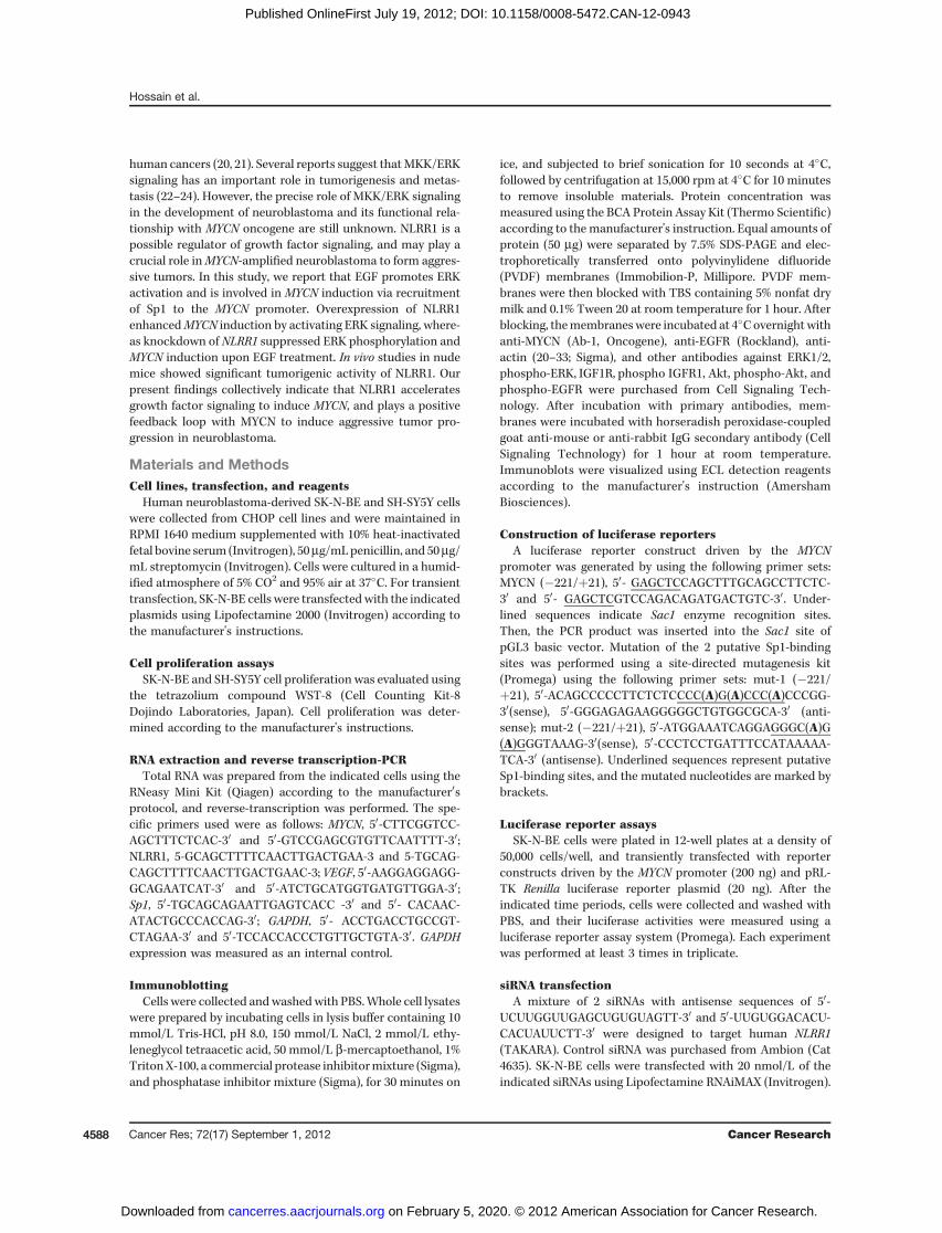
Figure 1. NLRR1 promotes EGF/IGF-mediated cell proliferation. A,quantification of SK-N-BE cell proliferation following EGF (left) and IGF(right) stimulation for the indicated time periods usingWST-8 assays. Thedata are represented as mean � SD. B, ectopic expression of NLRR1 inSK-N-BE cells was confirmed by immunoblotting (top). NLRR1-expressing SK-N-BE cells were treated with EGF (left) and IGF (right) forthe indicated time periods, and proliferation was measured by WST-8assays. C, NLRR1 knockdown by transfectionwith siRNAwas confirmedby immunoblotting (top). Growth curve of SK-N-BE cells transfected withcontrol siRNA and siRNA against NLRR1 in the presence of EGF (left) andIGF (right) was measured by WST-8 assays. For all WST-8 assays (A, B,and C), cells were cultured in 2% serum-containing medium, and growthfactors were used at a concentration of 50 ng/mL.
NLRR1 Modulates EGF-Mediated MYCN Induction
www.aacrjournals.org Cancer Res; 72(17) September 1, 2012 4589
on February 5, 2020. © 2012 American Association for Cancer Research. cancerres.aacrjournals.org Downloaded from
Published OnlineFirst July 19, 2012; DOI: 10.1158/0008-5472.CAN-12-0943

standard protocol of Cell signaling. Secondary biotinylateduniversal antibody from Vector Laboratories was appliedat a dilution of 1:400. Reactivity was visualized with an avi-din-biotin complex immunoperoxidase system using diami-nobenzidine as the chromagen and Hematoxylin as the coun-terstain (Vector Laboratories).
ResultsNLRR1 enhances EGF/IGF-mediated cell proliferation inneuroblastoma cells
Our previous report showed that NLRR1 is a direct tran-scriptional target of MYCN in neuroblastoma, and is asso-ciated with cell proliferation and survival (14). However, themechanism by which NLRR1 regulates cell proliferation wasstill unknown. Another member of leucine rich repeat pro-tein family, NLRR3, has been associated with the activationof ERK (15), and EGFR activation was reported to accelerateneuroblastoma cell proliferation (16), suggesting the possi-
bility that NLRR1 may activate EGFR to induce cell prolif-eration. To test this hypothesis, we first investigated neu-roblastoma cell proliferation upon EGF and IGF-I treatment.Consistent with their proliferative role (16, 17), both EGFand IGF-I also accelerated the proliferation of SK-N-BE cells(Fig. 1A). Similar data were also observed in neuroblastomaSH-SY5Y cells (data not shown). To determine whetherNLRR1 promotes EGF- and IGF-1–mediated cell prolifera-tion, we transiently transfected SK-N-BE cells with NLRR1expression plasmid and treated them with EGF and IGF-I.Interestingly, ectopic expression of NLRR1 enhanced the cellproliferation mediated by EGF and IGF-I (Fig. 1B). Over-expression of NLRR1 in SH-SY5Y showed similar promotionof proliferation (data not shown). To confirm the role ofNLRR1, we used an RNA interference approach to knockdown endogenous NLRR1. The data suggested that knock-down of NLRR1 suppressed the EGF- and IGF-1–mediatedcell proliferation (Fig. 1C). Collectively, the data suggested
p-ERK
EGF or IGF - -
p-EGFR
EGFR
ERK
pcDNA3 NLRR1
EGF IGF EGF IGF
-
control siRNA siNLRR1
p-ERK
p-Akt
p-EGFR
p-IGFIR
IGFIR
Akt
-
BA
Actin
NLRR1
Actin
NLRR1
EGF +
C
IGF - +
Control
U0126 (5 µmol/L)
Control
U0126 (5 µmol/L)
-
Re
lati
ve
nu
mb
er
of
ce
lls
Re
lati
ve
nu
mb
er
of
ce
lls
Figure 2. NLRR1 enhances EGF andIGF-mediated phosphorylation ofERK. A, SK-N-BE cells weretransiently transfected with mock(pCDNA3) and NLRR1-expressingplasmid. Forty-eight hours aftertransfection, cells were starvedwith serum-free medium for12 hours and then stimulated withEGF and IGF for 10 minutes. Wholecell lysates were used forimmunoblotting with specificantibodies (bottom). NLRR1overexpression was confirmed byimmunoblotting of the same lysates(top). B, siRNA-mediatedknockdown of NLRR1 suppressesphosphorylation of ERK mediatedby EGF and IGF (bottom). Theexperimental conditions are similarto those in A. Knockdownefficiency was confirmed byimmunoblotting (top). C, SK-N-BEcells transfected with NLRR1-expressing plasmids werepretreated with MEK1/2-specificinhibitor (U0126) and then culturedin the presence or absence of EGFand IGF; cell proliferation wasmeasured by WST-8 assays.
Hossain et al.
Cancer Res; 72(17) September 1, 2012 Cancer Research4590
on February 5, 2020. © 2012 American Association for Cancer Research. cancerres.aacrjournals.org Downloaded from
Published OnlineFirst July 19, 2012; DOI: 10.1158/0008-5472.CAN-12-0943

that NLRR1 enhances cell proliferation mediated by EGF andIGF-I.
NLRR1 enhances EGF and IGF-mediated activation ofERKBecause ERK is an important kinase that is often regulated
by EGF and IGF-1 growth factors to induce cell proliferation,we were interested to determine whether NLRR1 affected ERKphosphorylation upon EGF and IGF-I treatment. We examinedthe activation of ERK in NLRR1-overexpressing cells. Interest-ingly, ERK phosphorylation was enhanced compared with themock-transfected cells (Fig. 2A). The increased phosphoryla-tion of EGFRwas observed in the NLRR1 overexpressing SK-N-BE cells upon EGF stimulation (Fig. 2A). Similar data were alsoobserved in SH-SY5Y cells (data not shown). We also usedsiRNA studies to further elucidate the role ofNLRR1 in EGF andIGF-1 signaling. Consistent with the overexpression study, weobserved a reduction in ERK phosphorylation inNLRR1 knock-down cells upon both EGF and IGF-1 treatment comparedwiththe control siRNA-transfected cells (Fig. 2B). Interestingly,phosphorylation of EGFR and IGFR was found to be decreasedin NLRR1 knockdown cells (Fig. 2B). To elucidate whether theactivation of ERK is important for cell proliferation, we usedthe MEK1/2-specific inhibitor, U0126 (27), in the cell prolifer-ation assays. For the cell proliferation assays, we have used theminimum concentration of U0126 (5 mmol/L) required toinhibit ERK activation in the cells (Supplementary Fig. S1).The data showed that EGF- and IGF-I–mediated proliferationof NLRR1-overexpressing SK-N-BE cells was inhibited uponU0126 treatment (Fig. 2C).
EGF stimulation induces MYCN via ERKIGF-I stimulation induces endogenous MYCN expression in
neuroblastoma cells via MAPK activation (17). Therefore, it ispossible that EGF can induce MYCN, because EGF treatmentalso activatesMAPK. To investigate this hypothesis, we treatedSK-N-BE cells cultured in serum-free medium with increasingamounts of EGF. Consistent with our hypothesis, both mRNAand protein levels of MYCNwere induced upon EGF treatment(Fig. 3A). Time course experiments were also used to confirmthat EGF can induceMYCN.MYCNwas found to be induced at 6hours after EGF treatment (Fig. 3B). Under our experimentalconditions, EGF treatment successfully phosphorylated EGFR(Supplementary Fig. S1) and ERK (data not shown). We alsoobserved that EGF stimulation induced MYCN expression inother neuroblastoma cell lines, SH-SY5Y and NLF (data notshown). To verify our experimental data that EGF inducesMYCN, we used VEGF as positive control, which was reportedto be induced by EGF (28). AG1478 is a specific inhibitor forEGFR, and is sufficient to block EGFR-mediated activation ofERK (29, 30). We have confirmed the effect of the optimumconcentration of AG1478 to inhibit EGF-mediated phosphor-ylation of EGFR in the cells (Supplementary Fig. S1). Therefore,we used the optimum concentrations of AG1478 to elucidatethat EGF-mediated MYCN induction is dependent on EGFR-ERK signaling. Consistently, MYCN induction by EGF wassuccessfully blocked in AG1478-pretreated SK-N-BE cells (Fig.3C left).We also usedU0126 to prove that EGF-mediatedMYCN
induction is dependent on ERK (Fig. 3C, right panel). Thesedata collectively suggest that EGF-mediated MYCN inductionis dependent on the EGFR–ERK pathway in neuroblastomacells.
EGF enhances MYCN transcription via recruitment ofSp1 to the MYCN promoter
To confirm the EGF-mediated MYCN transcription, wegenerated a luciferase reporter plasmid containing a MYCNgenomic fragment spanning positions �221 to þ21 (Fig. 4A,left panel), where þ1 represents the transcriptional initiationsite. This promoter region contains both Sp1 and E2F1 tran-scriptional element sites (31, 32). We also used the emptycontrol vector pGL3basic to compare the EGF responses. SK-N-BE cells were transiently transfected with pGL3basic andpGL3-MYCN (�221, þ21) together with Renilla luciferasereporter plasmid. The data showed that EGF stimulationsignificantly enhanced the promoter activity of MYCN geneat 6 hours (Fig. 4A, right panel): pGL3-Basic reporter constructsdid not respond to EGF treatment (Fig. 4A, right), suggestingthat EGF treatment is specific to the MYCN promoter. MYCNhas been reported to be transcriptionally regulated by 2 majortranscription factors, Sp1 and E2F1 (31, 32). To determine theregulatory mechanism of EGF-mediated MYCN transcription,we performed ChIP assays. Both antibodies against Sp1 andE2F1 used for ChIP assays pulled down the specific
B
C
MYCN
Actin
AG1478
MYCN
Actin
25
EGF - + + + + +- - D
MS
O
5 10 U0126 10
EGF - + + + + +- - D
MS
O
1 5
AEGF 0 1 5 10 50 100 ng/mL
MYCN
GAPDH
VEGF
MYCN
Actin
EGF 0 1 5 10 50 100 ng/mL
MYCN
GAPDH
MYCN
Actin
EGF
0 6 12 24 h
EGF
0 6 12 24 h
VEGF
µmol/L µmol/L
Figure 3. EGF stimulation induces endogenousMYCN. A, serum-starvedSK-N-BE cells were treated with increasing doses of EGF for 12 hours.Total mRNA was used in reverse transcription (RT)-PCR to check theexpression ofMYCN and VEGF (top).GAPDHwas checked as an internalcontrol. Whole cell lysates were used in immunoblotting to detect theproteins (bottom). B, MYCN was induced by EGF in a time-dependentmanner. Serum-starved SK-N-BE cells were treated with 50 ng/mL ofEGF. Total RNA and whole cell lysates were collected at the indicatedtime points for RT-PCR (top) and Western blotting (bottom) assays tocheck the expression level of MYCN. C, SK-N-BE cells were cultured inserum-free medium for 12 hours with or without different concentrationsof AG1478 (left) and U0126 (right). Cells were then treated with EGF (50ng/mL) for 12 hours, and whole cell lysates were used to check MYCNprotein levels by immunoblotting. Actin was checked as an internalcontrol.
NLRR1 Modulates EGF-Mediated MYCN Induction
www.aacrjournals.org Cancer Res; 72(17) September 1, 2012 4591
on February 5, 2020. © 2012 American Association for Cancer Research. cancerres.aacrjournals.org Downloaded from
Published OnlineFirst July 19, 2012; DOI: 10.1158/0008-5472.CAN-12-0943

endogenous proteins (data not shown). Twelve hours after EGFstimulation, chromatin DNA from SK-N-BE cells was cross-linked and processed for ChIP assays. Sp1-derived pulled-downchromatin was amplified by a specific primer targeting theMYCN core promoter region (�221 toþ21) in the EGF-treatedcells (Fig. 4B top panel), suggesting that EGF enhances recruit-ment of Sp1 to the MYCN promoter. However, no change inE2F1 recruitment was observed between the EGF-treated anduntreated cells (Fig. 4B, bottom panel).
To identify the critical Sp1-binding region required for thetransactivation of MYCN, we mutated the Sp1-binding ele-ments (Fig. 4C, left panel) by PCR reactions, as described in theMaterials andMethods section. SK-N-BE cells were transientlytransfected with wild-type MYCN promoter as well as themutated luciferase constructs. Promoter assays showed thatthe pGL3MYCN (mut-1) construct had significantly (P < 0.001)less promoter activity (Fig. 4C,middle panel) than thewild typeand pGL3MYCN (mut-2) constructs. Furthermore, we tran-siently transfected the wild type, as well as the Sp1 site 1-deleted constructs, into the SK-N-BE cells to determine theeffects on EGF. The data showed that the deletion constructfailed to respond to EGF treatment (Fig. 4C, right panel),
suggesting that the Sp1-binding element 1 is important forEGF-mediated transactivation of MYCN.
To check whether Sp1 is critical for the expression ofMYCN, we knocked down Sp1 using siRNAs. Consistently,MYCN expression was suppressed in cells transfected withsiRNAs against Sp1 (Supplementary Fig. S2A). Both siRNAsagainst Sp1 reduced MYCN promoter activity (SupplementaryFig. S2B). Cell proliferation was also suppressed in the Sp1knockdown cells (Supplementary Fig. S2C). It has beenreported that EGF stimulation induces phosphorylation ofERK, and that this phosphorylation event may be importantfor Sp1 phosphorylation (33) and recruitment to the targetgene promoter. Similarly, our results also showed that EGFtreatment resulted in phosphorylation of Sp1, and this phos-phorylation event was inhibited in U0126- and calf intestinalphosphatase (CIAP)-treated cells (Supplementary Fig. S3A).CIAP treatment is reported to block recruitment of phospho-transcriptional factors on genomic DNA (35). We also usedMithramycin-A (Mit-A), a well-known Sp1 inhibitor (34). EGF-mediated MYCN induction was successfully inhibited in cellspretreated with Mit-A (Supplementary Fig. S3B) and CIAP(Supplementary Fig. S3C). Consistently, CIAP treatment also
A
Luc.- 221
+21
E2
F1
Sp
1
Sp
1
E2
F1
E2
F1
pG
L3
-Ba
sic
Fold activation
0 10 20 30 40 50 60 70
0 hr
1 hr6 h
12 h
0 h1 h
6 h
12 h
P < 0.01
C
Luc.
Luc.×
Luc.× Mut-2
Mut-1
Wt
pGL3-Basic
2 4 6 8 10 12
Fold activation
P < 0.001
Fold activation
Mut-2
Mut-2+EGF
Mut-1+EGF
Mut-1
4 8 12 16 2420
- 221 +21
B
IP: α-Sp1
IP: α-E2F1
IP: NRS
EGF - +
Input
IP: NRS
Input
E2
F1
Sp
1 (s
ite 1
)
E2
F1
E2
F1
Sp
1 (s
ite 2
)
Figure4. EGFenhancesSp1-mediated transactivation ofMYCN. A,MYCN core promoter region (�221 toþ21) cloned in pGL3Basic luciferase vector (left) wastransiently transfected into SK-N-BE cells for 36 hours, followed by culture in serum-freemedium for 12 hours. Cells were then treatedwith EGF (50 ng/mL) forthe indicated time periods, and luciferase assays were carried out to quantify the relative promoter activity. B, SK-N-BE cells were cultured inserum-freemedium for 12 hours, followed by treatment for 12 hours with EGF (50 ng/mL). Cross-linked chromatin was isolated from the cells and precipitatedwith Sp1- and E2F1-specific antibodies or with normal rabbit serum (NRS). C, Sp1 site 1-deletedMYCN core promoter failed to respond to EGF stimulation.Two Sp1-binding sites on the MYCN core promoter region were deleted by site-directed mutagenesis (left). Relative activity of the deleted promoterconstructs weremeasured by luciferase assays in SK-N-BE cells 24 hours after transfection (middle). SK-N-BE cells were transiently transfected by the 2 Sp1site deleted constructs, followed by culture in serum-free medium for 12 hours and then treatment with EGF (50 ng/mL). Twelve hours after EGF treatment,firefly luciferase activities were determined (right). Renilla luciferase was used as an internal control to standardize the transfection efficiency of the luciferasevectors.
Hossain et al.
Cancer Res; 72(17) September 1, 2012 Cancer Research4592
on February 5, 2020. © 2012 American Association for Cancer Research. cancerres.aacrjournals.org Downloaded from
Published OnlineFirst July 19, 2012; DOI: 10.1158/0008-5472.CAN-12-0943

reduced the recruitment of Sp1 to the MYCN promoter (Sup-plementary Fig. S3D). Collectively, our results suggest that Sp1recruitment to the MYCN promoter can enhance the transac-tivation of MYCN upon EGF treatment.
NLRR1 enhances MYCN inductionERK is reported to induce phosphorylation of Sp1 (33). In
our present findings, NLRR1 promotes phosphorylation ofERK upon EGF treatment, suggesting that NLRR1 mayinduce MYCN. Therefore, we overexpressed NLRR1 in SK-N-BE cells and found that endogenous MYCN was effectivelyinduced in a dose-dependent manner (Fig. 5A, left panel).NLRR1-mediated MYCN induction was inhibited in U0126-and AG1478-pretreated cells (Fig. 5A, right panel), suggest-ing that NLRR1 induced MYCN via EGFR-ERK signaling.Ectopic expression of NLRR1 in cells accelerated MYCNinduction upon EGF treatment compared with that ofmock-transfected (empty pcDNA3.1 vector) cells (Fig. 5B,left panel). Consistent with the overexpression study, knock-down of NLRR1 suppressedMYCN induction, suggesting thatNLRR1 accelerates EGF-mediated MYCN induction. Todetermine whether NLRR1 enhances Sp1 recruitment, weperformed ChIP assays. The data show that overexpressionof NLRR1 in SK-N-BE cells increased Sp1 recruitment to theMYCN promoter, which was further accelerated upon EGFtreatment (Fig. 5C).
Stable expression of NLRR1 in cells accelerates tumorgrowth in nude miceNLRR1 is highly expressed in aggressive MYCN-amplified
neuroblastoma (14), suggesting the possibility that NLRR1may have a potent tumorigenic role. NLRR1 overexpressionenhanced the colony formation ability of SH-SY5Y cells(Supplementary Fig. S4A). Consistent with our previousobservation that ectopic expression of NLRR1 enhanced cellproliferation and inhibited apoptosis, NLRR1-stably expres-sing SH-SY5Y clones proliferated faster than mock stableclones (Supplementary Fig. S4C). Furthermore, MYCNexpression was upregulated in NLRR1 stable clones com-pared with mock clones (Supplementary Fig. S4B), suggest-ing that NLRR1 has oncogenic potential. To elucidate thetumorigenic activity of NLRR1, we performed tumor xeno-graft studies in nude mice using NLRR1-stably expressingSH-SY5Y clones. Significant enhancement of tumor growthwas observed in mice bearing NLRR1-expressing xenograftscompared with the mock-expressing xenografts (P < 0.01; Fig.6A). Mice of each group were sacrificed when they becomemorbid, and the survival curve was analyzed using theKaplan–Meier method. The survival of mice with NLRR1-expressing xenografts was significantly shorter comparedwith mice-bearing mock xenografts (P ¼ 0.003; Fig. 6B).Immunohistochemical data showed that NLRR1-expressingtumors had increased numbers of Ki-67–positive cells anddecreased number of terminal deoxynucleotidyl transfer-ase–mediated dUTP nick end labeling (TUNEL) positivecells compared with mock tumors (Fig. 6C), indicating thatthere were more proliferative cells in the tumors derivedfrom NLRR1-expressing clones. To see the consistence in
vitro finding of NMYC induction by NLRR1, we carried outIHC assays using the tumor xenografts. Induction of NMYCand p-ERK was found in NLRR1 tumors whereas total ERKwas unchanged in both tumors (Fig. 6D). Collectively, ourdata suggest that NLRR1 induces NMYC in vivo and haspotent tumorigenic roles.
C
IP: α-Sp1
IP: NRS
Input
EGF - -+ +
Mock NLRR1
Rela
tiv
e r
ecru
itm
en
t
of
Sp
1
EGF - -+ +NLRR1Mock
0
10
20
30
40
EGF
si-
co
ntr
ol
si-
NL
RR
1
- -+ +
Mo
ck
NL
RR
1
B
NLRR1
MYCN
GAPDH
EGF
NLRR1
MYCN
GAPDH
- -+ +
A
NLRR1
MYCN
Actin
NLRR1
p-ERK
ERK
NLRR1
MYCN
Actin
NLRR1
U0126
AG1478 - - -- - -
++
- + + +
ERK
p-ERK
Figure 5. NLRR1 inducesMYCN in neuroblastoma cells. A, SK-N-BE cellswere transiently transfected with increasing amounts of NLRR1-expressing plasmids. Forty-eight hours after transfection,immunoblotting was carried out to detect NLRR1, p-ERK, total ERK, andMYCN expression (left). EGFR and ERK inhibitors prevent MYCNinduction in the NLRR1-overexpressing cells. SK-N-BE cells weretransiently transfectedwithNLRR1 expression plasmids for 48 hours andthen treated with or without AG1478 (20 mmol/L) and U0126 (10 mmol/L).Twelve hours after treatment, whole cell lysates were prepared.Immunoblotting data show the expression of NLRR1, p-ERK, total ERK,and MYCN (right). B, NLRR1 enhances MYCN induction upon EGFtreatment. Twenty-four hours after ectopic expression of NLRR1, SK-N-BE cells were cultured in serum-free medium for 12 hours followed byEGF (10ng/mL) treatment. RT-PCRwasperformed to checkendogenousMYCN expression (left). Forty hours after transfection with control siRNAand NLRR1-siRNA, SK-N-BE cells were serum starved for 12 hours andtreated with EGF (10 ng/mL). Twelve hours after EGF treatment, RT-PCRwas carried out to check the expression of NLRR1 and MYCN (right).C, crossed-linked chromatin from the SK-N-BE mock- and NLRR1-ectopically expressing cells treated with or without EGF (50 ng/mL)for 12 hours was used for pulldown by Sp1-specific antibody. Primerstargeting MYCN core promoter (�221, þ21) were used to amplify thepulled-down chromatin (left). The recruitment of Sp1 was quantified fromthe PCR band by image J software and plotted (right). The data representmean � SD.
NLRR1 Modulates EGF-Mediated MYCN Induction
www.aacrjournals.org Cancer Res; 72(17) September 1, 2012 4593
on February 5, 2020. © 2012 American Association for Cancer Research. cancerres.aacrjournals.org Downloaded from
Published OnlineFirst July 19, 2012; DOI: 10.1158/0008-5472.CAN-12-0943

DiscussionUpregulated MKK-ERK signaling is known to be involved in
the genesis of several cancers (18–21). Multidrug-resistanthuman neuroblastoma cells have three- to 30-fold more cellsurface EGFRs than the drug-sensitive parental cells (36),indicating that EGFRs may play an important role in theaggressiveness of human neuroblastoma. EGF stimuli activat-ed MAPK in neuroblastoma (16) and IGF-1 enhanced neuro-blastoma cell proliferation via activation of MAPK (17). NLRRfamily proteins are considered to modulate cellular signaling,especially that of MAPKs (15). Although it was reported thatoverexpression of NLRR1 enhances cell proliferation and inhi-bits cellular apoptosis in neuroblastoma, our previous studydid not rule out the involvement of NLRR1 with EGF and IGF-1signaling. Here, we first report that ectopic expression ofNLRR1 also enhanced EGF- and IGF-1–mediated cellular pro-liferation, which is inhibited in cells treated with MEK1/2inhibitor. This suggests that NLRR1-mediated promotion ofproliferation is at least in part dependent on activation of ERK.However, the method by which NLRR1 enhances EGFR andIGFR signaling still needs to be clarified. Another NLRR familyprotein, NLRR3, was reported to induce phosphorylation ofERK in response to EGF (15), suggesting the possibility that
NLRR1 that is 54% homology to NLRR3 may also induce ERKphosphorylation. Our results propose that NLRR1 is importantin enhancing ERK phosphorylation in cells upon EGF and IGFstimuli. Interestingly, NLRR1 affects p-ERK and p-AKT indifferent extent (Fig. 2B) that depends on cell lines (data notshown). The reason behind this selectivity is unknown andneed to be addressed further in the future.
ERK is known to phosphorylate many transcriptional fac-tors, including Sp1 (33, 37). Moreover, Sp1 and E2F1 are re-ported to transcriptionally regulate MYCN in neuroblastoma(31, 32). Therefore, there may be an important link betweenthe activation of ERK and MYCN induction, which is furthersupported by the evidence that IGF-1 can induce MYCN viaactivation of MAPK (17). However, in this report of EGF-1-mediatedMYCN induction, the involvement of Sp1 or E2F1wasnot explained. Here, we report that EGF inducesMYCN via theEGFR-ERK pathway. Several stimuli, including retinoic acidand TGF-b repress the MYCN gene, and are associated withrecruited Sp1 and E2F1 and their other cofactors (31, 38). Byusing ChIP assays, we observed that Sp1 but not E2F1 wasrecruited to theMYCN promoter in response to EGF. However,it has been reported that the presence of Sp1 is not alwayssufficient to activate a transcriptionally silentMYCN gene (32).
A
Tu
mo
r vo
lum
e (
mm
3)
Days after injection
70635649423528211470
100
200
300
400
Mock #1
Mock #2
Mock #3
NLRR1 #1
NLRR1 #2
NLRR1 #3
Mock NLRR1
DN
MY
CControl NLRR1
p-E
RK
ER
K
DA
PI
NL
RR
1 K
i67
Control NLRR1C
Rela
tive e
xp
ressio
n ki67
012345678
DA
PI
TU
NE
L
Rela
tive e
xp
ressio
n
TUNELControl NLRR1
B
Observation days
20 40 60 80 100 120
Su
rviv
al
pe
rce
nta
ge
100
80
40
NLRR1 (n = 21)
Mock (n = 21)
P = 0.0034
60
20
0
Figure 6. Neuroblastoma cellsstably expressing NLRR1accelerate tumor growth in nudemice. A, after subcutaneousinjection of mock- and NLRR1-stably expressing SH-SY5Y clonesinto nude mice, tumor volumeswere measured on the indicateddays. The data represent the meanvalue. Top panel showsrepresentative pictures of tumors70 days after injection. B, Kaplan–Meyer survival curve of mice-bearing mock- and NLRR1-stablyexpressing SH-SY5Y xenografts.C, detection of NLRR1 and Ki-67expression in the mock- andNLRR1-expressing tumorxenografts (top). TUNEL stainingshows that NLRR1 tumor hasreduced apoptotic cells (bottom).D, IHC data show the stainingimage ofNMYC, p-ERK, andERK inNLRR1 versus control tumors.
Hossain et al.
Cancer Res; 72(17) September 1, 2012 Cancer Research4594
on February 5, 2020. © 2012 American Association for Cancer Research. cancerres.aacrjournals.org Downloaded from
Published OnlineFirst July 19, 2012; DOI: 10.1158/0008-5472.CAN-12-0943
Therefore, we have further investigated whether Sp1 is impor-tant for MYCN induction in our experimental conditions.siRNA-mediated knockdown of Sp1-reducedMYCN expressionand also inhibited cell proliferation, suggesting that Sp1-mediated MYCN induction regulates cell proliferation. Usingluciferase reporter assays, we identified the responsive Sp1-binding element in the promoter region for MYCN induction.Sp1 phosphorylation is a prerequisite for the interaction withgenomic DNA (33). Therefore, we also investigated whetherEGF induces Sp1 phosphorylation and enhances its recruit-ment to the MYCN promoter. Western blotting data showedthat EGF treatment enhanced phosphorylation of Sp1, andthe MEK1/2 inhibitor U0126 inhibited this phosphorylationevent, supporting the previous report that ERK induces Sp1phosphorylation (33). Using the dephosphorylating agentCIAP, we further confirmed that phospho-Sp1 is involved inthe transactivation of MYCN by recruitment to the genomicDNA of the MYCN promoter. Collectively, this evidence sug-gests that EGF induces MYCN through phospho-Sp1 recruit-ment to the MYCN promoter. However, MYCN induction byIGF-1 was not investigated in the present study, but wespeculate that it may be by the same mechanism as EGF,because IGF-1 has been reported to activate MAPK inneuroblastoma.Another new finding of our current study is that MYCN is
induced after ectopic expression of NLRR1 in neuroblastomacells. Under our experimental conditions, EGF-mediatedMYCN induction was accelerated in NLRR1-overexpressingcells and inhibited in NLRR1-knockdown cells, suggesting thatNLRR1 enhances MYCN induction and activation of ERK. Inaddition, NLRR1 overexpression enhanced Sp1 recruitment tothe MYCN promoter. We previously reported that NLRR1transcriptionally regulated by MYCN is highly expressed inaggressive neuroblastoma (14). NLRR1 was also found to behighly expressed in MYCN-amplified tumors compared withthat of MYCN nonamplified neuroblastoma. Therefore, wesuggest that MYCN directly regulates NLRR1, and that NLRR1further inducesMYCN through the activation of the EGFR-ERKcascade, suggesting a positive feedback loop between NLRR1and MYCN that may lead to aggressive neuroblastoma. Tothis notion, we further determined the in vivo tumorigenicactivity of NLRR1 inMYCN nonamplified SH-SY5Y cells, which
are well known to form tumors in nude mice (39, 40). Ourresults show that SH-SY5Y cells stably expressing NLRR1rapidly proliferate in culture medium and in nude mice, andpromote decreased survival rates among inoculated mice,presumably by induced endogenous MYCN.
In conclusion, our results provide significant evidence thatthat NLRR1 enhances ERK signaling to induce MYCN, whichmay play major roles in the progression of aggressive neuro-blastoma. Taking note that MYCN highly expressing tumorsare often resistant to antitumor therapies, new drug discoveryblocking NLRR1-mediated ERK signaling to control MYCNexpression may be an attractive choice in treating aggressiveneuroblastoma.
Disclosure of Potential Conflicts of InterestNo potential conflicts of interest were disclosed.
Authors' ContributionsConception and design: S. Hossain, A. Takatori, A. NakagawaraDevelopment of methodology: S. Hossain, A. Takatori, Y. Suenaga, T. KamijoAcquisition of data (provided animals, acquired and managed patients,provided facilities, etc.): S. Hossain, A. Takatori, Y. NakamuraAnalysis and interpretation of data (e.g., statistical analysis, biostatistics,computational analysis): S. Hossain, A. TakatoriWriting, review, and/or revision of the manuscript: S. Hossain, A.NakagawaraAdministrative, technical, or material support (i.e., reporting or orga-nizing data, constructing databases): Y. Suenaga, Y. Nakamura, T. Kamijo, A.NakagawaraStudy supervision: A. Nakagawara
AcknowledgmentsWe thank Drs. M. Ohira, T. Ozaki, J. Akter, and K. Hasan (Chiba Cancer Center
Research Institute, Chiba, Japan) for the fruitful discussion and assistance.
Grant SupportThis work was supported by a Grant-in-Aid from the Ministry of Health,
Labour, andWelfare for Third TermComprehensive Control Research for Cancer(A. Nakagawara), a Grant-in-Aid for Scientific Research on Priority Areas fromthe Ministry of Education, Culture, Sports, Science, and Technology, Japan (A.Nakagawara), a grant from the Takeda Science Foundation, and a Grant-in-Aidfor Scientific Research from the Japanese Society for the Promotion of Science(A. Takatori and A. Nakagawara).
The costs of publication of this article were defrayed in part by the payment ofpage charges. This article must therefore be hereby marked advertisement inaccordance with 18 U.S.C. Section 1734 solely to indicate this fact.
Received March 16, 2012; revised May 21, 2012; accepted June 6, 2012;published OnlineFirst July 19, 2012.
References1. BrodeurGM.Neuroblastoma: biological insights into a clinical enigma.
Nat Rev Cancer 2003;3:203–16.2. Weinstei JL, Katzenstein HM, Cohn SL. Advances in the diagnosis and
treatment of neuroblastoma. Oncologist 2003;8:278–92.3. Brodeur GM, Nakagawara A. Molecular basis of clinical heterogeneity
in neuroblastoma. Am J Pediatr Hematol Oncol 1992;14:111–6.4. Bown N, Cotterill S, Lastowska M, O'Neill S, Pearson AD, Plantaz D,
et al. Gain of chromosome arm 17q and adverse outcome in patientswith neuroblastoma. N Engl J Med 1999;340:1954–61.
5. Frappaz D, Michon J, Coze C, Berger C, Plouvier E, Lasset C, et al.LMCE3 treatment strategy: results in 99 consecutively diagnosedstage 4 neuroblastomas in children older than 1 year at diagnosis. JClin Oncol 2000;18:468–76.
6. SchmidtML, Lukens JN,SeegerRC,BrodeurGM,ShimadaH,GerbingRB, et al. Biologic factors determine prognosis in infants with stage IV
neuroblastoma: a prospective Children's Cancer Group study. J ClinOncol 2000;18:1260–8
7. Felsher DW, Bishop JM. Reversible tumorigenesis byMYC in hemato-poietic lineages. Mol Cell 1999;4:199–207.
8. D'Cruz CM, Gunther EJ, Boxer RB, Hartman JL, Sintasath L, MoodySE, et al. c-MYC induces mammary tumorigenesis by means of apreferred pathway involving spontaneous Kras2 mutations. Nat Med2001;7:235–9.
9. Pelengaris S, Littlewood T, Khan M, Elia G, Evan G. Reversibleactivation of c-Myc in skin: induction of a complex neoplastic pheno-type by a single oncogenic lesion. Mol Cell 1999;3:565–77.
10. Taguchi A, Wanaka A, Mori T, Matsumoto K, Imai Y, Takagi T, et al.Molecular cloning of novel leucine-rich repeat proteins and theirexpression in the developing mouse nervous system. Brain Res MolBrain Res 1996;35:31–40.
NLRR1 Modulates EGF-Mediated MYCN Induction
www.aacrjournals.org Cancer Res; 72(17) September 1, 2012 4595
on February 5, 2020. © 2012 American Association for Cancer Research. cancerres.aacrjournals.org Downloaded from
Published OnlineFirst July 19, 2012; DOI: 10.1158/0008-5472.CAN-12-0943
11. Taniguchi H, Tohyama A, Takagi T. Cloning and expression of a novelgene for a protein with leucine-rich repeats in the developing mousenervous system. Brain Res Mol Brain Res 1996;36:45–52.
12. Hamano S, Ohira M, Isogai E, Nakada K, Nakagawara A. Identificationof novel human neuronal leucine-rich repeat (hNLRR) family genes andinverse association of expression of Nbla10449/hNLRR-1 andNbla1067/hNLRR-3 with the prognosis of primary neuroblsatoma. IntJ Oncol 2004;24:1457–66.
13. BandoT,SekineK,Kobayashi S,WatabeAM,RumpA, TanakaM, et al.Neuronal leucine-rich repeat protein 4 functions in hippocampus-dependent long-lasting memory. Mol Cell Biol 2005;25:4166–75.
14. Hossain MS, Ozaki T, Wang H, Nakagawa A, Takenobu H, Ohira M,et al. N-MYC promotes cell proliferation through a direct transactiva-tion of neuronal leucine-rich repeat protein-1 (NLRR1) gene in neuro-blastoma. Oncogene 2008;27:6075–82.
15. Fukamachi K, Matsuoka Y, Ohno H, Hamaguchi T, Tsuda H. Neuronalleucine-rich repeat protein-3 amplifies MAPK activation by epidermalgrowth factor through a carboxy-terminal region containing endocy-tosis motifs. J Biol Chem 2002;277:43549–52.
16. Ho R, Minturn JE, Hishiki T, Zhao H, Wang Q, Cnaan A, et al. Prolif-eration of human neuroblastomas mediated by the epidermal growthfactor receptor. Cancer Res 2005;65:9868–75.
17. MisawaA, Hosoi H, Arimoto A, Shikata T, Akioka S,Matsumura T, et al.N-Myc induction stimulated by insulin-like growth factor I throughmitogen-activated protein kinase signaling pathway in human neuro-blastoma cells. Cancer Res 2000;60:64–9.
18. ChangL,KarinM.MammalianMAPkinase signalling cascades.Nature2001;410:37–40.
19. Johnson GL, Lapadat R. Mitogen-activated protein kinase pathwaysmediated by ERK, JNK, and p38 protein kinases. Science 2002;298:1911–2.
20. Dunn KL, Espino PS, Drobic B, He S, Davie JR. The Ras-MAPK signaltransduction pathway, cancer and chromatin remodeling. BiochemCell Biol 2005;83:1–14.
21. Davies H, Bignell GR, Cox C, Stephens P, Edkins S, Clegg S, et al.Mutations of the BRAF gene in human cancer. Nature 2002;417:949–54.
22. Jeffers M, Fiscella M, Webb CP, Anver M, Koochekpour S, VandeWoude GF. The mutationally activated Met receptor mediates motilityand metastasis. Proc Natl Acad Sci U S A 1998;95:14417–22.
23. Webb CP, Taylor GA, Jeffers M, Fiscella M, Oskarsson M, Resau JH,et al. Evidence for a role of Met-HGF/SF during Ras-mediated tumor-igenesis/metastasis. Oncogene 1998;17:2019–25.
24. Ward Y, Wang W, Woodhouse E, Linnoila I, Liotta L, Kelly K. Signalpathways which promote invasion and metastasis: critical and dis-tinct contributions of extracellular signal-regulated kinase and Ral-specific guanine exchange factor pathways. Mol Cell Biol 2001;21:5958–69.
25. Ghos AK, Steele R, Ray RB. Carboxy-terminal repressor domain ofMBP-1 is sufficient for regression of prostate tumor growth in nudemice. Cancer Res 2005;65:718–21.
26. Konstantinidou G, Bey EA, Rabellino A, Schuster K, Maira MS, GazdarAF, et al. Dual phosphoinositide 3-kinase/mammalian target of rapa-mycin blockade in an effective radiosensitizing strategy for the treat-ment of non-small cell lung cancer harboring K-ras mutations. CancerRes 2009;69:7644–52.
27. Wang J, Zhou J, Wu GS. ERK-dependent MKP-1-mediated cisplatinresistance in human ovarian cancer cells. Cancer Res 2007;67:11933–41.
28. Maity A, Pore N, Lee J, Solomon D, O'Rourke DM. Epidermal growthfactor receptor transcriptionally up-regulates vascular endothelialgrowth factor expression in human glioblastoma cells via a pathwayinvolving phosphatidylinositol 30-kinase and distinct from that inducedby hypoxia. Cancer Res 2000;60:5879–86.
29. DavisII JW, Lauer FT,BurdickAD,HudsonLG,Burchiel SW.Preventionof apoptosis by 2,3,7,8-Tetrachlorodibenzo-p-dioxin (TCDD) in theMCF-10A cell line: correlation with increased transforming growthfactor a production. Cancer Res 2001;61:3314–20.
30. Zhou Y, Brattain MG. Synergy of epidermal growth factor receptorkinase inhibitor AG1478 and ErbB2 kinase inhibitor AG879 in humancolon carcinoma cells is associated with induction of apoptosis.Cancer Res 2005;65:5848–56.
31. Strieder V, Lutz W. E2F proteins regulate MYCN expression in neu-roblastomas. J Biol Chem 2003;278:2983–9.
32. Kramps C, Strieder V, Sapetschnig A, Suske G, Lutz W. E2F and Sp1/Sp3 Synergize but are not sufficient to activate the MYCN gene inneuroblastomas. J Biol Chem 2004;279:5110–7.
33. Merchant JL, Du M, Todisco A. Sp1 phosphorylation by Erk 2stimulates DNA binding. Biochem Biophys Res Commun 1999;254:454–61.
34. JiaZ, ZhangJ,WeiD,WangL,YuanP, LeX, et al.Molecular basis of thesynergestic sntiangiogenesis activity of Bevacizumab and Mithramy-cin A. Cancer Res 2007;67:4878–85.
35. Matsuoka T, Zhao L, Stein R. The DNA binding activity of the RIPE3b1transcription factor of insulin appears to be influenced by tyrosinephosphorylation. I Biol Chem 2001;276:22071–6.
36. Meyers MB, Shen WP, Spengler BA, Ciccarone V, O'Brien JP, DonnerDB, et al. Increased epidermal growth factor receptor in multidrug-resistant human neuroblastoma cells. J Cell Biochem 1988;38:87–97.
37. Milanini-Mongiat J, Pouyssegur J, Pages G. Identification of two Sp1phohphorylation sites for p42/44 mitogen-activated protein kinases. JBio Chem 2002;277:20631–9.
38. Ferreira R, Naguibneva I, Pritchard LL, Ait-Si-Ali S, Harel-Bellan A. TheRb/chromatin connection and epigenetic control: opinion. Oncogene2001;20:3128–33.
39. AoyamaM,Ozaki T, InzukaH, TomotsuneD,Hirato J,OkamotoY, et al.LMO3 interacts with neuronal transcription factor, HEN2, and acts asan oncogene in neuroblastoma. Cancer Res 2005:65:4587–97.
40. Eggert A, Grotzer MA, Ikegaki N, Liu XG, Evans AE, Brodeur GM.Expression of the neurotrophin receptor TrkA down-regulates expres-sion and fuinction of angiogenic stimulators in SH-SY5Y neuroblas-toma cells. Cancer Res 2002;62:1802–8.
Hossain et al.
Cancer Res; 72(17) September 1, 2012 Cancer Research4596
on February 5, 2020. © 2012 American Association for Cancer Research. cancerres.aacrjournals.org Downloaded from
Published OnlineFirst July 19, 2012; DOI: 10.1158/0008-5472.CAN-12-0943
2012;72:4587-4596. Published OnlineFirst July 19, 2012.Cancer Res Shamim Hossain, Atsushi Takatori, Yohko Nakamura, et al.
In Vivoand Accelerates Tumor Growth Induction in NeuroblastomaMYCNNLRR1 Enhances EGF-Mediated
Updated version
10.1158/0008-5472.CAN-12-0943doi:
Access the most recent version of this article at:
Material
Supplementary
http://cancerres.aacrjournals.org/content/suppl/2012/07/19/0008-5472.CAN-12-0943.DC1
Access the most recent supplemental material at:
Cited articles
http://cancerres.aacrjournals.org/content/72/17/4587.full#ref-list-1
This article cites 40 articles, 23 of which you can access for free at:
Citing articles
http://cancerres.aacrjournals.org/content/72/17/4587.full#related-urls
This article has been cited by 3 HighWire-hosted articles. Access the articles at:
E-mail alerts related to this article or journal.Sign up to receive free email-alerts
Subscriptions
Reprints and
To order reprints of this article or to subscribe to the journal, contact the AACR Publications Department at
Permissions
Rightslink site. Click on "Request Permissions" which will take you to the Copyright Clearance Center's (CCC)
.http://cancerres.aacrjournals.org/content/72/17/4587To request permission to re-use all or part of this article, use this link
on February 5, 2020. © 2012 American Association for Cancer Research. cancerres.aacrjournals.org Downloaded from
Published OnlineFirst July 19, 2012; DOI: 10.1158/0008-5472.CAN-12-0943