Embed Size (px)
Citation preview
New
born Care
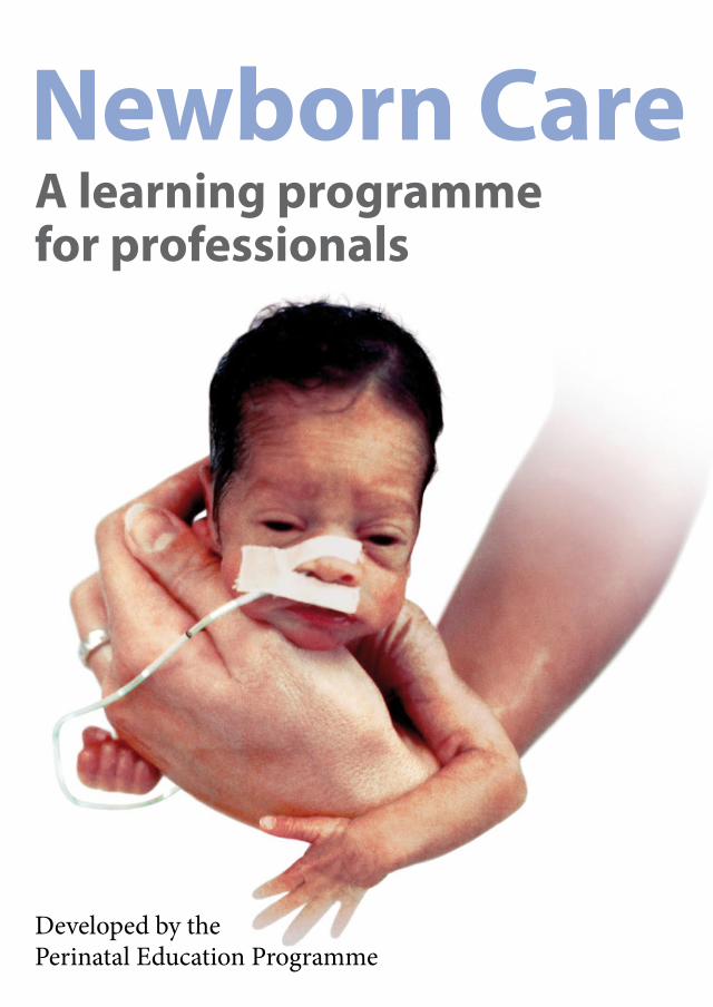
Newborn CareA learning programme for professionals
Take control of your own learning in this innovative new book for nurses and doctors. You’ll be taught to solve practical problems and to take a simple, logical approach to common disorders.
Clear learning objectives help you understand the most important lessons to be learned.
Theoretical knowledge is presented in an easy, problem-solving way.
Clear, step-by-step guides through definitions, causes, diagnoses, prevention, dangers and management.
Case studies in story-form let you apply your new knowledge to solve common problems.
Multiple-choice questions help you monitor your progress.
Skills workshops develop important clinical techniques.
This balanced and up-to-date guide to common and important clinical conditions can be used as a training course or as a reference manual for professionals and students.
•
•
•
•
•
•
Perinatal Education Programm
e
EBW Electric Book Workswww.ebwhealthcare.com
Part of the highly successful Perinatal Education Programme, which has brought effective continuing training to thousands of midwives, neonatal nurses and doctors.
ISBN 978 1 920218 28 7
Developed by thePerinatal Education Programme
A learning programme for professionals
Newborn Care

Newborn CareA learning programme for professionals
Developed by thePerinatal Education Programme
www.ebwhealthcare.com

VERY IMPORTANTWe have taken every care to ensure that drug dosages and related medical advice in this book are accurate. However, drug dosages can change and are updated often, so always double-check dosages and procedures against a reliable, up-to-date formulary and the given drug‘s documentation before administering it.
Newborn Care: A learning programme for professionals
Updated: 15 June 2010
First published by EBW Healthcare in 2009
Text © Perinatal Education Programme 2009
Cover photograph © Harris Steinman
Illustrations by Mary Hann and Anne Westoby
Getup © Electric Book Works 2009
ISBN (paperback): 978-1-920218-28-7
ISBN (PDF ebook): 978-1-920218-57-7
All text in this book excluding the tests and answers is published under the Creative Commons Attribution Non-Commercial No Derivatives License. You can read up about this license at http://creativecommons.org/licenses/by-nc-nd/3.0/.
The multiple-choice tests and answers in this publication may not be reproduced, stored in a retrieval system, or transmitted in any form or by any means without the prior permission of Electric Book Works, 87 Station Road, Observatory, Cape Town, 7925.
Visit our websites at www.electricbookworks.com and www.ebwhealthcare.com

Contents
Acknowledgements 7
Introduction 9
About the EBW Healthcare series 9Why decentralised learning? 9Books in the EBW Healthcare series 9Format of the courses 11Contributors 12Updating the course material 13Contact information 13
1 Neonatal asphyxia and resuscitation 15
Assessing the infant at birth 15Neonatal resuscitation 17Preventing meconium aspiration 23Neonatal encephalopathy 24Case study 1 26Case study 2 27Case study 3 28Case study 4 28
Skills workshop: Neonatal resuscitation 31
Assessing the Apgar score 31Giving mask ventilation 33Tracheal intubation 34Chest compressions 40
2 Assessing gestational age and size at birth 43
Assessing an infant’s gestational age at birth 43
Assessing an infant’s size at birth 45Grouping infants by their weight for
gestational age 45Case study 1 50Case study 2 51
Case study 3 51Case study 4 52Case study 5 52
Skills workshop: Gestational age and weight 53
Assessing the gestational age 53Measuring weight and head circumference
57Plotting weight and head circumference 57References 60
3 The routine care of normal infants 61
Managing normal infants 61Common minor problems 66Discharging a normal infant 68Case study 1 68Case study 2 69Case study 3 70Case study 4 70
Skills workshop: Clinical history and examination 72
Taking a perinatal history 72The physical examination of a newborn
infant 73The road-to-health card 81
4 Feeding normal infants 83
Introduction to infant feeding 83The benefits of breastfeeding 84Promoting breastfeeding 85Teaching mothers to breastfeed 86Managing breastfeeding problems 89Formula-feeding newborn infants 91The baby friendly initiative 93Feeding the HIV-exposed infant 93

� newborn care
Case study 1 94Case study 2 95Case study 3 95Case study 4 96
5 Care of high-risk and sick infants 97
Classification of infants on the basis of risk 97
Managing a sick infant 99Monitoring a high-risk or sick infant 100The management of shock 101The management of fits 102The management of acidosis 103Case study 1 105Case study 2 105Case study 3 106Case study 4 106
Skills workshop: Clinical notes and observations 107
Writing good clinical notes 107Recording routine observations 111Recording fluid intake and output 111
6 Feeding sick or high-risk infants 115
Fluid requirements 115Intravenous fluids 117Milk feeds for sick or high-risk infants 119Vomiting 121Case study 1 122Case study 2 122Case study 3 123Case study 4 123
Skills workshop: Feeding sick or high-risk infants 125
Passing a nasogastric tube 125Nasogastric feeding 126Preparation of formula feeds 126Starting a peripheral intravenous
infusion 127Using a fluid controller 129
7 Temperature control and hypothermia 131
Measuring body temperature 131Heat production and loss 132Hypothermia 133Pyrexia 137
Case study 1 137Case study 2 138Case study 3 138
Skills workshop: Temperature control and hypothermia 140
The telethermometer 140The closed incubator 141The overhead radiant warmer 144
8 Glucose control and hypoglycaemia 147
Glucose control 147Hypoglycaemia 150Hyperglycaemia 153Case study 1 153Case study 2 154Case study 3 154
Skills workshop: Glucose control and hypoglycaemia 156
Measuring the glucose concentration in capillary blood with reagent strips 156
Measuring the glucose concentration in capillary blood with a glucose meter 158
Inserting an umbilical vein catheter 159
9 Jaundice, anaemia and polycythaemia 163
Jaundice 163Haemolytic disease 167Phototherapy 170Anaemia 173Polycythaemia 174Case study 1 175Case study 2 175Case study 3 176Case study 4 176Case study 5 177
Skills workshop: Jaundice and phototherapy 178
Measuring the packed cell volume 178Using a phototherapy unit 180
10 Respiratory distress and apnoea 183
Respiratory distress 183Hyaline membrane disease 185Wet lung syndrome 187

�contents
Meconium aspiration syndrome 188Pneumonia 190Pneumothorax 191Heart failure and patent ductus
arteriosus 191Apnoea 193Case study 1 194Case study 2 195Case study 3 196Case study 4 196
Skills workshop: Respiratory distress and apnoea 198
Gastric aspirate shake test 198Using an apnoea monitor 200Transillumination of the chest 201Emergency needling of a pneumothorax
201Inserting a chest drain 202
11 Oxygen therapy 205
Oxygen therapy 205Measuring the amount of oxygen 206The advantages and disadvantages of
extra oxygen 207Administering oxygen safely 209Providing continuous positive airways
pressure (CPAP) 212Case study 1 214Case study 2 215Case study 3 215Case study 4 216
Skills workshop: Oxygen therapy 217
Using a flow meter with humidifier 217Using a blender or venturi 218Using an oxygen monitor 218Using a pulse oximeter (oxygen saturation
monitor) 219Providing nasal cannula oxygen 220Providing nasal CPAP 220
12 Infection 223
Preventing infection 223Minor infections 226Major infections 230Chorioamnionitis 233Chronic intra-uterine infection 234
Syphilis 235HIV infection 236Case study 1 238Case study 2 239Case study 3 239Case study 4 239Case study 5 240
13 Trauma and bleeding 241
Trauma 241Bleeding 245Case study 1 248Case study 2 249Case study 3 249Case study 4 250
14 Birth defects 251
Introduction to birth defects 251Common birth defects 252Serious birth defects 255Major neurological defects 257Important syndromes 257Managing parents of infants with a birth
defect 259Case study 1 259Case study 2 260Case study 3 260
15 Communication 263
Communication with parents 263Parental bonding 264Managing the family of a sick or dying
infant 265Bereavement 265Communicating with colleagues at other
hospitals and clinics 268Transferring newborn infants 270Assessing the perinatal health-care status
in your region 272Case study 1 274Case study 2 275Case study 3 276Case study 4 276
Tests 279

We acknowledge all the participants of Newborn Care courses who have made suggestions and offered constructive criticism. It is only through constant feedback from colleagues and participants that the content of Perinatal Education Programme courses can be improved.
The production costs of this book were generously funded by Eduhealthcare, a non-profit organisation dedicated to improving healthcare education.
Editor-in-Chief of the Perinatal Education Programme: Prof D L Woods
Editor of Newborn Care: Prof D L Woods
Contributors to Newborn Care: Dr D H Greenfield, Prof G Theron, Prof H de Groot, Ms H Louw, Dr N Rhoda, Ms M Petersen, Prof P Henning, Prof C Pieper, Dr A Horn, Dr M Hann.
Cover photograph: Dr Harris Steinman
Illustrations: Mary Hann and Anne Westoby
Acknowledgements


About the ebW heAlthCAre series
EBW Healthcare publishes an innovative series of distance-learning books for healthcare professionals, developed by the Perinatal Education Trust, Eduhealthcare, the Desmond Tutu HIV Foundation and the Desmond Tutu TB Centre, with contributions from numerous experts.
Our aim is to provide appropriate, affordable and up-to-date learning material for healthcare workers in under-resourced areas, so that they can manage their own continuing education courses which will enable them to learn, practise and deliver skillful, efficient patient care.
The EBW Healthcare series is built on the experience of the Perinatal Education Programme (PEP), which has provided learning opportunities to over 60 000 nurses and doctors in South Africa since 1992. Many of the educational methods developed by PEP are now being adopted by the World Health Organisation (WHO).
Why deCeNtrAlised leArNiNg?
Continuing education for healthcare workers traditionally consists of courses and workshops run by formal trainers at large central hospitals. These teaching courses are expensive to attend, often far away from the healthcare workers’ families and places of work, and the content frequently fails to address the real healthcare requirements of the poor, rural communities who face the biggest healthcare challenges.
To help solve these many problems, a self-help decentralised learning method has been developed which addresses the needs of professional healthcare workers, especially those in poor, rural communities.
books iN the ebW heAlthCAre series
Maternal Care addresses all the common and important problems that occur during pregnancy, labour, delivery and the puerperium. It covers the antenatal and postnatal care of healthy women with normal pregnancies, monitoring and managing
Introduction

10 newborn care
the progress of labour, specific medical problems during pregnancy, labour and the puerperium, family planning and regionalised perinatal care. Skills workshops teach clinical examination in pregnancy and labour, routine screening tests, the use of an antenatal card and partogram, measuring blood pressure, detecting proteinuria and performing and repairing an episiotomy.
Maternal Care is aimed at healthcare workers in level 1 hospitals or clinics.
Primary Maternal Care addresses the needs of healthcare workers who provide antenatal and postnatal care, but do not conduct deliveries. It is adapted from theory chapters and skills workshops from Maternal Care. This book is ideal for midwives and doctors providing primary maternal care in level 1 district hospitals and clinics, and complements the national protocol of antenatal care in South Africa.
Intrapartum Care was developed for doctors and advanced midwives who care for women who deliver in district hospitals. It contains theory chapters and skills workshops adapted from the labour chapters of Maternal Care. Particular attention is given to the care of the mother, the management of labour and monitoring the wellbeing of the fetus. Intrapartum Care was written to support and complement the national protocol of intrapartum care in South Africa.
Newborn Care was written for healthcare workers providing special care for newborn infants in regional hospitals. It covers resuscitation at birth, assessing infant size and gestational age, routine care and feeding of both normal and high-risk infants, the prevention, diagnosis and management of hypothermia, hypoglycaemia, jaundice, respiratory distress, infection, trauma, bleeding and congenital abnormalities, as well as communication with parents. Skills workshops address resuscitation, size measurement, history, examination and clinical notes, nasogastric feeds, intravenous infusions, use of incubators, measuring blood
glucose concentration, insertion of an umbilical vein catheter, phototherapy, apnoea monitors and oxygen therapy.
Primary Newborn Care was written specifically for nurses and doctors who provide primary care for newborn infants in level 1 clinics and hospitals. Primary Newborn Care addresses the care of infants at birth, care of normal infants, care of low-birth-weight infants, neonatal emergencies, and common minor problems in newborn infants.
Mother and Baby Friendly Care describes gentler, kinder, evidence-based ways of caring for women during pregnancy, labour and delivery. It also presents improved methods of providing infant care with an emphasis on kangaroo mother care and exclusive breastfeeding.
Saving Mothers and Babies was developed in response to the high maternal and perinatal mortality rates found in most developing countries. Learning material used in this book is based on the results of the annual confidential enquiries into maternal deaths and the Saving Mothers and Saving Babies reports published in South Africa. It addresses the basic principles of mortality audit, maternal mortality, perinatal mortality, managing mortality meetings and ways of reducing maternal and perinatal mortality rates. This book should be used together with the Perinatal Problem Identification Programme (PPIP).
Birth Defects was written for healthcare workers who look after individuals with birth defects, their families, and women who are at increased risk of giving birth to an infant with a birth defect. Special attention is given to modes of inheritance, medical genetic counselling, and birth defects due to chromosomal abnormalities, single gene defects, teratogens and multifactorial inheritance. This book is being used in the Genetics Education Programme which trains healthcare workers in genetic counselling in South Africa.

11introduction
Perinatal HIV enables midwives, nurses and doctors to care for pregnant women and their infants in communities where HIV infection is common. Special emphasis has been placed on the prevention of mother-to-infant transmission of HIV. It covers the basics of HIV infection and screening, antenatal and intrapartum care of women with HIV infection, care of HIV-exposed newborn infants, and parent counselling.
Childhood HIV enables nurses and doctors to care for children with HIV infection. It addresses an introduction to HIV in children, the clinical and immunological diagnosis of HIV infection, management of children with and without antiretroviral treatment, antiretroviral drugs, opportunistic infections and end-of-life care.
Childhood TB was written to enable healthcare workers to learn about the primary care of children with tuberculosis. The book covers an introduction to TB infection, and the clinical presentation, diagnosis, management and prevention of tuberculosis in children and HIV/TB co-infection. Childhood TB was developed by paediatricians with wide experience in the care of children with tuberculosis, under the auspices of the Desmond Tutu Tuberculosis Centre at the University of Stellenbosch.
Child Healthcare addresses all the common and important clinical problems in children, including immunisation, history and examination, growth and nutrition, acute and chronic infections, parasites, skin conditions, and difficulties in the home and society. Child Healthcare was developed for use in primary care settings.
Adult HIV covers an introduction to HIV infection, management of HIV-infected adults at primary-care clinics, preparing patients for antiretroviral (ARV) treatment, ARV drugs, starting and maintaining patients on ARV treatment and an approach to opportunistic infections. Adult HIV was developed by
doctors and nurses with wide experience in the care of adults with HIV, under the auspices of the Desmond Tutu HIV Foundation at the University of Cape Town.
FormAt oF the Courses
1. Objectives
The learning objectives are clearly stated at the start of each chapter. They help the participant to identify and understand the important lessons to be learned.
2. Pre- and post-tests
There is a multiple-choice test of 20 questions for each chapter at the end of the book. Participants are encouraged to take a pre-test before starting each chapter, to benchmark their current knowledge, and a post-test after each chapter, to assess what they have learned.
Self-assessment allows participants to monitor their own progress through the course.
3. Question-and-answer format
Theoretical knowledge is presented in a question-and-answer format, which encourages the learner to actively participate in the learning process. In this way, the participant is led step by step through the definitions, causes, diagnosis, prevention, dangers and management of a particular problem.
Participants should cover the answer for a few minutes with a piece of paper while thinking about the correct reply to each question. This method helps learning.
Simplified flow diagrams are also used, where necessary, to indicate the correct approach to diagnosing or managing a particular problem.
Each question is written in bold, like this, and is identified with the number of the chapter, followed by the number of the question, e.g. 5-23.

12 newborn care
4. Important lessons
Important practical lessons are emphasised by placing them in a box like this.
5. Notes
note Additional, non-essential information is provided for interest and given in notes like this. These facts are not used in the case studies or included in the multiple-choice questions.
6. Case studies
Each chapter closes with a few case studies which encourage the participant to consolidate and apply what was learned earlier in the chapter. These studies give the participant an opportunity to see the problem as it usually presents itself in the clinic or hospital. The participant should attempt to answer each question in the case study before reading the correct answer.
7. Practical training
Certain chapters contain skills workshops, which need to be practised by the participants (preferably in groups). The skills workshops, which are often illustrated with line drawings, list essential equipment and present step-by-step instructions on how to perform each task. If participants aren’t familiar with a practical skill, they are encouraged to ask an appropriate medical or nursing colleague to demonstrate the clinical skill to them. In this way, senior personnel are encouraged to share their skills with their colleagues.
8. Final examination
On completion of each course, participants can take a 75-question multiple-choice examination on the EBW Healthcare website, when they are ready to.
All the exam questions will be taken from the multiple-choice tests from the book. The content of the skills workshops will not be included in the examination.
Participants need to achieve at least 80% in the examination in order to successfully complete the course. Successful candidates will be emailed a certificate which states that they have successfully completed that course. EBW Healthcare courses are not yet accredited for nurses, but South African doctors can earn CPD points on the successful completion of an examination.
Please contact [email protected] or +27 021 44 88 336 when you are ready to take the exam.
CoNtributors
The developers of our learning materials are a multi-disciplinary team of nurses, midwives, obstetricians, neonatologists, and general paediatricians. The development and review of all course material is overseen by the Editor-in-Chief, emeritus Professor Dave Woods, a previous head of neonatal medicine at the University of Cape Town who now consults to UNICEF and the WHO.
Perinatal Education Trust
Books developed by the Perinatal Education Programme are provided as cheaply as possible. Writing and updating the programme is both funded and managed on a non-profit basis by the Perinatal Education Trust.
Eduhealthcare
Eduhealthcare is a non-profit organisation based in South Africa. It aims to improve health and wellbeing, especially in poor communities, through affordable education for healthcare workers. To this end it provides financial support for the development and publishing of the EBW Healthcare series.
The Desmond Tutu HIV Foundation
The Desmond Tutu HIV Foundation at the University of Cape Town, South Africa, is a centre of excellence in HIV medicine,

13introduction
building capacity through training and enhancing knowledge through research.
The Desmond Tutu Tuberculosis Centre
The Desmond Tutu Tuberculosis Centre at Stellenbosch University, South Africa, strives to improve the health of vulnerable groups through the education of healthcare workers and community members, and by influencing policy based on research into the epidemiology of childhood tuberculosis, multi-drug-resistant tuberculosis, HIV/TB co-infection and preventing the spread of TB and HIV in southern Africa.
updAtiNg the Course mAteriAl
EBW Healthcare learning materials are regularly updated to keep up with developments and changes in healthcare protocols. Course participants can make important contributions to the continual improvement of EBW Healthcare books by reporting factual or language errors, by identifying sections that are difficult to understand, and by suggesting additions or improvements to the contents. Details of alternative or better forms of management would be particularly appreciated. Please send any comments or suggestions to the Editor-in-Chief, Professor Dave Woods.
CoNtACt iNFormAtioN
EBW Healthcare
Website: www.ebwhealthcare.com
Email: [email protected]
Telephone: +27 021 44 88 336
Fax: +27 088 021 44 88 336
Post: 87 Station Road, Observatory, 7925, Cape Town, South Africa
Editor-in-Chief: Professor Dave Woods
Website: www.pepcourse.co.za
Email: [email protected]
Telephone: +27 021 786 5369
Fax: +27 021 671 8030
Post: Perinatal Education Programme, PO Box 34502, Groote Schuur, Observatory, 7937, South Africa

objectives
When you have completed this unit you should be able to:
Define neonatal asphyxia.Appreciate the importance of neonatal asphyxia.List which infants are likely to need resuscitation at birth.Prepare for resuscitation.Resuscitate an infant.Prevent meconium aspiration.List the dangers of hypoxia.Diagnose and manage neonatal encephalopathy.
••
•
•••••
AssessiNg the iNFANt At birth
1-1 What is neonatal asphyxia?
Newborn infants normally start to breathe well without assistance and often cry immediately after birth. By 1 minute after delivery most infants are breathing well or crying. If an infant fails to establish adequate, sustained respiration after birth, the infant is said to have neonatal asphyxia. About 10% of all newborn infants have neonatal asphyxia and require some assistance to start breathing well after birth.
Neonatal asphyxia is defined as the failure of an infant to cry or breathe well after delivery
Neonatal asphyxia will result in hypoxia if the infant is not rapidly resuscitated. Therefore neonatal asphyxia is an important cause of neonatal death if not managed correctly.
note The word ‘asphyxia’ causes an enormous amount of confusion as it is also used to indicate fetal hypoxia (i.e. ‘intrapartum asphyxia’). Neonatal asphyxia is sometimes called ‘neonatal birth
1Neonatal asphyxia and resuscitation

1� newborn care
depression’ in an attempt to get away from using the word asphyxia. Terms like ‘birth asphyxia’ and ‘perinatal asphyxia’ are of very little help as they are difficult to define and understand.
1-2 What is hypoxia?
Hypoxia is defined as too little oxygen in the cells of the body. Hypoxia may occur in the fetus or the newborn infant. If the placenta fails to provide the fetus with enough oxygen, hypoxia will result and cause fetal distress. Similarly, with failure to breathe well after delivery (i.e. neonatal asphyxia) the infant will develop hypoxia if not correctly managed. As a result of hypoxia, before or after delivery, the heart rate falls, central cyanosis develops and the infant becomes hypotonic (floppy) and unresponsive. Most fetal hypoxia occurs during labour (intrapartum hypoxia).
Hypoxia is defined as too little oxygen in the cells of the body
note Note that the definitions of neonatal asphyxia and hypoxia are not the same. However, fetal hypoxia may result in neonatal asphyxia while neonatal asphyxia will result in hypoxia if the infant is not rapidly resuscitated. Many infants with fetal hypoxia during labour still manage to cry well at birth and, therefore, do not have neonatal asphyxia. Hypoxaemia means too little oxygen in the blood. It is sometimes used instead of hypoxia.
1-3 What is the Apgar score?
The Apgar score is a method of assessing an infant’s clinical condition after delivery. The Apgar score is based on 5 vital signs:
Heart rateRespiratory effortPresence or absence of central and peripheral cyanosisMuscle toneResponse to stimulation
Each vital sign is given a score of 0 or 1 or 2. A vital sign score of 2 is normal, a score of 1 is mildly abnormal and a score of 0 is severely abnormal. The individual vital sign scores are
1.2.3.
4.5.
then totalled to give the Apgar score out of 10. The best possible Apgar score is 10 and the worst 0. An infant with a score of 0 shows no sign of life.
Normally the Apgar score is from 7 to 10. Infants with a score between 4 and 6 have moderate depression of their vital signs while infants with a score of 0 to 3 have severely depressed vital signs and are at great risk of dying unless actively resuscitated.
Due to the presence of peripheral cyanosis in most infants at delivery, it is unusual for a normal infant to score 10 at 1 minute. By 5 minutes most infants will have a score of 10. If the Apgar score is guessed, and not correctly assessed, too high a score is usually given. This is a common error in Apgar scoring.
A normal Apgar score is 7 or higher
note The Apgar score is named after the late Dr Virginia Apgar, an anaesthetist from the USA, who described the scoring method in 1953.
1-4 When should you determine the Apgar score?
The Apgar score should be performed on all infants at 1 minute after complete delivery to record the infant’s clinical condition after birth. This helps to identify infants who need resuscitation. If the 1 minute Apgar score is below 7, then the Apgar score should be repeated at 5 minutes to document the success or failure of the resuscitation efforts. If the 5 minute Apgar score is still low, it should be repeated every 5 minutes until a normal Apgar score of 7 or more is achieved. In many hospitals, the Apgar score is often routinely repeated at 5 minutes even if the 1 minute score was normal. This is not necessary and the infant should rather be handed to the mother. Apgar scoring is an important way to document the infant’s clinical condition and the response to resuscitation in the hospital or clinical records.

17neonatal asphyxia and resuscitation
All infants should receive an Apgar score at 1 minute to document the infant’s clinical condition after delivery
1-5 What causes a low Apgar score?
There are many causes of a low Apgar score. These include:
Fetal distress due to hypoxia before delivery (especially during labour)Maternal general anaesthesia or recent analgesia (e.g. morphine)Preterm infantDifficult or traumatic deliveryExcessive suctioning of the pharynx after deliverySevere respiratory distress
Note that fetal distress due to hypoxia during labour is only one of the many causes of neonatal asphyxia.
It is important to always try and find the cause of a low 1 minute Apgar score. If the Apgar score remains low at 5 minutes, despite good resuscitation efforts, the infant probably had fetal hypoxia before birth.
Intrapartum hypoxia is the most important cause of neonatal asphyxia
note A base deficit of 10 or more in a sample of umbilical artery blood strongly suggests that the infant has had significant hypoxia before delivery. This is very useful information in any delivery following a diagnosis of fetal distress. It is also very useful in infants who need resuscitation after delivery.
NeoNAtAl resusCitAtioN
1-6 What is neonatal resuscitation?
Resuscitation is a series of actions taken to establish normal breathing, heart rate, colour,
1.
2.
3.4.5.
6.
tone and activity in a newborn infant with depressed vital signs (i.e. a low Apgar score).
1-7 Which infants need resuscitation?
All infants who do not breathe well after delivery (i.e. infants with neonatal asphyxia) or have a 1 minute Apgar score below 7 need immediate resuscitation. The lower the Apgar score the more urgent is the need for resuscitation Therefore, it is important to formally asses the clinical condition of all infants after delivery. Any infant who stops breathing or has depressed vital signs at any time after delivery or in the nursery also requires resuscitation.
All infants with a 1 minute Apgar score below 7 require resuscitation
In practice, it may be obvious before 1 minute after delivery that an infant needs resuscitation. Therefore most protocols suggest that the need for resuscitation should be assessed as soon as possible. However, by the time the infant is dried, the umbilical cord is clamped and the infant is assessed, 1 minute after delivery has often been reached. Any infant who is not breathing well by 1 minute definitely needs resuscitation.
1-8 Can you anticipate who will need resuscitation at birth?
The following clinical situations often lead to the delivery of an infant with neonatal asphyxia (a low Apgar score at 1 minute):
Signs of fetal distress during labour (late decelerations)Delivery before 37 weeks of gestationAbnormal presentation of the fetus (e.g. breech)Difficult or traumatic delivery (e.g. forceps delivery)General anaesthesia or recent analgesia (pethidine or morphine within the last 4 hours)
1.
2.3.
4.
5.

18 newborn care
Remember that any infant can be born with neonatal asphyxia without prior warning. It is essential, therefore, to be prepared to resuscitate any newborn infant. Everyone who delivers an infant must be able to perform resuscitation.
Any infant can have neonatal asphyxia without warning signs during labour
1-9 What equipment do you need for basic infant resuscitation?
It is essential that you have all the equipment needed for basic infant resuscitation. The equipment must be in good working order and immediately available. The equipment must be checked daily.
A warm, well-lit corner of the delivery room should be available for resuscitation. A heat source, such as an overhead radiant warmer, is needed to keep the infant warm. Avoid draughts. A good light, such as an angle poise lamp, is required so that the infant can be closely observed during resuscitation. A firm, level working area is needed. A thin foam matress with a plastic covering can be easily cleaned.
The following essential equipment must be available in all hospitals and clinics where infants are delivered:
Suction apparatus: An electric or wall vacuum suction apparatus is ideal but the vacuum pressure should not exceed 200 cm water (i.e. 20 kPa or 200 mbar). Soft F10 end hole suction catheters are needed. Smaller catheters (F5 and F6) with side holes are of limited use as they block easily. They can be used for an orogastric tube or for umbilical vein catheterisation. Oxygen: Whenever possible a cylinder or wall source of 100% oxygen should be available. However, infants can be resuscitated in room air only without oxygen. A flow meter is needed and an air-oxygen blender is very useful. A pulse
1.
2.
oximeter (saturation monitor) is very helpful to identify infants who are hypoxic. Ventilation bag and mask: A neonatal self-inflating ventilation bag and mask (e.g. Laerdal, Ambu, Penlon or Cardiff) must be available to provide ventilation. A reservoir attached to the ventilation bag is needed if high concentrations of oxygen are required. Correct size face masks with a cushioned rim are important. Endotracheal tubes: 2.5 mm, 3.0 mm and 3.5 mm straight tubes must be available. Introducers are also needed. Cuffed endotracheal or shouldered tubes must not be used in newborn infants. Laryngoscope: A laryngoscope with a small, straight blade (size 0 and 1 blades). Spare batteries and bulbs must be kept with the laryngoscope. This is the only expensive piece of equipment that is essential for all hospitals and clinics where deliveries are done. Naloxone: Ampoules of naloxone (Narcan). Syringes and needles will be needed to administer the drug. Ampoules of 1:1000 adrenaline and normal saline. Wall clock or wrist watch: Needed to time the Apgar scoring. Disposable gloves. Always wear gloves when delivering or resuscitating an infant. Stethoscope.
All resuscitation equipment must be available and checked every day
note Ampoules of 4% sodium bicarbonate as well as equipment to start an intravenous infusion..
1-10 How should you stimulate respiration immediately after birth?
Immediately after birth all infants must be thoroughly dried in a warm towel and then placed in a second warm, dry towel before they are clinically assessed. This prevents rapid heat loss due to evaporation. Handling and rubbing the newborn infant with a dry towel is usually all that is needed to stimulate
3.
4.
5.
6.
7.
8.
9.
10.

19neonatal asphyxia and resuscitation
the onset of breathing. Most infants can be dried on the mother’s abdomen. There is no need to smack newborn infants to get them to breathe. However, gently flicking their feet may help. Never shake an infant. If the infant does not respond immediately to stimulation, the umbilical cord must be cut and clamped and the infant must be moved to the resuscitation area.
Dry to stimulate all infants immediately after delivery
Infants who are active and breathe well can stay with their mother. It is best to delay clamping their umbilical cord for 1 to 2 minutes if the infant does not need resuscitation. Then the infant should be placed in the kangaroo mother care position to keep warm. They should not be routinely suctioned as this is not necessary and suctioning sometimes causes apnoea. Infants born by caesarean section also need not be routinely suctioned. However, the infant’s mouth can be wiped with a clean towel if there are excessive secretions.
It is not necessary to routinely suction the mouth and nose of infants after delivery
1-11 How do you resuscitate an infant?
If the infant fails to respond to the stimulation of drying, then the infant must be actively resuscitated. The most experienced person, irrespective of rank, should resuscitate the infant. However, all staff who conduct deliveries must be able to resuscitate infants. It is very helpful to have an assistant during resuscitation.
There are 4 main steps in the basic resuscitation of a newborn infant. They can be easily remembered by thinking of the first 4 letters of the alphabet, i.e. ‘ABCD’: Airway – Breathing – Circulation – Drugs. Therefore the steps in neonatal resuscitation are:
Airway: Open the airway. Breathing: Start the infant breathing by providing adequate ventilation. Circulation: Obtain a good heart rate and circulation with chest compressions. Drugs: Give adrenaline to stimulate the heart and Narcan to reverse pethidine and morphine.
1-12 How should you open and clear the airway?
Open the airway by placing the infant on his back and then putting the infant’s head in the neutral position with the neck slightly extended. Do not flex or overextend the neck. It is best to place the infant on a firm surface so that the head is towards you. It is important to position the head correctly to open the airway before starting mask ventilation. Gently clear the throat. The infant may be unable to breathe because the airway is blocked by meconium or blood. Therefore, if the infant is covered with blood or meconium, gently suction the back of the mouth and throat with a soft end-hole F10 catheter. Excessive suctioning, especially if too deep in the region of the vocal cords, may result in apnoea and bradycardia by stimulating the vagal nerve. This can be prevented by holding the catheter 5 cm from the tip when suctioning the infant’s throat. Do not suction the nose before suctioning the mouth or throat as this often causes the infant to gasp and inhale mucus and blood. Never hold an infant upside down to clear secretions. Suctioning clear liquor from the airways is probably not needed. Remember to keep the infant warm. Stimulation and an open airway will start most infants breathing.
If positioning, suctioning and stimulation fail to start breathing, the infant needs mask ventilation. Do not waste time by giving oxygen, without also applying mask ventilation, if the infant does not breathe.
1.2.
3.
4.
1.
2.

20 newborn care
If an infant fails to breathe well after birth, mask ventilation should be started within one minute. The Apgar score should be determined at one minute to assess the infant’s condition.
Infants needing ventilation include:
Any infant who is not breathing well, is breathing poorly or gaspingAny infant who has central cyanosisAny infant who has a heart rate below 100 breaths per minute
Ventilation is indicated if the infant does not breathe well.
Most infants who breathe well will be centrally pink with a good heart rate. Free-flow mask oxygen alone, without ventilation, is only indicated in the few infants who breathe well with a good heart rate but remain centrally cyanosed. Even in infants who are warm and breathe well, peripheral cyanosis may take up to ten minutes to resolve.
1-13 How can you start the infant breathing by providing adequate ventilation?
Mask ventilation: If the infant fails to breathe adequately despite opening the airway and stimulation, some form of artificial ventilation (breathing) is required. Most infants can be adequately ventilated with a bag and mask. The mask must be held tightly over the infant’s nose and mouth. Make sure the head is in the correct position and the airway is open. Even if breathing is not started, most infants can be kept alive with face mask ventilation until help arrives. Intubation and ventilation are only needed if adequate chest movement cannot be achieved with mask ventilation. Good bag and mask ventilation is the most important step in resuscitation of an infant. Ventilate the infant at about 40 breaths per minute. If mask ventilation is needed for more than a few minutes, it is useful to pass a F8 orogastric tube to prevent abdominal distension.
1.
2.3.
1.
Intubation and ventilation: The most effective method of ventilation is via an endotracheal tube. All staff who frequently deliver infants should learn this simple technique. Infants who fail to respond to mask ventilation must be intubated. Ventilate the infant at a rate of about 40 breaths a minute. Make sure that the infant’s chest moves well with each breath and that good, bilateral air entry is heard over the sides of the chest. Abdominal distension or air entry heard over the abdomen suggests that the oesophagus has been intubated in error. Mouth-to-mouth ventilation and mouth suction should be avoided, as the infant’s mother may be HIV positive.
note Respiratory stimulants such as Vandid must not be used as they are dangerous and do not help.
Most infants can be adequately ventilated with a bag and mask
Ventilation is often given with supplementary oxygen until good breathing efforts are established. Set the flow meter at 5 litres per minute. Added oxygen should probably be stopped once the infant is centrally pink. It is very useful to have a blender and pulse oximeter so that the amount of oxygen can be monitored and controlled.
Remember that a self-inflating bag and mask will not deliver oxygen unless the bag is squeezed. A reservoir is needed to ventilate an infant with 100% oxygen.
Ventilation is the most important step in newborn resuscitation
Once adequate ventilation has been given for 30 seconds, the infant’s breathing, colour and heart rate must be assessed. Stop ventilation once the infant is pink and breathing well with a heart rate above 100 beats per second. If the heart rate remains below 60 beats per minute in spite of effective ventilation for 30 seconds, chest compressions are needed.
2.

21neonatal asphyxia and resuscitation
A good heart rate is the best indicator of adequate ventilation
note The question of using room air or oxygen in neonatal resuscitation is being hotly debated as additional oxygen may increase the risk of neonatal encephalopathy. Many experts suggest that room air should be used unless ventilation for 30 seconds does not correct central cyanosis.
1-14 How should you obtain a good heart rate with chest compressions?
Apply chest compressions (external cardiac massage) at a rate of about 90 times a minute. Three chest compressions should be followed by one ventilation (a breath). One or both hands can be used to give chest compressions.
Chest compressions are indicated if the heart rate is less than 60 beats per minute after 30 seconds of adequate ventilation
Once both effective ventilation and chest compressions have been given for 30 seconds, again assess the infant’s breathing, colour and heart rate. When the heart rate reaches above 60 beats per minute, chest compressons can be stopped. If the heart rate has not increased above 60 beats per minute, give adrenaline (epinephrine) to stimulate the heart.
1-15 How should you give adrenaline to stimulate the heart?
Adrenaline 1:10 000 should be given intravenously or placed down the endotracheal tube. The intravenous route is preferable as it is more effective. Adrenaline stimulates the myocardium and increases the heart rate. 1 ml of adrenaline 1:1000 must first be diluted with 9 ml normal saline to give a 1:10 000 solution. One ml of the diluted solution can then be given to term infants and 0.5 ml to preterm infants (recommended dose is 0.25 ml/kg of diluted adrenaline). Adrenaline is very important if the heart rate remains slow or if no heart beat can be detected. The dose can be repeated every 3 to 5 minutes
if the heart rate does not increase to above 60 beats per minute. Do not give adrenaline subcutaneously or by intramuscular injection.
Adrenaline is indicated if the heart rate is less than 60 beats per minute after 30 seconds of chest compressions
note 1:1000 adrenaline gives 1 mg/ml. Therefore 1 ml of 1:10 000 adrenaline gives 0.1 mg while 0.5 ml gives 0.0 5mg. A dose of 0.25 ml/kg of 1:10 000 adrenaline gives 0.025 mg/kg. It has been suggested that 0.5 ml/kg may be more effective if adrenaline is given via an endotracheal tube.
If the infant has a good heart rate and is centrally pink, but still does not breathe, consider giving Narcan if the mother has received an opiate analgesic (pethidine or morphine) in the 4 hours before delivery.
1-16 How can you give Narcan to reverse pethidine or morphine?
If the mother has received either pethidine or morphine during the 4 hour period before delivery, the infant’s poor breathing may be due to narcotic depression. If so, the depressing effect of the maternal analgesia on the infant’s respiration can be rapidly reversed with Narcan (1 ml ampoule contains 0.4 mg naloxone). Narcan 0.1 mg/kg (i.e. 0.25 ml/kg) can be given by intramuscular injection into the anterolateral aspect of the thigh. Do not use Neonatal Narcan as this preparation requires too big a volume. Narcan will not help resuscitate an infant if the mother has not received an opiate analgesic during labour, or has only received a general anaesthetic, barbiturates or other sedatives. Narcan is not a general respiratory stimulant. Never give Narcan before providing adequate ventilation.
Narcan must only be used after adequate ventilation has been provided
note Narcan acts more rapidly if injected directly into the umbilical vein. It should not be given down the endotracheal tube. Intramuscular

22 newborn care
Narcan may take a few minutes to reverse the effects of opiates. Flumazenil (Anexate) will reverse the depressant effect of benzodiazepines such as diazepam (Valium).
With experience and further training, additional medication can be given if the above steps fail to resuscitate the infant:
If the infant remains shocked with poor peripheral perfusion despite all other attempts at resuscitation, a plasma volume expander such as Normal Saline, Ringer’s lactate, stabilised human serum, Haemaccel or Plasmolyte B can be given intravenously via an umbilical vein catheter. The required volume is usually 10 ml/kg over 10 minutes. If needed this can be repeated once unless there has been severe blood loss.An injection of 4 ml/kg of 4% sodium bicarbonate into the umbilical vein to correct acidosis and stimulate the cardiorespiratory system. Sodium bicarbonate should only be given once adequate ventilation has been achieved. An 8% solution must never be used as it is extremely hypertonic. Never give sodium bicarbonate down the endotracheal tube. Ideally, sodium bicarbonate should only be given after confirming a severe metabolic acidosis.Only give extra glucose intravenously if the blood glucose concentration is low when measured with a reagent strip. Do not routinely give glucose during resuscitation. Usually a 10% glucose solution is adequate to correct any hypoglycaemia.
1-17 How can you assess whether resuscitation is successful?
The 4 steps in resuscitation are followed step by step until the 3 most important vital signs of the Apgar score have returned to normal:
A pulse rate above 100 beats per minute. Easily assessed by palpating the base of the umbilical cord or listening to the chest with a stethoscope. A good heart rate is the best indicator of adequate ventilation during resuscitation. It is useful to count the number of heart beats in 6 seconds and then multiply by 10 to give beats per minute. A good cry or good breathing efforts (not just gasping). This assures adequate
•
•
•
1.
2.
breathing. A good cry usually indicates that the infant has been successfully resuscitated. A pink tongue. This indicates a good oxygen supply to the brain. Do not rely on the colour of the lips or buccal mucosa.
1-18 When is further resuscitation hopeless?
Every effort should be made to resuscitate all infants that show any sign of life at delivery unless the infant’s gestational age, weight or severe birth defects indicate a very poor chance of survival.. The Apgar scores at 1 and 5 minutes are not a good indicator of the likelihood of hypoxic brain damage or the possibility of an unsuccessful resuscitation. If the Apgar score remains low after 5 minutes, efforts at resuscitation must be continued. It is important to keep repeating the Apgar score every 5 minutes until the score is normal or resuscitation is abandoned.
If the infant has not started to breathe, or only gives occasional gasps by 20 minutes, the chance of death or brain damage is extremely high. The exception is when the infant is sedated by maternal drugs. It is preferable if an experienced person decides when to abandon further attempts at resuscitation. Resuscitation can also be stopped if there are no signs of life after 10 minutes.
note Some people claim that resuscitating infants with severe neonatal asphyxia is contra-indicated as they survive with brain damage. Research has indicated that this claim is not correct as many infants with severe neonatal asphyxia, that are aggressively resuscitated and survive, recover completely.
1-19 What post-resuscitation care is needed?
All infants that require resuscitation must be carefully observed for at least 4 hours after delivery. Their temperature, pulse and respiratory rate, colour and activity should be recorded and their blood glucose concentration checked. Keep these infants
3.
23neonatal asphyxia and resuscitation
warm and provide fluid and energy either intravenously or orally. Usually these infants are observed in a closed incubator. Do not bath the infant until the infant has fully recovered.
If the infant has signs of respiratory difficulty, or is centrally cyanosed in room air after resuscitation, it is essential to provide oxygen while the infant is being moved to the nursery. Some infants may even require ventilation during transport.
Careful notes must be made describing the infant’s condition at birth, the resuscitation needed and the probable cause of the neonatal asphyxia.
preveNtiNg meCoNium AspirAtioN
1-20 Does the meconium-stained infant need special care?
Yes. All infants that have meconium-stained amniotic fluid (liquor) at birth need special care to reduce the risk of severe meconium aspiration after delivery. Whenever possible all these at-risk infants should be identified before delivery, especially infants with thick meconium in the amniotic fluid.
note Good intrapartum care will help to prevent fetal distress and meconium-stained liquor.
1-21 Why does the meconium-stained infant need special care?
As a result of hypoxia before delivery, the fetus may pass meconium before birth. Some hypoxic fetuses will also make gasping movements. As a result, the fetus can suck meconium into the upper airways together with amniotic fluid. Fortunately most of the meconium is unable to reach the fluid-filled alveoli of the fetus. Only after delivery, when the infant inhales air, does meconium enter the small airways and alveoli.
Meconium contains enzymes from the fetal pancreas that can cause severe lung damage
and even death if inhaled into the alveoli after delivery. Meconium also obstructs the airways. This results in respiratory distress due to meconium inhalation.
note Meconium often burns the infant’s skin and digests away the infant’s eyelashes! Therefore, imagine the damage meconium can cause to the delicate lining of the bronchi and alveoli.
1-22 How can you reduce the risk of meconium aspiration during delivery?
Many cases of meconium aspiration syndrome can be prevented with the correct care of the infant during delivery. A suction apparatus and a F10 end hole catheter must be ready at the bedside. If possible, the person conducting the delivery should have an assistant to suction the infant’s mouth when the head delivers.
After delivery of the head, the shoulders should be held back and the mother asked to pant to prevent delivery of the trunk. The infant’s face is then turned toward the assistant so that the mouth and pharynx can be well suctioned. Only when no more meconium can be cleared, should the infant be completely delivered. The same process should be followed if a meconium-stained infant is delivered by casarean section. Suctioning should not take more than 30 seconds.
Some infants develop apnoea and bradycardia as a result of the suctioning and, therefore, need ventilation after delivery.
Meconium-stained infants must be suctioned before delivery of the shoulders
note A recent study suggests that suctioning meconium-stained infants at delivery is not needed. However, these findings probably do not apply to services where monitoring in labour is poor, intrapartum hypoxia is an important cause of neonatal death, caesarean section rates are low and severe meconium aspiration syndrome is common. A meconium aspirator, which attaches between the endotracheal tube and bag, is very useful.

2� newborn care
1-23 How can you reduce the risk of meconium aspiration after delivery?
If the infant is vigorous and cries well after delivery, no further suctioning is needed. The mouth can be wiped with a towel and meconium can be removed from the skin during routine drying.
If a meconium-covered infant needs resuscitation, it is better to intubate the infant immediately to clear the airways. Once intubated, direct suction can be applied to the endotracheal tube. Withdraw the endotracheal tube slowly while applying suction. Repeat intubation and suction until no more meconium is obtained. This aggressive method of suctioning is very successful in preventing severe meconium aspiration. The pharynx can also be suctioned under direct vision using a laryngoscope, before ventilation is started. Do not use bag and mask ventilation before adequately suctioning meconium-stained infants as this can blow meconium from the pharynx into the lungs.
Only meconium-stained infants who require resuscitation need suctioning after delivery
1-24 What care should you give to meconium-stained infants in the nursery?
All heavily meconium-stained infants should be observed in the nursery for a few hours after delivery as they may show signs of hypoxic damage or meconium aspiration syndrome. Most meconium-stained infants have swallowed meconium before delivery. Meconium is a very irritant substance and causes meconium gastritis. This may result in repeated vomits of meconium-stained mucus.Infants with lightly meconium-stained amniotic fluid who appear well after delivery can be kept with their mothers.
Meconium gastritis may be prevented by washing out the stomach with normal saline or 2% sodium bicarbonate (mix 4% sodium bicarbonate with an equal volume
1.
2.
of sterile water). Five ml of normal saline or 2% sodium bicarbonate is repeated injected into the stomach via a nasogastric tube and then aspirated until the gastric aspirate is clear. Only heavily meconium-stained infants should have a stomach washout on arrival in the nursery. This should be followed by a feed of colostrum. Routine stomach washouts in all preterm infants or infants born by caesarean section are not needed. A stomach washout is also not needed if there is only lightly meconium-stained amniotic fluid.
Meconium-stained infants do not need to be washed or bathed immediately after delivery.
note Colostrum contains phagocytic cells that ingest any meconium that remains in the stomach. This reduces the chance of further vomiting.
A stomach washout is only needed if the infant is covered with thick meconium
NeoNAtAl eNCephAlopAthy
1-25 What is the danger of hypoxia before or after delivery?
If the cells of the fetus or newborn infant do not receive enough oxygen, many organs may be damaged. This may result in either:
Transient damage which will recover completely after deliveryPermanent damage that will not recover fully after birthDeath of the fetus or newborn infant
1-26 What organs are commonly damaged by hypoxia?
The brain needs a lot of oxygen and, therefore, is very sensitive to hypoxia either before or after delivery.The kidneys may be damaged, resulting in haematuria, proteinuria and decreased urine output for the first few days after
1.
2.
3.
1.
2.

2�neonatal asphyxia and resuscitation
delivery. Occasionally renal failure may result.The heart may be damaged causing heart failure. This presents with hepatomegaly, respiratory distress and poor peripheral perfusion.The gut may be damaged causing necrotising enterocolitis.The lungs may be damaged resulting in respiratory distress with pulmonary artery spasm (persistent pulmonary hypertension).
note At the onset of hypoxia, blood is shunted away from the kidneys, gut and lungs to protect the brain and heart. This may cause ischaemic damage to the kidneys, gut and lungs. The increased blood flow to the brain may cause intraventricular haemorrhage in preterm infants. With severe, prolonged hypoxia, cardiac output falls and as a result the brain and myocardium may also suffer ischaemic damage.
Fetal hypoxia may cause brain damage
1-27 What damage is done to the brain by hypoxia?
Different types of brain damage can occur depending on the gestational age of the fetus and the severity of the hypoxia:
In term infants and infants near to term, hypoxia and ischaemia of the brain during labour presents in the first 72 hours as neonatal encephalopathy (hypoxic ischaemic encephalopathy). This is common especially where monitoring and care of the fetus during labour is poor. Intraventricular haemorrhage. In preterm infants, hypoxia before delivery may damage small blood vessels around the ventricles of the brain. These immature vessels may bleed into the ventricles which can damage the surrounding brain. An intraventricular haemorrhage usually presents within the first 2 days after delivery. A mild haemorrhage is usually asymptomatic but a severe haemorrhage causes apnoea, shock and death. The
3.
4.
5.
1.
2.
clinical diagnosis of intraventricular haemorrhage can be confirmed with ultrasonography of the brain. Hypoxia may cause decreased blood flow which results in infarction (death) of part of the brain. In preterm infants this usually causes spastic diplegia (spastic weakness of both legs). In term infants hypoxia usually causes convulsions, mental retardation and cerebral palsy. Hypoxia may also cause blindness, deafness or learning and behaviour problems at school.
1-28 What are the clinical signs of neonatal encephalopathy?
Altered level of consciousness. Either depressed level of consciousness with poor feeding, or staring with increased irritability.Altered tone. Either increased tone or decreased tone (hypotonia).Poor feeding or abnormal breathing with apnoea.Fits (convulsions) or abnormal movements.Abnormal reflexes e.g. no or poor Moro relex.
Most infants with neonatal encephalopathy are clinically behaving abnormally in the first 12 hours after delivery. Most, but not all, cases of neonatal encephalopathy are due to intrapartum hypoxia (hypoxia during labour or just before delivery). Hypoglycaemia, meningitis and brain haemorrhage can also give neonatal encephalopathy.
note A number of scoring methods are available to assess the severity of neonatal encephalopathy on a daily basis for the first two weeks of life. This can help to predict the outcome. Infants with a normal score on day 7 will probably recover completely.
Neonatal encephalopathy presents with abnormal neurological signs soon after birth
3.
4.
1.
2.
3.
4.5.

2� newborn care
1-29 What are the results of neonatal encephalopathy?
The encephalopathy may recover completely and the child develops normally. This is common with mild encephalopathy when the infant appears normal by 7 days of age.The encephalopathy may recover slowly and the child survives but has permanent brain damage with cerebral palsy or mental disability or both. This is often seen when the signs of neonatal encephalopathy have not disappeared by 7 days of life.The encephalopathy may get worse and the infant dies during the first few days.
1-30 What is the management of an infant with neonatal encephalopathy?
The cause is severe hypoxia, which should be prevented, if possible, by good monitoring and care in labour. Once the hypoxic and ischaemic brain damage is done, there is little that can repair this.
Infants with neonatal encephalopathy should be given general supportive care to prevent hypoglycaemia or further hypoxia. If possible they should be referred to a level 2 or 3 hospital.It is very important that they do not become too hot as this may make the brain damage worse. Their abdominal skin temperature should not be allowed to increase above 35.5 °C and axillary temperature above 36 °C.Fluid intake is usually restricted to 60 ml/kg daily for the first 3 days to help prevent cerebral oedema.Fits are controlled with 20 mg/kg intravenous phenobarbitone.Ventilation may be needed.Monitor the vital signs and look out for hypoxic damage to other organs.Survivors must be followed up for signs of neurodevelopmental delay or cerebral palsy.
note Recent exciting studies show that the extent of brain damage in infants with moderate encephalopathy can be reduced if the infants are cooled for the first 72 hours after delivery. While
1.
2.
3.
1.
2.
3.
4.
5.6.
7.
this is still an experimental procedure, it promises some hope to many of these infants.
CAse study 1
After a normal pregnancy, an infant is born by elective caesarian section under general anaesthesia. Immediately after delivery the infant is dried and placed under an overhead radiant warmer. At 1 minute after birth the infant has a heart rate of 80 beats per minute, gives irregular gasps, has blue hands and feet but a pink tongue, has some muscle tone but does not respond when dried. Resuscitation is started and at 5 minutes the infant has a heart rate of 120 beats per minute and is breathing well. The tongue is pink but the hands and feet are still blue. The infant moves actively and cries well.
1. What is the infant’s Apgar score at 1 minute?
The Apgar score at 1 minute is 4: heart rate=1, respiration=1, colour=1, tone=1, response=0.
2. Does this infant have neonatal asphyxia? Give your reasons.
Yes, the infant has neonatal asphyxia because there was failure to establish adequate, sustained respiration. The diagnosis of neonatal asphyxia is supported by the low Apgar score at 1 minute.
3. What is the probable cause of the neonatal asphyxia?
The general anaesthetic. Both the intravenous drugs and the anaesthetic gases cross the placenta and may sedate the fetus. These sedated infants usually respond rapidly to resuscitation.
4. What is the most important step in resuscitating this infant?
If respiration cannot be stimulated by drying the infant, then ventilation must be started.entilation must be started.

27neonatal asphyxia and resuscitation
Most infants can be adequately ventilated with a bag and mask. If good chest movement cannot be obtained with mask ventilation, the infant must be intubated and ventilated.
5. What is this infant’s Apgar score at 5 minutes?
The Apgar score at 5 minutes is 9: heart rate=2, breathing=2, colour=1, tone=2, response=2. This indicates that the infant has responded well to resuscitation. Blue hands and feet (peripheral cyanosis) at 5 minutes are common.
6. Why is this infant very unlikely to have suffered brain damage due to hypoxia?
Because there is no history of fetal distress to indicate that this infant had been hypoxic before delivery. The rapid response to resuscitation also suggests that there was not fetal hypoxia. There is also no good reason why the fetus should be hypoxic as the mother has had an elective caesarean section and was not in labour. Most fetal hypoxia occurs during labour.
7. What should be the management of this infant after resuscitation?
The infant should be kept warm and be transferred to the nursery for observation for a few hours.
CAse study 2
After fetal distress has been diagnosed, an infant is delivered by a difficult vacuum extraction. At delivery the infant is covered with thick meconium. The infant starts to gasp. Only then are the mouth and pharynx suctioned for the first time. The Apgar score at 1 minute is 3. The infant is given face mask oxygen and by 5 minutes the Apgar score is 6. By 15 minutes the infant is active and crying well. It is decided to bath the infant and give a stomach washout in the labour ward before
transferring both mother and infant to the postnatal ward.
1. What are the probable causes of the low 1 minute Apgar score ?
Hypoxia resulting in fetal distress, as indicated by the passage of meconium before delivery. The difficult delivery by vacuum extraction may also result in a low Apgar score, while inhaled meconium may have blocked the airway.
2. What mistake was made with the management of this infant?
The infant’s mouth and pharynx should have been well suctioned before the shoulders were delivered. This will usually prevent severe meco-nium aspiration as the airway is cleared of meconium before the infant starts to breathe.
3. What size catheter would you have used to suction this infant’s mouth and pharynx?
A large catheter (F10) must be used as a small catheter will block with meconium. The catheter should have a hole at the end and not just at the side.
4. Should this infant be given a bath and stomach washout in the labour ward after it starts to breathe spontaneously?
No. A bath should not be done until the infant has been stable for a number of hours in the nursery. As there was thick meconium, the infant should be given a stomach washout with normal saline or 2% sodium bicarbonate in the nursery followed by a breastfeed.
5. What 2 complications is this infant at high risk of?
This infant may develop meconium aspiration syndrome as meconium was probably inhaled into the lungs after birth. The infant may also suffer brain damage or damage to other organs due to hypoxia causing fetal distress during labour.

28 newborn care
6. What does an Apgar score of 6 at 5 minutes suggest?
It suggests that the infant has not been correctly resuscitated. This infant needed intubation and suctioning followed by ventilation, and not just face mask oxygen.
CAse study 3
A woman with an abruptio placentae delivers at 32 weeks. Before delivery the fetal heart rate was only 80 beats per minute. The infant appeared dead at birth but was intubated and ventilated. Chest compressions were also given, and the heart rate remained slow after ventilation was started. The 1 minute Apgar score was 2. Despite further efforts at resuscitation, the Apgar score at 5, 10, 15 and 20 minutes remained 2.
1. What is the probable cause of neonatal asphyxia in this infant?
Fetal distress caused by fetal hypoxia. Abruptio placentae (placental separation before delivery) is a common cause of severe hypoxia and fetal distress.
2. Why is it possible to successfully resuscitate some infants that appear dead at birth?
If a fetal heart is heard just before delivery but the infant appears dead at birth, the duration of cardiac arrest has only been a few minutes. With ventilation and chest compressions, it is possible to resuscitate some of these infants. Many of the survivors do not suffer brain damage.
3. What is the significance of the low Apgar scores at 5, 10, 15 and 20 minutes?
Prolonged failure to respond well to resusci-tation suggests that the infant will die due to severe hypoxic damage to the brain and heart.
4. Which 5 organs are likely to be damaged by severe hypoxia?
The brain, heart, kidneys, gut and lungs.
5. What is neonatal encephalopathy?
Abnormal neurological behaviour of a newborn infant within hours of birth. The important features of neonatal encephalopathy are altered level of consciousness, abnormal muscle tone, poor feeding and breathing, depressed reflexes and convulsions. Neonatal encephalopathy is usually due to intrapartum hypoxia.
6. When should attempts at resuscitation be stopped?
If there is no heart beat after 10 minutes or no attempt at breathing after 20 minutes.
CAse study 4
After a normal labour and delivery at term, an infant cries well at birth. No maternal analgesia was needed and the amniotic fluid was not meconium stained. The infant is well suctioned after delivery as this is the routine practice in the clinic. Immediately after suctioning the infant stops breathing and becomes cyanosed. The 1 minute Apgar score is not done. The medical officer tries unsuccessfully for 5 minutes to intubate the infant. When an intramuscular injection of Narcan fails to stimulate respiration, further attempts at resuscitation are abandoned. The infant is centrally cyanosed, has a heart rate of 50 beats per minute and starts to gasp at 5 minutes. Face mask oxygen was given and eventually the infant cried weakly. No one at the clinic had been trained in basic neonatal resuscitation.
1. What was the first mistake that was made in managing this infant?
The infant’s mouth and throat should not have been suctioned as there was no clinical indication. The infant breathed well after

29neonatal asphyxia and resuscitation
delivery and was not meconium stained. Normal infants must not be routinely suctioned. Suctioning clear liquor from the mouth and throat before starting ventilation is probably not needed. The 1 minute Apgar score should have been done to document the infant’s clinical condition at this time.
2. Why did the infant stop breathing and become cyanosed?
Excessive, deep suctioning often causes apnoea. This is why routine suctioning has been stopped.
3. How should this infant have been resuscitated?
The head and neck should have been correctly positioned to open the airway. Then bag and mask ventilation should have been given. With this basic resuscitation, the infant would almost certainly have started to breathe normally and cry. The infant became more and more hypoxic while attempts were made to intubate the trachea. The Apgar should also have been done at 5 minutes and every 5 minutes thereafter to record the condition of the infant during the resuscitation attempt.
4. What is the value of giving Narcan to infants with neonatal asphyxia?
Narcan is useful in reversing respiratory depression in the newborn infant if the mother had received pethidine or morphine during the 4 hours before delivery. There was no indication for giving Narcan in this infant
as the mother had not received any analgesia. Narcan is not a respiratory stimulant.
5. Should attempts at resuscitation have been abandoned before 5 minutes?
No. Attempts should be continued for at least 20 minutes. An urgent telephone call to the referral hospital could have provided the correct advice needed. Some infants with neonatal asphyxia will eventually start gasping spontaneously even if the correct resuscitation is not given. However, during the period of inadequate resuscitation the infant becomes progressively more hypoxic. This can result in brain damage.
6. Who should be trained to give basic resuscitation to newborn infants?
All the medical and nursing staff who deliver infants. Often it cannot be predicted during labour which infants will not breathe well and need resuscitation. Clinics and hospitals should not deliver infants if they are not able to provide good resuscitation.
7. Should this infant have received chest compressions?
Only if the heart rate remained below 60 per minute after 30 seconds of effective ventilation. With early bag and mask ventilation the heart rate would almost certainly have increased and the cyanosis disappeared.
See Figure 1-1, the important steps in basic new-born resuscitation, on the following page.

Figure 1-1: The important steps in basic newborn resuscitation
infant needs resuscitation
1. Keep warm2. Position head3. Clear airway if needed
Assess breathing
Poor breathing
Bag and mask ventilationor
intubate and ventilate
Assess heart rate
Heart rate less than 60
Chest massage
Heart rate remains less than 60
Adrenaline
Assess heart rate
30 seconds
30 seconds
30 seconds

objectives
When you have completed this skills workshop you should be able to:
Perform an Apgar score.Mask ventilate an infant.Intubate an infant.Give chest compressions.
••••
AssessiNg the ApgAr sCore
The Apgar score determines the infant’s clinical condition after birth. It consists of scoring the infant’s heart rate, breathing, colour, tone and response to stimulation.
1-a Counting the heart rate
The heart rate can be counted by listening to the heart with a stethoscope, or by feeling the pulsations of the umbilical arteries at the base of the umbilical cord. The femoral, brachial and carotid arteries are difficult to feel immediately after birth. Usually the heart rate
is counted for 30 seconds and then multiplied by 2, or counted for 6 seconds and multiplied by 10. A wall clock with a second hand is needed in all delivery rooms.
The normal heart rate is 140 beats per minute with a range of 120 to 160. If the heart rate is 100 or more, a score of 2 is given. A score of 1 is given if the heart rate is less than 100, while a score of 0 is given if no heart beat can be detected.
1-b Assessing the respiratory effort
Observe the infant’s respiratory movements. If the infant breathes well or cries, a score of 2 is given. If there is poor or irregular breathing, or occasional gasping only, a score of 1 is given. A score of 0 is given if the infant does not make any attempt to breathe. If infants are being ventilated, stop the ventilation for a few seconds to assess any spontaneous respiration.
1-c Determining the presence or absence of cyanosis
The infant’s tongue must be examined to determine the presence or absence of central cyanosis (blue). Normally the tongue is pink. Do not look at the infant’s lips or mucous membranes of the mouth as their colour is not reliable. Also look at the infant’s hands and feet
Skills workshop: Neonatal resuscitation

32 newborn care
for peripheral cyanosis (blue or grey). Most infants have peripheral cyanosis for the first few minutes after birth. This is normal.
If the tongue, hands and feet are pink the infant is given a score of 2. If the tongue is pink but the hands and feet are cyanosed, a score of 1 is given. A score of 0 is given if the tongue, hands and feet are all cyanosed.
1-d Assessing muscle tone
The normal infant has good muscle tone at delivery. When lying face up, the arms and feet are moved actively in the air or are held in a flexed position against the body. If the tone and movement appear normal, a score of 2 is given. If there is some movement of the limbs but the tone appears decreased, then a score of 1 is given. With decreased tone the limbs are usually not flexed but lie in an extended position away from the body and resting on the towel. If the infant is completely limp and does not move at all, a score of 0 is given. Healthy, normal preterm infants often have poor tone and are given a score of only 1.
1-e Determining the response to stimulation
The infant can be stimulated by simply drying with a towel. There is no need to repeatedly flick the feet to assess a response to stimulation. If the infant responds well with a cry and movement of the limbs, a score of 2 is given. However, if the response is poor, a score of 1 is given. A score of 0 is given if there is no response to stimulation.
1-f The final Apgar score
The individual scores of the 5 criteria are now added up to give the Apgar score. The best way to learn how to perform an Apgar score accurately is to score infants with an experienced colleague. With practice the Apgar score can be accurately performed in less than a minute. Do not guess the Apgar score as this is usually higher than the correctly assessed score. Always record the Apgar score in the infant’s notes.
The individual scores and total Apgar score are recorded at 1 minute on a special form which should be attached to the infant’s notes. The score is repeated at 5 minutes if active resuscitation is required.
1 minute 5 minutesHeart rate per minute None 0 None 0
Less than 100 1 Less than 100 1More than 100 2 More than 100 2
Respiratory effort Absent 0 Absent 0Weak/irregular 1 Weak/irregular 1Good/cries 2 Good/cries 2
Colour Centrally cyanosed 0 Centrally cyanosed 0Peripherally cyanosed 1 Peripherally cyanosed 1Peripherally pink 2 Peripherally pink 2
Muscle tone Limp 0 Limp 0Some flexion 1 Some flexion 1Active/well flexed 2 Active/well flexed 2
Response to stimulation None 0 None 0Some response 1 Some response 1Good response 2 Good response 2
Total /10 /10
Figure 1-A: The Apgar scoring sheet.

33skills workshop: neonatal resuscitation
giviNg mAsk veNtilAtioN
1-g Position of the infant
The infant must lie supine (back down) on a firm, flat horizontal surface. A resuscitation unit, table or bed can be used. Ideally, the working surface should be at the height of the examiner’s waist. The infant’s neck should be slightly extended (in the ‘sniffing position’). Do not overextend the neck as this may obstruct the airway. If possible, a folded nappy or sheet should be placed under the infant’s shoulders to keep the head in the correct position.
If you pretend that you are offered a flower to smell, you would hold the flower in front of your nose, push your head slightly forward and slightly extend your neck. This is the position that you want the infant’s head and neck to be in as it keeps the upper airways open (and makes the vocal cords easier to see with a laryngoscope).
See Figure 1-B.
1-h Bag and mask ventilation
A self-inflating neonatal ventilation bag and mask is an essential piece of equipment. If possible a soft face mask with a cushioned rim should be used. The neonatal Laerdal, Cardiff or Ambu bags with moulded face masks are recommended. A Samson resuscitator should only be used if a bag and mask are not available. A ventilation bag and mask can also be used with an endotracheal tube.
The bag and mask can be dismantled and cleaned with soap and water. Shake and then allow to dry before reassembling. The mask can best be cleaned with an alcohol swab. However, if possible, the bag and mask should be gas sterilised after use. If additional oxygen is needed, make sure that the oxygen source is switched on at 5 litres per minute to ensure an adequate flow. Humidification is not necessary. A reservoir is needed if high percentages of oxygen need to be given. A bag and mask can be used with room air alone.
Remember that you can only provide supplementary oxygen via a bag and mask if the bag is regularly squeezed.
Figure 1-B: Position of head during mask ventilation.
Correct
Incorrect

3� newborn care
note A Neopuff infant resuscitator can be used to provide positive pressure ventilation. The percentage oxygen, rate and inflation pressure can be controlled.
1-i Position of the mask
The mask must be firmly placed over the infant’s nose and face (from the tip of the chin to the top of the nose but do not cover the eyes). Hold the mask tightly against the infant’s face so that there are no air leaks. The mask should be held in place with the left hand while the bag is compressed at about 40 times per minute with the right hand. If the little and ring fingers of the left hand are placed under the angle of the infant’s jaw, the jaw can be gently pulled upwards to help keep the airway open and the tongue from falling back. An inserted oral airway is not needed if mask ventilation is only needed for a few minutes.
When giving bag and mask ventilation, always watch for chest movement. Squeeze the bag hard enough to move the chest with each inspiration. Good, bilateral air entry over the sides of the chest (in the axilla) should be heard if ventilation is adequate. Most infants can be well ventilated with bag and mask if the airway is open and clear.
Most infants can be well ventilated with a bag and mask
trACheAl iNtubAtioN
1-j Equipment needed for intubation
A firm, level surface on which to place the infantA good light so that you can see the infantA source of heat, such as an overhead heater or a warm room, so that the infant does not get coldA source of oxygen, a flow meter and plastic tubing. An air/oxygen blender is useful to control the concentration of oxygen provided, if mechanical air is available. Usually a flow of 5 litres is used.Endotracheal tubes: 2.5, 3.0 and 3.5 mm (internal diameter). Straight tubes are safer and therefore should be used rather than shouldered tubes. A 2.5 mm tube is best for infants below 200 g; a 3.0 mm tube for infants 1000 to 2000 g; and a 3.5 mm tube for infants larger than 2000 g. Sometimes tubes are cut to 15 cm before use. Make sure that the connector has been inserted into the top of the endotracheal tube.
1.
2.3.
4.
5.
Figure 1-C: A bag and mask for resuscitation.
Valve
ReservoirMask
Bag

3�skills workshop: neonatal resuscitation
A ventilation bag and face mask (e.g. Laerdal or Ambu bag). A reservoir enables 100% oxygen to be given if needed.Introducers for the endotracheal tubes. Before intubating an infant, the introducer should be placed into the endotracheal tube. Make sure that the end of the introducer does not stick out beyond the tip of the endotracheal tube. It is important to bend a wire introducer at the top of the tube so that it does not slip out beyond the tip of the tube. With the introducer in place, bend the tip of the endotracheal tube slightly upward.A laryngoscope handle with small straight blades, size 0 (for small infants) and size 1 (for big infants). The blades must be cleaned or sterilised after use.Spare batteriesSpare bulbsSuction apparatus and tubing. The suction pressure most not exceed 200 cm water (20 kPa or 200 mbar).Suction catheters, sizes F5 and F6. A size F5 catheter will pass down a 2.5 mm
6.
7.
8.
9.10.11.
12.
endotracheal tube while a F6 catheter will pass down a 3.0 mm tube.A small stethoscopeA saturation monitor is very useful but not essential.
The equipment must be checked daily to make certain that everything is present and in good working order.
1-k Look for the vocal cords with the laryngoscope
Pull the laryngoscope blade into a 90 degree position so that the light is switched on. Make sure that the bulb is tightly screwed in.Hold the laryngoscope in your left hand (even if you are right handed).With the infant lying supine (back down), and the infant’s head towards you in the correct position for mask ventilation, place the blade into the infant’s mouth. Always keep the base of the blade to the left of the mouth with the tip of the blade in the midline of the tongue. Throughout the
13.14.
1.
2.
3.
45 6 7 8
910
1112
Figure 1-D: An endotracheal tube with an introducer in place.
Blunt end to introducer
Transparent endotracheal tube with measurement markings
Connector piece
Wire introducer

3� newborn care
Figure 1-F: The blade of the laryngoscope on the tongue.
Laryngoscope
Laryngoscope blade and bulbTongue
Uvula
Figure 1-E: A laryngoscope with a small, straight blade.
Unscrew base to replace batteries
BladeBulb
Handle

37skills workshop: neonatal resuscitation
procedure the tip of the blade must always remain in the midline. See Figure 1-F.Slowly move the tip of the blade along the back of the tongue until you can see the infant’s epiglottis. The epiglottis is about 1 cm long and is in the midline. It hangs down from the wall of the pharynx to cover the opening of the larynx (the glottis). If your view is obstructed by mucus, suction the pharynx with a catheter held in your right hand.Place the tip of the laryngoscope blade in the hollow just before the epiglottis (i.e. the vallecula). The epiglottis must always remain in view. One of the commonest mistakes is to push the blade in too far, beyond the epiglottis. It is important to initially look for the epiglottis rather than the vocal cords. See Figure 1-G.
4.
5.
Now use the laryngoscope to lift the epiglottis out of the way so that the vocal cords and glottis can be seen. It is important to lift the laryngoscope upwards and not to pull the handle back towards you, as this may damage the infant’s upper gum. Slight downward pressure on the infant’s throat with the little finger of your left hand may make the vocal cords and glottis easier to see. This is called cricoid pressure. See Figure 1-H.The larynx (vocal cords and glottis) is a triangular structure and, therefore, is easy to recognise. The two sides of the triangle are formed by the vocal cords. The vocal cords tend to move apart when the infant breathes out. If the cords are in spasm against one another, they can be separated by gently squeezing the infant’s chest. The
6.
7.
Figure 1-G: A view of the epiglottis.
Epiglottis
Laryngoscope

38 newborn care
most important step in intubation is to get a good view of the vocal cords. The opening between the vocal cords is the glottis. This is where the endotracheal tube must be inserted. See Figure 1-I.
1-l Introducing the endotracheal tube
Take the endotracheal tube, with the introducer in place, in your right hand and insert it towards the larynx from the right side of the mouth. This will allow you to keep the vocal cords in view all the time. Push the first 1 to 2 cm of the endotracheal tube between the vocal cords and into the glottis (to the black ‘vocal cord line’). Always make sure that you can see the vocal cords clearly, otherwise you will push the tube into the oesaphagus. Make sure that you do not push the tube in too far. Once the tube is correctly in place, the laryngoscope can be removed. Your left hand can now be used to hold the endotracheal tube in place. It is helpful to
1.
hold the endotracheal tube tightly against the infant’s hard palate. Note the length of the endotracheal tube at the infant’s lip.Remove the introducer with your right hand while the endotracheal tube is held in position with your left hand. Make sure that the endotracheal tube does not slip out of the larynx. See Figure 1-J.Attach the connector at the end of the endotracheal tube to the ventilation bag and ventilate the infant at about 40 breaths per minute using your right hand. Usually the face mask is removed before the ventilation bag is attached to the connector of the endotracheal tube.Listen to both sides of the chest and watch the chest movement:
The chest should move well with each inspiration and air should be heard to enter both sides equally when the chest is examined with a stethoscope. Misting of the inside of the endotracheal tube during expiration is a helpful sign that
2.
3.
4.
•
Figure 1-H: The laryngoscope is lifted upwards to see the vocal cords. Note that the tip of the blade is in the hollow just before the epiglottis.

39skills workshop: neonatal resuscitation
Figure 1-J: Introducing the endotracheal tube.
Figure 1-I: View of the larynx.
Cords
Wire introducer
Endotracheal tube
Laryngoscope

�0 newborn care
the tube is in the trachea and not the oesophagus.If the air entry is good on the right side but poor on the left side of the chest, then the endotracheal tube has been pushed in too far and has entered the right bronchus. Slowly pull the endotracheal tube back until good air entry is heard over the right chest.If the endotracheal tube has been placed into the oesophagus by mistake, then the air entry will be poor on both sides of the chest and the chest movement will also be poor. In addition, the stomach will become distended with air and air entry will be well heard over the abdomen. The tube must be removed and be replaced correctly.If the infant cannot be intubated within 20 seconds of attempting, remove the laryngoscope and mask ventilate for a minute to allow the infant to recover. Then try again. If a second attempt also fails, give mask ventilation and call for help.Once the infant has started to breathe well, the heart rate is above 100 beats per minute and the tongue is pink, the endotracheal tube can be pulled out.The laryngoscope and blade must be cleaned after use.
A plastic intubation head model or fresh stillborn infant can be used to learn the method of laryngeal intubation. The correct ‘tip to lip’ distance of an endotracheal tube with oral intubation is approximately the infant’s weight plus 6 cm (e.g. 2.3 + 6 = 8.3 cm for a 2.3 kg infant).
Chest CompressioNs
If the heart rate remains below 60 beats per minute after adequate ventilation has been achieved for 30 seconds, the infant should be given regular chest compressions (cardiac massage) to improve the circulation to the heart, brain and other organs.
•
•
•
•
•
1-m Giving chest compressions
An assistant ventilates the infant while you give chest compressions. The person giving chest compressions stands at the feet of the infant while the person ventilating the infant stands at the head. With the infant supine (back down) and the head facing away from you, place both of your hands under the infant’s chest. Both thumbs are now placed on the lower half of the infant’s sternum about 1 cm below the level of the nipples and 1 cm above the tip of the sternum. It is best to place one thumb over the other in a small infant as the sternum is very narrow. This will prevent you pushing on the infant’s ribs. Push down with both thumbs but do not squeeze the chest. This will depress the sternum about one third of the chest diameter (by about 2 cm). Keep your hands and thumbs in contact with the chest wall both when you are pushing down and while the chest is allowed to expand again. Push down on the sternum at about 90 times per minute. Continue with the cardiac massage until the infant’s heart rate increases to above 60 beats per minute.
Figure 1-K: The position of the hands when giving chest compressions.

�1skills workshop: neonatal resuscitation
Pressing on the sternum compresses the heart between the sternum and the spine. This squeezes blood out of the heart and into the circulation. When the sternum returns to the normal position, the heart fills again with blood. Therefore it is important that the chest be allowed time to expand fully after each compression. Repeated compression of the heart causes the blood to circulate throughout the body.
note The main aim of chest compressions is to perfuse the coronary arteries. This takes place when the compression on the chest is released (i.e. during diastole). Therefore, do not give chest compressions too fast.
1-n Co-ordinating ventilation with chest compressions
When ventilation and chest compressions are both being given, 30 breaths and 90 chest
compressions should be given each minute. This means 3 compressions to each ventilation. However, it is important to avoid giving a breath and a chest compression at the same time, especially with bag and mask ventilation.
Therefore chest compressions and breaths must be co-ordinated. This is best achieved if the person giving the chest compressions counts out aloud ‘one-and-two-and-three-and-breath-and-one-and-two-and- …’. At each number count (one-and-two-and-three) the chest is compressed and then allowed to relax. At the count of ‘breath’ the chest is not compressed but the infant is given a breath. Note that the ventilation rate is reduced to 30 breaths per minute in order to allow time for chest compressions. Once chest compressions are stopped the ventilation rate should be increased again to 40 breaths per minute.


objectives
When you have completed this unit you should be able to:
Define the normal range of gestational age.List the complications of preterm and postterm infants.Define the normal range of weight at birth.Divide infants into groups by gestational age and weight.List the causes and complications of underweight and overweight for gestational age infants.Recognise the signs of wasting.List the complications of wasted infants.
•
•
•
•
•
••
AssessiNg AN iNFANt’s gestAtioNAl Age At birth
2-1 What is gestational age?
Gestational age (or the duration of pregnancy) is measured in weeks from the first day of the mother’s last normal menstrual period to the day of delivery.
note ‘Gestational age’ or menstrual age differs from post-conceptual age, which is the duration from conception to birth. Post-conceptual age is 2 weeks less than gestational age. It is important to remember this when assessing the gestational age of infants conceived by in vitro fertilisation.
2-2 What is the average gestational age?
The average gestational age is 40 weeks (280 days). Not all women with normal pregnancies deliver at exactly 40 weeks however. A range of 37 weeks (259 days) to 42 weeks (294 days) is accepted as normal. Infants with a normal gestational age are called term infants. Most infants are born at
2Assessing gestational age and size at birth

�� newborn care
term and these infants have the lowest risk of problems in the newborn period.
Most infants are born between 37 and 42 weeks of gestation
2-3 When is the duration of pregnancy too short or too long?
Infants that are born before 37 weeks are called preterm infants. About 5% of all infants are born preterm in an affluent community and often more than 20% in a poor community. Preterm infants have a high risk of neonatal problems. Therefore, any pregnancy ending before 37 weeks is regarded as too short.Infants with a gestational age of 42 weeks or more are called postterm infants. About 5% of infants are born postterm. As these infants also have an increased risk of neonatal problems, a pregnancy of 42 weeks or more is regarded as too long.
Infants born before 37 weeks gestation are called preterm infants
note The words ‘premature’ and ‘postmature’ are no longer used as they are confusing and difficult to define.
2-4 How do you assess the gestational age of an infant?
If possible the gestational age should be determined before delivery from the mother’s menstrual history and clinical examination in early pregnancy. An ultrasound examination before 20 weeks is also an accurate method of determining the gestational age.If the duration of pregnancy is unknown or uncertain, the gestational age can be roughly estimated by simply observing the infant’s appearance and behaviour. This is an inaccurate method, however, unless the examiner is very experienced.
1.
2.
1.
2.
A clinical scoring test can be used to assess more accurately the infant’s gestational age. The Ballard method of scoring an infant’s gestational age is based on both the infant’s physical appearance and behaviour.
note Other scoring systems such as the Finnstrom and the Dubowitz methods can also be used to assess the gestational age of infants. A modification of the original Ballard method is now used to give a more accurate assessment of gestational age in infants less than 32 weeks. Wasted and growth-restricted infants tend to underscore.
2-5 What are the common complications of a preterm infant?
These infants have immature organs because they are born too soon. They are also small and fragile and can, therefore, be easily damaged at delivery.
The common neonatal complications in infants born preterm are:
Neonatal asphyxiaHypothermiaHypoglycaemiaHyaline membrane diseaseRecurrent apnoeaPoor feedingJaundiceInfectionAnaemiaIntraventricular haemorrhagePatent ductus arteriosusSeparation from parents
Preterm infants are therefore at high risk of many complications after birth and need special care. Many preterm infants die as a result of these complications.
Preterm infants have an increased risk of problems due to organ immaturity
2-6 What are the common complications of a postterm infant?
These infants usually are large, due to the prolonged period of intra-uterine growth
3.
1.2.3.4.5.6.7.8.9.10.11.12.
1.

��assessing gestational age and size at birth
and, therefore, may experience birth trauma due to cephalopelvic disproportion, e.g. fractured clavicle or Erb’s palsy.Commonly the placenta is unable to provide the large fetus with enough energy and oxygen during the last extra weeks of pregnancy. Therefore, there is an increased risk of fetal distress during labour and also soft tissue wasting with hypoglycaemia soon after birth.
Postterm infants often suffer trauma, hypoxia and hypoglycaemia
AssessiNg AN iNFANt’s size At birth
2-7 How do you determine an infant’s size after delivery?
The size of a newborn infant is usually determined by weighing the naked infant after birth. While weight is often the only measurement of size that is determined after birth, it is also useful to measure the infant’s head circumference. Sometimes the crown-heel length is also measured at birth. However, length is difficult to measure accurately and, therefore, is usually not recorded routinely.
2-8 Why should you determine an infant’s birth weight?
After delivery all infants should be weighed because:
Weight is commonly used to determine an infant’s size at birth as it is both easy and accurate to measure.Infants with an abnormally low or abnormally high birth weight have an increased risk of neonatal problems. Measuring birth weight is a simple method of identifying these high-risk infants.Birth weight is important in assessing subsequent weight gain or loss during the first week of life.
2.
1.
2.
3.
All infants must be weighed at birth because birth weight can be used to divide infants into high-risk and low-risk categories
2-9 How do you group infants by their birth weight?
Infants can be divided into groups by their birth weight. This is particularly useful if the gestational age is not known. Most infants weigh between 2500 g and 4000 g at birth. These infants are usually healthy and require only routine care. Infants weighing less than 2500 g and infants weighing 4000 g or more have an abnormal birth weight and are at an increased risk of neonatal problems. They therefore may require special care.
Infants weighing less than 2500 g are called low birth weight (LBW) infants.
Low birth weight infants weigh less than 2500 g, often have clinical problems in the newborn period and may need extra care
groupiNg iNFANts by their Weight For gestAtioNAl Age
2-10 How can you group infants by their weight for gestational age?
Weight for gestational age can also be used to group newborn infants into low-risk and high-risk categories.
It is, therefore, possible to group infants into low-risk and high-risk categories by their:
Gestational age alone.Weight alone.Weight for gestational age (weight and gestational age together).
Each method is useful as it tells you something different about an infant.
1.2.3.

�� newborn care
2-11 How do you determine weight for gestational age?
Once you have weighed an infant and assessed the gestational age, this information can be plotted on a weight for gestational age chart. Gestational age is plotted in weeks along the bottom of the chart and is divided into preterm, term and postterm categories. Birth weight in grams is plotted on the left hand margin of the chart. Note that the birth weight steadily increases with gestational age:
On the chart are 2 lines called the 10th and 90th centiles. Infants with a normal birth weight for their gestational age fall between the 10th and 90th centiles. As their weight is appropriate for their gestational age they are referred to as appropriate for gestational age infants (AGA infants). These infants have grown normally during pregnancy.
1.
Infants with a birth weight for their gestational age that falls above the 90th centile are called overweight for gestational age infants. These infants have grown faster than normal during pregnancy and weigh more than expected.Infants with a birth weight for their gestational age that falls below the 10th centile are called underweight for gestational age infants. These infants have grown slower than normal during pregnancy and weigh less than expected.
Underweight for gestational age infants weigh less than expected and have a birth weight which falls below the 10th centile
In a well-nourished community 80% of infants will be appropriate for gestational
2.
3.
Figure 2.1: Weight for gestational age chart.
5000
4000
3000
2000
1000
500
26 27 28 29 30 31 32 33 34 35 36 37 38 39 40 41 42 43 44
Birth weight (g)
Gestational age (weeks)

�7assessing gestational age and size at birth
age, 10% overweight and 10% underweight for gestational age. However, in a poor community there may be far more underweight and far fewer overweight for gestational age infants.
note The descriptions ‘small-for-gestational-age’ (SGA) and ‘large-for-gestational-age’ (LGA) are also used. However, overweight (OGA) and underweight for gestational age (UGA) are preferable as they specify that weight is being assessed. Many UGA infants have a normal length and head circumference. Therefore, they are not really small but rather thin or wasted.
Many weight for gestational age standards (charts) have been used. They differ depending on the nutritional status of the study population. The international weight for gestational age standard used in this unit is both simple and useful to screen infants. Head circumference and length standards may also be used to further classify infants.
2-12 Why do some infants weigh more than others?
You will now understand that a heavy infant may weigh more than usual at delivery because the infant is either:
Postterm.Overweight for gestational age.Both postterm and overweight for gestational age.
Likewise a low birth weight infant may weigh less than usual at delivery because the infant is either:
PretermUnderweight for gestational age, orBoth preterm and underweight for gestational age.
Low birth weight infants are not all born preterm
2-13 What is the value of plotting an infant’s weight for gestational age?
The exercise of classifying all newborn infants by their weight for gestational age is extremely useful as infants born overweight for gestational age and infants born underweight
1.2.3.
1.2.3.
for gestational age commonly have problems during the first weeks of life. It is important, therefore, to identify these infants as soon as possible after delivery. Infants that are born at term and are appropriate for their gestational age have the lowest risk of problems in the newborn period and, therefore, usually need routine care only.
Underweight and overweight for gestational age infants commonly have complications during the first weeks of life
An infant’s head circumference can also be plotted against gestational age on a head circumference for gestational age chart. The method is the same as that used for plotting weight for gestational age. Head circumferences between the 10th and 90th centiles are regarded as normal for gestational age. A head circumference above the 90th centile is abnormally large while a head circumference below the 10th centile is abnormally small for gestational age. A head circumference below the 10th centile indicates that the infant’s brain is small and has not grown at the normal rate.
2-14 What are the causes of an infant being born overweight for gestational age?
There are 2 main causes of a fetus growing faster than usual, resulting in an overweight for gestational age infant:
A heavy or obese mother.A diabetic mother.
However, in many cases the cause of the infant being overweight for gestational age is not known.
Infants born to diabetic women are often overweight for gestational age
1.2.

�8 newborn care
2-15 What are the complications of overweight for gestational age infants?
Overweight for gestational age infants are at an increased risk of neonatal problems:
They are usually large and obese and, therefore, may experience birth trauma due to cephalopelvic disproportion.Infants that are overweight for gestational age because their mothers are poorly controlled diabetics are at high risk of hypoglycaemia and respiratory distress after delivery. They also commonly have congenital abnormalities.
2-16 What are the causes of an infant being born underweight for gestational age?
There are both maternal and fetal causes of slow fetal growth resulting in the birth of an underweight for gestational age infant.
Maternal causes:Low maternal weightSmokingExcess alcohol intakeHypertension
Fetal causes:Multiple pregnancyChromosomal abnormalities, e.g. Down syndromeSevere congenital abnormalitiesChronic intra-uterine infections, e.g. syphilis
Maternal hypertension and smoking result in decreased blood flow to the placenta. However, in many cases no obvious cause can be found.
Pregnant women should not smoke or drink alcohol
note Placental causes are rare. Chronic hypertension, pre-eclampsia (gestational proteinuric hypertension) and smoking are maternal causes as the problem is in the spiral arteries not the placenta.
1.
2.
1.••••
2.••
••
2-17 What is the clinical appearance of an underweight for gestational age infant?
This will depend on whether the cause of slow fetal growth affected the fetus from early pregnancy or only during the last few weeks of pregnancy:
If intra-uterine growth restriction (IUGR) was present for many months, the infant will appear symmetrically small at delivery with a birth weight and head circumference (and length) below the 10th centile. These infants have been growing slowly for a long time.If the fetal growth was only affected during the last weeks of pregnancy, then the infant will be wasted at delivery. These infants appear to have recently lost weight and, therefore, look starved. They have suffered acute undernutrition for a few weeks. Therefore only the weight will be below the 10th centile. The head circumference (and length) will be above the 10th centile.Some infants will have the features of both intra-uterine growth restriction and wasting. These infants have grown slowly for months during pregnancy followed by weight loss during the last weeks before delivery. Although their weight and head circumference will both be below the 10th centile, weight will fall below head circumference on the chart.
note Some underweight for gestational age infants are not wasted but have simply grown slowly since early pregnancy. These infants usually have underweight mothers and grow slowly in order to protect themselves from starvation, which might occur towards term if they grew faster and had greater nutritional needs. In this case IUGR may be an appropriate response to a poor environment. These infants continue to grow slowly after birth.
2-18 What are the clinical signs of wasting in a newborn infant?
Wasting of the soft tissues, such as muscle and subcutaneous fat, may be recognised in the infant at birth by the following features:
1.
2.
3.

�9assessing gestational age and size at birth
Dry, peeling skinLoose, wrinkled skin and little muscle, especially in the upper legs, giving the appearance that the infant has recently lost weight
These clinical signs suggest that the fetus has been undernourished during the last weeks of pregnancy and, as a result, has very few energy stores at birth (i.e. little glycogen, fat and muscle).
2-19 What are the common complications of an underweight for gestational age infant?
All underweight for gestational age infants, whether they are wasted or not, are at an increased risk during the first weeks of life because they have often received too little food and oxygen during pregnancy. Underweight for gestational age infants, therefore, need special care after delivery.
The common complications of infants born underweight for gestational age are:
Neonatal asphyxiaOrgan damage due to lack of oxygen before delivery (hypoxia)Meconium aspirationHypothermiaHypoglycaemia
If the infant is born underweight for gestational age because of congenital abnormalities, chromosomal abnormalities (e.g. Down syndrome) or chronic intra-uterine infections (e.g. syphilis), then these causes will also result in clinical problems.
2-20 Are all wasted infants either postterm or underweight for gestational age?
No. Although many wasted infants are postterm or underweight for gestational age, some wasted infants are born at term with a birth weight that is appropriate for their gestational age. These infants have only been starved for a few days before delivery and, therefore, have not lost enough weight to become underweight for gestational age at
1.2.
1.2.
3.4.5.
birth. Therefore, all wasted infants, even if they are born at term and have a birth weight between the 10th and 90th centiles, are at an increased risk of those problems expected in underweight for gestational age infants, i.e. neonatal asphyxia, hypoxia, meconium aspiration, hypothermia and hypoglycaemia.
note When a wasted infant with a birth weight that is appropriate for gestational age is plotted on the chart, the infant’s weight will be lower than the head circumference, although both measurements still fall between the 10th and 90th centiles.
All wasted infants are at an increased risk of problems in the newborn period, even if they are not underweight for gestational age
2-21 Which infants need their gestational age and weight assessed at birth?
The gestational age should be clinically assessed and the weight measured in all infants at delivery. Many women will know their duration of pregnancy. With experience the gestational age can be roughly estimated by simple observation alone. As the Ballard score takes time to perform, it is not done routinely on all infants. However, the following high-risk infants should be scored by the Ballard method if the gestational age is uncertain. In addition their weight should be accurately measured and plotted on the weight for gestational age chart:
Infants who are preterm or postterm by dates or appearanceInfants with a birth weight below 2500 g or above 4000 gWasted infantsClinically ill infants
The risk of complications can be best assessed if both birth weight and gestational age are known and weight for gestational age determined.
Gestational age should be accurately determined in all high-risk infants
1.
2.
3.4.

�0 newborn care
2-22 Why are all infants not the same size at birth?
You should realise by now that all newborn infants are not the same size at birth because the gestational age and weight may vary widely and they may or may not be wasted. Similarly all infants do not have the same risk of neonatal problems. By dividing infants into separate groups, using gestational age, weight and wasting, you should be able to identify high-risk infants and also predict the sort of problems that a particular infant will develop during the first weeks of life. This is extremely important as many of these problems are preventable with correct management soon after delivery.
At the birth of every infant, the following 3 questions should be asked:
What is the gestational age?What is the birth weight?Does the infant have dry, peeling skin and appear starved?
If any of these 3 questions reveals an abnormal result, then you should ask whether the infant’s weight for the gestational age falls within the normal range (i.e. between the 10th and 90th centiles)? With all this information you will be able to answer the following questions:
Is the duration of pregnancy too short or too long?Does the infant have a low birth weight?Is the infant overweight or underweight for gestational age?Is the infant wasted?
Now you should be able to decide whether the infant is at high risk or low risk of problems and what level of care is needed during the first days of life.
A risk assessment should be done on all infants at birth
1.2.3.
1.
2.3.
4.
2-23 What is the value of measuring the head circumference at birth?
An accurate measurement of the head circumference at birth is very helpful and should be done routinely. A normal head circumference (and length) for gestational age at birth indicates that the infant has grown normally during pregnancy. A normal head circumference but low weight for gestational age suggests wasting. Knowing the head circumference at birth assists in the clinical assessment of an older child with developmental delay or cerebral palsy.
2-24 Do low birth weight infants grow normally after birth?
It depends on the reason for being low birth weight. Preterm, wasted or underweight for gestational age infants with a normal head circumference and length for gestational age at birth usually grow well during childhood. However, infants with a head circumference and length below the 10th centile at birth usually remain small for their age as children and become short adults.
CAse study 1
A woman presents in labour at a level 1 clinic. By her dates and abdominal palpation she is 32 weeks pregnant. After a short labour she delivers a male infant weighing 1400 g. The Ballard score confirms the gestational age. The infant’s weight falls between the 10th and 90th centiles.
1. How would you classify this infant by weight alone?
This is a low birth weight infant as the weight is less than 2500 g.
2. How would you classify this infant by gestational age alone?
The infant is preterm because the infant was born before 37 weeks of gestation.

�1assessing gestational age and size at birth
3. How would you classify this infant by weight for gestational age?
The infant should be classified as appropriate for gestational age because the weight falls within the normal range for gestational age.
4. What problems is this infant at high risk of in the first few weeks of life?
As a preterm, appropriate for gestational age infant, the important risks after delivery are neonatal asphyxia, hypothermia, hypoglycaemia, hyaline membrane disease, apnoea, poor feeding, jaundice, infection, anaemia, intraventricular haemorrhage, patent ductus arteriosus and separation from the parents. Management must, therefore, be aimed at preventing or treating these problems.
CAse study 2
A female infant weighs 2200 g at birth. The mother is unbooked and does not know the date of her last menstrual period. She smokes 20 cigarettes a day. The infant has loose, wrinkled, dry skin. The infant scores at 42.5 weeks on the Ballard score. When plotted on a weight for gestational age chart, the infant falls below the 10th centile.
1. What is the explanation for the appearance of this infant’s skin?
The loose, wrinkled, dry skin suggests soft tissue wasting due to an inadequate supply of food to the fetus during the last weeks of pregnancy.
2. Why was it important to score this infant?
Because the patient did not know the duration of pregnancy and because the infant weighed less than 2500 g and was wasted.
3. How would you classify this infant by gestational age alone?
The scored age of 42.5 weeks indicates that the infant was born postterm.
4. How would you classify this infant by weight for gestational age?
This infant is underweight for gestational age.
5. Why is it important to identify this infant as being both underweight for gestational age and wasted?
Because the diagnosis places the infant at high risk of neonatal asphyxia, meconium aspiration, hypothermia and hypoglycaemia. The infant may also have organ damage due to the lack of oxygen before delivery (prenatal hypoxia).
6. Are all postterm infants wasted or underweight for gestational age?
No, but postterm infants often are wasted or underweight for gestational age.
7. What is the probable cause of this infant being underweight for gestational age?
The mother’s heavy smoking.
CAse study 3
A woman who booked early and attended an antenatal clinic regularly delivers an infant at a level 1 clinic. She is 39 weeks by dates and the infant appears to be of normal size. The infant appears normal and feeds well. The birth weight is 3100 g.
1. Why should this infant be weighed?
All infants should be weighed routinely after delivery, even if they appear normal and healthy, in order to identify low birth weight infants. An accurate birth weight is also important to determine weight gain or loss during the first days and weeks after birth.
2. Should you score this infant’s gestational age?
There is no need to score this infant as the mother knows her menstrual dates and the infant appears normal.
�2 newborn care
3. How would you classify this infant by its weight and gestational age?
This is a term, appropriate-for-gestational-age infant and, therefore, is at low risk for problems in the newborn period. As the infant also appears normal and feeds well, the infant should receive routine, primary care at the level 1 clinic.
4. Are all infants who weigh 3100 g born at term?
Most will be term infants. However, some may be underweight for gestational age postterm infants while others may be overweight for gestational age preterm infants.
5. Should you measure this infant’s head circumference and length?
Head circumference should be measured routinely after birth. However, length is difficult to measure accurately and usually is not recorded.
CAse study 4
An obese diabetic patient delivers an infant at 36.5 weeks. The infant appears very fat and weighs 3700 g.
1. How would you classify this infant?
Preterm and overweight for gestational age as the infant was born before 37 weeks and must be above the 90th centile with a weight of 3700 g. This should be confirmed by plotting the infant’s weight for gestational age on a chart.
2. What is the probable cause of the infant being overweight for gestational age?
The mother’s diabetes and obesity.
3. Why should this infant receive more than just primary care?
Because infants of diabetic women, especially if they are preterm and overweight for gestational age, are at high risk of problems in the first weeks of life.
CAse study 5
A woman who has hypertension and proteinuria (i.e. pre-eclampsia) for the last month of pregnancy delivers at 38 weeks. The infant appears wasted but has a birth weight between the 10th and 90th centiles. The Apgar scores were low and the infant required resuscitation.
1. How would you classify this infant?
Term and appropriate for gestational age.
2. Why is this infant at high risk of problems during the first weeks of life?
Because the infant is wasted. Even though this infant is appropriate for gestational age and born at term, the risk of clinical problems is high because of the wasting.
3. What is the probable cause of the wasting?
The maternal pre-eclampsia. As a result of decreased placental blood flow, the fetus did not receive enough energy (calories) during the last few weeks of pregnancy and, therefore, became wasted due to weight loss.
4. What was the probable cause of the neonatal asphyxia?
Hypoxia during labour. Wasted infants are at high risk of fetal hypoxia.

objectives
When you have completed this skills workshop you should be able to:
Assess an infant’s gestational age using the Ballard scoring method.Accurately measure weight and head circumference.Plot weight and head circumference for gestational age.
•
•
•
AssessiNg the gestAtioNAl Age
2-a Assessing gestational age by simple inspection
There are a number of easily observable clinical signs that can help you decide whether an infant is term or preterm:
Term PretermSucks well Yes NoFlexes arms and legs Yes NoVeins seen under skin No Yes
Nipple clearly seen Yes NoPalpable breast bud Yes NoDescended testes Yes NoCovered labia minora Yes No
Postterm infants can usually be recognised by their long finger nails. With experience, most preterm infants can be identified by their general appearance and behaviour on clinical inspection.
2-b Scoring gestational age
To obtain a more accurate idea of the gestational age, the Ballard scoring method can be used. The accuracy of the method depends on the experience of the examiner. With practice and careful attention to detail, the infant’s correct gestational age can be estimated with an accuracy of about 2 weeks. If the scored age is within 2 weeks of the gestational age suggested by the mother’s dates, then accept her dates as correct. However, if the scored age is more than 2 weeks higher or lower than the mother’s dates, then her dates are probably incorrect and the scored age should be used. The scored gestational age can also be used to decide whether the gestational age, determined by obstetric assessment, is correct or not.
Skills workshop: Gestational age and weight

�� newborn care
Other scoring methods such as the Finnstrom method and the Dubowitz method can also be used.
2-c Use of the Ballard method
The Ballard scoring method uses both neurological features and external features. The descriptions given below describe how to assess the features illustrated in Figure 2.A. Each feature is given a score and these individual scores are added up to give a final total score. This final total score can then be converted to an estimated gestational age by consulting the table in Figure 2.A. Where possible, examine both the left and right sides of the body when doing the Ballard score and give the average score observed on either side. Half scores can be used. Note that some features have negative scores for very preterm infants.
2-d Neurological features
All 6 neurological features are assessed with the infant lying supine (the infant’s back on the bed). The infant should be awake but not crying.
Posture: Handle the infant and observe the position of the arms and legs. More mature infants (with a higher gestational age) have better flexion (tone) of their limbs.
Score 0 if both arms and legs are fully extended.Score 1 if there is slight flexion of the legs only.Score 2 if there is moderate flexion of the legs.Score 3 if the legs are flexed to 90° and the arms are partially flexed.Score 4 if all limbs are fully flexed against the body.
Square window: Gently press on the back of the infant’s hand to push the palm towards the forearm. Observe the degree of wrist flexion. More mature infants have greater wrist flexion.
Score –1 if the wrist cannot be flexed to 90°.Score 0 if the wrist can only be flexed to 90° only, giving the appearance of a ‘square window’.
•
•
•
•
•
••
Score 1 if the wrist can be flexed to 60°.Score 2 if the wrist can be flexed halfway to the forearm.Score 3 if the wrist can be flexed to 30°.Score 4 if the palm of the hand can be pressed against the arm.
Arm recoil: Fully bend the arm at the elbow so that the infant’s hand reaches the shoulder, and keep it flexed for 5 seconds. Then fully extend the arm by pulling on the fingers. Release the hand as soon as the arm is fully extended and observe the degree of flexion at the elbow (recoil). Arm recoil is better in more mature infants. Note that a score of 1 is not given.
Score 0 if there is no arm recoil at all.Score 2 if there is some arm recoil.Score 3 if the arm recoil is good and the arm is flexed halfway back to the shoulder.Score 4 if there is a brisk arm recoil and the infant pulls the arm back almost to the shoulder.
Popliteal angle: With your one hand hold the infant’s knee against the abdomen. With the index finger of the other hand gently push behind the infant’s ankle to bring the foot towards the face. Observe the angle formed behind the knee by the upper and lower legs (the popliteal angle). More mature infants have a smaller popliteal angle with less extension of the knee.
Score –1 if the leg can be fully extended to form an angle of 180°.Score 0 if the knee can only be extended to 160°.Score 1 if the knee can only be extended to 140°.Score 2 if the knee can only be extended to 120°.Score 3 if the knee can only be extended to 100°.Score 4 if the knee can be extended to 90°.Score 5 if the knee cannot be extended to 90°.
Scarf sign: Take the infant’s hand and gently pull the arm across the front of the chest and around the neck like a scarf. With your other hand gently press on the infant’s elbow to help
••
••
•••
•
•
•
•
•
•
••

��skills workshop: gestational age and weight
the arm around the neck. In more mature infants the arm cannot be easily pulled across the chest.
Score –1 if the arm can be wrapped tightly around the neck (like a scarf).Score 0 if the elbow can only be pulled beyond the chest but not fully wrapped around the neck.Score 1 if the elbow reaches the other side of the chest but cannot be pulled beyond the chest.Score 2 if the elbow can reach the midline of the chest.Score 3 if the elbow cannot reach the midline of the chest.Score 4 if the elbow cannot be pulled as far as the side of the chest.
Heel to ear: Hold the infant’s toes and gently pull the foot towards the ear. Allow the knee to slide down at the side of the abdomen. Unlike the illustration, the infant’s pelvis may be allowed to lift off the bed. Observe how close the heel can be pulled towards the ear. More mature infants have less flexion of the hips and, therefore, you cannot bring the heel towards the ear.
Score –1 if the heel can easily be pulled to the ear.Score 0 1 if the heel can almost reach the ear.Score 1 if the heel gets close to the ear.Score 2 if the heel can be pulled just beyond halfway to the ear.Score 3 if the heel can be pulled halfway to the ear.Score 4 if the heel cannot not be pulled halfway to the ear.
2-e External features
Six external features are examined. The infant has to be turned over to examine the amount of lanugo on the back. If the infant is too sick to be turned over, then the amount of lanugo is not scored.
•
•
•
•
•
•
•
•
••
•
•
Skin: Examine the skin over the front of the chest and abdomen, and also look at the limbs. More mature infants have thicker skins.
Score –1 if the skin is sticky and transparent.Score 0 if the skin appears very thin, red and gelatinous (jelly-like).Score 1 if the skin is thin and smooth with many small blood vessels visible.Score 2 if the skin is thicker with only a few blood vessels seen. Fine peeling of the skin is often noticed, especially around the ankles.Score 3 if the skin is pale and slightly dry with only a few bigger blood vessels seen.Score 4 if the skin is dry and cracked with no blood vessels visible.Score 5 if the skin is very thick and looks like leather.
Lanugo: This is the fine, fluffy hair that is seen over the back of small infants. Except for very immature infants that have no lanugo, preterm infants have a lot of lanugo and this decreases with maturity.
Score –1 if no lanugo is seen in a very small infant.Score 0 if there is only some lanugo in a very small infant.Score 1 if the lanugo is thick and present over most of the back.Score 2 if the lanugo is thinning, especially over the lower back.Score 3 if there are bald areas with no lanugo.Score 4 if very little lanugo is seen. These are always bigger infants.
Plantar creases: Use your thumbs to stretch the skin on the bottom of the infant’s foot. Only note definite creases and not very fine wrinkles, that disappear when the skin is stretched. More mature infants have more creases. To measure the length of the foot in very small infants place a ruler on the sole and measure the distance in mm from the back of the heel to the tip of the big toe.
Score –2 if there are no creases at all (there may be fine wrinkles) and the heel-toe distance is less than 40 mm.
••
•
•
•
•
•
•
•
•
•
•
•
•
�� newborn care
Score –1 if there are no creases at all and the heel-toe distance is 40 to 50 mm.Score 0 if shallow, red creases are present, especially over the anterior sole, and the heel to toe distance is more than 50 mm.Score 1 if shallow, red creases are present, especially over the anterior sole.Score 2 if deeper creases are present on the anterior third of the sole only.Score 3 if deep creases are present over two thirds of the sole.Score 4 if the whole sole is covered with deep creases.
Breast: Both the appearance of the breast and the size of the breast bud are considered. Palpate for the breast bud by gently feeling under the nipple with your index finger and thumb. More mature infants have a bigger areola and breast bud.
Score –1 if the areola (pink skin around the nipple) cannot be seen.Score 0 if the areola is very small but can be seen.Score 1 if the areola is small and flat, and no breast bud can be felt.Score 2 if the breast bud can just be felt and the areola is stippled (has fine bumps).Score 3 if the areola is raised above the surrounding skin and the breast bud is easily felt (3–4 mm).Score 4 if the areola appears distended and the breast bud is the size of a pea (5–10 mm).
Ears and eyes: Both the shape and thickness of the external ear are considered. With increasing maturity the edge of the ear curls in. In addition, the cartilage in the ear thickens with maturity so that the ear springs back into the normal position after it is folded against the infant’s head. The eyelids separate with increasing maturity.
Score –2 if the eyelids are tightly fused (stuck together).Score –1 if the eyelids are still partly fused.Score 0 if the eyelids are open and the ear is soft and flat and stays folded.
•
•
•
•
•
•
•
•
•
•
•
•
•
••
Score 1 if the ear slowly unfolds, and the upper margin of the ear (pinna) has started to curl in.Score 2 if the upper margin of the ear is well curled and the ear unfolds quickly. Areas of cartilage still feel soft, especially towards the edge of the ear.Score 3 if the cartilage feels firm throughout the ear, and the ear springs back rapidly if folded.Score 4 if the ear feels stiff and the whole ear margin is well curled in.
Genitalia: Male and female genitalia are scored differently. With maturity the testes descend in the male and the scrotum becomes wrinkled. In females the labia majora increase in size with maturity. Note that a score of 1 is not given.
Males:
Score –1 if the scrotum is very small, flat and smooth with no testes palpable.Score 0 if the scrotum has faint wrinkles (rugae) with no testes palpable.Score 1 if there are a few wrinkles on the scrotum and the testes are felt high in the groin.Score 2 if there are a few wrinkles and the testes are felt high in the scrotum.Score 3 if the testes are in the scrotum and the skin of the scrotum has a lot of wrinkles.Score 4 if the scrotum hangs low with fully descended testes.
Females:
Score –1 if the clitoris is prominent and the labia flat.Score 0 if the clitoris is prominent and the labia minora (inner labia) still small.Score 1 if the clitoris is prominent and the labia minora are larger.Score 2 if the labia majora (outer labia) and labia minora are of equal size.Score 3 if the labia majora are bigger than the labia minora.Score 4 if the labia majora cover the clitoris and labia minora.
Each separate criteria is given a score after examining that sign on the infant. These
•
•
•
•
•
•
•
•
•
•
•
•
•
•
•
•

�7skills workshop: gestational age and weight
separate scores are then added together to give a total score. From the total score the estimated gestational age can be read off the table.
meAsuriNg Weight ANd heAd CirCumFereNCe
2-f Weighing an infant
The naked infant is weighed, to the nearest 10 g, on a scale. Usually a digital scale is used. If a spring scale is used, it should be standardised with a known weight every month. If possible, the infant should always be weighed on the same scale. The birth weight must be recorded on the infant record card.
See Figure 2.B for a weight for gestational age chart.
note Measurements made on a spring scale are called weight while measurements recorded with a balance scale are called mass. The result is the same and both are read in grams (g). In this programme all measurements are called weight.
2-g Measuring head circumference
The occipito-frontal head circumference is measured with a tape measure or a special plastic head circumference tape to the nearest 1 mm. The largest head circumference must be measured around the forehead and back of the occiput. Usually the head circumference is measured after delivery when the weight is recorded. However, the measurement of head circumference should be postponed for 24 hours if marked moulding or severe caput are present at birth as they may result in an incorrect reading. If possible, the head circumference should be recorded on the infant record card.
The crown-heel length is usually not measured routinely as this is very inaccurate unless a special measuring box is used. Infant length is measured only in special circumstances, e.g. when dwarfism is suspected or for research on growth.
plottiNg Weight ANd heAd CirCumFereNCe
2-h Plotting weight for gestational age
On the chart in Figure 2.B an infant’s birth weight of 3000 g and gestational age of 39 weeks have been recorded. Note that lines have been drawn from the given weight and gestational age. The weight for gestational age is recorded at the point where these 2 lines meet.
Practise plotting weight for gestational age on the above chart by recording the following infants’ weight and gestational age. Decide whether each infant is overweight, appropriate weight, or underweight for gestational age. Remember that the centile lines mark the outer limit of the normal (or appropriate) weight for gestational age.
Weight 1500 g and gestational age 30 weeks.Weight 1500 g and gestational age 34.5 weeks.Weight 3950 g and gestational age 39 weeks.Weight 4000 g and gestational age 42.2 weeks.Weight 3000 g and gestational age 43 weeks.
2-i Plotting head circumference for gestational age
Practise plotting head circumference for gestational age by recording the following infants’ head circumference and gestational age on the chart in Figure 2.C. Decide whether each infant’s head is large, appropriate or small for gestational age.
Head circumference 27 cm and gestational age 29.5 weeks.Head circumference 25.5 cm and gestational age 29 weeks.Head circumference 30 cm and gestational age 32 weeks.Head circumference 30 cm and gestational age 35.7 weeks.Head circumference 36 cm and gestational age 38 weeks.
1.2.
3.4.
5.
1.
2.
3.
4.
5.

Figure 2.A: The Ballard scoring method
–1 0 1 2 3 4 5Posture
Square window (wrist) >90° 90° 60° 45° 30° 0°Arm recoil
180° 140–180° 110–140° 90–110° <90°Popliteal angle
180° 160° 140° 120° 100° 90° <90°Scarf sign
Heel to ear
Physical maturity–1 0 1 2 3 4 5
Skin Sticky, friable, trans-parent
Gelatinous, red, trans-lucent
Smooth, pink, visible veins
Superficial peeling and/or rash, few veins
Cracking pale areas, rare veins
Parchment, deep cracking, no vessels
Leathery, cracked, wrinkled
Lanugo None Sparse Abundant Thinning Bald areas Mostly bald
Plantar surface
Heel–toe40–50 mm: –1<40 mm: –2
>50 mmNo crease
Faint red marks
Anterior transverse crease only
Creases anterior 2/3
Creases over entire sole
Breast Imper-ceptible
Barely per-ceptible
Flat areola, no bud
Stippled areola1–2 mm bud
Raised areola3–4 mm bud
Full areola5–10 mm bud
Eye/ear Lids fusedloosely: –1tightly: –2
Lids open; pinna flat, stays folded
Slightly curved pinna, soft, slow recoil
Well-curved pinna; soft but ready recoil
Formed and firm, instant recoil
Thick cartilage, ear stiff
Genitals: male
Scrotum flat, smooth
Scrotum empty, faint rugae
Testes in upper canal, rare rugae
Testes descending, few rugae
Testes down, good rugae
Testes pendulous, deep rugae
Genitals: female
Clitoris prominent, labia flat
Prominent clitoris, small labia minora
Prominent clitoris, enlarging minora
Majora and minora equally prominent
Majora large, minora small
Majora cover clitoris and minora
Maturity ratingScore –10 –5 0 5 10 15 20 25 30 35 40 45 50Weeks 20 22 24 26 28 30 32 34 36 38 40 42 44

Figure 2.C: Head circumference for gestational age chart
Figure 2.B: Weight for gestational age chart
5000
4000
3000
2000
1000
500
26 27 28 29 30 31 32 33 34 35 36 37 38 39 40 41 42 43 44
Birth weight (g)
Gestational age (weeks)
37363534333231302928272625242322
26 27 28 29 30 31 32 33 34 35 36 37 38 39 40 41 42 43 44
Birth weight (g)
Gestational age (weeks)

�0 newborn care
reFereNCes
The Ballard scoring method – J Pediatr 1991; 119: 417–423.
Weight for gestational age chart – Acta Paediatr Scand Suppl 1985; 31: 180.
Head circumference for gestational age chart – Pediatr Res 1978; 12: 987.

objectives
When you have completed this unit you should be able to:
Manage a normal infant at delivery.Assess a newborn infant after delivery.Give routine care to a healthy infant. Advise a mother about care of a normal infant.Appreciate the importance of the road-to-health card.
••••
•
mANAgiNg NormAl iNFANts
3-1 What is a normal infant?
A normal infant has the following characteristics:
Pregnancy, labour and delivery were normal.The infant is born at term.
1.
2.
The 1 minute Apgar score is 7 or more and no resuscitation is needed after birth.The infant weighs between 2500 g and 4000 g at birth.The birth weight falls between the 10th and 90th centiles.There is no soft tissue wasting.On physical examination the infant appears healthy with no congenital abnormalities or abnormal clinical signs.The infant feeds well.There have been no problems with the infant since delivery.
Normal infants are at low risk of developing problems in the newborn period and, therefore, require primary care only. About 80% of all infants are normal.
Normal newborn infants are at low risk of developing problems and, therefore, require only primary care
3-2 What care should you give a normal infant immediately after delivery?
Dry the infant in a warm towel then transfer the infant to a second warm,
3.
4.
5.
6.7.
8.9.
1.
3The routine care of normal infants
�2 newborn care
dry towel. This will prevent hypothermia caused by evaporation after delivery. Drying also stimulates the infant to cry.Assess the Apgar score at 1 minute. The normal infant will have an Apgar score of 7 or more and, therefore, does not need any resuscitation. It is not necessary to suction the nose and pharynx of a normal infant at birth. If the infant has a lot of secretions, turn the infant onto the side for a few minutes.An initial, brief physical examination should be done to assess the infant for size, gender, gross congenital abnormalities or other obvious clinical problems. This is usually carried out at the same time as the 1 minute Apgar score.
Gloves must be worn by the nurse or doctor who delivers the infant and assesses the infant immediately after birth.
3-3 When should you clamp the umbilical cord?
The cord is usually clamped with surgical forceps immediately after birth. However, it is preferable to allow the infant to cry well a few times before clamping the cord, as this allows the infant to receive some extra blood from the placenta. The extra blood may help prevent iron deficiency anaemia later in the first year of life. Therefore, it is probably best to clamp the cord as soon as the infant has been well dried and the 1 minute Apgar score has been assessed.
The umbilical cord must be clamped or tied about 3 to 4 cm from the infant’s abdomen. Once the infant has been dried and assessed, the surgical forceps can be replaced with a sterile, disposable cord clamp or a sterile cord tie.
note A recent study shows that the umbilical cord normally stops pulsating about 2 minutes after birth. Delaying cord clamping until this time increases iron stores and reduces the risk of anaemia at 6 months.
2.
3.
3-4 When should you give the infant to the mother?
It is essential for the mother to see and hold her infant as soon as possible after delivery. If the infant appears to be normal and healthy, the infant can be given to the mother after the 1 minute Apgar score has been assessed, the umbilical cord clamped and the initial examination made. After delivery, both the infant and mother are in an alert state. The infant’s eyes are usually wide open and looking around.
The mother will usually hold the infant so that she can look at the face. She will talk to her infant and touch the face and hands. This initial contact between a mother and her infant is an important stage in bonding. Bonding is the emotional attachment that develops between mother and child, and is an important step towards good parenting later. Where possible, it is important that the father be present at the delivery so that he can also be part of this important phase of the bonding process.
Give the infant to the mother as soon as possible after the delivery
3-5 When should the normal infant be put to the breast?
If possible the mother should put the infant to her breast as soon as the infant has been dried and assessed at 1 minute because:
Studies have shown that the sooner the infant is put to the breast, the greater is the chance that the mother will successfully breastfeed.Nipple stimulation by suckling may speed up the third stage of labour by stimulating the release of maternal oxytocin which causes the uterus to contract.It reassures the mother that her infant is healthy.
Some women want to hold and look at their infants but do not want to breastfeed immediately after delivery. Their wishes
1.
2.
3.

�3the routine care of normal infants
should be respected. Mothers should be encouraged to start kangaroo mother care when they are given their infant. During a complicated third stage or during the repair of an episiotomy some mothers would rather not hold their infants.
3-6 When do you identify the infant?
Once the parents have had a chance to meet and inspect their new infant, formal identification by the mother and staff must be done. Labels with the mother’s name and folder number, together with the infant’s sex, date and time of birth are then attached to the infant’s wrist and ankle. Twins must be labelled ‘A’ and ‘B’. Once correctly identified, other routine care can then be given. Do not identify the infant before the mother has had a chance to meet her newborn infant.
3-7 Should all infants be given vitamin K?
Yes. It is essential that all infants be given 1 mg of vitamin K1 (Konakion) by intramuscular injection into the anterolateral aspect (side) of the mid-thigh after delivery. Never give the vitamin K into the buttock as it may damage nerves or blood vessels that are very superficial in infants. Vitamin K will prevent haemorrhagic disease of the newborn. Be very careful not to give the infant the mother’s oxytocin (Syntocinon) in error. To avoid this mistake, some hospitals give vitamin K in the nursery or postnatal ward and not in the labour ward. Do not use oral vitamin K as it has to be repeated to be effective.
note An injection of oxytocin or ergometrine into the infant by mistake results in severe apnoea after a few hours. As a result, the infant may require ventilation.
3-8 Should antibiotic ointment be placed in the eyes?
Yes, it is advisable to place tetracycline, chloromycetin or erythromycin ointment or drops routinely into both eyes to prevent Gonococcal conjunctivitis. The use of
erythromycin or tetracycline will also decrease the risk of conjunctivitis due to Chlamydia.
3-9 Should all infants be weighed and measured?
Yes, it is important to measure the infant’s weight and head circumference after birth. The parents are usually anxious to know the infant’s weight. An assessment of the gestational age should also be made, especially if the infant weighs less than 2500 g. Usually head circumference is also measured and recorded. In low birth weight infants (less than 2500 g), these measurements should be plotted on a size for gestational age chart. It is difficult to measure length accurately without a measuring board.
The routine management of the newborn infant (identification, vitamin K, eye prophylaxis and measurement) does not have to be done immediately after birth. The infant should be given to the mother to hold and put to the breast. Once the third stage is completed, these routines can be carried out.
3-10 What care and management should be documented?
Accurate notes should be made after the infant has been delivered. It is important to document the following observations and procedures:
Apgar scoreAny action taken to resuscitate the infantEstimated gestational age, especially if the infant appears to be smallWhether the infant looks healthy or sickAny abnormality or clinical problem noticedIdentification of the infantAdministration of vitamin KWhether prophylactic eye ointment was givenBirth weight and head circumference
1.2.3.
4.5.
6.7.8.
9.

�� newborn care
3-11 Should the infant stay with the mother after delivery?
Yes. If the mother and infant are well, they should not be separated. The infant can stay with the mother in the labour ward and should be transferred with her to the postnatal ward. Kangaroo mother care (skin-to-skin care) should be encouraged. If the infant is cared for by the mother, the staff will be relieved of this additional duty. Most mothers want their infants to stay with them.
If at all possible, the mother and her infant should not be separated
3-12 Should all normal infants room-in?
Yes, all normal infants should room-in. ‘Rooming-in’ means that the infant stays with the mother and does not get cared for in the nursery. The infant is given kangaroo mother care or nursed in a cot (bassinet) next to the mother’s bed.
The advantages of rooming-in are:
The mother can be close to her infant all the time and get used to caring for her infant. This strengthens bonding.It encourages demand feeding and avoids all the complications of schedule feeding.It promotes kangaroo mother care.It prevents the infant being exposed to the infections commonly present in a nursery.It reduces the number of staff needed to care for infants.It builds up the mother’s confidence in her ability to handle her infant.Each infant will receive individual attention.
The disadvantages of rooming-in are that the infant may keep the mother awake and that the excessive crying of some infants may disturb other mothers. In practice this can be avoided by removing an occasional infant for a short while. However, this is seldom necessary. Rooming-in is the modern way of providing good care. It is not dangerous for the infant to sleep with the mother.
1.
2.
3.4.
5.
6.
7.
3-13 When should the infant receive the first bath?
There is no need to routinely bath all infants after delivery to remove the vernix. Vernix will not harm the infant and disappears spontaneously after a day or two. Vernix protects the skin and kills bacteria. Many infants also get cold if they are bathed soon after delivery. The only indication for an infant to be washed or bathed soon after birth is severe meconium staining or contamination with blood or maternal stool. A sick or high-risk infant should never be bathed soon after delivery.
It is, however, important that all primiparous mothers learn how to bath an infant before they are sent home. If these infants have to be bathed on the first day of life, it is preferable that this be delayed until they are a few hours old. A carbolic soap (e.g. Lifebouy) is suitable as it kills bacteria. Make sure the room is warm and the infant is well dried immediately after the bath.
3-14 What is the appearance of a newborn infant’s stool?
For the first few days the infant will pass meconium, which is dark green and sticky. By day 5 the stools should change from green to yellow, and by the end of the first week the stools have the appearance of scrambled egg. The stools of breastfed infants may be soft and yellow-green but should not smell offensive.
Some infants will pass a stool after every feed while others may not pass a stool for a number of days. As long as the stool is not hard, the frequency of stools is not important.
3-15 How many wet nappies should an infant have a day?
A normal infant has at least 6 wet nappies a day. If the infant has fewer than 6 wet nappies a day, you should suspect that the infant is not getting enough milk.

��the routine care of normal infants
3-16 Should the mother breastfeed her infant?
Yes. There are many benefits to both the mother and her infant from breastfeeding, especially exclusive breastfeeding. HIV-positive mothers should be counselled about their feeding options before the infant is born.
3-17 What routine cord care is needed?
The umbilical cord stump is soft and wet after delivery and this dead tissue is an ideal site for bacteria to grow. The cord should, therefore, be dehydrated as soon as possible by 6 hourly applications of surgical spirits. It is important to apply enough spirits to run into all the folds around the base of the cord. There is no need to use antibiotic powders. If the cord remains soft after 24 hours, or becomes wet or smells offensively, then the cord should be treated with surgical spirits every 3 hours. Do not cover the cord with a bandage. Usually the cord will come off at between 1 and 2 weeks after delivery.
note Alcohol is not used to clean the cord in some first world communities where cord sepsis and neonatal tetanus are rare. This practice is not appropriate in poor communities.
3-18 When should the normal infant be fully examined?
It is an important part of primary care to carefully examine all normal infants within 24 hours of delivery. The examination should be done after the mother and infant have recovered from the delivery, which usually takes about 2 hours. The infant must be examined in front of the mother so that she is reassured that the infant is normal. It also gives her a chance to ask questions about her infant. A quick look to exclude major abnormalities is done when the infant is dried immediately after delivery.
3-19 Is it normal for an infant to lose weight after birth?
Yes. Most breastfed infants will lose weight for the first few days after birth due to the
small volume of breast milk being produced. Colostrum, however, will meet the infant’s nutritional needs. Once the breast milk ‘comes in’, between days 3 and 5, the infant will start to gain weight. Most breastfed infants regain their birth weight by day 7. This weight loss is normal and does not cause the infant any harm. The normal infant does not lose more than 10% of the birth weight. Formula-fed infants may not show this initial weight loss.
It is normal for an infant to lose some weight during the first few days
note To prevent dehydration during the first few days of life, when the mother’s breast milk production is still limited, all infants have physiological oliguria.
3-20 Is it necessary to weigh a normal infant every day?
No. The normal infant should be weighed at delivery and again on days 3 and 5 if still in hospital. Weight at discharge must be recorded. At every clinic visit the infant’s weight should be measured and recorded. Test weighing is not needed in normal infants. After the first week most infants gain about 25 g per day.
3-21 How should the infant be dressed?
It is important that the infant does not get too hot or too cold. Usually an infant wears a cotton vest and a gown that ties at the back. A disposable or washable nappy is worn. If the room is cold, a woollen cap should be worn. Woollen booties are sometimes also worn. It is important that the clothing is not too tight. Infants should be dressed so that they are comfortable and warm. Usually a single woollen blanket is adequate.
3-22 Should an infant sleep in the mother’s bed?
If the room is cold then an infant can be kept warm by sleeping with the mother. Sharing a bed does not increase the risk of a ‘cot death’.
�� newborn care
Low birth weight infants should be given kangaroo mother care.
3-23 Must the birth be notified?
The birth of every infant must be notified by the hospital, clinic or midwife. The parents later must register the infant’s name with the local authority.
3-24 Should all infants receive a ‘road to health’ card?
Yes. All newborn infants must be given a ‘road to health’ card as this is one of the most important advances in improving the health care of children. The relevant information must be entered at birth. Mothers should be instructed as to the importance of the card. Explain the idea of the ‘road to health’ to her. She must present the card every time the infant is seen by a health-care worker. It is essential that all immunisations are entered on the card. A record of the infant’s weight gain is also very important as poor weight gain or weight loss indicates that a child is not thriving.
All infants must be given a road-to-health card
3-25 Should newborn infants be immunised?
The schedule of immunisations varies slightly in different areas of southern Africa but most newborn infants are given B.C.G. and polio drops within 5 days of delivery. It is safe to give polio drops to preterm infants but BCG may cause problems in some HIV-infected infants. Sick infants and preterm infants are given B.C.G. and polio drops when they are ready to be discharged home.
note The use of BCG in HIV-exposed infants is controversial as local BCG infection can result in HIV-infected infants. It has been suggested that the decision on BCG immunisation should be postponed until after PCR testing at 6 weeks.
CommoN miNor problems
3-26 Can a vaginal discharge be normal in an infant?
Yes. Many female infants have a white, mucoid vaginal discharge at birth which may continue for a few weeks. Less commonly the discharge may be bloody. Both are normal and caused by the secretion of oestrogen by the infant before and after delivery.
3-27 May normal infants have enlarged breasts?
Yes. Many infants, both male and female, have enlarged breasts at birth due to oestrogen secreted by the fetus. The breasts may enlarge further after birth. Breast enlargement is normal and the breasts may remain enlarged for a few months after delivery. Some enlarged breasts may secrete milk. It is very important that these breasts are not squeezed as this may introduce infection resulting in mastitis or a breast abscess.
3-28 Are erections of the penis normal in infants?
Yes. All newborn, male infants have erections of the penis. They also have larger testes than older infants. These signs are due to the secretion of male hormones by the fetus and usually disappear within a few months.
3-29 Should the foreskin of an infant’s penis be pulled back?
No. The foreskin is usually attached to the underlying skin and, therefore, should not be pulled back to clean the glans. There are no medical indications to routinely circumcise all male infants.
3-30 Which birth marks are normal?
A blue patch over the sacrum is very common and is called a ‘mongolian spot’.
1.

�7the routine care of normal infants
It is seen in normal infants and is due to the delayed migration of pigment cells in the skin. It is not a sign of Down syndrome (mongolism). Sometimes similar patches are seen over the back, arms and legs and may look like bruises. They need no treatment and disappear during the first few years of life. Unlike bruises, these patches do not change colour after a few days.It is common for an infant to have a few small pink or brown marks on the skin at birth. These are normal and do not fade if they are pressed gently for a few seconds. Some will disappear.Many infants also have pink areas on the upper eyelid, the bridge of the nose and back of the neck that become more obvious when the infant cries. These marks are called ‘angel’s kisses’, ‘salmon patches’ or ‘stork bites’. They are also normal and usually disappear during the first few years.
3-31 Are cysts on the gum or palate normal?
Small cysts on the infant’s gum or palate are common and almost always normal. They do not need treatment and disappear with time. They must not be opened with a pin or needle as this may introduce infection.
3-32 Can infants be born with teeth?
Yes, some infants are born with teeth. These are either primary teeth or extra teeth. Primary teeth are firmly attached and should not be removed. Extra teeth are very small and usually very loose. A tooth that is very loose, and is only attached by a thread of tissue, should be pulled out. It will be replaced later by a primary tooth.
3-33 Should ‘tongue tie’ be treated?
Many infants have a web of mucous membrane under the tongue that continues to the tip. As a result the infant is not able to stick the tongue out and, therefore, is said to have ‘tongue tie’. This does not interfere with sucking and usually corrects itself with time. Do not cut the membrane as this may cause
2.
3.
severe bleeding. Refer the child to a surgeon if the tongue does not appear normal by 2 years. It is very rare for tongue tie to interfere with speech development.
3-34 Does an umbilical hernia need treatment?
Infants commonly develop a small umbilical hernia after the cord has separated. This does not cause problems and usually disappears without treatment when the infant starts to walk. If the hernia is still present at 5 years the child should be referred for possible surgical correction.
3-35 What is a coccygeal dimple?
Many normal infants have a small dimple or sinus in the skin at the top of the cleft between the 2 buttocks. If you put your finger on the dimple or sinus you will feel the ridge of the coccyx underneath. Both a dimple and sinus are normal and do not need to be removed.
note A sacral dimple or sinus is situated in the midline over the sacrum. These infants must all be referred urgently to a neurosurgeon as they are at high risk of developing meningitis or abnormalities of the spinal column.
3-36 Do normal infants commonly have a blocked nose?
Yes, a blocked nose is common due to the small size of the nose in a newborn infant. Normal infants cannot blow their nose but can sneeze. Usually a blocked nose does not need treatment provided the infant appears generally well and can still breathe and feed normally. However, some infants may develop apnoea if both nostrils become completely blocked. Nose drops containing drugs can be dangerous as they are absorbed into the blood stream. Normal saline or 2% sodium bicarbonate nose drops can be used.

�8 newborn care
3-37 Are wide fontanelles and sutures common?
Many normal infants have wide fontanels and sutures. This is particularly common in preterm and underweight for gestational age infants. The anterior fontanel may also pulsate. If the fontanelle feels full and the head circumference is above the 90th centile, the infant must be referred to a level 2 or 3 hospital as hydrocephaly is probably present.
3-38 Are extra fingers or toes normal?
Extra fingers that are attached by a thread of skin are common and occur in normal infants. There is often a family history of extra fingers. These extra fingers should be tied off as close to the hand as possible with a piece of surgical silk. If extra fingers or toes contain cartilage or bone and are well attached, they must not be tied off. These infants have a high risk of other abnormalities and, therefore, should be referred to a level 2 or 3 hospital. The extra digits are removed surgically.
3-39 Should an infant’s nails be cut?
If an infant’s finger nails become long they may scratch the face. Long nails should, therefore, be cut straight across with a sharp pair of scissors. Do not cut the nails too short. Never bite or tear the nails. Nail clippers are dangerous.
disChArgiNg A NormAl iNFANt
3-40 When can a normal infant be discharged from the hospital or clinic?
Most normal newborn infants can be discharged after 6 hours.
Before discharging an infant from either a hospital or clinic, you should ask yourself the following questions:
Does the infant appear normal, active and healthy?
1.
Does the infant feed well?If the infant is more than 5 days old, is it gaining weight?Can the mother feed and care for her infant?Has the infant been immunised?Does the infant weigh 2000 g or more?
3-41 What advice should the mother be given about a normal infant at discharge?
Before discharge all mothers must be advised about:
Feeding their infantBathing and dressing their infantFollow-up appointments and arrangementsReporting immediately if the infant appears ill or behaves abnormally (danger signs)The importance of the ‘road to health’ card
3-42 Should normal infants be followed up after discharge?
If the infant is discharged before 7 days of age, the infant should be seen at home or at a clinic on days 2 and 5 to assess whether:
The infant appears healthy or sick.The infant is feeding well and receiving enough milk.The mother is managing to care for her infant.The cord is clean and dry.The infant is jaundiced.The mother has any problems with her infant.
After the age of one week, the normal infant should be followed at the local ‘well baby’ clinic to assess the infant’s weight gain and general development, and to receive the required immunisations. These details must be noted on the road-to-health card (preschool health card).
CAse study 1
An infant is delivered to a primigravid mother by spontaneous vertex delivery at term. Immediately after birth the infant cries well and appears normal. The cord is clamped and
2.3.
4.5.6.
1.2.3.4.
5.
1.2.
3.
4.5.6.
�9the routine care of normal infants
cut and the infant is dried. The infant has a lot of vernix and a blue mark is noticed over the lower back. The infant is placed in a cot and sent to the nursery for a bath. It is noticed that the child has a white vaginal discharge.
1. When should the infant be given to the mother?
As soon as the infant is dried, the cord cut, the Apgar score determined and a brief examination indicates that the infant is a normal, healthy term infant. The father should also be present to share this exciting moment. The infant should not have been sent to the nursery as the mother and infant should not be separated.
2. When should the mother be encouraged to put the infant to her breast?
As soon as she wants to. This is usually after she has had a chance to have a good look at her infant. She should be encouraged to use the kangaroo mother care position of nursing her infant, skin to skin, between her breasts. Many mothers put their infant to the breast before the placenta is delivered.
3. What is the blue mark over the infant’s back?
This is common and normal. It is important to explain to the mother that it is not a bruise. It disappears over a few years.
4. Should the vernix be washed off immediately after delivery?
Infants should not be bathed straight after delivery, as they often get cold, while vernix should not be removed as it helps protect the infant’s skin from infection. It would be better to bath the infant the following day, in the mother’s presence, by which time most of the vernix will have cleared. She then has an opportunity to learn how to bath her infant.
5. Should the infant stay with the mother after delivery?
Yes, if possible the mother and her infant should be kept together after delivery.
6. Is a white vaginal discharge in a newborn infant a sign of infection?
No. This is normal and common.
CAse study 2
A normal infant weighs 3000 g at birth. By day 4 the infant’s weight has dropped to 2850 g. The infant has tongue tie and the mother thinks that this is preventing the infant from sucking well. The policy in the hospital is to keep all normal infants in the nursery where the mothers can visit at feeding time.
1. Is the weight loss of 150 g normal for this infant?
Yes. An infant may normally lose up to 10% of the birth weight in the first 5 days after delivery.
2. Does tongue tie prevent an infant from sucking normally?
Tongue tie does not prevent an infant from sucking normally. It usually causes no problems and improves spontaneously. It does not require treatment.
3. Why is it important to assess whether an infant sucks well if the weight gain after birth is poor?
If an infant sucks poorly and loses weight, it suggests that the infant is not normal.
4. What do you think of normal infants being kept in the nursery?
Normal infants should room-in with their mothers. This is safer than remaining in the nursery where the risk of infection is higher.

70 newborn care
5. When should this infant be immunised?
BCG and polio drops should be given before the infant is discharged. Later it will receive the other routine immunisations at the well baby clinic.
6. When can this infant be discharged home?
When the mother is ready for discharge. Usually a healthy mother and her normal infant can be discharged 6 hours after delivery. Some hospitals may keep both for 1 or 2 days.
CAse study 3
Starch powder is sprinkled onto the umbilical cord of a newborn infant twice a day to hasten drying. The cord is then covered with a linen binder. The mother is worried that the infant has enlarged breasts. As the ward is cold at night, she puts the infant into her bed. The grandmother says this is dangerous as she may roll onto the infant during the night.
1. What do you think of the method of cord care in this infant?
The cord should be dried with surgical spirits and not covered with starch powder. Covering the umbilical cord with a binder is incorrect as it prevents the cord drying out.
2. What treatment is needed for the infant’s enlarged breasts?
No treatment is needed and the mother must not squeeze the breasts. The mother must be reassured that breast enlargement resolves spontaneously in a few months.
3. What would you advise the mother about sleeping with her infant?
If the ward is cold and there is no simple way of keeping the infant warm, then the infant should sleep with the mother. It is important that infants do not get cold. The ideal is to give
kangaroo mother care. It is not dangerous if the infant sleeps with the mother.
CAse study 4
A mother delivers an active infant weighing 2400 g at a private hospital. Vitamin K is not given as the infant ‘is too small’. The staff forget to give eye prophylaxis. The mother is not given the infant to hold after delivery and only visits her infant for the first time the following day. The hospital does not allow rooming-in so that the mothers can sleep well and have a rest. The mother is worried because the infant has a blocked nose at times and also has small cysts on the gums.
1. Is this infant too small to be given vitamin K?
No. All infants must be given vitamin K to prevent haemorrhagic disease. Vitamin K is best given by intramuscular injection into the side of the thigh.
2. Why is it important that ‘eye prophylaxis’ is not forgotten?
Tetracycline, chloromycetin or erythromycin ointment should be placed in both eyes after birth to prevent severe conjunctivitis due to Gonococcus.
3. Should the mother and infant be separated after delivery to give her a chance to rest?
No. Every effort must be made to keep the mother and her infant together. Most mothers want their infants to stay with them.
4. Do you think that private hospitals should practise rooming-in?
Yes. Rooming-in promotes bonding and breast-feeding and helps the mother become confident in caring for her infant. Many progressive private hospitals practise rooming-in because it is the best way of providing good care.

71the routine care of normal infants
5. Should a doctor be called to examine the infant as it has a blocked nose?
No. Many normal infants have a blocked nose. Saline or 2% sodium bicarbonate nose drops can be used if necessary. A blocked nose is only a problem if the infant cannot feed or breathe properly.
6. What is the correct management of gum cysts?
Do nothing. Gum cysts are common and disappear with time. Never attempt to open a gum cyst as you may introduce infection.

objectives
When you have completed this skills workshop you should be able to:
Take a perinatal history.Perform a physical examination on a newborn infant.Complete an examination chart.Issue a preschool health card.
••
••
The complete examination of a newborn infant consists of:
The perinatal historyThe physical examinationThe assessment of the findings
tAkiNg A periNAtAl history
3-a The importance of a perinatal history
Before examining a newborn infant, it is important to first take a careful perinatal history. The history should be taken from the mother, together with the maternal and
1.2.3.
infant record. Discussion with the staff who have cared for the mother and infant is also important. The history will often identify clinical problems and suggest what clinical signs to look for during the examination. A general examination is not complete if a history is not taken.
3-b The sections of a perinatal history
1. The maternal background:
The mother’s age, gravidity and parity.The number of infants that are alive and the number that are dead. The cause of death and age at death.The birth weight of the previous infants.Any problems with previous infants, e.g. neonatal jaundice, preterm delivery, congenital abnormalities.The home and socioeconomic status.Family history of congenital abnormalities.
2. The present pregnancy:
Gestational age based on menstrual dates, early obstetric examination and ultrasound examination.Problems during the pregnancy, e.g. vaginal bleeding.
••
•
•
••
•
•
Skills workshop: Clinical history and examination

73skills workshop: clinical histor y and examination
Illnesses during the pregnancy, e.g. rubella.Smoking, alcohol or medicines taken.VDRL (or RPR) and TPHA (or FTA) results. Treatment if syphilis diagnosed.HIV status and CD4 count if HIV positive.Antiretroviral prophylaxis or treatment.Blood groups.Assessment of fetal growth and condition.
3. Labour and delivery:
Spontaneous or induced onset of labour.Duration of labour.Method of delivery.Signs of fetal distress.Problems during labour and delivery.Medicines given to the mother, e.g. pethidine, antiretroviral therapy.
4. Infant at delivery:
Apgar score and any resuscitation needed.Any abnormalities detected.Birth weight and head circumference.Estimated gestational age.Vitamin K given.Placental weight.
5. Infant since delivery:
Time since delivery.Feeds given.Urine and meconium passed.Any clinical problems, e.g. hypothermia, respiratory distress, hypoglycaemia.Contact between infant and mother.
3-c Assessment of history
It is a valuable exercise to make an assessment of the potential and actual problems after taking the history and before examining the infant. This helps you to look for important clinical signs that may confirm or exclude problems suggested by the history.
•
••
•
•••
•
•••••
•
•••••
••••
•
the physiCAl exAmiNAtioN oF A NeWborN iNFANt
3-d Requirements for the examination
Whenever possible the infant’s mother should be present. This gives her the chance to ask questions. She can also be reassured by the examination. The examiner should use the opportunity to teach the mother about caring for her infant.A warm environment is essential to prevent the infant becoming cold. The room should be warm or a source of heat must be used, e.g. an overhead radiant heater. Prevent draughts of cold air by closing doors and windows. Do not place the infant on a cold table top. Use a towel or blanket if necessary.A good light is important so that the examiner can see the infant well.Wash your hands before examining the infant to prevent the spread of infection.The infant should be completely undressed. A full examination is impossible with the infant partially dressed.
A basic general examination should be done on all infants. A more detailed general examination is needed in ill infants.
3-e The order of examination
The physical examination should always be performed in a fixed order so that nothing is forgotten. Usually the following steps are followed:
1. Measurements:
The infant’s weight and head circumference are measured and recorded.An assessment of the infant’s gestational age should be made. If necessary, the weight and head circumference measurements can now be plotted against the gestational age on weight and head circumference for gestational age charts.
1.
2.
3.
4.
5.
•
•

7� newborn care
Often the infant’s skin or axillary temperature is measured at this stage of the examination.
2. General inspection:
A general inspection is made of the infant, paying special attention to the infant’s appearance, nutritional state and skin colour.
3. Regional examination:
The infant is examined in regions starting at the head and ending with the feet. The examination of the hips is usually left until last as this often makes the infant cry.
4. Neurological status.
5. Examination of the hips.
6. Examination of the placenta (if available).
7. An assessment:
An assessment is made using all the information from the history and the physical examination.
The physical examination of the newborn infant is not easy and requires a lot of practice. The correct method of examination should be taught at the bedside by an experienced doctor or nurse. It is not possible to learn how to examine an infant simply by reading an explanation of the method of examination.
3-f Recording the findings of the physical examination
Usually a form is used to remind the nurse or doctor which clinical signs to look for and also to record the results of the physical examination. The important observations needed are listed together with the possible normal and abnormal results. The normal results are given on the left hand side of the form while the abnormal results are given on the right hand side. The normal and abnormal results are separated by a bold vertical line. A tick should be placed in the appropriate blocks to indicate which physical signs are present. At a glance any abnormality will be noticed
• on a completed examination form as it will be recorded to the right of the solid line.
3-g Assessment of the complete examination
When the history has been taken and the physical examination completed, an overall assessment of the infant must be made. The examiner must decide whether the infant is normal or abnormal. In addition, a list of the problems identified must be drawn up. The management of each problem can then be addressed in turn. A perinatal history and physical examination are of little value if an assessment is not made.
Figure 3.A: On the next page, see a form used to record the results of the physical examination. It can also be used as a guideline for a basic general examination.
General Well SickAppearance Well nourished Obese Wasted DysmorphicBehaviour Responsive Lethargic Irritable JitteryColour Pink Pale Plethoric CyanosedSkin Normal Rash Jaundice Purpura BruisesOdour Normal OffensiveHead shape Normal Asymmetrical Caput Cephal-
haematomaFontanelles Normal Bulging LargeSutures Mobile Overriding FusedFace Symmetrical Asymmetrical AbnormalEyes Normal Small Large Slanting DischargeNose Patent BlockedMouth Normal Smooth philtrum Cleft lipPalate Normal Cleft Tongue Normal Large ProtrudingChin Normal RecedingEars Normal Abnormal Low slungNeck Normal Swellings WebbedClavicles Intact Swellings CrepitusNipples Normal AccessoryRespiratory rate 40 – 60/minute Fast SlowChest movements Symmetrical Asymmetrical ShallowRecession Absent Costal SternalBreath sounds Quiet Grunting NoisyHeart rate 120 – 160/minute Tachycardia BradycardiaPulses Present No femoralArms Normal Not moving FracturePalmar creases Normal Single creaseFingers Normal Polydactyly Syndactyly Extra fingersAbdomen Normal Distended ScaphoidUmbilicus Normal Moist Flare Bleeding Meconium
stainedHips Normal Dislocated DislocatableLegs Normal Not movingFeet position Normal Positional
deformityClubbed
Toes Normal Polydactyly SyndactylyBack Normal Scoliosis Meningocoele Sacral dimple Tuft of hairGenitalia male Testes descended Undescended Fluid hernia Inguinal hernia HypospadiasGenitalia female Normal AmbiguousAnus Patent ImperforateMoro reflex Present and equal Asymmetrical AbsentSucking reflex Present Weak AbsentGrasp reflex Present Weak AbsentMuscle tone Normal Hypotonic HypertonicCry Normal High pitched Hoarse
Assessment: Examined by:
Date and time:

7�skills workshop: clinical histor y and examination
Often the infant’s skin or axillary temperature is measured at this stage of the examination.
2. General inspection:
A general inspection is made of the infant, paying special attention to the infant’s appearance, nutritional state and skin colour.
3. Regional examination:
The infant is examined in regions starting at the head and ending with the feet. The examination of the hips is usually left until last as this often makes the infant cry.
4. Neurological status.
5. Examination of the hips.
6. Examination of the placenta (if available).
7. An assessment:
An assessment is made using all the information from the history and the physical examination.
The physical examination of the newborn infant is not easy and requires a lot of practice. The correct method of examination should be taught at the bedside by an experienced doctor or nurse. It is not possible to learn how to examine an infant simply by reading an explanation of the method of examination.
3-f Recording the findings of the physical examination
Usually a form is used to remind the nurse or doctor which clinical signs to look for and also to record the results of the physical examination. The important observations needed are listed together with the possible normal and abnormal results. The normal results are given on the left hand side of the form while the abnormal results are given on the right hand side. The normal and abnormal results are separated by a bold vertical line. A tick should be placed in the appropriate blocks to indicate which physical signs are present. At a glance any abnormality will be noticed
• on a completed examination form as it will be recorded to the right of the solid line.
3-g Assessment of the complete examination
When the history has been taken and the physical examination completed, an overall assessment of the infant must be made. The examiner must decide whether the infant is normal or abnormal. In addition, a list of the problems identified must be drawn up. The management of each problem can then be addressed in turn. A perinatal history and physical examination are of little value if an assessment is not made.
Figure 3.A: On the next page, see a form used to record the results of the physical examination. It can also be used as a guideline for a basic general examination.
General Well SickAppearance Well nourished Obese Wasted DysmorphicBehaviour Responsive Lethargic Irritable JitteryColour Pink Pale Plethoric CyanosedSkin Normal Rash Jaundice Purpura BruisesOdour Normal OffensiveHead shape Normal Asymmetrical Caput Cephal-
haematomaFontanelles Normal Bulging LargeSutures Mobile Overriding FusedFace Symmetrical Asymmetrical AbnormalEyes Normal Small Large Slanting DischargeNose Patent BlockedMouth Normal Smooth philtrum Cleft lipPalate Normal Cleft Tongue Normal Large ProtrudingChin Normal RecedingEars Normal Abnormal Low slungNeck Normal Swellings WebbedClavicles Intact Swellings CrepitusNipples Normal AccessoryRespiratory rate 40 – 60/minute Fast SlowChest movements Symmetrical Asymmetrical ShallowRecession Absent Costal SternalBreath sounds Quiet Grunting NoisyHeart rate 120 – 160/minute Tachycardia BradycardiaPulses Present No femoralArms Normal Not moving FracturePalmar creases Normal Single creaseFingers Normal Polydactyly Syndactyly Extra fingersAbdomen Normal Distended ScaphoidUmbilicus Normal Moist Flare Bleeding Meconium
stainedHips Normal Dislocated DislocatableLegs Normal Not movingFeet position Normal Positional
deformityClubbed
Toes Normal Polydactyly SyndactylyBack Normal Scoliosis Meningocoele Sacral dimple Tuft of hairGenitalia male Testes descended Undescended Fluid hernia Inguinal hernia HypospadiasGenitalia female Normal AmbiguousAnus Patent ImperforateMoro reflex Present and equal Asymmetrical AbsentSucking reflex Present Weak AbsentGrasp reflex Present Weak AbsentMuscle tone Normal Hypotonic HypertonicCry Normal High pitched Hoarse
Assessment: Examined by:
Date and time:
General Well SickAppearance Well nourished Obese Wasted DysmorphicBehaviour Responsive Lethargic Irritable JitteryColour Pink Pale Plethoric CyanosedSkin Normal Rash Jaundice Purpura BruisesOdour Normal OffensiveHead shape Normal Asymmetrical Caput Cephal-
haematomaFontanelles Normal Bulging LargeSutures Mobile Overriding FusedFace Symmetrical Asymmetrical AbnormalEyes Normal Small Large Slanting DischargeNose Patent BlockedMouth Normal Smooth philtrum Cleft lipPalate Normal Cleft Tongue Normal Large ProtrudingChin Normal RecedingEars Normal Abnormal Low slungNeck Normal Swellings WebbedClavicles Intact Swellings CrepitusNipples Normal AccessoryRespiratory rate 40 – 60/minute Fast SlowChest movements Symmetrical Asymmetrical ShallowRecession Absent Costal SternalBreath sounds Quiet Grunting NoisyHeart rate 120 – 160/minute Tachycardia BradycardiaPulses Present No femoralArms Normal Not moving FracturePalmar creases Normal Single creaseFingers Normal Polydactyly Syndactyly Extra fingersAbdomen Normal Distended ScaphoidUmbilicus Normal Moist Flare Bleeding Meconium
stainedHips Normal Dislocated DislocatableLegs Normal Not movingFeet position Normal Positional
deformityClubbed
Toes Normal Polydactyly SyndactylyBack Normal Scoliosis Meningocoele Sacral dimple Tuft of hairGenitalia male Testes descended Undescended Fluid hernia Inguinal hernia HypospadiasGenitalia female Normal AmbiguousAnus Patent ImperforateMoro reflex Present and equal Asymmetrical AbsentSucking reflex Present Weak AbsentGrasp reflex Present Weak AbsentMuscle tone Normal Hypotonic HypertonicCry Normal High pitched Hoarse
Assessment: Examined by:
Date and time:

7� newborn care
3-h Guidelines for a detailed examination
Measurements Normal AbnormalBirthweight 2500 g to 4000 g. Between 10th
and 90th centile for gestational age.
Low birthweight (below 2500 g). Underweight (below 10th centile) or overweight (above 90th centile) for gestational age.
Head circumference Between 10th and 90th centile for gestational age.
Small head (below 10th centile) or large head (above 90th centile for gestational age).
Gestational age Physical and neurological features of term infants (37–42 weeks).
Immature features in preterm infant (below 37 weeks). Postterm infants (42 weeks and above) have long nails.
Skin temperature Abdominal wall (36–36.5 °C) or axilla (36.5–37 °C).
Hypothermia (below 36 °C).
GENERAL INSPECTIONWellbeing Active, alert. Lethargic, appears ill.Appearance No abnormalities. Gross abnormalities. Abnormal
face.Wasting Well nourished. Soft tissue wasting.Colour Pink tongue. Cyanosis, pallor, jaundice, plethora.Skin Smooth or mildly dry. Vernix
and lanugo. Stork bite, mongolian spots, milia, erythema toxicum, salmon patches.
Dry, marked peeling. Meconium staining. Petechiae, bruising. Large or many pigmented naevi. Capillary or cavernous haemangioma. Infection. Oedema.
Regional examinationHEADShape Caput, moulding. Cephalhaematoma,
subaponeurotic bleed. Asymmetry, anencephaly, hydrocephaly, encephalocoele.
Fontanelle Open, soft fontanelle with palpable sutures.
Full or sunken anterior fontanelle. Large or closed fontanelles. Wide or fused sutures.
EYESPosition Wide or closely spaced.Size Small or abnormal eyes.Lids Mild oedema common after
delivery.Marked oedema, ptosis, bruising.
Conjunctivae May have small subconjunctival haemorrhages.
Pale or plethoric. Conjunctivitis. Excessive tearing when nasolacrimal duct obstructed.

77skills workshop: clinical histor y and examination
Cornea, iris and lens Cornea clear, regular pupil, red reflex.
Opaque cornea, irregular pupil, cataracts, no red reflex, squint, abnormal eye movements.
NOSEShape Small and upturned. Flattened in oligohydramnios.Nostrils Both patent. Easy passage of
feeding catheter.Choanal atresia. Blocked with dry secretions.
Discharge Mucoid, purulent or bloody secretions.
MOUTHLips Sucking blisters. Cleft lip. Long smooth upper lip in
fetal alcohol syndrome.Palate Epstein’s pearls. High arched or cleft palate.Tongue Pink. Cyanosed, pale, or large.Teeth None at birth. Extra or primary teeth.Gums Small cysts. Tumours.Mucous membranes Pink, shiny. Thrush, ulcers.Saliva Excessive if poor swallowing or
oesophageal atresia.Jaw Smaller than in older child. Very small.EARSSite Ears vertical. Low-set ears.Appearance Familial variation. Skin tag or sinus. Malformed ears.
Hairy ears.NECKShape Usually short. Webbing, torticollis.Masses No palpable lymph nodes or
thyroid.Cystic hygroma. Goitre. Sternomastoid tumour.
Clavicle Swelling or fracture.BREASTSAppearance Breast bud at term 5 to 10 mm.
Enlarged, lactating breasts. Extra or wide spaced nipples. Mastitis.
HEARTPulses Brachial and femoral pulses
easily palpable. 120–160 beats per minute.
Pulses weak, collapsing, absent, fast or slow or irregular.
Capillary filling time Less than 4 seconds over chest and peripheries.
Prolonged filling time if infant cold or shocked.
Blood pressure Systolic 50 to 70 mm at term. Hypertensive or hypotensive.Precordium Mild pulsation felt over heart
and epigastrium.Hyperactive precordium.
Apex beat Heard maximally to left of sternum.
Heard best in right chest in dextrocardia.

78 newborn care
Murmurs Soft, short systolic murmur common on day 1.
Systolic or diastolic murmurs.
Heart failure Oedema, hepatomegaly, tachy-pnoea or excessive weight gain.
LUNGSRespiration rate 40-60 breaths per minute.
Irregular in REM sleep. Periodic breathing with no change in heart rate or colour.
Tachypnoea above 60 breaths per minute. Gasping. Apnoea with drop in heart rate, pallor or cyanosis.
Chest shape Symmetrical. Hyperinflated or small chest.Chest movement Symmetrical. Asymmetrical in pneumothorax
and diaphragmatic hernia.Recession Mild recession in preterm infant. Severe recession in respiratory
distress.Grunting Expiratory grunt in respiratory
distress.Stridor Inspiratory stridor a sign of upper
airway obstruction.Percussion Resonant bilaterally. Dull with effusion or
haemothorax. Hyperresonant with pneumothorax.
Air entry Equal air entry over both lungs. Bronchovesicular.
Unequal or decreased.
Adventitious sounds Transmitted sounds. Crackles, wheeze or rhonchi.ABDOMENUmbilicus 2 arteries and 1 vein. 1 artery, 1 vein. Infection. Bleeding
or discharge. Hernia. Exomphalos.Skin Periumbilical redness or oedema.Shape Distended or hollow.Liver Palpable 1 cm below coastal
margin, soft.Enlarged, firm, tender.
Spleen Not easily felt. Enlarged, firm.Kidneys Often felt but normal size. Enlarged, firm.Masses No other masses palpable. Full
bladder can be percussed.Palpable mass.
Bowel sounds Heard immediately on auscultation.
Few or absent.
Anus Patent. Absent or covered.Stools Meconium passed within
48 hours of birth. Yellow stools by day 5. Breastfed stool may be green and mucoid.
Blood in stool. White stools in obstructive jaundice. Offensive watery stools.
SPINEAppearance Coccygeal dimple or sinus.
Straight spine.Sacral dimple or sinus. Scoliosis. Meningomyelocoele.
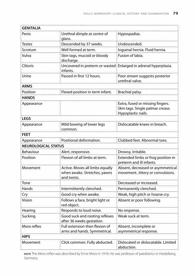
79skills workshop: clinical histor y and examination
GENITALIAPenis Urethral dimple at centre of
glans.Hypospadias.
Testes Descended by 37 weeks. Undescended.Scrotum Well formed at term. Inguinal hernia. Fluid hernia.Vulva Skin tags, mucoid or bloody
discharge.Fusion of labia.
Clitoris Uncovered in preterm or wasted infants.
Enlarged in adrenal hyperplasia.
Urine Passed in first 12 hours. Poor stream suggests posterior urethral valve.
ARMSPosition Flexed position in term infant. Brachial palsy.HANDSAppearance Extra, fused or missing fingers.
Skin tags. Single palmar crease. Hypoplastic nails.
LEGSAppearance Mild bowing of lower legs
common.Dislocatable knees in breach.
FEETAppearance Positional deformation. Clubbed feet. Abnormal toes.NEUROLOGICAL STATUSBehaviour Alert, responsive. Drowsy, irritable.Position Flexion of all limbs at term. Extended limbs or frog position in
preterm and ill infants.Movement Active. Moves all limbs equally
when awake. Stretches, yawns and twists.
Absent, decreased or asymmetrical movement. Jittery or convulsions.
Tone Decreased or increased.Hands Intermittently clenched. Permanently clenched.Cry Good cry when awake. Weak, high pitch or hoarse cry.Vision Follows a face, bright light or
red object.Absent or poor following.
Hearing Responds to loud noise. No response.Sucking Good suck and rooting reflexes
after 36 weeks gestation.Weak suck at term.
Moro reflex Full extension then flexion of arms and hands. Symmetrical.
Absent, incomplete or asymmetrical response.
HIPSMovement Click common. Fully abducted. Dislocated or dislocatable. Limited
abduction.
note The Moro reflex was described by Ernst Moro in 1918. He was professor of paediatrics in Heidelberg, Germany.

80 newborn care
3-i Examination of the hips
The hips must be examined in all newborn infants to exclude congenital dislocation or an unstable hip.
The infant is examined lying supine (back on the bed) with the hips flexed to a right angle and knees flexed.
Barlows test demonstrates both a dislocated and a dislocatable (unstable) hip: One hand immobilises the pelvis (thumb over pubic ramus, fingers over sacrum) while the other hand moves the opposite thigh into mid-abduction. If the hip is dislocatable, backward pressure on the inner side of the thigh with the thumb causes the femoral head to slip backwards out of the acetabulum. Conversely forward pressure on the outer side of the thigh with the fingers would tend to cause the head to spring forwards, back into the acetabulum. The same procedure is then carried out for the opposite side.
Ortolani test for a dislocated hip: Both thighs are held so that the examiner’s fingers are over the outer side of each thigh (greater trochanter) and his thumbs rest on the inner side of each thigh (lesser trochanter). Both thighs are then abducted. If a hip is dislocated, a ‘clunk’ can be felt and heard as the femoral head slips forward into its normal position in the acetabulum.
3-j Examination of the placenta
Every placenta should be carefully examined after birth as this can provide valuable information about the infant. Usually the gross placental weight is measured and recorded (placenta, membranes and umbilical cord). As gestation progresses the weight of the placenta increases. An infant of 3000 g usually has a placenta weighing about 600 g (between 450 and 750 g). Therefore, at term the gross placental weight is about a fifth that of the fetus. Infants who are underweight for gestational age have both an absolutely and relatively small placenta. In contrast, infants of poorly controlled diabetics, and infants who
have suffered a chronic intrauterine infection (e.g. syphilis) or fetal hydrops have placentas that weigh more than expected.
There are three layers to the placental membranes. The amnion on the inside (prevents the fetus sticking to the membranes), the chorion in the middle (to provide strength), and the decidua on the outside. The amnion is usually smooth and shiny. If the healthy amnion is peeled away from the rest of the membranes, it is completely clear and transparent. A cloudy or opaque amnion suggests infection (chorioamnionitis) while a granular surface (amnion nodosum) suggests too little amniotic fluid (oligohydramnios). The membranes should not smell offensive.
The umbilical cord normally has one large vein and two thick walled arteries. The more the pull (e.g. when a cord is relatively short due to it being wrapped around the fetal neck) the longer the cord will grow. A short cord suggests very poor fetal movement. The cord becomes stained green once the amniontic fluid has been contaminated with meconium for a few hours. A single umbilical artery is associated with congenital malformations. The umbilical vein has one-way valves (‘false’ knots). A true knot may kill the fetus.
The shape of the placenta is not important. Most are oval. Usually the umbilical cord is inserted into the centre of the placenta with arteries and veins radiating out in all directions over the chorionic plate. A peripheral insertion is of no clinical importance. However, insertion into the membranes in a low-lying placenta can result is severe haemorrhage from a fetal vessel when the membranes rupture (vasa praevia). Arteries always cross over veins. Fetal vessels torn off at the placental edge indicate that an extra piece of placenta has been retained (accessory lobe). Pale patches on the fetal surface are due to fibrin deposits and are not clinically important.
The maternal surface of the placenta is dark maroon in preterm infants but becomes grey towards term. A pale placenta suggests

81skills workshop: clinical histor y and examination
anaemia. Calcification is not important and reflects a good maternal calcium intake. The maternal surface is divided into lobes (cotyledons). Make sure that the placenta is complete as a retained lobe can result in postpartum haemorrhage or infection. Firmly attached blood clot, especially if it lies over an area of compressed placenta, suggest placental abruption. Fresh infarcts are best identified on palpation as they form a hard lump. Old infarcts are yellow or grey and easily seen, especially if the placenta is sliced. It is of no help to simple describe a placenta as ‘unhealthy’.
It is particularly important to examine the placentas of twins. Unlike-sexed (boy and girl) twins are always non-identical (dizygous). Liked-sex twins are definitely identical (monozygous) if they share a single placenta (monochorionic twins). Monochorionic placentas always have fetal blood vessels on the chorioninic place which run from one umbilical cord to the other. Monochorionic placentas have one chorion and usually two amniotic sacs. Two placentas fused together (dichorionic placentas) may be mistaken for a single placenta. However, there are never fetal blood vessels linking the two umbilical cords. Dichorionic placentas can be seen in both identical and non-identical twins. The separating membranes of dichorionic twins always include both amnion and chorion.
Pathological examination with histology should be requested if an abnormality of the placenta is identified. Placental ischaemia, chronic intrauterine infection and chorioamnionitis are easily identified on histology.
the roAd-to-heAlth CArd
Use of the road-to-health card (preschool health card) is advocated by the World Health Organisation as one of the main methods of improving child health, especially in a
developing country. The card is widely used throughout southern Africa.
After delivery each newborn infant is issued with a road-to-health card which forms the primary health-care record until the infant starts school by the age of 6 years. The infant’s mother keeps the card in a plastic cover and should present the card whenever the infant is taken to a clinic or hospital. The infant’s perinatal history, growth, immunisations and childhood illnesses are recorded on the card. Usually the infant’s HIV status and management are also recorded on the card.
3-k Completing the road-to-health card after delivery
After delivery the clinic or hospital staff must enter the perinatal details onto the road-to-health card. The details which are usually entered onto the card are:
1. Maternal information:
The mother’s nameThe mother’s hospital numberThe mother’s home address
2. Pregnancy and delivery information:
The duration of pregnancyThe result of the VDRL or other creening test for syphilis and HIVThe maternal blood groupAny pregnancy complicationsThe method of deliveryThe date and place of birth
3. Neonatal data:
The Apgar scoresThe birth weight (mass), head circumference (and sometimes length)The name and sex of the infantThe date, infant weight and method of feeding at discharge
Details of the information recorded on the preschool health card vary slightly from one region to another. Sometimes additional information is also recorded after delivery.
•••
••
••••
••
••
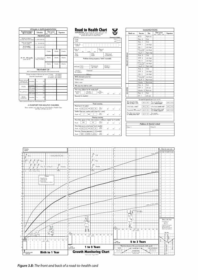
Figure 3.B: The front and back of a road-to-health card
3,5
4,5
4
6
5,5
5kg
7
6,5
8,5
8
9,5
9
11
10,5
11,5
13
14,5
15kg
14
12
13,5
15,5
16
16,5
2,5kg
4,5
6
5,5
5kg
7
6,5
7,5
8,5
8
10kg
9
11
10,5
11,5
13
12,5
14,5
15kg
14
12
13,5
16
16,5
18
18,5
19 kg
17,5
17
BCG
9mths
2
1,5
1
0,5
0
3
3,5
4,5
4
6
5,5
5kg
7
6,5
7,5
8,5
8
10kg
9,5
9
11
10,5
11,5
13
12,5
14,5
15
14
12
13,5
15,5
16
16,5
17
2,5kg
Child'sname:Date for next visit
Write on the chart- Any illness e.g. ~ diarrhoea, ~ ARI, etc.- Admission to hospital,- Solids introduced,- Breastfeeding stopped,- Birth of next child, etc.
like this:
Chart revised: August 2003
19
18
17
16
15
14
13
12
11
10
20
1
9
8
7
5
3
2
4
6
Birth to 1 Year
1 to 2 Years
Growth Monitoring Chartwrite birth monthand year
writebirth month
2 to 3 Yearswritebirth month
age in monthsBirth
weight1 2 3 54 6 7 8 9 1110
12 13 14 15 1716age in months
18 19 20 21 2322
24 25 26 27 2928age in months
30 31 32 33 3534
Watch the direction of the curve showing the child's growth
3rd centile
50th centile
97th centile
IMMUNISATIONS
60% of 50th centile
18mths
Measles
Polio
I can talk
DANGER SIGNNot gaining weight.
Find out why.
GOODMeans the child is
growing well.
VERY DANGEROUSChild may be ill,needs extra care.
Wo
rm m
edic
ine
A R
I Dia
rrh
oea
Ad
mit
ted
to h
osp
ital
(2-7
Sep
tem
ber
)
jjb
day month yearnr.
17,5
17
Extr
a m
eals
giv
en
23
22
21
24
0
14wks10wks6wks
Hib, DTP & Hep B1 2 3
Polio1 2 3
2Measles
1
4
4
3
birth
12,5
10kg
7,5
9,5
15,5
1year
2years
17,5
18
Discuss:BreastfeedingChild spacingFood intakeOral rehydration solution
DTP
12
13
15
16
18
19
17
3 to 4 Years
50th centile
97th centi
le
60% of 50th centile
write birth month
24
4 to 5 Years
5
DT
38 40 42 44 4846 50 52 54 56 5837 39 41 43 4745 49 51 53 55 595736age in months
Road to Health ChartIMPORTANT: always bring this chart when you visit
any health clinic, doctor or hospital andpresent the chart on school entry
Child'sname
Child's IDnumber
Date ofbirth
Place ofbirthday month year
Birthweight
Birthlength
Birth headcircumference
Problems during pregnancy / birth / neonatally
How many children has the mother had?Number
bornDate
informationgiven:
RtHC information given by:
Who does the child live with?
Mother's name:
Father's name:
IMMUNISATIONS
Vaccine Site Date given Signatureday month year
PRIM
ARY
SCH
EDU
LEBO
OST
ERS
BCG
Polio 0
Polio 1
DTP 1
Hib 1
Hep B 1
Polio 2
DTP 2
Hib 2
Hep B 2
Polio 3
DTP 3
Hep B 3
Hib 3
Measles 1
Polio 4
Measles 2
Polio 5
DT 1
DTP 4
Right arm
Oral
Oral
Left thigh
Left thigh
Right thigh
Oral
Left thigh
Left thigh
Right thigh
Oral
Left thigh
Left thigh
Right thigh
Right thigh
Oral
Left arm
Right arm
Oral
Left arm
Numberalive now
boygirl
Was the baby lessthan 2,5kg at birth
Are there any reasonsfor taking extra care?
Is the baby a twin? Is the baby bottle fed?
Are any brothers orsisters underweight?
Household TB contact? Does the mother needmore family support?
In need of special care (mark with X)
(for example:single parent etc.)
yes no
yes no
yes no
yes no
yes no
yes no
yes no
GW 8/123
Clinic 1:
Address of clinic(s) visitedDoes baby turn to a loud noise? (at 6 months)
L: R: yes noyes no dd mm yyDate
tested:Result:
143rd centile
10
11
3years 4years
10kg
write birth month
60
5years
Polio
1
9
13
12
15kg
14
16
17
20
19
22
21
24
23
10,5
11,5
12,5
13,5
15,5
16,5
17,5
18,5
19,5
20,5
21,5
22,5
23,5
5 years
14,5
18
23
11
Snellen Chart test: conduct with E-chart (5> years)
dd mm yyDate
tested:Result: L: R:/ /
Does baby appear to listen when someone is talking or singing? (at 3 months)
dd mm yyDate
tested:Result: yes no
Voice test: Hearing impairment (>12 months)
dd mm yyDate
tested:Result:
Gestationalage (wks)
Mother'sSerologyAPGAR 1 min.
5 min.
Mother'sfile numbers
Delivery:: Antenatal:
: :
dd mm yy
Reason(s) for death(s):
Pencil test (>6 weeks)
L: R: yes noyes no dd mm yyDate
tested:Result:
Hearing screening
Visual screening
Normalhearing
Moderateimpairment
Severeimpairment
Clinic 2:
Department of Health
Batch no:
9,5
9
DTP 2 / Hip 2 Left thigh(combined)
DTP 1 / Hip 1 Left thigh(combined)
DTP 3 / Hip 3 Left thigh(combined)
A PASSPORT FOR HEALTHY CHILDRENShow mothers you value the use of the Road to Health Chart
and they will take care of it
VITAMIN A SUPPLEMENTATIONSupplementation:
age in months Schedule Date given Signatureday month year
Mother at delivery (not later than 6-8 weeks)
Infant not breastfed(at 6 weeks)
1 x 200 000 IU
1 x 50 000 IU
PROPHYLAXIS
1 x 100 000 IU
At 12 - 60 months(mark with X)
1 x 200 000 IUevery 6 months
18mths 24mths
30mths 36mths 42mths
48mths 54mths 60mths
Persistent diarrhoea/
Xerophthalmia
Measles
Dosage according to following age group: 2-5mths: 50 000IU6-11mths: 100 000IU
12-60mths: 200 000IU(See IMCI classification)
TREATMENT OF :
Immediate
Immediate
24h repeat
Immediate
24h repeat
1 x ............IU
1 x ............IU
1 x ............IU
1 x ............IU
1 x ............IU
(NOT if prophylactic dose was given within previous month)
Severemalnutrition Immediate 1 x ............IU
Diarrhoea withsevere dehydration
At 6 months*(up to 11mths)
12mths
* Allow a period of at least one month between doses

objectives
When you have completed this unit you should be able to:
List the benefits of breastfeeding.Promote breastfeeding.Teach a mother how to breastfeed her infant.Manage common problems with breast-feeding.Treat cracked nipples and engorged breasts.Formula feed an infant.
•••
•
•
•
iNtroduCtioN to iNFANt FeediNg
4-1 What are the nutritional needs of a normal infant?
Like the adult, the infant needs the following nutrients to grow and develop normally:
WaterCarbohydrates such as lactose (milk sugar) and glucoseProteins (made up of amino acids)FatsVitaminsMinerals (such as sodium, potassium, chloride, calcium, magnesium, phosphate)Trace elements (such as iron)
The carbohydrates, proteins and fats provide the infant with 440 kilojoules per kg per day (105 kilocalories/kg/day) needed to grow normally.
4-2 What milk can be given to a normal infant?
Breast milk. Human breast milk meets all the nutritional needs of a healthy term infant.Formula feeds. These feeds are made from cow’s milk or soya bean and are modified to have similar constituents to breast milk.
Whenever possible mothers should breastfeed their infants
1.2.
3.4.5.6.
7.
1.
2.
4Feeding normal infants

8� newborn care
the beNeFits oF breAstFeediNg
4-3 What are the benefits of breastfeeding?
Breastfeeding provides many benefits to both the infant and mother. The main benefits are:
1. Benefits to the infant:
Breast milk is the perfect feed for term infants as it provides all the nutrients in the correct amount and proportion for normal growth and development until the age of 6 months. Thereafter breast milk should still provide a lot of the protein in the infant’s diet until 2 years of age.Breast milk is easily digested and absorbed.
note The protein in breast milk (lactalbumin) does not form a curd and, therefore, is easily digested. Most fatty acids in breast milk are unsaturated and, therefore, easily absorbed. The main sugar in breast milk is lactose.
Breast milk is clean and warm, and avoids the dangers of a contaminated water supply, inadequately sterilised bottles and teats, and lack of refrigeration facilities.Breast milk avoids the danger of diluted or concentrated formula.Breast milk contains many anti-infective factors, such as antibodies, living cells (lymphocytes and macrophages) and complement. Breast milk also encourages the growth of beneficial bacteria in the infant’s bowel. These properties of breast milk help to prevent gastroenteritis, the major cause of infant deaths in many poor communities.Breast milk decreases the risk of allergy in the infant, especially if there is a strong family history of allergy.
Breastfeeding decreases the incidence of gastroenteritis and therefore lowers the infant mortality rate in poor communities.
•
•
•
•
•
•
2. Benefits to the mother:
Breastfeeding is much cheaper than buying formula feeds.Breast milk is instantly available at all times. No sterilising of bottles and teats, and preparation of formula is needed.It is emotionally satisfying for the mother to successfully breastfeed her infant and this helps to form a strong bond between mother and infant.Breastfeeding helps the involution of the uterus and reduces the amount of bleeding during the puerperium.Breastfeeding helps the mother to lose excessive weight gained during pregnancy. Pregnancy, not breastfeeding, alters the shape of a woman’s breasts.Exclusive breastfeeding reduces the risk of the mother falling pregnant again.Breastfeeding may reduce the risk of breast cancer.
Breastfeeding saves money and ensures an adequate, safe supply of food. It therefore decreases the incidence of malnutrition.
Prolonged breastfeeding is one of the most successful ways of reducing infant mortality in poor communities.
4-4 Why do some mothers not breastfeed successfully?
Breastfeeding is not always easy as some mothers and infants have to learn the ‘natural art of breastfeeding’.
Some mothers do not breastfeed or fail to breastfeed successfully because:
They do not know the advantages of breastfeeding.They believe that they do not have enough milk because they do not understand that infants need frequent, small feeds for the first few days. It takes a number of days before the supply of milk increases.They think that their milk is too thin, or their breasts too large or too small.
•
•
•
•
•
•
•
1.
2.
3.
8�feeding normal infants
They develop cracked nipples or engorged breasts due to an incorrect method of feeding.They want to return to work and do not realise that many working mothers can continue to breastfeed successfully.They are afraid of breastfeeding.They have flat or inverted nipples which are often managed incorrectly.Traditional beliefs may result in unsuccessful breastfeeding, e.g. incorrect beliefs that colostrum is not good for the infant, intercourse spoils the milk, and delayed feeding causes the milk to become sour in the breast.Poor sleeping or excessive crying by the infant is blamed on the inadequate quality or supply of the breast milk.
Some HIV-positive mothers may elect not to breastfeed once they have been counselled.
promotiNg breAstFeediNg
4-5 How can breastfeeding be promoted?
Breastfeeding should be promoted as the normal, natural method of feeding an infant. This can be achieved by:
Encouraging a positive attitude towards breastfeeding in the home during childhood and adolescence by seeing other infants being breastfed.Teaching the advantages of breastfeeding in schools.Promoting breastfeeding in the media (radio, TV, books).Teaching the advantages and method of breastfeeding in all antenatal clinics.Starting breastfeeding groups run by mothers who have themselves breastfed.Encouraging breastfeeding in hospitals and clinics.Discouraging bottle feeding in hospital by using cups instead by women who formula feed.
4.
5.
6.7.
8.
9.
1.
2.
3.
4.
5.
6.
7.
Teaching mothers how to express their milk.
Further information on breastfeeding can be obtained from a local breastfeeding support group or local branches of the Breastfeeding Association of South Africa and La Leche League.
Breast is best
4-6 How can breastfeeding be encouraged in hospitals and clinics?
Staff should be trained in the many advantages that breastfeeding has for the mother and infant, they should feel comfortable and not embarrassed when speaking to patients about breastfeeding, and they must have the knowledge and skills to teach mothers how to breastfeed. Breastfeeding must be promoted during all visits to antenatal clinics.
Infants should be placed on the mother’s abdomen immediately after birth and put to the breast when ready to start breastfeeding. Rooming-in must be encouraged, giving mothers unlimited access to her infant to demand feed. Mothers must be helped individually with kindness and patience.
4-7 What is the value of a local breastfeeding support group?
Often the best person to advise and help a breastfeeding mother is someone who has herself breastfed an infant. A number of mothers who have breastfed and are interested in helping others to breastfeed can form a local support group. With help and training by knowledgeable midwives and doctors they can provide a very helpful service.
8.

8� newborn care
teAChiNg mothers to breAstFeed
4-8 What preparation does a mother need for breastfeeding?
The decision to breastfeed should ideally be taken before her infant is born. If she is undecided at delivery, she should be encouraged to breastfeed.
No preparation of breasts and nipples before delivery is necessary.
A good, supportive bra should be worn. Breast size or shape and nipple size are no indication of a woman’s ability to breastfeed.
Routine breast preperation during pregnancy is not needed
4-9 How can you treat flat or inverted nipples?
If a woman’s’ nipples appear flat or inverted during pregnancy, she may need additional help with breastfeeding. Hoffman’s exercises and nipple shields are not helpful and are no longer recommended.
Many flat or inverted nipples will correct once breastfeeding starts. The infant is the best treatment for flat or inverted nipples.
4-10 Should the infant be put to the breast immediately after delivery?
Yes. The mother should breastfeed her infant as soon as possible after delivery as the infant’s sucking drive is usually strongest in the first hour or two after birth. Early suckling promotes bonding between the mother and infant. It also stimulates milk production and encourages successful breastfeeding. The small amount of colostrum satisfies the infant and is very rich in antibodies. It is very important not to give formula feeds on the first day as this may interfere with the establishment of breastfeeding.
Practising kangaroo mother care (skin-to-skin care) is a very powerful way of promoting successful breastfeeding. Most mothers can be given their infant within minutes of birth, before the placenta is delivered. When placed on the mothers abdomen the infant will start looking for the nipple.
note If a patient cannot breastfeed her infant soon after delivery because she has had a caesarean section under general anaesthesia, then she should express some colostrum before the caesarean section. This can be used for the first few feeds. With an epidural or spinal anaesthetic, the infant can be given to the mother immediately after the surgery.
The infant should not be put to the breast immediately after delivery if:
Polyhydramnios was present. A nasogastric tube must be passed first to exclude an oesophageal atresia.The infant had low Apgar scores and required resuscitation.Severe maternal distress or illness is present.The mother has decided not to breastfeed.The infant is to be adopted.
Some stable preterm infants with an assessed gestational age of less than 35 weeks can be given to the mother for skin-to-skin care after delivery even if they are too immature to breastfeed.
4-11 Why are clear feeds not necessary?
It is not necessary to give clear feeds of sterile water or dextrose water to an infant before starting breast or formula feeds on day 1. A breastfed infant does not need additional clear feeds. If the mother chooses to breastfeed, no bottle feeds should be given to the infant as a teat can confuse the infant and cause it to reject the nipple.
Routine clear feeds are not needed on day 1
note Unlike a bottle-fed infant that sucks the milk out of the teat, a breastfed infant holds the nipple against the hard palate and compresses the milk
1.
2.
3.
4.5.

87feeding normal infants
ducts under the areola with the gums. This is called suckling.
4-12 What is the best position to hold an infant while feeding?
The correct position of the infant while feeding is important. The mother should be warm, relaxed and comfortable. Usually she sits up and holds her infant across her body in front of her. The infant is held in one arm, and should lie on its side with its mouth facing the nipple. The breast being offered to the infant is held in the other hand.
Mothers should be encouraged to try different feeding positions in order to find which is most comfortable. Some mothers prefer to lie down while they feed. Other mothers prefer to tuck the infant under an arm like a rugby ball.
4-13 How should an infant latch at the breast?
One of the commonest mistakes made when breastfeeding is that the infant is not held and latched correctly to the breast. Sucking or chewing on the nipple due to incorrect latching causes the mother pain and damages the nipple.
To latch the infant to the breast correctly the mother should hold the infant in a comfortable position facing her. With a hand holding her breast she should tickle the infant’s mouth with her nipple and wait for the infant to open its mouth (this is called ‘rooting’). The mother should then pull the infant towards the breast so that the infant takes the nipple and as much of the pigmented areola into the mouth as possible. She should feel no pain when the infant starts to suckle. If she feels pain she should gently remove the infant from the breast and latch the infant correctly. Make sure that the infant’s nose is not covered by the breast.
The infant must take the whole nipple and most of the areola into the mouth when latching to the mother’s breast
4-14 Should infants be demand fed?
Yes. Whenever possible infants should be demand fed. This means that the infant is put to the breast whenever hungry. A normal breastfed infant will usually feed every 2 to 3 hours during the day for the first few weeks. Demand feeding helps to encourage a good milk supply and prevent engorged breasts. Demand feeding is easy in hospital if mothers room-in and use skin-to-skin care.
4-15 What is the let-down reflex?
When an infant is put to the breast, the pituitary gland in the mother’s brain responds by producing the hormones prolactin and oxytocin. Prolactin stimulates the breast to secrete milk while oxytocin produces the ‘let-down reflex’. This reflex produces a tingling feeling in the breast, and results in milk being pumped into the infant’s mouth by the contraction of muscle cells that surround the milk ducts in the areola. At the same time, oxytocin causes the uterus to contract. Milk may leak from the other nipple. The release of oxytocin helps the uterus to involute but may produce abdominal pain during feeding for the first few days after birth. Reassure the mother that abdominal pain with feeding is normal. Tension, anxiety and a lack of sleep may inhibit the let-down reflex.
4-16 How do you manage leaking breasts?
Milk leaking from the breasts is common in the first few weeks of feeding. Leaking of the opposite breast during feeding is normal and can be stopped by pressing on that nipple. Cotton handkerchiefs or pads can be used for leaking between feeds. They should be changed frequently as dampness may cause sore nipples.
4-17 Can a mother’s milk be too strong or too weak?
No, but the appearance of breast milk varies. There are 3 different types of breast milk:
Colostrum. This is a milky fluid produced in small quantities for the first few days
1.

88 newborn care
after birth. However, it contains a lot of protein and provides all the infant’s nutritional needs. After 3 to 5 days the milk supply suddenly increases (‘comes in’) and the breasts feel full. This is due to the production of mature milk, which consists of foremilk and hind milk.Foremilk. This is produced at the start of each feed. It appears very ‘weak’ and ‘thin’ as it consists mainly of water with little fat. On a hot day a thirsty infant will take frequent, small feeds of foremilk.Hindmilk. This is only produced towards the end of a feed. It looks thick and rich, and contains a lot of fat. A hungry infant will empty the breast to obtain the hind milk.
It is best to empty one breast first before putting the infant to the opposite breast. This ensures that the infant gets the rich hind milk. However, on some days the infant may wish to feed on both breasts while on other days may want to feed on one breast only. Start each feed on alternate breasts.
4-18 Do all mothers produce enough breast milk?
All healthy mothers can produce enough milk for their infant if breastfeeding is managed correctly. Unfortunately many mothers stop breastfeeding during the first 5 days because they are incorrectly advised that they ‘do not have enough milk’ or because the infant is losing weight. Milk supply is normally poorest in the late afternoon and early evening.
A mother is probably not producing enough milk if her breasts do not feel full before a feed, especially in the mornings after day 5.
4-19 How can you improve a mother’s milk supply?
Reassurance, support and encouragement that she will be able to breastfeed. Also ensure that she is getting enough sleep and is not under too much stress, as anxiety is a major cause of poor milk production. Anxiety also inhibits the let-down reflex.
2.
3.
1.
Many mothers are more relaxed in their own homes.Make sure that she is latching the infant correctly to the breast and that the infant is sucking correctly. Pain during feeding suggests that the infant is not latching correctly.Put the infant to the breast frequently during the day until a good milk supply is established. If the infant is not demand feeding 2 to 3 hourly, it should be woken for feeds. The best stimulus to milk production is the infant sucking frequently and for prolonged periods.The mother should rest for a while in the afternoon and drink adequate fluids.Stop any formula or water feeds.
Reassurance, support and encouragement are important for successful breastfeeding
note If the milk supply has not improved after 5 days and the infant has not started to gain weight despite the above steps, the mother can be given metoclopramide (Maxolon) 10 mg 8 hourly or sulpiride (Eglonyl) 50 mg 8 hourly for a week to stimulate the secretion of prolactin. The dose should then be gradually reduced and stopped over a further week. Prolonged use of these drugs may be dangerous for the mother and infant.
4-20 How do you know that an infant is getting enough breast milk?
The infant is probably latching well and getting enough milk if:
The mother’s breasts feel full before feeds and empty after a feed.The infant is content at the end of a feed and falls asleep.The infant loses less than 10% of its birth weight.The infant starts gaining weight by day 5.The infant has 5 or more wet nappies a day after day 5.
Weight gain is best determined over a few days. Many mothers stop breastfeeding simply
2.
3.
4.
5.
1.
2.
3.
4.5.

89feeding normal infants
because their infant cries a lot and they think that the infant is not getting enough milk.
note If you are worried that an infant is not getting enough milk, then the infant can be test weighed before and after a feed. After day 5, most term infants will gain about 25 g per day.
Infants that gain weight normally are getting enough feed
4-21 Should infants be routinely test weighed?
No, there is no need to test weigh most infants. The amount of milk an infant takes varies widely between feeds. A small feed, which is common in the afternoon or when the mother is tired, may cause maternal anxiety.
4-22 What is the appearance of the stools of a breastfed infant?
The stools of a breastfed infant may be yellow or green, and may be loose or firm. The infant may have several stools a day or only pass stool every few days. The stools should not smell offensive. The number of stools a day does not indicate whether the infant is getting adequate feeds.
mANAgiNg breAst-FeediNg problems
4-23 What should you do if an infant refuses the breast?
Some infants may reject the breast and refuse to latch on the nipple. Common causes are a sore mouth due to thrush, the infant being ill or upset, or the milk flow being too fast. These problems should be looked for and treated. As infants get older they spend less time feeding. As long as the infant is gaining weight the mother should be reassured.
Do not hold the infant’s head too tightly or push the face towards the breast as the infant
will turn towards your hand instead of the nipple. It helps to squeeze a little breast milk onto the nipple before latching the infant.
4-24 What causes the infant to choke while feeding?
Sometimes the mother may have too much milk and the milk may flow too fast causing the infant to choke or gag when feeding. As a result the infant may refuse to feed and become restless. It may help for the mother to lie back at the start of the feed with the infant across her chest so that the milk has to flow uphill against gravity. The mother may have to express a bit to soften the areola before starting the feed. Only offer one breast per feed and only when the infant appears hungry and the milk supply will settle with time.
4-25 How should you manage swollen or painful breasts?
A normal, full breast feels tense and heavy, but is not painful and is relieved by feeding. Breasts that are swollen, tender, hard, lumpy and painful are caused by either engorgement or mastitis. Both engorgement and mastitis result from an obstruction in milk flow.
1. Engorged breasts:
Usually both breasts are swollen, hard and painful but the mother does not feel ill. The milk does not flow freely. Engorged breasts usually occur between days 3 and 5 when the mother’s milk suddenly ‘comes in’. Engorged breast are common if the infant does not feed correctly, if the mother does not room-in and if the mother does not demand feed.
Treatment consists of encouraging the infant to suckle frequently. The infant should be fed on the most painful breast first. Placing ice onto the breasts between feeds will help to reduce the swelling and a warm shower relieves the discomfort, while a mild analgesic like paracetamol (Panado) is helpful. Often the infant is not able to latch correctly if the breast is engorged as the nipples become flattened by the swelling. If some milk is first expressed
90 newborn care
from the breast to soften the areola, the infant will usually latch well. Breast engorgement is prevented by frequent feeds.
Sometimes only a segment of one breast is painful due to a blocked milk duct. Changing the infant’s position during the feed and gently massaging the breast towards the nipple usually clears the obstruction.
2. Mastitis (milk fever):
Mastitis is an inflammation of the breast due to blocked milk ducts and seepage of milk into the surrounding tissues. It causes a swollen, painful, red area of one breast. The mother feels ill and may have a temperature.
Treat with rest, warm compresses and a mild analgesic. It is most important that the infant continues to suckle frequently on the affected breast as this will help the milk to flow. Altering the feeding position often helps to drain the affected area. The breast milk is not dangerous for the infant. If the signs and symptoms do not improve within 24 hours an antibiotic (flucloxacillin) is prescribed for 10 days. If a fluctuant mass develops then a breast abscess has formed. This should be surgically drained. Because of the pain of a breast abscess, feeding may have to be stopped on that breast for a few days. Usually feeding can start again once the oozing from the abscess has stopped. Breastfeeding from the other breast must continue.
4-26 How do you prevent painful nipples?
It is most important that the infant is correctly latched at the breast so that the nipple is not chewed. Remember that infants breastfeed and do not nipple feed. Nipples should not be painful, even in the first few days, if the infant is correctly latched to the breast. Make sure that the infant has all of the nipple and most of the areola in the mouth when feeding. When removing the infant from the breast, the mother should insert her little finger into the corner of the infant’s mouth to break the suction.
Instead of protecting the nipples with lanolin cream, petroleum jelly (Vaseline) or masse
cream, it is suggested that a little colostrum or hind milk be left to dry on the nipples after each feed. The milk has anti-infective properties and the fat protects the nipples. Do not use alcohol on the nipples. Avoid vigorous washing or soap on the nipples.
Correct latching of the infant at the breast will help to prevent painful nipples
4-27 How should you treat painful nipples?
Cracked nipples are very painful and should be prevented by correctly latching the infant to the breast and avoiding engorged breasts. Treat cracked nipples with breast milk spread onto the nipple between feeds. Usually with correct latching to the breast the mother will feel no pain and the crack will heal within a day. However, should the cracked nipple be too sore to continue feeding, express the affected breast. Feed the infant on the other breast and after the feed give the expressed breast milk by cup.
4-28 Do breastfed infants need complementary feeds?
Most breastfed infants do not need complementary (additional) feeds of formula. Complementary feeds decrease the time the infant spends on the breast and, thereby, reduce the production of breast milk. Bottle feeds may confuse the infant. Only if an infant continues to lose weight and the mother has inadequate lactation should complementary feeds be used. The mother should be encouraged to express her breasts to increase milk production. Some mothers will give complementary feeds if they have to leave their infant for more than a few hours. However, expressing milk into a sterile container for the missed feed would be preferable.
Expressed breast milk can be safely stored up to 6 hours in a cool place or for 48 hours in a fridge. Breast milk can be safely frozen and stored for 2 weeks in a fridge freezer or 6 months in a deep freeze. Frozen milk should be thawed slowly by placing the container in

91feeding normal infants
warm (not hot) water. Expressed breast milk should be given by cup.
4-29 Can working mothers continue to breastfeed?
Yes. Mothers can continue to breastfeed for many months while working. Breastfeeds can be given in the morning and again when she returns home. Feeding over the weekend should not be a problem. Breast milk can be expressed at work and this can be stored and then given to the infant during the following day. Alternatively, formula can be given while the mother is away at work and then breastfeeds given when she is home. Ideally it should be possible to take the infant to work or place the infant in a creche at or near the place of work.
4-30 Do drugs cross into the breast milk?
Almost all drugs that the mother takes by mouth will cross into the breast milk but only in very small quantities that will not affect the infant. Breastfeeding mothers should only take medication that is necessary. There is no evidence that antituberculous drugs or antiviral drugs that cross in the breast milk are dangerous to the infant.
note Thyroxine, antithyroid preparations, Warfarin, antidepressants, anticonvulsants, mild analgesics and most antibiotics can safely be given to a lactating mother. Tetracycline and cytotoxics should not be used if the patient is breastfeeding. Avoid breastfeeding for 24 hours after radioactive iodine is given to treat thyrotoxicosis.
FormulA-FeediNg NeWborN iNFANts
4-31 When should an infant be formula fed?
If the mother has decided that she definitely does not want to breastfeed, then the infant should be fed formula. If possible the formula feed should be given by cup rather than by bottle.
1.
If a mother is unable to breastfeed because she is separated from her infant, she should express her milk, manually or with a breast pump, for the infant. Only if this is not practical should the infant be formula fed.If the mother has an inadequate milk supply despite advice and support, and if the infant is not gaining weight by 7 days, then complementary feeds should be started to ensure normal growth. However, the mother should be encouraged to continue to express her breast milk to increase her milk production.If the mother is very ill.If an HIV-infected mother decides to exclusively formula feed.
note Maternal tuberculosis is not a contraindication to breastfeeding unless the mother is very ill. The infant should be given prophylactic treatment for 3 months whether breast or formula fed.
4-32 What formula should be used for a term infant?
A number of starter formula feeds are available for term infants (e.g. NAN 1, S26 1, SMA 1). They are very similar and, therefore, the milk available at the local clinic or the cheapest milk should be bought. Unaltered cows milk, evaporated milk and skimmed milk are not suitable for infants under 6 months of age. Milk creamers must never be used to feed infants. Follow-up formulas are used from 6 months (e.g. NAN 2, S26 2, Lactogen 2).
Formula-fed infants should be fed on demand. If fed according to a schedule, most infants will need to be fed 5 times a day, at 06:00, 10:00, 14:00, 18:00 and 22:00. Most term infants will take about 100 ml per feed after the first week.
4-33 How is formula made up?
Usually a level scoop of milk powder (scraped level with a knife and not packed down) is added to every 25 ml of water in a cup or feeding bottle (read the instructions on the tin). The water should have been boiled beforehand
2.
3.
4.5.

92 newborn care
and allowed to cool. Mix the formula well before feeding the infant. The bottle and teat must have been cleaned and sterilised by boiling or standing in a disinfecting agent (Milton or half diluted Jik). A feeding cup can be cleaned with soap and water.
One of the great dangers of formula feeds is to make the mixture too strong or too weak. If too much milk powder is added, the infant may receive too much salt and protein which can be dangerous. If too little milk powder is added, the infant may become malnourished. Another danger is gastroenteritis caused by infected water or dirty bottles and teats. These and other problems of formula feeds can be avoided by breastfeeding.
Many of the dangers of infection when using bottles and teats can be avoided if cup feeds are used instead.
Formula-fed infants may be offered a few clear feeds daily if the weather is very hot. Bottle-fed infants must be held while feeding. The bottle should never be propped.
4-34 What are the advantages of cup feeding over bottle feeding?
If an infant cannot be breastfed it is better to cup feed than to bottle feed. The greatest advantage of cup feeding is that a cup can be easily cleaned with soap and water. A cup also dries easily, especially if placed in the sun which helps to sterilise the cup. This is most important when clean or boiling water is not available for washing bottles. A cup feed usually takes less time than a bottle feed. It is also easier to wean a preterm infant from tube feeds onto cup feeds than onto bottle feeds. Infants drink the milk out of the cup. The milk should not be poured into the infant’s mouth. Any small plastic cup or dish can be used to feed an infant. Breast milk can be expressed directly into the cup before a feed. Mothers who do not breastfeed should be shown how to cup feed correctly before they are discharged home after delivery.
In some infants bottle feeding may cause problems with breastfeeding as the mechanism of sucking from a bottle is different from feeding at a breast. This is often called ‘nipple confusion’.
The ideal feeding cup for formula can be used for measuring the correct amount of water, mixing in the powder, storing and finally giving the feed. It should also be easy to clean. Keep the cup away from flies and dust.
Cup feeding has many advantages over bottle feeding
4-35 Are iron and vitamin supplements needed?
A normal term infant born to a healthy mother on a good, mixed diet and regularly exposed to sunlight does not need supplements in the first 6 months of life. Additional iron and vitamin supplements may, however, be of benefit in poor communities when iron drops 0.3 ml (or syrup 5 ml) and multivitamin drops 0.3 ml (or syrup 5 ml) can be given daily. Supplements given to well term infants are not harmful. Remember that all preterm infants need supplements.
note Vitamin D deficiency in unsupplemented breastfed infants is not uncommon during winter, especially in dark-skinned infants, in countries far from the tropics. In some areas with very little fluoride in the drinking water, supplementary fluoride drops can be given to reduce the risk of dental caries.
4-36 When should solids be introduced?
Normally breast milk or formula feeds will meet all the infant’s nutritional needs until 6 months of age. Thereafter milk alone is not enough and solids are usually introduced. If possible, an infant should be exclusively breastfed for 6 months. Even if the mother can only breastfeed for a few weeks or months, this will be of benefit to both her and her infant. Introducing solids reduces the anti-infectious properties of breast milk.

93feeding normal infants
Some mothers continue to partially breastfeed up to 2 years. It is best to continue breastfeeding after solids have been introduced. This practice is particularly important in poor communities as breast milk provides the infant with a good source of protein. Weaning should be done over a few weeks by dropping one feed per week. In practice solids are often introduced early, especially with formula feeding.
Whenever possible infants should be exclusively breastfed for 6 months
4-37 How should lactation be suppressed if indicated?
Cold or warm compressors help relive the discomfort. Breast binding is no longer used. Oral pyridoxine 200 mg three times a day for 5 days may help. Only express a little milk if the engorgement is very uncomfortable. Fluid intake should not be reduced.
Bromocriptine (Parlodel) is not safe and should not be used. Oestrogen is contra-indicated as it increases the risk of deep-vein thrombosis.
note Cold cabbage leaves or ice packs will help to relieve the discomfort of engorgement if placed on the breast.
the bAby FrieNdly iNitiAtive
4-38 What is a baby friendly hospital?
The idea of a ‘Baby Friendly Hospital’ was introduced by the World Health Organisation to promote the advantages of breastfeeding. UNICEF is the agency which assesses and registers hospitals as baby friendly. To become registered as a Baby Friendly Hospital all the ‘Ten steps to successful breastfeeding’ have to be implemented. Clinics should also be baby friendly.
4-39 What are the ten steps to successful breastfeeding?
Have a written breastfeeding policy that is frequently communicated to all the health-care staff.Train all the health-care staff in the skills needed to implement successful breast-feeding.Inform all pregnant women about the benefits of breastfeeding.Help mothers to start breastfeeding within 30 minutes of delivery.Show mothers how to breastfeed and teach them how to maintain lactation even if they are separated from their infants.Do not give newborn infants formula or water feeds unless this is indicated for medical reasons.Allow mothers and their infants to remain together all the time from delivery to discharge.Encourage demand feeding.Discourage the use of dummies, teats and nipple shields.Promote the formation of breastfeeding support groups and refer mothers to these groups on discharge from hospital or clinic.
FeediNg the hiv-exposed iNFANt
4-40 How should HIV-positive women feed their infants?
The decision to exclusively breastfeed or exclusively formula feed should be made by all HIV-infected women well before they deliver. The choice should be made after the mother has been carefully counselled about the advantages and disadvantages as well as the risk of HIV transmission of each feeding method. It is most important that HIV-infected mothers do not give mixed breastfeeds (breast milk as well of formula or solid feeds) as this carries the highest risk of HIV transmission from mother to infant.
1.
2.
3.
4.
5.
6.
7.
8.9.
10.

9� newborn care
Mixed breastfeeding carries the highest risk of HIV transmission
4-41 When is it best to formula feed HIV-exposed infants?
The World Health Organisation recommends that HIV-positive women exclusively formula feed their infants only if all of the following criteria can be met:
It should be acceptable to her family and friends. There are social and cultural barriers to formula feeding in many poor communities. In some communities women may be afraid to formula feed and not breastfeed.It should be feasible to formula feed. The mother must have the knowledge and skills to make up formula correctly.It must be affordable to formula feed. Formula is expensive. Free formula may be provided in some areas.It should be sustainable. Formula must be available. Mothers often live far from shops in rural areas.It should be safe. Clean water must be available. The mother should be able to prepare feeds hygienically and be able to clean the bottles, teats and cups. Access to primary health care is particularly important if infants are formula fed.
If any of these criteria cannot be met, it would be better for women to exclusively breastfeed as the risk of HIV transmission in breast milk is probably less than the dangers of formula feeding. Women who decide to formula feed must be taught how to prepare and give formula correctly. A cup rather than a bottle should be used as cups are easier to clean.
Women should exclusively breastfeed unless the risk of HIV transmission in breast milk is greater than the dangers of formula feeding
note WHO uses the acronym AFASS for acceptable, feasible, affordable, sustainable and safe.
1.
2.
3.
4.
5.
4-42 Is the danger of HIV infection with breastfeeding not unacceptibly high?
No. In poor, underserved rural or peri-urban communities the risk of not breastfeeding (gastroenteritis and malnutrition) is often greater than the risk of HIV transmission via breastfeeding. With exclusive breastfeeding for 6 months the risk of HIV transmission through breast milk is less than 5%. If the mother is receiving antiretroviral treatment the risk is almost nil. Therefore, in many poor communities, exclusive breastfeeding is the safest option.
CAse study 1
A woman who bottle fed her first infant, delivers at term and wants to breastfeed this infant. However, she is concerned as she has small breasts and flat nipples. She asks the staff what are the benefits of breastfeeding.
1. Should she try to breastfeed this infant if she formula fed her first child?
Yes, many mothers are able to breastfeed successfully even if they did not breastfeed their previous children. The use of prolonged skin-to-skin care helps to promote breast-feeding. She needs to be shown how to hold and latch her infant correctly. She also needs a lot of support and encouragement. It is important to keep the mother and her infant together if possible.
2. Why are some women unable to breastfeed successfully?
The commonest cause of failure to breastfeed successfully is that mothers are not managed correctly and not fully informed about the advantages and method of breastfeeding.
3. Can women with small breasts feed their infants?
Yes. The size of a woman’s breasts is determined by the amount of fat and not

9�feeding normal infants
glandular tissue and, therefore, does not influence her ability to breastfeed.
4. How should flat nipples be treated?
No treatment is needed during pregnancy. However, it is important to identify women with flat or inverted nipples so that extra help can be given to get the infant to latch correctly at the breast.
5. What are some of the benefits of breastfeeding?
Breast milk is easily digested and meets all the infant’s nutritional needs.The risk of infection in the infant is much less with breastfeeding.Breastfeeding is convenient and cheap.Breastfeeding promotes bonding.
6. When should women decide whether to breastfeed or not?
This decision should be made during pregnancy and preferably before the infant is born.
CAse study 2
A mother has breastfed her infant for 3 days. The infant’s birth weight was 3200 g but he now weighs only 3000 g. When she squeezes her nipple, her milk appears very watery.
1. Is the infant’s weight loss normal?
Yes. Normal infants can lose up to 10% of their birth weight, which would be 320 g for this infant. The weight loss of 200 g will not cause the infant any harm.
2. When should an infant start gaining weight after birth?
Most infants start gaining weight between 3 days and 5 days after birth when breast-feeding is established and the mother’s milk supply increases.
•
•
••
3. Should the mother give complementary feeds of formula?
These are not needed. Complementary feeds will decrease the time the infant spends sucking at the breast and this will reduce the mother’s milk supply.
4. How can this mother increase her milk supply?
She should put the infant to the breast frequently to stimulate her milk supply. She can also be encouraged to express her milk after feeds. Usually there is no need for a mother to take drugs to increase her milk supply.
5. Why does her milk appear too weak?
Foremilk looks ‘too weak’. This is normal and contrasts with the hind milk which is much richer with more fat.
6. If the weather is hot should clear feeds be given?
There is no need to give clear feeds to breastfed infants.
CAse study 3
A mother who has breastfed her infant for 5 days develops painful, cracked nipples after her breasts became engorged. She feeds her infant every 5 hours and is taking flucloxacillin for an infected caesarean section wound.
1. How can cracked nipples be prevented?
With prevention of breast engorgement and careful attention to latching the infant correctly, cracked nipples should not occur.
2. How can breast engorgement cause painful nipples?
If the infant is unable to latch correctly due to swelling of the areola and breast, the nipple can be damaged by the infant sucking incorrectly.

9� newborn care
3. Why is it advisable for mothers to demand feed rather than schedule feed?
Demand feeding whenever the infant is hungry helps prevent engorged breasts which can lead to cracked nipples.
4. How should you treat cracked nipples?
By showing the mother how to latch correctly. Breast milk helps the nipple to heal.
5. Should the mother continue breast-feeding as she is taking an antibiotic? Give reasons for your answer.
Yes, she should continue breastfeeding. Like most other antibiotics, flucloxacillin will cross into the breast milk in small quantities only. This should not be dangerous to the infant.
6. How can she continue to breastfeed if she plans to return to work?
She can still breastfeed in the morning before going to work and again when she gets home. She can also breastfeed throughout the weekend and express her breast milk at work to be stored and fed by cup during the next day. Alternatively, the infant can be formula fed while she is at work. Ideally she should be able to take the infant to work with her.
CAse study 4
An HIV-positive woman decides to formula feed her infant. She is discharged from the clinic 6 hours after delivery without any clear instructions.
1. What milk formula should she buy?
She can buy any term formula. There is no need to buy the more expensive formulas. Fresh cows milk, evaporated milk or skimmed milk are not suitable for small infants.
2. How is formula made up?
Most formulas are made up by adding one level scoop of milk powder to every 25 ml of water. The water should first be boiled and allowed to cool. The formula must be mixed well. Making the formula too strong or too weak is dangerous.
3. What is the best way to formula feed an infant?
It is better to give formula by cup than by bottle as cups are easier to clean. This is particularly important in poor communities where gastroenteritis is common.
4. How should bottles and teats be cleaned?
The bottle and teat must be cleaned and sterilised by boiling or standing in a disinfect-ing agent (Milton or half diluted Jik). Teats should be scrubbed both inside and outside with a brush.
5. When should solids be started?
It is preferable to only introduce solids at 6 months. However, solids are often started earlier in formula-fed infants.
6. Should iron and vitamin supplements be given to healthy, term infants?
These are not routinely needed in term infants.

objectives
When you have completed this unit you should be able to:
Recognise high-risk infants and sick infants.Provide general care to sick infants.List the observations needed in these infants.Diagnose and treat shock.Diagnose and treat fits.Diagnose and treat acidosis.
•
••
•••
ClAssiFiCAtioN oF iNFANts oN the bAsis oF risk
5-1 How can infants be classified on the basis of risk?
All newborn infants can be classified into 1 of 3 groups:
Well infants.High-risk infants.Sick infants.
To decide which group an infant falls into, the history must be reviewed and the infant examined.
5-2 What is a well infant?
A well (or low-risk) infant has all of the following features:
Born at termWeight appropriate for gestational age and not wastedThe history of the pregnancy, labour, delivery and the post delivery period are normal.
1.2.3.
••
•
5Care of high-risk and sick infants
98 newborn care
The vital signs are normal and the infant appears normal on examination.
Infants that do not have all of the above features are either high-risk infants or sick infants. Well infants only require routine level I care. This can be provided in a clinic or at home.
5-3 What is a high-risk infant?
A high-risk infant is an infant that appears well but has a much greater chance than most infants of developing a clinical problem, such as hypothermia, hypoglycaemia, apnoea, infection, etc. in the newborn period.
High-risk infants appear clinically well on examination.
High-risk infants appear well but have an increased risk of complications
5-4 Which infants should be regarded as high risk?
An infant that appears well but has any of the following features should be regarded as high risk and, therefore, likely to develop a problem during the newborn period:
Infants that are born preterm or postterm.All low birth weight infants.Infants who are underweight or overweight for gestational age.Wasted infants.Infants who have a low 1 minute Apgar score (i.e. need resuscitation after birth).Infants who are born to mothers with a complicated pregnancy, labour or delivery.Infants who have had one or more clinical problems since delivery.Infants who were sick but have now recovered.
A high-risk infant often falls into more than one of the above categories.
•
1.2.3.
4.5.
6.
7.
8.
5-5 What should you do for a high-risk infant?
It is essential to identify the clinical problem that the infant is at risk of developing, so that the problem can be anticipated.Every effort should then be made to prevent this problem occurring.If this is not possible then the infant must be carefully monitored so that the problem can be identified as soon as it develops. This allows for early treatment.Once the problem occurs, it must be treated as early as possible.
You should aim at anticipating and preventing problems in high-risk infants, so as to avoid having to treat them. High-risk infants usually do not need immediate treatment.
It is important to anticipate problems in high-risk infants so that steps can be taken to prevent these problems occurring
This stepwise process of identifying a possible problem, taking steps to prevent it, monitoring the infant for early signs of the problem, and finally having to treat the problem only if the earlier steps fail, forms the basis of good patient care.
5-6 What is a sick infant?
A sick infant does not appear well and has abnormal clinical signs. The infant may previously have been well or may previously have been identified as a high-risk infant.
Therefore, if a well or high-risk infant develops one or more abnormal clinical signs, or the infant appears ill, then it is reclassified as a sick infant.
The most important clinical signs that indicate that an infant is sick are:
Heart rate. The infant may have a:Tachycardia (a heart rate more than 160 beats per minute)
1.
2.
3.
4.
1.•

99care of high-risk and sick infants
Bradycardia (a heart rate less than 120 beats per minute)
Respiration rate and pattern. Abnormal signs are:
Slow, shallow, irregular respirationRapid respiration (tachypnoea) more than 60 breaths per minuteGrunting, recession or gaspingApnoea
Colour. The infant may be:PalePlethoric (very red)Cyanosed. Centrally or peripherallySeverely jaundiced
Temperature. The infant may be hypothermic (cold) or pyrexial (hot). Activity. The infant may be:
Lethargic and respond poorly to stimulationHypotonic and less active than beforeFeeding poorlyJittery with abnormal movements or fits
You should recognise these most important or vital signs that are usually monitored routinely in a sick or high-risk infant. The sick infant may also be recognised by other, less common but abnormal signs on clinical examination, e.g. bleeding, oedema, abdominal distension, loose stools.
The recognition of a sick infant is one of the most important clinical skills that nurses and doctors must learn
Infants that have a congenital abnormality but are otherwise well are often grouped together with sick infants when management is planned.
5-7 What are the causes of a sick infant?
Of the many causes of a sick infant, the most important are:
Infection.Hypoxia.HypothermiaHypoglycaemia.Acute blood loss.
•
2.
••
••
3.••••
4.
5.•
•••
1.2.3.4.5.
Anaemia.Trauma.Marked hyperbilirubinaemia.Intraventricular haemorrhage.
mANAgiNg A siCk iNFANt
5-8 What should you do for a sick infant?
Resuscitate the infant if needed.Immediately treat the abnormal signs, e.g. give oxygen for cyanosis.Attempt to make a diagnosis of the cause of the clinical signs.Treat the cause if possible.Give the infant general supportive care.Monitor the vital signs.Discuss the problem with the parents.Decide whether to transfer the infant to a level 2 or 3 nursery.
5-9 How do you resuscitate a sick infant?
The method of resuscitating a sick infant is similar to that of resuscitating an infant with a low Apgar score at birth. The most important steps are:
Clear the airway by suction, especially if the infant has vomited.Provide a source of oxygen if the infant is cyanosed.Stimulate respiration if the infant is not breathing adequately. Ventilation by face mask or endotracheal tube may be needed.Assess whether the infant is shocked. Treat if signs of shock are present.
5-10 How do you diagnose the cause of the problem?
Review the history.Examine the infant carefully.Do any special investigations that are indicated, such as:
Measure the blood glucose concentration.Determine the packed cell volume or haemoglobin concentration.
6.7.8.9.
1.2.
3.
4.5.6.7.8.
1.
2.
3.
4.
1.2.3.
•
•
100 newborn care
Determine the acid base status by measuring the blood gases if possible.Order a chest X-ray.
5-11 What general supportive care is needed by a sick infant?
Maintain adequate respiration and circulation.Maintain a normal body temperature.Handle the infant as little as possible.Provide extra oxygen only if needed.Observe the infant carefully, paying special attention to the vital signs.Provide fluid and energy by giving intravenous fluid. Usually the stomach is emptied via a nasogastric tube.Prevent infection by washing your hands or spraying them with a disinfectant before touching the infant.
5-12 What should you not do if the infant is sick?
Do not feed the infant by mouth as this may cause vomiting or apnoea. Most sick infants are given an intravenous infusion.Do not handle the infant unless it is necessary.Do not bath the infant.Do not take the infant out of oxygen for a procedure, e.g. a chest X-ray.
5-13 Why is excessive handling bad for sick infants?
Handling a sick infant, such as changing the nappy, may precipitate an apnoeic attack or bradycardia. The infant should not be suctioned unless this is indicated. The PaO2 (concentration of oxygen in arterial blood) and SaO2 (oxygen saturation) often fall when the infant is handled or suctioned. It is, therefore, important to handle a sick infant as little as possible. Only essential observations and procedures should be done. If possible, all needed procedures should be done at the same time rather than repeatedly handle the infant.
•
•
1.
2.3.4.5.
6.
7.
1.
2.
3.4.
moNitoriNg A high-risk or siCk iNFANt
5-14 How should you monitor a high-risk or sick infant?
The infant’s vital signs should be observed and recorded with as little interference as possible to the infant. Electronic monitors, e.g. a respiratory, heart rate and oxygen saturation monitor, should be used whenever possible. The infant should be handled and moved as little as possible when observations are made.
5-15 What observations should you make in a high-risk or sick infant?
You should observe the vital signs:Heart rateRespiratory rate and patternColourTemperature of the infant and the incubatorActivity of the infant
Look for any other abnormal signs that are present, such as vomiting, loose stools, etc.The blood glucose concentration is usually monitored.The type and volume of the fluid intake (both oral feeds and intravenous fluids) are recorded.The frequency with which the infant passes urine must be charted. In severely ill infants the urine is collected in a urine bag or urethral catheter so that the volume of the urine passed can be measured.The content of the urine (protein, blood and glucose) is determined with a reagent strip.The blood pressure should be recorded in severely ill infants in level 2 or 3 nurseries.If the infant is receiving oxygen, then the FiO2 (fraction or percentage of inspired oxygen) must be noted. The SaO2 should also be recorded if the infant is being monitored with an oxygen saturation monitor.
1.••••
•2.
3.
4.
5.
6.
7.
8.

101care of high-risk and sick infants
The packed cell volume or haemoglobin concentration should be measured if the infant has lost blood.The infant’s weight should be recorded every day.
5-16 What is the importance of weighing a sick infant daily?
A weight loss of more than 10% of the birth weight or the previous day’s weight suggests that the infant is dehydrated.
5-17 How often should the observations be made?
Observations should be frequent enough to ensure that any change in the infant’s condition will be detected as soon as possible. The sicker the infant, the more frequently observations will be needed. Very sick infants should be observed continuously. The use of monitoring equipment, such as apnoea alarms and heart rate monitors, is of great help in continuous monitoring. Routine observations in sick infants are usually made every 30 minutes. Three-hourly observations are usually adequate in high-risk infants.
5-18 How should the observations be documented?
It is very important that the observations be carefully recorded on an observation chart together with the time of the observations. Not only is the observation record very helpful to monitor the infant’s progress but it also provides an important hospital record. All abnormal observations must be acted upon. It is of little value to record an abnormal observation (e.g. hypoglycaemia) if the problem is not managed.
5-19 Which observation chart should be used?
A chart that is specially designed for the recording of observations on newborn infants should be used. The observation chart must have columns for the recording of the vital signs
9.
10.
and other important observations. A column must also be provided for the date and time, the observer’s name and a space for comments.
The observation chart must be kept at the infant’s cot or incubator so that it is easily accessible to both nurses and doctors.
5-20 What additional records are important?
Progress notes must be written by the nursing and medical staff describing the infant’s condition and the management given. The progress notes can be written on the observation chart or in a separate folder. Both nursing and medical staff should use the same records. The notes should be problem orientated.
5-21 What are problem-orientated notes?
Every time the infant is examined, you should ask yourself ‘what are the infant’s problems?’ Each problem must then be listed, together with the clinical observations, investigations and management of that problem. This system forces you to pay careful attention to each problem, and makes it very easy for another person to understand your notes. Each problem must be carried over in the notes from day to day until that problem has been resolved.
the mANAgemeNt oF shoCk
5-22 How do you diagnose shock?
Shock is the failure of the circulation to provide an adequate blood supply to the tissues. The signs of shock are:
Decreased capillary filling timeCold hands and feet in spite of a warm trunkTachycardia or bradycardiaLow blood pressurePallor or cyanosis
1.2.
3.4.5.

102 newborn care
In clinical practice, a decreased capillary filling time is the easiest sign of shock to detect.
note The blood pressure in an infant is best measured with an automatic machine that uses the principles of oscillometry or Doppler shift. A cuff is placed around the upper arm to measure the blood pressure (mean, systolic and diastolic) in the brachial artery. The width of the cuff must be two thirds the length of the upper arm and the balloon must be long enough to extend completely around the arm. The normal range of systolic blood pressure in term infants on day 1 is 50 to 70 mm Hg while that for diastolic blood pressure is 25 to 50 mm Hg. The normal range increases with the birth weight and gestational age of the infant. It also increases during the first week of life. The mean blood pressure is similar to the infant’s gestational age, e.g. 38 mm Hg at 38 weeks gestation.
5-23 How can you measure the capillary filling time?
Normally the capillaries in the skin fill immediately after they have been emptied. Due to the poor peripheral blood flow in shock, there is a delay in the time it takes to fill the capillaries.
The method of determining the capillary filling time is as follows:
Press firmly over the skin of the infant’s heel or anterior chest with your thumb or finger for 3 seconds. The skin under your thumb will become pale as the capillary blood is pushed out.Remove your thumb and immediately start counting. Stop counting when the blood has returned to the compressed area and the pallor has disappeared.To ensure that you count at the correct speed of one number per second, it is helpful to count ‘1 potato, 2 potatoes, 3 potatoes’, etc.
The normal capillary filling time is 3 seconds or less. If the capillary filling time is more than 3 seconds then the infant is shocked. A false reading may be obtained if the infant is cold, as this may also cause poor perfusion of the skin.
1.
2.
3.
5-24 What are the causes of shock?
Hypoxia is the commonest cause of shock in newborn infants.Excessive vagal stimulation caused by suctioning the pharynx.Haemorrhage.Dehydration.Septicaemia.Heart failure.
5-25 How do you treat shock?
The treatment of shock is aimed at:
Correcting the cause of the shock, e.g. septicaemia or hypoxia.Correcting the poor peripheral circulation by giving intravenous resuscitation fluids, such as normal saline, fresh frozen plasma, stabilised human serum or Haemaccel. Ten to 20 ml/kg of fluid is given over 10 to 20 minutes. This should correct the blood pressure and capillary filling time.
note An intravenous infusion of dopamine at a rate of 5 µg/kg/minute may be used to increase cardiac output if fluid alone does not correct the shock.
the mANAgemeNt oF Fits
5-26 How can you recognise a fit in a newborn infant?
A fit (i.e. a convulsion) is caused by excessive activity of a group of nerve cells in the brain. A fit may present as:
Twitching of part of the body (e.g. a hand), one side of the body, or the whole body (a generalised fit).Extension (spasm) of part of the body (e.g. an arm) or the whole body.Abnormal movements (e.g. mouthing movements, deviation of the eyes to one side or cycling movements of the legs).Apnoea alone.
It is often very difficult to recognise a fit in a newborn infant as infants usually do not have a typical grand mal fit (generalised extension
1.
2.
3.4.5.6.
1.
2.
1.
2.
3.
4.

103care of high-risk and sick infants
followed by jerking movements) as seen in older children and adults.
Jitteriness and the movements normal infants make while asleep must not be confused with fits. Unlike fits, jitteriness can be stimulated by handling the infant. In addition, jitteriness can be stopped by holding that limb.
5-27 What are the important causes of fits?
The common causes of fits in the newborn infant are:
Hypoxia, especially neonatal encephalo-pathy (hypoxic ischaemic encephalopathy) due to severe intrapartum hypoxia)HypoglycaemiaMeningitisCongenital malformations of the brainTrauma or bleeding in the brain
note Hypocalcaemia and hypomagnesaemia may also cause fits.
5-28 How do you treat a fit?
1. Make sure that the infant is getting enough oxygen:
Clear the mouth and pharynx by suction and remove any vomited feed.Give oxygen by face mask.If the infant remains cyanosed or does not breathe, ventilate the infant by bag and mask or via an endotracheal tube.Empty the stomach by a nasogastric tube to prevent vomiting.
2. Stop the fit.
The 2 drugs usually used are either:
Phenobarbitone intravenously 20 mg/kg.Diazepam (Valium) 0.5 mg/kg by slow intravenous injection or rectally. Intravenous Valium may cause apnoea. Rectal Valium is given by a syringe and nasogastric tube. Valium is usually only used if phenobarbitone fails to stop the fit.
note If available, paraldehyde 0.3 ml/kg by intramuscular injection can also be used. Do not
1.
2.3.4.5.
•
••
•
••
keep paraldehyde in a plastic syringe for more than 2 minutes as it may react with the plastic.
3. Look for and treat the cause of the fit.
Important steps in diagnosing the cause of the fit are:
Always check the blood glucose concentration as hypoglycaemia is a common cause of fits.A history of fetal distress or a low Apgar score will suggest hypoxia before delivery.A lumbar puncture should be done to exclude meningitis.
If a cause is found, it should be treated. Unfortunately there is no standard method yet of treating hypoxic brain damage once it has already occurred.
4. Prevent further fits.
Usually a single intravenous dose of phenobarbitone will not only stop a fit but also prevent further fits. However, if there is a further fit, the dose of phenobarbitone can be repeated intravenously. Only if there are recurrent fits should a maintenance dose of 5 mg/kg oral phenobarbitone daily be started. All infants that have had a fit should be transferred to a level 2 or 3 nursery for further investigation and management. However, it is essential that hypoglycaemia is corrected and good respiration ensured before the infant is moved.
the mANAgemeNt oF ACidosis
5-29 What is acidosis?
Normally acid and alkali are present in equal amount in the body and are therefore in balance. Acidosis is an excess of acid in the body. This is determined by measuring the pH (hydrogen ion concentration) of the arterial blood, using a blood gas analyser (an ‘Astrup machine’). Normally the arterial blood pH
•
•
•

10� newborn care
in a newborn infant is 7.30–7.40. If the pH is below 7.30 then there is too much acid in the blood and the infant is said to be acidotic. In contrast, if the pH is above 7.40 there is too little acid in the body and the infant is said to be alkalotic. Alkalosis is less common than acidosis and is usually not as dangerous.
note Capillary blood from a warmed heel may also be used to measure the pH.
5-30 What types of acidosis are important?
There are 2 types of acidosis:
Metabolic acidosis. This is the common and dangerous type of acidosis seen in sick infants. It is due to the accumulation of lactic acid which is formed during hypoxia, septicaemia, dehydration and shock when the cells of the body do not receive enough oxygen. If the cells receive too little oxygen, some energy can still be produced by converting a lot of glucose into lactic acid (anaerobic metabolism). The increased production of lactic acid lowers the pH, resulting in a metabolic acidosis.Respiratory acidosis. This type of acidosis is caused by the accumulation of carbon dioxide in the blood during respiratory distress and apnoea. Because the lungs are unable to get rid of carbon dioxide, the excess carbon dioxide dissolves in the plasma to form carbonic acid.
Therefore, acidosis may be caused by too much lactic acid or carbonic acid.
5-31 How does the body try to correct a metabolic acidosis?
The body tries to correct the low pH by hyperventilating (excessive breathing). This lowers the PaCO2 (carbon dioxide concentration in arterial blood) and, thereby, reduces the amount of carbonic acid.
The excess lactic acid is bound to bicarbonate (base) in the blood. This lowers the serum bicarbonate concentration, producing a base deficit.
1.
2.
5-32 How is acidosis diagnosed?
Although there are clinical signs which may suggest acidosis in older children and adults, these are of little use in infants. Acidosis, therefore, is diagnosed by measuring the pH of a sample of arterial blood. It can further be decided whether the acidosis is metabolic or respiratory by determining the base deficit and measuring the carbon dioxide concentration (PaCO2) in the blood specimen.
With a metabolic acidosis the pH is below 7.30 and the base deficit is greater than 5 (normal = 0).With a respiratory acidosis the pH is below 7.30 and the carbon dioxide concentration (PaCO2) is above 6.8 kPa (50 mm Hg).
The pH, the base deficit and carbon dioxide concentration are determined with a blood gas analyser which also calculates the base deficit.
With a metabolic acidosis the ph is low and the base deficit is high
5-33 How do you treat metabolic acidosis?
Always try to find and correct the cause, e.g. giving oxygen for hypoxia, treating septicaemia with antibiotics, and correcting shock with intravenous fluids.
If the pH is below 7.20 then 4% sodium bicarb-onate must be given intravenously. Usually 2 ml/kg is given slowly over 10 minutes. The correct amount of 4% sodium bicarbonate that should be given can be calculated from the base deficit:
The volume of 4% sodium bicarbonate (in ml) = base deficit × the infant’s weight in kg × 0.6.For example: If the base deficit was 10 and the infant weighs 1.5 kg then the volume of 4% sodium bicarbonate needed would be 9 ml (10 × 1.5 × 0.6).
Never use 8% sodium bicarbonate in newborn infants as the concentration is dangerously high. If 4% sodium bicarbonate is not
1.
2.
•
•

10�care of high-risk and sick infants
available, then 8% sodium bicarbonate must be diluted with an equal volume of sterile water to make up a 4% solution.
It is of little help to give sodium bicarbonate if the cause of the metabolic acidosis is not removed as the acidosis will simply recur. Always make sure that the infant’s respiration is adequate before giving sodium bicarbonate.
5-34 How do you treat respiratory acidosis?
The infant must be ventilated with a mask and bag or ventilator. When the high PaCO2 (above 6.8 kPa or 50 mm Hg) is corrected, the pH will return to normal. Sodium bicarbonate will not correct a respiratory acidosis.
CAse study 1
A preterm infant of 1500 g has a normal Apgar score and appears healthy after delivery. The infant is transferred to the nursery for further care.
1. Should this infant be classified as a well, high-risk or sick infant?
The infant should be classified as high risk as the infant, although appearing well, is preterm and low birth weight.
2. List the 4 steps in the management of a high-risk infant.
Identify the clinical problems that are likely to occur.Take steps to prevent these problems occurring.Monitor the infant, specifically looking out for these problems.Treat any problems that do occur.
3. Do high-risk infants need immediate care?
No. Unlike sick infants, high-risk infants do not need immediate care. However, steps to prevent possible problems should be taken as soon as possible.
1.
2.
3.
4.
4. Would you expect the vital signs of a high-risk infant to be normal or abnormal?
The vital signs of a high-risk infant are normal. If any of the vital signs become abnormal then the infant must be reclassified as a sick infant.
CAse study 2
A two day old term infant becomes lethargic and develops abdominal distension. A clinical diagnosis of septicaemia is made. The heart rate is 180 beats per minute, the respiration and temperature are normal and the infant is peripherally cyanosed. The capillary filling time of the skin over the anterior chest is 5 seconds.
1. Is this a high-risk infant or a sick infant?
This is a sick infant as some of the vital signs are abnormal and the infant appears sick.
2. Which vital signs are abnormal?
The infant has a tachycardia (heart rate more than 160 beats per minute).The infant is peripherally cyanosed.The infant is lethargic.
3. What other clinical signs are abnormal?
Abdominal distensionThe capillary filling time is prolonged
4. What is a normal capillary filling time?
The normal capillary filling time is 3 seconds or less.
5. What is the clinical significance of a prolonged capillary filling time?
It indicates that the infant is shocked.
6. How should the shock be treated?
By giving 10 to 20 ml of normal saline (or fresh frozen plasma, stabilised human serum or Haemaccel) per kilogram by intravenous infusion over 10 to 20 minutes. The
1.
2.3.
1.2.

10� newborn care
septicaemia, which has caused the shock, must also be treated with parenteral antibiotics.
CAse study 3
A term infant is delivered by caesarean section after a diagnosis of fetal distress is made. The Apgar scores are 2 at 1 minute and 5 at 5 minutes. After resuscitation the infant appears lethargic and at 2 hours has a generalised fit. The blood glucose concentration is normal.
1. What is the probable cause of the fit?
Prenatal hypoxia which resulted in the fetal distress and low Apgar scores.
2. What are the clinical presentations of fits in a newborn infant?
Twitching of part or the whole bodyExtension (spasm) of part or the whole bodyAbnormal movements such as mouthing or deviation of the eyes to one sideApnoea
3. What are the 4 important steps in treating fits?
Make sure that the infant is getting enough oxygen.Stop the fit.Look for and treat the cause of the fits.Prevent further fits.
4. How should the fits be stopped?
Phenobarbitone 20 mg/kg is usually used to stop the fit. Intravenous (or rectal) diazepam (Valium) 0.5 mg/kg is also effective but may cause apnoea.
5. What drug can be used to prevent further fits?
Phenobarbitone 20 mg/kg by intravenous or intramuscular injection will not only stop most fits but should prevent further fits.
1.2.
3.
4.
1.
2.3.4.
CAse study 4
A week old preterm infant of 1500 g develops loose stools and loses 50 g in weight overnight. Gastroenteritis with dehydration is diagnosed. A blood gas analysis on a sample of arterial blood gives the following result: pH 7.16; PaCO2 5.1 kPa (37 mm Hg); base deficit 15. Nasogastric feeds are stopped, an antibiotic is prescribed and an intravenous infusion is started.
1. Is the infant acidotic?
Yes, because the pH is below 7.3.
2. Does the infant have a metabolic or respiratory acidosis?
A metabolic acidosis because the base deficit is greater than 5. There is not a respiratory acidosis as the PaCO2 is normal, i.e. not above 6.8 kPa (50 mm Hg).
3. What is the cause of the metabolic acidosis?
Dehydration due to the loose stools.
4. How should the metabolic acidosis be treated?
Remove the cause of the acidosis by correcting the dehydration with intravenous fluids.Give intravenous 4% sodium bicarbonate as the pH is below 7.2.
5. How much sodium bicarbonate should be given to this infant?
The infant has a base deficit of 15 and weighs 1450 g at the time that acidosis is diagnosed. Therefore, the infant should be given (15 × 1.450 × 0.6) 13 ml of 4% sodium bicarbonate. Remember that 8% sodium bicarbonate is not used in newborn infants.
1.
2.

objectives
When you have completed this skills workshop you should be able to:
Write good clinical notes.Record routine observations.
••
WritiNg good CliNiCAl Notes
Good clinical notes, which form the patient record, should be accurate, brief and easy to read. In addition, they must be systematic. Therefore, they should be written in an orderly, logical way so that all staff members can understand them.
5-a The date and time
Whenever notes are written it is important to give the date and the time that the record is made. It is then possible to know when the observation was made or care was given.
5-b Always sign your notes
Every time you write clinical notes you should sign (and write) your name. The rest of the health team then knows who wrote the notes.
5-c The ‘soap’ method of writing notes
When an infant is examined for the first time the clinical notes should include:
The story (i.e. the history)The observations (i.e. the physical examination and investigations)The assessmentThe plan
In order to remember these important steps in writing clinical notes, remember the word ‘SOAP’. The letters in SOAP stand for Story–Observations–Assessment–Plan.
5-d The story
Good notes should always start with the history (i.e. the history of the pregnancy, labour, delivery and events after delivery). A history should always be taken before examining an infant.
1.2.
3.4.
Skills workshop: Clinical notes and observations

108 newborn care
5-e The observations
The observations include the findings of the physical examination and the results of any additional investigations done, e.g. packed cell volume or chest X-ray.
5-f The assessment
Once you have recorded the results of the history, the physical examination and the investigations, you must make an assessment of the infant’s condition. For example, you should ask yourself:
Is the infant sick or well?Is the infant at high risk or low risk for clinical problems?What clinical problems does the infant have at present?
The assessment must not be forgotten as a carefully recorded history and examination are of little value if you are unable to assess what the results mean. The management depends on an accurate assessment of the infant’s problems. If you cannot identify the problems you will not be able to plan the correct treatment. Assessing an infant’s problems correctly takes a lot of practice.
5-g The problem list
When the assessment is made, it is very helpful to compile a problem list. Each clinical problem that you identify from the story and observations must be listed separately. A typical problem list looks like this:
Unmarried, teenage mother.Preterm delivery.Jaundice.
You now have a good idea of the clinical problems that require management.
Read the following case history and draw up your own problem list:
After a normal vaginal delivery at 40 weeks, an infant has Apgar scores of 3 and 8 and requires mask ventilation. The birth weight is 2300 g. The infant is not put to the breast
1.2.
3.
1.2.3.
after delivery and at 45 minutes after birth the blood glucose concentration, measured with a reagent strip, is 1.5 mmol/l. While starting an intravenous infusion, the infant’s skin temperature falls to 34.5 °C.
You should be able to identify at least 4 problems. Each will have to be managed.
5-h The management
Finally the management of the infant must be planned. The management consists of the nursing care, the observations needed, the medical treatment, and the management of the parents.
5-i An example of good ‘soap’ notes
14-1-2008 10:30
S:
18 year old primip. Booked. Spontaneous
preterm labour. 35 weeks by dates and
palpation. No signs of fetal distress.
NVD 06:15. Apgar scores 4 and 9.
Intubation and ventilation needed for
3 minutes. Thereafter infant moved to
nursery.
O:
Male infant. Weight 2000 g.
Assessed gestational age 36 weeks.
Active. No congenital abnormalities.
Skin temperature 36 oC.
RS – Respiratory distress with recession
and a respiratory rate of 65 breaths
per minute. Infant needs 50% head box
oxygen to remain pink.
CVS – Heart rate 150/min. Well perfused.
GIT – Abdomen normal.
CNS – Appears normal. Fontanelle flat.
Blood glucose 3.0 mmol/l. PCV 60%.
A:
1. Preterm delivery.
2. Neonatal asphyxia.
3. Respiratory distress.

109skills workshop: clinical notes and obser vations
P:
1. Incubator.
2. Neonatalyte IVI at 4 dpm.
3. Nasogastric tube. Nil per mouth.
4. Routine observations.
5. Head box oxygen.
6. Speak to parents.
7. Arrange transfer to level 2 hospital.
Signed: Sr. Mowtana
These brief notes give all the important information in a simple and systematic manner. Try to write your notes using the SOAP method.
5-j An example of poor notes
No antenatal care. Antepartum
haemorrhage.
Normal delivery. 2000 g. Female, term
infant.
Good Apgar scores. Vitamin K given.
Temp. 36 oC. Infant looks pale. Blood
glucose normal.
No respiratory distress. Heart rate
200/min.
Abdomen normal. Sucks poorly.
Keep nil per mouth. Neonatalyte infusion
started at 5 dpm.
Hb. 10 g/dl. Blood taken for cross match.
Nurse in incubator.
Although most of the information is given, these notes are not systematic and, therefore, they are difficult to understand. Notice how the history, examination and investigations are mixed up in a disorganised way. There is no problem list so that the reader is not sure what problems have been identified. There is also no date or signature. Try to rewrite these notes using the SOAP method. Do not forget to draw up a problem list.
5-k Problem-orientated patient record
When writing follow-up notes, the SOAP system can be applied to each problem in turn. This method is known as the problem-orientated patient record. It is very useful in a nursery where infants may need ongoing care for days or weeks. Each day the problem list of the previous day is examined. You must decide which problems remain unresolved and, therefore, must be carried over to the next day. Resolved problems can be dropped from the list. After reviewing the record for the past 24 hours and examining the infant, any new problems are added to the previous list.
For example, on day 2, the infant described in 5-i is doing well. The respiratory distress has improved slightly but the infant has developed a mild conjunctivitis. The problem list for day 2 should, therefore, be:
Preterm infant.Respiratory distress.Conjunctivitis.
The problem of neonatal asphyxia has been removed from the problem list, as it has resolved and no longer has any effect on the infant, while the new problem of conjunctivitis has been added to the list.
Again the SOAP system can be used, but now it is applied to each problem in the problem list. For example:
15/1/08 09:00
1. Preterm infant.
S:
No problems during the night. Passed
meconium. No apnoea.
O:
Active. Abdomen normal. Not pale. Blood
glucose and temperature normal.
A:
No change.
P:
1. Keep in incubator.
1.2.3.

110 newborn care
2. Start 2 x 12 feeds of expressed breast
milk.
3. Continue Neonatalyte at 4 dpm.
2. Respiratory distress.
S:
Oxygen requirements came down slightly
during the night.
O:
Mild recession. Respiratory rate 55/min.
Chest clear with good air entry. Pink
with normal oxygen saturation in 40%
head box oxygen. Blood gases normal.
A:
Improving. Diagnosis probably hyaline
membrane disease.
P:
1. Continue head box oxygen.
2. Repeat blood gas analysis at lunch
time.
3. Conjunctivitis.
S:
Eyes became sticky during the night.
Swabbed with saline.
O:
Mild purulent discharge from both eyes.
Eyelids not swollen.
A:
Probably Gonococcal conjunctivitis.
P:
1. Pus swab for laboratory.
2. Clean eyes every 2 hours.
3. Chloromycetin eye drops 2 hourly.
4. Ceftriaxone 100 mg IMI.
Signed: Dr A. Smith
This example shows how simple, short, problem-orientated notes can give a very clear record of the patient’s progress. This is far better than pages and pages of jumbled notes. Each day, after the infant has been carefully examined and the observations chart read, the problem
list should be drawn up and the SOAP method used to write notes under each problem.
5-l A common patient record
It is far more efficient if both the medical and nursing staff use the same patient notes. In all clinics and hospitals the records should be shared. All members of the health team should learn how to keep systematic patient records.
5-m Abbreviations
To save time and space, abbreviations are often used in the patient record. A list of the commonly used abbreviations in your nursery should be drawn up and displayed in the nursery. Below is a list of some of the commonly used abbreviations in the notes of newborn infants:
AFIS Amniotic fluid infection syndrome, i.e. chorioamnionitis
AGA Appropriate weight for gestational age
CNS Central nervous system
CPAP Continuous positive airways pressure
CVS Cardiovascular system
EBM Expressed breast milk
FAS Fetal alcohol syndrome
GIT Gastro-intestinal tract
Hb Haemoglobin
HC Head circumference
HMD Hyaline membrane disease
IDM Infant of a diabetic mother
IMI Intramuscular injection
IV Intravenous
LBW Low birth weight
NEC Necrotising enterocolitis
PCV Packed cell volume
PDA Patent ductus arteriosus
RDS Respiratory distress syndrome

111skills workshop: clinical notes and obser vations
RS Respiratory system
TSB Total serum bilirubin
UGA Underweight for gestational age
reCordiNg routiNe observAtioNs
5-n The observation chart
Routine observations made on sick infants by nurses or doctors must be recorded on a special chart. The usual observations are:
Heart (pulse) rate.Respiratory rate.Skin or axillary temperature.Incubator temperature (if the infant is in an incubator).Percentage oxygen given (FiO2).Pattern of respiration (recession, grunting, shallow or irregular).Colour.Apnoea.Blood glucose concentration.
5-0 Using an observation chart
The names of the different observations are listed along the top of the chart at the head of separate columns. Each time an observation is made, the date and time must be recorded as well as the observer’s name. The result of the observation is then recorded in the correct column. A column is also available for comments to be written. It is very important that the person recording the observation knows whether the result is normal or abnormal. Some people prefer to write abnormal results in red. The record on the observation chart is started when observations on a sick infant begin. Usually a new page is started each day, most commonly in the morning when the day staff take over duty from the night staff.
Different observation charts are used in different hospitals. However, they all use
1.2.3.4.
5.6.
7.8.9.
the same principle for recording clinical observations.
See Figure 5.A, an example of a chart used for the routine observations of sick infants.
reCordiNg Fluid iNtAke ANd output
The total amount of fluid given to a sick infant (the intake) and lost by a sick infant (the output) should be carefully recorded on an intake and output chart so that the fluid balance can be calculated each day.
5-p Recording fluid intake
The fluid may be given by mouth, nasogastric or orogastric tube, or by intravenous infusion. The type, volume and time of each oral or tube feed must be noted on the chart by the nurse who has given the feed. The type of intravenous fluid given, together with the time it was started, the time it was completed and the volume received, must also be carefully recorded.
The daily volume of each type of fluid intake is recorded separately and then added together to give the total intake for the 24 hour period.
It is essential that clear instructions are given each day for both milk and intravenous fluids. The type of oral or tube feed to be given, together with the volume and frequency of feeds, must be clearly written on the intake chart. In addition, the type of intravenous fluid and the drip rate must also be stated.
5-q Recording fluid output
Fluid may be lost in the urine, stool, vomitus or may be aspiration from a nasogastric or orogastric tube. Less commonly, fluid may be lost via a drain from the chest or other site. Some forms of fluid loss, such as in the stools and from the lungs and skin, cannot be measured easily and therefore are not routinely recorded. If necessary, they can be measured or calculated. Even very small

112 newborn care
volumes of fluid loss may be important in a small infant.
Urine has to be collected in a urine bag, aspirated via a catheter and measured with a plastic syringe if an accurate record of urine output is to be kept. This is often difficult, especially in a female infant, as the urine tends to leak out of the bag. In addition, removing a urine bag may damage the infant’s skin. Disposable nappies can be weighed dry and wet with urine to calculate output. This is usually done in a level 3 nursery. Therefore, an accurate record of the volume of urine passed is only kept when there is a clinical indication, e.g. possible dehydration or renal failure. Most infants pass about 2 ml/kg/hour. Oliguria in a newborn infant is defined as a urine output of less than 1 ml/kg/hour.
In many small infants, only a record of the frequency of wet nappies is kept. Most infants have about 8–10 wet nappies a day.
The number of vomits, and whether they are large or small, must be carefully recorded. If the stomach is aspirated before feeds, an accurate record of the volume of fluid aspirated should also be kept.
The number and appearance of stools passed is recorded. Loose stools may contain a lot of fluid and, therefore, must be recorded carefully.
Each type of fluid loss is recorded separately and then added up at the end of the 24 hour period to give the total measured output. The difference between the intake and the output over 24 hours is called the daily fluid balance.
See Figure 5.B, an example of an intake and output chart.

Figure 5.A: An example of a chart used for the routine observations of sick infants.
New
born
Obs
erva
tion
Char
tN
ame:
Hos
pita
l no.
:W
eigh
t:D
ate:
7:00
8:00
9:00
10:0
011
:00
12:0
013
:00
14:0
015
:00
16:0
017
:00
18:0
019
:00
20:0
021
:00
22:0
023
:00
00:0
001
:00
02:0
003
:00
04:0
005
:00
06:0
0
Resp
irato
ry ra
teG
runt
ing
Rece
ssio
nA
pnoe
aH
eart
rate
Tem
p: in
fant
Tem
p: in
cuba
tor
Colo
urO
xyge
n %
Oxy
gen
satu
ratio
nBl
ood
gluc
ose
Rem
arks
:

Figure 5.B: An example of an intake and output chart.
Doc
tor’s
ord
ers
Inst
ruct
ions
intr
aven
ous
inta
keD
rops
per
min
ute
Sign
atur
eIn
stru
ctio
ns o
ral/t
ube
feed
ing
1. 2. 3. 4. 5. 6.
Nam
e of
pat
ient
Fo
lder
num
ber
Sex
Race
Age
W
ard
TPN
ord
ers
Tim
eO
ral
inta
ke o
r fe
ed ty
pe
Volu
me
Posi
tion
chec
ked
Flus
hed
Tim
eG
astr
ic
aspi
rate
Vom
itus
Uri
neO
ther
dr
aina
geB.
A.
SIG
N.
Put u
pG
iven
Vol.
S.G
.
07:0
007
:00
08:0
008
:00
09:0
009
:00
Nur
ses
reco
rdIn
trav
enou
s in
take
10:0
010
:00
11:0
011
:00
Type
of
fluid
Tim
e pu
t up
Tim
e co
m-
plet
edVo
lum
e gi
ven
Sign
.12
:00
12:0
0
13:0
013
:00
14:0
014
:00
15:0
015
:00
16:0
016
:00
17:0
017
:00
18:0
018
:00
19:0
019
:00
20:0
020
:00
21:0
021
:00
22:0
022
:00
Nur
ses
reco
rd T
PN in
take
23:0
023
:00
00:0
000
:00
01:0
001
:00
02:0
002
:00
03:0
003
:00
04:0
004
:00
05:0
005
:00
06:0
006
:00
Tota
l int
rave
nous
(IV
)
Tota
l per
M
outh
Tube
Tota
ls
IV
O
ral/T
ube
Tota
l int
ake
+
=
M
LSSi
gnat
ure
of n
urse
To
tal o
utpu
t M
LS

objectives
When you have completed this unit you should be able to:
Describe the fluid needs of infants.Give maintenance intravenous fluid.Feed preterm infants.Feed sick infants.Manage an infant that vomits feeds.
•••••
Fluid requiremeNts
Before you plan the fluid and energy require-ments of an infant, you must understand the role of fluid in the body and the importance of maintaining a normal amount and composition of the body fluid.
6-1 What is body fluid?
About 70% of the weight of a term infant is fluid. This is called body fluid and it consists of:
Water.1.
Electrolytes (such as sodium, potassium, chloride and bicarbonate).Proteins.Nutrients such as glucose, amino acids, fatty acids and vitamins.Gases such as oxygen and carbon dioxide.Waste products such as urea and acids.Trace elements.‘Messengers’ such as hormones.
These substances are called the constituents of body fluid.
6-2 Where do you find body fluid?
Body fluid is found in different areas (body spaces) and is divided into:
Intracellular fluid which is present inside the cells of the body. Intercellular fluid which is present between the cells. Intravascular fluid which is present in the blood vessels.
The concentration of the various constituents of body fluid varies between these different areas.
2.
3.4.
5.6.7.8.
1.
2.
3.
6Feeding sick or high-risk infants

11� newborn care
6-3 Can body fluid move from one area to another?
Yes, there is a continual movement of fluid from one area to another. Water tends to move to the area which has the highest concentration of electrolytes and protein.
6-4 What is the function of body fluid?
Intracellular fluid is essential to maintain the health of cells and to move constituents of body fluid from one part of the cell to another.Intercellular fluid is needed to move the constituents of body fluid from one cell to another, and between the blood vessels and the cells.Intravascular fluid (serum or plasma) is needed to move blood cells and the constituents of body fluid from one area of the body to another.
Too much or too little body fluid, or an imbalance in the amount or constituents of body fluid between the different areas, can prevent the body from functioning normally and may even result in death.
6-5 Does body fluid need to be replaced?
Yes. Water and all the constituents of body fluid are continually being lost in the urine, stool and sweat, and, therefore, need to be replaced. This can be done:
By mouth (orally)By nasogastric tubeBy intravenous infusion (IV drip)
6-6 What determines an infant’s fluid needs?
The daily fluid requirement of the infant depends on:
The body weight. Fluid requirements are expressed in ml per kg of body weight. Heavy infants, therefore, need more fluid than do light infants.
•
•
•
•••
1.
The age after delivery. Fluid requirements increase gradually from birth to day 5 and then remain stable.
Both the infant’s body weight and age after delivery are used to calculate the total amount of fluid that is needed each day
6-7 Do infants need more fluid than adults?
Yes, infants need more fluid per kg than adults because:
They lose more fluid through their thin skin as ‘insensible water loss’.They lose more fluid from their lungs due to more rapid respiration.Their kidneys do not concentrate urine well and, therefore, need more fluid to excrete waste products.
All these ways of losing fluid are more exaggerated in preterm infants.
6-8 How much fluid do most infants need?
Most infants need:
60 ml/kg on day 175 ml/kg on day 2100 ml/kg on day 3125 ml/kg on day 4150 ml/kg on day 5 and thereafter
Note that the fluid requirements are given in ml per kg body weight, and that they increase gradually from day 1 to day 5. After day 5 there is no further increase in the daily fluid needs per kg body weight. As the infant’s weight increases after day 5, the total amount of fluid a day will increase although the amount of fluid per kg remains constant at 150 ml/kg.
The fluid requirements per day increase from 60 ml/kg on day 1 to 150 ml/kg on day 5
The fluid needs given above for each day after birth are a guide. Some infants may need more while others need less.
2.
1.
2.
3.
•••••

117feeding sick or high-risk infants
6-9 Why do the daily fluid needs increase during the first 5 days?
For the first few days after delivery the mother’s breasts do not produce a lot of milk. To prevent dehydration, the kidneys of the newborn infant, therefore, produce little urine during this period. The infant also has an additional store of extracellular fluid at birth. As a result the infant does not need a lot of fluid in the first few days of life and is well adapted to surviving while the mother’s milk supply slowly increases.
The infant’s fluid needs gradually increase from day 1 to 5. By day 5 the kidneys are functioning well and a lot more urine is passed. Giving 150 ml/kg during the first 4 days to term infants may result in overhydration and oedema.
6-10 Why do infants lose weight after birth?
Because the fluid intake is reduced for the first few days. This weight loss is normal as long as it is not greater than 10% of the birth weight.
6-11 Which infants need extra fluid?
Some infants need more than the amount of fluid given above:
Infants weighing less than 1500 g need 75 ml/kg on both the first and second days of life.Infants under a radiant warmer need an extra 25 ml/kg a day to replace the additional fluid lost through their skin. Infants receiving phototherapy do not usually need extra fluid if their temperature remains normal and a perspex sheet is placed under the phototherapy lights to absorb most of the heat.Infants with diarrhoea or vomiting.Some preterm infants need between 150 and 180 ml/kg after day 5 before they obtain enough energy for growth. However, most preterm infants will grow on 150 ml/kg feeds.
Weighing the infant daily is the best method of assessing whether enough fluid is being given. If an infant looses more than 10% of their
1.
2.
3.4.
birth weight, then the daily fluid intake must be increased.
note Very small infants, weighing less than 1000 g, need even more extra fluid during the first 4 days. Usually 100 ml/kg is given on days 1 to 3.
6-12 What are the energy needs of an infant?
Feeds of 150 ml/kg/day of breast milk or standard formula provide the infant with approximately 440 kJ/kg/day (105 kcal/kg/day) which is usually enough energy to allow for adequate growth.
note Some preterm infants, however, need up to 500 kJ/kg/day before they gain weight. This requires milk feeds of up to 180 ml/kg/day if breast milk or a standard formula is used or up to 170/ml/day if a special preterm formula is used. Instead of increasing the volume of the feeds, however, extra energy can be added to the milk by giving 1 ml Liprocil (medium-chained triglyceride oil giving 38 kJ) before each feed.
iNtrAveNous Fluids
6-13 What types of intravenous fluid are used?
There are 4 main types of intravenous fluid based on their constituents, particularly the type and amount of electrolytes, and the clinical circumstances under which they are used:
Resuscitation fluid.Maintenance fluid.Replacement fluid.Total parenteral nutrition.
6-14 What is resuscitation fluid and when is it used?
Resuscitation fluid is used to resuscitate an infant that is shocked due to hypoxia, septicaemia, blood loss or severe dehydration. As the fluid is given rapidly, it must be isotonic with blood (i.e. it must contain the same concentration of electrolytes as blood). If hypotonic fluid (i.e. it has a lower
1.2.3.4.

118 newborn care
concentration of electrolytes than blood) is given rapidly, the water in the fluid leaves the blood stream and enters cells causing oedema and brain swelling.
Resuscitation fluids are:
Normal saline (0.9%)BloodStabilised human serum (SHS)Fresh frozen plasmaPlasmalyte BRinger’s lactateHaemaccel
The choice of fluid depends on which fluid is available and the cause of shock. Normal saline is usually used. Collapse due to hypoxia or septicaemia can also be treated with stabilised human serum or Plasmalyte B. Acute blood loss is best treated with blood or stabilised human serum.
Resuscitation fluid is given 10–20 ml/kg over 10–20 minutes until normal perfusion and blood pressure are achieved. In severe shock it must be given as fast as possible.
6-15 What is maintenance fluid and when is it used?
Maintenance fluid is used to supply the daily requirements of water and electrolytes. It also supplies some, but not all, of the infant’s energy needs. Maintenance fluid is given to infants that cannot be fed by mouth or nasogastric tube.
The commonly used maintenance fluid for newborn infants in South Africa is Neonatalyte. It contains 10% dextrose (glucose) as well as electrolytes.
Maintenance fluid will provide the infant with the correct amount of electrolytes if the correct volume per kg a day is given. It is dangerous to use replacement fluid or resuscitation fluid for maintenance as it will provide too much sodium.
A neonatal giving set should be used to provide intravenous fluids to infants. A drip rate of 1 drop per minute will provide
1.2.3.4.5.6.7.
approximately 25 ml per day. Therefore, a drip rate of 4 will provide about 100 ml a day.
note The daily electrolyte needs of the infant are:Sodium: 1–3 mmol/kgPotassium 1–2 mmol/kgChloride 1–4 mmol/kg
Breast milk provides the term infant with 1 mmol sodium/kg/day. Some preterm infants need more sodium per kg body weight than do term infants, varying from 3–6 mmol/kg. The more immature the infant the greater the sodium loss in the kidneys and, therefore, the higher the daily sodium requirement.
6-16 What is replacement fluid and when is it used?
Replacement fluid is used to correct the fluid and electrolyte balance after excess fluid and electrolyte have been lost in the stool, urine, sweat or by vomiting. Therefore, replacement fluid is used to correct dehydration provided the infant is not shocked.
Replacement fluids are:
Half-strength Darrow’sHalf-normal saline
Replacement fluids contain 3 times more sodium than maintenance fluids. In addition, half strength Darrow’s contains a lot of potassium and is, therefore, used to replace fluid losses in gastroenteritis. Half-normal saline does not contain potassium and is, therefore, used to replace fluid lost by vomiting and excessive sweating where sodium and water but not potassium are lost.
6-17 What is total parenteral nutrition and when is it used?
Total parenteral nutrition (or TPN) is used to meet all the fluid, electrolyte and nutritional requirements of an infant who cannot be fed by mouth or nasogastric tube for more than a few days. Total parenteral nutrition is only used in hospitals with special expertise in parenteral feeding.
•••
1.2.

119feeding sick or high-risk infants
milk Feeds For siCk or high-risk iNFANts
6-18 What milk feeds should be given to a preterm infant?
Whenever possible, every effort should be made to feed a preterm infant with BREAST MILK. Preterm infants are at great risk of gastroenteritis and have difficulty digesting cow’s milk due to their immaturity. Both these problems can be largely prevented by using fresh breast milk. Pasteurised mother’s milk or pasteurised donor milk can be used for preterm infants of HIV-positive mothers.
Whenever possible, breast milk should be used for preterm infants
6-19 What milk formula should be used for preterm infants if breast milk is not available?
If breast milk is not available, then formula (powdered milk) should be used. Infants weighing 1500 g or more can be given a standard newborn formula (such as Nan 1). However, infants weighing less than 1500 g should be given a special preterm formula (such as PreNan). While the nutrients and chemical make up of these special formulas are designed to meet the needs of preterm infants, they lack most of the anti-infection factors found in breast milk.
If the correct volume of breast milk or formula is given, the infant will receive the correct amount of nutrients and energy. Diluted feeds must not be used.
6-20 What route should be used to feed a preterm infant?
Most preterm infants born after 35 weeks are able to suck well and, therefore, take all their feeds by mouth. If possible, they should be breastfed. If skin-to-skin care is used, even younger preterm infants will often breastfeed.
A cup rather than a bottle should be used to give feeds of expressed breast milk.
Preterm infants that are not able to suck should be fed via a nasogastric or orogastric tube. They usually start to suck well when 36 weeks is reached, i.e. a 32 week preterm infant should suck by 4 weeks after delivery.
If the infant is fed via a nasogastric or orogastric tube, the mother must manually express her milk every 4 hours during the day. A breast pump, if available, can also be used. The milk can be safely stored for 48 hours in a household fridge. It should stand at room temperature for 15 minutes to warm before feeding.
6-21 How often should a preterm infant be fed?
If below 1500 g: feed every 2 hours (i.e. 12 feeds a day).If 1500–1800 g: feed every 3 hours (i.e. 8 feeds a day).If 1800–2000 g: feed every 4 hours.(i.e. 6 feeds a day).If over 2000 g: feed every 4 hours or on demand.
note Infants weighing less than 1000 g are usually fed hourly (i.e. 24 feeds a day). Very small infants may need continuous feeds.
6-22 How should feeds in preterm infants be planned?
The method of feeding depends on the weight of the infant:
1. Infants weighing less than 1500 g at birth
Start with an intravenous infusion of maintenance fluid only on day 1. From day 2 tube feeds are gradually introduced and intravenous fluid is gradually stopped over a number of days. The smaller and sicker the infant, the more gradually milk feeds are introduced. If milk feeds are tolerated well, without vomiting, abdominal distension or apnoea, then the volume per feed can be increased.
1.
2.
3.
4.

120 newborn care
An example of the introduction of milk feeds to a small, preterm infant is given below:
Day Intravenous fluid (ml/kg/day)
Milk feed (ml/kg/day)
Total fluid intake (ml/kg/day)
1 75 Nil 752 50 25 753 50 50 1004 50 75 1255 50 100 1506 25 125 1507 Nil 150 150
In many infants the rate of increase in the volume of tube feeds can be faster allowing the intravenous infusion to be stopped on day 3 or 4. These are guidelines only and each infant must be individually assessed.
2. Infants weighing 1500g or more at birth
They can be started on milk feeds from birth and usually do not need an intravenous infusion. Tube feeds of breast milk or formula are started at 60 ml/kg on day 1 and can be gradually increased to 150 ml/kg/day by day 5.
6-23 How should you feed sick infants?
The choice of feeding method will depend on:
The birth weight and gestational age.Whether the infant has respiratory distress.Whether the infant will tolerate oral or tube feeds.Whether bowel sounds are present.How ill the infant is.
Sick infants are usually fed as you would feed an infant weighing less than 1500 g, i.e. start with an intravenous infusion of maintenance fluid only and then gradually introduce milk feeds and reduce intravenous fluids as the infant’s clinical condition improves. If milk feeds are given to sick infants, the infant’s stomach should be aspirated before each feed.
1.2.3.
4.5.
6-24 Which infants should be given intravenous maintenance fluid?
Infants weighing less than 1500 g. Note that not all preterm infants and low birth weight infants need intravenous maintenance fluid.Infants who are too sick to tolerate milk feeds, e.g. infants with severe respiratory distress, abdominal distension with decreased bowel sounds, and severely ill infants. Small volume feeds can usually be started when the clinical condition of the infant improves. If milk feeds cannot be given for more than 72 hours, then total parenteral nutrition must be considered. These sick infants should be managed in an intensive care unit.
6-25 What are the dangers of milk feeds in sick infants?
Vomiting, which may cause apnoea or aspiration pneumonia.Abdominal distension, which may cause apnoea or make respiratory distress worse.
To avoid these complications, sick infants are often kept nil per mouth for a few days. If sick infants are fed by tube, the stomach should be aspirated before each feed. Feeds should be stopped if the aspirate exceeds 2 ml/kg body weight. Feeds must also be stopped if the infant vomits or develops abdominal distension. Always record the volume of feed aspirated.
When tube feeds are given to sick infants, the infant’s stomach should be aspirated before each feed
6-26 How should you feed a wasted or underweight for gestational age infant?
As both these groups of infants are at high risk of hypoglycaemia, milk feeds or an intravenous infusion of maintenance fluid must be started within an hour of delivery. If possible milk feeds should be given, as they contain more energy than maintenance fluid. The volume of
1.
2.
1.
2.

121feeding sick or high-risk infants
milk per feed can usually be increased much faster than in preterm or sick infants.
note Breast milk and formula contain 290 kJ/100 ml (70 kcal/100 ml) while maintenance fluid (10% dextrose) only contains 168 kJ/100 ml (40 kcal/100 ml).
6-27 What supplements are needed by preterm infants?
Multivitamin drops 0.3 ml daily should be started on day 5 if the infant is tolerating milk feeds. This should be increased to 0.6 ml daily at term and then continued at 0.6 ml daily until the infant is 6 months old.Iron drops (ferrous lactate) 0.3 ml daily. They should be started when the infant is two weeks old. When the infant reaches term the dose is increased to 0.6 ml daily and continued at 0.6 ml daily until the infant is 6 months old.
Both the multivitamin and iron drops can be added to the formula or breast milk if nasogastric or cup feeds are used. They can be given directly into the infant’s mouth if the infant is breastfeeding. Vitamins and iron are given to build up the infant’s nutrient reserves and, thereby, prevent vitamin deficiency and iron deficiency anaemia when the infant is a few months old. There is usually no need to introduce solids until the preterm infant is 4–6 months old.
All preterm infants should receive multivitamin and iron supplements
vomitiNg
6-28 When is vomiting abnormal?
Many normal infants occasionally vomit because the volume of the feed is too big, due to swallowing too much air with the feed or due to excessive handling after the feed. These infants appear well, gain weight well, and do not need any treatment.
1.
2.
Vomiting is abnormal if any of the following features are present:
The infant vomits frequently, or vomits most of the feed, and as a result fails to gain weight or loses weight.If the vomitus contains bile (green).
Bile-stained vomiting is an emergency. Feeds must be stopped and the stomach aspirated. An intravenous infusion must be started and the infant must be urgently referred to a level 2 or 3 hospital for investigation of a possible bowel obstruction.
Bile-stained vomiting is always abnormal
6-29 What are the important causes of excessive vomiting?
The volume of feed is too big or the feeding tube is placed too high.Gastro-oesophageal reflux. This is common in preterm infants, where the muscle sphincter in the lower oesophagus is immature and allows milk to pass up the oesophagus from the stomach.Infection, especially septicaemia, meningitis and necrotising enterocolitis.Congenital abnormalities such as oesophageal atresia, duodenal atresia or obstruction of the small or large intestine.Gastritis due to meconium or blood.
6-30 How should you prevent vomiting?
If possible remove or treat the cause.Give smaller, more frequent feeds.Nurse the infant prone (on the abdomen) and raise the head of the mattress. This reduces the frequency of vomiting and helps to prevent milk aspiration.Replace the nasogastric or orogastric tube to ensure that it is in the correct position.In infants who vomit repeatedly, aspirate the stomach before each feed. If more than 2 ml/kg is aspirated the volume of the feed should be reduced.
•
•
1.
2.
3.
4.
5.
1.2.3.
4.
5.

122 newborn care
If the above fails to stop the vomiting, then the infant must be transferred to a level 2 or 3 hospital for further management. Cup feeds can be thickened with 5 ml Nestagel/100 ml milk when other more serious causes of vomiting have been excluded.
note Gaviscon can also be used to thicken feeds. Unlike Nestagel, Gaviscon only thickens the feed when the feed reaches the stomach. Therefore, Gaviscon can also be used with nasogastric feeds.
6-31 Should all infants be nursed on their abdomen?
No. Only infants below 1800 g, infants with apnoea or respiratory distress and infants who vomit frequently are nursed on their abdomens. This position reduces the risk of vomiting and apnoea while improving respiration. Other infants should be nursed on their back as this decreases the risk of cot death.
CAse study 1
A 1900 g, healthy preterm infant is born by normal vertex delivery after a 34 week gestation.
1. What method should be used to feed this infant?
Tube feeds should be started within an hour of delivery. There is no indication to give intravenous fluids. As soon as the infant starts to suck, breast or cup feeds can be introduced.
2. What type of feed would you choose?
Breast milk should be used if possible. If this is not available, then a standard infant formula (e.g. Nan 1) should be given.
3. What volume of feed will be needed on day 1?
60 ml/kg = 60 × 1.9 = 114 ml over 24 hours. Thereafter the volume will be increased daily until 150 ml/kg is reached on day 5.
6. 4. How often should feeds be given to this infant?
4 hourly (i.e. 6 feeds a day). Therefore, the volume of each feed will be 114/6 = 19 ml.
5. What supplements does this infant need?
A multivitamin liquid 0.3 ml daily should be started on day 5, while iron drops 0.3 ml should be started after two weeks. Both should be increased to 0.6 ml daily when 37 weeks (term) is reached (i.e. 3 weeks after delivery in this infant) and continued for 6 months.
CAse study 2
A preterm, underweight for gestational age infant is delivered by caesarean section at 32 weeks and weighs 1100 g at birth. The Apgar scores are normal and on examination the infant appears clinically well.
1. What fluids will this infant need on day 1?
An intravenous infusion of a maintenance fluid such as Neonatalyte. Milk feeds should not be started yet.
2. What volume of fluid is needed on day 1?
75 ml/kg = 75 × 1.1 = 82.5 ml. This amount of fluid must be infused over 24 hours.
3. When can milk feeds be started?
Small tube feeds of expressed breast milk (or preterm formula if breast milk is not available) can be started on day 2.
4. What volumes of milk and intravenous fluid will be needed on day 2?
The total fluid intake on day 2 should be 75 ml/kg, as on day 1. This should consist of:
Breast milk 25 ml/kg = 25 × 1.1 = 27.5 ml. Therefore, 2.3 ml (27.5 ml ÷ 12) should be given every 2 hours.
1.

123feeding sick or high-risk infants
Intravenous maintenance fluid 50 ml/kg = 50 × 1.1 = 55 ml.
After day 2 the volume of milk is gradually increased as the volume of intravenous fluid is gradually decreased to give a steady increase in total volume until day 5 when 150 ml/kg milk feeds should be given.
5. When should breastfeeds be started?
Usually a preterm infant starts to suck after 35 weeks. Therefore breast (or cup) feeds can be started when this infant is about 36 weeks, i.e. 4 weeks after delivery. Some infants will start to suck earlier, especially if they receive skin-to-skin (kangaroo mother) care.
CAse study 3
A 2100 g infant with hyaline membrane disease is given headbox oxygen and nursed under an overhead radiant warmer. As the infant has severe respiratory distress and an ileus, it is decided that the infant is too sick to be given milk feeds.
1. What volume of intravenous fluid should be given on day 1?
60 ml/kg = 60 × 2.1 = 126 ml. However, an additional 25 ml/kg must be given as the infant will have increased fluid losses due to the overhead heater. Therefore an extra 25 ml/kg (= 25 × 2.1 = 52.5 ml) must be added to the daily volume. The total day one volume will be 126 + 52.5 = 178.5 ml.
2. What type of intravenous fluid should be given?
A maintenance fluid such as Neonatalyte. This contains 10% dextrose (glucose) to provide energy.
3. When can milk feeds be started?
When the respiratory distress starts to improve and bowel sounds are present. This will usually be about day 3. If the infant
2. remains very distressed, has a distended abdomen or poor bowel sounds, then the introduction of milk feeds will have to be delayed further and the maintenance fluid replaced with parenteral nutrition.
4. What type of fluid is maintenance fluid?
A hypotonic solution as the concentration of electrolytes is less than that of blood.
5. Why are rehydration and replacement fluids not suitable for maintenance?
Because they are isotonic fluids, having the same concentration of electrolytes as blood. This would give the infant excess electrolytes if used for maintenance.
CAse study 4
A 1600 g, preterm infant of 33 weeks is 1 week old and is cared for in a level 1 hospital. The infant appears well but vomits after most feeds and has not started to gain weight yet.
1. Why is the vomiting important?
Because the infant is not gaining weight. Also, the vomited milk may be inhaled causing apnoea or milk aspiration.
2. What is the most likely cause of the vomiting in this infant?
Gastro-oesophageal reflux.
3. What other common or important causes of vomiting should you always think of when any infant has repeated vomits?
The volume of feed is too big.The infant is being handled too much after a feed.An incorrectly placed nasogastric or orogastric tube (too high).Infection.Congenital abnormalities of the bowel.Gastritis due to meconium or blood.
1.2.
3.
4.5.6.

12� newborn care
4. How should you manage an infant with gastro-oesophageal reflux?
Give frequent, small feeds. As the infant needs 240 ml per day (150 ml × 1.6 kg), feeds of 20 ml can be given every 2 hours (20 × 12 = 240 ml).
Nurse the infant prone (on the abdomen) and slightly raise the head of the mattress. If the infant continues to vomit and fails to gain
weight, refer to a level 2 hospital. Nestagel can be added to cup feeds if other causes of vomiting have been excluded.
5. Why is bile-stained vomiting important?
Because it suggests that the infant may have a bowel obstruction. These infants should be referred urgently for further investigation.

objectives
When you have completed this skills workshop you should be able to:
Pass a nasogastric tube.Give a nasogastric feed.Prepare a formula feed.Start a peripheral intravenous infusion.Use a fluid controller.
•••••
pAssiNg A NAsogAstriC tube
A nasogastric tube, which passes through a nostril to reach the stomach, is used in infants who are too preterm or too ill to suck and yet need milk feeds. In very small infants and infants with respiratory distress, an orogastric tube, which is passed through the mouth instead of the nose, may be used instead of a nasogastric tube.
6-a Choosing the correct size tube
A sterile plastic feeding ‘tube’ (catheter) must be used. It will be packed in a wrapper which is
labelled with the size of the tube. A size F 5 tube is used for infants weighing less than 1500 g and a size F 6 tube is used for infants weighing between 1500 g and 2500 g. For infants above 2500 g a size F 8 tube is usually used.
6-b Measuring the correct length of the tube
Before removing the tube from the package, it is important to wash your hands. The length of the tube from the nostril to the stomach can be determined by measuring the distance from the suprasternal notch to the xiphisternum (the length of the infant’s sternum), doubling this distance and then adding 2.5 cm. The correct length can be marked on the tube with a felt-tipped or ball point pen or a narrow strip of coloured plastic tape.
6-c Inserting a nasogastric tube
Flex the infant’s neck slightly and gently pass the required length of tube through one nostril. If the tube does not slide easily into the nostril, try inserting the tube into the other nostril. Never force the tube into the nostril.The tube is then fixed in place with a strip of zinc oxide plaster. Strap the tube to the
1.
2.
Skills workshop: Feeding sick or high-risk infants

12� newborn care
infant’s upper lip and not the nose. Make sure that the other nostril is patent.Aspirate a few drops of gastric fluid with a small syringe. Place a drop of the gastric aspirate onto a strip of blue litmus paper. If the litmus paper turns pink then the fluid is acidic and confirms that the end of the tube is in the stomach. If the litmus paper remains blue then either the tube has not been pushed far enough down the oesophagus or is not in the oesophagus at all but is in the trachea or curled up in the pharynx. If the end of the tube is in the oesophagus instead of the stomach, the infant may vomit or develop apnoea when a feed is given, due to the reflux of milk. If acid fluid is not obtained after pushing the tube in a little further, remove the tube completely and try inserting it again. It is very important that the tube is in the correct place. A nasogastric tube can be left in place for up to 7 days. If the tube blocks or slips out, it should be replaced with a new tube.
If an orogastric tube is needed, the length of the tube is measured as for a nasogastric tube. However, the tube is pushed into the mouth rather than through a nostril. The tube is fixed with strapping to the side of the mouth.
NAsogAstriC FeediNg
61-d Giving a tube feed
Breast milk or formula feeds can be injected into the tube with a sterile syringe or the feed can be allowed to drain into the tube from a funnel. A sterile syringe barrel without a plunger can be used as a funnel. Usually the barrel of a 10 or 20 ml syringe is used. The feed should be given over 10 to 15 minutes. If the milk does not flow out of the funnel, place a sterile rubber teat into the open end of the funnel and push your thumb into the teat. This will increase the pressure in the funnel and start the milk flowing through the tube. A staff member should always be present while a tube
3.
feed is being given. If the mother is taught what to do, she can also help with feeds. The funnel can be held by hand or allowed to hang through the hole in the top (hood) of a closed incubator.
prepArAtioN oF FormulA Feeds
If formula feeds are to be given by mouth, it is very important that the bottles and teats or cup, as well as the formula, are prepared properly. It is far better to use a cup than bottle for formula feeds.
6-e Cleaning bottles and teats
The bottles and teats must be cleaned very carefully as unsterile bottles are a common source of infections.
Before use the bottle should be well washed out with water and detergent or soap. A bottle brush can be used to remove milk sediment from the bottom of the bottle.If possible, the bottles should be sterilised by autoclaving or boiling them for 15 minutes.Bottles can also be cleaned by soaking them in a solution of sodium hypochlorite (Milton or Jik) for at least 15 minutes. If Jik is used it must first be diluted with water by adding 1 tablespoon of Jik to 2 litres of clean water. A fresh solution of Milton or Jik should be made up every day. It is not necessary to rinse the bottles with water before using them.The teats should be washed after use and then scrubbed inside and outside with a brush or rubbed with salt to remove any dried milk. If possible, teats should also be boiled, sterilised or soaked in Milton or Jik.Clean bottles and teats should be stored dry.
It is easier, safer and more hygienic to use a cup rather than a bottle and teat. A cup can also be soaked in Milton or Jik, or simply washed well with soap and water and then allowed to dry. The advantage of a cup is that the bottom can easily be reached with a finger
1.
2.
3.
4.
5.

127skills workshop: feeding sick or high-risk infants
while cleaning. It also does not have grooves that are difficult to keep clean while a teat is not needed. Whenever possible, cup feeds should be used rather than bottle feeds. Special cups are available to measure the water, mix, store and give the feed.
It is better to use a cup than a bottle for formula feeds
6-f Making up formula
The bottles and teats must be clean.It is very important that the water to be used is sterile. This is best achieved by boiling the water. Wait until the water is cool before adding the milk powder. Chlorinated tap water can be used for older infants. Dirty water is a common cause of gastroenteritis.When making up most formulas, a level scoop (25 ml) of powder must be added to 100 ml of water. Do not heap or pack the scoop with powder. Fill the scoop and scrape off the excess powder with a clean knife. If too little powder is added the infant will not gain weight adequately. Alternatively, too much powder is dangerous as the infant will receive too much sodium which may cause hypernatraemia. Always read the instructions on the tin.
stArtiNg A peripherAl iNtrAveNous iNFusioN
A peripheral intravenous infusion is usually referred to as a ‘drip’ or an ‘I.V. line’. The infusion is given into a peripheral vein using a small needle or a peripheral cannula (IV catheter).
6-g Equipment needed to start an intravenous infusion
Alcohol swabs, or sterile swabs and surgical spirits.
1.2.
3.
1.
A 23 gauge ‘scalp vein set’. Alternatively, a 24 gauge cannula (intravenous catheter), may be used.A solution administration set (a ‘giving set’) which supplies 60 drops/ml. Occasionally a controlled volume administration set, with a chamber to measure the volume of fluid, is used.The neonatal maintenance fluids to be infused (usually Neonatalyte or Neolyte).Zinc oxide plaster 5 mm wide (‘pink strapping’). The plaster should be cut into lengths of about 10 cm.
Only use an administration set that supplies 60 drops/ml
note A central cannula is used when a ‘long line’ is needed in a very small infant or when an intravenous infusion is needed for many days, e.g. to give parenteral nutrition. A central cannula has dangers but avoids the risk of tissue damage due to local infiltration.
6-h Common sites for giving an intravenous infusion
A scalp vein is often used especially if the infusion is to be given via a scalp vein set. The advantage of this site is that the needle or cannula can be easily and firmly secured. The disadvantage is that an area of scalp has to be shaved. It may take months before the hair fully regrows.A vein over the back of the hand or the top of the foot. Either a scalp vein set or a cannula can be used, but the advantage of a cannula is that it does not have to be changed as frequently as a scalp vein set. A splint may be needed to keep the hand or foot still.
Sites such as the forearm or the front of the elbow can also be used. The femoral vein, jugular vein and fontanelles must not be used.
2.
3.
4.
5.
1.
2.

128 newborn care
6-i Starting an intravenous infusion via a scalp vein set
Collect all the equipment that is needed and take it to the infant’s incubator or cot side.Attach the infusion set to the bag of infusion fluid (usually Neonatalyte) and then attach the scalp vein set to the end of the infusion set.Allow the fluid to fill the infusion set and the scalp vein set. Make sure that all air bubbles are cleared.Wash your hands well with soap and water or a disinfectant.Find a suitable vein and clean the skin with alcohol.Compress the limb above the vein if a hand or foot vein is to be used. If a scalp vein is used, press over the vein below the site chosen for the infusion. Always press between the chosen vein and the infant’s heart. This will obstruct the flow of blood making the vein easier to see.The needle can now be slowly pushed into the vein. As soon as the needle is in the correct position a little blood will be seen to flush back into the tubing at the base of the needle. Do not push the needle any further into the vein.Make sure that the fluid runs in well without any swelling under the skin at the end of the needle.The needle must now be secured in place with strips of zinc oxide strapping or plaster of Paris.
The technique of inserting a scalp vein needle or cannula is best learned by personal tuition by someone experienced in the skill.
6-j Starting an intravenous infusion via a cannula
Collect the equipment needed and prepare the infusion set as described above. Wash your hands and then clean the skin with alcohol. Select a suitable vein and distend it.Remove the cannula set from the sterile package.
1.
2.
3.
4.
5.
6.
7.
8.
9.
1.
2.
Remove the cannula set from the protective cover.Insert the cannula with the needle still in place into the chosen vein.When the cannula set is correctly in place the transparent hub will fill with blood.While keeping the needle still, push the cannula forward to advance it beyond the tip of the needle. Continue pushing the cannula up the vein until the coloured hub of the cannula reaches the skin around the puncture site.Withdraw the needle and attach the fluid-filled giving set to the coloured hub of the cannula.Allow the fluid in the giving set to run into the cannula for a few seconds to make sure that the cannula is correctly in the vein. Make sure that swelling does not develop at the site of the cannula tip. If the fluid does not run freely, or swelling develops at the end of the cannula, then the cannula is not correctly in the vein and must be removed.Fix the cannula in place using the same method as that for fixing a scalp vein needle.
It is very important not to prick yourself with the needle after it is used, because of the risk of infection with HIV. A sharps container must always be used for needles. Place the used needle in the sharps container as soon as possible. Most needle stick accidents happen when the equipment is being cleared up after the infusion has been safely started.
6-k Complications of a peripheral infusion
Obstruction of the needle or cannula, or thrombosis of the vein, causing the infusion to ‘block’.Leakage of the infusion fluid into the tissues (‘the drip goes into the tissues’). This results in swelling (oedema) around the infusion site which usually resolves in a few hours once the infusion is stopped. If an infusion pump, rather than a controller, is used then the leakage into the tissues can be marked. Leakage of certain drugs or hypertonic fluids may cause local necrosis and subsequent ulceration and scarring.
3.
4.
5.
6.
7.
8.
9.
1.
2.

129skills workshop: feeding sick or high-risk infants
Infection. This presents as swelling, redness and tenderness around the infusion site (thrombophlebitis).The needle or cannula may be accidentally pulled out of the skin. The infusion fluid now leaks onto the skin and strapping. Bleeding is rarely a problem.With any infusion one of the greatest dangers is to give too much fluid too quickly. This may cause heart failure.
usiNg A Fluid CoNtroller
A fluid controller (infusion controller) is a machine which controls the rate at which intravenous fluid is infused into an infant. Gravity alone provides the pressure needed to infuse the fluid. A fluid controller differs from an infusion pump which actively pumps fluid in the infant. The most commonly used fluid controller is manufactured by Ivac. This machine will, therefore, be used to illustrate the use of a fluid controller.
A number of different fluid controllers are available. Some controllers measure the fluid in drops per minute while others use ml per minute. For more details of the use of a specific make of controller read the instruction manual or consult the local sales representative.
A fluid controller consists of a sensor and the controller itself.
6-l The sensor
The sensor shines a beam of light through the drip chamber of the infusion set and counts the drops of fluid given. The sensor must be clipped around the drip chamber so that the
3.
4.
5.
beam of light crosses about 2 mm above the surface of the fluid. If the sensor is placed too low it cannot detect the drops and as a result the controller will sound an alarm.
6-m The controller
The controller determines the rate at which fluid flows down the infusion set. The method of setting up the controller is as follows:
Start the peripheral infusion as described. The clamp on the infusion tubing should be closed and the bag of fluid must be at least 1 metre above the infant.Make sure that the power plug on the wall is in position and switched on.Open the door of the controller and place the tubing of the infusion set into position between the guides. When the tubing is correctly placed, close the controller door.Set the dial of the controller to give the drip rate that is required, e.g. 1 drop per minute which will give 1 ml of fluid per hour or about 25 ml per day. Always use an administration set that supplies 60 drops/ml.The controller can now be switched on by pressing the ‘on/off ’ button.Release the clamp on the infusion tubing.Press the ‘start’ button to start the infusion. Every time a drop falls the ‘drop’ light flashes.
The alarm will sound and the ‘alarm’ light will flash if the needle or cannula is blocked, if the fluid bag is empty or if the sensor is incorrectly placed. Correct the problem and press the ‘start’ button to switch off the alarm and to restart the infusion. If the power fails the controller has its own rechargeable battery that should provide power for a few hours.
1.
2.
3.
4.
5.
6.7.


objectives
When you have completed this unit you should be able to:
Keep infants warm.Explain why infants can develop hypothermia.Recognise the signs and list the dangers of hypothermia.Prevent and treat hypothermia.List the causes and complications of pyrexia.
••
•
••
meAsuriNg body temperAture
7-1 How do you measure an infant’s temperature?
An infant’s skin temperature, rather than the oral or rectal temperature, is usually measured. As infants commonly become cold rather than
hot, it is preferable to measure axillary (arm pit) or abdominal skin temperature as the skin is the first part of the body to cool down.
note In contrast, the oral or rectal temperature is useful in detecting a fever in older children and adults as the body core (centre) is the first part of the body to heat up.
Skin temperature can be measured with:
A digital thermometer: The digital thermometer is placed in the infant’s axilla (armpit) for 2 minutes before the reading is taken. Thermometers should be stored dry when not in use to prevent cross-infection. A low reading glass mercury thermometer: Used and stored the same as a digital thermometer. Mercury thermometers should no longer be used in children due to the risk of mercury poisoning if the thermometer should break in the mouth. A telethermometer: (electrical thermometer). If a telethermometer is used, the probe is usually placed over the left, lower abdomen or the lower back. Avoid the right, upper abdomen as the liver produces a lot of heat and this may
1.
2.
3.
7Temperature control and hypothermia

132 newborn care
give too high a reading. Telethermometers should be calibrated regularly.
7-2 What is the normal range of body temperature?
This depends on the site where the temperature is measured:
The normal axillary temperature is 36.5–37 °C.The normal abdominal skin temperature is 36–36.5 °C.
All newborn infants have the same range of normal body temperature.
note The normal oral temperature is 37–37.5 °C and rectal temperature is 37.5–38 °C. Neither are routinely used in newborn infants.
heAt produCtioN ANd loss
7-3 What determines body temperature?
The body temperature depends on a balance between:
The rate of heat production (how fast heat is produced).The rate of heat loss (how fast heat is lost).
If the rate of heat production is low or the rate of heat loss is high, then the body temperature may fall. Similarly, excessive heat production or reduced heat loss causes an increased body temperature (fever or pyrexia).
7-4 How do newborn infants produce heat?
Adults and older children are able to increase their heat production by shivering and doing physical exercise. Newborn infants cannot shiver or exercise. However, during the first few weeks of life the infant is able to break down (metabolise) brown fat which releases large amounts of heat. Brown fat is a special tissue laid down in the neck, chest and abdomen of the fetus during the last weeks of pregnancy. It is brown in colour, due to the
1.
2.
1.
2.
presence of many nerves and blood vessels, and differs in many ways from the ordinary white fat that is found under the skin. When the body temperature drops, the infant breaks down brown fat and, thereby, produces heat to correct the body temperature.
To a lesser degree the infant is also able to use other energy stores to produce heat, such as:
White fat which is found under the skin.Glycogen which is stored in the liver.Milk feeds.
7-5 Which infants produce too little heat?
The following infants are often unable to produce enough heat to maintain a normal body temperature:
Preterm infants. They are born before adequate stores of brown fat have been deposited. Underweight for gestational age or wasted infants. They have used up their stores of brown fat before delivery. Infected or hypoxic infants. Generalised infection or severe hypoxia prevents the normal breakdown of brown fat and, thereby, decreases the production of heat. Infected and hypoxic infants, therefore, commonly present with a drop rather than a rise in body temperature.
Infection in newborn infants causes a fall rather than a rise in body temperature
7-6 How do infants lose heat?
Infants lose heat from the skin to the environment by the following methods:
Convection. This is the loss of heat from the infant’s skin to the surrounding air. Infants lose a lot of heat by convection when exposed to cold air or draughts. Conduction. This is the loss of heat when the infant lies on a cold surface. Infants rapidly lose heat by conduction when placed naked on a cold table, weighing
•••
1.
2.
3.
1.
2.

133temperature control and hypothermia
scale or X-ray plate, or are wrapped in a cold blanket or towel. Evaporation. This is the loss of heat from an infant’s wet skin to the surrounding air. Infants lose heat by evaporation after delivery or after a bath. Even an infant in a wet nappy can lose heat by evaporation. Radiation. This is the loss of heat from an infant’s skin to distant cold objects, such as cold windowpanes, walls and the incubator hood. Many people find radiation difficult to understand as the loss of heat from a warm to a distant cold object is not affected by the temperature of the surrounding air. Even if the room and incubator are warm, an infant may still radiate heat to a cold windowpane. The closer the infant is to the cold window, the more heat will be lost. Curtains reduce radiant heat loss at night when windows are coldest.
7-7 Which infants lose too much heat?
The following infants commonly lose too much heat and, therefore, may drop their body temperature:
Small infants. All small infants have a large surface area in relation to their body weight. Therefore, preterm and underweight for gestational age infants tend to lose heat rapidly.Infants with little subcutaneous fat. Preterm, underweight for gestational age and wasted infants all have very little WHITE fat under their skin (subcutaneous fat) to insulate their body against heat loss. Most white fat is deposited under the skin during the last weeks of pregnancy. Therefore, preterm infants are born before they are able to build up stores of white fat. Underweight for gestational age and wasted infants also have little white fat as they have used up their white fat before delivery.Infants in a cold environment may lose heat by conduction, convection, evaporation or radiation. Heat loss is greatest if an infant is left naked and not covered.
3.
4.
1.
2.
3.
Wet infants lose heat by evaporation. Infants are wet after delivery, after a bath, and when lying in a wet nappy.Infants with poor muscle tone. Hypotonic infants, such as preterm or ill infants lie with their arms and legs spread out. They, therefore, expose a greater area of skin for heat loss than do well, term infants who hold their arms and legs flexed against the body.
hypothermiA
7-8 What is hypothermia?
An abdominal temperature below 36 °C or an axillary temperature below 36.5 °C is abnormally low. These infants need to be warmed. Therefore, a working definition of hypothermia (low body temperature) is a body temperature below these values. Once the body temperature falls below 35 °C the infant is in danger of complications related to being too cold. As the rectal temperature is normally higher than that at other sites, a rectal temperature below 35 °C is particularly dangerous.
7-9 Which infants are at the greatest risk of hypothermia?
Infants who produce too little heat or lose too much heat are at the greatest risk. These high-risk infants are:
Preterm infantsUnderweight for gestational age infantsWasted infantsInfants who have not been fedInfected infantsHypoxic infantsWet infantsInfants exposed to a cold environmentInfants who are nursed naked and not coveredInfants nursed close to a cold window
4.
5.
1.2.3.4.5.6.7.8.9.
10.

13� newborn care
7-10 How do you prevent hypothermia?
Identify all infants at high risk of hypothermia. This includes all infants who are likely to produce too little heat or lose too much heat. Provide energy (calories) by oral, nasogastric tube or intravenous feeding. This is very important in infants who are born with little brown and white fat. Early feeding with breast milk or milk formula feeds helps to reduce the incidence of hypothermia by providing the infant with energy needed to produce heat. Provide a warm environment for all infants. The smaller the infant, the warmer the required environment. Most infants under 1800 g need some source of warmth. You should:
Use skin-to-skin care (kangaroo mother care) whenever possible.Never place an infant in a cold incubator.Keep the incubator ports closed.Always wrap an X-ray cassette in a towel before use.Warm and humidify oxygen whenever possible.Do not nurse an infant near a cold window.Have curtains in the nursery.Not bath small or sick infants.
Insulate the infant. Dress the infant and use a woollen cap. The head of the newborn infant loses a lot of heat by radiation as the surface area of the scalp is large, the brain produces a lot of heat and there is little hair for insulation. A woollen cap is more effective than booties or leggings. It is best if all these are used. A woollen cap is particularly important if the infant is receiving headbox oxygen which has not been warmed. Most infants in incubators should wear a woollen cap. All wet infants must be dried immediately and then wrapped in another, warm, dry towel. Do not leave an infant in a wet towel. Remember to dry the infant’s head. Treat any infection or hypoxia.
1.
2.
3.
•
•
••
•
•
••
4.
5.
6.
Monitor the skin or axillary temperature in all infants who are at an increased risk of hypothermia. It is essential to detect any drop in temperature as soon as possible.
A woollen cap prevents radiant heat loss from the infant’s head
7-11 What is the best environmental temperature?
The best environmental (e.g. room or incubator) temperature depends on:
The weight and gestational age of the infant. The lower the weight and the earlier the gestational age, the higher is the required environmental temperature. Infants that are underweight for gestational age or wasted also need a higher environmental temperature.The postnatal age of the infant. The greater the postnatal age the lower is the required environmental temperature, i.e. as the infant gets older, a lower environmental temperature is needed. Illness. Sick infants need a higher environmental temperature.
For example, a 1000 g preterm infant on day 1 may need an environmental temperature of 37 °C to keep warm while a healthy term infant on day 5 may need an environmental temperature of only 20 °C.
In clinical practice each infant must be handled as an individual and the above factors, which influence the infant’s temperature needs, must be regarded only as guidelines. The environmental temperature for each infant should be adjusted in order to give a normal abdominal skin or axillary temperature. This can be achieved automatically if a servo-controlled incubator or radiant warmer is used.
The infant’s energy and oxygen needs are lowest when the skin temperature is normal and the infant is nursed at the correct environmental temperature. Both energy and oxygen needs increase if the infant’s skin
7.
1.
2.
3.

13�temperature control and hypothermia
temperature is either above or below normal. Infants gain weigh fastest when they are kept at the correct environmental temperature.
The environmental temperature should be adjusted to give a normal axillary or skin temperature
note The neutral thermal environment (best room or incubator temperature) is that environmental temperature at which the skin temperature is normal and the infant’s metabolic rate is at its lowest. In this state the infant uses the least amount of oxygen and energy. The energy in feeds, therefore, can be used for growth rather than for generating heat. It is important to ensure that all infants are nursed as close as possible to their own neutral thermal environment.
7-12 How do you keep an infant warm?
There are a number of ways to keep an infant warm:
Maternal body heat (skin-to-skin care). Infants can very easily and effectively be kept warm by placing them naked against the mother’s bare breasts. The infant should wear a woollen cap and nappy. Both mother and infant should be covered. The mother’s body heat will keep the infant warm. This simple method is an important part of kangaroo mother care (KMC).
Kangaroo mother care is the best method of keeping a well infant warm
A closed incubator. This is the traditional way of nursing most small or sick infants as the temperature can be carefully controlled. Today more and more infants are being nursed with KMC rather than in an incubator.
Radiant warmers (overhead radiant heaters). A radiant warmer is used for resuscitating an infant or for nursing a very sick infant in an intensive care unit. Water loss by evaporation is higher than in a closed incubator. A thick plastic sheet or
1.
2.
3.
‘bubble wrap’ over the infant reduces water loss. As soon as ill infants have improved they should be moved out of a radiant heater and into a closed incubator or KMC.
Warm room. Most healthy, term infants can be nursed in a cot or bassinet in a warm nursery, ward or home. The room temperature should be about 20 °C. The infant should be dressed to prevent heat loss by radiation to cold windows or walls.
Hot room. Many low birth weight infants can be kept warm in a bassinet if they are nursed in a room where the temperature is kept at 25–30 °C. The smaller the infant, the higher the required room temperature will be. However, keeping the mother and infant together with KMC is preferable.
Dressing the infant. The infant can be kept warm by covering the body with an insulating layer and, thereby, preventing heat loss by convection to cold air and radiation to cold objects in the room. This is done by dressing the infant in a nappy, jacket, woollen hat and booties. A woollen cap is most important in preventing heat loss by radiation. Often infants in closed incubators are dressed.
Thermal blanket. An infant can be kept warm for hours if wrapped in a thermal blanket, silver swaddler or heavy gauge aluminium foil normally used for cooking. This is an effective method of preventing heat loss during transport if KMC or a transport incubator is not available. The infant must be warm and dry before being wrapped in a thermal blanket. Never put a cold infant into a thermal blanket or use a thermal blanket in an incubator.
Perspex heat shield. A transparent perspex shield can be placed over an infant in an incubator to reduce heat loss by radiation.
A woollen cap and perspex heat shield reduces heat loss by radiation in infants nursed in an incubator
4.
5.
6.
7.
8.

13� newborn care
The most appropriate method should be chosen for each individual. There is no excuse for an infant ever becoming hypothermic because hypothermia is preventable. Skin-to-skin care by the mother, father, family member, nurse, doctor or paramedic is always available.
Hypothermia is preventable
7-13 When does a small infant no longer need an incubator?
Most small infants are able to maintain their body temperature in a warm room when they reach a weight of 1800 g. However, many small infants can maintain their body temperature much sooner with KMC. Most well infants can be moved from an incubator to KMC by 1600 g.
7-14 How do you recognise a hypothermic infant?
Hypothermic infants present with the following signs:
They are cold to the touch.They are lethargic, hypotonic, feed poorly and have a feeble cry.Their hands and feet are usually pale or blue, but their tongue and cheeks are pink. Note that they are not centrally cyanosed. The pink cheeks may incorrectly suggest that the infant is well.Peripheral oedema or sclerema (a woody or plastic feel to the skin).Shallow, slow respiration or signs of respiratory distress.Bleeding from the mouth, nose or needle punctures. Hypothermic infants often die of massive pulmonary haemorrhage.
The more severe the hypothermia (especially if the body temperature falls below 35 °C) the more clinical signs will be present.
1.2.
3.
4.
5.
6.
7-15 What metabolic problems are common in hypothermic infants?
Hypoglycaemia. This is a common cause of death in cold infants and the most important complication of hypothermia. Cold infants use a lot of energy in an attempt to warm themselves. As a result they use up all their energy stores, resulting in hypoglycaemia. Hypoxia. When haemoglobin becomes cold it takes up, but will not release, oxygen. The oxygen is trapped in the haemoglobin and not released to the body cells. The cold infant, therefore, appears centrally pink even while dying of hypoxia. Hypothermia also increases the oxygen needs of the body and this make the hypoxia worse. Metabolic acidosis. Due to poor peripheral perfusion, blood does not carry enough oxygen to the cells. The resulting hypoxia causes a metabolic acidosis.
note A cold infant increases its metabolic rate to produce heat and rapidly breaks down glucose. This in turn increases the oxygen needs of the cells, aggravating any hypoxia. The resultant anaerobic metabolism of glucose causes an excess lactic acid production. Disseminated intravascular coagulopathy is also common in marked hypothermia.
Hypothermic infants often die of hypoglycaemia
7-16 How do you treat hypothermia?
Warm the infant in a closed incubator, overhead radiant warmer or warm room. Skin-to-skin care is a very effective method of warming a cold infant. The incubator temperature should be set at 37 °C until the skin temperature returns to normal. Warm water (37 °C) has also been used to correct hypothermia. Provide energy while the infant is being warmed. Hypoglycaemia may occur during warming. Energy can be given as oral or nasogastric milk, or intravenous
1.
2.
3.
1.
2.

137temperature control and hypothermia
maintenance fluid containing 10% dextrose water (e.g. Neonatalyte). Provide oxygen. Although centrally pink, cold infants are often hypoxic. Therefore, give 30% oxygen (FiO2 0.3) while the infant is being warmed. A normal oxygen saturation in a cold infant does to exclude tissue hypoxia as oxygen is trapped in the red cells. Give 4% sodium bicarbonate. Most hypothermic infants have a metabolic acidosis. If intravenous fluid is given, add 10 ml 4% sodium bicarbonate to 100 ml of maintenance fluid (Neonatalyte). Obtain a blood gas analysis if possible and half correct any base deficit. Observations. Monitor and record the infant’s temperature, pulse, respiration, skin colour and blood glucose concen-tration until they are normal and stable. Antibiotics. Give parenteral antibiotics if there are any signs of infection.
pyrexiA
7-17 What is pyrexia?
Pyrexia or fever (high body temperature) is defined as an abdominal skin temperature of 37 °C or more, or an axillary temperature of 37.5 °C or more. As newborn infants can only sweat a little, they are unable to cool themselves and, therefore, easily become too hot.
Pyrexia may be caused by:
A high environmental temperature. This is usually due to the incubator or room being too hot for the infant’s needs, or the infant being placed in the sun or too close to a heater. Infection. However, most infants become hypothermic when infected.
7-18 Is pyrexia dangerous?
Yes. Pyrexia is an important cause of recurrent apnoea which can result in death if the infant
3.
4.
5.
6.
1.
2.
is not cooled. Prolonged pyrexia can also lead to dehydration and increases the body’s oxygen and energy needs.
CAse study 1
A preterm female infant is brought to the nursery from the labour ward wrapped in a wet towel. The axillary temperature is 32.5 °C. The infant’s estimated gestational age is 35 weeks. The cheeks and tongue are pink but the hands and feet are grey and feel cold. The infant is lethargic.
1. Does the infant have hypothermia? Give your reasons.
Yes. An axillary temperature below 36 °C is below the normal range and is defined as hypothermia.
2. What is the probable cause of the peripheral cyanosis in this infant?
The peripheral cyanosis was almost certainly caused by hypothermia, and should, therefore, disappear when the infant’s temperature returns to normal. Cold infants are often centrally pink even if they are hypoxic.
3. Why do you think this infant is cold?
Because the infant was not well dried after birth and wrapped in a second warm, dry towel. This is a common error. The labour ward may also have been cold. Therefore body heat would be lost by both evaporation and convection. In addition, the infant is preterm. Preterm infants lose heat rapidly as they have little subcutaneous fat.
4. How should this infant have been kept warm in the labour ward?
The easiest way to have kept this infant warm after delivery would have been to dry her well and then place her in the KMC position, naked against the mother’s breasts. The mother’s skin would have kept the infant warm.

138 newborn care
5. What should be the management of this infant?
The infant should be removed from the wet towel and dried well. Do not forget to dry the infant’s head. Then place the infant in a prewarmed closed incubator set at 37 °C or under an overhead radiant heater. If neither is available, skin-to-skin care (KMC) or a warmed room can be used. Give 30% head box oxygen while the infant is being warmed. Energy must be given intravenously as an infusion of maintenance fluid (e.g. Neonatalyte). As soon as possible, nasogastric milk feeds must be started to prevent hypoglycaemia. Careful observations should be kept until the infant is warm and appears clinically normal.
6. What investigations do you think should be done when the infant arrives in the nursery?
The blood glucose concentration must be determined and the temperature must be carefully monitored with a digital or low reading thermometer until the infant is warm. Any hypoglycaemia must be treated.
CAse study 2
A 5 day old male term infant is bathed in a cold ward. Afterwards the infant appears well but feels cold. A telethermometer reading over the right upper abdomen gives a result of 34 °C. The infant, which weighed 2400 g at birth and is clinically wasted, is rapidly warmed by placing it next to a wall heater.
1. Give 3 probable reasons why this infant became hypothermic.
The infant is underweight for gestational age and is also wasted. Both these conditions may cause hypothermia as the infants have little white and brown fat. In addition the infant probably became cold after the bath because he was not well dried and the room was cold.
2. What error was made when the infant’s temperature was determined?
The temperature should not be taken over the liver as this is a very warm organ. The skin temperature should have been taken over the left side of the abdomen. An axillary temperature could also have been taken.
3. When should this infant be fed?
As this infant is at high risk of hypoglycaemia it should be given a feed as soon as possible. Check the blood glucose concentration.
4. How should the infant be kept warm during the next few days?
It should be dressed and given a woollen cap. If the room becomes cold at night, the infant can be kept warm in the mother’s bed or be given KMC.
CAse study 3
A term female infant is brought to an outlying clinic on a cold winters day. The mother delivered 30 minutes before and has to be referred to hospital because of a retained placenta. The infant’s axillary temperature is 34.5 °C but the infant appears active. Neither the clinic nor the ambulance has an incubator.
1. How can you warm this infant in the clinic?
You can use a heater, warm room or warm water to correct the infant’s temperature. One of the staff or a family member could give skin-to-skin care. The infant can also be warmed by placing her skin-to-skin against the mother and wrapping both in blankets.
2. When should the infant be moved to hospital?
If possible, it is best to warm the infant first before moving it to hospital.

139temperature control and hypothermia
3. How can the infant be kept warm in the ambulance?
The infant should be warmly dressed if you have clothes. If not, provide skin-to-skin care or wrap the infant in a blanket. A thermal blanket (or aluminium foil) can also be
wrapped around the infant. Remember that the infant must be warmed before it is placed in a thermal blanket. Skin-to-skin care can be provided by the ambulance crew if necessary. This is a simple but very effective method of keeping an infant warm during transport

objectives
When you have completed this skills workshop you should be able to:
Use a telethermometer.Determine the best temperature for a closed incubator.Use a closed incubator.Use a radiant warmer.
••
••
the telethermometer
7-a The components of a telethermometer
The electrical telethermometer (tele-thermometer = to measure temperature at a distance) is attached to a temperature (skin) probe by a cable. The telethermometer should be calibrated every month and contains a battery which should be replaced every year.
7-b Using a telethermometer
Check the calibration by unplugging the cable from the skin probe and then switching on the telethermometer. The
1.
needle should lie directly over the red line indicating that the calibration is correct. Only if the calibration is incorrect should you use a screw driver to turn the adjusting screw until the needle lies over the red line. An incorrect calibration usually indicates that the battery is almost flat.Plug the cable from the temperature probe into the telethermometer.Attach the temperature probe to the infant’s skin.Switch the telethermometer on.Allow the needle to settle and then read the infant’s skin temperature.
7-c Attaching a temperature probe to the skin
The temperature probes of a telether-mometer, a servocontrolled closed incubator and a radiant warmer are all attached to the infant’s exposed skin using the same method. Rectal probes are not usually used. If the infant lies supine (back down) attach the temperature probe over the left side of the abdomen. Avoid the area over the liver on the right side as this is warmer than other areas of the abdomen. If the infant lies prone (back up) attach the temperature probe over the back to the left of the spine at the level of the
2.
3.
4.5.
1.
Skills workshop: Temperature control and hypothermia

1�1skills workshop: temperature control and hypothermia
lower ribs. The infant should not lie on the temperature probe as this will give too high a reading (similar to a rectal temperature) and may damage the infant’s skin.Clean the area of skin where the probe is to be placed. This removes any vernix and allows the probe to be firmly attached. Alcohol can be used to clean the skin. The skin can also be prepared with Skin prep which helps to protect sensitive skin.The probe is best attached with a non-irritant tape such as Dermical. The temperature probe must be firmly taped to the skin so that it does not come loose. Do not cover the temperature probe with clothing or a nappy and do not allow the infant to lie on the temperature probe. When the temperature probe is to be removed, care must be taken in removing the tape. Do not injure the skin as this may cause infection. The preparation Uni-solve can be used to remove the tape without damaging the skin.
7-d Problems with a telethermometer
The temperature probe may come loose.The cable may be damaged if it is crushed by the incubator hood.The battery may run flat.The calibration may not be accurate.The skin may be damaged if the infant lies on the temperature probe or when the strapping is removed.The reading may be much higher than the temperature of the rest of the infant’s skin if the infant lies on the temperature probe or if the temperature probe is covered by clothing or a nappy.
If you suspect that the telethermometer reading is incorrect, check the skin temperature with another telethermometer or measure the axillary temperature with a digital or low reading mercury thermometer. An axillary reading should be about 0.5 °C higher than the telethermometer reading over the left side of the abdomen.
2.
3.
4.
1.2.
3.4.5.
6.
the Closed iNCubAtor
There are many different makes of closed incubator but the basic operating principles are the same. In a closed incubator, air is warmed by an electrical heater and then circulated through the incubator by a fan to heat the infant by convection. The temperature of the circulating air in the incubator can be adjusted manually or automatically (i.e. servocontrolled).
7-e Determining the correct temperature setting for a closed incubator
The correct temperature setting in a closed incubator will give the infant a normal skin temperature and keep the infant’s oxygen and energy needs as low as possible. The correct incubator temperature is known as the neutral thermal environment. Infants gain weight and grow best in a neutral thermal environment. This is, therefore, the ideal incubator temperature for an infant.
The neutral thermal environment decreases as the infant’s body weight, gestational age and age since delivery increases. Newborn infants with a very low body weight and gestational age, therefore, need a higher incubator temperature than heavier, more mature infants who are older.
7-f Using an incubator temperature chart
Usually the infant’s birth weight and age after delivery is used to determine the recommended incubator temperature. If you look at Figure 7-A you will notice that the incubator temperature is lower for heavier infants. The recommended incubator temperature also falls as the infant becomes older. In practice, once the infant is stable, the incubator temperature can be changed manually to keep the infant’s temperature within the normal range if a servocontrolled incubator is not used.

1�2 newborn care
Figure 7.A: The recommended environmental tem-peratures (for warm rooms or closed incubators) for naked infants on the day of birth.
Birth weight Temperature (°C)1000 g 35.01500 g 34.52000 g 34.0
Most dressed infants can be nursed in a warm room (28 °C at 2000 g and 26 °C at 3000 g).
7-g The components of a closed incubator
The hood (or cover) which is made of transparent Perspex. Some incubators have a double hood to reduce radiant heat loss. Doors in the hood for gaining access to the infant. The doors (port holes) have a perspex cover or are lined with a rubber gasket and can be closed with a plastic cuff. Sometimes the whole side of the incubator can be opened to get better access to the infant for procedures. The mattress and floor which can be tilted into the head-up position.The heating coil and fan that warm and circulate the air.The thermostat which controls the temperature of the incubator.The control panel where the required temperature is set. On servocontrolled incubators, the infant’s temperature and the incubator temperature are also displayed.The water reservoir which can be used to humidify the air in the incubator. Often water is not used in incubators as dangerous bacteria may colonise the water and infect the infant. Not using the water reservoir does not effect the functioning of the incubator.The incubator housing which holds the heating coil, fan and thermostat.The temperature probe. Servocontrolled incubators have a skin probe that measures the infant’s temperature.The thermometer which measures the air temperature in the incubator.
1.
2.
3.
4.
5.
6.
7.
8.
9.
10.
The stand which supports the incubator. The stand may have shelves or drawers.
7-h Using a manually operated closed incubator
With these older incubators the staff have to manually control the temperature of the air in the incubator. If the skin temperature of the infant is too low then the incubator temperature must be manually increased by turning up the temperature setting on the control panel. This will increase the temperature of the heating coil. In contrast, the temperature of the incubator must be turned down if the infant is too hot. The temperature of the incubator and the infant’s skin temperature have to be measured regularly and carefully recorded on the observation sheet by the staff. The temperature of the incubator often has to be increased at night when the nursery temperature drops. During the day the incubator temperature usually has to be turned down.
The method of using a manually controlled incubator is as follows:
Before placing an infant in a closed incubator, the incubator must be clean and correctly assembled. Place a clean linen sheet over the mattress.Plug the power lead into the wall fitting and switch on the power at the wall.Switch on the incubator and adjust the incubator temperature to 37 °C. Always keep a warm incubator available in the nursery for a new admission. Never place an infant in a cold incubator.Open the hood for as short a time as possible when placing the infant in the incubator. If available, open the side panel rather then the whole hood. This will prevent cooling the incubator.From the infant’s birth weight and age since delivery determine the recommended incubator temperature (neutral thermal environment).Read the incubator temperature. Increase or decrease the incubator temperature
11.
1.
2.
3.
4.
5.
6.
1�3skills workshop: temperature control and hypothermia
until the recommended temperature is reached.Measure the temperature of the infant and incubator after 30 minutes and adjust the incubator temperature if the infant’s temperature is not in the normal range (36–36.5 °C for skin and 36.5–37 °C for axilla).The infant and incubator temperature should be read and recorded as part of the routine observations. Alter the incubator temperature whenever the infant’s temperature is outside the normal range.If the infant remains cold in spite of the recommended incubator setting then the cause is either:
The room is too cold or the incubator is too close to a cold window. Warm the room if necessary and move the incubator away from the cold window. Place a perspex heat shield over the infant and put on a woollen cap to prevent radiant heat loss.The infant is infected. Septicaemic infants often become hypothermic.The incubator is malfunctioning.
7-i Using a servocontrolled closed incubator
These modern incubators automatically control (servocontrol) the temperature of the air in the incubator to keep the infant’s skin temperature within the normal range. The required skin temperature of the infant must be set on the control panel. A temperature probe, which is attached to the infant’s skin, sends information via a cable to the thermostat in the incubator. The thermostat then automatically increases or decreases the temperature of the heating coil to keep the infant at the required temperature.
The method of using a servocontrolled incubator is as follows:
Clean and re-assemble the incubator, plug in the power lead at the wall and switch on the wall plug and incubator as mentioned above for manual closed incubators.
7.
8.
9.
•
•
•
1.
Switch the controls to manual (AIR) and preheat the incubator to 37 °C.Place the infant in the warm incubator and attach the temperature probe to the infant’s skin. Make sure that the cable from the skin probe is correctly plugged into the incubator.Switch the incubator controls from manual (AIR) to servocontrolled (SKIN). Set the required skin temperature to 36.5 °C on the control panel. The actual skin temperature will be displayed on the panel. If the incubator gets too hot an alarm will sound.After 30 minutes check that the infant’s skin temperature is the same as the required temperature. If not, then the skin probe is not correctly applied or the incubator is malfunctioning. If the skin probe comes loose the incubator will continue to warm up and the infant may become too hot (hyperthermic).
If required, a servocontrolled incubator can be used under manual control.
7-j Advantages and disadvantages of a servocontrolled closed incubator
The advantages of a servocontrolled closed incubator are:
The incubator temperature does not have to be repeatedly adjusted by the staff. Therefore, the infant can be maintained at a constant temperature which results in the best possible weight gain.The skin temperature can be read off the incubator. Therefore, a telethermometer, digital or low reading mercury thermometer does not have to be used.
The disadvantages of a servocontrolled closed incubator are:
They are more expensive than a manually controlled incubator.It is more difficult to identify an infant with infection as they cannot become hypothermic or pyrexial.
2.
3.
4.
5.
1.
2.
1.
2.

1�� newborn care
7-k Cleaning a closed incubator
The incubator should be wiped down daily, and thoroughly cleaned weekly or after an infant is moved out of the incubator. This is important to prevent infection in the infants. A dirty incubator encourages the growth of bacteria.
Each day that the infant is in the incubator, the inside walls should be wiped with a detergent solution to keep it clean. Any urine, stool, blood or vomitus in the incubator must be immediately cleaned away with a detergent solution. Detergent (soapy) solutions commonly used to clean incubators include Savlon (diluted 1 in 200), Teepol (5 ml in 5 litres) or Patagon (5 ml in 5 litres) diluted in warm water.
The following should be done when an incubator is thoroughly cleaned:
Move the incubator to a suitable area of the nursery.Dismantle the incubator removing the mattress, floor, the port hole cuffs and gaskets (rubber or plastic linings), and the hood gasket. Note the exact position of the gaskets before removing them.Soak the cuffs and gaskets in a detergent solution for 1 hour.Wipe both the inside and the outside walls of the hood and the base of the incubator with a detergent solution. Make sure that all dirt is removed.Allow the incubator to dry completely before re-assembling it. Allow the gaskets and cuffs to drip dry and replace them.
The principle of cleaning an incubator is to wash it thoroughly with a detergent (soapy) solution to remove any contaminating material. Once dry it should be free of harmful bacteria, fungi and viruses. Bacteria will not grow on a clean, dry incubator surface.
All closed incubators should be serviced every 3 months by a qualified technician.
1.
2.
3.
4.
5.
the overheAd rAdiANt WArmer
The overhead, infra-red radiant warmer heats the infant by radiation. A number of different makes (models) of radiant warmer are available.
7-l Components of a radiant warmer
The platform on which the mattress liesThe overhead heating unitThe control panel which contains the on/off switch, the thermostat, a display of the infant’s skin temperature and the temperature controlsThe temperature (skin) electrode
7-m Using a radiant warmer
The mattress and platform must be clean. Cover the mattress with a clean linen sheet.Plug the power lead into the wall and switch the wall plug on.Switch on the radiant warmer.Usually the radiant warmer does not need to be preheated as it warms almost instantly. However, should you want to warm the mattress, set the controls to manual mode and set the temperature to 37 °C. If a manual mode is not available, simply leave the probe on the mattress and switch the warmer on.Usually the warmer is switched on when the infant is placed on the mattress. The infant must be nursed naked so that the skin can absorb the radiant heat. Very small infants can be covered with a single layer of thin plastic sheeting, ‘bubble plastic’ or a Perspex heat shield to prevent draughts cooling the infant. Plastic sheeting will also reduce the amount of water lost through the infant's skin. Always keep the sides of the platform up as they reflect heat and also reduce draughts.Attach the temperature probe to the infant's skin. Make sure that the lead to the temperature probe is correctly plugged into the warmer.
1.2.3.
4.
1.
2.
3.4.
5.
6.

1��skills workshop: temperature control and hypothermia
Adjust the servocontrol setting on the control panel to 36.5 °C. The control panel will display the infant's skin temperature.After 10 minutes check that the infant's skin temperature is the same as that set on the control panel. If not, then the temperature probe is loose or the radiant warmer is malfunctioning.Infants under a radiant warmer should receive an extra 25 ml/kg of fluid a day, either as milk or intravenous maintenance fluid to replace the extra fluid lost by evaporation.
7-n Advantages and disadvantages of a radiant warmer
Advantages of a radiant warmer when compared to a closed incubator:
It is very easy to handle and examine an infant under a radiant warmer. Radiant warmers are particularly useful for resuscitating infants, for sick infants who
7.
8.
9.
1.
need a lot of care, and for many procedures in an intensive care unit.Hypothermia can be rapidly corrected.It is easier for the parents to see and touch their infant.They are easy to clean.
Disadvantages of a radiant warmer when compared to a closed incubator:
Infants, especially very small infants during the first days of life, lose a lot of water by evaporation when nursed under a radiant warmer. In contrast, the temperature and humidity in a closed incubator are more stable and this improves the growth rate of the infant. Most small infants should, therefore, be nursed in a closed incubator.The infants are exposed to the droplet spread of bacteria and viruses.Infants under radiant warmers are often excessively handled by the staff due to the ease of access.The radiant warmers are large and take up a lot of space in the nursery.
2.3.
4.
1.
2.
3.
4.


objectives
When you have completed this unit you should be able to:
Explain why the body needs glucose.Define hypoglycaemia.List the dangers of hypoglycaemia.Diagnose hypoglycaemia.Identify infants at risk of hypoglycaemia.Prevent and treat hypoglycaemia.Discuss the causes and management of hyperglycaemia.
•••••••
gluCose CoNtrol
8-1 What is glucose?
Glucose is a simple sugar. It is obtained from the diet by the breakdown of more complex carbohydrates (such as starch) and from the conversion of other dietary sugars (such as lactose in milk). Fat and protein in the diet
can also be converted by the liver into glucose. Glucose is an essential source of energy to many cells of the body, especially the brain. Glucose is absorbed from the gut and stored in the body as:
Glycogen in the liverProtein in musclesFat under the skin
Glycogen, protein and fat form the body’s energy stores. They can all be converted back into glucose by the liver if needed.
The amount of glucose available to the cells can be assessed by measuring the concentration of glucose in the blood. Glucose is the same as dextrose.
note The fetus gets most of its energy from the mother in the form of glucose which crosses the placenta. The higher the mother’s blood glucose concentration, the more glucose the fetus will receive.
8-2 How is glucose measured in the blood?
The concentration of blood glucose can be measured by different methods:
The quickest, cheapest and easiest method in the nursery to measure the blood
1.2.3.
1.
8Glucose control and hypoglycaemia

1�8 newborn care
glucose concentration is to use a reagent strip such as Haemo-Glukotest (Dextrostix is no longer available). The colour of the reagent strip is then compared to the colour range on the bottle to determine the blood glucose concentration. Unfortunately reading the result by eye is not reliable while reagent strips may give a false low reading if the method is not done correctly.A more accurate method to screen for hypoglycaemia is to use a glucose meter (a reflectance meter) such as reading Haemo-Glukotest strips with a Reflolux meter or AccuChek Active strips with a Glucotrend meter or AccuChek Active meter. This is much better than simply reading the reagent strip by eye. It is essential that the correct meter is used with the reagent strips designed for that meter.In a laboratory the serum glucose concentration can be measured using a more complicated method. The laboratory method is more accurate than reagent strips but takes longer, is more expensive and requires more blood.
The blood glucose concentration in the nursery is usually measured with a reagent strip and a glucose meter
8-3 What is the normal concentration of glucose in the blood?
The normal concentration of glucose in the blood of newborn infants is 2.0 mmol/l (35 mg/dl) to 7.0 mmol/l (120 mg/dl). This is called normoglycaemia (normo = normal; glycaemia = blood glucose). Most newborn infants have a blood glucose concentration in the middle of the normal range, about 3 to 5 mmol/l. The normal range for older children and adults is higher than this. It is preferable to use the metric units of mmol/l rather than the old units of mg/dl.
Note that the normal blood glucose concentration, as measured with reagent strips,
2.
3.
is 0.5 mmol/l lower than the serum glucose concentration, as measured in the laboratory.
The normal range of blood glucose concentration in newborn infants is 2.0 mmol/l to 7.0 mmol/l
note One mmol/l of glucose equals 18 mg/dl.
8-4 What is hypoglycaemia?
A blood glucose concentration below 2.0 mmol/l (35 mg/dl) is abnormal and, therefore, defined as hypoglycaemia (hypo = low; glycaemia = blood glucose). Mild hypoglycaemia is defined as a blood glucose concentration between 1.5 to 2.0 mmol/l while severe hypoglycaemia is defined as a blood glucose concentration of less than 1.5 mmol/l (25 mg/dl). Whenever a reagent strip gives a reading below 1.5 mmol/l, a sample of blood should be taken, if possible, to confirm the diagnosis of hypoglycaemia by a laboratory measurement. The definition of hypoglycaemia when serum is sent to the laboratory is a concentration below 2.5 mmol/l (blood glucose is lower than serum glucose concentrations).
note The definition of hypoglycaemia in newborn infants remains debatable with textbooks often giving different cut-off points.
Hypoglycaemia is defined as a blood glucose concentration below 2.0 mmol/l
8-5 What are the dangers of hypoglycaemia?
Hypoglycaemia is extremely dangerous especially when the blood glucose concen-tration is below 1.5 mmol/l. When the blood glucose concentration is low the cells of the body, particularly the brain, do not receive enough glucose and cannot produce energy for their metabolism. As a result the brain cells can be damaged or die, causing cerebral palsy, mental retardation or death.
Hypoglycaemia may cause brain damage or death

1�9glucose control and hypoglycaemia
8-6 When are infants at risk of developing hypoglycaemia?
Infants are at an increased risk of developing hypoglycaemia when:
They have reduced energy stores.They have increased energy needs.
8-7 Which infants have reduced energy stores?
The supply of glucose into the blood is reduced when the body’s energy stores are low, such as reduced glycogen in the liver, protein in muscles, and fat under the skin.
The following newborn infants do not have adequate energy stores to convert into glucose:
Preterm infants. They are born before adequate amounts of glycogen, protein and fat are stored in their tissues. The fetus gets most of its energy stores in the last 6 weeks of pregnancy. Therefore, most preterm infants have very small energy stores. Underweight for gestational age or wasted infants. They have either not built up energy stores or have used up most of their energy stores before delivery because they have not been getting enough glucose from their mother. Starved infants. Infants that are not fed, either orally or intravenously soon after delivery, rapidly use up their energy stores. Stressed infants, such as infants who are infected or who have suffered hypoxia, may be unable to convert their energy stores into glucose. This includes infants who need active resuscitation at birth. Infants with liver damage, such as hepatitis, often have low stores of liver glycogen and also are unable to convert other energy stores into glucose.
8-8 Which infants have increased energy needs?
The following infants have increased energy needs and, therefore, rapidly use up their energy stores:
1.2.
1.
2.
3.
4.
5.
Infants with respiratory distress. Their respiratory muscles are doing a lot of work and require large amounts of glucose to provide the energy needed for respiration. Hypothermic infants. These infants use large amounts of glucose and fat to produce heat in an attempt to correct their body temperature. Infants of diabetic mothers. Before delivery these infants receive excess glucose across the placenta, especially if the maternal diabetes is poorly controlled. The higher the maternal glucose concentration, the more glucose the infant receives. This large supply of glucose makes the fetus obese and stimulates the fetal pancreas to secrete extra insulin. At delivery the supply of glucose from the mother suddenly stops when the umbilical cord is clamped. However, the stimulated fetal pancreas continues to secrete excessive amounts of insulin after delivery, and the high insulin concentration in the blood of the newborn infant causes hypoglycaemia. Overweight for gestational age infants. Some of the mothers of these infants may be undiagnosed diabetics. Think of maternal diabetes in all obese infants. Polycythaemic infants. Their increased number of red cells use a lot of glucose.
Hypothermia causes hypoglycaemia
note With maternal diabetes, the mother is diabetic but the infant is not. In fact the problem in the infant is exactly the opposite to that in the mother. While the mother secretes too little insulin and, therefore, has a high blood glucose, the newborn infant secretes too much insulin and, therefore, becomes hypoglycaemic. Insulin acts as a fetal growth hormone. Excess fetal insulin, therefore, results in macrosomia.
1.
2.
3.
4.
5.
1�0 newborn care
hypoglyCAemiA
8-9 Which infants have an increased risk of hypoglycaemia?
Those infants with a decreased supply of glucose or an increased demand for glucose (i.e. infants with small energy stores or large energy needs):
Low birth weight infants (either preterm or underweight for gestational age)Wasted infantsInfants where there is a delay in the onset of feeding (infants who have not been fed)Hypoxic infants and infants who need active resuscitation at birthInfected infantsInfants with liver diseaseInfants with respiratory distressHypothermic infantsInfants of diabetic mothersOverweight for gestational age infantsPolycythaemic infants
Low birth weight infants and starved infants are at high risk for hypoglycaemia
8-10 What are the clinical signs of hypoglycaemia?
Hypoglycaemia may produce no clinical signs or present with non-specific signs only. This makes the clinical diagnosis of hypoglycaemia very difficult. When present, the signs of hypoglycaemia are:
Depression of brain function. The infant may be lethargic and hypotonic, feed poorly, have a weak cry, apnoea, cyanosis or an absent Moro reflex. Overstimulation of brain function. The infant may be jittery with a high-pitched cry, a fixed stare and fisting, have abnormal eye movements or convulsions. Excessive sweating. This sign may not be present, however, especially in preterm infants.
1.
2.3.
4.
5.6.7.8.9.10.11.
1.
2.
3.
Often an infant has some signs of brain stimulation (such as jitteriness )and other signs of brain depression (such as poor feeding) at the same time. Therefore, while some parts of the brain may be stimulated other parts may be depressed by hypoglycaemia. As a result, the clinical presentation of hypoglycaemia is very variable, making the clinical diagnosis of hypoglycaemia very unreliable.
Hypoglycaemic infants may have no abnormal clinical signs
8-11 How can you diagnose hypoglycaemia?
The clinical diagnosis is difficult and often missed. Therefore, it is essential that all infants at risk of hypoglycaemia, and infants with clinical signs that may be caused by hypoglycaemia, be screened with reagent strips. Whenever possible, use a reflectance meter such as an Accu-chek or Glucotrend meter rather than reading the reagent strip by eye. Ideally a diagnosis of hypoglycaemia made with reagent strips should be confirmed with a laboratory serum glucose measurement.
An infant’s blood glucose concentration will fall into one of the following groups:
2.0 mmol/l or more. Remember that the normal range of blood glucose in newborn infants is 2.0 mmol/l to 7.0 mmol/l.Between 1.5 mmol/l and 2.0 mmol/l. This is mild hypoglycaemia. These infants’ blood glucose concentration is abnormally low and they are at high risk of developing severe hypoglycaemia.Less than 1.5 mmol/l. This is the definition of severe hypoglycaemia, which is very dangerous.
8-12 How can you prevent hypoglycaemia?
The following steps must be taken to prevent hypoglycaemia:
Identify all infants at high risk of developing hypoglycaemia.
1.
2.
3.
1.

1�1glucose control and hypoglycaemia
Monitor the blood glucose concentration of these infants with reagent strips so that a falling blood glucose can be detected before hypoglycaemic levels are reached.Feed all infants as soon as possible after delivery, especially preterm, underweight for gestational age and wasted infants, as well as infants of diabetic women.Whenever possible, milk feeds should be given. Both clear feeds orally and oral dextrose feeds should not be used in newborn infants as they are low in energy and may result in hypoglycaemia.If milk feeds cannot be given, then an intravenous infusion of 10% glucose (e.g. Neonatalyte) should be started.Prevent hypothermia.
Early feeding with milk helps to prevent hypoglycaemia
8-13 How should you treat an infant with mild hypoglycaemia?
These infants, with a blood glucose concen-tration between 1.5 mmol/l and 2.0 mmol/l, need milk feeds or intravenous glucose urgently to prevent severe hypoglycaemia:
If they tolerate oral or nasogastric feeds, give 10 ml/kg breast milk or milk formula immediately. Do not give 5% or 10% dextrose orally as the energy content is less than that of breast milk or milk formula.Repeat the blood glucose measurement 30 minutes after the feed to determine whether the blood glucose concentration has returned to the normal range. If it is still in the mild hypoglycaemia range, repeat the feed with an added 5 ml sugar (one teaspoon) per 30 ml milk and repeat the blood glucose measurement after a further 30 minutes.When the blood glucose concentration has returned to normal, continue with regular milk feeds and continue to monitor with reagent strips hourly for 3 hours. If the blood glucose concentration remains low
2.
3.
4.
5.
6.
1.
2.
3.
despite 2 milk feeds, start an intravenous infusion.If the blood glucose concentration falls below 1.5 mmol/l at any time, treat for severe hypoglycaemia.If the infant is too small or too ill to tolerate milk feeds, start a 10% intravenous infusion (e.g. Neonatalyte). Monitor the blood glucose concentration with reagent strips and start milk feeds as soon as possible.
Remember that mild hypoglycaemia may rapidly progress to severe hypoglycaemia if not correctly treated.
Mild hypoglycaemia can usually be corrected with milk feeds
8-14 How should you treat an infant with severe hypoglycaemia?
All infants with a blood glucose concentration below 1.5 mmol/l have severe hypoglycaemia. This is a medical emergency and must be treated immediately. Do not wait for the result of the laboratory measurement before starting treatment. The management of severe hypoglycaemia consists of the following steps:
The treatment of choice is to start an intravenous infusion of 10% dextrose (or Neonatalyte) at a drip rate calculated to give 60 ml/kg in the first 24 hours. Infants older than 24 hours can be given a larger volume calculated for their age.
note A 10% dextrose infusion at 60 ml/kg/24 hours will provide 0.22 mmol (4 mg) glucose/kg/minute which will meet most infants’ energy needs. To increase a 5% to a 10% glucose solution, add 10 ml of 50% dextrose to 100 ml 5% dextrose. Some infants will need a 15% glucose solution, however, to maintain a normal blood glucose concentration.
If you cannot rapidly establish a peripheral intravenous line, insert an umbilical vein catheter so that intravenous fluids can be given.
4.
5.
1.
2.

1�2 newborn care
Once an intravenous line has been estab-lished, give 2 ml/kg of 10% glucose as a bolus. It is not advisable to inject a bolus of 25% or 50% dextrose as it is extremely hypertonic.If the blood glucose concentration still has not returned to normal within a further 15 minutes, give 5 mg hydrocortisone intravenously.
note Take 5 ml of blood from these infants for glucose and insulin estimation before giving the hydrocortisone. This is very important in identifying the correct cause of the hypoglycaemia. Glucagon 0.3 mg/kg IM or IV can be used if hydrocortisone fails to correct the blood glucose concentration.
In an emergency, if you are unable to give intravenous dextrose, give the infant 10 ml/kg breast milk or formula (or cow’s milk if neither is available) by mouth or via a nasogastric tube. You can add 5 ml (a teaspoon) of sugar, or 5 ml of 50% dextrose, per 10 ml feed to increase the energy concentration. Do not give pure 50% dextrose as it will cause vomiting.Start regular milk feeds as soon as possible. Extra sugar can be added to the milk feeds if necessary.As the volume of milk feeds are increased the rate of the intravenous infusion can be reduced. Never suddenly withdraw intravenous dextrose as this may precipitate hypoglycaemia, as commonly happens if the drip infiltrates the tissues. Reduce the drip rate gradually when oral feeds are introduced.Keep the infant warm.Once the blood glucose concentration has returned to normal, monitor the blood glucose concentration hourly until full volume feeds have been established.
note Repeated or unresponsive hypoglycaemia may be due to a rare metabolic cause and urgent specialist medical advice must be sought. A sample of venous blood should be taken for further investigations.
3.
4.
5.
6.
7.
8.9.
The treatment of severe hypoglycaemia is an intravenous infusion of 10% dextrose
8-15 How frequently should you measure the blood glucose concentration?
The blood glucose concentration should be closely monitored in infants at risk of hypoglycaemia and in infants who have had hypoglycaemia:
In most infants at high risk of hypogly-caemia, the blood glucose concentration should be measured hourly with for the first 3 hours, then 2 hourly for the next 3 hours. Thereafter the blood glucose should be monitored every 3 hours until 100 ml/kg/day milk feeds have been established which is usually in 24 to 48 hours.Infants with mild hypoglycaemia should be monitored every 30 minutes until the blood glucose concentration has returned to the normal range. Readings should then be made hourly for 3 hours to ensure that the blood glucose concentration does not fall again. Thereafter, measure the blood glucose concentration every 2 hours until 100 ml/kg milk feeds are established.Infants with severe hypoglycaemia should have their blood glucose concentration measured every 15 minutes until it has increased above 1.5 mmol/l. Then measure the blood glucose concentration as for infants with mild hypoglycaemia.
The greater the risk of hypoglycaemia the more frequently the blood glucose concentration should be monitored.
8-16 What is the prognosis after hypoglycaemia?
The risk of brain damage depends on the severity, duration and number of hypoglycaemic attacks. The prognosis is worst if the hypoglycaemia has produced clinical signs, especially convulsions. The risk of permanent brain damage is probably low if the hypoglycaemia is asymptomatic. However,
1.
2.
3.
1�3glucose control and hypoglycaemia
asymptomatic hypoglycaemia remains dangerous and must be treated urgently as clinical signs may suddenly develop.
hyperglyCAemiA
8-17 What is hyperglycaemia?
Hyperglycaemia (hyper = high; glycaemia = blood glucose) is defined as a blood glucose concentration above 7.0 mmol/l (120 mg/dl). Usually hyperglycaemia does not cause problems until the blood glucose concentration increases above 10 mmol/l.
8-18 What is the cause of hyperglycaemia?
Hyperglycaemia is usually due to a 10% dextrose or Neonatalyte infusion given to a preterm infant during the first few days of life. Some immature infants are not able to remove glucose fast enough from the blood stream.Hyperglycaemia may be caused by a severe intraventricular haemorrhage.The stress of hypoxia or infection may increase or decrease the blood glucose concentration.
note Transient or permanent neonatal diabetes is a rare cause of hyperglycaemia.
8-19 What are the dangers of hyper-glycaemia?
A high blood glucose concentration results in a lot of glucose being excreted in the urine (glycosuria), which in turn may cause polyuria and lead to dehydration. Mild glycosuria is common in preterm infants and does not require treatment.Severe hyperglycaemia increases the risk of intraventricular haemorrhage in preterm infants.
8-20 How should you treat hyperglycaemia?
The raised blood glucose concentration usually can be lowered into the normal range by
1.
2.
3.
1.
2.
simply changing the intravenous solution from Neonatalyte or 10% dextrose to a 5% dextrose solution. Once milk feeds are established, hyperglycaemia usually returns to normal.
CAse study 1
A term infant is brought to a rural clinic after having been born at home. The infant is cold and wasted but otherwise appears well. A reagent strip reading is between 1.5 and 2.0 mmol/l when the colour is matched against the container.
1. What is your interpretation of the blood glucose concentration?
The infant has mild hypoglycaemia. This should be confirmed with a reflectance meter if possible as reading a reagent strip by eye is not very accurate.
2. What is the danger of mild hypo-glycaemia?
The infant is at high risk of developing severe hypoglycaemia.
3. Why does this term infant have a low blood glucose concentration?
Because the infant is cold. Hypothermic infants increase the rate at which they break down glucose in order to produce heat. Eventually the energy stores become depleted and hypoglycaemia may result. In addition this infant is wasted and, therefore, has reduced energy stores.
4. Why is this infant at risk of brain damage?
Because the infant has mild hypoglycaemia. This may progress to severe hypoglycaemia if not correctly managed. Remember that even without clinical signs, hypoglycaemia is still dangerous.

1�� newborn care
5. How would you treat this infant at the clinic?
Give the infant a feed of breast milk, formula milk or sweetened cow’s milk. The infant must also be warmed. The blood glucose concentration should have returned to normal in 30 minutes. If not, repeat the feed and arrange urgent transport to the nearest hospital. If the infant develops severe hypoglycaemia, or is to be transported, an infusion with Neonatalyte or 10% dextrose must be started. It is very important to start treatment before referring the infant to hospital. The blood glucose concentration must be carefully monitored during transport.
CAse study 2
A preterm infant of 1500 g is born in a level I hospital. The infant is nursed in a closed incubator but no feed is given for 2 hours. At 1 hour after birth the Haemo-Glukotest reading with a Reflolux meter is normal but at 2 hours after birth the reading indicates severe hypoglycaemia. The infant is jittery with a poor Moro reflex.
1. Why is this infant hypoglycaemic?
The infant is preterm and, therefore, has little energy store. In addition the infant has not been fed for 2 hours after birth. The normal blood glucose concentration at 1 hour indicates that the infant had energy stores to last 1 but not 2 hours.
2. How could the hypoglycaemia have been prevented?
Breast or formula feeds via a nasogastric tube or an intravenous infusion should have been started within an hour of delivery.
3. Could the hypoglycaemia have caused the jitteriness and poor Moro reflex?
Yes. The brain uses glucose to obtain energy. Therefore, hypoglycaemia interferes with the
normal functioning of the brain and may cause both depression of brain function resulting in a poor Moro reflex and overstimulation of the brain resulting in jitteriness.
4. How would you treat this infant?
An intravenous infusion of 10% dextrose or Neonatalyte must be started immediately at a rate to give 60 ml/kg/day. Add 2 ml/kg of 10% dextrose as a bolus. Repeat the reagent strip measurement after 15 minutes. If it is still low give a dose of 5 mg hydrocortisone intravenously. Start milk feeds every 2 hours as soon as possible. If the milk feeds are tolerated and the blood glucose concentration returns to normal, then the rate of the 10% dextrose infusion can be slowly reduced. Monitor the blood glucose concentration carefully.
5. Why could this infant not be treated with 5% dextrose orally?
Because 5% dextrose does not contain enough glucose to rapidly correct hypoglycaemia.
6. Has this infant already suffered brain damage?
It is possible as the infant has symptomatic hypoglycaemia. With immediate treatment there is a good chance that this infant will not suffer permanent brain damage.
CAse study 3
An infant weighing 4500 g is born to a patient whose diabetes was poorly controlled during pregnancy. The infant is sweating a lot and has a convulsion. The blood glucose concentration is 0 mmol/l. Attempts to give 10% dextrose water via a scalp vein needle fail as the staff cannot find a suitable vein.
1. Why is this infant hypoglycaemic?
Because the mother is a poorly controlled diabetic. Excessive glucose crosses the placenta to the fetus and this stimulates the fetal

1��glucose control and hypoglycaemia
pancreas to secrete excessive insulin. Soon after birth the infant becomes hypoglycaemic as a result of the large amount of insulin still being secreted by the infant’s pancreas.
2. Can hypoglycaemia cause sweating and convulsions?
Yes, in both infants and adults hypoglycaemia may present with sweating and convulsions. The convulsions are worrying as they suggest that the function of the brain has been severely affected.
3. What should the staff do if they cannot find a suitable vein?
Give 10% dextrose or Neonatalyte via an umbilical vein catheter.
4. What should be done if the hypoglycaemia cannot be corrected with an infusion of 10% dextrose?
Give 5 mg hydrocortisone intravenously.
5. How could the hypoglycaemia have been prevented?
The maternal diabetes should have been well controlled. As this infant is at very high risk of hypoglycaemia due to the poor diabetic control and high birth weight, milk feeds should have been given straight away and a 10% dextrose or Neonatalyte infusion started. Once feeds are tolerated and the reagent strip readings are normal, the infusion can gradually be slowed.
Flow diagram 8.1: The acute management of an infant with hypoglycaemia.
Blood glucose less than 2.0 mmol/l
Blood glucose 1.5–2.0 mmol/l (mild
hypoglycaemia)
Blood glucose less than 1.5 mmol/l (severe
hypoglycaemia)
• Give 10 ml/kg milk feed • Measure blood glucose in
30 minutes
• Milk feed with added sugar
• Measure blood glucose in 30 minutes
Treat as for severe
hypoglycaemia
• Normal feeds • Measure blood glucose 3 hourly
• Start milk feeds • Measure blood glucose hourly
Give 5 mg hydrocortisone intravenously
• 5 ml 30% dextrose bolus • Measure blood glucose in
15 minutes
• 10% dextrose infusion • Measure blood glucose in
15 minutes
Normal Normal
Low
Low Low
Low
Normal Normal

objectives
When you have completed this skills workshop you should be able to:
Measure the glucose concentration of capillary blood with reagent strips.Measure the glucose concentration of capillary blood with a glucose meter.Insert an umbilical vein catheter.
•
•
•
meAsuriNg the gluCose CoNCeNtrAtioN iN CApillAry blood With reAgeNt strips
8-a The equipment that is needed
An alcohol swab or gauze swab soaked with surgical spiritsA sterile lancetA container for ‘sharps’, i.e. for lancetsReagent strips such as Haemo-GlukotestCotton wool swab to stop the bleedingA clock or watch
1.
2.3.4.5.6.
Plastic squeeze bottle of waterPaper towel and cotton wool
8-b Description of reagent strips
An infant’s blood glucose concentration can be simply, cheaply and rapidly measured using a reagent strip. This consists of a plastic strip to which is attached a block of paper (the reagent area) containing the required chemical reagents. A commonly used reagent strip for newborn infants is Haemo-Glukotest. Reagent strips are packed, together with a desiccant, in a plastic container. The desiccant keeps the reagent strips dry while the container prevents damage to the reagent strips by bright light. The reagent strips should be stored at room temperature, not in a fridge. Keep away from direct sunlight. The cap must be replaced immediately after a reagent strip is removed. Once the bottle or container is opened, the reagent strips must not be used beyond the expiry date. Always replace the stopper of the container as soon as a strip is removed.
note The reagent strip contains the enzymes glucose oxidase and peroxidase, together with a colour indicator. The glucose oxidase reacts specifically with glucose, releasing hydrogen peroxide. This in turn is broken down by the peroxidase, to oxidise the colour agent. The
7.8.
Skills workshop: Glucose control and hypoglycaemia

1�7skills workshop: glucose control and hypoglycaemia
higher the concentration of glucose in the blood the greater is the colour change. The reagent strips, therefore, are specific for glucose.
8-c How to obtain a capillary blood sample
While capillary blood is usually used, a sample of venous or arterial blood is also suitable. Capillary blood is usually obtained from the infant’s heel:
Clean the skin over the side of the heel with an alcohol swab.Allow the skin to dry and then pierce the skin with a lancet to obtain 1 large drop of blood. Usually the skin is pierced in the areas shown below. Recent research has shown that piercing the skin over the middle and back of the heel with a lancet is safe and very unlikely to penetrate the periosteum of the bone and cause osteitis.Put the lancet into a container for ‘sharps’.Transfer the drop of blood onto the reagent strip and stop the bleeding by compressing the puncture site for a few minutes.
8-d Do not prick your finger by mistake
Be very careful not to prick your finger by mistake when obtaining a sample of capillary blood from an infant. Immediately after piercing the infant’s skin, the lancet must be placed in a special container for ‘sharps’. It is very important to dispose of the lancet before transferring the drop of blood onto the reagent strip. Most people prick themselves while removing the used equipment after the procedure. Therefore, never leave a lancet or needle lying exposed. Viruses such as hepatitis B and HIV can be transmitted as a result of a finger prick with a lancet or needle if the patient is infected.
Place the lancet in a special container for ‘sharps’ immediately after piercing the skin
1.
2.
3.4.
Figure 8.A: The areas of the heel (shaded) that are usually used to obtain capillary blood.
8-e The types of reagent strips available
Most strips show a colour change which can be read by eye. They can also be read with a special device (a reflectance meter) which gives a more objective reading. Common examples of reagent strips are Haemo-Glukotest and Accu-Check Active.
8-f How to use a haemo-glukotest reagent strip
Place a large drop of blood onto the two reagent areas, which are found on the printed side of the plastic strip. Both reagent areas must be covered with blood.Wait exactly 60 seconds then quickly wipe the blood off the reagent area with a piece of cotton wool. Do not touch the reagent area with your finger.After a further 60 seconds compare the colour of the reagent areas by holding the strip against the colour chart on the container. A good light is essential.The reagent area, closer to the end of the reagent strip that you hold, gives the more accurate measurement if the blood glucose concentration is in the normal or low range. The colour chart consists of 7 colour
1.
2.
3.
4.

1�8 newborn care
blocks ranging from yellow through grey-green to dark blue, which indicate blood glucose concentrations from 1 mmol/l (20 mg/dl) to 17 mmol/l (300 mg/dl) and above. The reagent area may match a colour block or fall between 2 adjacent colour blocks.
It is essential that the method described is strictly adhered to. Otherwise an incorrect reading may be obtained. For a more accurate measurement of the blood glucose concentration, a blood glucose meter (Reflolux) can be used to read the Haemo-Glukotest strips.
8-g Common errors with reagent strips
The reagent strips must be fresh. Age, temperature, humidity or light may cause the reagents to deteriorate. This may result in a falsely low reading. The reagent strips must also be discarded if the expiry date on the bottle has been reached.The whole reagent area must be covered with blood to prevent difficulty reading the colour change. A large drop of blood is, therefore, needed.The blood must be wiped off after exactly 60 seconds. Waiting less than 60 seconds will give a falsely low reading while waiting too long will give a falsely high reading. It is advisable to use a watch or clock with a second hand to measure the 60 seconds accurately.Do not attempt to wipe or wash off or rub off any pieces of adherent, dry blood as excessive washing will give a falsely low reading.A low packed cell volume (PCV) may give a falsely high reading while a high PCV may give a false low reading.If possible, all readings below 2 mmol/l (40 mg/dl) should be confirmed with a blood glucose meter or by a laboratory measurement of the blood glucose concentration in venous or arterial blood.
1.
2.
3.
4.
5.
6.
meAsuriNg the gluCose CoNCeNtrAtioN iN CApillAry blood With A gluCose meter
8-h How to use a Reflolux blood glucose meter
A blood glucose meter (such as Reflolux) allows the reagent strip to be read more accurately than is possible with the eye. This is particularly important when the blood glucose concentration is below 2 mmol/l.
Press the ‘On/Off ’ button of the Reflolux meter to switch the instrument on.Obtain a drop of blood as described above.Transfer the drop of blood onto the Haemo-Glukotest strip making sure that both blocks of paper are covered.Immediately press the ‘Time’ button on the Reflolux meter.After 60 seconds wipe the blood off the Haemo-Glukotest strip with a piece of cotton wool.Insert the Haemo-Glukotest strip into the opening of the Reflolux meter with the blocks of paper facing the ‘On/Off ’ button.After 120 seconds the blood glucose concentration is displayed on the Reflolux meter.Remove the Haemo-Glukotest strip after noting the reading.Press the ‘On/Off ’ button to switch the instrument off.
When a new container of Haemo-Glukotest strips is opened the Reflolux meter must be calibrated with the bar code enclosed in the container. Insert the code strip into the Reflolux meter and switch the instrument on. The code will be read and displayed on the panel. This will calibrate the meter.
Accu-Chek Active test strips can also be read by eye but are best used with an Accu-Chek Active, Accu-Chek Plus or Glucotrend
1.
2.3.
4.
5.
6.
7.
8.
9.

1�9skills workshop: glucose control and hypoglycaemia
monitor. The method of using the strips and the monitor are given in the package insert.
iNsertiNg AN umbiliCAl veiN CAtheter
8-i Common indications for inserting an umbilical vein catheter
An umbilical vein catheter should be considered in any infant of less than 48 hours of age when an urgent intravenous line is needed and a peripheral vein infusion cannot be started. Usually a sterile, end-hole umbilical vein or artery catheter is used to catheterise the umbilical vein. If this is not available, a sterile nasogastric tube may be used.
The common indications for an umbilical vein catheter are:
Need for urgent correction of hypoglycaemia.Need for urgent intravenous fluids at resuscitation.Need for plasma volume expanders in a shocked infant.Inability to start a peripheral infusion in a small or sick infant.Exchange transfusion.
8-j The equipment needed
A sterile size F5 umbilical vein or artery catheter (or nasogastric tube).A 5 ml syringe.An ampoule of normal saline.A sterile scalpel blade.Surgical spirits.Sterile swabs.A sterile linen cord tie.Narrow adhesive strapping.An aerosol can of acrylic spray (plastic skin).A container for ‘sharps’.
These items should be packed and ready in a surgical tray. Remember that inserting an umbilical vein catheter is a sterile procedure.
1.
2.
3.
4.
5.
1.
2.3.4.5.6.7.8.9.
10.
8-k How to insert an umbilical vein catheter
Put on a face mask.Wash your hands and forearms well. Dry with a sterile paper towel and put on sterile gloves.Open the sterile pack and fill the sterile syringe with normal saline. Attach the catheter to the syringe and fill the catheter with saline to displace any air.The infant should be placed naked on its back in a warm environment. A good light is essential.While the assistant lifts up the umbilical cord stump with an artery forcep, clean the cord and peri-umbilical skin well with surgical spirits.Place a sterile towel around the umbilicus.Loosely tie a sterile tape around the base of the umbilical cord. This is done so that the tape can be quickly tied if the cut umbilical vessels start to bleed.
Figure 8.B: Loosely tie a tape around the base of the umbilical cord.
Grasp the base of the cord between the thumb and index finger of one hand. Do not release the pressure on the base of the cord until the catheter has been inserted and the tape around the base of the cord has been firmly tied. This is very important to prevent bleeding from the cord vessels.
1.2.
3.
4.
5.
6.7.
8.

1�0 newborn care
While squeezing the base of the cord, ask the assistant to hold up the cord away from the infant’s abdominal wall. Now cut the cord off about 2 cm from the skin. Immediately after cutting the umbilical cord, the scalpel blade must be dropped into a special container for ‘sharps’.On the cut end of the cord you will now see the one umbilical vein and two umbilical arteries. The umbilical vein is thin walled and situated towards the infant’s head (at
9.
10.
12 o’clock) while the umbilical arteries have thick walls and are situated towards the infant’s feet (at 4 and 8 o’clock).Insert the tip of the saline-filled catheter into the umbilical vein. Only gentle pressure is needed to introduce the catheter. Feed about 10 cm of the catheter into the vein, when you should be able to aspirate blood.Tighten the knot of the tape around the umbilical cord to prevent bleeding from
11.
12.
Figure 8.C: Squeeze the base of the umbilical cord to prevent any possible bleeding from the umbilical vessels when they are cut.
Figure 8.D: The cut surface of the umbilical cord has one thin-walled vein and two thick walled arteries.

1�1skills workshop: glucose control and hypoglycaemia
the arteries. Only now should you release your grip on the base of the umbilical cord.Spray the area of skin around the umbilicus with acrylic spray (plastic skin) and allow to dry.Cut 2 lengths of strapping about 15 cm each. Fold them as shown in Figure 8.F and attach to the prepared skin on either side of the umbilical cord to form the ‘uprights’ of a ‘soccer goal post’.
13.
14.
Use a piece of strapping to fix the catheter to the ‘uprights’ as shown.The catheter is now ready to be used and can be attached to a syringe, intravenous giving set or to a 3 way tap.
The method of inserting a catheter into the umbilical vein can be practised on the umbilical cord still attached to a placenta after delivery.
15.
16.
Figure 8.E: The catheter has been inserted into the umbilical vein and the tape has been tied tightly around the base of the umbilical cord.
Figure 8.F: Two lengths of strapping are folded as illustrated.

1�2 newborn care
8-l Problems with umbilical vein catheters
Bleeding is the most serious complication. Bleeding can occur if the base of the cord is not tightly held during the procedure. Bleeding can also occur after the catheter is inserted if the tie is not tightly in place or if the catheter is dislodged from the vein.If the procedure is not conducted in a sterile manner, infection can be introduced.Air may be introduced via the vein if the syringe and catheter are not first filled with saline.It may not be possible to insert the catheter if the cord is cut too long, if an attempt
1.
2.
3.
4.
is made in error to catheterise one of the arteries or if the infant is more than a few days old and the vein has dried out.Dextrose 50% and sodium bicarbonate 8% should never be given via the umbilical vein as they are very hypertonic and may cause liver damage.Rarely portal vein thrombosis may complicate umbilical vein catheterisation.The catheter may be inserted into an umbilical artery by mistake.
Do not insert an umbilical vein catheter if it is possible to put up a peripheral vein infusion (drip) as this is less dangerous.
5.
6.
7.
Figure 8.G: The pieces of strapping are stuck to the prepared skin on either side of the umbilical cord.
Figure 8.H: A piece of strapping is used to fix the catheter in place.

objectives
When you have completed this unit you should be able to:
Define jaundice and hyperbilirubinaemia.Describe the excretion of bilirubin.List the causes of jaundice.List the dangers of hyperbilirubinaemia.Understand haemolytic disease of the newborn.Treat jaundice.List the common causes and treatment of anaemia.List the common causes and treatment of polycythaemia.
•
••••
••
•
JAuNdiCe
9-1 What is jaundice?
Jaundice is the yellow colour of the skin and sclerae caused by deposits of bilirubin. In newborn infants the sclerae (white of the eye) are difficult to see and, therefore, the skin colour is used to detect jaundice. Jaundice is a clinical sign and not a laboratory measurement.
Neonatal jaundice is a yellow colour of the skin caused by bilirubin
9-2 What is bilirubin?
Red blood cells contain a red pigment called haemoglobin which carries oxygen. Red cells in the fetus and newborn infant live for 3 months only. Therefore, the body is continually forming new red cells in the bone marrow to replace the old red cells which are broken down in the liver and spleen.
When red cells die, their haemoglobin is changed into a yellow pigment called bilirubin. This unconjugated bilirubin is carried by
9Jaundice, anaemia and polycythaemia
1�� newborn care
albumin in the blood stream to the liver where it is first conjugated (joined to another substance) and then excreted in the bile. If the concentration of bilirubin in the serum (blood) rises, it becomes visible in the skin causing jaundice. Newborn infants normally have a high haemoglobin concentration and, therefore, produce a lot of bilirubin.
Normal newborn infants produce a lot of bilirubin
9-3 What is hyperbilirubinaemia?
Hyperbilirubinaemia is defined as concen-tration (level) of total serum bilirubin (TSB) that is higher than the normal range. Normally the bilirubin concentration in the serum is low at birth, less than 35 µmol/l. It then climbs steadily for the first few days before returning again to an adult level of less than 35 µmol/l by 2 weeks. The total serum bilirubin concentration in term infants usually does not rise above 200 µmol/l (12 mg/dl).
The upper limit of the total serum bilirubin concentration (TSB) in most healthy term infants is approximately:
Days after birth 0 0.5 1TSB (µmol/l) 35 75 125
Days after birth 2 3 4 5TSB (µmol/l) 150 175 200 200
note The upper limit of the normal total serum bilirubin concentration is very controversial. In healthy, term, breastfed infants the upper limit may be as high as 275 µmol/l.
9-4 How is bilirubin measured?
It is both difficult and inaccurate to assess the concentration of bilirubin in the serum by clinical examination of the degree of jaundice, especially in an infant with a dark skin. Therefore, it is important to measure the bilirubin concentration of the serum if an infant is jaundiced. Usually a sample of blood is collected into a capillary tube and spun down
to separate the serum from the red cells. The total serum bilirubin (TSB) is then measured with a bilirubinometer and expressed in µmol/l. The TSB includes both unconjugated and conjugated bilirubin. However, in the newborn infant the TSB usually consists mainly of unconjugated bilirubin.
The total serum bilirubin (TSB) cannot be estimated accurately by clinically assessing the degree of jaundice in the skin
note The degree of jaundice can be measured with a transcutaneous bilirubinometer. However, the monitors are expensive, easily damaged and of limited accuracy once phototherapy has been started. They are useful in screening infants for phototherapy.
9-5 How is bilirubin excreted in the adult?
Unconjugated bilirubin, formed by the breakdown of red cells, is carried by albumin in the blood stream to the liver. In the liver cells enzymes combine the unconjugated bilirubin with glucuronic acid to form conjugated bilirubin. This chemical process, called conjugation, makes the bilirubin water soluble. Only when the bilirubin is water soluble can the liver cells excrete it into the small bile ducts. From here the conjugated bilirubin is carried in the bile to the intestine where it is further converted by bacteria into the brown pigment, stercobilin. In the adult, bilirubin is not reabsorbed from the intestine. Therefore the final breakdown products of bilirubin are passed in the stool.
Conjugation of bilirubin in the liver is essential before it can be excreted into the bile
9-6 How is bilirubin excreted in the fetus?
The fetus is unable to excrete conjugated bilirubin via the stool as it usually does not pass meconium and also lacks bacteria in the intestines to convert bilirubin into stercobilin. Instead, fetal bilirubin, is excreted by the

1��jaundice, anaemia and polycythaemia
placenta into the mother’s blood. However, the placenta can only remove fat soluble, unconjugated bilirubin and not water soluble, conjugated bilirubin. To ensure that most of the fetal bilirubin remains unconjugated, the enzyme system that controls bilirubin conjugation in the liver functions very slowly. The small amount of conjugated bilirubin that is excreted into the bile is carried to the fetal intestine. Here a special fetal enzyme deconjugates the bilirubin, which is then reabsorbed back into the fetal blood stream as unconjugated bilirubin. In this way the fetus ensures that all the bilirubin can be excreted by the placenta.
Bilirubin in the fetus is excreted by the placenta rather than the liver
9-7 How is bilirubin excreted in the newborn infant?
During the first week of life the enzyme system that conjugates bilirubin with glucuronic acid in the liver functions slowly, as in the fetus. Therefore, unconjugated bilirubin accumulates in the serum as the placenta is no longer present to remove it. As a result, newborn infants commonly become jaundiced due to an increased concentration of unconjugated bilirubin in the serum. After a few days the rate of liver conjugation increases and the bilirubin concentration in the serum slowly returns to the normal adult range of less than 35 µmol/l. Jaundice at birth or in the first 24 hours is unusual, however, as the bilirubin is adequately excreted by the placenta up until the time of delivery.
Some of the bilirubin that is conjugated and excreted by the liver in the first week of life is broken down by the fetal enzyme in the intestine which continues to function for a few weeks after birth. This unconjugated bilirubin is reabsorbed by the intestine, adding to the increase of the TSB.
Jaundice during the first 24 hours is always abnormal
note The reabsorption of bilirubin from the intestine back into the blood stream of newborn infants (enterohepatic circulation of bilirubin) is due to the enzyme β glucuronidase in the bowel wall. This enzyme is also present in breast milk resulting in a higher TSB in breastfed infants. As a result, some normal breastfed infants remain jaundiced for more than two weeks (breast milk jaundice).
9-8 What are the causes of jaundice?
The main causes of jaundice in the newborn infant are:
Increased production of bilirubinSlow bilirubin conjugation in the liverDecreased excretion of bile
9-9 What are the causes of an increased production of bilirubin?
There are many causes of an increased bilirubin production:
The normal newborn infants produces a lot of bilirubin due to a high haemoglobin concentration. Cephalhaematoma or bruising. Haemoglobin which has escaped out of damaged blood vessels is rapidly broken down into bilirubin which is absorbed back into the blood stream. Polycythaemia. Infants with a very high packed cell volume or haemoglobin concentration have excess haemoglobin and, therefore, produce a lot more bilirubin than normal. Infection. General infections such as septicaemia and syphilis cause haemolysis (breakdown of red cells). The released haemoglobin is converted into bilirubin. Haemolytic disease of the newborn. Excess haemolysis causes an increased level of unconjugated bilirubin.
note Excessive haemolysis may rarely be due to deficiency of a red cell enzyme (e.g. glucose 6 phosphate dehydrogenase deficiency), an
1.2.3.
1.
2.
3.
4.
5.

1�� newborn care
abnormal red cell membrane (e.g. spherocytosis) or an abnormal haemoglobin (e.g. alpha thalassaemia).
9-10 What are the causes of slow bilirubin conjugation?
Normal, healthy, term infants have a slow bilirubin conjugation for the first week after delivery. The liver of the newborn infant during the first few days of life, therefore, functions like that of the fetus.About 10% of clinically healthy, term infants have a TSB that increases above the normal range. They have a greater than usual delay in the maturation of their liver enzymes responsible for conjugation.Preterm infants also commonly have a TSB that rises above the normal range due to immaturity of their liver enzymes. This is known as jaundice of immaturity. Jaundice is commoner in preterm than in term infants.Congenital hypothyroidism due to the absence of a thyroid gland in the infant may cause prolonged jaundice due to slow maturation of the liver enzymes. Although rare, it is important as these children become severely mentally retarded if not diagnosed and treated soon after birth. They need lifelong thyroxine treatment.
In all these conditions, with an increased production or slow conjugation of bilirubin, the serum bilirubin is unconjugated.
Prolonged jaundice may be due to hypothyroidism
note In some countries all newborn infants are screened for hypothyroidism. Thyroid stimulating hormone (TSH) and thyroxine (T4) are measured in infant blood collected at birth or within a few days after delivery. A high TSH and low T4 suggests hypothyroidism.
9-11 What causes a decreased excretion of bilirubin?
Healthy breastfed infants have a decreased excretion of bilirubin for a few weeks after
1.
2.
3.
4.
1.
birth as they reabsorb some unconjugated bilirubin from the intestine back into the blood stream. As a result, jaundice with a raised unconjugated bilirubin is common in breastfed infants. Hepatitis due to septicaemia, viral infection or syphilis. Swelling of the liver cells obstructs the flow of bile in the small bile ducts. Biliary atresia is destruction of the bile ducts caused by a viral infection during the first weeks of life.
Diseases of the liver (hepatitis and biliary atresia) prevent the excretion of conjugated bilirubin into the bile. Conjugated bilirubin is, therefore, reabsorbed into the blood stream, resulting in jaundice. This is called obstructive jaundice, and can be diagnosed by finding a high concentration of conjugated bilirubin in the serum. If bilirubin cannot be excreted in the bile, the stools become pale. Some of the excess conjugated bilirubin is excreted by the kidneys, giving dark urine. Jaundice due to decreased excretion of bilirubin is far less common in newborn infants than jaundice due to the excessive production or decreased conjugation of bilirubin.
Pale stools and dark urine suggest that the jaundice is due to liver disease
9-12 What is physiological jaundice?
All healthy newborn infants have a total serum bilirubin concentration (TSB) higher than in adults. This is due to the normal increase in production, slow conjugation and decreased excretion of bilirubin. As a result, 50% of normal, term infants have mild jaundice during the first 2 weeks of life. However, they are clinically well, their jaundice disappears within 14 days and their TSB does not rise above 200 µmol/l (12 mg/dl). This is known as physiological jaundice. Mild jaundice is, therefore, very common in normal infants.
2.
3.

1�7jaundice, anaemia and polycythaemia
Mild jaundice in healthy infants is very common in the first two weeks of life
note Some studies suggest that in physiological jaundice the TSB may rise as high as 275 µmol/l.
hAemolytiC diseAse
9-13 What is haemolytic disease of the newborn?
Haemolytic disease of the newborn is the condition where antibodies (immunoglobulins) from the mother cross the placenta into the fetal blood stream. Here these antibodies destroy the fetal red cells (haemolysis) causing anaemia and an increased production of bilirubin in the fetus and newborn infant.
The 2 most important causes of haemolytic disease of the newborn are:
ABO haemolytic diseaseRhesus haemolytic disease
There are also other uncommon forms of haemolytic disease in newborn infants.
9-14 What are blood groups?
Red cells have proteins on their surface called antigens, which determine a person’s blood group. The red cell antigens of a fetus are inheri-ted from both parents and, therefore, may differ from that of the mother. The most important red cell antigens are the ABO antigens and the D (Rhesus) antigen. If the A antigen is present on a person’s red cells the blood group will be A. Similarly, the presence of the B antigen makes the blood group B. If both antigens are present the blood group will be AB while if both antigens are absent the blood group will be O. Most people are blood group O.
The D antigen is inherited separately from the ABO antigens. The presence of the D antigen on the red cells makes a person Rhesus positive. If the D antigen is missing, then the person is Rhesus negative.
1.2.
Therefore, a person with both A and D antigens will have the A positive blood group while another with neither A, B nor D antigens will be blood group O negative.
9-15 What is ABO haemolytic disease?
ABO haemolytic disease occurs when the mother is blood group O and her fetus is blood group A or B, the fetus having inherited these blood groups from the father. For reasons unknown, some group O mothers start producing anti-A or anti-B antibodies which cross the placenta and cause fetal haemolysis by attacking the fetal red cells. The haemolysis is not severe enough to damage the fetus but may cause severe jaundice in the newborn infant. ABO haemolytic disease may occur in a first pregnancy or any later pregnancy.
The maternal antibodies, which stick to the fetal red cells, give a positive Coomb’s test in the newborn infant, while the haemolysis results in a low packed cell volume and haemoglobin, and a raised TSB. An infant with ABO haemolytic disease usually appears normal at delivery, as the placenta has been able to remove the excess bilirubin produced during pregnancy. However, the infant becomes jaundiced within the first 24 hours after birth. The TSB may increase rapidly and reach dangerous levels. Due to the haemolysis, the infant becomes anaemic. Unfortunately ABO haemolytic disease is not preventable nor can it be diagnosed accurately before delivery. ABO haemolytic disease is the commonest cause of severe jaundice in term infants.
ABO haemolytic disease is the commonest cause of severe jaundice in term infants
note In ABO haemolytic disease the mother produces IgG antibodies to A or B. These small antibodies can cross the placenta and, therefore, differ from the large IgM antibodies to A and B which are present in the serum of all group O adults. It is not understood why some women produce IGg antibodies to A and B antigens.

1�8 newborn care
9-16 How do you diagnose ABO haemolytic disease at birth?
The mother is blood group O.The father is blood group A, B or AB.The infant is blood group A or B.The Coomb’s test is positive in the infant.The TSB is often high (above 35 µmol/l) while the haemoglobin and packed cell volume is often low (below 45%) in the cord blood.The infant commonly develops jaundice within 24 hours of delivery. The jaundice usually increases rapidly and the TSB may reach dangerous levels during the first week.The TSB at 6 hours after delivery is often above 80 µmol/l.
Jaundice on day 1 suggests haemolytic disease
9-17 What is Rhesus haemolytic disease?
Rhesus haemolytic disease is haemolytic disease of the newborn caused by maternal antibodies to the D antigen. With Rhesus haemolytic disease, the mother is always Rhesus negative (Rh negative or Rh –ve) while the fetus and infant (and father) are always Rhesus positive (Rh positive or Rh +ve).
Normally the fetal red blood cells do not enter the maternal circulation during pregnancy or delivery. However, if the fetal capillaries in the placenta are damaged, fetal red blood cells may cross into the maternal blood. If the fetal red cells have the D antigen (Rh positive) but the mother’s do not (Rh negative), then the fetal cells may be recognised by the mother’s immune system as foreign. As a result the Rh-negative mother will respond by producing antibodies (anti-D) against these foreign red cells. This process is known as sensitisation. Rarely an Rh-negative woman may also be sensitised against the D antigen if she receives an incompatible blood transfusion with Rh positive red cells.
Rhesus haemolytic disease is more severe than ABO haemolytic disease. Rhesus haemolytic disease is rare in first pregnancies
1.2.3.4.5.
6.
7.
as it only occurs if fetal blood crosses the placenta (a fetomaternal bleed) to reach the mother’s blood and, thereby, sensitises her into producing anti-D antibodies. Rhesus haemolytic disease becomes progressively worse with each further pregnancy. Unlike ABO haemolytic disease, with Rhesus haemolytic disease the degree of fetal haemolysis is severe and the fetus may go into heart failure with resulting generalised oedema (called hydrops) due to anaemia and die. Fortunately Rhesus haemolytic disease is not common, because most people are Rh positive. Rhesus haemolytic disease can be prevented.
note The Rhesus blood group is named after the Rhesus monkey used in early experiments with red cell antigens. Rhesus haemolytic disease can also be caused by other Rhesus antigens (C, c, E and e). These forms are less severe than Rhesus haemolytic disease due to the D antigen (Rh D haemolytic disease).
Fetal red cells may cross the placenta into the mother’s blood:
At delivery (the most common)During a miscarriageWith abruptio placentaeDuring amniocentesisDuring external cephalic version
The fetus can die of Rhesus haemolytic disease
9-18 How can you prevent Rhesus haemolytic disease?
The Rh positive fetal red cells that cross the placenta can be destroyed, before they sensitise the mother, by giving her 100 µg (4 ml) anti-D immunoglobulin by intramuscular injection within 72 hours. This is usually given after delivery of an Rh-negative mother. However, anti-D immunoglobulin must also be given after any of the above complications of pregnancy. Unfortunately it is useless giving the mother anti-D immunoglobulin if she has already been sensitised and has developed her own anti-D antibodies.
1.2.3.4.5.

1�9jaundice, anaemia and polycythaemia
Give all Rhesus-negative mothers anti-D immunoglobulin after delivery
9-19 How can you diagnose Rhesus haemolytic disease during pregnancy?
Mother’s blood group is Rhesus negative.Father’s blood group is Rhesus positive.Mother has anti-D antibodies in her blood.Mother may have had a previous infant with jaundice or a past obstetric history which suggests a fetomaternal bleed. Usually the mother would not have received anti-D immunoglobulin after her previous deliveries.
The blood group of all pregnant women should be determined at the start of antenatal care. If you suspect that the pregnancy is complicated by Rhesus haemolytic disease (i.e. a Rh-negative patient with anti-D antibodies), the mother must be referred urgently to a hospital where specialist care is available.
All women must have their blood groups identified during pregnancy
Rhesus haemolytic disease should be considered if the infant is jaundiced, pale and oedematous in the first 24 hours of life. Severe Rhesus haemolytic disease affecting the fetus can be diagnosed during pregnancy if antenatal ultrasound examination shows signs of fetal oedema and heart failure (hydrops fetalis).
Rhesus haemolytic disease should be diagnosed during pregnancy
note Antenatal Doppler ultrasound can be used to identify anaemic fetuses while new technology can determine the fetal blood group on a sample of maternal blood.
9-20 Is jaundice dangerous?
Jaundice can become dangerous when the concentration of unconjugated bilirubin in the blood becomes very high. Unconjugated
1.2.3.4.
bilirubin may then enter the brain of the newborn infant and cause bilirubin encephalopathy (kernicterus). Conjugated bilirubin is not toxic to the brain. In clinical practice the TSB is used to assess whether the bilirubin is reaching dangerous concentrations as the TSB in newborn infants usually consists almost entirely of unconjugated bilirubin.
The risk of bilirubin encephalopathy depends on:
The total serum bilirubin concentration. The higher the TSB, the greater is the chance that unconjugated bilirubin will cross the blood brain barrier into the brain cells.The gestational age. The more preterm the infant the higher the risk due to an immature blood brain barrier.The postnatal age. A high TSB in the first few days has a greater chance of increasing to dangerous levels than the same TSB at a week of age.Factors that may make the blood brain barrier more permeable to bilirubin, such as hypoxia, hypothermia, hypoglycaemia and infection.
note The blood brain barrier is a complex mech-anism that prevents toxic substances like bilirubin crossing from the blood into the brain cells.
In well, term infants the TSB becomes dangerous above 350 µmol/l (20 mg/dl) while in preterm infants the TSB becomes dangerous above 250 µmol/l (15 mg/dl). The dangerous level is lower if factors that make the blood brain barrier more permeable to bilirubin are also present.
A high serum concentration of unconjugated bilirubin can damage the brain
9-21 How do you recognise bilirubin encephalopathy?
The infant is very jaundiced.The TSB is very high.
1.
2.
3.
4.
1.2.

170 newborn care
At first the infant is lethargic, hypotonic, has a weak cry, poor Moro reflex and feeds poorly with vomiting due to depressed brain function.Later the infant becomes irritable with a high-pitched cry, jitteriness, opisthotonus and convulsions due to brain irritation.
Many infants with bilirubin encephalopathy die while the survivors are usually deaf and mentally retarded with hypotonic cerebral palsy. Therefore, every effort must be made to prevent bilirubin encephalopathy.
9-22 How can you prevent bilirubin encephalopathy?
By not allowing the TSB to reach dangerous levels. A number of methods can be used to reduce the TSB:
Give early milk feeds to reduce the amount of bilirubin that is reabsorbed from the intestine.Prevent preterm delivery.Give anti-D immunoglobulin to all Rhesus-negative mothers after delivery, a miscarriage, amniocentesis, abruptio placentae or external cephalic version.Give phototherapy when the TSB approaches dangerous levels.Do an exchange transfusion when phototherapy cannot keep the TSB below dangerous levels or when dangerous levels have already been reached.
Early milk feeds help to lower the total serum bilirubin concentration
phototherApy
9-23 What is phototherapy?
Phototherapy uses white or blue light (i.e. visible light) to change unconjugated bilirubin in the skin into a water-soluble form of bilirubin. The water-soluble bilirubin is then carried by albumin in the blood to the liver,
3.
4.
1.
2.3.
4.
5.
from where it can easily be excreted without first having to be conjugated. Phototherapy is, therefore, able to rapidly lower the TSB. Bright light is able to change the shape but not the chemical composition of the bilirubin molecule. Ultraviolet light, which causes sunburn and severe tissue damage, could kill an infant and is not used for phototherapy.
note With phototherapy, unconjugated bilirubin is converted by the process of photoisomerisation into photobilirubin and lumirubin which are water soluble and, therefore, easily excreted in the stool and urine. Phototherapy does not conjugate bilirubin. Blue light (at a wavelength of ) 455 nm is the most effective.
9-24 What is used to give phototherapy?
Phototherapy is usually given with a phototherapy unit which consists of a row of fluorescent tubes. Daylight tubes (SABS No. 5) or white tubes (SABS No. 2) are used. They should be changed after 1000 hours use as, despite still appearing bright, their effectiveness decreases with time and they produce ultraviolet light which is dangerous. A perspex (clear plastic) sheet must be placed below the tubes to reduce heat. A perspex sheet also protects the infant if a fluorescent tube breaks or comes loose. Sometimes a special white halogen spot light or blue LED (light emitting diode) spotlight is also used to provide phototherapy.
Although exposure to sunlight also lowers the TSB, an infant placed in the sun may rapidly become hyperthermic. Therefore, this form of phototherapy must be used with great caution, if at all.
note The amount of light given out by the phototherapy unit can be measured with a photometer. Only light in the blue part of the spectrum is measured and the energy output is given in μWatts/cm2/nm. An output above 7μWatts is needed for effective phototherapy. A phototherapy blanket can add to the amount of phototherapy given by phototherapy lights. However, it can only provide a limited amount of phototherapy if used alone. An advantage of LED spot lights is that they emit no ultraviolet light or heat.

171jaundice, anaemia and polycythaemia
9-25 When should you give phototherapy?
Phototherapy can be used either therapeutically or prophylactically.
Therapeutic phototherapy should be given whenever the TSB is above the normal range and approaches dangerous levels. In practice a simple chart is used to decide when to give therapeutic phototherapy. If the TSB for the infant’s age reaches the phototherapy line, treatment should be started. Phototherapy is usually started earlier in preterm or sick infants. Phototherapy should not be given to healthy, term infants who are jaundiced with a TSB below the phototherapy line.
All infants born to women who are blood group O should have their TSB measured at 6 hours after birth. If the TSB is above 80 µg/dl phototherapy should be started.
With phototherapy it is possible to avoid almost all exchange transfusions.
Prophylactic phototherapy is given when the TSB is still below the phototherapy line but either the TSB is expected to increase rapidly or the infant is at an increased risk of bilirubin
encephalopathy. Therefore, prophylactic phototherapy is started immediately after birth if haemolytic disease of the newborn is suspected or diagnosed. Prophylactic phototherapy in preterm infants is usually started when the TSB reaches 125 µmol/l in infants weighing less than 1250 g, at 150 µmol/l in infants less than 1500 g, and at 200 µmol/l in infants weighing less than 2000 g.
9-26 How do you give phototherapy?
Switch on the phototherapy unit and make sure the tubes are all working. Check the age of the tubes and ensure that the perspex sheet is in position.Place the infant naked in an incubator or bassinet so that the mattress is about 40 cm from the phototherapy tubes. The infant must not wear a nappy. Instead, a nappy can be placed under the infant.Cover the infant’s eyes with pads as the bright light worries the infant and may possibly damage the retina. Remove the eye pads during feeding so that the eyes can be checked for infection and to allow the infant and mother to see each other.
1.
2.
3.
Figure 9-1: Simple phototherapy guide for term infants showing the phototherapy line.
Age in days
TSB µmol/1
350
300
250
200
150
100
50
0
0 1 2 3 4 5 6 7

172 newborn care
Turning the infant over every hour does not make the phototherapy more effective.Feed the infant milk, at least every 3 to 4 hours. Breastfeed if possible. Add an extra 25 ml/kg/day if the infant is demand fed with formula. The lights may be switched off during feeds or the infant may be removed from the phototherapy unit when fed.Monitor the infant’s temperature hourly, weigh twice a day and measure the TSB daily or more frequently if it approaches dangerous levels.Allow the mother unrestricted visiting. If possible, the infant should be given phototherapy next to the mother in the postnatal ward.
9-27 For how long should you give phototherapy?
Continue phototherapy until the TSB has been under the phototherapy line for 24 hours. Sometimes the TSB rises above the line again after the phototherapy has been stopped and treatment has to be restarted. However, once the TSB drops it usually stays down. Most phototherapy can be stopped after 3 days.
9-28 What are the dangers of phototherapy?
The infant may become too hot or too cold. It is essential to monitor skin temperature very carefully during phototherapy.The infant may pass loose green stools, due to the large amount of bilirubin excreted into the gut. The infant may also sweat more than usual. This may lead to excessive weight loss due to dehydration. When under phototherapy, breastfed infants should be allowed to feed at least every 4 hours while bottle-fed infants should receive an extra 25 ml/kg/day as milk feeds. There is no need to give extra clear feeds. All infants under phototherapy should have their weight monitored.The infant’s eyes should be covered with pads to prevent excessive exposure to bright light. The pads prevent the mother and infant seeing each other, however,
4.
5.
6.
7.
1.
2.
3.
and this may interfere with bonding. Conjunctivitis may also be hidden by the pads. Therefore, it is advisable to remove the eye pads every time the infant is fed. Replace the pads after the feed.Visible jaundice rapidly disappears under phototherapy even if the TSB remains high or continues to increase. The TSB must be monitored in all infants receiving phototherapy.Other changes in skin colour may occur. After a few days the infant may become tanned. Erythema (a pink rash) may result from excessive heat if a perspex sheet is not placed below the tubes. Most skin rashes are aggravated by phototherapy, and phototherapy given in error to an infant with conjugated hyperbilirubinaemia gives a grey/green colour to the skin known as bronzing.Phototherapy separates the infant from the mother and, therefore, should not be given unless there is a good reason. The separation caused by phototherapy results in maternal anxiety and also may prevent the establishment of breastfeeding.
The total serum bilirubin should be measured in all infants receiving phototherapy
9-29 Can phenobarbitone be used to treat jaundice?
There is little evidence that giving oral or intramuscular phenobarbitone adds to the effectiveness of phototherapy. It may also cause lethargy and poor feeding. Phenobarbitone is, therefore, not recommended to treat neonatal jaundice. It should not be used instead of phototherapy.
note Phenobarbitone 5 mg/kg intravenously may help lower the TSB in infants with severe jaundice. Clearing the meconium from the colon with a glycerine suppository may also help. In infants with Rh or ABO haemolytic jaundice, immunoglobulin intravenously stops the haemolysis.
4.
5.
6.

173jaundice, anaemia and polycythaemia
Routine use of phenobarbitone to reduce jaundice is not advised
9-30 When is an exchange transfusion needed?
Except for infants with severe Rhesus haemolytic disease, an exchange transfusion is rarely needed today if phototherapy is used correctly when indicated. However, an exchange transfusion may still be needed if there is a delay in starting phototherapy and the progressive increase in TSB cannot be controlled.
The indications for an exchange transfusion are:
A TSB above 400 µmol/l (20 mg/dl) in well term infants despite phototherapy.A TSB above 250 to 350 µmol/l (15 mg/dl) in well preterm infants depending on the weight of the infant. The smaller the infant the lower the TSB at which an exchange transfusion is indicated.A lower TSB if the infant is or has been hypoxic, hypothermic, hypoglycaemic or infected.A rapidly rising TSB and falling packed cell volume on day 1 in an infant with Rhesus haemolytic disease.
note The following weight categories are a useful guide to the need for an exchange transfusion:
Below 1500 g 1500–1999 g
Well infant 250 µmol/l 300 µmol/l
Sick infant or infant with haemolysis
200 µmol/l 250 µmol/l
2000–2499 g 2500 g
Well infant 350 µmol/l 400 µmol/l
Sick infant or infant with haemolysis
300 µmol/l 350 µmol/l
All infants who may need an exchange transfusion must be transferred urgently to a hospital with the staff and equipment needed. Send parental consent and a tube of clotted maternal blood with the infant for cross-
1.
2.
3.
4.
matching. Give phototherapy in the meantime. Remember that an exchange transfusion has its dangers and side-effects.
note In an exchange transfusion, twice the infant’s blood volume is exchanged with fresh compatible group O negative donor blood (160 ml/kg). Usually the exchange is done via a catheter placed in the umbilical vein and 10–20 ml blood is exchanged at a time. Care should be taken to keep the infant warm and to monitor vital signs during the procedure.
ANAemiA
9-31 What is anaemia?
To determine whether an infant has anaemia, either of the following can be measured:
Packed cell volume (PCV), which is expressed as a percentage.Haemoglobin concentration (Hb), which is expressed in g/dl.
Anaemia is defined as a PCV or Hb that falls below the normal range for the postnatal age of the infant.
The normal PCV at birth is 45–65% and the Hb 15–25 g/dl. After delivery the PCV and Hb in term infants falls steadily until about 8 weeks of age and then slowly increases. The PCV should not fall below 35% and the Hb below 12 g/dl in a term infant. The life span of the red cell in the infant is only 90 days (3 months) compared to 120 days in the adult.
It is important that the PCV or Hb is measured on a sample of venous or arterial blood, or blood collected from a warm heel. Capillary blood sampled from a cold heel will give a falsely high reading.
note Anaemia (which is diagnosed technically) is not the same as pallor (the clinical sign of pale skin, nail beds and mucous membranes). While anaemic infants are pale, there are many other causes of pallor (e.g. cold or shock).
1.
2.

17� newborn care
9-32 What are the common causes of anaemia in the infant?
Anaemia of prematurityRepeated removal of small volumes of blood for special investigations(e.g. blood gases)Haemolytic disease of the newbornHaemorrhage before delivery (fetomaternal haemorrhage)Haemorrhage at or after delivery (e.g. bleeding from the umbilical cord)Infection (e.g. septicaemia and syphilis).
Iron deficiency is a very rare cause of anaemia in the newborn period. However, iron deficiency anaemia is common after 3 months of age especially in preterm infants who have their umbilical cord clamped immediately after delivery and do not receive regular iron supplements.
9-33 What is anaemia of prematurity?
The PCV and Hb of the preterm infant are normal at birth but fall faster and to a lower level than those in the term infant. The more preterm the infant the faster and lower will be the fall in PCV and Hb. In most preterm infants the PCV falls to 30% and the Hb to 10 g/dl. A Hb or PCV below these levels is common, especially in very preterm infants, and is called ‘anaemia of prematurity’. Anaemia of prematurity is often made worse by repeated blood sampling. Oral iron supplements do not prevent or correct anaemia of prematurity.
note The cause of anaemia of prematurity is failure of the immature kidney to secrete erythropoeitin when the PCV and Hb fall below the normal range. As a result the bone marrow is not stimulated and does not release red cells into the blood stream.
9-34 When should you treat anaemia in the newborn infant?
An infant should be transfused with packed cells if the PCV is below 45% or the Hb below 15 g/dl at birth or during the first 24 hours. Thereafter, term infants are usually considered for transfusion at a PCV below 30% or a Hb below 10 g/dl.
1.2.
3.4.
5.
6.
Packed cells should be given to an infant with anaemia of prematurity if the PCV falls below 25% or the Hb below 8.5 g/dl. An earlier transfusion is indicated if the PCV is below 30% or the Hb below 10 g/dl and the infant develops respiratory distress, signs of heart failure or patent ductus arteriosus, fails to grow or has severe infection.
The packed cells must be fully cross-matched with the infant. Usually 10 ml/kg are given over 4 hours. Very small infants may need a number of transfusions during the first few months of life before their PCV and Hb returns to normal spontaneously. The PCV should be checked a week after the transfusion.
polyCythAemiA
9-35 What is polycythaemia?
Polycythaemia (too many red blood cells) is defined as a packed cell volume above 65% or a haemoglobin concentration above 25 g/dl. It is important that the blood is venous, arterial, or capillary blood from a warm foot.
Polycythaemic infants appear very red (plethoric). Usually polycythaemia causes no major problems although the infant may become jaundiced. However, if the PCV and Hb are very high, the blood becomes thick and sticky causing neurological signs, respiratory distress, hypoglycaemia and heart failure.
9-36 What are the causes of polycythaemia in infants?
Chronic fetal hypoxia which is common in:Underweight for gestational age infantsWasted infants
Maternal diabetesOvertransfusion:
Transfusion of blood from one identical twin to the other before delivery via a monochorionic placenta (twin-to-twin transfusion)Accidental overtransfusion with blood after delivery. This can occur if the
1.••
2.3.
•
•

17�jaundice, anaemia and polycythaemia
infant is held at a level far below the mother (placenta) before the umbilical cord is clamped.
9-37 What is the treatment of polycythaemia?
If the polycythaemia causes no clinical problem it does not need to be treated. During the first few weeks the PCV and Hb gradually return to normal. However, if the infant has neurological signs (very jittery, convulsions or feeds poorly), respiratory distress, hypoglycaemia or heart failure due to the polycythaemia, then it must be treated by partial plasma exchange transfusion. The method is the same as an exchange transfusion for jaundice except that the infant is given normal saline, fresh frozen plasma or stabilised human serum. The exchange is limited to 20 ml/kg.
CAse study 1
A well, breastfed, term infant develops jaundice on day 3 and the TSB (total serum bilirubin) is 120 µmol/l. Both the mother and infant are blood group 0+ve. The infant's packed cell volume is 60% (haemoglobin 20 g/dl) and the Coomb's test is negative.
1. What is the probable cause of this infant’s jaundice?
This infant probably has physiological jaundice caused by the normally high bilirubin production, slow bilirubin conjugation by the liver, and increased bilirubin reabsorption by the intestines.
2. Why does the infant not have jaundice caused by ABO or Rhesus haemolytic disease?
Because both the mother and infant have the same ABO blood groups and are both Rhesus positive, the infant’s Coomb’s test is negative, and the PCV (Hb) is normal. With haemolytic disease, the TSB would probably be much higher and the PCV low.
3. Does this infant have hyperbili-rubinaemia? Give reasons for your answer.
No, this infant does not have hyperbili-rubinaemia because the TSB falls within the normal range for a 3 day old infant.
4. What is the correct management of this infant?
The infant should be managed as for a healthy, normal infant except that the TSB should be repeated daily until it starts to fall.
5. Should this infant receive phototherapy?
No. There is no reason for phototherapy.
6. Should the mother stop breastfeeding?
No. she should continue to breastfeed. Although breastfeeding may result in a slightly higher TSB, it is not necessary to stop breast-feeding.
CAse study 2
An infant scores at 32 weeks and weighs 1600 g at birth. The infant has bilateral cephalhaematomas and becomes jaundiced on day 2 with a TSB of 190 µmol/l. No treatment is given. On day 5 the infant becomes lethargic and hypotonic with a weak cry. The TSB is now 370 µmol/l.
1. Why do you think this infant became jaundiced?
Because the infant was born preterm and has an immature liver with slow conjugation of bilirubin. In addition there is an increased production of bilirubin by the breakdown of haemoglobin in the cephalhaematomas.
2. How should this infant have been treated on day 2?
Phototherapy should have been started as soon as the TSB was above the phototherapy line.
17� newborn care
3. Why should you be worried if a jaundiced infant becomes lethargic and hypotonic with a weak cry?
Because these are early signs of bilirubin encephalopathy. Remember that they may also be early signs of other problems such as septicaemia.
4. How would you treat this infant’s hyperbilirubinaemia?
The TSB is so high that the infant must be given an exchange transfusion as soon as possible. In the meantime, phototherapy must be started. Do not drain the cephal-haematomas.
CAse study 3
A term infant becomes jaundiced at 12 hours. The mother is blood group O+ve and the infant is blood group B+. Phototherapy is started and after 24 hours the infant no longer appears jaundiced.
1. What is the likely cause of this infant’s jaundice?
Haemolytic disease as the jaundice was noticed within the first 24 hours. The blood groups suggest ABO haemolytic disease (i.e. mother group O and infant blood group B). A positive Coomb’s test would confirm the diagnosis of haemolytic disease.
2. What investigation is needed?
The infant’s TSB must be measured.
3. Do you think that the phototherapy can be stopped safely on day 2 as the jaundice had cleared? Explain your answer.
No. Phototherapy may clear the jaundice although the TSB remains high.
4. When should the phototherapy be stopped?
When the TSB has been below the photo-therapy line for 24 hours. Thereafter, the TSB should still be monitored for a few days as the TSB may increase again due to continuing haemolysis.
CAse study 4
A preterm infant who weighed 1200 g at birth is now a month old. For the past week the infant has not gained weight but otherwise appears to be well. The PCV is 22%. The infant is being treated with iron supplements. As the infant now weights 1800 g, the mother wants to take him home.
1. Does this infant have anaemia?
Yes, because the PCV is below 30%.
2. What is your diagnosis?
Anaemia of prematurity. Many preterm infants fail to produce red cells for a few weeks after birth.
3. How should this infant be treated?
The infant needs a transfusion with packed cells. Normally, 10 ml/kg of blood is given over 4 hours.
4. Why should this infant not be taken home yet?
Because the PCV (and Hb) may still continue to fall. Once the infant has been transfused he can be discharged. He should be brought back to the clinic or hospital to have his PCV checked after a week.
5. Should this infant receive iron supplements?
Yes. Iron supplements will help to prevent iron deficiency in a few months time. However,

177jaundice, anaemia and polycythaemia
iron supplements will not prevent or correct the anaemia of prematurity.
CAse study 5
A term infants is born at home and then taken to the nearest hospital. On examination the infant appears well but is noted to be very plethoric. A capillary sample of blood is taken. The PCV is 70%.
1. What is the diagnosis?
The infant has polycythaemia. This is suggested clinically by the red colour and confirmed by the high PVC.
2. What is a normal PCV at birth?
At birth the normal PCV is 45 to 65%. During the first few weeks the PCV gradually drops.
3. What precautions should be taken if a sample of capillary blood is collected for a PCV?
The heel must be warm and should not be squeezed. Otherwise the PCV result will be falsely high.
4. How should this infant be managed?
As the infant appears well there is no special treatment needed. However, the infant should be observed for jaundice.
340320300280260240220200180160140120100
80604020
6h 12h 24h 36h 48h 60h 72h 84h 96h 108h120h0
Mic
ro m
ol/L
TSB
(tot
al se
rum
bili
rubi
n)
Time (age of baby in hours)
38+ wks or 3000+ g35–37w 6d or 2500–2999 g34–34w 6d or 2000–2499 g32–33w 6d or 1500–1999 g30–31w 6d or 1250–1499 g28–29w 6d or 1000–1249 g< 28w or < 1000 g
Figure 9-2: Detailed phototherapy chart for infants in different birth weight and gestational age categories (e.g. 34 to 34 weeks and 6 days or 34 to less than 35 weeks). (From: A R Horn: Neonatal Medicine, University of Cape Town.) Start intensive phototherapy when the TSB is above the line according to gestation or weight.

objectives
When you have completed this skills workshop you should be able to:
Collect a sample of capillary blood.Use a microcentrifuge.Measure the packed cell volume.Use a phototherapy unit.
••••
meAsuriNg the pACked Cell volume
The packed cell volume (PCV or haematocrit) is the percentage of red cells in a sample of whole blood. The normal packed cell volume is 45 to 65% at birth (i.e. just over half the volume of blood consists of red cells). In the newborn nursery the packed cell volume, rather than the haemoglobin concentration, is measured as it is more accurate. It is also more convenient as often the blood has to be spun in order to measure the total serum bilirubin concentration. The packed cell volume is approximately 3 times the haemoglobin
concentration (Hb), i.e. a PCV of 30% would be expected if the Hb was 10 g/dl.
9-a Equipment that is needed to collect a sample of capillary blood
A plastic dish of warm water or a warm, wet towel.An alcohol swab or gauze swab soaked with surgical spirits.A jar of Vaseline (petroleum jelly).A sterile lancet.A container for sharps.A heparinised 75 mm capillary tube.Plasticine.A dry swab to stop the bleeding.A pair of gloves if HIV is common in the community.
9-b Collecting a blood sample to measure the packed cell volume
The packed cell volume may be measured on a sample of arterial, venous or capillary blood. If capillary blood is used, very careful attention must be paid to the correct method of collecting the sample. Usually blood is sampled from the infant’s heel. The infant’s foot must be warm so that the blood flows easily without the need to squeeze the heel. If the heel is cold and has to be squeezed, an
1.
2.
3.4.5.6.7.8.9.
Skills workshop: Jaundice and phototherapy

179skills workshop: jaundice and phototherapy
incorrectly high-packed cell volume reading may be obtained.
The method of obtaining a capillary blood sample for a packed cell volume measurement is as follows:
If possible, the infant’s foot should be placed in a plastic dish of warm (not hot) water or wrapped in a warm towel for 1–2 minutes.Dry the foot, clean the skin with an alcohol swab and smear a thin layer of Vaseline over the heel.Pierce the skin with a lancet and then immediately place the lancet in the sharps container.A large drop of blood should form on the skin. Touch the drop of blood with a heparinised glass capillary tube and the blood will run into the tube if it is held horizontally or slightly downwards.Close one end of the capillary tube with plasticine.Stop the bleeding by applying pressure with a dry swab for a few minutes.
9-c Do not prick your finger by mistake
Be very careful not to prick your finger by mistake when obtaining a sample of capillary blood from an infant. Immediately after piercing the infant’s skin, the lancet must be placed in a special container for ‘sharps’. It is very important to dispose of the lancet as soon as possible as it is very easy to prick yourself while removing the used equipment after the procedure. Therefore, never leave a used lancet or needle lying exposed. Viruses such as hepatitis B and HIV can be transmitted as a result of a finger prick with a lancet or needle if the patient is infected.
Place the lancet in a special container for ‘sharps’ immediately after piercing the skin
If possible, clear plastic rather than glass capillary tubes should be used. This avoids the risk of a tube breaking in the microcentrifuge. Blood-stained broken glass is dangerous as it may cut the operator’s finger and spread HIV.
1.
2.
3.
4.
5.
6.
It is best to always use gloves when collecting a blood sample.
A safety lancet is expensive and only used once. However, it avoids the risk of a ‘needle stick injury’.
9-d Using a microcentrifuge
The sample of blood in the capillary tube must be spun down in a microcentrifuge for 2 minutes. All level 2 and 3 nurseries should have a microcentrifuge.
A microcentrifuge should be used as follows:
The power lead can be left plugged in and switched on all the time.Open the lid of the microcentrifuge and unscrew the cover.Place the capillary tube in one of the radiating grooves in the centrifuge plate so that the end of the tube, which is blocked with plasticine, is right up against the outside edge of the plate. Many capillary tubes can be centrifuged (spun) at the same time if needed. The capillary tube must be balanced by another tube (filled with water if necessary) placed in the groove opposite it.Replace the cover and screw it tightly closed.Close the lid.Set the timer for 2 minutes.Switch the microcentrifuge on.After 2 minutes the microcentrifuge will automatically switch off. Allow it to stop. Some microcentrifuges can be manually slowed down with a brake.When the microcentrifuge has stopped completely, open the lid, unscrew the cover and remove the spun capillary tube.
You will notice that the red cells have all been spun to one end of the tube. The rest of the tube is filled with serum. Where the red cells and the serum meet, you will see a 1 mm white band. This is formed by the white blood cells.
1.
2.
3.
4.
5.6.7.8.
9.

180 newborn care
9-e Determining the packed cell volume
A special instrument called a packed cell volume reader (or a micro-haematocrit reader) is used to measure the packed cell volume. Two different types of PCV reader are available. One type measures the PCV while the capillary tubes are still in the microcentrifuge while the other type is completely separate from the microcentrifuge.
Measuring the packed cell volume on the microcentrifuge:
Centrifuge (spin) the capillary tube as described above.Place the perspex reader over the plate holding the capillary tubes.While holding the plate still with one hand so that it does not turn, twist the knob on the reader with the other hand until the baseline (i.e. 0) crosses the capillary tube at the point where the red cells meet the plasticine.Now hold the knob still with one hand and rotate the perspex reader with the other hand until the top line (i.e. 100%) crosses the capillary tube at the top of the serum (not the top of the tube).Determine which line crosses the capillary tube at the point where the red cells meet the serum. Follow that line along to either the left or the right and read the PCV.
Measuring the packed cell volume off the microcentrifuge:
Remove the capillary tube from the micro-centrifuge and place it in the vertical groove of the reader so that the junction of the plasticine and the red cells lies on the bottom line.Slide the capillary tube holder to the left or the right until the top line falls on the top of the serum (not the top of the tube).Move the perspex arm up so that the line falls on the junction of the red cells and the serum.Read the PCV.
1.
2.
3.
4.
5.
1.
2.
3.
4.
9-f Using a bilirubinometer
Before the total serum bilirubin (TSB) can be measured with a bilirubinometer, a sample of blood has to be collected into a capillary tube and spun down as described above. A number of different types of bilirubinometers are available. Some measure the TSB in the serum while still in the capillary tube. Others require that the tube has to be snapped at the junction of the serum and the red cells so that the serum can be run into a special glass measuring chamber. This type is dangerous if the HIV prevalence in the community is high as it is very easy to cut one’s finger when snapping a glass tube. It is safest to use clear plastic capillary tubes with a bilirubinometer which measures the TSB in intact tubes. Both types of bilirubinometer are electrically powered, expensive and have to be carefully standardised at least once a week. The care and use of a bilirubinometer, therefore, should be the responsibility of a trained medical technologist. Staff wishing to use a bilirubinometer should get personal instructions from the local technologists. The method is simple but differs depending on the model of bilirubinometer available.
usiNg A phototherApy uNit
A phototherapy unit is used to provide a source of bright light to treat jaundice in newborn infants. The light alters the bilirubin in the skin of the infant allowing the infant’s liver to excrete the bilirubin which is now water soluble.
9-g Components of a phototherapy unit
A number of commercial or hospital-made phototherapy units are available. Usually they have fluorescent tubes although some have white halogen or blue LED (light emitting diode) spot lights. The main components of a fluorescent tube phototherapy unit are:
The fluorescent tubes: Most phototherapy units contain 4 or more white or blue
1.

181skills workshop: jaundice and phototherapy
fluorescent tubes. Increasing the number of fluorescent tubes improves the efficiency of the phototherapy. Ultraviolet tubes are never used as they will burn the infant very seriously. Usually 60 cm long white ‘Daylight’ tubes are used (labelled SABS No. 2; SABS stands for South African Bureau of Standards). Daylight tubes are also used in hospitals for lighting rooms and passages. To increase the amount of blue light produced by the phototherapy unit, Daylight tubes are often mixed with special blue tubes (Tl 20 Watt/03T produced by Philips). Fluorescent tubes have a limited life span and, therefore, must be replaced every 1000 hours (or 6 months if the hours in service are not recorded). A special photometer can be used to measure the light output of a phototherapy unit. This helps in deciding when to replace the tubes. The light box: The fluorescent tubes are fixed into a special light box which usually has a fan to keep the tubes cool. Otherwise the tubes and the infant will overheat. Most light boxes have a time counter which indicates how many hours the tubes have been used.
A thick sheet (1 cm) of clear perspex (plastic) must be placed under the tubes to protect the infant from falling glass if a tube explodes. The perspex does not lessen the effect of the phototherapy but it does reduce the amount of heat reaching the infant.
The stand: All phototherapy units have some form of stand to support the lighting box. Usually the stand allows the height of the light box to be raised or lowered. The tubes usually are placed 40 cm above the infant. The closer the tubes are to the infant the more effective is the treatment as more light is provided. Therefore the tubes are often moved closer if the infant is severely jaundiced. However, the infant may overheat if the tubes are brought too
2.
3.
close. Some phototherapy units also have a platform on which to stand the bassinet. Others can be moved over the incubator or bassinet. The stand should be as small as possible so as not to take up too much space in the nursery or ward. Sometimes more than one phototherapy unit is used for an infant with a high TSB.
Some overhead radiant heaters (intensive care cribs) have white halogen spot lights or blue LED spotlights which can be used to give phototherapy.
9-h Setting up for phototherapy
The infant must be fully undressed. Do not leave the nappy on as this covers a large area of skin. It is safe to leave the genitalia exposed under phototherapy.The eyes should be covered for comfort. It is safe to remove the eye covers during feeds even if the infant remains under phototherapy.Do not cover the infant with a sheet or blanket.Make sure that the infant is not in a draft (near an open door or window).The phototherapy lights are usually set at about 40 cm above the infant.Milk feeds should be continued unless there is a contraindication. Extra clear feeds are not necessary. Unless the TSB is very high, breast milk feeds can be continued.The infant’s skin temperature should be recorded every 3 hours. Lying undressed under phototherapy can result in either hypothermia or hyperthermia.The infant should be weighed every 12 hours. Weight loss is the best clinical guide to dehydration.The infant is often turned onto the abdomen or back after each feed. However, this has not been shown to increase the effectiveness of phototherapy.Make sure that the mother can visit or stay with her infant during phototherapy.
1.
2.
3.
4.
5.
6.
7.
8.
9.
10.


objectives
When you have completed this unit you should be able to:
Diagnose respiratory distress.Diagnose apnoea.List the causes of respiratory distress and apnoea.Diagnose the different causes of respiratory distress and apnoea.Prevent respiratory distress and apnoea.Manage an infant with respiratory distress or apnoea.
•••
•
••
respirAtory distress
10-1 What is respiratory distress?
Respiratory distress in a newborn infant presents as a group of clinical signs which
indicate that the infant has difficulty breathing. The 4 most important clinical signs of respiratory distress are:
Tachypnoea. A respiratory (breathing) rate of 60 or more breaths per minute (normal respiratory rate is less than 60). Central cyanosis. A blue tongue in room air. Recession. The in-drawing of the ribs and sternum during inspiration (also called retractions). Grunting. A snoring noise made in the throat during expiration.
If an infant has 2 or more of the above clinical signs, the infant is said to have respiratory distress. Most infants with respiratory distress have central cyanosis.
An infant has respiratory distress if two or more of the important clinical signs of difficult breathing are present
note Respiratory distress is not a complete diagnosis as there are many different causes.
1.
2.
3.
4.
10Respiratory distress and apnoea

18� newborn care
10-2 What are the important causes of respiratory distress?
Respiratory distress in newborn infants has many pulmonary (lung) as well as extra-pulmonary (outside the lungs) causes.
The most important pulmonary causes of respiratory distress are:
Hyaline membrane diseaseWet lung syndromeMeconium aspirationPneumonia
The important extra-pulmonary causes of respiratory distress are:
PneumothoraxHeart failureHypothermiaMetabolic acidosisAnaemiaPolycythaemia
note Less common pulmonary causes of respiratory distress include pulmonary haemorrhage, hypoplastic lungs and chronic lung disease while less common extra-pulmonary causes include diaphragmatic hernia and persistent pulmonary hypertension.
There are many different causes of respiratory distress
Always look for the cause if an infant has respiratory distress. Simply saying that an infant has respiratory distress is not enough.
10-3 How should you manage an infant with respiratory distress?
The principles of general care are the same, irrespective of the cause of the respiratory distress. Therefore, all infants with respiratory distress should receive the following general management:
Keep the infant warm, preferably in a closed incubator or under an overhead radiant heater.Handle the infant as little as possible, because stimulating the infant often
1.2.3.4.
1.2.3.4.5.6.
1.
2.
increases the oxygen requirements. There is no need to routinely suction the airways.Provide energy, preferably by giving an infusion of maintenance fluid (e.g. Neonatalyte).Record the following important observations every hour and note any deterioration:
Respiratory ratePresence or absence of recession and gruntingPresence or absence of cyanosisPercentage of inspired oxygen (FiO2)Oxygen saturation (SaO2) by pulse oximeterHeart rateBoth the abdominal skin (or axilla) and incubator temperatures
Treat central cyanosis by giving oxygen by head box, nasal cannula or nasal prongs. An air/oxygen blender or venturi must be used. Monitor the percentage (fraction) of inspired air (FiO2) and oxygen saturation (SaO2). If this is not possible, give just enough oxygen to keep the infant’s tongue pink.Take a chest X-ray.If possible measure the infant’s arterial blood gases (pH, oxygen and carbon dioxide).Consult the nearest level 2 or 3 hospital as the infant may need to be transferred. This is particularly important in hyaline membrane disease.The infant may need continuous positive airways pressure (CPAP) via nasal prongs if oxygen alone fails to keep the infant pink.If the infant develops recurrent apnoea or if continuous positive airways pressure fails to keep the infant pink, then intubation and ventilation are indicated.
In addition to the general management of respiratory distress, any specific treatment of the cause of the respiratory distress must be given, e.g. antibiotics for pneumonia or surfactant for hyaline membrane disease.
note Spasm of the pulmonary arteries may be caused by excessive handling, hypothermia,
3.
4.
••
•••
••
5.
6.7.
8.
9.
10.

18�respirator y distress and apnoea
acidosis or hypoxia. Pulmonary blood is then shunted away from the lungs making the hypoxia much worse. It is, therefore, essential to avoid these aggravating factors.
hyAliNe membrANe diseAse
10-4 What is hyaline membrane disease (HMD)?
At term the fetal alveoli are mature and ready to be inflated with air after delivery. These mature alveoli secrete a substance called surfactant that prevents them collapsing completely at the end of each expiration. This allows the infant to breathe air in and out with very little physical effort.
In contrast, infants with immature lungs do not have adequate amounts of surfactant at birth. As a result their alveoli collapse with expiration and the infant has difficulty expanding them again during inspiration. Collapsed alveoli, due to the lack of surfactant, result in respiratory distress. This condition with too little surfactant is known as hyaline membrane disease (HMD).
Hyaline membrane disease is caused by the lack of enough surfactant in immature lungs
note The lungs of infants with hyaline membrane disease have 3 major problems:
Generalised alveolar collapse due to inadequate amounts of surfactantThe collapsed alveoli fill with a protein-rich fluid that forms hyaline membranes, giving the condition its nameSpasm of the pulmonary arteries which results in blood being shunted away from the lungs via the foramen ovale and ductus arteriosus
These abnormalities all result in respiratory failure with poor oxygenation of the blood.
•
•
•
10-5 Which infants do not have adequate surfactant?
Preterm infants often have immature lungs with inadequate surfactant. Therefore, the more preterm the infant, the greater is the risk of hyaline membrane disease.
Hyaline membrane disease is a major cause of death in preterm infants
10-6 How can you tell whether an infant has adequate amounts of surfactant?
The presence or absence of surfactant in the fetal lung can be determined before delivery by doing a bubbles test on a sample of amniotic fluid obtained by amniocentesis.Similarly the shake test on a sample of gastric aspirate obtained within 30 minutes after delivery will indicate whether adequate amounts of surfactant are present in the lungs of a newborn infant.
note Fetal lung fluid is both swallowed and passed out of the mouth into the amniotic fluid during pregnancy. A sample of amniotic fluid before delivery, or gastric aspirate immediately after delivery can, therefore, be used to test whether surfactant is being produced by the alveoli.
10-7 How do you diagnose hyaline membrane disease?
The infant is almost always preterm. Only occasionally does a term infant develop hyaline membrane disease. Term infants with hyaline membrane disease are usually born to women with poorly controlled diabetes or after severe intrapartum hypoxia.The bubbles test on amniotic fluid or the shake test on gastric aspirate is negative indicating inadequate surfactant.The infant develops respiratory distress at or soon after delivery. The signs of respiratory distress gradually become worse during the first 48 hours after birth if surfactant treatment is not given.
1.
2.
1.
2.
3.

18� newborn care
The infant is usually inactive and commonly develops peripheral oedema.The chest X-ray is abnormal and shows small lungs with granular lung fields. These findings are the result of alveolar collapse. A typical chest X-ray is needed to make a definite diagnosis of hyaline membrane disease.Oxygenation improves dramatically if surfactant treatment is given.
note The typical X-ray findings of severe hyaline membrane disease include small lung volume (best seen on the lateral view) with air bronchograms extending beyond the cardiothymic shadow, granular opacities extending out to the periphery of the lungs, poor distinction between the cardiothymic shadow and the lungs, and usually a large thymus. The X-ray features of hyaline membrane disease may not be typical in the first few hours after birth and less marked in infants with mild hyaline membrane disease..
10-8 What is the clinical course in hyaline membrane disease?
The degree of respiratory distress gets worse and the concentration of inspired oxygen needed to keep the infant pink increases for the first 2 to 3 days after birth. During this time some infants will die of hyaline membrane disease. Otherwise the respiratory distress gradually improves after 48 to 72 hours and the oxygen can usually be stopped after 5 to 10 days of age. Once fully recovered the infant’s lungs are usually normal, although repeated episodes of bronchiolitis during the first year of life are common. This natural course of hyaline membrane disease is changed and shortened with surfactant treatment.
note The clinical signs of respiratory distress get worse after delivery as the alveolar surfactant is gradually used up. However, after 2 to 3 days of breathing air the lungs start to produce surfactant again and the signs of respiratory distress, therefore, improve.
Hyaline membrane disease gets worse before it gets better
4.
5.
6.
10-9 How do you prevent hyaline membrane disease?
If possible, preterm delivery should be prevented. Unfortunately this is often not possible.If the patient is in preterm labour between 26 and 34 weeks gestation, labour should be suppressed if there are no contra-indications. Steroids (intramuscular betamethasone in 2 doses given 24 hours apart) should then be given to a mother to accelerate maturation of the fetal lungs. If possible, delivery should be delayed for 48 hours to allow the full benefit of steroids.Move the mother to a hospital where there is a neonatal unit able to manage small infants.All preterm infants must be adequately resuscitated. Avoid using high pressures during ventilation.Prevent hypothermia, hypoglycaemia and hypoxia after birth as they can all decrease the production of surfactant.Giving steroids to the newborn infant does not prevent hyaline membrane disease.
Maternal steroids during preterm labour can prevent hyaline membrane disease in many infants
note In preterm labour, it is possible to assess whether the fetal lungs are mature by doing a bubbles test on a sample of amniotic fluid obtained by amniocentesis. However, this is usually no longer done.
10-10 How do you manage an infant with hyaline membrane disease?
Provide the general supportive management needed by all infants with respiratory distress. If the infant can be kept alive for the first 72 hours, recovery usually occurs when spontaneous surfactant production increases.It is important to diagnose hyaline membrane disease as soon as possible after birth because these infants need urgent transfer to a level 2 or 3 hospital with a newborn intensive care unit. Whenever
1.
2.
3.
4.
5.
6.
1.
2.

187respirator y distress and apnoea
possible, all infants at high risk of hyaline membrane disease should be delivered in a level 2 or 3 hospital.Give oxygen correctly and safely to prevent hypoxia.Provide continuous positive airways pressure (CPAP) via nasal prongs. This has greatly improved the management of infants with hyaline membrane disease and should be started as soon as the diagnosis is made. It is best started as soon as the infant has been resuscitated.If nasal CPAP and oxygen fail to keep the infant pink, intubation and ventilation are needed. However, CPAP can usually prevent the need for ventilation in most infants with hyaline membrane disease.The early use of artificial surfactant has shortened the course and lessened the severity of hyaline membrane disease. It has also decreased the mortality. Fortunately artificial surfactant is less expensive than before.
The early use of continuous positive airways pressure and artificial surfactant has greatly improved the management and survival of infants with hyaline membrane disease
10-11 When and how is artificial surfactant given?
Artificial surfactant is usually given within the first few hours to infants with hyaline membrane disease who cannot be adequately oxygenated with a FiO2 of 0.4 using nasal prong CPAP alone. These infants are intubated and the surfactant is instilled rapidly down the endotracheal tube. After a minute of gentle bag ventilation the endotracheal tube can be withdrawn and the infant is placed back on nasal prong CPAP. The use of artificial surfactant is usually restricted to level 2 and 3 units. It is important that staff receive special training before attempting to use this form of treatment.
note Survanta and Curosurf are two commonly used artificial surfactants in South Africa. Usually a
3.
4.
5.
6.
single dose is adequate. The dosing regime differs between the two products but their effectiveness is similar.
10-12 What are the complications of hyaline membrane disease?
All the other problems of the preterm infant are common in these infants, especially jaundice, apnoea of immaturity, hypothermia and hypoglycaemia.Hypoxic brain damage if the infant cannot be kept pinkSecondary bacterial pneumonia if the infant is intubatedIntraventricular haemorrhagePneumothoraxPatent ductus arteriosusChronic lung disease
Wet luNg syNdrome
10-13 What is the wet lung syndrome?
Before delivery the fetal lungs are not collapsed but the alveoli and bronchi are filled with lung fluid. At vaginal delivery, most of this fluid is squeezed out of the lungs as the chest is compressed in the birth canal. After birth the remaining fluid is coughed up or is absorbed into the capillaries and lymphatics of the lung within a few minutes. In some infants this rapid removal of fetal lung fluid does not take place resulting in the wet lung syndrome which presents as respiratory distress. The wet lung syndrome (also called ‘wet lungs’ or transient tachypnoea of the newborn) is the commonest cause of respiratory distress. It is also important because during the first day of life it can easily be confused with hyaline membrane disease.
The wet lung syndrome is the commonest cause of respiratory distress
1.
2.
3.
4.5.6.7.

188 newborn care
10-14 Which infants commonly develop the wet lung syndrome?
In the following conditions the normal clearance of lung fluid is often delayed for many hours resulting in the wet lung syndrome:
Caesarean section, especially if the mother has not been in labour and the membranes have not been ruptured before delivery (elective caesarean section)Fetal hypoxia or severe neonatal asphyxia (need for resuscitation after delivery)Maternal sedationPolyhydramnios
In some infants, however, the above risk factors are not present and the cause of the wet lung syndrome is not known.
note Excessive secretion of pulmonary fluid, poor respiratory efforts, damaged pulmonary capillaries and poor contraction of the left ventricle probable all can result in the wet lung syndrome.
10-15 How can you diagnose the wet lung syndrome?
These infants may be born at or before term.They develop respiratory distress soon after delivery.They often have an overinflated chest and usually do not need more than 50% oxygen (FiO2 of 0.5) to correct the central cyanosis.Their clinical signs gradually improve after birth and usually disappear by 72 hours.The shake test on the gastric aspirate is positive, which excludes hyaline membrane disease.The chest X-ray in the wet lung syndrome shows hyperinflated (large) lungs, which is different from the small lungs seen in hyaline membrane disease.
The wet lung syndrome is important because it can be confused with hyaline membrane disease
note After 6 hours of age the chest X-ray in the wet lung syndrome is typical with hyperexpanded lungs (due to air trapping caused by oedematous small airways), increased parahilar vascular mark-ings (due to dilated lymphatics and capillaries)
1.
2.
3.4.
1.2.
3.
4.
5.
6.
and clear peripheral lung fields. However, the chest X-ray in the first few hours after delivery may be similar to that of hyaline membrane disease due to the presence of alveolar fluid. It is, therefore, best to wait a few hours before taking a chest X-ray if hyaline membrane disease and wet lung syndrome are to be differentiated.
10-16 What is the clinical course of the wet lung syndrome?
Respiratory distress caused by the wet lung syndrome presents at or soon after birth and can mimic hyaline membrane disease for the first few hours after delivery. However, infants with the wet lung syndrome gradually improve during the first 24 hours, and oxygen is usually needed for 2 to 3 days only. Therefore the clinical course of the wet lung syndrome is very different from that of hyaline membrane disease.
Wet lung syndrome steadily improves after delivery
10-17 How should you manage infants with the wet lung syndrome?
The management of an infant with the wet lung syndrome is the same as the general management of all infants with respiratory distress. However, oxygen alone or continuous positive airways pressure is usually all that is needed to prevent cyanosis, and only rarely is there a need to transfer the infant to a level 2 or 3 unit as the condition can be expected to steadily improve. Three hourly feeds by nasogastric tube, rather than an intravenous infusion, can usually be given.
meCoNium AspirAtioN syNdrome
10-18 What is the meconium aspiration syndrome?
If the fetus is hypoxic in utero it may pass meconium and make gasping movements

189respirator y distress and apnoea
which suck the meconium-stained liquor into the larynx and trachea. If the airways are not well suctioned after the head is delivered, the meconium can be inhaled into the smaller airways and alveoli with the onset of breathing, resulting in the following lung damage:
Meconium contains enzymes (from the fetal pancreas) which damage the epithelial lining of the bronchi and bronchioles.The enzymes in meconium also cause severe alveolar damage.Meconium plugs partially or completely block the airways resulting in some areas of collapsed lung and other areas of over-expanded lung.
Most meconium-stained infants have not gasped and inhaled meconium before delivery. Therefore they will not develop the meconium aspiration syndrome.
note Severe meconium aspiration causes a chemical pneumonitis and almost always results in pulmonary hypertension with shunting of blood away from the lungs via the foramen ovale and ductus arteriosus. This causes severe hypoxaemia.
10-19 How do you diagnose the meconium aspiration syndrome?
The infant is usually born at term or postterm but only rarely preterm.The amniotic fluid is meconium stained. The thicker the meconium the greater is the risk of severe meconium aspiration syndrome.Meconium may be suctioned from the mouth and upper airways at birth and the infant is usually meconium stained.Respiratory distress is present and the chest usually appears hyperinflated (over-expanded).The chest X-ray shows hyperinflation with many white areas of collapsed lung.
note The contradictory combination of marked hyperinflation (due to partially blocked airways) together with diffuse patches of collapsed lung (due to completely blocked airways and chemical pneumonitis) is typical of the meconium aspiration syndrome.
1.
2.
3.
1.
2.
3.
4.
5.
10-20 What is the clinical course of the meconium aspiration syndrome?
From birth the meconium-stained infant has respiratory distress which, in severe aspiration, gets progressively worse and may kill the infant. Milder cases will gradually recover over days or weeks. Infants who survive severe meconium aspiration often have damaged lungs that may take months to recover.
10-21 Can you prevent the meconium aspiration syndrome?
Yes, most cases of severe meconium aspiration syndrome can be prevented by carefully suctioning the upper airways of all meconium-stained infants before they start to breathe at birth. Therefore, it is essential to clear the airways before the infant’s shoulders are delivered.
Severe meconium aspiration can usually be prevented by suctioning the upper airways immediately after the infant’s head has been delivered
Meconium may be passed into the amniotic fluid and then sucked into the large airways by a fetus who suffers hypoxia during labour. Because the fetal alveoli are filled with lung fluid before delivery, very little meconium can get into the small airways and alveoli until the infant inhales air at delivery.
If the infant requires active resuscitation after delivery, suction the airway well once more before starting bag and mask ventilation. There is no need for further suctioning after delivery if the infant cries well and does not need resuscitation.
The most effective way to prevent meconium aspiration syndrome is to improve the care during labour to avoid fetal hypoxia.
note Reports from trials conducted in countries with high rates of caesarean section and low rates of neonatal death due to meconium aspiration syndrome suggest that early suctioning is not needed. These results must be accepted with

190 newborn care
caution in countries with inadequate obstetric services where severe meconium aspiration is common. Other studies suggest that amniotic fluid washout before delivery may reduce the risk of meconium aspiration.
10-22 How should you manage an infant with the meconium aspiration syndrome?
Management consists of the supportive care needed by any infant with respiratory distress.Unfortunately there is no specific treatment for the infant with respiratory distress caused by meconium aspiration. The value of steroids, to decrease the inflammation, and prophylactic antibiotics remains unproved and, therefore, they are usually not given.Continuous positive airways pressure or mechanical ventilation may be needed to correct hypoxaemia.A stomach washout with 2% sodium bicarbonate or half normal saline helps to prevent gastritis caused by meconium. The phagocytes in colostrum feeds also help in the removal of meconium from the stomach.Look for complications.
It is far better to prevent than have to treat meconium aspiration syndrome
10-23 What are the complications of meconium aspiration?
Pneumothorax and pneumomediastinum are common due to rupture of areas of over-expanded lung.Hypoxic damage to other organs, such as the brain, due to the intrapartum hypoxia that caused the fetus to pass meconium.Meconium gastritis which presents with repeated vomiting of meconium-stained mucus.
note Persistent pulmonary hypertension (damage and spasm of the pulmonary arteries) often complicates meconium aspiration syndrome and causes severe hypoxia. This condition may be
1.
2.
3.
4.
5.
1.
2.
3.
present and result in respiratory distress even if good suctioning prevents meconium aspiration.
pNeumoNiA
10-24 What is the cause of pneumonia in newborn infants?
An infant may be born with bacterial pneumonia (congenital pneumonia) as a complication of chorioamnionitis.Infants may develop pneumonia in the days or weeks after birth (acquired pneumonia) due to the spread of bacteria by the hands of staff or parents (nosocomial infection).Congenital syphilis may also cause pneumonia if mothers are not routinely screened for syphilis during pregnancy.
10-25 How is pneumonia diagnosed and treated in newborn infants?
The diagnosis of congenital pneumonia resulting from chorioamnionitis is suggested by seeing pus cells and bacteria in a Gram stain of the gastric aspirate after delivery. These infants are often preterm and develop respiratory distress soon after birth. Usually their mothers have no clinical symptoms or signs of infection.
In contrast, infants with acquired pneumonia usually only become ill 2 or 3 days after delivery. Acquired pneumonia is common in infants receiving ventilation. Every effort must be made to prevent pneumonia in newborn nurseries by practising good aseptic techniques (clean hands).
The clinical diagnosis of pneumonia can be confirmed by a chest X-ray which usually shows areas of collapsed or consolidated lung. Treatment of pneumonia is supportive care plus parenteral antibiotics, e.g. penicillin and gentamicin, or ceftriaxone. Infants with congenital syphilis are treated with penicillin.
Clean hands can prevent many cases of pneumonia in a newborn care unit
1.
2.
3.

191respirator y distress and apnoea
pNeumothorAx
10-26 What is a pneumothorax?
A pneumothorax (pneumo=air; thorax=chest) is a collection of air in the pleural cavity surrounding the lung. It is caused by the rupture of one or more alveoli which allows air to escape from the lung. The pneumothorax compresses the lung and prevents normal lung expansion during inspiration. Usually a pneumothorax occurs on one side only but it may be bilateral (pneumothoraces).
10-27 Who is at risk of pneumothorax?
All infants with respiratory distress, whatever the causeInfants with meconium aspirationInfants that need ventilation at resuscitationInfants that are intubated and are ventilated in the nursery
10-28 How do you diagnose a pneumothorax?
The clinical diagnosis of pneumothorax in the newborn is often very difficult as the classical signs may not be present. The following signs are helpful however:
Sudden unexpected collapseRapidly increasing oxygen needs in respiratory distressPoor breath sounds with little movement on one side of the chestAn easily palpable liver in a right-sided pneumothoraxPoor heart sounds or heart sounds best heard on the right of the sternum in a left-sided pneumothorax (the heart is pushed to the right)
The suspected clinical diagnosis can be confirmed by:
Transillumination of the chest. The chest wall on the side of the pneumothorax transilluminates well while the chest wall on the normal side does not.
1.
2.3.4.
1.2.
3.
4.
5.
1.
A chest X-ray which will show air in the pleural space.
10-29 How do you treat a pneumothorax?
If the infant has mild respiratory distress with a small pneumothorax and is not cyanosed in headbox oxygen, the infant can be closely observed. Many small pneumothoraces will reabsorb without drainage. However, infants requiring oxygen should be transferred to a level 2 or 3 hospital where a chest drain can be inserted, if necessary.If the infant develops severe respiratory distress due to a pneumothorax, is receiving continuous positive airways pressure or is on a ventilator, a chest drain must be inserted immediately.If a chest drain cannot be inserted due to lack of equipment or a trained person, the pleural space can be aspirated with a needle and syringe as an emergency procedure. This is a first aid measure only and must be followed as soon as possible with a chest drain.
heArt FAilure ANd pAteNt duCtus Arteriosus
10-30 What are the common causes of heart failure in the newborn infant?
There are many different causes of heart failure. The common causes in the newborn infant are:
Patent ductus arteriosusCongenital malformation of the heartInfusion of excessive amounts of intravenous fluidHypoxiaAnaemia
Heart failure in many of these conditions presents as respiratory distress due to pulmonary oedema.
2.
1.
2.
3.
1.2.3.
4.5.

192 newborn care
10-31 What is a patent ductus arteriosus (PDA)?
The ductus arteriosus is a large artery that joins the aorta and pulmonary artery in the fetus. Because the lungs do not function before birth, blood from the pulmonary artery by-passes the fetal lungs via the ductus arteriosus to the aorta. After delivery the ductus arteriosus normally closes and blood then passes from the pulmonary artery to the lungs. In preterm infants the ductus often does not close normally but remains open (patent) for a few weeks. As a result, blood flows backwards from the aorta into the pulmonary artery, flooding the lungs with blood and causing heart failure. This usually presents 3 or more days after delivery and may be precipitated by increasing a preterm infant’s feeds to more than 150 ml/kg/day. If the ductus is large then it will cause pulmonary oedema and present with signs of respiratory distress.
10-32 How is a patent ductus arteriosus diagnosed?
The clinical diagnosis of a patent (open) ductus arteriosus can be made by observing the following signs:
A heart murmurCollapsing pulses (the pulses are very easy to feel)In severe cases the infant will also have signs of respiratory distress.
The clinical diagnosis is easily confirmed by ultrasonography.
note The heart murmur is typically pansystolic and heard best under the left clavicle or to the left of the sternum. Usually the heart beat can also be easily felt by placing your hand over the infant’s lower sternum. On chest X-ray the lungs appear congested.
10-33 How should you treat a patient with a patent ductus arteriosus?
If the infant has no signs of heart failure, then the feeds should not exceed 150 ml/kg/day and the infant should be carefully observed.
1.2.
3.
In most cases further treatment is not needed and the ductus closes spontaneously when term is reached.
However, if the infant has signs of respiratory distress:
The infant must be referred to a level 2 or 3 hospital.Restrict fluid intake to 120 ml/kg/day.Furosemide (Lasix) 1 mg/kg must be given orally or by intramuscular or intravenous injection.Transfuse very slowly with packed cells (10 ml/kg) if the PCV is below 30% (Hb below 10 g/dl).
If the infant fails to respond to this management then the infant must be transferred to a level 3 hospital for ultrasound examination to confirm the clinical diagnosis. Oral or intravenous treatment with indomethacin (Indocid) or ibuprofen (Brufen) is used to close the patent ductus arteriosus. Rarely surgical closure may be needed.
note Indomethicinan or ibuprofen blocks the synthesis of prostaglandins which keep the ductus arteriosus patent. Both drugs have side effects, however, including renal failure.
10-34 How can you differentiate between the common causes of respiratory distress?
Factors in the history, physical examination and investigations may suggest a particular cause for the respiratory distress.
History:
Hyaline membrane disease in a preterm infant or an infant of a diabetic motherWet lung syndrome in an infant born by elective caesarean sectionMeconium aspiration if the infant is meconium stainedCongenital pneumonia if there has been maternal pyrexia or offensive liquorPatent ductus arteriosus in a preterm infant who is a few days or weeks old
Examination:
Preterm infant in hyaline membrane disease
1.
2.3.
4.
1.
2.
3.
4.
5.
1.

193respirator y distress and apnoea
Hyperexpanded chest in the wet lung syndromeMeconium staining in the meconium aspiration syndromeOffensive smell at birth in congenital pneumoniaAsymmetrical chest movement and breath sounds in pneumothoraxMurmur and full pulses in an infant with a patent ductus arteriosus
Investigations:
No surfactant on shake test in hyaline membrane diseasePus cells and bacteria in the gastric aspirate in congenital pneumoniaTypical chest X-ray in hyaline membrane disease, wet lung syndrome, meconium aspiration syndrome, pneumonia and pneumothoraxTransillumination in pneumothoraxUltrasonography to diagnose patent ductus arteriosus
ApNoeA
10-35 What is apnoea?
Apnoea is the arrest (stopping) of respiration for long enough to cause bradycardia together with cyanosis or pallor. The oxygen saturation falls with apnoea. Usually apnoea for 20 seconds or longer is needed to produce these clinical signs. The infant may have a single apnoeic attack but usually the episodes of apnoea are repeated.
Infants with apnoea stop breathing for long enough to result in bradycardia and cyanosis
Apnoea should not be confused with periodic breathing, which is a normal pattern of breathing in preterm and some term infants. These infants have frequent short pauses in their respiration (less than 20 seconds each). With periodic breathing, the arrest of breathing movements does not last long enough to cause bradycardia, cyanosis or pallor.
2.
3.
4.
5.
6.
1.
2.
3.
4.5.
Periodic breathing does not cause bradycardia or cyanosis
10-36 How should you diagnose apnoea?
The diagnosis of apnoea is usually made by observing the breathing pattern, colour and heart rate of an infant.Apnoea can also be diagnosed with the aid of an apnoea monitor which is usually set to trigger if the infant does not breathe for 20 seconds. A solid sensor pad is placed under the infant, or electrodes are attached to the infant’s chest. The sensor pad or electrodes are attached via a connecting lead to the monitor unit.A cardiorespiratory monitor, that measures and displays both the respiratory and heart rate, can also be used to detect apnoea.A pulse oximeter will indicate a fall in oxygen saturation when the infant has apnoea.
note Obstruction of the airways can similarly present with bradycardia and cyanosis even though respiratory efforts are still being made. However, there is no movement of air in and out of the lungs (obstructional apnoea).
10-37 What are the causes of apnoea?
Apnoea is a clinical sign that has many causes:
The commonest cause is apnoea of immaturity.Respiratory distress of any cause may result in apnoea.Infection, especially pneumonia, septi-caemia and meningitis may cause apnoea.Hypoxia, hypothermia or hypoglycaemia.Hyperthermia due to overheating in an incubator or overhead radiant heater is an important and easily correctable cause of apnoea.Intraventricular haemorrhage.A large feed, vomit or gastro-oesophageal reflux.Convulsions may present with recurrent apnoea only.
1.
2.
3.
4.
1.
2.
3.
4.5.
6.7.
8.

19� newborn care
Maternal analgesia or sedation during labour, e.g. pethidine, morphine or diazepam (Valium).Anaemia, especially if the infant also has a patent ductus arteriosus.
10-38 What is apnoea of immaturity?
In some preterm infants the respiratory centre in the brain stem is immature and this results in repeated attacks of apnoea. These infants are usually under 34 weeks of gestation. The more preterm the infant, the greater is the risk of apnoea of immaturity. Apnoeic attacks usually start after 48 hours of age, and occur especially after a feed.
10-39 How should you manage apnoea?
Always look for a cause of the apnoea and treat the cause if possible.Apnoea of immaturity can be prevented and treated with the use of oral theophylline or caffeine.Nursing newborn infants slightly head up on their abdomen (the prone position) decreases the incidence of apnoea. However, older infants should be nursed on their backs to prevent sudden infant death.Keeping the infant’s abdominal skin temperature strictly between 36 and 36.5 °C helps to prevent apnoea.Monitor infants for apnoea, or infants with a high risk of apnoea, using an apnoea monitor, cardiorespiratory monitor or pulse oximeter.During an attack of apnoea, breathing can be restarted in most cases by simply stimulating the infant. Touching the feet is usually adequate to restart breathing.Headbox or nasal cannula oxygen, at a concentration not higher than 25% (FiO2 0.25), may prevent repeated apnoea. Giving a higher concentration of oxygen to prevent apnoea is extremely dangerous as it can cause retinopathy of prematurity.Continuous positive airways pressure via nasal prongs is used to prevent repeated apnoea if theophylline fails. It can usually be given without oxygen.
9.
10.
1.
2.
3.
4.
5.
6.
7.
8.
In more severe apnoea, resuscitation with bag and mask ventilation is needed. If oxygen is used, the concentration must be reduced to 25% or less as soon as breathing is established and the cyanosis corrected. These infants may need intubation and ventilation to prevent further apnoea.Apnoea needing ventilation usually is not due to immaturity but is caused by some other more serious problem.
Apnoea of immaturity can be prevented by oral theophylline
10-40 How is theophylline administered?
Theophylline is usually given via a nasogastric tube, e.g. as Nuelin liquid. With oral theophylline, a loading dose of 5 mg/kg is given, followed by a maintenance dose of 2.5 mg/kg every 12 hours. Prophylactic theophylline is given routinely to all infants born below 35 weeks of gestation. It can usually be stopped at 35 weeks or when a weight of 1600 g is reached. Monitor the infant for apnoea for 48 hours after stopping theophylline. An overdose of theophylline can cause tachycardia, vomiting or convulsions.
Oral caffeine is very effective but has to be made up by the hospital pharmacy. An oral loading dose of 10 mg/kg is followed by a daily dose of 2.5 mg/kg.
note Theophylline can also be given intravenously at the same dose as oral theophylline. The serum concentration of theophylline can be measured to determine whether the correct dose is being given. The correct range to prevent apnoea of immaturity is 5–10 µg/ml.
CAse study 1
A male infant is born at 32 weeks gestation in a level 1 hospital. Soon after delivery his respiratory rate is 80 breaths per minute with recession and expiratory grunting. The infant’s tongue is blue in room air. The gastric aspirate
9.
10.

19�respirator y distress and apnoea
collected 10 minutes after delivery contains no pus cells or bacteria on Gram stain but the shake test is negative.
1. What are the infant’s clinical signs which indicate that he has respiratory distress?
Tachypnoea, recession, grunting and central cyanosis in room air.
2. What is the probable cause of the respira-tory distress? Give reasons for your answer.
The infant probably has hyaline membrane disease due to immature lungs. This is common in infants born preterm. The diagnosis is supported by the negative shake test. The normal Gram stain suggests that the infant does not have congenital pneumonia as a complication of chorioamnionitis.
3. Should this infant remain at the level 1 hospital?
No, he should be moved as soon as possible to a level 2 or 3 hospital with staff and facilities to care for sick infants. Hyaline membrane disease deteriorates for 2 to 3 days before improving. Therefore, this infant will need more intensive care during the next 72 hours.
4. What would you expect to see on a chest X-ray of this infant?
The lungs will appear small and granular due to collapsed alveoli.
5. How would you manage this infant before transfer to a larger hospital?
Keep the infant warm and give just enough oxygen via a head box or nasal cannula to keep the tongue pink. Handle the infant as little as possible after starting an intravenous infusion of maintenance fluid (e.g. Neonatalyte). Carefully observe his respiration rate and pattern, colour, heart rate and temperature.
6. How is hyaline membrane disease treated in a neonatal intensive care unit?
The use of artificial surfactant and continuous positive airways pressure (CPAP) via nasal prongs has greatly improved the management on infants with respiratory distress due to hyaline membrane disease. Ventilation via an endotracheal tube may be needed.
7. What is the best way to determine whether this infant is receiving the correct amount of oxygen?
By measuring his oxygen saturation with a pulse oximeter.
CAse study 2
A preterm infant with mild hyaline membrane disease is treated with nasal cannula oxygen in the intensive care unit of a level 2 hospital. On day 5 the respiratory distress becomes much worse and the amount of oxygen (FiO2) has to be increased.
1. Give 3 important conditions that may complicate hyaline membrane disease on day 5.
Pneumonia, pneumothorax and a patent ductus arteriosus.
2. How would you diagnose a pneumo-thorax?
The chest may move poorly with decreased breath sounds on the side of the pneumothorax. An easily palpable liver suggests a right-sided pneumothorax while poorly heard heart sounds suggest a left-sided pneumothorax. However, the clinical diagnosis is difficult and transilluminating the chest is the quickest way to diagnose a pneumothorax. A chest X-ray will also confirm the diagnosis.
3. How is a pneumothorax treated?
Usually a chest drain must be inserted for a few days. In an emergency, the air in the

19� newborn care
pleural space can be aspirated with a syringe and needle while waiting for staff and equipment to insert a chest drain.
4. What clinical signs would suggest a patent ductus arteriosus?
A heart murmur and collapsing pulses (i.e. very easy to feel).
5. What investigation can confirm a patent ductus arteriosus?
Ultrasonography.
6. What drug can be used to close a patent ductus arteriosus?
Indomethicin or ibrufen.
CAse study 3
A 2900 g infant is delivered in a clinic and appears normal at birth. However, at 30 minutes of age the infant has tachypnoea and mild central cyanosis. There is no meconium staining. The infant improves markedly in oxygen and is transferred to a level 1 hospital.
1. Does this infant have enough clinical signs to diagnose respiratory distress?
Yes, as the infant has 2 of the 4 important signs of respiratory distress, i.e. tachypnoea and central cyanosis in room air.
2. What is the probable cause of the respiratory distress?
Wet lung syndrome. The birth weight suggests that the infant is not preterm while the lack of meconium staining makes meconium aspiration unlikely. Congenital pneumonia cannot be excluded.
3. What test on the gastric aspirate after birth would help to diagnose congenital pneumonia?
A Gram stain showing pus cells and bacteria.
4. Why is a patent ductus arteriosus unlikely to be the cause of the respiratory distress in this infant?
Because a patent ductus arteriosus rarely causes respiratory distress in a term infant and usually does not present the first few days of life.
5. Should this infant be transferred to hospital?
Yes. As the wet lung syndrome usually resolves in 48 hours, this infant need only be transferred to a level 1 hospital, provided that there are adequate facilities to give and monitor oxygen via a headbox or nasal cannulas. Careful observations are essential. The infant must be transferred to a level 2 or 3 hospital if the signs of respiratory distress become worse as this would suggest that the cause is not wet lung syndrome.
CAse study 4
An infant with a gestational age of 30 weeks has 3 apnoeic attacks on day 3. Clinically the infant is well with no signs of respiratory distress or infection. The infant is nursed in a closed incubator and fed by nasogastric tube.
1. What are the likely causes of the apnoea?
Apnoea of immaturity, big volume feeds, or the incubator temperature being too high.
2. What is apnoea of immaturity?
Apnoea which is common in healthy preterm infants, due to immaturity of the respiratory centre.
3. How do you differentiate apnoea from periodic breathing?
In periodic breathing the infant stops breathing for less than 20 seconds and does not develop bradycardia, cyanosis or pallor. The oxygen saturation does not drop.

197respirator y distress and apnoea
4. How would you treat this infant?
Give the infant oral theophylline (e.g. Nuelin liquid) 5 mg/kg as a loading dose via a naso-gastric tube, then 2.5 mg/kg every 12 hours. The theophylline can usually be stopped when the infant reaches 1800 g or 35 weeks. If available. Oral caffeine could also be used. Observe the infant carefully with an apnoea monitor.
5. Should this infant be given oxygen?
Infants with apnoea but no respiratory distress usually do not need oxygen. If oxygen is used for apnoea alone, the FiO2 must not be higher than 0.25. If possible a pulse oximeter should be used to make sure too much oxygen is not given.
objectives
When you have completed this skills workshop you should be able to:
Perform a gastric aspirate shake test.Use an apnoea monitor.Transilluminate an infant’s chest.Needle a pneumothorax in an emergency.Insert a chest drain (if adequately trained).
•••••
gAstriC AspirAte shAke test
In the fetus, lung fluid is either swallowed or passes out of the mouth into the amniotic fluid. A sample of gastric aspirate collected from a newborn infant within 30 minutes after delivery consists mainly of swallowed lung fluid and amniotic fluid. Therefore, gastric aspirate can be used to assess whether surfactant is present in the infant’s lungs at birth. If the gastric aspirate shake test indicates that surfactant is present, then the infant’s lungs are mature and hyaline membrane disease is very unlikely. The gastric aspirate
shake test should be done on all infants who develop respiratory distress within the first 30 minutes after delivery and in all infants who weigh less than 1500 g at birth (i.e. likely to be less than 35 weeks gestation).
Note that the gastric aspirate shake test is similar to, but not the same as, the bubbles test performed on amniotic fluid obtained by amniocentesis. In the bubbles test the amniotic fluid is not diluted and a different concentration of alcohol is used.
10-a The equipment needed for the gastric aspirate shake test
A F5 or F6 nasogastric tubeThree 1 ml plastic syringesA clean glass test tubeAn ampoule of normal salineA bottle of 95% alcohol with a tight-fitting topA rubber stopper or piece of Parafilm
10-b Collection of the gastric aspirate
A nasogastric tube is passed after delivery and before the first feed is given. The stomach contents are aspirated into a plastic syringe. The sample of gastric aspirate must be collected within 30 minutes of birth. Thereafter the stomach contents consist of
1.2.3.4.5.
6.
Skills workshop: Respiratory distress and apnoea

199skills workshop: respirator y distress and apnoea
gastric secretion rather than swallowed lung fluid and amniotic fluid and, therefore, may give an incorrect result with the shake test.
10-c The method of doing the shake test
Inject 0.5 ml of gastric aspirate from the syringe into a clean glass test tube.Aspirate 0.5 ml of saline into a second clean syringe and inject the saline into the test tube containing the gastric aspirate.Close the end of the test tube with a rubber stopper or piece of Parafilm and shake the 1 ml mixture of gastric aspirate and saline well for 15 seconds. Remove the stopper or Parafilm.Aspirate 1 ml of 95% alcohol into the third clean syringe and inject the alcohol into the test tube containing the gastric aspirate-saline mixture.Again close the test tube with the stopper or Parafilm and shake the 2 ml mixture of gastric aspirate, saline and alcohol well for a further 15 seconds.Let the test tube stand upright for 15 minutes and then examine the surface of the fluid to decide the result of the shake test.
10-d Evaluating the result of the shake test
The result of the shake test is determined by observing the number of bubbles present on the surface of the mixture after it has been allowed to stand for 15 minutes:
If no bubbles are present then the test is negative. This result indicates that the infant’s lungs are probably immature and that very little surfactant is present. As a result, the infant is at high risk of developing hyaline membrane disease.If bubbles are seen around the top of the fluid but not enough bubbles are present to completely cover the surface, then the test is intermediate. This result indicates that only some surfactant is present in the lungs and the infant may still develop mild hyaline membrane disease.
1.
2.
3.
4.
5.
6.
1.
2.
If bubbles are present right across the surface of the fluid, then the test is positive. This indicates that the lungs are mature and are producing adequate amounts of surfactant. Any respiratory distress that the infant might develop is very unlikely to be due to hyaline membrane disease.
Figure 10-A: The method of evaluating the shake test.
10-e Problems with the shake test
The gastric aspirate must be collected within 30 minutes after delivery. If collected later than this the shake test may give an incorrect result. If a preterm infant or infant with respiratory distress is to be transferred to a level 1 or 2 unit, it is advisable to collect a sample of gastric aspirate soon after delivery and to send it with the infant.The exact amount of gastric aspirate, saline and alcohol must be measured correctly.The test tube must be clean.The test tube must be closed with a rubber stopper or piece of Parafilm before shaking. Covering the opening with your finger may give a false positive result, which means that no surfactant is present even though the test is positive.
3.
1.
2.
3.4.
Negative Intermediate Positive

200 newborn care
The alcohol must be 95%. Always close the top of the alcohol bottle immediately after use as this keeps the alcohol concentration constant. If the top is left off, the alcohol absorbs moisture from the air. As a result, the concentration of the alcohol will fall.You must always mix the gastric aspirate with saline first before adding the alcohol or you will get an incorrect result.You must wait for 15 minutes before reading the result. Examining the number of bubbles earlier may give a false positive result. Reading the result too early is the commonest mistake made when doing the shake test.If the gastric aspirate is blood or meconium stained you should not do the shake test as the result may be falsely positive.
usiNg AN ApNoeA moNitor
An apnoea monitor (or apnoea alarm) is an electronic apparatus used to detect apnoea in a newborn infant. If the infant stops breathing the monitor will alarm to attract the attention of the nursery staff. The apnoea can then be treated immediately.
10-f The components of an apnoea monitor
A number of different types of apnoea monitor are available but they all have a similar function and consist of 3 parts:
1. The monitor unit
The monitor unit is powered by electricity via a power cable which is plugged into a wall plug. It also has a battery which should be replaced periodically. The monitor can be switched on or off. A jack is present, usually at the back of the monitor, to plug in the lead from the sensor pad. When the monitor alarms a red light flashes and a high-pitched noise is made. Some monitors also indicate the respiratory rate or have a yellow light that flashes with each breath. At the back of the monitor the duration of apnoea needed
5.
6.
7.
8.
to activate the alarm can be set at 10, 15 or 20 seconds. Usually the monitor is set to alarm after 20 seconds of apnoea.
2. The sensor pad
The infant lies prone (chest down) or supine (back down) on a flat, solid sensor pad. The modern sensor pad is easier to use than the earlier air-filled mattress. In some models skin electrodes are used instead of a sensor pad. The sensor pad should be cleaned with a detergent solution before it is used on another infant.
3. The lead connecting the sensor pad to the monitor unit
The monitor unit is attached to the sensor pad by a thin wire lead. If the infant is in a closed incubator, take the connecting lead out of an incubator port. Do not let the hood rest on the lead as this may damage the lead.
10-g Using an apnoea monitor
The method of using an apnoea monitor with a sensor pad is as follows:
Plug the connecting lead of the sensor pad into the monitor.Plug the power cable into the power source and switch the wall plug on.Place the sensor pad in the incubator or bassinet so that it will lie under the infant’s chest or back. Cover the pad with a thin sheet or blanket only.Set the apnoea period to 20 seconds.Switch on the monitor.Make sure that the monitor registers the infant’s breathing.The monitor can be tested by removing the sensor pad from under the infant. After 20 seconds the alarm should register.
10-h Common problems with an apnoea monitor
If the infant is only partially on the pad or has moved off the pad, the alarm will repeatedly trigger.If the apnoea period is set at less than 20 seconds the alarm may trigger
1.
2.
3.
4.5.6.
7.
1.
2.

201skills workshop: respirator y distress and apnoea
repeatedly during normal periodic breathing.Often the alarm is switched off while the infant is removed for a feed and is not switched on again when the infant is placed back in the incubator or bassinet.If the sensor pad is covered with a thick blanket it may not detect breathing movements and, therefore, may trigger repeatedly.
Examine the apnoea monitors in your nursery and identify the different components. If you are still not able to operate an apnoea monitor after completing this skills workshop, please get a senior staff member or maintenance technician to help you.
trANsillumiNAtioN oF the Chest
Transillumination of the chest is a simple and easy method of diagnosing or excluding a large pneumothorax. It is far quicker than waiting for a chest X-ray.
10-i The transillumination light
A very bright, mobile, cold light (fibre-optic light) is needed. The light shines through the end of a flexible tube. The tube is attached to a light box which is powered by a cable from an electrical wall plug. Although expensive, a transillumination light is an important piece of equipment in a level 2 or 3 nursery.
10-j Method of chest transillumination
Make the nursery as dark as possible by switching off the ceiling lights and closing the curtains or blinds. If this is not possible, use a black cloth of 1 metre by 1 metre. The cloth can be used to cover the infant and the examiner’s head thereby producing a miniature dark room.Turn the infant into the supine position (chest upwards).
3.
4.
1.
2.
Switch on the cold light to the brightest setting.Hold the light firmly against the infant’s skin in line with the axilla (armpit) and about halfway down the chest.Observe whether that side of the chest transilluminates well (lights up). Normally only the skin about 1 cm around the light will transilluminate and form a halo.Repeat by holding the light against the infant’s skin in the midclavicular line and halfway down the chest.Always transilluminate both sides of the chest and compare the degree of transillumination on both sides.
If only one side of the chest transilluminates well then a pneumothorax is present on that side of the chest. If both sides of the chest transilluminate equally poorly then a pneumothorax is probably not present. A small pneumothorax may be missed especially if it is not possible to darken the nursery and the infant is term with thick skin.
An obvious pneumothorax on trans-illumination should be treated immediately. Do not wait for a chest X-ray to confirm the diagnosis. However, if the result of the trasillumination is uncertain and the infant is not severely distressed a chest X-ray should first be asked for.
emergeNCy NeedliNg oF A pNeumothorAx
Inserting a needle to relieve a pneumothorax is an emergency procedure which should only be done if you are certain that a pneumothorax is present, if you have been trained in the procedure and if the infant has severe respiratory distress.
This procedure relieves the severe respiratory distress caused by a pneumothorax and is used while preparations are being made to insert a chest drain. Chest needling should only be done if the infant’s life appears to be in danger. It should not be used to diagnose a
3.
4.
5.
6.
7.

202 newborn care
pneumothorax as the needle may puncture the lung and actually cause a pneumothorax.
10-k The method of needling a pneumothorax
Clean the skin over the side of the infant’s chest with an alcohol swab.Attach a 20 ml syringe to a scalp vein set.Insert the needle in the midaxillary line between the forth and fifth intercostal spaces. Push the needle through the skin and just above a rib. Remember that the blood vessels lie immediately below the ribs. When the needle enters the pleural space you will feel a ‘pop’ as the needle punctures the pleura. Do not push the needle in any further.Aspirate as much air as possible. If the syringe fills up with air, pinch closed the tubing of the scalp vein set, detach and empty the syringe, and again aspirate as much air as possible. Rather than having to remove the syringe, a 3-way tap can be used to expel the aspirated air. The infant’s clinical condition should improve rapidly and the oxygen saturation increase after the air is aspirated. Some people prefer to hold the end of the scalp vein set under water in a small plastic dish rather than use a syringe. If a pneumothorax is present a gush of air escapes into the water followed by a steady stream of bubbles.Once the air has been aspirated from the pneumothorax remove the needle before it damages the underlying lung. If the infant’s respiratory distress again gets worse before a chest drain can be inserted, repeat the above procedure.A chest X-ray should always be done after the chest has been needled to confirm or exclude the presence of a pneumothorax.
iNsertiNg A Chest drAiN
Treatment of a pneumothorax by inserting a chest drain is an invasive procedure and
1.
2.3.
4.
5.
6.
should only be done by a doctor who is trained and experienced in the technique. However, this section should still be read even if you are not going to learn the procedure.
Inserting a chest drain is the correct way to treat most pneumothoraces as needling the chest usually only improves the respiratory distress for a short time. Inserting a chest drain is a sterile procedure and, therefore, the person must scrub as for any minor surgical operation. The surgical equipment needed should be kept in a sterile pack and stored in the nursery so that it is always available in an emergency.
10-l Equipment needed to insert a chest drain
Mask, gloves and a sterile drapeChlorhexidine (Hibitane) and providone iodine (Betadine) solution together with 2 small dishes and small swabsA pointed scalpel bladeA small pair of curved forceps (mosquito forceps)A large pair of straight artery forceps000 suture material attached to a curved, cutting needleA needle holderF10 and F12 intercostal drains. A trochar is not needed. If present, it must be removed and discarded.A sterile suction bottle with plastic tubing to connect to the chest drain. A plastic connector should be inserted into the end of the tubing so that it can be easily attached to the intercostal drain.If local anaesthetic is to be used, an ampoule of 1% lignocaine, a 2 cm syringe and a 26 gauge needle will be needed.
10-m Method of inserting a chest drain
First prepare the underwater drainage bottle. Half fill with sterile water so that the drainage pipe is about 1 cm below the surface. Make sure that the connecting piping is correctly fitted to the bottle, and that the plastic connector is in place. Do not attach to the suction apparatus yet.
1.2.
3.4.
5.6.
7.8.
9.
10.
1.

203skills workshop: respirator y distress and apnoea
The infant should lie supine (chest up) with the side of the pneumothorax facing the operator. Undress the infant and keep the infant warm in an incubator, or preferably under a radiant warmer. The infant’s arm should be held above the head to expose the side of the chest. It is useful to place a small, folded towel under the infant’s back to turn the side of the chest slightly away from the operator. Usually the infant will need oxygen via a face mask or head box.The operator should mask, scrub and wear surgical gloves. Clean the side of the chest with chlorhexidine and alcohol.If local anaesthetic is used, infiltrate the skin between the forth and fifth rib in the midaxillary line. Do not use more than 0.5 ml of 1% lignocaine. If possible, always use local anaesthetic.Make a small (0.5 cm) incision parallel and between the forth and fifth rib in the midaxillary line. Make sure that you are at least 2 cm away from the nipple as the breast bud may be damaged. A common mistake is to make too long an incision which later has to be stitched closed.By repeatedly opening and closing the small, curved forceps push through the intercostal muscle just above the forth rib. Remember that the blood vessels lie immediately below the rib. A ‘pop’ will be felt when the pleura is pierced. Do not remove the forceps when the pleural space is entered.With your other hand pick up the chest drain. Open the small forceps as wide as possible and push the tip of the drain through the hole in the chest wall. Insert the drain 5 cm. If you are not able to get the drain in place, remove the small forceps and use them to pick up the drain so that the tip of the forceps is parallel to and holds the tip of the drain. Now push the tip of the forceps, still holding the
2.
3.
4.
5.
6.
7.
drain, into the pleural space. If you are still not successful, enlarge the incision in the skin and try again. Do not use the trochar.When the chest drain is correctly placed remove the forceps.Attach the chest drain to the connecting tubing. The drain should ‘swing’ well if correctly in place. If not, remove the drain and insert it again.Often the skin incision will not need to be sutured closed. If the incision was too big, however, close it with 1 or 2 interrupted sutures. There is no need to tie the sutures to the drain or to put on a dressing. Never use a purse-string suture as it may cause necrosis of the trapped skin.Fix the chest drain in place with a ‘soccer post’ made of adhesive strapping. (This method is fully explained in the skills workshop in chapter 8).When the drain has been successfully inserted, obtain a chest X-ray to determine whether the pneumothorax has been completely emptied. If the infant is on a ventilator or if the pneumothorax is not completely emptied, the underwater drainage bottle should be suctioned at –200 cm water pressure.
Usually the chest drain is left in place until the drain fills with serum and does not swing. Then clamp the drain for a further 6 hours. If the infant’s clinical condition remains stable, the drain can be removed. Loosen the strapping and remove any sutures present. Pull the drain out and immediately cover the hole with a piece of sterile gauze that has been smeared with a thin layer of Vaseline. This provides an airtight seal and prevents an air leak through the incision. The gauze should be held in place for 24 hours with a strip of strapping. Usually there is no need to close the incision with a suture. The wound should heal rapidly and leave only a small scar.
8.
9.
10.
11.
12.


objectives
When you have completed this unit you will be able to:
Explain why the body needs oxygen.List the indications for oxygen therapy.Describe the dangers of oxygen administration.Understand the methods used to give oxygen safely.List what equipment is needed to administer oxygen.Understand the advantages of continuous positive airways pressure.
•••
•
•
•
oxygeN therApy
11-1 What is oxygen?
Oxygen is one of the many gases that make up the earth’s atmosphere. It is produced by green plants (photosynthesis) and used by all
animals. Oxygen is essential for many living organisms.
11-2 Why does the body need oxygen?
Energy for all the vital functions of the body is obtained by either aerobic or anaerobic metabolism:
Aerobic metabolism releases energy from carbohydrates, proteins and fats by the process of oxygenation. Aerobic metabolism is used by most cells, as it produces large amounts of energy for prolonged periods of time, but requires the presence of oxygen.Anaerobic metabolism, in contrast, does not need oxygen but is far less efficient as it produces small amounts of energy and only functions for short periods of time.
Therefore the body needs oxygen to produce the large amount of energy required for most body functions such as moving, breathing, eating and digestion.
In the body, oxygen is carried by haemoglobin in red blood cells from the lungs to all the other organs. When loaded with oxygen the haemoglobin and red blood cells are red in colour and as a result the infant appears pink.
1.
2.
11Oxygen therapy

20� newborn care
However, if the red blood cells carry too little oxygen they become blue in colour and the infant appears cyanosed.
Oxygen is needed by the body to release large amounts of energy stored in carbohydrates, proteins and fats
11-3 What is cyanosis?
This is the blue colour of the body due to too little oxygen. The cyanosed infant may have central cyanosis, when the tongue is blue, or peripheral cyanosis, when the hands and feet are blue.
11-4 What is hypoxia?
Hypoxia is the lack of enough oxygen in the tissues. It causes cyanosis. Hypoxaemia is too little oxygen in the blood. Hypoxaemia results in hypoxia.
meAsuriNg the AmouNt oF oxygeN
11-5 How is the amount of oxygen in the atmosphere measured?
The amount of oxygen in the atmosphere is determined by measuring its concentration or partial pressure:
The concentration of oxygen is given as a percentage (e.g. 40%) or as a fraction (e.g. 0.40).The partial pressure of oxygen is measured in kilopascals (kPa) or millimetres of mercury (mm Hg).
The concentration of oxygen in room air is usually called the fraction of inspired oxygen, and abbreviated to FiO2. For example, the FiO2 is 0.40 if the percentage oxygen is 40%. It is preferable to speak about the fraction rather than the percentage of oxygen in inspired air (breathed in air) as the latter is often confused
1.
2.
with the percentage of oxygen saturation in the blood.
11-6 How much oxygen is present in room air?
The atmosphere of earth consists of a mixture of many gases such as nitrogen, oxygen and carbon dioxide. Oxygen forms 21% of the gas in the atmosphere. Therefore the fraction of oxygen in room air is 0.21. This is true, both at sea level and at high altitudes, and is adequate to meet the needs of aerobic metabolism in adults and newborn infants.
note The atmosphere exerts a pressure as demonstrated by the collapse of a soap bubble. The total pressure in the atmosphere is approximately 100 kPa (760 mm Hg) at sea level; 21% of this total pressure is produced by oxygen. Therefore, the partial pressure of oxygen in the atmosphere is 21% of 100 kPa (760 mm Hg), which is 21 kPa (160 mm Hg).
The FiO2 (fraction of inspired oxygen) of room air is 0.21
11-7 How do you determine the amount of oxygen in the blood?
This can be roughly assessed clinically as the infant appears peripherally and centrally cyanosed if there is not enough oxygen in the red cells. This clinical method is often inaccurate and should, whenever possible, be confirmed by measuring either the saturation of oxygen or the partial pressure in arterial blood.At the bedside the saturation of oxygen in arterial blood can be measured with a pulse oximeter (a saturation monitor), which simply clips onto the infant’s hand or foot and measures the oxygen saturation through the skin. A pulse oximeter can be used to accurately screen for hypoxaemia.In a laboratory the partial pressure of oxygen can be measured accurately in a sample of arterial blood using a machine called a blood gas analyser, which measures the pH and concentration
1.
2.
3.

207oxygen therapy
of oxygen and carbon dioxide. The partial pressure of oxygen in venous or capillary blood is usually not used as it does not reflect accurately the amount of oxygen reaching the tissues. Taking an arterial blood sample if often painful and distressing to the infant.
note A pulse oximeter determines the saturation of oxygen in the arterial blood entering the capillaries by assessing the colour of the red cells. The red cell colour is determined through the skin and does not require a sample of blood. A sample of capillary blood can be used to measure the partial pressure provided that the puncture site (e.g. heel) is warmed and the blood allowed to run without squeezing. The result is very similar to arterial blood provided the sample is collected correctly.
11-8 How much oxygen is needed by the normal infant?
Healthy, normal infants (and adults) need 21% oxygen in the air they breathe (i.e. a FiO2 of 0.21).
11-9 What is the normal partial pressure of oxygen in arterial blood?
Normally a FiO2 of 0.21 in the inspired air (i.e. room air) produces a partial pressure of oxygen in the arterial blood of 8–10 kPa (60–75 mm Hg). The partial pressure of oxygen in arterial blood is referred to as the PaO2. Therefore a PaO2 of 8–10 kPa is normal and adequate to fully load the haemoglobin in the circulating red blood cells with oxygen. In South Africa kPa is the unit usually used to express partial pressure.
The normal PaO2 (partial pressure of oxygen in arterial blood) is 8–10 kPa
11-10 What is the normal saturation of oxygen in arterial blood?
The normal saturation of oxygen in arterial blood is 86–92% in newborn infants breathing room air. At this oxygen saturation the
haemoglobin in arterial blood is fully loaded with oxygen. The degree of saturation of arterial blood with oxygen is referred to as the SaO2. Therefore, at a FiO2 of 0.21 the SaO2 in a newborn infant is normally 86–92%.
The normal SaO2 (saturation of oxygen in arterial blood) is 86–92%
note The SaO2 is determined by the PaO2 and the haemoglobin’s ability to take up oxygen. The SaO2 increases in a linear manner as the PaO2 rises until the SaO2 reaches 92%. Thereafter there is a poor correlation between the two measurements. This explains why a SaO2 above 92% is potentially dangerous as the PaO2 may be very high. While the normal range of SaO2 is slightly higher in term than preterm infants, for practical reasons a common normal range of 86–92% is used. The normal range of SaO2 in older children and adults is above 95% (as they have adult haemoglobin).
the AdvANtAges ANd disAdvANtAges oF extrA oxygeN
11-11 When does an infant need extra oxygen?
If the PaO2 in both term and preterm infants falls below 8 kPa and the SaO2 falls below 86%. At these levels (hypoxaemia) the red cells will not be adequately loaded with oxygen. The infant may now appear cyanosed and the cells of the body will not receive enough oxygen for aerobic metabolism (hypoxia). Therefore, extra oxygen is needed in the inspired air (i.e. a FiO2 of more than 0.21) if:
The infant has central cyanosis (a blue tongue).The PaO2 drops below 8 kPa.The SaO2 falls below 86%.
11-12 Is too little oxygen dangerous?
Yes. If the cells of the body do not receive enough oxygen they can be damaged or die.
1.
2.3.

208 newborn care
Without adequate oxygen, cells are forced to change from aerobic to anaerobic metabolism. This markedly reduces the amount of energy the cells can produce. Toxic substances, such as lactic acid, are also produced as a by-product of anaerobic metabolism. This causes a metabolic acidosis. The cells of many organs, but particularly the brain, are affected by these metabolic changes.
Too little oxygen in the blood can cause brain damage
11-13 Which infants are usually given extra oxygen?
Infants with respiratory distress due to clinical conditions such as hyaline membrane disease, pneumonia and meconium aspiration. Extra oxygen may also be needed by some infants who require resuscitation at birth.
A pulse oximeter is very helpful when deciding whether an infant needs extra oxygen.
A pulse oximeter is very helpful in deciding whether extra oxygen is needed
11-14 Which infants do not need extra oxygen?
Infants with normal Apgar scores at birth. Do not give oxygen to infants who do not need resuscitation.Many infants with low Apgar scores can be successfully resuscitated with room air. Not all infants needing resuscitation require extra oxygen.Some infants with peripheral but not central cyanosis. If there is peripheral cyanosis only, the cause is usually cold hands and feet with poor perfusion, rather than hypoxia.Many infants with recurrent apnoea. If oxygen is given during resuscitation, it should be stopped once spontaneous respiration has started. Infants with
1.
2.
3.
4.
recurrent apnoea but no respiratory distress usually do not need oxygen.Small, preterm infants with a normal PaO2 and SaO2.
A normal SaO2 indicates that extra oxygen is not needed.
Only give extra oxygen when there is a good clinical indication
11-15 When indicated, how much oxygen should you give?
The FiO2 should be increased until:
Central cyanosis is corrected (the tongue is pink).The PaO2 is 8–10 kPa or SaO2 is 86–92%.
The required FiO2 to keep different infants pink may vary from 0.22 to 1 (i.e. 21 to 100%). For example, an infant with severe lung disease may need a FiO2 of 0.9 while another with mild lung disease may need only 0.25 to achieve a normal PaO2 and SaO2.
11-16 Can you give an infant too much oxygen?
Yes. If the FiO2 is increased too much, the PaO2 and SaO2 will rise above the normal range. If the PaO2 is above 10 kPa or SaO2 above 92%, the excessive amount of oxygen in the blood may damage the infant.
If a particular infant needs an FiO2 of 0.35 to give a normal PaO2 and SaO2, increasing the FiO2 to 0.50 will be of no additional help to the infant and may be dangerous. Therefore, do not give oxygen unless it is needed. Also do not give more oxygen than is required. In an emergency, oxygen should be given for as short a time as possible. Giving oxygen can be dangerous when it is not required.
note A SaO2 above 96% is safe if the infant is in room air and not receiving oxygen. They will always have a normal PaO2.
5.
1.
2.

209oxygen therapy
Too much oxygen is dangerous as it may damage the infant
11-17 When is the concentration of inspired oxygen too high?
Any FiO2 that increases the PaO2 or SaO2 above the normal range is too high. It is impossible to tell by clinical examination alone that the FiO2 is too high. The risk of oxygen damage is determined by the PaO2 or SaO2 and not by the FiO2. A high FiO2 is not dangerous if the PaO2 or SaO2 are normal (e.g. with severe respiratory distress). A high FiO2 is most dangerous if there are no lung or heart problems, e.g. oxygen given to healthy preterm infants during transport.
11-18 What are the dangers of too much oxygen in the blood?
If the PaO2 is too high, the retina of the infant’s eyes can be damaged causing retinopathy of prematurity. However, if a very high FiO2 is needed to maintain a normal PaO2 and SaO2, the retina is not likely to be damaged. Therefore, it is the raised PaO2 and not the increased FiO2 that causes retinopathy. The longer the period during which the PaO2 is too high, the greater is the risk of retinopathy.A high FiO2 for a long time, especially if the infant is intubated and on a ventilator, may damage the alveoli and small bronchi of the lung resulting in chronic lung disease (bronchopulmonary dysplasia).
A high PaO2 in a preterm infant may cause retinopathy of prematurity
11-19 What is retinopathy of prematurity?
The immature blood vessels in the retina of preterm infants constrict (go into spasm) when exposed to a high PaO2. This causes retinal ischaemia and haemorrhage with healing by fibrosis. This important eye problem is called retinopathy of prematurity. Mild degrees
1.
2.
of retinopathy recover and vision is not affected. However, severe retinopathy with a lot of fibrosis causes a condition known as retrolental fibroplasia which can permanently impair vision and even result in blindness.
The lower the gestational age the greater is the risk of retinopathy of prematurity. The risk of retinopathy is greatest in infants under 32 weeks gestation. At term the risk of oxygen toxicity to the retina is much less. Retinopathy is diagnosed by examining the eye with an ophthalmoscope.
11-20 How can you prevent retinopathy of prematurity?
Most cases of retinopathy can be prevented by adjusting the FiO2 so that the PaO2 and SaO2 are within the normal range. If these investigations are not available, give just enough oxygen to correct central cyanosis, i.e. just enough to keep the tongue pink.
note Unfortunately the cause of retinopathy is not fully understood and some very immature infants may still get eye damage despite careful oxygen control. Infants of less than 32 weeks gestation should be screened for retinopathy at 6 weeks by direct fundoscopy.
AdmiNisteriNg oxygeN sAFely
11-21 What is a safe concentration of inspired oxygen?
No FiO2 above 0.21 can be regarded as safe unless the PaO2 or SaO2 are measured and found to be in the normal range. Even a slightly raised FiO2 in an infant with normal lungs will give a high PaO2 and SaO2. An increased FiO2 is most dangerous in a preterm infant with recurrent apnoea but no respiratory distress, as the PaO2 can become very high while they are breathing well.

210 newborn care
11-22 How can you safely administer the correct amount of oxygen?
As there are dangers in giving too much or too little oxygen, the following principles must be followed to ensure that oxygen administration is safe:
It is very unusual for an infant to need a FiO2 of 1.0 (100% oxygen). At all times the FiO2 must be matched to the infant’s needs.The FiO2 must be adjusted to give a PaO2 of 8–10 kPa or a SaO2 of 86–92%.If monitoring and laboratory facilities are not available, give just enough oxygen to correct central cyanosis. This clinical assessment is not accurate, however, so it is best to determine the PaO2 or SaO2 if at all possible.The easiest method of monitoring oxygen therapy is with a pulse oximeter to measure the SaO2 repeatedly or continuously. If continuous monitoring is not available, the SaO2 must be measured, at least every 6 hours. As the infant’s clinical condition improves or deteriorates, the required FiO2 may need to be changed.Never give oxygen therapy unless it is indicated. Stop the oxygen therapy as soon as it is no longer needed.
Monitoring the percentage oxygen saturation with a pulse oximeter is very important
11-23 What methods can you use to administer oxygen?
Oxygen can be given by short nasal cannulas. The cannulas are about 1 cm long and lie just inside the nostrils. This is a simple and effective way of providing extra oxygen. It is useful but not essential to use an air/oxygen blender. Warmed humidification and routine suctioning of the nose are not needed. Low flow rates of 0.5 to 1 litres per minute are needed so little oxygen, a scarce resource, is used. However, with pure oxygen an FiO2 of only 0.4 can be reached.
1.
2.
3.
4.
5.
1.
Oxygen is can be given into a perspex head box if facilities are not available to use nasal cannulas. This is a simple and cheap method but is not as effective as nasal cannulas and uses a lot of oxygen. However, an FiO2 of well above 0.4 can be provided. It is a useful method if medical air or a blender is not available. With an oxygen monitor the FiO2 can be measured. A warm humidifier is not needed and there is no risk of nasal obstruction or gastric distension.Oxygen can be given via nasal prongs when continuous positive airways pressure is needed. This is particularly effective in infants with respiratory distress. However, medical air, a blender and warmed humidifier are required. It is also important to have adequate, experienced nursing.An endotracheal tube is used for most infants receiving oxygen via a ventilator. The oxygen must be warmed, humidified and blended with medical air.A bag and mask are often used during resuscitation. Some infants need oxygen during resuscitation.
There are advantages and disadvantages to each method of administering oxygen.
Nasal prongs are the best method of giving oxygen to newborn infants
11-24 What methods should not be used to administer oxygen?
Oxygen should not be given directly into a closed incubator as this method is wasteful, high concentrations of oxygen cannot be reached and the concentration of oxygen drops every time an incubator port is opened.Long nasal catheters are rarely used as they are often blocked with secretions.Giving 100% oxygen via a cardboard cup or face mask is extremely dangerous as it is almost impossible to control the FiO2 accurately. This method should be used as the last resort only.
2.
3.
4.
5.
1.
2.
3.

211oxygen therapy
Gastric oxygen via a nasogastric tube is valueless and dangerous.
11-25 Should you always humidify oxygen and medical air?
Oxygen or medical air direct from a cylinder or wall piping is very dry and cold. It irritates the airways and can drop the infant’s temperature, especially at high flow rates. Therefore, oxygen and medical air should be bubbled through water at room temperature (a ‘bubbler’) if possible when giving cannula or head box oxygen.
Oxygen and medical air should always be humidified and warmed if it is being given at high flow rates via nasal prongs or an endotracheal tube. Warmed humidification is not necessary if oxygen and medical air is given into a head box or by nasal cannulas as a low flow is inspired through the nasal passages, where it can be warmed and humidified. Dangers of humidifiers include overheating, drowning and infection.
note Warmed humidification is needed at high flow rates (more than 2 litres per minute) which dry out the nasal mucus and mucous membranes. While warmed humidification is not needed at low flow rates (less than 2 litres per minute).
11-26 How should you control the concentration of oxygen given?
The best way to control the FiO2 is with an air-oxygen blender. A blender accurately mixes pure oxygen with medical air to give the required FiO2. A supply of both oxygen and medical air is needed for a blender.
If a supply of medical air or a blender is not available, a venturi can be used with a head box. Some venturis mix pure oxygen with room air to give any required FiO2 while others only give a fixed FiO2 (e.g. 40%). The flow rate must not be used to control the concentration of oxygen given as it is far too inaccurate.
Without medical air and a blender the FiO2 cannot be controlled if nasal cannulas, nasal prongs or an endotracheal tube is used.
4. A blender or venturi should be used to control the concentration of oxygen given
note A venturi is a simple apparatus that uses a jet of oxygen to suck in a fixed amount of room air. The resultant mixture of gases gives a known percentage of oxygen.
11-27 What flow rate of oxygen is best?
When oxygen is given via nasal cannulas the flow rate should be set at 0.5 to 1 litre per minute. Do not use higher flow rates as this only dries out the nose.When oxygen is given into a headbox, either directly or via a blender or venturi, the flow should be at least 5 litres per minute to prevent carbon dioxide accumulation. It is also very difficult to accurately control the FiO2 by altering the flow rate when low rates are used. Alternately a high flow rate, such as 10 litres, wastes oxygen and cools the infant. With few exceptions, a flow rate of 5 litres per minute is best.
11-28 Should the oxygen concentration in a head box be monitored?
Yes. The concentration of inspired oxygen should, whenever possible, be measured with an oxygen monitor. This is the most accurate way of knowing what concentration of oxygen the infant is breathing from a head box. If an oxygen monitor is not available, the concentration of oxygen set on the air-oxygen blender or venturi is a good guide provided that the flow rate is 5 litres per minute or more.
11-29 How long should an infant receive oxygen?
Only as long as it is required to prevent central cyanosis and maintain a normal PaO2 and SaO2. Tachypnoea alone is not an indication for supplementary oxygen. Whenever possible the FiO2 should be reduced. Stop as soon as possible. The time that oxygen is required varies widely from one infant to another.
1.
2.

212 newborn care
11-30 Are fluctuations in the oxygen concentration important?
Yes. Even small fluctuations in the FiO2 may cause a change in the PaO2 and SaO2. With the correct equipment a stable FiO2 can be maintained.
11-31 How rapidly should you reduce the oxygen concentration?
The FiO2 must never be reduced suddenly in a single big step. Instead it should be reduced in small steps at a time (e.g. an FiO2 decrease of 0.05 every 15 minutes). A sudden, large drop in FiO2 may cause severe hypoxia and collapse. Never stop the oxygen, even for a short time (e.g. to take a blood sample), in an infant who still needs oxygen. A pulse oximeter is very helpful when the FiO2 is being reduced.
note Flip-flop is the name given to the clinical situation where a sudden, large drop in the FiO2 causes a dangerous drop in the PaO2 with collapse and sometimes death. Increasing the FiO2 back to the original level fails to correct the cyanosis. This is because of the development of pulmonary hypertension with a right to left shunt in response to the low PaO2.
Never remove an oxygen-dependent infant from oxygen, even for a short period of time
11-32 What oxygen sources can be used?
Piped oxygen and medical air is the best source and should be available in all new-born intensive care and special care units.Gas cylinders should be available in primary care units. A small oxygen cylinder can be used in emergencies in home deliveries.An oxygen concentrator.
Some source of oxygen should be available for emergencies in all deliveries and in all nurseries.
1.
2.
3.
11-33 Is it safe to use an oxygen concentrator?
In areas where piped or bottled (cylinder) oxygen is not available, an oxygen concentrator can be used to concentrate oxygen from room air. Modern concentrators are very efficient and can supply high concentrations of oxygen.
11-34 What equipment do you need to give oxygen safely?
If oxygen is given without CPAP or ventilation:
A source of pure (100%) oxygen. Either piped or cylinder oxygen is usually used in hospitals. The cylinder must have a reducing valve and a gauge that measures the amount of gas present.A source of medical air if possible. Either piped or from a cylinder.Plastic tubingAn oxygen flow meterAn oxygen-air blender if possibleA venturi for head box oxygen if medical air or a blender is not availableA ‘bubbler’ to humidify the oxygenNasal cannulas or a perspex head boxAn oxygen monitor if possible when head box oxygen is used.A pulse oximeter (saturation monitor) if possibleA blood gas analyser in level 2 or 3 hospitals
providiNg CoNtiNuous positive AirWAys pressure (CpAp)
11-35 What is continuous positive airways pressure?
Continuous positive airways pressure (CPAP) is a method of providing respiratory support by allowing the infant to breathe out against pressure. The wider clinical use of CPAP has made a major difference to the management of infants with respiratory distress, especially
1.
2.
3.4.5.6.
7.8.9.
10.
11.

213oxygen therapy
those with hyaline membrane disease. Usually oxygen is given with CPAP but sometimes CPAP is used with room air only (e.g. in infants with apnoea).
11-36 How does CPAP work to improve respiratory function?
Normally the alveoli of the lungs remain open and do not collapse with expiration. However, in some respiratory complications in newborn infants the alveoli tend to collapse and these infants are not strong enough to expand them again during every inspiration. As a result the infant is not able to breathe normally and becomes cyanosed (hypoxic) and may die. CPAP prevents alveoli collapse and also helps to stimulate breathing, especially in infants with apnoea.
CPAP is not a form of mechanical ventilation. Therefore the infant must be able to breathe spontaneously while receiving CPAP.
CPAP helps to keep the alveoli expanded
11-37 Which infants benefit from CPAP?
Infants who suffer from mild or moderate:
Hyaline membrane diseaseWet lung syndromeMeconium aspirationRecurrent apnoea of prematurity
11-38 When should CPAP not be used?
CPAP must not be used in infants with severe respiratory distress or severe recurrent apnoea. These infants need mechanical ventilation, especially if they have severe recession and grunting or need an FiO2 of over 0.6 to keep their SaO2 in the normal range.
CPAP is also not helpful in infants with neonatal asphyxia or cyanotic heart disease.
note CPAP is very useful after extubation from mechanical ventilation and may also be helpful in infants with pneumonia and large ductus arteriosus.
1.2.3.4.
11-39 When should CPAP be started in infants with respiratory distress?
CPAP is indicated in most infants needing extra oxygen, especially preterm infants with hyaline membrane disease. It is better to start CPAP early to prevent deterioration in their respiratory distress than wait until they need high percentages of oxygen. The early use of CPAP prevents many infants needing mechanical ventilation.
The early use of CPAP often prevents the need for mechanical ventilation
11-40 How is CPAP given?
CPAP is usually given via nasal prongs with a special CPAP apparatus. This is a machine which is designed to control and deliver CPAP. It includes a blender, flow meter, warm humidifier and pressure gauge. The device is linked by tubes (pipes) to a nose piece which has nasal prongs that are placed into the infant’s nostrils. There are 3 sizes of nasal prongs so that the nose piece can fit all newborn infants. A Flow Driver is a commercial device to deliver CPAP. CPAP can also be given with a ventilator set on CPAP mode. Do not try to give CPAP with nasal cannulas as this is very unreliable and ineffective.
CPAP must be given in a newborn nursery (usually in a level 2 hospital) where the correct equipment is available and the staff have been trained to give CPAP safely.
note CPAP can be given without a Flow Driver if the special nosepiece is available. However, it is preferable to use a Flow Driver.
11-41 How much CPAP is needed?
Usually 4 to 5 cm water pressure is given. This usually requires a flow rate of 6 to 8 litres per minute. The FiO2 should be increased until the SaO2 is 86 to 92%. Some infants with recurrent apnoa may need CPAP with air and no added oxygen.

21� newborn care
11-42 When is CPAP successful?
Most infants on CPAP will soon settle down without severe recession or apnoea. The FiO2 should fall to below 0.4 with a normal SaO2.
note With successful CPAP the pH should be above 7.25 and the PaC02 should be below 7.5 kPa.
11-43 Can infants on CPAP be fed?
An orogastric tube should be inserted and left open to drain. This prevents CPAP distending the stomach with air. As a result infants on CPAP usually are not given milk feeds but require an intravenous infusion. The infant’s mouth acts as a natural safety valve if the CPAP pressure is too high. Therefore the mouth must not be taped closed.
11-44 Can surfactant be used with CPAP?
CPAP and surfactant are often used together in infants with hyaline membrane disease. This prevents alveolar collapse and avoids the need for mechanical ventilation in many of these infants. Usually the infant is intubated to give the surfactant and then extubated and placed on CPAP. Infants with severe HMD need surfactant and mechanical ventilation.
Surfactant and CPAP are often used together to treat infants with mild hyaline membrane disease
11-45 What are the problems with CPAP?
It must be given in a newborn nursery by staff who have all the required equipment and are trained in the technique of giving CPAP correctly. It is dangerous if given by inexperienced staff. In this situation nasal cannula or headbox oxygen is safer.Nasal obstruction as the result of secretions or the prongs not being correctly positioned. Warmed humidified oxygen helps to prevent excessive nasal secretions. Routine nasal suctioning is not needed.The nasal prongs can be displaced (come out of the nostrils). Attaching the nose piece correctly to the cap is important.
1.
2.
3.
Damage to the nasal mucous membrane or cartilage (pressure necrosis). This is usually caused by using prongs that are too big and do not fit correctly.PneumothoraxAbdominal distension and vomiting. This can be prevented by an open orogastric tube.Water collecting in the tubing
Most of these complications can be avoided with correct care and careful monitoring.
11-46 When has CPAP failed?
When CPAP does not correct severe respiratory distress, apnoea or hypoxia. Infants with severe recession, recurrent apnoea or a FiO2 above 0.6 need mechanical ventilation.
note Infants with a PaCO2 above 7.5 kPa or pH below 7.25 are in respiratory failure and need ventilation.
11-47 When can CPAP be stopped?
Once the infant is clinically improving the FiO2 can be slowly reduced. When the FiO2 reaches 0.25 the CPAP can be slowly reduced in steps of 1 cm water at a time. Stop the CPAP and remove the nose piece when the pressure is less than 2 cm water and the FiO2 is 0.21. It is important to monitor the SaO2 carefully while weaning an infant off CPAP.
CAse study 1
A preterm infant is nursed in a closed incubator in room air. The doctor asks that the infant’s SaO2 be measured. When this is found to be low, she starts extra oxygen via nasal cannulas. The nurse is then asked to record the FiO2.
1. How much oxygen is present in room air?
There is 21% oxygen in room air. Nitrogen forms most of the air we breathe.
4.
5.6.
7.

21�oxygen therapy
2. What does SaO2 mean?
The SaO2 is the saturation of oxygen in arterial blood, i.e. what percentage of the haemoglobin in the red cells are saturated (filled) with oxygen.
3. How is the SaO2 measured?
With a pulse oximeter (a saturation monitor) which clips onto the infant’s hand or foot.
4. What is the normal range of SaO2 in a newborn infant?
85 to 92%
5. What do you understand by FiO2?
The FiO2 is the fraction of oxygen in room air (how much of air the infant is breathing is made up of oxygen). The FiO2 of room air is 0.21 (i.e. 21%). As more and more oxygen is added to the air the infant receives, the FiO2 will increase. The FiO2 will give you an accurate measurement of how much oxygen the infant is breathing in.
6. How is the FiO2 measured in a head box?
With an oxygen monitor. This is better than just reading the percentage oxygen on the air-oxygen blender or venturi and far better than using the reading on the flow meter to guess the percentage of oxygen in the inspired air.
7. What is the value of knowing all these measurements?
Knowing how much oxygen is being breathed in and how much oxygen in present in the arterial blood is important information as it indicates whether there are problems in the infants lungs and heart. It also helps to assess how severe the problems are. The more oxygen that is needed to provide a normal saturation, the more severe is the problem.
CAse study 2
A 3 day old, term infant has pneumonia in a level 1 hospital and is nursed in an incubator. The infant is cyanosed in room air and needs oxygen therapy.
1. What equipment should be used to administer the oxygen?
Oxygen could be given via nasal cannulas or into a perspex head box. Giving oxygen directly into the incubator is unsatisfactory as it uses a lot of oxygen. In addition, high concentrations of oxygen cannot be reached with this method and the amount of oxygen in the incubator drops if a porthole is opened.
2. How should you control the fraction of oxygen given?
With an oxygen-air blender or a venture (in a head box).
3. Why should the oxygen or oxygen/air mixture be humidified?
Because unhumidified gas is very dry and will irritate the linings of the nose, throat and airways.
4. What volume of oxygen/air mixture should be given into the head box?
A flow rate of 5 litres per minute is best. This is measured on the flow meter.
5. What oxygen sources can be used if the hospital does not have piped oxygen?
Bottled oxygen or an oxygen concentrator.
CAse study 3
A sick infant with respiratory distress is receiving oxygen via nasal cannulas. The FiO2 is 0.75. Both the tongue and peripheries are pink.

21� newborn care
1. What does an FiO2 of 0.75 mean?
It means that the infant is receiving 75% oxygen.
2. Why should you be unhappy to decide the correct FiO2 by simply examining the colour of the infant’s tongue?
Central cyanosis indicates that the infant does not have enough oxygen in its red cells and, therefore, needs a higher FiO2. However, the tongue will be pink whether the infant is receiving the correct amount of oxygen or too much oxygen. The FiO2 of 0.75 may, therefore, be much too high for this infant.
3. How should you determine whether this infant is receiving the correct concentration of oxygen?
The SaO2 (saturation of oxygen in arterial blood) or the PaO2 (partial pressure of oxygen in arterial blood) must be measured.
4. How is the PaO2 measured?
A blood gas analyser is used to measure the PaO2 on a sample of blood (usually arterial).
CAse study 4
An infant, born after 28 weeks gestation, has hyaline membrane disease and is receiving oxygen by nasal prongs which give CPAP of 7 mm. The FiO2 is 0.55, the SaO2 is 98% and the PaO2 is 20 kPa (150 mm Hg).
1. What do you think about the SaO2 reading?
It is too high as the normal range is 86–92%. This indicates that this infant is receiving too much oxygen.
2. What is the normal range for the PaO2?
The PaO2 should be between 8 and 10 kPa (60–75 mm Hg). Therefore, the reading in this infant is above the normal range.
3. How would you change the management of this infant?
The FiO2 must be reduced by adjusting the oxygen/air mixture on the blender. The FiO2 should be reduced by 0.05 (5%) every 15 minutes while watching the SaO2. The FiO2 is correct when the SaO2 falls within the normal range.
4. What is the danger of too much oxygen in this infant?
Retinopathy of prematurity. The high PaO2 damages the immature retina and this may cause blindness.
5. What is the greatest danger of giving this infant too little oxygen?
Brain damage
6. What is the advantage of using CPAP?
It helps to prevent collapse of the alveoli and reduces the need for ventilation.

objectives
When you have completed this skills workshop you should be able to:
Use a flow meter with humidifier.Use an air/oxygen blender.Use a venturi.Use an oxygen monitor.Use a pulse oximeter (saturation monitor).Provide cannula oxygen.Provide nasal prong CPAP.
•••••••
usiNg A FloW meter With humidiFier
11-a The flow meter
It is important to measure the flow rate of gas given to an infant with a flow meter. The flow meter is usually plugged into an oxygen/air blender. However, the flow meter can also be plugged directly into an oxygen wall plug or the reducing value of an oxygen cylinder.
The flow of gas is measured in litres per minute and can be adjusted by turning an adjusting wheel. A flow rate of 5 litres per minute is usually used into a head box. A high flow rate wastes gas and cools the infant while a low flow rate may allow carbon dioxide to accumulate in the head box.
11-b The humidifier
It is also important to use a humidifier together with the flow meter so that water vapour can be added to the dry gas (oxygen, medical air or a mixture). If a humidifier is not used the infant will breathe very dry gas which may damage the airways.
A simple humidifier (‘water bubbler’ at room temperature) is usually used to add water vapour to the dry gas if a head box or nasal cannulas are used. Sterile or boiled water (which has been allowed to cool) is added to the humidifier bottle until the water level reaches the full mark. When the water level approaches the empty mark more water must be added. The water must be changed and the humidifier must be cleaned every day or when the humidifier is to be used for another infant. Dangerous bacteria such as Pseudomonas can grow well in water and, therefore, the humidifier should only be filled with water
Skills workshop: Oxygen therapy

218 newborn care
when it is being used. The humidifier should be cleaned with detergent or soap and water, and be allowed to drip dry. The switch on the humidifier must be kept on ‘bubbles’ and not ‘jet’. The humidifier must be dry during storage.
Some humidifiers both warm and humidify the gas. These are expensive and are usually used with a blender. When infants are given nasal prong CPAP or are ventilated via an endotracheal tube (except during resuscitation), warmed, humidified gas must be used as the high flow rates can cool and dry out the mucosa.
usiNg A bleNder or veNturi
Except during an emergency resuscitation, 100% oxygen from a cylinder or piped source should not be used as pure oxygen is toxic to many tissues, especially the retina of the eye. Whenever possible oxygen should be mixed (blended) with medical air using a blender or with room air using a venturi.
11-c The components of an oxygen/air blender
The plastic gas pipes: The pipe for oxygen is usually white while the pipe for medical air is usually black. Each pipe ends in a steel connector that must be plugged into a wall gas fitting or a reduction valve on a gas cylinder. The shape of the 2 connectors differs to prevent the pipe being connected to the incorrect source. The oxygen connector is 6 sided while the medical air connector has 2 flat sides and 2 curved sides. The wall fitting for oxygen is white and the wall fitting for medical air is grey.The blender unit: This, with the gas pipes, is usually attached to a supporting rail on the wall. The blender also has emergency escape valves which operate if the gas pressure gets too high. An alarm will sound if one of the pipes is not plugged in properly, or the pressure of oxygen or
1.
2.
air is too low. The dial which controls the mixture of oxygen and air can be set at any combination from 21% oxygen (i.e. pure medical air) to 100% oxygen (pure oxygen).The flow meter with humidifier (either room temperature or warmed).
11-d Using a venturi
If a blender is not available, a venturi can be used with a head box. A venturi is cheaper than a blender but not as accurate. The venturi is a short plastic tube to which a pipe supplying oxygen is attached. The oxygen passing through the venturi sucks in room air and, thereby, mixes the 2 gases. The venturi is usually attached to a head box (oxygen hood). Some venturis provide a fixed concentration of oxygen while others can be used to give the concentration required. The latter are preferred. When using a venturi attached to a head box, an oxygen flow rate of 5 litres must be used. If possible the percentage of oxygen in the head box should still be accurately measured with an oxygen monitor.
usiNg AN oxygeN moNitor
Whenever an infant is given oxygen into a head box the FiO2 (fraction of inspired oxygen) must be measured with an oxygen monitor as too high or too low a concentration of oxygen may be dangerous for that infant if it results in too much or too little oxygen in the blood. The FiO2 cannot be controlled accurately with a flow meter alone. If an oxygen monitor is not available then a blender or venturi should be used to determine the approximate FiO2, provided a flow of 5 litres or more is used.
11-e The components of an oxygen monitor
The monitoring unit: This is usually attached to a rail or stands on a shelf. On the front of the unit is an on/off switch, a display of the FiO2, high and low settings,
3.
1.

219skills workshop: oxygen therapy
a calibration knob and an alarm light. The monitor is powered by batteries that have to be replaced at intervals. Most models have a ‘low battery’ display to warn that the battery is getting flat. The oxygen sensor: This is attached to the monitoring unit by a thin cable. The sensor is placed in the head box next to the infant’s head.
11-f Calibrating the oxygen monitor
Place the sensor in room air and switch on the monitor. The display should read 21%. If not, adjust the calibration knob until the display reads 21%. The monitor should always be calibrated before it is used. It should also be calibrated at least daily while in use.
11-g Using the oxygen monitor
First calibrate the monitor with room air. Then place the sensor into the head box. The display should now give the FiO2 in the head box. Set the high and the low alarm limits to 5% above and 5% below the required FiO2. If the display falls outside these limits, the red alarm light will come on and the alarm buzzer will sound. Silence the alarm by correcting the air/oxygen mixture to the required FiO2. The display should be read and recorded on the observation chart at regular intervals while the infant is receiving extra oxygen. Remember that the monitor measures the FiO2 but does not control the FiO2. The FiO2 cannot be changed by simply adjusting the oxygen monitor!
usiNg A pulse oximeter (oxygeN sAturAtioN moNitor)
A pulse oximeter (also called an oxygen saturation monitor) measures the saturation (amount) of oxygen in the red cells of small arteries under the skin. The result is expressed as a percentage and the normal saturation of oxygen (SaO2) in a newborn infant is 86–92%.
2.
A SaO2 above 92% is safe only if the infant is breathing room air.
A saturation below this range may be dangerous to the infant. The measurement is made by shining a bright light through the skin and then determining the colour of the transmitted light on the other side with a sensor. If the blood is red (well saturated) the SaO2 reading will be normal or high. A low reading will be obtained if the blood is cyanosed. The monitor also measures the pulse rate by detecting the arterial pulsations in the small vessels in the skin.
11-h Components of a pulse oximeter
The monitor is attached to a skin sensor by a thin cable. The monitor is powered by electricity (via a power cable which plugs into a wall fitting) or battery and displays a pattern of the pulse wave together with the percentage saturation and pulse rate. A number of different designs of sensor are available. One type looks like a clothes peg and can be clipped onto the infant’s hand, foot or ear lobe. Another type can be strapped onto a hand or foot with tape, while an adult finger sensor can, with difficulty, be slipped over the infant’s foot. A regular pulse wave indicates that the skin sensor is correctly positioned. The pulse wave may be displayed as a moving line on a screen or a digital display of vertically arranged lights.
11-i Using a pulse oximeter
Attach the sensor to the infant’s hand, foot or ear and then switch on the monitor. It may take a short while before it displays the pulse wave on the screen.A good, regular pulse wave should be displayed. If not, adjust the position of the sensor slightly or move the sensor to another part of the body.Set the upper and lower limits for the SaO2 and pulse rate. This is usually done by simply pressing the limit buttons. The SaO2 limits are usually set at 86% and 92% while the pulse rate limits are usually set to 120–160 beats per minute.
1.
2.
3.

220 newborn care
You should now be able to read both the SaO2 and the pulse rate on the display panel. If the pulse wave is poor or the SaO2 or pulse rate is abnormal the alarm will sound. Press the alarm button to switch off the alarm and take the necessary action.
11-j Problems with a pulse oximeter
If the infant moves a lot it may not be poss-ible to obtain a good pulse wave reading and the monitor will alarm repeatedly.If the infant’s perfusion is poor it is best to attach the sensor to the hand or ear rather than the foot.If the infant is receiving phototherapy or is under a bright light, it is preferable to cover the sensor with a nappy or piece of cloth as the light may interfere with the function of the sensor.
The pulse oximeter should be used when the measurement of SaO2 is needed on a sick infant. The sensor can be left attached for continuous monitoring or the sensor can be attached at regular intervals for a single reading. The monitor should not be used simply to obtain the pulse rate. If the pulse rate recorded by the monitor differs from the correct heart rate, then the monitor is not functioning properly and, as a result, the SaO2 displayed may be incorrect. When moving the sensor from one infant to another, the sensor should first be wiped with an alcohol swab to prevent the spread of infection.
note A red and infrared light is used in a pulse oximeter to measure the colour of red cells. Well-oxygenated haemoglobin absorbs more infrared light while poorly oxygenated haemoglobin absorbs more red light. The barr graph indicates when the pulse of arterial blood enters the capillaries. The oximeter reading is taken at the height of the pulse and, therefore, reflects the oxygen saturation of arterial blood. A good pulse is needed to get an accurate reading.
4.
1.
2.
3.
providiNg NAsAl CANNulA oxygeN
This is the best way of providing an infant with extra oxygen if CPAP or ventilation is not required.
11-k Setting up the equipment needed
Source of oxygen and medical air which is mixed in a blender. If a blender is not available, 100% oxygen can be used.A flow meter. The flow rate is set between 0.5 and 1 litre per minute. Do not use high flow rates.A humidifier (bubbler) at room temperatureConnecting tubingA nasal cannula set. This consists of a loop of tubing with two short nasal cannulas at the centre of the loop. The nasal cannula set is plugged into the connecting tubing from the blender or oxygen source.It is very useful to have a pulse oximeter to make sure that the correct percentage oxygen is being given.
11-l Attaching nasal cannulas
The nasal cannula set is slipped over the infant’s head so that both short cannulas sit comfortably in the nostrils. The two tubes are then gently pulled together at the back of the head. Usually the tubing is taped to the infant’s face on either side of the nose. This will keep the nasal cannulas in place and prevent them pulling out.
providiNg NAsAl CpAp
It is important not to attempt to provide nasal CPAP unless the medical and nursing staff have been trained in the correct method to apply this management.
11-m CPAP apparatus
CPAP is given to the infant with a CPAP apparatus. This may be made up of individual
1.
2.
3.4.5.
6.

221skills workshop: oxygen therapy
parts or bought as a Flow Driver, which is a commercial device designed specially for providing CPAP.
The components of a CPAP apparatus are:
A blender with air and oxygen pipes to connect to the gas source (wall plugs or cylinders). This will allow a choice of FiO2 between 0.21 and 1.0. An oximeter is very useful as it accurately measures the FiO2 being provided.A flow meter to control the flow of mixed air and oxygen in litres per minute. Setting the flow rate controls the amount of CPAP provided.A warmed humidifierA pressure gauge. This allows the pressure (CPAP) to be measured.Tubing (pipes for the circuit) to connect the humidifier to the nose piece. The single tube from the humidifier divides into 2 smaller tubes, one going to each nasal prong.A special nose piece with interchangeable nasal prongs. Three sizes of prongs are needed. Two small tubes carry the blended and humidified air and oxygen mixture to the nose piece while a single larger tube allows the infant to exhale through the nose piece. The temperature probe from the humidifier plugs into the tubing at the point where the single tube divides into 2 tubes.
A cotton or woollen cap with tapes to hold the nose piece in position, as well as strapping to attach the tubing, is needed.
11-n Setting up the CPAP apparatus
Place the infant supine (back lying on the bed) under an overhead radiant heater or in a close incubator.It is useful to place a small rolled-up nappy under the infant’s shoulders to get the head and neck in the correct position.The infant should not be fed and an orogastric tube should be passed and kept on open drainage to prevent abdominal distension. An intravenous infusion is needed.
1.
2.
3.4.
5.
6.
1.
2.
3.
A well-fitting cotton or woollen cap should be put in place so that it fits snugly over the back of the infant’s head.Choose the correct size nasal prongs which fit comfortably into the infant’s nostrils. It is very important to choose the correct size of nasal prongs which are not too tight as this can cause damage to the infant’s nose.Connect the nasal prongs to the nosepiece.The CPAP nose piece is put into position and firmly attached.The temperature of the humidifier must be set at 37 °C.The required FiO2 is set and the flow adjusted to provide CPAP of 5 cm water.Monitor the infant carefully with regular observation. This is very important as the nasal prongs can easily be dislodged. Routine suctioning is not needed. If possible the infant should be monitored with a pulse oximeter.
11-o Attaching the nose piece
A cotton or woollen cap is placed on the infant’s head. Tapes attached to the cap are then tied to the nose piece so that the nose piece is held in place. Tape the ties between the cap and nose piece to the sides of the infant’s face. It helps if a piece of Stomadhesive, about 1 by 2 cm, is cut and stuck over the infants cheekbones in front of the ears on both sides of the face. The tubing can now be taped to the Stomadhesive pieces. This protects the infant’s skin.
The 2 inflow tubes should rest on a roll of cotton stocking placed on top of the infant’s head. The cotton roll is taped to the top of the cotton cap. The small tubes can now be strapped to the cap to help keep the nose piece in place if the infant moves her head.
11-p Managing an infant on nasal CPAP
Record the infant’s respiratory rate, heart rate, colour, presence or absence of recession or apnoea. Record the pulse oximeter reading if available.
4.
5.
6.7.
8.
9.
10.
1.

222 newborn care
Check that the nasal prongs are in position and make sure the nasal prongs are not too tight.Check the temperature and water level of the humidifier and remove any excess water from the tubing.Adjust the FiO2 if needed.
2.
3.
4.
Only suction if needed.If necessary the CPAP pressure can be increased to 8 cm water.Wean both the CPAP pressure and FiO2 as the infant improves clinically.
Do not provide nasal CPAP unless the staff have received appropriate training.
5.6.
7.

objectives
When you have completed this unit you should be able to:
Explain why infection is common in newborn infants.Prevent infection.List the common minor infections.List the major infections.Treat both minor and major infections.Diagnose chorioamnionitis at birth.Diagnose and treat congenital syphilis.Manage an infant born to an HIV-positive woman.
•
•••••••
preveNtiNg iNFeCtioN
12-1 What is infection?
Infection is the invasion of the body by organisms such as bacteria, viruses, fungi, spirochaetes and protozoa. This may result in
disease by causing inflammation, abnormal growth, damage or death of tissues. In contrast, colonisation is simply the growth of organisms on a body surface, such as the skin, gut or airways, without the invasion of tissues.
12-2 How does the body prevent infection?
The immune system, which helps protect the body against infection, can be divided into 4 different parts:
Antibodies (immunoglobulins), such as IgG, IgA and IgM, which damage organisms and attract phagocytic cells.Lymphocytes that produce antibodies and kill organisms.Phagocytic cells, such as macrophages and polymorphs, that ingest and, thereby, kill organisms.Complement. This is a group of proteins that help antibodies to damage organisms and attract phagocytic cells.
These different parts of the immune system all function together to destroy invading organisms and, thereby, protect the infant from infection.
1.
2.
3.
4.
12Infection

22� newborn care
12-3 Is infection common in newborn infants?
Yes. Newborn infants often become infected as the risk of infection in the newborn infant is much higher than in older children or adults. Infection is important as it is one of the commonest causes of death in infants during the first few months of life. Infection is particularly common and dangerous in preterm infants.
Infection is an important cause of death in young infants
12-4 Why do newborn infants often become infected?
Infection is common in newborn infants because their immune system is immature. The following deficiencies in the immune system make newborn infants susceptible to infection:
The antibodoes IgA and IgM are too big to cross the placenta from the mother. The fetus and newborn infant, therefore, do not have maternal IgA and IgM to protect them from infection. However, during the first 6 months of life the infant gradually produces its own IgA and IgM.The antibody IgG does cross the placenta but most only crosses in the last weeks of pregnancy. Preterm infants, therefore, have very little IgG. After birth the infant starts to produce its own IgG as the amount of maternal IgG decreases.Both term and preterm infants have lymphocytes. However, these lymphocytes are immature and, therefore, do not function well. A mature lymphocyte needs previous contact with a specific organism before it is able to recognise and kill it. The fuction of lymphocytes improves as the infant gets older.The concentration of complement in the blood is low in newborn infants, especially if born preterm.The phagocytic cells are present in both term and preterm infants but they do
1.
2.
3.
4.
5.
not function normally due to the low concentrations of complement.
With all these deficiencies in the immature immune system of the newborn infant, especially if it is born preterm, it is not surprising that infections are common. By the age of a few months the immune system functions better and the growing infant becomes less susceptible to infections as it gets older.
12-5 How can you prevent infection in newborn infants?
There are many simple ways in which infections can be prevented in the newborn infant:
Hand spraying and hand washing before touching an infant is the most important method of preventing infection in the nursery. Hands should be washed with a carbolic soap (e.g. Lifebouy) when entering the nursery or when soiled with stool. Before handling an infant in the nursery, spray your hands with an antiseptic spray containing chlorhexidine and alcohol (e.g. D-Germ). It is best to handle infants in the nursery as little as possible as most infections are spread by hands. There is no evidence that gowns or masks reduce cross-infection.
Everyone must always wash or spray their hands before handling an infant
Breastfeeding. Breast milk contains antibodies, lymphocytes, phagocytes and complement and, therefore, protects the gut of the infant from infections. Breast milk also encourages the growth of harmless bacteria in the gut and, thereby, lessens the growth of harmful bacteria.
Breast milk protects infants against infections
The aseptic preparation of formula feeds, and the boiling of bottles and teats, in the milk kitchen is essential to prevent
1.
2.
3.

22�infection
contaminated feeds. Clean preparation of formula at home is also important. It is better to use a cup rather than a bottle as it is easier to clean.Vernix has antibacterial properties and, therefore, should not be washed off routinely at delivery. After a few hours it is absorbed by the skin.
Do not routinely wash off vernix after delivery
Bathing infants with a carbolic soap (e.g. Lifebouy) or chlorhexidine (e.g. Bioscrub) reduces colonisation with harmful bacteria. Sweet smelling, white or coloured soaps are often not antibacterial.Stethoscopes and other instruments should be sprayed with an antiseptic spray (e.g. D-Germ) before an infant is examined.Routine care of the umbilical stump with alcohol (surgical spirits) prevents infection.Immunisation of all pregnant women with tetanus toxoid prevents neonatal tetanus complicating cord infection.Routine prophylactic eye care after delivery with tetracycline, chloromycetin or erythromycin ointment or providone-iodine drops prevents conjunctivitis resulting from colonisation with Gonococci during delivery.Avoid kissing newborn infants as this may spread harmful viruses such as Herpes simplex. Parents or staff with active herpes infection of the lips must be very careful when handling infants.Avoid overcrowding in nurseries by keeping normal infants with their mothers whenever possible.
The risk of cross-infection in a nursery increases with overcrowding
Skin-to-skin care (kangaroo mother care) to colonise the infant with the mother’s bacteria (rather than the hospital bacteria) is an important method of reducing serious infection.
4.
5.
6.
7.
8.
9.
10.
11.
12.
Skin-to-skin care reduces serious infections in hospital
Isolation of infected infants is usually not needed if a policy of frequent hand washing is practised in the nursery. However, infants with gastroenteritis should not be nursed near well infants. If possible, newborn infants should not be nursed in a general children’s ward but rather in a special newborn nursery.
It is not necessary to restrict visits of parents and family in the nursery provided that strict hand washing and hand spraying are enforced. There is no need for visitors to wear masks or gowns. Children with coughs and colds should not visit.
12-6 What are the sources of infection?
The infant may be colonised or infected:
Before delivery. This may be due to infection crossing the placenta from the mother’s blood stream to cause a chronic intra-uterine infection (non-bacterial) in the fetus, e.g. syphilis and HIV, or due to an acute infection (bacterial) spreading from the vagina into the membranes and liquor, i.e. chorioamnionitis.During delivery. The infant is colonised as it passes through the cervix and vagina during delivery and may present with infection hours or days after delivery, e.g. Gonococcal conjunctivitis.After delivery. When the newborn infant becomes colonised and may later be infected in the home or nursery, e.g. Staphylococcal infection of the umbilical cord.
note Nosocomial infections are infections acquired in hospital when organisms are spread from one infant to another. They usually present at 72 hours or more after birth. Earlier infections usually result from colonisation during labour or delivery.
Infants that are born at home and then brought to hospital soon after delivery are
13.
14.
1.
2.
3.

22� newborn care
often incorrectly regarded as infected and, therefore, not allowed into the nursery. These infants are only rarely infected and do not spread infection to the infants born in hospital. They should be cared for in the nursery and not in a general ward with older children.
12-7 How are infections classified?
For convenience, infections in the newborn infant can be divided into:
Minor acute infections, which usually do not kill the infant.Major acute infections, which may kill the infant.Chronic intra-uterine infections where the fetus has been infected across the placenta.
miNor iNFeCtioNs
12-8 What are the common, minor infections?
The common minor acute infections in the newborn infant are:
ConjunctivitisInfection of the umbilical cordSkin infectionOral thrush
12-9 What are the signs of conjunctivitis?
Conjunctivitis presents in one or both eyes with:
An exudate (discharge) from the eyesRedness of the conjunctivaeOedema of the eyelids
The degree of conjunctivitis can be divided into mild, moderate and severe:
Mild conjunctivitis consists of a slight muco-purulent discharge causing a dry exudate on the eyelashes. The eyelids tend to stick together.Moderate conjunctivitis presents with redness of the conjunctivae with an
1.
2.
3.
1.2.3.4.
1.2.3.
1.
2.
obvious purulent discharge. Pus is present in the eye when the lids are separated.Severe conjunctivitis has a marked purulent discharge with oedema of the eyelids. Pus spurts from the eye and runs down the cheeks when the eyelids are opened. In the most severe cases, it is not possible to separate the eyelids due to the oedema.
Mild conjunctivitis is the most common while severe conjunctivitis the least common form of conjunctivitis.
12-10 What are the causes of conjunctivitis?
In the newborn infant conjunctivitis is usually caused by:
Chalamydia trachomatis. It is sexually transmitted and causes infection of the cervix. During vaginal delivery the eyes of the infant may be colonised with Chlamydia as the infant passes through the cervix. Chlamydial conjunctivitis, which is usually mild, develops in one or both eyes a few days after delivery. The infection lasts a few weeks and then resolves spontaneously if not treated. Chlamydia is probably the commonest cause of conjunctivitis in the newborn infant.
note In some infants the Chlamydia organism may spread and infect the upper airways via the nasolacrimal duct. From here the infection spreads to the lungs and can cause pneumonia a few weeks after birth. Another strain of Chlamydia causes trachoma. Chlamydia is an unsusal organism with features of both bacteria and viruses. It responds to antibiotics like bacteria but can only be grown in cell culture like a virus.
Gonococcus (Neisseria gonorrhoeae). This bacteria causes mild, moderate or severe conjunctivitis. Severe conjunctivitis is most important as it can result in blindness. Like Chlamydia, the Gonococcus is sexually transmitted and causes a cervicitis. The eyes of the infant are colonised during vaginal delivery and conjunctivitis develops hours or days thereafter.
3.
1.
2.

227infection
The Gonococcus causes severe conjunctivitis which may result in blindness
Staphylococcus. This, and other bacteria acquired in the nursery after delivery, can also cause conjunctivitis.
It is very difficult to identify the cause of the conjunctivitis by clinically examining the eye, although most cases of severe infection are caused by the Gonococcus. Gonococci and Staphylococci can be seen on a Gram stain of pus wiped from the eye. They can also be cultured in the laboratory. Unfortunately Chlamydia is not seen on a Gram stain and is very difficult to culture. The clinical diagnosis of Chlamydia conjunctivitis, therefore, is rarely confirmed.
note Chlamydial infection can be confirmed by an immunofluorescent test performed on pus swabbed from the eye.
12-11 What is the management of conjunctivitis?
The choice of treatment depends on the severity of the conjunctivitis as the causative organism is often not known at the time of diagnosis.
Mild conjunctivitis can usually be treated by cleaning the eye with saline or warm water if the lashes become sticky. A local antibiotic is frequently not needed. However, if the infection does not recover in a few days, tetracycline or chloromycetin ointment should be used 6 hourly for 5 days. Tetracycline, chloromycetin and erythromycin ointment will kill Gonococcus but only erythromycin and tetracycline will treat Chlamydia.Moderate conjunctivitis should be treated by cleaning the eye and then instilling tetracycline or chloromycetin ointment 3 hourly or more frequently if needed. Usually 5 days treatment is needed.Severe conjunctivitis is a medical emergency as it can lead to blindness if not promptly and efficiently treated. The infection is usually due to the Gonococcus
3.
1.
2.
3.
and treatment consists of irrigating the eye and giving intramuscular ceftriaxone.
The pus must be washed out of the eye with saline or warm water. This must be started immediately and repeated frequently enough to keep the eye clear of pus. The simplest way of irrigating the eye is to use a vacolitre of normal saline via an administration set.Intramuscular Ceftriaxone daily for 3 days must be given. Many strains of Gonococcus are now resistant to penicillin. Therefore intramuscular or intravenous penicillin should only be used if cefriaxone is not available. Local antibiotic drops alone are inadequate for treating a severe conjunctivitis as the infection may have already spread to involve the whole eye.Only when this treatment has been started should the infant be referred urgently to hospital for further management.If possible a pus swab should be taken before treatment is started to confirm the diagnosis of Gonococcal conjunctivitis. When positive, the mother and her partner must be treated. Also look for other sexually transmitted diseases such as syphilis.
Intramuscular cefriaxone is used to treat severe conjunctivitis
12-12 What are the signs of an infected umbilical cord?
A healthy umbilical cord stump is white and soft at delivery. With good cord care it becomes dark brown and dehydrated within a few days, and at no stage does it smell offensive or have an exudate.
Infection of the umbilical cord (omphalitis) presents with:
An offensive (smelly) discharge over the surface of the cord.Failure of the cord to become dehydrated (i.e. the cord remains wet and soft).
•
•
•
•
1.
2.

228 newborn care
Erythema of the skin around the base of the cord (a flare).
The commonest site of infection is at the base where the cord meets the skin. There is no oedema of the skin around the base of the cord with an uncomplicated cord infection. The infant is generally well when the infection is localised to the cord only.
Umbilical cord infection may spread to the anterior abdominal wall (cellulitis) from where it may cause a peritonitis or septicaemia. Signs that the infection of the umbilical cord has extended to the abdominal wall are:
Redness and oedema of the skin around the base of the cord.Abdominal distension often with decreased bowel sounds and vomiting (peritonitis).The infant is generally unwell with the features of septicaemia.
Cellulitis, peritonitis and septicaemia are not minor but major infections and the infant may die if not treated immediately.
Infection of the umbilical cord may also cause tetanus in the newborn infant if the mother has not been fully immunised.
12-13 What are the causes of umbilical cord infection?
Infection of the umbilical cord usually is caused by:
Bacteria that colonise the infant’s bowel such as Escherichia coliStaphylococcus Clostridium tetani that causes tetanus
12-14 How do you treat umbilical cord infection?
With good preventative cord care, infection of the umbilical cord should not occur. Prevention consists of routine applications of alcohol (surgical spirits) to the cord every 6 hours until it is dehydrated. Antibiotic powder is not used. Never cover the cord as this keeps it moist.
3.
1.
2.
3.
1.
2.3.
If the infection is localised to the umbilical cord, and there are no signs of cellulitis, peritonitis, septicaemia or tetanus, then treatment consists simply of cleaning the cord frequently with surgical spirits. Neither local nor systemic antibiotics are needed. The cord should be carefully cleaned with a swab and adequate amounts of spirits every 3 hours to clear the infection and hasten dehydration. Special attention must be paid to the folds around the base of the cord which often remain moist. Within 24 hours the infection should have resolved. Keep a careful watch for signs that the infection may have spread beyond the umbilicus.
Cellulitis of the abdominal wall around the base of the cord (redness and oedema of the skin), peritonitis or septicaemia must be treated with parenteral antibiotics.
12-15 What is tetanus?
Tetanus in the newborn infant (tetanus neonatorum) is caused by the bacterium, Clostridium tetani, which infects dead tissues such as the umbilical cord. Clostridium tetani usually occurs in soil and faeces, which may be placed on the cord or other wounds as a traditional practice. It produces a powerful toxin that affects the nervous system.
Tetanus presents with:
Increased muscle tone, especially of the jaw muscles and abdomen.Generalised muscle spasms and convul-sions, often precipitated by stimulation such as handling or loud noises.Respiratory failure and death in untreated infants, due to spasm of the respiratory muscles.
12-16 How do you manage tetanus?
Tetanus can be prevented by:
Good cord careImmunising all pregnant women with tetanus toxoid if tetanus is common in the region.
1.
2.
3.
1.2.

229infection
The emergency treatment of tetanus consists of:
Keeping the airway clear and giving oxygen.Not stimulating the infant.Stopping spasms with 1 mg diazepam (Valium) intravenously or rectally, repeatedly until the spasms stop. You may have to mask ventilate the infant. Transferring the infant urgently to the nearest level 2 or 3 hospital.
note Hospital management of tetanus includes penicillin, human anti-tetanus immunoglobulin, tracheotomy, paralysis and ventilation.
12-17 What are the signs and causes of skin infection?
The 2 commonest forms of skin infection in the newborn infant are:
Bullous impetigo caused by the Staphylo-coccus which presents as pus-filled blisters usually seen around the umbilicus or in the nappy area.A rash caused by the fungus Candida albicans. This almost always occurs in the nappy area and presents as a red, slightly raised, ‘velvety’ rash which is most marked in the skin creases.
Rashes that frequently mimic skin infections are:
Erythema toxicum which usually appears on day 2 or 3 after delivery as red blotches which develop small yellow pustules in the centre. The rash is most marked on the face and chest and disappears after about a week. The cause is not known, the infants remain generally well and no treatment is needed. This rash is important as it may look like a Staphylococcal infection.Nappy rash is due to irritation of the skin by stool and urine and, unlike a Candida rash, usually spares the creases.Sweat rash may present as small, clear blisters on the forehead or a fine red rash on the neck and trunk. Both are due to excessive sweating when an infant is kept too warm. Blisters are caused by the droplets of sweat that are not able to get
1.2.3.
4.
1.
2.
1.
2.
3.
through the upper layer of the skin while the red rash is due to the irritant effect of the salty sweat on the skin. Treat both rashes by washing the infant, to remove the sweat, and prevent overheating.Pustular melanosis is usually present at birth as small blisters that soon burst to leave a small, peeling, pigmented area of skin. Sometimes the blisters have already burst before delivery. The infants are well and the rash slowly disappears without treatment.
note The blisters in bullous impetigo are filled with Gram-positive cocci and pus cells while the pustules in erythema toxicum are filled with eosinophils only.
12-18 How do you treat skin infections?
If vernix is not routinely washed off immediately after birth and if strict attention is paid to hand washing and spraying, skin infection should not be a problem in a nursery.
Bullous impetigo is treated by washing the infant in chlorhexidine (e.g. Bioscrub) twice a day for 5 days. Do not cover the infected area with a nappy. Treat any cord infection. Wash hands well after handling the infant to prevent the spread of infection to other infants. If the infant remains generally well, local or systemic antibiotics are not needed. However, if the infant should become unwell and show any signs of septicaemia, then urgent treatment with parenteral antibiotics is indicated.Candida rash should be treated with topical mycostatin (Nystatin) cream and the area should not be covered. Allow the infant to sleep prone on a nappy to keep the infected area of skin exposed to the air. A little sunshine will also help but do not let the infant get too hot or sunburned. If the rash does not improve in 48 hours, give oral mycostatin drops also to decrease the number of Candida spores in the stool.
4.
1.
2.

230 newborn care
12-19 What is the cause and clinical presentation of oral thrush?
Oral thrush (candidiasis or moniliasis) is caused by the fungus Candida albicans, which may also cause skin infections. Oral thrush presents as patches of white coating on the tongue and mucous membrane of the mouth. Unlike a deposit of milk curds, sometimes seen after a feed, thrush cannot be easily wiped away. The degree of infection varies from mild to severe:
With mild infection there are only scattered areas of thrush with the remainder of the mucous membrane appearing healthy. The infant also sucks well. Mild thrush is very common, especially in breastfed infants.In contrast, with severe infection there are extensive areas of thrush. The tongue and mucous membrane are red and the infant feeds poorly due to a painful mouth. The infant appears miserable and may lose weight or even become dehydrated.
Repeated, severe oral thrush in a young infant should always suggest AIDS.
12-20 How would you treat oral thrush?
The treatment of oral thrush depends on the degree of infection:
Mild thrush usually does not need to be treated as it does not cause discomfort and the infant feeds normally. The infection usually clears spontaneously. Sometimes the infection may become severe.Severe thrush requires treatment as it interferes with feeding. The treatment of choice is 1 ml mycostatin drops (Nystatin) into the mouth after each feed. Mycostatin ointment can also be used and should be wiped onto the oral mucous membrane with a swab or clean finger. Treatment should be continued for a week. Gentian violet can be used if mycostatin is not available. It is very messy, however, and may occasionally cause mucosal damage.
1.
2.
1.
2.
It is essential to also look for and treat the source of infection:
In a breastfed infant the source usually is Candida colonisation of the mother’s nipples. Mycostatin ointment should be smeared on the nipple and areolae after each feed. If the mother has a monilial vaginal discharge, this should be treated with mycostatin vaginal cream to reduce skin colonisation with Candida.In bottle-fed infants, the bottles and teats must be boiled after the feed. Disinfectant solutions such as Milton and Jik are very useful to prevent bacterial contamination of bottles but may not kill Candida. Rather use a cup than a bottle for feeding as it is easier to clean. Dummies should be boiled.
If the infant is treated, but the source is not correctly managed, the oral thrush will return once the treatment is stopped.
mAJor iNFeCtioNs
12-21 What are the major infections in newborn infants?
The most frequent major acute infections in newborn infants are:
SepticaemiaPneumoniaMeningitisNecrotising enterocolitis
Other less common major infections include urinary tract infections and osteitis.
12-22 What causes septicaemia?
Septicaemia is infection of the blood stream with bacteria which may have colonised the infant before, during or after birth. Septicaemia is often a complication of a local infection, e.g. pneumonia, umbilical cord or skin infection.
Septicaemia can be caused by either Gram-positive bacteria (e.g. Staphylococcus and
1.
2.
1.2.3.4.

231infection
Group B Streptococcus) or Gram-negative bacteria (e.g. Escherichia coli, Klebsiella and Pseudomonas).
note Bacteria are divided into 2 groups depending on their appearance under the microscope after exposure to Gram’s stain. If they take up the stain and appear purple, they are called Gram-positive. In contrast, Gram-negative bacteria do not take up the stain and, therefore, appear pink.
12-23 What are the clinical signs of septicaemia?
The clinical signs of septicaemia are often non-specific, making the early diagnosis of septicaemia difficult. The common clinical signs are:
Lethargy. The infant appears less active than before and is generally unwell. This usually is the earliest sign of septicaemia but unfortunately needs experience to recognise and may be caused by many other conditions.Poor feeding or poor sucking. The infant may also fail to gain weight or may even lose weight. These signs are of particular importance if the infant had previously been feeding well.Abdominal distension, vomiting and decreased bowel sounds (ileus).Pallor. This is only partially explained by anaemia.Jaundice, which may be due to a raised concentration of both unconjugated and conjugated bilirubin in the blood.Purpura (petechiae) due to too few platelets. Often also bleeding from puncture sites (due to a disseminated intravascular coagulopathy). This indicates that the infant is severely ill.Recurrent apnoea.Hypothermia. Fever is far less common.Oedema or sclerema (a woody feel to the skin).
The infant may also have signs of a local infection, e.g. umbilical cord infection, pneumonia or meningitis.
1.
2.
3.
4.
5.
6.
7.8.9.
A septicaemic infant may present with one or more of these signs. Once most of the clinical signs are present and the diagnosis is easily made, it is often too late to save the infant. An early clinical diagnosis is, therefore, essential.
An early diagnosis of septicaemia in a newborn infant is often difficult
The diagnosis of septicaemia is confirmed by blood culture. Treatment must be started immediately, however, as it may be a few days before the blood culture results are available.
note Unfortunately laboratory investigations are not of much help in making an early diagnosis of septicaemia. A positive blood culture should occur by 48 hours. A total white count of less than 5000, or an immature to total neutrophil ratio of more than 20%, is highly suggestive of septicaemia. A normal CRP (C-reactive protein) does not exclude septicaemia as it may take many hours to become positive. Testing the urine for Streptococcal group B antigen (Wellcogen Strep B kit) is only of limited help in diagnosing septicaemia as it may simply indicate colonisation.
12-24 How should you treat septicaemia?
Management of septicaemia consists of:
General supportive care of a sick infant. Often transfer to a level 3 unit is needed.Antibiotics. When culture and sensitivity results are available, the most appropriate antibiotic is chosen. While awaiting these results, however, the antibiotics most commonly used are either:
Benzyl penicillin 50 000 units/kg/dose plus gentamicin (Garamycin) 5 mg/kg/dose; or cloxacillin 50 mg/kg/dose plus amikacin (Amikin) 5 mg/kg/dose. These are usually the first drug combinations of choice.Cefotaxime (Claforan) 50 mg/kg/dose or ceftriaxone (Rocephin) 50 mg/kg/dose. They are usually the second choice of antibiotic.
Penicillin, cloxacillin and cefotaxime are given in divided doses either intravenously
1.
2.
•
•

232 newborn care
or intramuscularly every 12 hours for infants under one week and every 8 hours after one week of age. Ceftriaxone has the advantage of being given once a day intravenously or intramuscularly. Gentamicin and amikacin are given also intravenously daily. Antibiotics should be continued for 10 days.
12-25 What causes pneumonia?
Pneumonia may be acquired as the result of colonisation of the upper airways before, during or after delivery:
Before delivery the fetus may be infected by inhaling liquor that is colonised by bacteria that have spread from a chorioamnionitis.The lungs may be infected by organisms that colonise the infant’s upper airways during delivery.Most pneumonia in the nursery is due to bacteria that are spread to the infant on the hands of the mother and staff.
note Anaerobic bacteria, E. coli and the group B Streptococcus are the commonest organisms infecting the fetus before delivery while the group B Streptococcus and Chlamydia may colonise the infant during delivery. In the nursery, Staphylococcus aureus and bowel organisms are important.
12-26 How should you diagnose pneumonia?
The diagnosis is usually made by observing typical clinical signs.
The infant develops signs of respiratory distress (tachypnoea, cyanosis, recession and grunting).Signs of pneumonia may or may not be heard with a stethoscope. Listening to the chest is not a reliable method of diagnosing pneumonia in infants.There are usually also signs of septicaemia.
A chest X-ray will show the typical features of pneumonia with areas of consolidation.
1.
2.
3.
1.
•
•
•
2.
12-27 How should you treat pneumonia?
General supportive care is important. Tranfer to a level 3 unit may be needed.Usually oxygen is needed.Give intravenous or intramuscular anti-biotics. Usually cefotaxime or cefriaxone, or penicillin and gentamicin are given.
12-28 How do you diagnose bacterial meningitis?
The diagnosis of meningitis in the newborn infant is often very difficult as it usually does not present with the signs of neck stiffness, full fontanelle, photophobia, vomiting and headache common in older children with meningitis. The infant is usually generally ill and may have signs of septicaemia. In addition the infant may:
Be irritable with a high-pitched cry.Have abnormal movements or convulsions.Tend to stare and keep the fists clenched.Have recurrent apnoea or cyanotic spells.
If meningitis is suspected, a lumbar puncture must be done to confirm or exclude the diagnosis. The sample of cerebrospinal fluid (CSF) must be examined for chemistry and cells, and it must be cultured for bacteria.
note In the first week of life the normal values are:
Protein 0,.25–2.5 g/lGlucose 2.2–3.3 mmol/lPolymorphs 0–15 per mm3
Lymphocytes 0–15 per mm3
12-29 How do you treat bacterial meningitis?
Bacterial meningitis in the newborn infant is usually caused by Gram-negative bacilli (e.g. E. coli or Klebsiella) or the Group B Streptococcus. The choice of antibiotics must cover both these groups of bacteria and also cross well from the blood stream into the cerebrospinal fluid. The drugs usually used are either cefotaxime 50 mg/kg/dose intravenously or intramuscularly every 12 hours or ceftriaxone 100 mg/kg/dose as a daily dose.
1.
2.3.
1.2.3.4.
••••
1.
233infection
The antibiotic should be given for 14 days. Half the infants with bacterial meningitis die despite treatment while half the survivors have permanent brain damage. Deafness, convulsions and cerebral palsy are common complications.To prevent or treat convulsions give phenobarbitone 20 mg/kg intravenously or intramuscularly, then follow with 5 mg/kg orally daily until the infant is clinically well.These ill infants need good supportive care and may need transfer to a level 3 unit.
12-30 What is necrotising enterocolitis?
Necrotising enterocolitis (NEC) is necrosis (death) of part or all of the small and large intestine. It is usually seen in 2 groups of newborn infants:
Term infants who have had severe intrapartum hypoxia which has caused ischaemia and damage to the gut.Preterm infants who have been infected in the nursery. This form of necrotising enterocolitis may occur in epidemics.
12-31 What are the clinical signs of necrotising enterocolitis?
Either ischaemia or infection damages the bowel wall, and the infant presents with:
Signs of septicaemia and often shock.Abdominal distension and ileus. The abdomen is tender when palpated.Vomiting which is often bile stained.Blood in the stool. This may only be detected when the stool is tested for occult blood.
An X-ray of the abdomen may show air in the bowel wall. This finding will confirm the clinical diagnosis of necrotising enterocolitis. All infants with one or more clinical signs of necrotising enterocolitis should have an X-ray taken of the abdomen.
Always think of necrotising enterocolitis when an infant presents with a distended abdomen
2.
3.
1.
2.
1.2.
3.4.
While some infants with necrotising enterocolitis recover with treatment, others develop complications that can lead to death:
Bowel perforationMassive haemorrhage from the gutSepticaemiaMalabsorption and multiple strictures as late complications after the acute illness
12-32 What is the management of necrotising enterocolitis?
These are extremely ill infants who must be referred to a level 2 or 3 hospital. Before transferring them, the following management is needed:
A nasogastric tube must be passed to relieve the bowel distension.Keep nil per mouth and start an intravenous infusion. Stabilised human serum or fresh frozen plasma may be needed to treat shock.Give general supportive care.Give penicillin, gentamicin and metro-nidizole intravenously.
note At the referral unit, parenteral nutrition is usually needed for a week or 2 while the damaged gut recovers. Bowel resection may be needed for extensive necrosis or perforation. Mortality following surgery is high. Metronidizole (Flagyl) 25 mg/kg/day is given orally or intravenously 8 hourly.
ChorioAmNioNitis
12-33 What is chorioamnionitis?
Chorioamnionitis is a common acute inflammation of the chorion, amnion and placenta. Normally the intra-uterine cavity is sterile during pregnancy. However, bacteria from the vagina sometime spread through the cervical canal and infect the chorion, amnion and placenta, resulting in chorioamnionitis. The infection may then spread to the amniotic fluid (amniotic fluid infection syndrome) and colonise the fetus. Fortunately most bacteria
1.2.3.4.
1.
2.
3.4.

23� newborn care
causing chorioamnionitis (anaerobes) colonise but usually do not infect the fetus. However, some bacteria, such as E. coli and the group B Streptococcus, may infect the fetus causing pneumonia and septicaemia.
Chorioamnionitis may occur with intact membranes, although it is most common after prolonged rupture of the membranes. Chorioamnionitis weakens the membranes and, therefore, is often the cause rather than the complication of preterm or prelabour rupture of the membranes.
Most bacteria causing chorioamnionitis stimulate the chorion and amnion to produce prostaglandins and, thereby, induce labour. Chorioamnionitis is the commonest cause of preterm labour and, therefore, should be suspected in all preterm infants born after the spontaneous onset of labour or prelabour rupture of the membranes.
Chorioamnionitis can cause preterm labour and prelabour rupture of the membranes
12-34 How can you diagnose chorioamnionitis after delivery?
Chorioamnionitis is usually asymptomatic in the mother and, therefore, is often not diagnosed before delivery. Only if the infection is severe will the mother develop fever, abdominal tenderness and possibly an offensive vaginal discharge. If the infection has spread to the amniotic fluid, the infant may smell offensive at delivery. Most of these colonised infants will be clinically well but some will develop signs of infection at or soon after delivery. Severe chorioamnionitis may also cause placental oedema and result in fetal hypoxia.
note The diagnosis of chorioamnionitis can be made at birth by examining a sample of gastric aspirate, collected within 30 minutes of delivery, under the microscope. The presence of pus cells indicates chorioamnionitis while bacteria on a Gram stain indicates amniotic fluid colonisation as well. The stripped amnion appears cloudy.
Whether the infant is infected or only colonised must be decided on clinical examination.
12-35 How do you manage an infant with chorioamnionitis?
Usually the infant appears well and does not need to be treated. However, parenteral antibiotics should be given if:
The infant is clinically ill with signs of pneumonia or septicaemia.The infant weighs less then 1500 g.
note A gastric aspirate with Gram-positive cocci in pairs suggests infection with the group B Streptococcus. These infants should be treated with parenteral ampicillin for 48 hours if the infant appears well, and for a full 5 day course if clinically ill.
ChroNiC iNtrA-uteriNe iNFeCtioN
12-36 What is a chronic intra-uterine infection?
A chronic intra-uterine infection is an infection of the fetus that is present for weeks or months before delivery and is caused by organisms that cross the placenta from the mother to the fetus during pregnancy. The infection may result in either:
MiscarriageStillbirthAn ill infant that may die after delivery. Ill infants that survive may recover completely or have permanent damage.An apparently healthy infant that is, however, infected and will develop signs of disease weeks or months after delivery.
12-37 What causes chronic intra-uterine infections?
The important causes of chronic intra-uterine infection are:
Syphilis
1.
2.
1.2.3.
4.
1.

23�infection
Rubella (German measles), which is very important because it causes congenital malformations if the infection takes place during the first 16 weeks of pregnancy (e.g. heart defects, deafness and blindness). Thereafter it only causes fetal infection with damage to many organs. As there is no treatment for congenital rubella, it must be prevented by immunising all children especially girls before puberty.Cytomegalovirus (CMV) infection is usually asymptomatic. However, it may cause infection of many organs, especially severe damage to the fetal brain resulting in mental retardation and cerebral palsy. Congenital CMV infection is more common if the mother has AIDS.Toxoplasmosis, which is rare and causes the same problems as CMV infection.
syphilis
12-38 What is congenital syphilis?
Congenital syphilis is a chronic intra-uterine infection caused by the spirochaete, Treponema pallidum. If the mother has untreated syphilis during pregnancy, the fetus has a 50% chance of becoming infected.
Syphilis causes infection and damage to many organs but, unlike rubella infection, does not cause congenital malformations. Stillbirth and neonatal death are common.
Maternal treatment for syphilis consists of 2.4 million units of benzathine penicillin given intramuscularly weekly for 3 weeks.
12-39 What are the signs of congenital syphilis?
An infant born with congenital syphilis may have one or more of the following signs:
Low birth weightBlisters and peeling of the hands and feetEnlarged liver and spleenPallor due to anaemia
2.
3.
4.
1.2.3.4.
Petechiae due to too few plateletsJaundice due to hepatitisRespiratory distress due to pneumoniaA heavy, pale placenta weighing more than a fifth of the weight of the infantAn X-ray of the legs showing osteitis of the bones around the knee
note Osteitis (a metaphysitis) of the lower femur, upper tibia and upper fibula, is a very common X-ray finding and useful diagnostic sign in congenital syphilis.
12-40 Do all infants with congenital syphilis have clinical signs of disease at birth?
No. Some infants with syphilis infection late in pregnancy may have no clinical signs and a normal X-ray of the legs at birth. If untreated, most of these asymptomatic infants will develop clinical signs of syphilis within a few months after delivery.
12-41 How can you confirm the clinical diagnosis of congenital syphilis in an infant after birth?
If the infant has clinical or X-ray signs of syphilis and the VDRL, RPR or syphilis rapid test is positive in either the mother or infant, then the clinical diagnosis of congenital syphilis is confirmed.It is often difficult to confirm a diagnosis of congenital syphilis if the infant appears clinically well at delivery and the X-ray is normal. If the mother has untreated or partially treated syphilis, or syphilis treated during the last few months of pregnancy, the VDRL, RPR and syphilis rapid test will be positive in both the mother and the infant at delivery. Even if the infant has not been infected, the maternal IgG antibodies that give positive tests cross the placenta. A positive result in an asymptomatic infant, therefore, indicates that the infant has been exposed to maternal syphilis but does not prove that the infant has congenital syphilis.If the VDRL, RPR or syphilis rapid test is negative in the mother or infant, then congenital syphilis is excluded.
5.6.7.8.
9.
1.
2.
3.

23� newborn care
A positive antibody test confirms that the infant has been exposed to syphilis
note A positive VDRL (or RPR or syphilis rapid test) plus TPHA (or FTA) in the mother or infant confirms that the mother has syphilis. A false-negative test may occur if the mother was only infected in the last few weeks of pregnancy. Special tests on the infant’s blood for IgM antibodies, such as the total IgM or the rheumatoid factor (i.e. IgM against the anti-spirochaetal IgG), are only of limited help in diagnosing congenital syphilis as there are many false-positive and false-negative results. A specific IgM test will show whether the infant is producing antibodies against the Treponema as maternal IgM antibodies do not cross the placenta. If tests for specific IgM antibodies are positive in the infant then infection of the infant is confirmed. However, some infants with early infection may not produce IgM.
12-42 How do you treat congenital syphilis?
The method of treatment depends on whether an infant with a positive antibody test for syphilis has or has not clinical signs of congenital syphilis:
If the infant has clinical signs of syphilis give 50 000 units/kg of procaine penicillin daily by intramuscular injection for 10 days. Ten days of treatment should be given to these infants even if the mother has been fully treated. Benzathine penicillin is not adequate to treat infants with clinical signs of congenital syphilis. These infants are often very sick and need good general supportive care in a level 2 hospital.If the mother has untreated syphilis, has not received a full course of treatment, or was only treated in the last month of pregnancy and the infant has no clinical signs of syphilis, then the infant can be treated with a single intramuscular dose of 50 000 units of benzathine penicillin.If the mother has received a full course of penicillin and the infant has no signs of syphilis, then the infant usually requires no treatment.
1.
2.
3.
note Occasionally infants with no clinical signs of syphilis have a metaphysitis on X-ray. These infants should be treated with 10 days of procaine penicillin. Erythromycin given to the mother for syphilis does not cross the placenta and treat the fetus.
Do not forget to also treat the parents if an infant has congenital syphilis. Always look for other sexually transmitted diseases.
Infants with clinical signs of congenital syphilis must be treated with 50 000 units of procaine penicillin intramuscularly daily for 10 days
hiv iNFeCtioN
12-43 What is HIV and AIDS?
AIDS (Acquired Immune Deficiency Syndrome) is a severe illness caused by the Human Immunodeficiency Virus (HIV). In adults the virus is usually sexually transmitted and causes asymptomatic infection for months or years before the clinical signs of chronic HIV infection appear. Only when HIV infection causes severe illness is it called AIDS. HIV infection usually is confirmed in adults and older children by finding antibodies to the virus in the patient’s blood. In South Africa, more than 25% of pregnant women are infected with HIV.
Common clinical signs of HIV infection in pregnant women include:
Weight loss and general lethargyChronic feverGeneralised lymphadenopathy (enlarged lymph nodes)Persistent diarrhoeaRepeated acute bacterial infections (e.g. pneumonia), tuberculosis or unusual (opportunistic) infectionsRecurrent oral thrushDementia (loss of memory and changes in behaviour)
1.2.3.
4.5.
6.7.

237infection
HIV infection cannot be cured but this chronic illness can be successfully controlled for many years with antiretroviral treatment while many of the complicating infections can be treated. Every effort must be made to prevent the sexual spread of HIV. Education, safer sex practices and the use of condoms are important.
12-44 Can an infant become infected with HIV?
Yes. If a woman with HIV infection falls pregnant, or gets infected with HIV during preg-nancy or while breastfeeding, then her fetus or newborn infant may also become infected. If the correct management is not given:
The risk of HIV crossing the placenta from the mother to her fetus during pregnancy is 5%.The risk that the infant will be infected by contact with the virus in maternal blood and secretions during vaginal delivery is 15%.The risk of HIV infection in the infant from mixed breastfeeding (breast milk plus other liquids or solids) for two years is 15% (5% for the first 6 months, 5% for the second 6 months and 5% for the second year). The risk from exclusive breastfeeding (breast milk only) for 6 months only is much less.
12-45 How can mother-to-child transmission of HIV be prevented?
The risk of mother-to-child transmission (MTCT) can be reduced from 20% (if exclusively formula fed) or 35% (if mixed breastfed) to less than 5% with antiretroviral prophylaxis and formula feeding. Usually two antiretroviral drugs are used:
AZT (zidovudine) to the mother daily from 28 weeks gestation and also during labour.AZT daily to the infants for the first week of life.A single dose of nevirapine to the mother when labour starts and a single dose to the infant after delivery.
1.
2.
3.
1.
2.
3.
Antiretroviral treatment (HAART) from early pregnancy gives the fetus almost complete protection.
HIV infection of infants can be prevented
12-46 How can you tell if an infant has HIV infection?
An infant with HIV infection usually appears normal and healthy at delivery. However, between 2 months and 2 years after birth, most infants infected with HIV will present with failure to thrive and repeated infections. Most of these infants will die before 3 years of age if they are not correctly managed with antiretroviral treatment.
All HIV-exposed infants (i.e. infants born to a mother with HIV infection) will have a positive HIV screening test up to 18 months of age. Therefore a positive PCR (polymerase chain reaction) test at 6 weeks (or 6 weeks after the last breastfeed is given) is used to identify infants infected with HIV. Fortunately most HIV-exposed infants are not HIV infected.
12-47 What is the management of an HIV-exposed infant?
It is important to identify HIV-exposed infants by screening all women for HIV early in pregnancy.Antiretroviral prophylaxis must be given to these infants after delivery. If the mother has not received correct antiretroviral prophylaxis, AZT should be given to the infant daily for a month in addition to the single dose of nevirapine after delivery.The mother must decide, after counselling, whether to breastfeed or formula feed.The PCR test should be used to identify HIV-infected infants. Infants who are HIV exposed but not infected can be followed at a well baby clinic.HIV-infected infants must be started on co-trimoxazole prophylaxis and be referred to an HIV clinic for further management. Many of these infants are now being
1.
2.
3.
4.
5.

238 newborn care
started on antiretroviral treatment while still clinically well.
12-48 How should HIV-infected women feed their infants?
In most urban communities these mothers will receive free formula milk for 6 months. They should use a cup rather than a bottle for feeding. However, in a poor community the advantages of exclusive breastfeeding for 6 months are greater than the risk of HIV infection. Therefore exclusive breast-feeding is still recommended in HIV-positive women from poor communities, especially in rural areas with inadequate health-care services. Each mother should be individually counselled so that she can choose the most appropriate method of feeding her infant.
12-49 Can the medical and nursing staff be infected with HIV at delivery?
Yes. The maternal blood and vaginal secretions are infectious if the woman has HIV infection. Proper infectious precautions must be taken for vaginal examinations and deliveries. Gloves should be worn when handling both the infant and placenta at birth. Drying all infants well after delivery will reduce the risk of staff becoming infected. There is no need to bath these infants immediately after birth.
12-50 Can the staff be infected with HIV from a newborn infant?
Yes. The blood of an infant who has HIV infection, even if there are no clinical signs of illness, is infectious. Staff can, therefore, become infected with HIV if they prick themselves with a needle or lancet (a ‘sharp’) that has been used to obtain a blood sample from an infected infant.
Special care is needed when blood is sampled from either an HIV-infected mother or infant. Immediately after the needle or lancet has been withdrawn from the skin, it must be placed in a sharps container. Never leave the needle or lancet lying next to the patient as the nurse or doctor may prick themselves when cleaning
up after the procedure. Always have a sharps container at the bedside when collecting a blood sample. While the wearing of gloves for procedures is advised, this will not always protect the person from needle pricks.
All needles and lancets must be placed in a sharps container immediately after use
CAse study 1
An infant is delivered at home by the grandmother. On day 3 the infant develops bilateral purulent conjunctivitis. When the infant is brought to the local clinic the eyelids are swollen due to oedema.
1. What is the probable cause of the conjunctivitis?
Gonococcus (Neisseria gonorrhoeae). This is the commonest cause of a purulent conjunctivitis (conjunctivitis with pus). Most infants are infected during delivery.
2. How could the conjunctivitis have been prevented?
By placing tetracycline or chloromycetin ointment into the infant’s eyes after delivery.
3. Is the conjunctivitis mild, moderate or severe? Give your reasons.
Severe, as the eyelids are swollen.
4. What is the danger of a severe purulent conjunctivitis?
The cornea may become soft and perforate, causing blindness.
5. What is the correct treatment of a severe conjunctivitis?
The eyes must be washed out with saline, water or penicillin drops to remove the pus. They should then be washed out or irrigated repeatedly until the pus stops forming. In

239infection
addition, cefotaxime or ceftriaxone must be given by intramuscular injection daily for 3 days. Only when the eyes are clean and the first dose of antibiotic has been given should the infant be referred to hospital for further treatment.
CAse study 2
A breastfed infant is brought to the clinic on day 10. The mother reports that the infant refuses the breast and cries when she tries to feed. On examination the infant is generally well but has a white coating of the tongue and mucous membrane of the mouth.
1. What is the diagnosis?
Severe oral thrush (or candidiasis) caused by the fungus Candida albicans. The infant is hungry but will not feed because of a sore mouth. Oral thrush must always be differentiated from milk curds, which can easily be wiped off, leaving a healthy mucous membrane underneath.
2. What is the danger of severe thrush?
The infant can become dehydrated due to not feeding.
3. What is the correct treatment?
Mycostatin (Nystatin) drops 1 ml should be placed in the mouth and repeated after every feed. Within a few hours the thrush should be improving. Continue treatment for a week. If the infant is dehydrated, nasogastric feeds or intravenous fluid may be needed for a few hours. The mother should put mycostatin cream on her nipples to prevent reinfecting her infant.
CAse study 3
A 5 day old preterm infant becomes lethargic and has a short apnoeic attack. The abdomen
is mildly distended and bowel sounds are absent. The skin temperature is 35 °C.
1. What do you think is wrong with this infant?
The infant probably has septicaemia as this can present with lethargy, apnoea, an ileus and hypothermia. However, it may also have meningitis which can present with apnoea, or necrotising enterocolitis which can present with lethargy and an ileus.
2. What investigations are needed?
A blood culture to diagnose septicaemia, a lumbar puncture to diagnose bacterial meningitis, and an abdominal X-ray and stool examination for occult blood to diagnose necrotising enterocolitis are essential.
3. What is the management of septicaemia?
Benzyl penicillin 50 000 units/kg/day and gentamicin 7.5 mg/kg/day are usually given in divided doses intravenously or intramuscularly every 8 to 12 hours. Cefotaxime or ceftriaxone can also be used. The choice of antibiotic may be changed when the sensitivity results of the blood culture are obtained. Antibiotics are usually continued for 10 days.
Good supportive care is also essential. This infant will need intravenous fluids, nasogastric drainage, incubator care and careful obser-vations. Skin temperature and respiration rate must be carefully monitored in this infant. The infant will need to be transferred to a level 2 or 3 hospital.
CAse study 4
An unbooked patient delivers a 2000 g infant with peeling skin on the hands and feet and an enlarged liver and spleen. The placenta is pale and weighs 680 g.

2�0 newborn care
1. What is the clinical diagnosis?
Congenital syphilis. This is suggested by the peeling rash on hands and feet, the hepato-splenomegaly and the heavy, pale placenta in a low birth weight infant. Syphilis should also be suspected in all unbooked patients.
2. How would you confirm the diagnosis?
The VDRL and TPHA in both mother and infant will be positive. An X-ray of the legs will almost certainly show the typical features of syphilic osteitis.
3. What is the treatment of an infant with clinical signs of congenital syphilis?
Procaine penicillin 50 000 units/kg intramuscularly each day for 10 days.
4. What is the treatment if the infant appears well but the mother has untreated syphilis?
If the infant has no clinical signs of syphilis the treatment is a single dose of 50 000 units benzathine penicillin.
5. How can congenital syphilis be prevented?
All pregnant women must be screened by using VDRL (or RPR) and TPHA (or FTA) blood tests, in the first trimester if possible, and be fully treated with benzathine penicillin if found to have syphilis.
CAse study 5
A mother who is known to be HIV positive has a vaginal delivery at term. The infant appears clinically normal but develops mild jaundice on day 5. A sample of blood is taken from the infant’s heel for a total serum bilirubin measurement.
1. What is the chance that this infant has been infected with the HIV virus?
About 20% if both mother and infant are not given antiretroviral prophylaxis. The risk is less than 5% with the correct management.
2. Would you expect clinical signs of HIV at birth in this infant?
No. Infants infected with HIV usually appear healthy at birth and remain well for the first few months of life.
3. How is HIV infection diagnosed in a newborn infant?
All HIV-exposed infants will have a positive HIV screening test. However, the PCR test is positive only in HIV-infected infants. The PCR test is usually done at 6 weeks in infants who are not breastfed. It may not be reliable before that time.
4. Should this mother be advised to breastfeed or formula feed?
Each mother should be counselled so that she can make the best decision for herself and her infant. However, it is important that she should not give mixed breastfeeds as this will have the highest risk of HIV transmission.
5. What is the danger to the staff if a sample of blood is collected from this infant?
If the infant is infected with HIV, the nurse or doctor may also become infected with HIV if they prick their finger after collecting a sample of the infant’s blood.
6. How can the staff protect themselves?
By placing the needle or lancet into a sharps container immediately after it has been used.
objectives
When you have completed this unit you should be able to:
Name the important forms of trauma that occur during delivery.Manage infants with birth trauma.Name the important causes of bleeding.Understand haemorrhagic disease of the newborn.Treat the different causes of bleeding.
•
•••
•
trAumA
13-1 What is trauma?
Trauma means damage or injury. During delivery the infant may be damaged by pressure on the body as it passes down the birth canal. The infant may also be damaged by the person conducting the delivery, either during a vaginal birth or caesarean section.
13-2 Which infants are at an increased risk of trauma?
Preterm infants who have delicate tissues that are easily damaged.Large infants where there is difficulty delivering the head and shoulders.Malpresentations, e.g. breech delivery where the infant has to be manipulated.Forceps or vacuum delivery where traction or suction is applied to the head.Unassisted deliveries where the infant may fall after delivery.Precipitous deliveries when the infant is delivered very fast.
13-3 What are the major types of trauma?
Caput (i.e. caput succedaneum)CephalhaematomaSubaponeurotic haemorrhageFacial palsyBrachial palsyBruisingFracturesLacerations
1.
2.
3.
4.
5.
6.
1.2.3.4.5.6.7.8.
13Trauma and bleeding

2�2 newborn care
13-4 What is caput?
Caput (or caput succedaneum) is oedema of the presenting part caused by pressure on the presenting part during a vaginal delivery. It is usually of no clinical importance and disappears during the first 48 hours after delivery. You should explain this to the parents especially if the caput is large.
More severe caput, often with damage to the skin, may be present on the infant’s head after a vacuum extraction (when it is called a chignon) or on the buttocks after a breech delivery. The caput is caused by the suction cup.
13-5 What is a cephalhaematoma?
A cephalhaematoma is a collection of blood under the periosteum of the parietal bone of the skull. It is common, may be unilateral or bilateral, and appears within hours of delivery as a soft, fluctuant swelling on the side of the head. A cephalhaematoma never extends beyond the edges of the bone and, therefore, never crosses suture lines.
Bleeding is caused by damage to capillaries under the periosteum of the parietal bone. This may occur during a normal vaginal delivery, but is more common with cephalopelvic disproportion or an assisted delivery.
13-6 What is the treatment of a cephal-haematoma?
Cephalhaematomas are usually small and need no treatment. The reabsorption of blood may cause jaundice, however, which may require treatment by phototherapy. It can take up to 3 months before the cephalhaematoma disappears. A bony ridge may form at the edge of the healing haematoma but this also eventually disappears without treatment. Never aspirate or drain a cephalhaematoma as this may introduce infection.
Never aspirate or drain a cephalhaematoma
13-7 What is a subaponeurotic haemorrhage?
A subaponeurotic haemorrhage is a collection of blood under the aponeurosis of the scalp. The aponeurosis is a sheet of fibrous tissue connecting the muscle over the forehead with that over the occiput (back of the head). The subaponeurotic space is large and can contain a lot of blood. Fortunately a subaponeurotic haemorrhage is not common.
A subaponeurotic haemorrhage results from trauma to blood vessels crossing this space from the skull to the overlying scalp. It is almost always caused by a forceps delivery or vacuum extraction.
A subaponeurotic haemorrhage presents with:
Shock and pallor. Shock presents with tachycardia, a low blood pressure and delayed capillary filling time. Within 30 minutes of the haemorrhage the haemoglobin and packed cell volume start to fall rapidly. There is a great danger that the infant will die of blood loss.A boggy (soft) swelling of the head. As the subaponeurotic space crosses the sutures, the blood is able to track over the whole head. A subaponeurotic haemorrhage gives a diffuse swelling of the head in contrast to the localised swelling in a cephalhaematoma. Sutures usually are not palpable. The amount of blood under the scalp is far more than is estimated. Within 48 hours the blood tracks between the fibres of the occipital and frontal muscles causing bruising behind the ears, along the posterior hair line and around the eyes.
It is important to differentiate between caput, a cephalhaematoma and a subaponeurotic haemorrhage
13-8 What is the treatment of a subaponeurotic haemorrhage?
The treatment consists of transfusing the infant with blood to replace the blood
1.
2.

2�3trauma and bleeding
which has been lost into the subaponeurotic space. While waiting for the donor blood to arrive, transfuse with normal saline (or stabilised human serum or fresh frozen plasma or Haemaccel) to correct the shock. Give Konakion 1 mg by intramuscular or intravenous injection to assist the liver to replace clotting factors which are lost with the haemorrhage. Infants with a subaponeurotic haemorrhage may die of blood loss if there is any delay in resuscitation and treatment.
A subaponeurotic haemorrhage requires emergency treatment to replace the blood loss
note A subdural haemorrhage is a collection of blood in the subdural space. Severe moulding and marked traction on the head during delivery causes a tear in the large veins and sinuses draining blood from the brain. These vessels then bleed into the subdural space. A subdural haemorrhage is uncommon and is usually seen after a difficult forceps delivery or vacuum extraction in a woman with cephalopelvic disproportion. This form of trauma should be prevented.
A subdural haemorrhage presents with:
Shock and/or anaemia due to blood loss.Neurological signs due to brain compression, e.g. convulsions, apnoea, a dilated pupil or a depressed level of consciousness.A full fontanelle and splayed sutures due to raised intracranial pressure.
The infant may die of blood loss or brain compression. Management consists of replacing the blood lost and transferring the infant urgently to the nearest level 2 or 3 hospital where the subdural haemorrhage may need to be drained.
13-9 What is a facial palsy?
Facial palsy is muscle weakness of one side of the face due to trauma to the facial nerve. This is almost always caused by pressure from a forceps blade on the facial nerve just in front of the ear.
The affected side of the face droops and the infant is unable to close the eye tightly on that side. When crying the mouth is pulled across
••
•
to the normal side. Fortunately the weakness usually recovers spontaneously in a few days or weeks and no treatment is needed.
Facial palsies usually recover spontaneously within days of delivery
note The asymmetric crying face syndrome mimics a facial palsy but the infant is able to close the eye on the weak side. Usually the weakness of the lower face is only seen when the infant cries and the mouth is pulled to the normal side. The weakness is due to congenital absence of the muscles on one side of the face and does not improve with time.
13-10 What is a brachial palsy?
Brachial palsy (or Erb’s palsy) is usually caused by excessive traction on the head and neck during a difficult vertex delivery. The infant is usually large and born at term with difficulty in delivering the shoulders. Brachial palsy may also complicate a poorly managed breech delivery. By stretching the neck, the brachial plexus of nerves in the neck is stretched and damaged.
Immediately after birth it is noticed that the infant does not move one arm due to weakness at the shoulder and elbow. The arm is fully extended, rotated inwards and held beside the body (the ‘porter’s tip’ position). The infant will not be able to flex the elbow but movement of the hand and fingers is normal. The infant also has a markedly asymmetrical Moro reflex. Unless there is an associated fracture, there is no tenderness, pain or swelling of the arm.
13-11 What is the treatment of brachial palsy?
Usually the weakness is much better by a week and full movement and power return. If the nerves in the neck have been torn, however, the infant will still not be able to flex the elbow after a week and some weakness will remain permanently. There is little hope of

2�� newborn care
spontaneous recovery if the arm is not better by 6 weeks.
Splints and keeping the arm above the head will not help recovery. Every time the nappy is changed, however, the arm should be put through a full range of movements to prevent the development of contractures. If weakness remains by 6 weeks, the infant must be referred to a level 3 hospital for investigation and possible surgery to repair the torn nerves.
A brachial palsy may result in permanent weakness of the arm
13-12 What causes bruising?
Bruising is common after difficult deliveries, especially breech delivery in a preterm infant. The bruise is due to bleeding into the skin and muscle caused by the rupture of small blood vessels. A tight umbilical cord around the neck commonly causes severe congestion and bruising of the face.
The bruise is present at or within hours of delivery and usually does not cause problems although the area may be tender for a few days. The bruise fades after a week or two and needs no treatment. The reabsorbed blood is converted into bilirubin and may cause jaundice, requiring phototherapy.
Bruising appearing after the first day is serious and suggests a bleeding problem or intentional (non-accidental) trauma (battering).
13-13 What fractures are seen in the newborn infant?
The clavicle is the bone most commonly fractured at birth. Fractures of the humerus, femur and skull are fortunately uncommon.
Fractures are usually caused by very difficult and traumatic deliveries. The fracture may be heard during delivery and bony crepitus (a grating feeling) may be felt after birth when the bone is palpated.
If the humerus or femur is fractured, the limb is usually swollen, tender and bruised. If the clavicle or humerus is broken, the infant will not move that arm, and will cry if the arm is moved. An asymmetric Moro reflex due to a fractured clavicle or humerus may mimic a brachial palsy. Fracture of a femur may cause shock due to blood loss. The clinical diagnosis of a fracture can be confirmed by X-ray.
Rarely the skull can be indented in a depressed fracture after a difficult delivery.
13-14 How are fractures treated?
Fracture of the clavicle needs no treatment and heals well.
With fracture of the humerus, the upper arm should be immobilised to lessen the pain by strapping it to the side of the chest.
A fractured femur is treated with gallows traction after the infant has been resuscitated. Paracetamol (Panado syrup 2.5 ml every 6 hours) can be given in all fractures if pain relief is needed.
If a depressed fracture of the skull does not correct spontaneously in 24 hours, or if the infant develops neurological signs, it must be referred urgently for the skull to be surgically elevated.
13-15 What is the management of lacerations?
Lacerations (cuts) are usually made during a caesarean section, when the infant is cut by mistake as the uterus is opened. The infant may also bleed after a fetal scalp blood sample has been taken. Small cuts can be held closed with strapping. Large cuts must be sutured as soon as possible after delivery.

2��trauma and bleeding
bleediNg
13-16 What makes a wound stop bleeding?
Normally bleeding from a wound stops spontaneously because:
Arteries go into spasm. This is particularly important when bleeding is from the umbilical cord or severe lacerations.The blood contains platelets which pack together and block the hole in the blood vessel.The blood contains clotting factors which are proteins that cause the blood to clot. Many of these proteins are produced in the liver and most require vitamin K for their production. Each clotting factor acts on another in a chain reaction to produce the clot. If one or more is missing then the blood will not clot.
13-17 What causes excessive bleeding?
There are 4 main causes of excessive bleeding in a newborn infant:
Damage to blood vesselsToo few plateletsAbnormal function of plateletsDecreased amount of clotting factors
13-18 When does damage to the blood vessels cause bleeding?
Damage to capillaries causes bruising or purpura (small pin-point pink or blue bruises) especially in preterm infants that have very delicate vessels in the skin. The bruising is usually localised and due to rough handling at delivery or a tight umbilical cord around the neck.
The commonest site of haemorrhage from a large vessel is the umbilical cord. If the cord clamp or ligature slips after the cord is cut, a serious or fatal haemorrhage may result. Haemorrhage from the umbilical vein can also occur if an intravenous umbilical vein catheter is displaced. If the cord snaps at delivery, haemorrhage occurs from both the
1.
2.
3.
1.2.3.4.
umbilical vein and arteries. Bleeding from a large blood vessel can be stopped by clamping or tying off the vessel. A transfusion of whole blood is needed if the infant is shocked. After a big haemorrhage the packed cell volume and haemoglobin concentration may be normal for the first hour, before starting to fall. Therefore, a normal haemoglobin concentration or packed cell volume immediately after a big bleed does not exclude severe blood loss.
13-19 When does a decreased number of platelets cause bleeding?
The number of platelets in the blood is normally more than 100 000 per mm3 (100 × 109/l) in both preterm and term infants. A platelet concentration of less than 100 000 per mm3 is abnormal and is called thrombocytopaenia (thrombocyte = platelet; paenia = deficiency in the blood).
Thrombocytopaenia is an abnormally low number of platelets in the blood
A decreased number of platelets may be caused by:
A decreased production of platelets by the bone marrow as a result of:
Septicaemia.Syphilis.
An increased destruction of platelets in the blood stream as a result of:
Disseminated intravascular coagulation (DIC), when problems such as hypoxia, hypothermia or infection cause excessive clotting within the blood stream. This results in the using up (consumption) of large numbers of platelets and large amounts of clotting factors.Antibodies against platelets crossing the placenta from the mother to the fetus (immune thrombocytopaenia).
Infection may cause both a decreased production and an increased destruction of platelets resulting in thrombocytopaenia
1.
••
2.
•
•

2�� newborn care
13-20 When does abnormal platelet function cause bleeding?
The concentration of platelets may be normal but their function may be abnormal. The commonest cause of decreased platelet function is the maternal ingestion of large amounts of aspirin during pregnancy. Aspirin crosses the placenta and may interfere with the normal function of the fetal platelets. These infants commonly are born with generalised purpura. Therefore, aspirin should not be taken in large amounts during pregnancy.
note Rarely, an inherited abnormality of platelets can cause abnormal platelet function.
13-21 How does a decreased platelet number or abnormal platelet function present clinically?
Both too few platelets and an abnormality of platelet function present with generalised purpura (also called petechiae). This is a rash of small pink or blue spots over the whole body, but especially areas where the infant is handled, such as the arms and legs. Localised purpura (purpura of one part of the body only) is usually due to trauma (e.g. cord around the neck).
Bleeding due to a decreased number of platelets is managed by removing the cause, if possible, and transfusing packs of platelet, if indicated.
Generalised purpura suggests too few platelets or abnormal platelet function
13-22 When does a decrease in clotting factors cause bleeding?
There are many separate clotting factors (numbered 1 to 13) which are needed for the normal clotting of blood. If one or more is missing, blood will not clot normally. Clotting factors are decreased in:
Haemorrhagic disease of the newbornInherited deficiencies of a single clotting factor, e.g. haemophiliaPreterm infants
1.2.
3.
Disseminated intravascular coagulopathy (DIC)Liver diseaseMaternal warfarin treatment. If this anticoagulant drug is given to the mother during pregnancy it will cross the placenta and may cause severe bleeding in the infant at delivery. Heparin does not cross the placenta and therefore does not cause bleeding in the fetus and newborn infant.
Except for disseminated intravascular coagulo-pathy (DIC), all these conditions result in a decreased production of clotting factors by the liver. In disseminated intravascular coagulopathy (DIC) clotting factors are used up (consumed) too fast.
Bleeding due to decreased clotting factors is managed by removing the cause, if possible, and replacing the missing factor with fresh frozen plasma and/or factor 8 concentrate.
13-23 What is haemorrhagic disease of the newborn?
Vitamin K is needed by the liver to produce most of the clotting factors. In adults, bacteria in the gut produce vitamin K while vitamin K is also available in a balanced adult diet.
The fetus receives very little vitamin K from the mother during pregnancy and there is not much vitamin K in breast milk. At birth the infant, therefore, has very limited stores of vitamin K for the production of clotting factors. If added vitamin K is not given at birth, the levels of some clotting factors start to fall and usually reach their lowest levels by day 4. Thereafter they slowly return to normal as bacteria colonise the gut and start to produce vitamin K. The fall in clotting factors is most marked and the recovery slowest in preterm infants as they have an immature liver.
Between days 4 and 7 after delivery, when the concentration of some clotting factors is low, the infant may present with bleeding. This is called haemorrhagic disease of the newborn.
4.
5.6.
2�7trauma and bleeding
Haemorrhagic disease is caused by low concen-trations of some clotting factors during the first week of life
13-24 How do you prevent haemorrhagic disease of the newborn?
All newborn infants must be given 1 mg vitamin K1 (0.5ml Konakion) within 1 hour after birth by intramuscular injection. This is best given into the lateral thigh (not into the buttock). It is preferable to give the vitamin K when the infant reaches the nursery or ward rather than in the labour ward or theatre as the infant may in error be given oxytocin or ergometrine prescribed for the mother. Giving oral Konakion is not recommended as it has to be repeated once or more, especially in breastfed infants, to be effective.
All babies must be given intramuscular Konakion after delivery
note Giving Konakion will not cause or aggravate neonatal jaundice. Ergometrine, given in error to the infant, causes severe apnoea lasting a few days.
13-25 What is the clinical presentation of haemorrhagic disease of the newborn?
Infants with haemorrhagic disease present with bleeding in the first week of life. At the start of the bleeding they appear generally well but later they may become pale and shocked due to blood loss. They will not have received vitamin K1 at delivery. Bleeding usually starts in the gut and presents as haematemesis (vomiting blood) or melaena (blood in the stools). Infants may also develop generalised bruising or bleed from the umbilical cord. They may also bleed excessively from a heel prick, needle punctures or circumcision wound. If the delivery was traumatic, they may develop a subaponeurotic haemorrhage. Rarely they may bleed into the brain. Unless treated, they may die of blood loss.
Haemorrhagic disease usually presents with vomiting blood or blood in the stool
A decrease in clotting factors due to causes other than haemorrhagic disease also presents with generalised bruising, bleeding into internal organs and bleeding from lacerations, needle prick sites or operation sites.
Bleeding from many sites is usually caused by a decreased concentration of one or more clotting factors
note Infants with haemorrhagic disease of the newborn have an abnormal partial thromboplastin time (PTT) and an abnormal prothrombin time or international normalised ratio (INR).
13-26 How should you treat haemorrhagic disease of the newborn?
Give the infant 1 mg vitamin K1 (Konakion) intravenously. An intramuscular injection may cause a haematoma.Start an intravenous infusion with Neonatalyte.Treat shock with fresh frozen plasma, stabilised human serum, Haemaccel or normal saline. Fresh frozen plasma is preferable as it will also replace some of the missing clotting factors.Cross-match fresh blood and transfuse if necessary.Monitor the haemoglobin concentration or packed cell volume.Watch for further bleeding or signs of shock.
note Konakion will correct the deficit of clotting factors in haemorrhagic disease within a few hours. Fresh frozen plasma will replace factors 2, 7, 9, 10 (vitamin K-dependent factors) which are very low in haemorrhagic disease.
13-27 How do you differentiate between maternal and infant blood?
Vomited blood or blood in the stool may be either:
1.
2.
3.
4.
5.
6.

2�8 newborn care
Maternal blood, swallowed at delivery or from a bleeding nipple.Infant blood from a bleed in the mouth, upper airways or gastrointestinal tract.
It is essential to differentiate between maternal and infant blood as swallowed maternal blood causes few problems while bleeding by the infant requires urgent management.
Fresh (red) blood can be identified as maternal or infant blood using the Apt (sodium hydroxide) test:
Add the sample of blood (vomitus or stool) to some water in a test tube and shake well to give a pink solution. If the solution is brown and not pink the Apt cannot be done. To do an Apt test the blood must be red and the solution pink.If the pink solution is not clear, centrifuge the sample and then transfer the clear solution to another test tube.Add 5 ml of the pink solution to 1 ml of 1% sodium hydroxide in a second test tube and shake well.Examine the colour of the mixture at 1 minute. If the mixture stays pink then the blood is from the infant. However, if the mixture turns brown the blood is maternal. If the test is read after 1 minute an incorrect result may be obtained as infant blood will eventually also turn brown.
13-28 What is haemophilia?
Occasionally infants bleed because they are missing a single clotting factor. This is an inherited abnormality and the commonest example is haemophilia, caused by the lack of clotting factor 8. Haemophilia is seen almost always in boys and may present as excessive bleeding after circumcision. A family history of bleeding in boys may be obtained. Girls are usually not affected by haemophilia but may pass the clotting abnormality on to their sons. Factor 8 deficiency cannot be corrected by giving vitamin K. All these infants must be referred urgently to a haematology unit in a level 2 or 3 hospital where they can be treated with factor 8. Haemophilia cannot be cured
1.
2.
1.
2.
3.
4.
and is a lifelong problem. However, individual bleeds can be controlled with factor 8 infusions.
note Infants with haemophilia have an abnormal partial thromboplastin time (PTT) but a normal prothrombin time or international normalised ratio (INR).
13-29 How can you diagnose the cause of the bleeding?
The cause of bleeding due to a severe laceration is obvious.Localised bruising or purpura is usually due to trauma.Infants with generalised purpura who are otherwise well usually have thrombocytopaenia, or abnormal platelet function due to maternal aspirin ingestion.Infants with generalised purpura, blood in the stools or vomitus, or excessive bleeding from the cord, lacerations, needle prick sites or operation wounds usually have a deficiency of one or more clotting factors.Bleeding in infants who have not been given routine Konakion is probably due to haemorrhagic disease.Generally sick infants usually have a disseminated intravascular coagulopathy as the cause of bleeding.Male infants who are generally well but bleed excessively may have haemophilia.
All infants with bleeding problems must be referred urgently to a level 2 or 3 hospital for measurements of platelets and clotting factors.
CAse study 1
An infant is delivered by difficult vacuum extraction. A few hours after delivery the infant appears pale, has a heart rate of 180 beats per minute and is noticed to have a boggy swelling of the whole scalp. The packed cell volume and haemoglobin concentration are low.
1. What is the diagnosis?
The swelling of the whole scalp, the tachycardia and the anaemia indicate that the infant has
1.
2.
3.
4.
5.
6.
7.

2�9trauma and bleeding
had a subaponeurotic haemorrhage. This was probably caused by the vacuum extraction.
2. Why is this not a cephalhaematoma?
A cephalhaematoma is usually unilateral and never extends over the whole scalp. Neither is it large enough to cause anaemia and shock.
3. What is the correct treatment of a subaponeurotic haemorrhage?
A subaponeurotic haemorrhage is an emergency as the infant can easily die of blood loss. Blood must be cross-matched urgently. While waiting for the blood, give fresh frozen plasma, stabilised human serum or Haemaccel to treat the shock. Fresh frozen plasma will also provide clotting factors. Also give 1 mg of Konakion to help the liver to replace the clotting factors lost in the haemorrhage.
4. Why are infants with a subaponeurotic haemorrhage not always anaemic?
The haemoglobin concentration and packed cell volume may remain normal immediately after the onset of a large subaponeurotic haemorrhage as it may take an hour for the tests to become abnormal. Therefore a normal Hb or PCV does not excude a bleed.
CAse study 2
An infant weighing 5 kg is born in a level 2 hospital. The shoulders are delivered with great difficulty. After birth it is noticed that the infant does not move her right arm much and has an asymmetrical Moro reflex. The infant is also very bruised.
1. What do you think is wrong with her arm?
She probably has a brachial palsy (Erb’s palsy) caused by excessive downward traction on the neck during the difficult delivery of the shoulders.
2. How would you confirm this diagnosis?
The infant will have weakness of the shoulder and elbow and will be unable to lift her arm off the bed or flex the elbow against gravity. Movement and power in the hand will be normal. Unless there is a fracture, there should be no tenderness.
3. Will the weakness recover?
Usually the weakness is much improved by a week. If not, then full recovery may not take place.
4. What is the correct treatment?
No treatment is needed. However, the mother should flex the elbow and shoulder a few times a day to prevent contractures developing. If weakness remains at 6 weeks the infant must be referred to a level 3 hospital for further investigation.
5. Why do infants bruise easily?
Because the small blood vessels in the skin are easily damaged. Bruising is usually due to a difficult delivery. A tight cord around the neck may cause bruising of the face. Extensive bruising may result in jaundice.
CAse study 3
A term infant is noticed to have extensive purpura over the trunk and limbs soon after a normal vaginal delivery. The infant is clinically well and breastfeeds.
1. What is purpura?
Purpura (or petechiae) are small pink or blue spots (bruises) cause by bleeding into the skin.
2. What are the causes of generalised purpura?
Either too few platelets or an abnormality of the platelets.

2�0 newborn care
3. What is thrombocytopaenia?
Too few platelets, i.e. less than 100 000 per mm3 (100 × 109/l).
4. What are the common causes of thrombocytopaenia in a newborn infant?
A decreased production of platelets, e.g. septicaemia or syphilisAn increased destruction of platelets, e.g. DIC or maternal antibodies against the infant’s platelets
5. What is a common cause of abnormal platelet function?
Aspirin which the mother has taken during the last few days of pregnancy.
CAse study 4
A preterm infant weighing 1500 g is born at home. The infant is transferred to hospital but the staff forget to give Konakion. On day 5 the infant passes a lot of fresh blood in the stool, has a small dark brown vomit and appears pale.
1. What is Konakion?
Vitamin K1. This must be given to all infants after birth by intramuscular injection into the thigh. Oral vitamin K should not be used as it has to be given repeatedly, especially in breastfed infants.
1.
2.
2. Why does this infant have blood in the vomitus and stool?
The infant probably has haemorrhagic disease of the newborn due to lack of vitamin K. This is commoner in preterm than in term infants.
3. What is the correct management of this infant?
Start an intravenous infusion with Neonatalyte and cross-match blood urgently. Give 1 mg Konakion intravenously. An intramuscular injection may cause a large haematoma. Give fresh frozen plasma followed by blood if the infant is shocked.
4. Is haemorrhagic disease of the newborn preventable?
Yes. This condition should not be seen if Konakion is given routinely after birth to all infants.
5. What diagnosis must always be thought of if bleeding presents in a male infant who is otherwise well?
Haemophilia.
6. What is a common cause of bleeding in a severely ill infant?
Disseminated intravascular coagulopathy (DIC) due to septicaemia or hypoxia.

objectives
When you have completed this unit you should be able to:
Recognise the common and important birth defects.Decide which infants must be referred for treatment.Provide emergency treatment where indicated.Manage the parents of an infant with a birth defect.
•
•
•
•
iNtroduCtioN to birth deFeCts
14-1 What is a birth defect?
A birth defect (congenital abnormality) is an abnormality in body structure or function that is present at birth. While most birth defects can be recognised at birth, unfortunately
some internal abnormalities (e.g. of the heart) or functions (e.g. haemophilia) can sometimes only be diagnosed weeks or months after birth. About 3% of all infants have a birth defect. These may be minor and not important, or serious enough to make the infant appear abnormal or to be the cause of the infant’s death.
About 3% of infants have a birth defect
note Intrauterine infections which do not cause structural defects (e.g. congenital syphilis) and recent acute insults (e.g. intrapartum hypoxia) are usually not regarded as birth defects.
14-2 What are the causes of birth defects?
There are many different causes of birth defects. The main causes are:
Chromosomal abnormalities, e.g. Down syndrome, in which there is an extra chromosome and Turner syndrome where there is a missing chromosome.Gene abnormalities. These are often inherited from either one parent or both parents (e.g. autosomal dominant,
1.
2.
14Birth defects

2�2 newborn care
autosomal recessive and X-linked recessive).Teratogens. These are substances in the environment which can damage the fetus, e.g. alcohol (fetal alcohol syndrome) and rubella (German measles).Multifactorial causes (interaction of an environmental and a genetic factor), e.g. neural tube defects.Maternal diabetes. The high blood glucose concentration damages the fetus.Compression of the fetus due to oligohydramnios.
Factors that may alter the intra-uterine environment, such as infections, teratogens and maternal diabetes, have a far greater chance of causing birth defects if they are present during the first trimester when the fetal organs are still forming.
Unfortunately the cause of many birth defects is not known.
note Birth defects may be due to failure of the normal development of one or more parts of the body in early pregnancy (malformation) or pressure on part of the body during later pregnancy (deformity). With autosomal dominant inheritance the risk of a birth defect is 50% while the risk is 25% with autosomal recessive inheritance. X-linked recessive inheritance affects males only with a risk of 50%.
All infants should be carefully examined after delivery for congenital abnormalities
14-3 When should you anticipate a birth defect?
If there is a family history of birth defects.Maternal illness in the first trimester, e.g. rubella (German measles).Maternal diabetes. With poorly controlled diabetes during the first trimester the risk of birth defects in the fetus increases 10 times.If the pregnant woman drinks excessive alcohol.Maternal drugs in the first trimester, e.g. warfarin or anticonvulsants.
3.
4.
5.
6.
1.2.
3.
4.
5.
Maternal age 35 years or above. In these older mothers the risk of Down syndrome is increased.If there has been polyhydramnios or oligohydramnios, look for birth defects. With polyhydramnios think of oesophageal atresia or neural tube defects (the fetus does not swallow) while with oligohydramnios think of renal abnormalities (the fetus passes very little urine).Persistent breech presentation.Twins, especially if they are identical.Underweight for gestational age infants, especially if no obvious maternal cause.
Many pregnant women are now being screened for major birth defects by having an ultrasound examination of the fetus and screening blood tests at 12–20 weeks.
CommoN birth deFeCts
14-4 What should you do if an infant has extra fingers?
It is not uncommon for an infant to be born with extra fingers (or toes). Extra fingers are usually attached to the side of the hand with a narrow thread of skin. Often the mother or father also had extra fingers at birth. After getting the parents’ consent, these extra fingers can be tied off with a piece of cotton or surgical silk. An assistant must gently pull the finger away from the hand so that you can tie the thread as close to the skin as possible.
note This is an example of autosomal dominant inheritance where a parent and infant have the same abnormality.
Less commonly the extra finger or toe contains bone or cartilage. This is often associated with other major congenital abnormalities. These fingers or toes cannot be simply tied off and the infant must be referred to a level 2 or 3 hospital for further investigation. The fingers or toes will later be removed surgically.
6.
7.
8.9.10.

2�3birth defects
14-5 What are clubbed feet and how should they be managed?
Many infants have feet that are slightly twisted inward due to the position the fetus lies in during pregnancy. These feet are not abnormal as they can easily be turned into a normal position by gentle pressure.
Some infants have one or both feet which are twisted inward and cannot be turned into a normal position. These are clubbed feet. The cause may be familial or due to oligohydramnios (pressure on feet during pregnancy). Often the cause is unknown. These infants must be referred to an orthopaedic clinic within a few days of delivery, as early treatment with strapping or serial plaster of Paris casts can correct the abnormality. They may also need a minor operation later. The result of treatment is good and these children can walk normally. Without correct treatment, clubbed feet result in permanent deformity and crippling.
14-6 How should you diagnose and treat dislocated hips?
At birth the upper end of an infant’s femur (the femoral head) is normally in the hip joint and cannot be pushed out (dislocated). However, occasionally one or both hips are dislocated or are dislocatable. If they are dislocated, the femoral head is not in the hip joint. If the hip is dislocatable then the femoral head can easily be moved out of the joint. The hips of all infants should be examined at birth (Barlow’s test) to detect either a dislocated or dislocatable joint. If the early diagnosis is missed, the infant may start to walk late and will have an abnormal waddling gait. The surgical results are poor with late treatment.
If a hip is dislocated, then the infant must be referred to an orthopaedic clinic at a level 2 or 3 hospital for treatment within a few days of delivery. Once the clinical diagnosis is confirmed with an X-ray or by ultrasonography, the infant’s legs should be placed in a plaster of Paris splint. With the correct, early treatment most children with
a dislocated hip will walk normally although arthritis in adult life is common.
If the hip is only dislocatable, the infant should be examined again after 2 weeks. If the hip remains dislocatable, the infant must be referred as above. However, most dislocatable hips return to normal within 2 weeks and need no further treatment.
The hips of all infants should be examined after birth
14-7 Should an undescended testis be treated?
By term, both testes should have descended normally into the scrotum. If a testis is not in the scrotum and cannot be gently pushed into the scrotum, then it is undescended. Many undescended testes will move into the scrotum spontaneously during the first 3 months. Thereafter, surgery is usually needed to bring down the undescended testis. The operation is usually done at about 1 year. With bilateral undescended testes, an earlier operation is important to reduce the risk of infertility. All undescended testes have an increased risk of malignancy in adulthood even if they were corrected in infancy.
14-8 What is hypospadias and how should it be managed?
Normally the urethral opening in a male infant is at the end of the penis. If the opening is on the underside of the penis or at the base of the scrotum, then the infant has hypospadias. These infants also have a curved rather than a straight penis and at birth appear to have been partially circumcised.
It is important to refer these infants to a urological clinic within a few weeks of birth. The hypospadias can be corrected surgically when the infant is a few months old. These infants must not be circumcised as the foreskin may be needed to correct the urethra. It is important to reassure the parents that

2�� newborn care
the abnormality can be corrected and that the infant’s sexual function will be normal when he grows up.
14-9 What are ambiguous genitalia?
Ambiguous genitalia means that the external sex organs are not typically male or female. It is, therefore, difficult to decide on the sex of the infant. There are many causes of ambiguous genitalia. Some of these infants are male and others female. They should all be referred urgently to a level 3 hospital for investigation, as one of the common causes of ambiguous genitalia results in a lack of important adrenal hormones that may cause hypoglycaemia and dangerous changes in the serum sodium and potassium concentrations. This can be fatal in the first few days of life if not correctly treated. It is also important to determine the correct sex of the infant and to tell the parents as soon as possible whether the infant should be brought up as a boy or girl. This may be a very difficult decision and may take some time. These infants will need corrective surgery later during childhood.
14-10 What is an inguinal hernia and how should it be managed?
Normally the inguinal canal closes after the testes have descended into the scrotum at about 36 weeks of gestation. However, if the canal does not close normally, bowel will push (herniated) into the scrotum resulting in an inguinal hernia. This presents as an oval-shaped mass in one or both sides of the scrotum. The mass may be firm or soft, often changes in size as the bowel moves in and out of the scrotum, and usually becomes bigger when the infant cries. Peristalsis may be felt in the hernia. The hernia does not transilluminate. Inguinal hernias are very common in infants who were born preterm.
The danger of an inguinal hernia is that the bowel may become trapped (incarcerated) in the scrotum. This will cut off the blood supply to that portion of the gut resulting in bowel obstruction, death of the bowel wall
(gangrene) and perforation. A trapped hernia presents as a hard, red, tender and tense mass in the scrotum. The abdomen may also become distended and the infant may vomit repeatedly. This is a surgical emergency and requires urgent referral.
To prevent this complication, inguinal hernias should be repaired when the infant is well enough to have a general anaesthetic and weighs more than 2500 g. Usually inguinal hernias are repaired before the infant is discharged home.
note Torsion of a testis can also present as a red, tender and swollen scrotum.
14-11 What is a fluid hernia?
This is an inguinal hernia where the opening from the abdominal cavity into the scrotum is only big enough to allow through fluid but not bowel (hydrocoeles). Like an inguinal hernia, it also presents as a one-sided or bilateral scrotal swelling. However, the scrotum transilluminates very well (the scrotum lights up if a torch is held against it). This sign is used to differentiate between typical inguinal and fluid hernias.
Most fluid hernias disappear after a few months and need no treatment. However, some fluid hernias, especially if they are very big, do not disappear and require surgical correction at about 3 months.
14-12 What is a birth mark?
A birth mark (a naevus) is a mark on the skin at or soon after birth caused by increased pigment or an abnormal collection of blood vessels. There are 3 important types of birth mark:
About 10% of infants have 1 or more raised, red marks on the skin which appear in the first few weeks of life. They are never present at birth. These are known as ‘strawberry marks’ and are formed by an abnormal collection of large veins. They become bigger for a few months then gradually fade in 1 to 5 years. Unless they
1.

2��birth defects
become very big they usually do not need any treatment.Far less commonly, infants are born with a large pink mark, usually on the face. This is known as a ‘port wine stain’ and is formed by an abnormal collection of small veins. These marks are always present at birth and do not fade. They become worse with age. The pigmented area can be covered with cosmetic cream. Laser treatment can remove the mark.Some infants are born with a large dark brown birth mark, often on the back. It is due to excessive pigment cells and, therefore, does not change colour when pressed. It becomes more marked with age and may become hairy. When these children are older the area of affected skin should be removed by a plastic surgeon as this birth mark can become malignant in adulthood.
14-13 Is it abnormal for an infant to have only one umbilical artery?
Yes. Most infants have 2 umbilical arteries and 1 umbilical vein. If the infant has a single umbilical artery, there is a much higher than normal chance that the infant also has other birth defects. These infants, therefore, must be carefully examined after delivery.
If you find one abnormality, always look for other abnormalities
serious birth deFeCts
14-14 What is the management of a cleft lip?
A cleft lip may occur alone or together with a cleft palate. Infants with a cleft lip look very abnormal and therefore the parents must be reassured that the cleft can be repaired. They must be referred to a plastic surgery clinic at a level 2 or 3 hospital. The lip is usually repaired at about 3 months. These infants
2.
3.
usually feed and gain weight well. It is very helpful to show the parents a photo of a child with a repaired cleft lip.
14-15 What is the management of a cleft palate?
This may be on one or both sides of the mouth and is usually seen together with a cleft lip. These infants have difficulty sucking. They must be referred within a day or 2 to a plastic surgery clinic at a level 3 hospital. Sometimes a plastic plate is fitted against the palate to help correct the position of the gums and the sides of the palate. A plate also makes feeding easier. Some infants with a cleft palate breastfeed well. Otherwise cup feeding or bottle feeding with a large hole in the teat helps. The cleft lip is usually repaired at 3 months but the cleft palate is repaired later, possibly after a few years. Speech and hearing problems are common. A multidisciplinary team at a combined assessment clinic is needed for the best results (plastic surgeon, dentist, audiologist and paediatrician).
14-16 What is the presentation and emergency management of oesophageal atresia?
Oesophageal atresia is an obstruction of the oesophagus due to a section of the oesophagus which is missing. It is usually associated with a connection (fistula) between the lower oesophagus and the bronchi of the lungs. Polyhydramnios is almost always present during pregnancy as the fetus cannot swallow. After birth these infants also cannot swallow as the oesophagus ends in a blind pouch. They dribble saliva. Feeds cause choking, cyanosis and collapse as the feed, which cannot be swallowed, is inhaled into the lungs. Gastric acid passes from the stomach into the bronchi, via a fistula, especially when the infants lie down. Both inhaled feeds and the reflux of gastric acid result in respiratory distress.
Do not feed any infant that you suspect of having an oesophageal atresia. The diagnosis is confirmed by the inability to pass a nasogastric

2�� newborn care
tube. Any aspirate will test alkaline with litmus paper as the tube is not in the stomach. Whenever polyhydramnios is diagnosed, a nasogastric tube must be passed at birth to exclude oesophageal atresia before the first feed is given.
Infants with oesophageal atresia must be nursed head up to prevent acid reflux, they must not be fed and the mouth should be repeatedly suctioned. They must be urgently referred to a level 3 hospital and this emergency treatment should be continued during the transfer. As the infant is kept nil per mouth, an intravenous infusion of maintenance fluid (e.g. Neonatalyte) may be needed.
note Barium must not be injected down the oesophagus in an attempt to confirm the diagnosis at it may enter and damage the lungs. A straight antero-posterior chest X-ray usually shows the air-filled upper oesophageal pouch which contains the coiled up nasogastric tube.
Polyhydramnios always suggests oesophageal atresia
14-17 How do you diagnose and manage duodenal atresia?
Duodenal atresia is an obstruction of the duodenum. Polyhydramnios may have been present and the amniotic fluid may also be bile stained due to the fetus vomiting. The infant may have Down syndrome. Soon after delivery the infant starts vomiting. The vomitus is often bile stained. The diagnosis is easily confirmed by an abdominal X-ray that shows 2 bubbles of air only in the bowel. These infants must be kept nil per mouth, the stomach should be emptied via a nasogastric tube, and they should be referred urgently to a level 3 hospital for surgery.
Other forms of small bowel obstruction may present in a similar way.
14-18 What should you do if no anus is present?
It is important to examine all newborn infants to make sure that an anus is present. The anus may simply be covered with skin or the absent anus may indicate a major abnormality of the large bowel. Some of these infants can pass meconium via a fistula into the vagina or bladder, but soon they develop abdominal distension due to bowel obstruction. They should be kept nil per mouth and referred urgently to a level 3 hospital for investigation. A covered anus can be corrected with a simple operation. Major defects of the large bowel require a colostomy followed later by complicated surgical correction.
14-19 What is exomphalos and how should it be managed?
An infant with exomphalos (omphalocoele) has no abdominal wall muscle around the base of the umbilical cord. The normal abdominal wall is replaced by a thin membrane through which the bowel may be seen. In a large exomphalos the bowel bulges into the umbilical cord. The covering membrane may burst at delivery. After birth the cord should be clamped well away from the exomphalos. The abnormality should be covered with sterile gauze or plastic wrapping. Whether the exomphalos is big or small, all these infants must be transferred urgently to a level 2 or 3 hospital for management. Infants with exomphalos often have other major abnormalities. An exomphalos is not the same as an umbilical hernia which is covered with skin and does not need to be treated.
note A gastroschisis is similar to an exomphalos but the defect in the abdominal wall is not central but to the side of the umbilical cord. Loops of bowel are not covered by a membrane and fall out of the gastroschisis. The bowel is usually abnormal as it is exposed to the amniotic fluid during pregnancy. Other birth defects are uncommon. Urgent surgery is needed.

2�7birth defects
14-20 What clinical signs would suggest a congenital heart abnormality?
Central cyanosis, especially if there is little or no respiratory distress and the cyanosis is not corrected by 100% oxygenA heart murmurAbsent femoral pulsesSigns of heart failure: hepatomegaly, excessive weight gain, oedema, respiratory distress
There are many different types of congenital heart abnormality. Any infant with any of the above signs should be urgently referred to a level 2 or 3 hospital for further investigation.
mAJor NeurologiCAl deFeCts
14-21 What is anencephaly?
In these infants the top of the skull is absent, exposing a poorly formed brain. They all die in a few hours or days. These infants should be kept warm and comfortable in the nursery until they die. They can be fed if necessary.
14-22 What is a meningomyelocoele and how is it managed?
A meningomyelocoele is a major abnormality of the spine, usually in the lumbar area. A flat area of the spinal cord is exposed on the skin. Sometimes a thin-walled sac is also present and this may rupture with delivery. The legs are usually paralysed and hydrocephalus is common. The infants also dribble urine due to a paralysed bladder. Polyhydramnios is common with anencephaly or meningomyelocoele.
The meningomyelocoele should be covered with a piece of sterile gauze or plastic wrapping and the infant referred urgently to a level 3 hospital for possible closure of the area. Many of these infants die and most of the survivors have major orthopaedic and
1.
2.3.4.
urological problems. They often also have other major abnormalities.
14-23 Can these neurological defects be prevented?
Most cases of anencephaly and meningo-myelocoele (also called neural tube defects) can be prevented if the mother takes 0.5 mg folic acid daily for a few weeks before and after falling pregnant. Maize meal and wheat flour should be fortified with folic acid. This is very important in women who have previously had a child with either anencephaly or meningomyelocoele as both these birth defects are more common in some families.
Folic acid supplements reduce the risk of major neural defects
14-24 What is hydrocephalus?
Hydrocephalus is an excessive amount of cerebrospinal fluid in the ventricles of the brain. Hydrocephalus may be mild or severe and has many causes. The prognosis depends on the cause rather than the severity. Marked hydrocephalus should be operated on (shunted) to relieve the pressure in the brain. All infants with hydrocephalus must be referred to a level 3 hospital for further investigation.
Ultrasonography during pregnancy can diagnose hydrocephalus, anencephaly and meningmyelocoele.
importANt syNdromes
14-25 What is a syndrome?
This is a collection of abnormalities that form a clinical pattern which can be recognised. Therefore all children with the same syndrome look alike. Most experienced doctors and nurses can recognise an infant with Down syndrome or fetal alcohol syndrome soon after birth.

2�8 newborn care
14-26 What is Down syndrome?
Down syndrome is caused by an extra number 21 chromosome (trisomy 21) and presents at birth with a number of recognisable signs:
A typical flat face with downward slanting eyes and a wide nasal bridge.The head is round and the back of the head (occiput) is flat.The tongue appears big and frequently sticks out.The ears are small.The hands are short and wide, often with a single crease on the palm. A single palmer crease is, however, not uncommon in normal infants.The feet are also short and wide, often with a wide gap between the big and second toe (a sandal gap).The infant is floppy (hypotonic) when handled.The infant feeds poorly.
Infants with Down syndrome often have major abnormalities, especially heart defects and duodenal atresia. They are all mentally retarded and, therefore, develop slowly.
The diagnosis must always be confirmed by a genetics laboratory where the extra chromosome 21 in white cells, obtained from a sample of blood, can be identified.
14-27 Can Down syndrome be prevented?
The risk of Down syndrome in the general population is about 1 in 600. However, the risk increases to about 1 in 200 for mothers at 35 years and 1 in 100 at 40 years. The older the mother the higher is the risk. Ideally all pregnant women, especially women of 35 years or more, should be screened. An amniocentesis at 16 weeks of pregnancy should be offered to women who are identified at high risk with the screening tests. Chromosome analysis on the cells of the amniotic fluid will diagnose Down syndrome. A termination of pregnancy can then be offered to the parents. Ultrasonography and a blood test early in pregnancy can identify
1.
2.
3.
4.5.
6.
7.
8.
most women at high risk of having a fetus with Down syndrome.
note In South Africa a termination of pregnancy is legal if there is a substantial risk of severe damage to the fetus. Terminations should not be done after 24 weeks as the infant may be viable.
14-28 How should you manage an infant with Down syndrome?
It is important to make the diagnosis and tell the parents as soon as possible after birth. The parents should be told what it means to have Down syndrome. These infants must be carefully examined for signs of major congenital abnormalities, especially heart abnormalities and duodenal atresia. If these are present, they must be referred to a level 3 hospital for further investigation. Infants with Down syndrome must be followed up to monitor their development. If possible the parents should be put in contact with other families with a Down syndrome infant. The Down Syndrome Association or other groups of parents of Down syndrome infants are very helpful. With a caring, stimulating home many children with Down syndrome are progressing far better than before.
14-29 What is the fetal alcohol syndrome?
Infants with this syndrome have been damaged by excessive alcohol intake by the mother during pregnancy. They have typical faces with a long, smooth upper lip. The eyes appear small due to a narrow palpebral fissure (opening between the eyelids). In addition, they are growth retarded with small heads and are often born preterm. Many also have abnormalities of the heart or limbs. They remain small for their age after birth and are mentally retarded. All pregnant women should be advised not to drink alcohol at all. Unfortunately fetal alcohol syndrome is common in South Africa.
Pregnant women should not drink alcohol
2�9birth defects
mANAgiNg pAreNts oF iNFANts With A birth deFeCt
14-30 How should you manage parents of an infant with birth defects?
When telling parents that their infant has a birth defect, there are a number of important points to remember:
If possible speak to the parents together.The sooner they are told of the abnormality the better.Always be honest with parents, although all the details of the abnormality and the full implications of the prognosis need not be told immediately. Do not try to give all the details at once.Be kind and tell the parents that you care.Be understanding. Parents are often angry with the staff and family when told that their infant is not normal.Be patient, as parents often need to be told again and again. Explain the problem in simple, easy to understand language. If needed, get an interpreter to help you. Shocked parents often forget what they have been told.Do not make the parents feel that it is their fault that the infant is abnormal. Many parents of an abnormal infant feel very guilty.Allow the parents to see and hold their infant. Point out the normal as well as the abnormal parts of the infant. By the way you handle the infant, indicate that you accept the infant and do not reject the infant as a ‘monster’.If possible, try to be optimistic and encouraging about the prognosis.Allow the parents to speak and ask questions.Speak about the risk of an abnormal infant in following pregnancies.Give details of the future management of the infant.
1.2.
3.
4.5.
6.
7.
8.
9.
10.
11.
12.
Always keep the infant comfortable in the nursery even if the infant is going to die. Never let parents feel that the staff have abandoned their infant.Consent for operation may be needed.
CAse study 1
A patient delivers an infant at term after a pregnancy complicated by polyhydramnios. The infant appears normal but dribbles a lot of saliva.
1. What should you suspect if the mother has polyhydramnios during pregnancy?
The infant may have a birth defect, especially oesophageal atresia, anencephaly or meningo-myelocoele.
2. What birth defect may present with excess saliva and dribbling?
Oesophageal atresia. These infants cannot swallow because the oesophagus ends in a blind pouch.
3. How can you confirm the diagnosis of oesophageal atresia?
Attempt to pass a nasogastric tube. Usually the tube will curl back into the mouth if oesophalgeal atresia is present. The aspirate will be alkaline when tested with litmus paper as the nasogastric tube is not in the stomach. A chest X-ray will usually show the air-filled, blind oesophageal pouch containing the coiled-up feeding tube.
4. What is the emergency treatment of this infant?
Keep the infant’s head raised to prevent gastric acid refluxing up the fistula into the lungs, do not feed the infant by mouth, keep the mouth well suctioned to prevent the saliva being inhaled into the lungs.
13.
14.

2�0 newborn care
5. What further management is needed?
The infant must be transferred to a level 3 hospital for surgical correction of the abnormality. The emergency treatment must be continued while the infant is being transported. As the infant is kept nil per mouth, an intravenous infusion may be needed. Speak to the parents and obtain consent for operation.
CAse study 2
An unbooked patient of 42 years old delivers an unusual looking infant which is very floppy. The hands and feet appear wider than usual. The infant’s tongue appears large and cyanosed. After the second feed the infant vomits green fluid.
1. What is the probable diagnosis?
Down syndrome. These infants typically have an abnormal looking face, broad hands and feet, and hypotonia.
2. How is this diagnosis confirmed?
Phone the nearest genetics laboratory and arrange for a sample of blood to be sent to them for chromosome analysis.
3. What chromosomal abnormality will confirm the clinical diagnosis of Down syndrome?
An extra chromosome 21.
4. Why was this mother at high risk of delivering an infant with Down syndrome?
Because she is 42 years old (older than 34). She should have booked early and been offered screening.
5. What associated birth defect does this infant probably have?
A congenital heart abnormality, as suggested by the central cyanosis, and duodenal atresia,
as suggested by the bile-stained vomit. Both these abnormalities are common in infants with Down syndrome.
6. What long-term supportive care should be offered to these parents?
They should be referred to the Down Syndrome Association or similar group in your area.
CAse study 3
An infant is born preterm with an abnormal foot that is twisted inward and cannot be turned back into a normal position. On careful examination it is noticed that the infant also has a swelling in the left side of the scrotum.
1. What is wrong with this infant’s foot?
The infant has a clubbed foot. A foot that is simply twisted inward, due to the position before delivery, is easily turned back into a normal position.
2. What management is needed to correct this foot?
The infant should be referred as soon as possible to an orthopaedic clinic where the foot will be placed in serial plaster of Paris casts until it is straight.
3. What are the two common causes of swelling of the scrotum in a newborn infant?
Inguinal hernia and fluid hernia (hydrocoele).
4. How can you differentiate between these conditions?
A fluid hernia transilluminates very well as it contains clear fluid while an inguinal hernia does not transilluminate because it contains bowel.

2�1birth defects
5. What is the correct management of this infant’s inguinal hernia?
It should be surgically corrected within the first few days if the infant is well. A delay in surgery may result in the bowel becoming trapped with resultant gangrene.


objectives
When you have completed this unit you should be able to:
Communicate better with parents.Encourage and promote parental bonding.Manage bereaved parents.Communicate better with colleagues in your health-care region.Arrange transport of an infant to a hospital or clinic.Assess the perinatal health status in your region.
••
••
•
•
CommuNiCAtioN With pAreNts
15-1 Why is it important that you are able to communicate well with the parents of a newborn infant?
Most parents are excited and thrilled to meet their healthy newborn infant. For many months they have been imagining what their infant will look like and how the infant will behave. The first few days after delivery are a very special time for parents, therefore, and it is a pleasure for the nurses and doctors to share this experience with them.
However, if the infant is not normal and healthy, then the parents are anxious, afraid and confused. They need a lot of help from the nurses and doctors caring for their infant. To give this care to the parents you must be able to communicate well with them. Poor communication makes this unhappy experience all the more difficult and unpleasant.
Parents of unplanned (often unwanted) infants also need extra help with bonding.
15Communication

2�� newborn care
15-2 What can you do to improve your communication skills?
Make time to speak to parents.Be honest when you tell parents about their infant.Listen to what they say and ask.Use simple language.Allow parents to ask questions.Look at the parents when you speak to them.Address the parents by name.Watch, listen and learn when more experienced colleagues speak to parents.Try to understand what the parents are feeling.Be kind and helpful.Find a place where the parents can speak to you in private.
pAreNtAl boNdiNg
15-3 What is parental bonding?
Bonding is the special emotional relationship that parents develop with their infant. Bonding starts during early pregnancy, especially after the mother first feels her fetus move. Bonding can be compared to ‘falling in love’. Every effort must be made to ensure that bonding takes place, especially in teenage mothers and mothers who do not want the pregnancy. Bonding is often poor with preterm infants when the parents are separated from their newborn infant. Anxiety about a sick infant or an infant with a birth defect can also interfere with the normal bonding process.
15-4 How can you encourage the bonding process?
During pregnancy you should encourage the parents to speak about their unborn fetus. They should think of possible names. Most prospective parents will imagine what their infant will look like. When available, an antenatal ultrasound
1.2.
3.4.5.6.
7.8.
9.
10.11.
1.
photograph of the fetus strengthens bonding.Allow the mother to hold her infant and put the infant to the breast as soon as possible after birth. The father should also see and hold the infant. If possible, the father should be present during the labour and delivery.Let the mother room-in with her infant and encourage her to demand feed.Practising skin-to-skin care (kangaroo mother care) is a very powerful way of promoting and strengthening bonding with both parents.The infant should be given a name soon after delivery.Take a photograph of the infant for the parents if the mother and the infant cannot be together.If the infant is small or ill and has to be cared for in the nursery, the parents must be allowed to visit their infant whenever they want. After washing their hands, they can touch their infant. They can also help with simple nursing procedures such as changing nappies and giving nasogastric feeding. Intermittent kangaroo mother care (KMC) can be used with small infants in an intensive care unit once they are stable.Parents should be encouraged to bring greeting cards and toys for their infant. Mothers can also bring clothes for the infant. This helps them realise that it is their infant and that the infant ‘does not belong to the hospital’.
It is very important to promote parental bonding
15-5 Should grandparents and siblings be allowed to visit a newborn infant?
Grandparents should be encouraged to visit the newborn infant, especially if the grandmother is going to help care for the infant. This is particularly important with single mothers. Brothers and sisters should also be allowed to visit the infant. They can even touch the infant if they first wash their
2.
3.
4.
5.
6.
7.
8.

2��communication
hands. However, visiting children must not be allowed to become a nuisance in the nursery.
15-6 How can parents be encouraged to bond with an infant who has a birth defect?
The sooner the parents are told of the abnormalities the better. If possible, tell the parents together.Encourage them to handle the infant. Point out the normal as well as the abnormal features.Handle the infant yourself as if you care and are not afraid to touch the infant.If possible, try to be optimistic. Explain the implications of the abnormalities and stress what can be done to correct them. Tell the parents what the management will be.Where applicable, show photographs of a corrected abnormality, e.g. a repaired cleft lip and palate.
mANAgiNg the FAmily oF A siCk or dyiNg iNFANt
15-7 How should bad news be told to parents?
If a fetus or newborn infant has died or is very sick, it is important that the parents be told as soon as possible. Sedation for the parents is usually not needed.A member of staff who knows the parents, or the most experienced member of staff, should give the bad news. Never delegate this responsibility to a junior staff member.If possible, tell the parents together. Allow them to cry if they so wish.Make sure that the parents have some privacy. Even a screen around the bed is helpful.Give the parents the best explanation possible for the cause of the death. Use simple language and always be honest. These details may have to be repeated over a few days.
1.
2.
3.
4.
5.
1.
2.
3.
4.
5.
Give the parents a momento such as a name band, piece of hair or a Polaroid photograph.Prepare the parents for having to break the news to any other children and other family and friends.
15-8 Should parents visit and touch a sick or dying infant?
Yes, parents of a sick infant should be encouraged to visit as often as possible. They must be allowed to touch their infant and, if possible, to help with the nursing care. Many parents want to be present when their infant dies. If an infant is dying on a ventilator, the endotracheal tube can be removed and the infant given to the mother to hold. Intravenous lines can be disconnected and the infant can be wrapped in a blanket. Kangaroo mother care can be used with terminaly ill infants.
Parents must be allowed time with terminally ill infants
15-9 Should the other children in the family be told that the infant has died?
Yes, it is very important that the parents tell the siblings the truth. They should be given a simple explanation and be told that the infant’s death has made the whole family sad. Siblings often feel jealous about the new infant and, therefore, feel guilty when the infant dies. Children need to be reassured that it is not their fault and that they will not also die.
bereAvemeNt
15-10 What is bereavement?
Bereavement (or mourning) is the normal emotional process that a person experiences when a close family member or friend dies. Bereavement is the same after a miscarriage, stillbirth or neonatal death as when an older child or adult dies. Bereavement lasts from a
6.
7.

2�� newborn care
few weeks in some people to many months in others. As death and bereavement are often taboo subjects, their correct management is commonly not discussed or taught. Many doctors and nurses feel distressed, threatened and inadequate when discussing death and, therefore, avoid the subject.
There are 5 major stages in bereavement:
Denial. At first the parents cannot believe that their fetus or newborn infant has died. They often ask if there has not been a mistake; ‘it cannot be true’. The parents may appear shocked and dazed, and do not seem to understand what the doctors and nurses tell them. This phase usually lasts a few hours. Anger. After the initial denial, parents often express their distress as anger. They may believe that the nurses or doctors are the cause of their infant dying. One parent may blame the other parent. They may even blame themselves for something that they did or did not do during the pregnancy. Parents often feel very guilty and believe that they are responsible for the infant’s death. Bargaining. Parents often bargain with themselves, e.g. ‘if the infant is not really dead I promise that I will never …’ Depression. The following are common features of depression after a stillbirth or neonatal death:
The parents feel very sad and distressed.They cry a lot.They often feel restless and cannot sleep at night.They lose their appetite.They have difficulty concentrating at work.Life seems empty and hopeless.They keep thinking about the infant all the time.They often dream about the infant.They may even think that they hear the infant crying, and fear that they are going mad.
Acceptance. After a varying amount of time, most parents eventually accept that
1.
2.
3.
4.
•••
••
••
••
5.
their infant has died and that nothing can be done to bring the infant back. They realise that life must continue and that they have responsibilities to other members of the family. With time they think about the infant less often as other needs and problems of day to day living take up their time.
Some parents do not pass through all the above stages of bereavement, while others often move backwards and forwards from one stage to another. However, most bereaved parents gradually progress from denial, anger and bargaining, through depression, to eventual acceptance. The time it takes for different people to work through the bereavement process varies. Often one parent takes longer than the other. Each person’s personality, outlook on life and religious convictions influence the process of bereavement. Some parents do not complete the mourning process but develop a severe, chronic depression and need professional help.
Bereavement is the normal emotional process that a person experiences when a close family member or friend dies
15-11 What are the goals of bereavement counselling?
Every effort should be made to help the parents and family to progress through and complete the normal mourning process. With the correct management, parents can experience bereavement without suffering permanent emotional damage. For the successful achievement of this goal, however, the parents must be encouraged to accept that they have had an infant who has died. In the past the opposite was practised by doctors, nurses, family and friends who tried to prevent bereavement by advising the parents to forget about the painful experience and to even pretend that it never took place. It was also thought that the suffering would be less if the parents did not bond with their infant.

2�7communication
The mother, therefore, was not shown her dead infant, the subject was not discussed or even mentioned, and the parents were told to ‘put the loss behind them’ and to ‘get on with their lives’. Every attempt was made to protect the parents from sadness and stress. Unfortunately, these well-intentioned actions often interfered with the normal bereavement process because the infant’s death was emotionally denied.
Today, parents who have had a stillbirth or neonatal death should still be supported with kindness and understanding but, at the same time, must be helped to accept the reality of the dead infant.
15-12 What can be done to help parents during bereavement?
Tell them that you are sorry that their infant has died. A hand on the shoulder, a hug or even a handshake makes physical contact with the parent and helps to indicate to them that you care. If possible, speak to the parents together.Make yourself available to listen to them, to explain the process of bereavement and to be sympathetic. Do not avoid grieving parents.Remember that people from different cultural and religious groups sometimes have different beliefs about death. These attitudes must always be respected.Allow the patient to decide whether she wants a private room or to be with other mothers. If she is still in hospital, try to discharge the patient as soon as possible.Allow parents to cry.If necessary, the mother’s breasts can be strapped with a crepe bandage to help suppress milk production.Sedatives are usually not helpful, but a hypnotic to help parents sleep for the first few nights is sometimes needed.Allow the parents to keep a memento of their dead infant, such as a name band, piece of hair or a Polaroid photograph.
1.
2.
3.
4.
5.6.
7.
8.
Contact a local person or group that is experienced in helping bereaved parents, e.g. a minister of religion or social worker.Ensure that the paper work (notification of birth and death certificates) and funeral arrangements are completed rapidly and efficiently.Encourage parents to contact you if they would like to discuss the infant’s death or their own feelings after the patient is discharged.Advise them not to plan another pregnancy for at least 6 months, or until the mourning process is completed, so that they can fully recover from the death. Never suggest that they should have another infant as soon as possible to replace the dead infant.Start a local support group that can discuss the management of bereavement and offer help to bereaved parents. A support group may already be available.If possible, the parents should be seen again in 6 weeks time to assess whether the mourning process is progressing normally. Signs such as persistent insomnia, loss of appetite and depression suggest that further counselling is needed. This meeting allows parents to ask further questions and the doctor or nurse to provide guidance and the results of any outstanding investigations.
15-13 What should you not say to bereaved parents?
‘It does not matter.’‘I understand how you feel’. Unless you have had a perinatal death yourself, you cannot know what they are feeling.‘It is better that the infant died than survived with brain damage.’ While this might be true, the parents are still sad that their infant has died.‘You can always fall pregnant again’. They can never replace the infant that has died.‘Try to forget about the infant.’‘You are lucky to have other healthy children.’
9.
10.
11.
12.
13.
14.
1.2.
3.
4.
5.6.

2�8 newborn care
‘You must pull yourself together and stop crying.’‘You are lucky that your infant died now rather than later.’ Parents mourn the death of an infant even if they did not have the opportunity of getting to know the infant.‘It is your fault that your infant died.’ Even if this might be true, it is very cruel to blame the parents. Rather suggest that the pregnancy might be successful the next time if they take your advice.
15-14 Should parents see and hold their dead infant?
Yes. The parents should be allowed to spend some time with their dead infant, alone if they wish. It is important that they see and hold the body. Although distressing to both parents and staff at the time, most parents are very grateful for the opportunity to say farewell to their infant. Even infants with severe birth defects can be dressed and shown to parents. Always stress the normal parts of the body, e.g. hands, feet and genitalia in an anencephalic infant. The imagined malformation is often worse than the real thing. However, if parents do not want to see and hold their dead infant, they must never be forced to do so.
Parents should be allowed to see and hold their dead infant
CommuNiCAtiNg With ColleAgues At other hospitAls ANd CliNiCs
15-15 How should perinatal services be organised?
Health care is usually planned on a regional basis, especially in urban and peri-urban areas (towns and their surroundings). The health region is then divided into districts. Each region and district must be well defined and
7.
8.
9.
take into consideration the best transport routes, distances from health facilities and municipal boundaries. Therefore, all aspects of preventive, promotive and curative care for pregnant women and their newborn infants in a given region should be planned and managed by a single authority. All levels of care in that region should be the responsibility of the regional authority which then co-ordinates care provided within districts. This requires excellent communication between all areas and levels of care.
This contrasts with the pure district model which is very useful in an underdeveloped country or in rural areas where only primary care is available. Here all health care is planned, funded and managed within health districts. A combination of district and regional health-care models may also be used where health care is controlled within districts but a number of districts are then grouped and co-ordinated into a health-care region. This model is useful when only level 1 and 2 care is available. When level 3 care is available, a regional model is essential to co-ordinate health-care activities between and within districts.
A regional model of health care is an effective method of providing perinatal services within urban and peri-urban areas
15-16 How can communication in a health-care region be improved?
Each clinic must be linked to a referral hospital. This may be either a district or regional hospital. The clinic staff should contact this hospital for help or advice and patients with problems must be referred to this hospital. The staff at the referral hospital should provide training for the clinic staff and draw up guidelines for management and referral. Regular meetings of clinic and hospital staff must be arranged. Hospital staff should help with mortality and referral audits in the clinic. Management guidelines and referral
1.

2�9communication
criteria should be agreed upon by both clinic and hospital staff.It is important for the nursing, medical and administrative staff in the region to appreciate that they are all members of the same health team working to provide the best possible care for mothers and infants. Therefore, the responsibility for all mothers and infants is shared. Ideally, nursing staff should be rotated between the hospital and clinics for training. It is of particular importance that the clinic and its referral hospital work together as a unit and not regard themselves as separate services.Good notes must always accompany infants who are transferred between different parts of a health-care region.
The staff at the clinic and referral hospital must always work as a team
One of the major reasons why primary health care fails is because of poor teamwork and inadequate communication between hospitals and clinics.
15-17 How should clinic staff communicate with the referral hospital?
A telephone or 2-way radio is essential so that the clinic staff and the hospital staff can speak directly to each other. Mobile (cells) phones have made an enormous difference in improving communication. It is so much easier if the clinic staff know the staff at the hospital.Clear guidelines are needed to indicate which infants should be referred to hospital. If the clinic staff are uncertain whether an infant needs referral, they must discuss the problem with the staff of the referral hospital. When in doubt, ask. They should not be afraid to seek help when it is needed.The staff at each clinic must know which hospital to contact if they need help. The hospital’s telephone number must be displayed next to the clinic’s telephone.The clinic staff must collect all the relevant information, e.g. birth weight, temperature,
2.
3.
1.
2.
3.
4.
blood glucose concentration, signs of respiratory distress, etc. before they contact the hospital. It is essential that the clinic staff identify the infant’s problems.When speaking to the hospital staff, stress the important information and summarise the problem. State clearly where advice is needed.Always give your name and rank and ask who you are speaking to. If necessary, insist that you speak to a senior staff member if you are not satisfied with the advice you receive.Good, systematic notes are essential and these must be sent with the infant. Good notes are one of the most effective methods of communication.
15-18 How can a referral hospital improve communication with the clinic?
A telephone line for incoming calls only (a ‘hot line’) should be available in the nursery so that the clinic staff can contact the nursery staff without delay.The most senior and experienced nurse or doctor should receive the call. Each day and night someone should be allocated to answer the clinic calls.Listen carefully, be patient, and try to obtain a clear idea of the problem. Try to put yourself in the position of the colleague asking for help.Ask for important information that has not been provided.It is better to admit the infant if there is any doubt about the infant’s condition.Arrange the transfer. This is often best done by the referral hospital rather than by the clinic.Suggest any emergency treatment needed before or during transfer.Always inform the clinic after the infant has arrived at the hospital. A reply slip can be used to give the patient’s condition on arrival, the diagnosis made by the hospital staff and the infant’s response to treatment. Feedback to the referring clinic or hospital is essential. It is a good way of learning.
5.
6.
7.
1.
2.
3.
4.
5.
6.
7.
8.

270 newborn care
When infants have recovered they can be transferred back to the clinic. The clinical notes and a referral letter must accompany the infant. The transfer must be arranged with the clinic.All infants transferred from a clinic must be reviewed every month. In this way problems with referrals can be identified and corrected.
These principles of good communication apply as well when mothers are transferred from a clinic to hospital.
trANsFerriNg NeWborN iNFANts
15-19 Why should newborn infants be transferred?
If pregnant women are correctly categorised into low-risk, medium-risk and high-risk groups during pregnancy and labour, infants should be delivered at clinics or hospitals with the necessary staff and equipment to care for them. However, when maternal categorisation is incorrect, when unexpected problems present during or after delivery or when a mother with a complicated pregnancy or labour arrives in advanced labour at a clinic, then the infant may need to be transferred to a hospital with a level 2 or 3 nursery. All women should be offered care at the most appropriate health facility. It is not in the best interests of the mother or the service if her clinical need and the level of care are mismatched, e.g. a normal mother delivering in a level 2 or 3 facility or a mother at high risk of problems delivering at a level 1 facility.
If possible, it is almost always better for the infant to be transferred before delivery than after birth. The mother is the best incubator during transfer.
It is better to transfer the mother before delivery than to transfer the infant after birth
9.
10.
15-20 What is the aim of caring for the infant during transfer?
The aim is to keep the infant in the best possible clinical condition while it is moved from the clinic to the hospital. This is achieved by providing the following:
A warm environmentAn adequate supply of oxygen if neededA source of energyCareful observations
This greatly increases the infant’s chance of survival without damage.
15-21 Which infants should be transferred from a clinic to a hospital?
All infants that need management which cannot be provided at the clinic must be referred to the nearest hospital with a nursery. The following infants should be transferred:
Preterm infants, especially infants less than 36 weeks gestation.Infants with a birth weight under 2000 g. Most infants between 2000 g and 2500 g do not need to be referred to a hospital and can be sent home.Infants that will not suck well.Infants with respiratory distress.Infants with neonatal asphyxia that require ventilation during resuscitation.Any sick infant may need to be transferred to hospital.Infants with major birth defects, especially if urgent surgery is needed.
Each region should establish its own clearly understood referral criteria so that the staff know which infants need to be transferred. All facilities in the region must agree with these referral criteria. For example, if KMC is used it may be possible to keep some small but healthy infants for a few days at the clinic before discharge home.
A list of referral criteria for infants must be available at all level 1 facilities
1.2.3.4.
1.
2.
3.4.5.
6.
7.

271communication
15-22 Why should the infant be resuscitated before being transferred?
It is very important that sick infants be fully resuscitated before being transferred. The infant must be warm, well oxygenated and given a supply of energy before being moved. Transferring a collapsed infant will often kill the infant. The clinic staff and the transfer personnel should together assess the infant and ensure that the infant is in the best possible condition to be moved.
Infants must be in the best possible condition before transfer
15-23 How should the transfer be arranged?
If possible, the hospital that will receive the infant should make the transfer arrangements. The hospital staff can then advise on management during transfer and be ready to receive the infant in the nursery. The unexpected arrival of an infant at the hospital must be avoided. The clinical notes and a referral letter must be sent with the infant. A sample of gastric aspirate, collected soon after delivery for microscopy and the shake test, is very helpful, especially in preterm infants, infants with respiratory distress and infants with suspected congenital pneumonia. Consent for surgery should also be sent if a surgical problem is diagnosed. The emergency management and plan for transfer must be discussed between the referring facility and the receiving facility before the infant is moved. Often the problem can be managed at the clinic following advice from the hospital.
The infant must be discussed with the hospital staff before transfer
15-24 What are the greatest dangers during transfer?
Hypothermia: Infants must be kept warm during transfer and their skin or axillary
1.
temperature should be regularly measured. A transport incubator is the best way to keep the body temperature normal. If an incubator is not available, kangaroo mother care can be used to prevent hypothermia. Ambulance or nursing staff or the father can give KMC if the mother does not get transferred with her infant. Hypothermia can also be avoided in a warm infant by dressing the infant and then wrapping the infant in a silver swaddler (space blanket) or heavy gauge tin foil. No transferred infant should ever by cold on arrival. Hypoxia: It is essential that oxygen is available during transfer, but only given if this is needed. All the equipment required for the safe administration of oxygen should be available. Infants who do not need extra oxygen must not be given oxygen routinely while being transferred. Some infants with respiratory distress or apnoea may need CPAP or ventilation during transfer. A pulse oximeter is very useful to monitor oxygenation during transfer. Hypoglycaemia: Some supply of energy must be provided during transfer. Either milk feeds or intravenous fluids should be given. The blood glucose concentration should be regularly measured with reagent strips.
15-25 Who should transfer a sick infant?
Vehicles to transfer patients must be provided by the local authority in each region. Ideally an ambulance should be used. If possible, ambulance personnel should be trained to care for sick infants during transfer. When this service is not available, the referral hospital should provide nursing or medical staff to care for the infant while it is being moved from the clinic to the hospital. A transport incubator, oxygen supply and emergency box of essential resuscitation equipment should always be available at the referral hospital for use in transferring newborn infants. Only as a last resort should the clinic provide a vehicle and staff to transfer a sick infant to hospital.
2.
3.

272 newborn care
In contrast, well infants being transferred from a hospital back to a clinic can usually be safely transported in a car or van. KMC is very useful to keep these infants warm.
15-26 Should the mother also be transferred to hospital?
Yes, whenever possible, the mother should be transferred to hospital with her infant. Do not separate the mother and her infant if at all possible.
AssessiNg the periNAtAl heAlth-CAre stAtus iN your regioN
15-27 How can the perinatal health-care status be assessed?
A very important method of measuring the perinatal health-care status within a health region, and comparing the status between health regions, is to determine the low birth weight rate, stillbirth rate, early neonatal mortality (death) rate and calculate the perinatal mortality rate of each region. This information is very useful if you want to improve the standard of perinatal care in your region.
The results of pregnancy outcome are usually given for a district, health region, province or whole country. The results for developing countries are similar to most developing communities within developed countries.
Perinatal information (data) is usually divided into 500 g categories.
The low birth weight, stillbirth and early neonatal mortality rates help to assess the perinatal health-care status of a region
15-28 What is the low birth weight rate?
The low birth weight rate is the number of infants weighing less than 2500 g at birth per 1000 deliveries. It is usually expressed as a percentage. In a developed country the low birth weight rate is usually less than 10% while in a developing country the low birth weight rate is usually much more than 10%. In South Africa the low birth weight rate is about 15%. This is similar to many developing countries.
In South Africa the low birth weight rate is about 15 per cent
15-29 What is the stillbirth rate?
The stillbirth rate is the number of stillborn infants per 1000 total deliveries (i.e. liveborn and stillborn). The international definition of stillbirth, used for collecting information on perinatal mortality, is an infant that is born dead and weighs 500 g or more (i.e. about 22 weeks gestation or more). In a developed country the stillbirth rate is about 5 per 1000. In a developing country, however, the stillbirth rate is usually more than 20 per 1000. In South Africa the stillbirth rate is about 24/1000, typical of a developing country.
note The legal definition of stillbirth in South Africa is an infant born dead after ‘6 months of intra-uterine life’ (i.e. 26 weeks since conception or 28 weeks since the start of the last period). When the gestational age is not known, 1000 g is often used as the cut off. Only legally defined stillborn infants require a stillbirth certificate and must be buried or cremated. However, for the collection of information on perinatal mortality, the inter-national definition of stillbirth (500 g) is used.
15-30 What is the early neonatal mortality rate?
An early neonatal death occurs if a liveborn infant dies during the first 7 days after delivery. Therefore, the early neonatal mortality rate is the number of infants that die in the first week of life per 1000 liveborn deliveries. A liveborn infant is defined as an infant that

273communication
shows any sign of life at birth (i.e. breathes or moves). However, liveborn infants below 500 g at birth are sometimes regarded as abortions, especially if they die soon after birth. The early neonatal mortality rate in a developed country is usually about 5 per 1000. In a developing country the early neonatal mortality rate is usually more than 10 per 1000. In South Africa the early neonatal mortality rate is about 12/1000 (half the stillbirth rate).
In a developing country the stillbirth rate is about double the early neonatal mortality rate. In contrast, the stillbirth and early neonatal mortality rates are about the same in most developed countries.
Most developing countries have a high stillbirth and early neonatal mortality rate
note The neonatal mortality rate is the number of infants that die in the first 4 weeks (28 days) of life per 1000 liveborn deliveries. The neonatal mortality rate is divided into early and late neonatal mortality rates. Most neonatal deaths occur during the first week of life. The late neonatal death rate is the number of infants that die between 8 and 28 days after delivery per 1000 liveborn deliveries.
15-31 What is the perinatal mortality rate?
The perinatal mortality rate is the number of stillbirths plus the number of early neonatal deaths per 1000 total deliveries (i.e. both stillborn and liveborn). The perinatal mortality rate is about the same as the stillbirth rate plus the early neonatal mortality rate. Most developed countries have a perinatal mortality rate of about 10/1000 while most developing countries have a perinatal mortality rate of more than 30/1000. South Africa has a perinatal mortality rate of about 36/1000.
Note that the early neonatal mortality rate is expressed per 1000 livebirths while the low birth weight rate, stillbirth rate and perinatal mortality rates are expressed per 1000 total births (i.e. livebirths plus stillbirths).
15-32 What is the value of knowing these rates?
It is very important to know the low birth weight, stillbirth, early neonatal and perinatal mortality rates in your region as these rates reflect the living conditions, standard of health, and quality of perinatal health-care services in that region. It is far more important to know the mortality rate for the whole region than simply the rates for one clinic or hospital in the region.
An increased low birth weight rate and high stillbirth rate suggests a low standard of living with many socio-economic problems such as undernutrition, poor maternal education, hard physical activity, poor housing and low income in the community. A high early neonatal mortality rate, especially if the rate of low birth weight infants is not high, usually indicates poor perinatal health services. Therefore, both a poor standard of living and poor health services will increase the perinatal mortality rate.
An increased low birth rate usually reflects poor socio-economic conditions while a high early neonatal mortality rate usually indicates poor perinatal health services
The low birth weight rate of 15% and stillbirth rate of 24/1000 in South Africa suggests a low standard of living while the early neonatal death rate of 12/1000 suggests that the standard of perinatal care can be improved.
note Worldwide four million infants younger than one month die each year. One million die on day one while a further one million die between days 2 and 7. Of these neonatal deaths 99% are in developing countries. 40% of the 10 million under 5 deaths annually are neonatal deaths. Therefore it is essential to lower the neonatal death rate if the under 5 death rate is to be reduced.
15-33 What are the main neonatal causes of early neonatal death?
In a developing country, the main causes of early neonatal death are:

27� newborn care
Preterm deliveryIntrapartum hypoxiaInfection
These deaths are usually the result of pregnancy and labour complications such as intra-uterine growth restriction, maternal hypertension, placental abruption and syphilis. The causes of stillbith are very similar. Many of these causes can be prevented or be identified and correctly managed with good perinatal care. It is essential that you determine the common causes of perinatal death in your area. The preventable causes of perinatal death can then be addressed.
15-34 What are avoidable factors?
An avoidable factor is something which could have caused the perinatal death and yet was potentially avoidable. If that event or condition was not present, the death may not have occurred. Avoidable factors include missed opportunities and substandard care.
Avoidable factors include no antenatal care, no fetal monitoring in labour and inadequate resuscitation after birth. Not screening the mother for syphilis and not giving vitamin K to the newborn infants are missed opportunities while substandard care is poor care before, during or after delivery which may have resulted in the perinatal death.
Avoidable factors may be associated with the mother (e.g. did not report poor fetal movements), the service (e.g. not enough well trained staff) or the health-care workers (e.g. did not follow standard protocols).
It is important to identify the avoidable factors before planning ways to improve maternal and newborn care.
Avoidable factors, missed opportinities and substandard care must be looked for in each perinatal death
1.2.3.
15-35 What is a perinatal mortality meeting?
This is a regular meeting of staff to discuss all stillbirths and early neonatal deaths at that clinic or hospital. Perinatal mortality meetings are usually held weekly or monthly. The aim of a perinatal mortality meeting is to identify causes of death and avoidable (modifiable) factors. Ways of preventing these problems in future must be discussed. Care must be taken to review the management of perinatal deaths so that lessens can be learned rather than to use the meeting to blame individuals for poor care. The disciplining of staff should be done privately and never at a perinatal mortality meeting.
Some causative factors are avoidable (e.g. hypothermia) while others are not avoidable (e.g. abruptio placentae). Avoidable factors should be looked for whenever there is a stillbirth or neonatal death. Only by identifying avoidable factors can plans be made to improve perinatal care.
The perinatal care can only be improved if the causes of poor care are identified
CAse study 1
An infant of 1500 g has mild neonatal asphyxia after a vaginal delivery. After resuscitation the infant is taken to the nursery and not shown to the mother. Only the mother, who is unmarried, is later allowed into the nursery but she is not allowed to touch her infant. The rest of the family can only view the infant through the nursery windows. As the infant will need to spend a few weeks in an incubator, the mother is discharged home on the second day after delivery. She is told to bind her breasts to suppress her milk.

27�communication
1. What should have been done to improve bonding in the delivery room?
The mother should have been shown her infant before it was moved to the nursery. Even if the infant is too small or too sick to be held and put to the breast, the parents should briefly see their infant.
2. What do you think about the visiting policy in the nursery?
The father of the infant and the grandparents should also be allowed to visit the infant in the nursery. This is particularly important if the mother is unmarried, as she needs her parents’ support. The grandparents must also bond with the infant as they often have to care for the infant when the mother returns to work.
3. Why should the mother be allowed to touch the infant?
This is a very important part of bonding. If a mother washes her hands first, there is very little risk of spreading infection to her infant. She can also help with simple nursing tasks such as changing the nappy and giving nasogastric feeds.
4. How could kangaroo mother care have helped?
The mother should have been encouraged to give KMC as soon as the infant was stable. Probably within the first few hours with this infant. KMC in the labour ward may have been possible.
5. Do you think that it was a good idea to discharge the mother and to suppress her ` lactation?
The mother should be kept in hospital with her infant for as long as possible. Mothers and infants should not be separated. In many hospitals, mothers stay until their infant is discharged. She should have been encouraged to express her breast milk for nasogastric feeds until the infant was old enough to start
breastfeeding. Suppressing her milk will prevent her breastfeeding.
6. What may be the result of this bad bonding experience?
The mother, father and their families may not bond as well with this infant as they would have if the hospital policies had been different. The unmarried mother may abandon the infant.
CAse study 2
An infant with severe intrapartum hypoxia dies when attempts at resuscitation fail. The body is immediately wrapped up and not shown to the parents. Only hours later is the mother told that her infant has died. The father is very angry when she tells him the news as he feels that the nursing staff are to blame for the infant’s death. No arrangements are made for the burial.
1. Is it better for the mother if she does not see her dead infant?
No. Most parents want to see their infant. The parents should have been allowed to spend some time with the dead infant before it was taken away.
2. When should the parents have been told of the infant’s death?
As soon as possible. If the father was at the delivery, both parents could have been told together when it was realised that the infant was dying.
3. Why was the father angry with the nursing staff?
Anger is a common reaction to news of an infant’s death and is part of the normal mourning process. Staff must realise that the anger is usually not directed personally at them.

27� newborn care
4. Should the hospital staff help with the funeral arrangements?
Yes. They should issue a notification of death certificate as quickly as possible and advise the family about arranging the burial.
CAse study 3
A 1700 g infant is born at a peripheral clinic. The clinic staff call for an ambulance to take the infant to the nearest hospital. The hospital is not contacted. The infant, who appears well, is wrapped in a blanket and not given a feed. The mother is kept at the clinic. The note to the hospital reads ‘Please take over the management of this small infant’.
1. How should the transfer of this infant have been arranged?
The clinic staff should have contacted the referral hospital and discussed the problem with them. The hospital staff should have advised the clinic staff as to further management. Only then should the infant have been transferred. With advice, the problem can often be managed at the clinic and the infant need not be transferred to hospital.
2. What was wrong with the management of the infant at the clinic?
The infant should have been fed before referral. A transport incubator, KMC or silver swaddler should have been used to prevent hypothermia on the way to hospital.
3. Why was the referral note inadequate?
The referral letter should give all the necessary details of the pregnancy, the delivery and the infant’s clinical condition.
4. Should the mother have also been sent to hospital?
Yes. If at all possible, the mother and infant should be kept together. She could have given her infant KMC on the way to hospital.
CAse study 4
It is decided to determine the perinatal care status of a region. Therefore, all the birth weights of all infants, together with the number of livebirths and perinatal deaths in the hospitals, clinics and home deliveries in that region are recorded for a year. Only infants with a birth weight of 500 g or more are included in the survey. Of the 2000 births, 50 were stillborn and 1950 were born alive. There were 25 infants born alive who died in the first week of life. One hundred and twenty infants weighed less than 2500 g at birth.
1. Why were infants between 500 g and 1000 g not also excluded?
Because many of these infants are salvageable. Therefore, all infants with a birth weight of 500 g or more must be included in a perinatal survey.
2. What was the stillbirth rate for this region?
There were 50 stillbirths and 2000 total births. Therefore, the stillbirth rate was 50/2000 × 1000 = 25 per 1000.
3. Is this stillbirth rate typical of a developed or developing country?
A developing country, which usually has a stillbirth rate above 20/1000. In contrast, a developed country usually has a stillbirth rate of about 5/1000. Therefore the stillbirth rate of 25/1000 suggests a developing country.
4. What was the early neonatal mortality rate?
Of the 1950 infants who were born alive, 25 died during the first week of life. Therefore, the early neonatal mortality rate was 25/1950 × 1000 = 12.8 per 1000.

277communication
5. What is the expected early neonatal mortality rate for a developing country?
Above 10/1000. Therefore, the rate of 12.8/1000 is what you would expect in a developing country. Note that the stillbirth rate of 25/1000 is about twice the early neonatal mortality rate of 12.8/1000. This is again what you would expect in a developing country.
6. What was the perinatal mortality rate for this region?
There were 50 stillbirths and 25 early neonatal deaths with 2000 total deliveries. Therefore, the perinatal mortality rate was 50 + 25/2000 × 1000 = 37.5 per 1000. Note that the perinatal mortality rate is similar but not exactly the same as the stillbirth rate plus the early neonatal death rate.
7. What is the low birth weight rate for this region?
Of the 2000 infants born during the year, 120 weighed less than 2500 g at delivery. Therefore, the low birth weight rate was 120/2000 × 100 = 6%.
8. Is the low birth weight rate typical of a developing country?
No. Most developing countries have a low birth weight rate of more than 10% (100/1000).
9. How do you interpret the finding of a high perinatal mortality rate with a low birth weight rate of only 6%?
It suggests that the living conditions of the mothers in the study region are satisfactory but the perinatal services are poor. Every effort must be made, therefore, to improve these services. Finding the common causes of perinatal death and the avoidable factors would be very useful in planned ways of improving care.

Tests
This free online ebook does not contain the multiple-choice pre- and post-tests and answers. To take the tests, please visit http://exam.ebwhealthcare.com or purchase the full ebook or print book from any of several online retailers, including:
Amazon
Barnes & Noble
Kalahari.net
Loot.co.za
Diesel-ebooks
ebooksabouteverything.com

Books in this seriesFind out about our other books and how to order them online at www.ebwhealthcare.com