Embed Size (px)
Citation preview

Neurobiology of Disease
Trem2 Deletion Reduces Late-Stage Amyloid PlaqueAccumulation, Elevates the A�42:A�40 Ratio, andExacerbates Axonal Dystrophy and Dendritic Spine Loss inthe PS2APP Alzheimer’s Mouse Model
William J. Meilandt,1 Hai Ngu,2 Alvin Gogineni,3 Guita Lalehzadeh,1 Seung-Hye Lee,1 Karpagam Srinivasan,1
Jose Imperio,1 Tiffany Wu,1 X Martin Weber,1 Agatha J. Kruse,3 Kimberly L. Stark,1 Pamela Chan,5 Mandy Kwong,5
Zora Modrusan,6 Brad A. Friedman,4 Justin Elstrott,3 Oded Foreman,2 Amy Easton,1 Morgan Sheng,1
and X David V. Hansen1
Departments of 1Neuroscience, 2Pathology, 3Biomedical Imaging, 4Bioinformatics, 5Biochemical and Cellular Pharmacology, and 6Molecular Biology,Genentech, Inc., South San Francisco, California 94080
TREM2 is an Alzheimer’s disease (AD) risk gene expressed in microglia. To study the role of Trem2 in a mouse model of �-amyloidosis,we compared PS2APP transgenic mice versus PS2APP mice lacking Trem2 (PS2APP;Trem2 ko) at ages ranging from 4 to 22 months.Microgliosis was impaired in PS2APP;Trem2 ko mice, with Trem2-deficient microglia showing compromised expression of proliferation/Wnt-related genes and marked accumulation of ApoE. Plaque abundance was elevated in PS2APP;Trem2 ko females at 6 –7 months; butby 12 or 19 –22 months of age, it was notably diminished in female and male PS2APP;Trem2 ko mice, respectively. Across all ages, plaquemorphology was more diffuse in PS2APP;Trem2 ko brains, and the A�42:A�40 ratio was elevated. The amount of soluble, fibrillar A�oligomers also increased in PS2APP;Trem2 ko hippocampi. Associated with these changes, axonal dystrophy was exacerbated from 6 to 7months onward in PS2APP;Trem2 ko mice, notwithstanding the reduced plaque load at later ages. PS2APP;Trem2 ko mice also exhibitedmore dendritic spine loss around plaque and more neurofilament light chain in CSF. Thus, aggravated neuritic dystrophy is a moreconsistent outcome of Trem2 deficiency than amyloid plaque load, suggesting that the microglial packing of A� into dense plaque is animportant neuroprotective activity.
Key words: Alzheimer’s disease; amyloid plaque; microglia; microgliosis; neuritic dystrophy; TREM2
IntroductionSince the discovery of TREM2 (triggering receptor expressed onmyeloid cells 2) variants as genetic risk factors for Alzheimer’sdisease (AD) (Guerreiro et al., 2013; Jonsson et al., 2013), TREM2
biology has become a focal point in research efforts to betterunderstand how the innate immune system impacts AD andother neurodegenerative diseases (Jay et al., 2017b; Ulrich et al.,2017; Yeh et al., 2017). However, whether Trem2 exerts protec-
Received Aug. 1, 2019; revised Dec. 8, 2019; accepted Dec. 23, 2019.Author contributions: W.J.M., K.L.S., J.E., A.E., M.S., and D.V.H. designed research; W.J.M., A.G., G.L., S.-H.L., K.S.,
J.I., T.W., M.W., A.J.K., P.C., M.K., and Z.M. performed research; W.J.M., H.N., A.G., S.-H.L., P.C., B.A.F., O.F., andD.V.H. analyzed data; W.J.M., H.N., M.W., A.E., M.S., and D.V.H. edited the paper; W.J.M. and D.V.H. wrote the paper;H.N. contributed unpublished reagents/analytic tools; D.V.H. wrote the first draft of the paper.
We thank Melissa Gonzales Edick and Joanna Yung for imaging support; members of the Genentech animal carestaff, FACS laboratory, and RNA sequencing laboratory for other research support; and Chris Bohlen for helpfulcritique of the manuscript.
All authors are or were employees of Genentech, Inc., with interests in developing novel therapeutics for neuro-degenerative diseases at the time of performing work for this manuscript.
Significance Statement
Genetic studies indicate that TREM2 gene mutations confer increased Alzheimer’s disease (AD) risk. We studied the effects ofTrem2 deletion in the PS2APP mouse AD model, in which overproduction of A� peptide leads to amyloid plaque formation andassociated neuritic dystrophy. Interestingly, neuritic dystrophies were intensified in the brains of Trem2-deficient mice, despitethese mice displaying reduced plaque accumulation at later ages (12–22 months). Microglial clustering around plaques wasimpaired, plaques were more diffuse, and the A�42:A�40 ratio and amount of soluble, fibrillar A� oligomers were elevated inTrem2-deficient brains. These results suggest that the Trem2-dependent compaction of A� into dense plaques is a protective microglialactivity, limiting the exposure of neurons to toxic A� species.
1956 • The Journal of Neuroscience, February 26, 2020 • 40(9):1956 –1974

tive or detrimental functions in mouse models of AD-relatedneuropathology has been rather unclear (Gratuze et al., 2018;Hansen et al., 2018; Ulland and Colonna, 2018).
In transgenic models of cerebral �-amyloidosis, plaque loadhas been reportedly increased, decreased, or unchanged in micethat lack Trem2, depending on the model, age, and brain regionbeing analyzed (Jay et al., 2015, 2017a; Wang et al., 2015, 2016;Yuan et al., 2016; Parhizkar et al., 2019). Trem2 deletion in amy-loidosis models has also been reported to either increase or de-crease phosphorylation of the endogenous tau protein (Jay et al.,2015; Wang et al., 2016). Similarly, studies in neurodegenerationmodels driven by transgenic expression of the human tau proteinhave suggested disparate roles of Trem2. In the hTau model (An-dorfer et al., 2003), Trem2 deletion increased the amounts of tauphosphorylation and aggregation detected (Bemiller et al., 2017).In contrast, in the PS19 model (Yoshiyama et al., 2007), Trem2deletion had a protective effect, preventing tau-driven synapticloss and atrophy in the hippocampus and entorhinal cortex, re-spectively (Leyns et al., 2017).
Transcriptional profiling studies have defined a disease/damage-associated microglial (DAM) activation state that iscommonly observed in the brains of neurodegeneration models(Deczkowska et al., 2018; Friedman et al., 2018). The acquisitionof the DAM state is Trem2-dependent in mouse models of AD,amyotrophic lateral sclerosis, and demyelinating disease (Polianiet al., 2015; Wang et al., 2015; Keren-Shaul et al., 2017; Krase-mann et al., 2017). Some have argued that this state of microglialactivation or alarm is fundamentally protective (Keren-Shaul etal., 2017), whereas others have argued that this state is damagingand that returning microglia to their normal “homeostatic” statewould be beneficial (Krasemann et al., 2017). It is conceivablethat microglial TREM2 activity may be either protective or detri-mental, depending on the disease stage and types of pathologypresent.
To clarify the role of Trem2 in �-amyloid-driven AD models,we studied the effects of Trem2 deletion on microglial activation,plaque accumulation, and neuronal pathology in the PS2APPmodel across a wide range of ages and in both sexes. PS2APP micedevelop amyloid plaque and attendant gliosis pathologies thatincrease with age, with female mice accumulating the pathologymore rapidly than males (Ozmen et al., 2009). Here we reportthat the effects of Trem2 deficiency on plaque load varied with ageand sex, but notably, plaque accumulation was reduced at olderages in both female and male Trem2 KO (PS2APP;Trem2 ko) micecompared with age-matched Trem2 WT (PS2APP;Trem2wt) mice.We consistently observed that the A�42:A�40 ratio was elevated,plaque morphology was more diffuse, and neuritic dystrophyhistopathology was more marked in PS2APP;Trem2 ko mice, evenat older ages when total plaque was reduced. Additional analysesof PS2APP;Trem2 ko mice at 12 months of age revealed ApoE-laden microglia, increased levels of soluble fibrillar oligomericA�, and elevated neurofilament-L in the CSF. By RNAseq, weobserved that proliferation-related transcripts in PS2APP;
Trem2 ko microglia were reduced, particularly those encodingcertain components and regulators of Wnt-related signaling.Collectively, our data indicate that Trem2-dependent microglialproliferation and activation attenuate the toxic effects of A� to-ward neurons (i.e., the DAM state is mainly protective) and thatmeasurements of neuronal pathology are more informative thanplaque load as readouts of microglial modulation in models of�-amyloidosis.
Materials and MethodsAnimals. All animal care and handling procedures were reviewed andapproved by the Genentech Institutional Animal Care and Use Commit-tee and were conducted in full compliance with regulatory statutes, In-stitutional Animal Care and Use Committee policies, and NationalInstitutes of Health guidelines. Animals were housed in specific pathogen-free conditions with 12 h light/12 h dark/d and maintained on regularchow diets. The Trem2 tm1(KOMP)Vlcg null allele (C57BL/6N background)was crossed into the PS2APP model (C57BL/6J background). To gener-ate experimental cohorts, all breeding mice were homozygous carriers ofthe PS2APP transgene and heterozygous carriers of the Trem2-null alleleto allow maximal use of littermates between Trem2 WT (Trem2 wt) andTrem2 KO (Trem2 ko) PS2APP groups. We designed our study to analyzefemale cohorts at early (4 months), intermediate (6 –7 months), and late(12 months) stages of pathology. We also analyzed males at the age of 6 –7months, a common age for us to examine PS2APP histopathology, tocheck whether any observed effects of Trem2 deletion were sex-specific.The 4 and 6 –7 month cohorts were processed together; the 12 and 19 –22month cohorts were separate batches. When we observed less plaque in12 month PS2APP;Trem2 ko females, we decided to also analyze their stillaging male counterparts to learn whether that effect was reproducible.(By that time, the numbers of aged males were somewhat depleted, so weincluded available Trem2 het mice to round out the analysis.) For den-dritic spine analysis, animals also carrying the Thy1:GFP-M transgene(Jackson ImmunoResearch Laboratories, stock #007788) were used.
Flow cytometry and FACS. Animals were anesthetized with ketamine/xylazine and transcardially perfused with 30 ml of ice-cold PBS beforedissection of cortex � hippocampus. Care was taken to remove the cho-roid plexus and as much of the meninges as possible before dissociationand sorting. Tissues were dissociated and cell suspensions prepared asdescribed previously (Srinivasan et al., 2016). All steps were performedon ice or at 4°C to prevent artifactual microglial activation.
For quantifying CD45 immunoreactivity and collecting brain-residentmyeloid cells by FACS, cell suspensions from 7 PS2APP;Trem2 wt (5females, 2 males) and 7 PS2APP;Trem2 ko (6 females, 1 male) mice at14 –15 months of age were stained with the following antibodies inHibernate-A medium for 20 min at 4°C on a rotator: APC-conjugatedanti-CD11b (BD Biosciences, 561690, 1:200), PE/Cy7-conjugated anti-CD45 (BD Biosciences, 552848, 1:500), FITC-conjugated anti-Ly6g(Tonbo Biosciences, 35-5931, 1:200), and PE-conjugated anti-Ccr2(R&D Systems, FAB5538P, 1:200). Samples were briefly washed andstained with DAPI before FACS sorting. Myeloid cells were selected bygating live (DAPI-negative) cells for CD11b and CD45 immunoreactiv-ity. To avoid the presence of peripheral myeloid cells in the flow analysisand FACS collections, Ccr2 � cells (peripheral monocytes/macrophages)and Ly6g � cells (neutrophils) were excluded. The total population ofbrain-resident myeloid cells (defined as CD11b �CD45 �Ccr2 �Ly6g �),consisting almost entirely of microglia but including CD45 high perivas-cular macrophages (�2% of total), from each sample was collected inHibernate-A. Collected cells were pelleted at 5000 rcf for 8 min, and RNAwas extracted from cell pellets using QIAGEN RNeasy Micro kits. Tomeasure CD45 immunoreactivity of brain myeloid populations duringFACS, CD45 low and CD45 high gates were defined using a nontransgenicanimal as a control to identify microglia in their normal state (CD45 low).The same gates were transposed onto cells from PS2APP animals sortedon the same day in the same machine to ensure accuracy and consistencyin determining percentages of CD45 low and CD45 high populations. Gen-erally, animal pairs, including one PS2APP;Trem2 wt and one PS2APP;Trem2 ko animal, were processed together from perfusion through
Correspondence should be addressed to David V. Hansen at [email protected] or William J. Meilandt [email protected].
K. Srinivasan’s present address: Alector, South San Francisco, CA 94080.A.J. Kruse’s present address: University of Washington, Seattle, WA 98195.M. Sheng’s present address: Broad Institute of Massachusetts Institute of Technology and Harvard, Cambridge,
MA 02142.https://doi.org/10.1523/JNEUROSCI.1871-19.2019
Copyright © 2020 Meilandt et al.This is an open-access article distributed under the terms of the Creative Commons Attribution License
Creative Commons Attribution 4.0 International, which permits unrestricted use, distribution and reproduction inany medium provided that the original work is properly attributed.
Meilandt et al. • Trem2 Mitigates A� Neurotoxicity in PS2APP Mice J. Neurosci., February 26, 2020 • 40(9):1956 –1974 • 1957

sorting. Data for nontransgenic animals in Figure 1F include the non-transgenic animals used to define the CD45 gates used during PS2APPcell sorting, as well as additional age-matched control animals from an-other cohort processed at another time (due to animal availabilityconstraints).
For measuring microglial cells with �-amyloid content, animals at�12 months (n � 3 PS2APP;Trem2 wt and 2 PS2APP;Trem2 ko) wereintraperitoneally injected with methoxy-X04 (10 mg/kg) 24 h beforetissue collection. Animals were processed as described above, and disso-ciated cells were stained for CD11b. Cells were also incubated withCalcein-AM (eBioscience, 65-0853-39, 1:1000) just before flow cytom-etry to label live cells using the 488 nm excitation channel. Live CD11b �
cells were gated as X04 � or X04 � using the DAPI excitation channel todetermine the percentage of microglia with ingested amyloid content.Transgenic animals injected with PBS were also used as negative controls(data not shown).
RNA sequencing, differential expression, and gene set analysis. RNAsamples from sorted brain myeloid cells of 7 PS2APP;Trem2 wt (5 fe-males, 2 males) and 6 PS2APP;Trem2 ko (5 females, 1 male) mice (14 –15months old) were selected for sequencing. The concentration of RNAsamples was determined using DS-11 spectrophotometer (DeNovix),and the integrity of RNA was determined by 2100 Bioanalyzer (AgilentTechnologies). Approximately 1–5 ng of total RNA was used as an inputmaterial for the library generation using SMART-seq v4 Ultra Low InputRNA kit (Clontech). Size of the libraries was confirmed using 4200TapeStation and High Sensitivity D1K screen tape (Agilent Technolo-gies), and their concentration was determined by qPCR-based methodusing KAPA Library Quantification Kit. The libraries were multiplexedand then sequenced on HiSeq4000 (Illumina) to generate 30 M of single-end 50 bp reads. Sorted cell RNA-Seq data were analyzed as describedpreviously (Srinivasan et al., 2016). Briefly, Illumina adapters, low-quality sequences, and rRNA reads were first discarded. Remaining readswere aligned to the GRCm38 genome with GSNAP aligner (Wu et al.,2016), and reads overlapping each gene were quantified. Normalizationwas based on the nRPKM method, which is proportional to size factornormalization of DESeq (Love et al., 2014). Differential expression wasperformed using voom�limma (Law et al., 2014). Raw RNA-Seq datahave been deposited to NCBI GEO under accession number GSE140744.
Heat maps in Figure 2A and Fig. 2-3 (available at https://doi.org/10.1523/JNEUROSCI.1871-19.2019.f2-3) were generated using gene setsof interest to compare transcriptional responses in multiple datasets. zscores were calculated as follows. First, log2-scale expression matriceswere calculated as max(log2(nRPKM), �4). Then, each gene was cen-tered and scaled to give z scores: for a given gene/sample combination,the z score represents distance of nRPKM value in SDs from the meanlog2-scale expression value for that gene across all samples within a da-taset. Rows (genes) were organized hierarchically using the Euclideandistance function. Columns (sorted microglia samples) were organizedby project and genotype.
Gene ontology (GO) query was submitted on the PANTHER Classifi-cation System version 14.1 (Mi et al., 2019) at the website (http://pantherdb.org/) using the following inputs: Gene list � genes differen-tially expressed between Trem2 ko and Trem2 wt PS2APP microglia (foldchange � 2, adjusted p value � 0.05); Organism � Mus musculus; Anal-ysis � statistical overrepresentation test; Annotation set � GO biologicalprocess complete; Reference list � Mus musculus whole genome genes;Test type � Fisher’s exact; Correction � Calculate false discovery rate(FDR).
For gene set enrichment analyses in Figure 2D and Fig. 2-2 (available athttps://doi.org/10.1523/JNEUROSCI.1871-19.2019.f2-2), each samplewas assigned a gene set score using log2(nRPKM) values for each gene inthe set. A sample’s gene set score reflected the average difference, for allgenes in the set, between that sample’s measured log2(nRPKM) value fora given gene and the average log2(nRPKM) value for the same gene acrossall samples. In cases when one or more samples had no transcripts de-tected for a given gene, an imputed log2(nRPKM) value was assignedequal to one log2 step below the lowest log2(nRPKM) value detected forthat gene in that sample set. We compared gene set scores between geno-
type groups using two-tailed t tests assuming unequal variance betweengroups.
Sectioning, histological and immunological staining. Single-sex cohortsof animals used for histological and biochemical analyses included 4month females (n � 11 nontransgenic Trem2 wt, 14 nontransgenicTrem2 ko, 10 PS2APP;Trem2 wt, and 12 PS2APP;Trem2 ko), 6 –7 monthfemales (n � 11 PS2APP;Trem2 wt and 13 PS2APP;Trem2 ko), 6 –7month males (n � 16 PS2APP;Trem2 wt and 14 PS2APP;Trem2 ko), 12month females (n � 15 PS2APP;Trem2 wt and 15 PS2APP;Trem2 ko), and19 –22 month males (n � 12 PS2APP;Trem2 wt, 8 PS2APP;Trem2 het, and7 PS2APP;Trem2 ko). Animals were deeply anesthetized with 2.5% tri-bromoethanol (0.5 ml/25 g body weight) and transcardially perfusedwith PBS. One brain hemisphere was drop-fixed in 4% PFA for 2 d at 4°Cwith agitation and then transferred to PBS for histopathological analyses.The other hemisphere was subdissected into cortical and hippocampal tis-sues that were frozen and stored at �80°C for biochemical assays.Immersion-fixed hemi-brains were cryoprotected, embedded up to 40 perblock in a solid matrix, and coronally sectioned at 35 �m (MultiBrain pro-cessing by NeuroScience Associates) as previously described (Wang et al.,2011; Kallop et al., 2014). Sheets of sections were stored in cryoprotectant(30% glycerol, 30% ethylene glycol in PBS) at �20°C until use.
Immunohistochemical (IHC) stains for Iba1, CD68, and Gfap wereperformed at NeuroScience Associates as described previously (Wang etal., 2011), and CD68-stained sections were counterstained with Nissl(0.05% thionine/0.08 M acetate buffer, pH 4.5). Silver stains for amyloidplaque (Campbell-Switzer stain) (Switzer et al., 1993) and neuronaldamage/degeneration (amino cupric silver) or “disintegrative degenera-tion” stain (de Olmos et al., 1994) were also performed at NeuroScienceAssociates. The bases for these silver stains are given by Switzer (2000)and described at the NeuroScience Associates website (https://www.neuroscienceassociates.com/technologies/staining/). IHC and silverstains spanned a broad rostral-caudal range, including 8 –11 sections peranimal. Stained slides were returned to Genentech for imaging and quan-titation, and unused sections were also returned to Genentech for cryo-protected storage until used for additional stains.
For X-34 stains, sheets were mounted onto slides and completely dried.Slides were incubated with 10 �M X-34 in PBS containing 40% ethanol and0.02 N NaOH for 10 min, followed by 3 quick washes in PBS, differentiationin 80% ethanol for 1 min, and additional 3 quick PBS washes. After applyingProLong Diamond Antifade Mountant (Thermo Fisher Scientific, P36961),slides were covered with no. 1 coverslips. Two sections per animal werestained, with all cohorts stained and analyzed simultaneously.
For costaining of plaque, microglia, and ApoE or dystrophic axons,sheets encompassing 2 or 3 sections per animal containing regions of therostral and caudal hippocampus were washed in PBS and then PBS plusTriton X-100 (PBST, 0.1%) and then blocked in PBST (0.3%) with 5%BSA and 5% normal donkey serum, then incubated overnight with pri-mary antibodies diluted in PBST (0.3%) plus 1% BSA at 4°C. Microgliawere labeled with rabbit anti-Iba1 (Wako, 019-19741, 1:000) or goatanti-Iba1 (Abcam, ab5076, 1:1000), ApoE with a rabbit monoclonal an-tibody (Abcam, ab183597, 1:4000), and dystrophic neurites with rat anti-Lamp1 (Abcam, ab25245, 1:2000). Primary antibody incubation wasfollowed by three 10 min washes in PBST, followed by incubation withsecondary antibodies for 2 h at room temperature. Donkey anti-rabbitIgG-Alexa555, anti-rat IgG-Alexa647, and anti-goat IgG-Alexa647(Thermo Fisher Scientific, 1:500) were used as secondary detection re-agents. Following the stain, tissue sheets went through three 10 minwashes in PBST (0.1%) and three quick washes in PBS. Sheets weremounted onto slides with 0.1% gelatin in PBS and allowed to dry andadhere to the slide at room temperature. To label plaque, slides were thenincubated with 10 �M methoxy-X04 in 40% ethanol in PBS for 10 min,washed briefly in PBS, differentiated in 0.2% NaOH in 80% ethanol for 2min, washed, and then allowed to dry. Slides were coverslipped withadded ProLong Gold Antifade Mountant (Thermo Fisher Scientific,P36961). All cohorts were stained and analyzed simultaneously.
Imaging and quantitation of stained sections. Brain tissue samples pro-cessed by NeuroScience Associates were imaged on the SCN400 whole-slide scanning system (Leica Microsystems) at 200� magnification.MATLAB (The MathWorks) running on a high-performance computing
1958 • J. Neurosci., February 26, 2020 • 40(9):1956 –1974 Meilandt et al. • Trem2 Mitigates A� Neurotoxicity in PS2APP Mice

cluster was used for all whole-slide image analyses performed in a blindedmanner. Quantification of CD68 or Iba1 staining and enlarged darkcluster areas was performed using morphometric-based methods as pre-viously described (Le Pichon et al., 2013; Kallop et al., 2014). The largedark “cluster” of CD68 or Iba1 � cells coincided with the presence ofamyloid plaques. Analysis of amino cupric staining was performed usingcolor thresholds and morphological operations. Plaque area was ana-lyzed from slides stained using the Campbell-Switzer method withplaques appearing with a black or amber hue. Multiple color classifiersspanning narrow ranges in RGB and HSV space were created for positiveand negative features. Plaques were segmented using these classifiers andapplying adaptive thresholding, Euclidean distance transform, morpho-logical operations, and reconstruction. The percentage plaque load,amino cupric, Iba1, CD68, or Gfap positivity for the entire section werecalculated by normalizing the positive pixel area to tissue section area andaveraged from 8 –11 sections/animal. All images, segmentation overlays,and data were reviewed by a pathologist.
Image acquisition of immunofluorescent slides costained for plaque,microglia, and either ApoE or dystrophic neurites was performed at200� magnification using the Nanozoomer S60 or XR (Hamamatsu)digital whole-slide scanner. Ideal exposure for each channel was deter-mined based on samples with the brightest intensity and set for the wholeset of slides to run as a batch. Total tissue area was detected by threshold-ing on the Iba1 signal and merging and processing of the binary masks bymorphological operations. Methoxy-X04, Lamp1, ApoE, and Iba1 stain-ing was analyzed using a top-hat filter and local threshold followed bymorphological opening and closing. For Lamp1 and methoxy-X04 stain-ing, shape factor, roundness, and solidity features were used to eliminateelongated objects. In addition, a minimum size of 34 �m 2 was applied toexclude small areas of staining. The detected plaques were used as mark-ers in a marker-controlled watershed segmentation to create watershedlines of separation. The plaque mask was then dilated by 17 �m butconstrained to be within watershed lines to prevent merging of plaques inclose proximity during dilation. Total Lamp1-positive staining was nor-malized to the whole tissue area. Plaque-associated Lamp1 and Iba1staining was constrained to be within the mask of dilated area aroundplaque and was normalized to the same area. Plaque-associated ApoEstaining was constrained to be within the mask of plaque � dilated areaand normalized to plaque area. Data were averaged from 2 or 3 sectionsper animal.
For X-34 stains, images were collected with a confocal laser scanningmicroscope LSM780 (Carl Zeiss) using Zen 2.3 SP1 software (Carl Zeiss).Eleven z-stack images at 1 �m intervals were collected with Plan-Apochromat 20�/0.8 M27, and maximum intensity projection imageswere created using Zen software. Images were collected and processedblind to genotypes. Image analysis was performed using MATLAB in ablinded fashion on the maximum intensity projection of the confocalz-stack. Control images that did not have X-34 positive staining wereused to determine an initial threshold to exclude background. A thresh-old that is �99.99% of all pixel intensities in the control images wasapplied to all images to determine an initial segmentation mask. Thebinary masks were then smoothed out using morphological opening andclosing. A minimum size of 9 �m 2 was applied to exclude small areas ofstaining. A threshold corresponding to the 80th and 50th intensity per-centile for the pixels within the segmentation mask of all positive imageswas applied to analyze compact and diffuse area, respectively. Post-threshold morphological operations and size exclusion were performedas described above. The plaque diffuseness index was calculated as fol-lows: (Areadiffuse�compact � Areacompact)/Areadiffuse�compact. For each an-imal, data were averaged from two sections per animal, with 3 or 4 imagesper section consisting of two fields from cortex: one field from dorsalsubiculum and/or one field from dentate gyrus molecular layer.
For analysis of methoxy-X04 or ApoE colocalization within Iba1 �
microglia, images were collected from the costained slides describedabove for the 12 month female cohort of animals using confocal laserscanning microscope LSM780 (Carl Zeiss) with Zen 2.3 SP1 software(Carl Zeiss). Eleven z-stack images at 1 �m intervals were collected fromthe cortex with Plan-Apochromat 20�/0.8 M27. To determine colocal-ization of ApoE and Iba1 staining, or methoxy-X04 and Iba1 staining, we
calculated the Mander’s colocalization coefficients using the ImageJ pl-ugin JACoP, as described previously (Dunn et al., 2011). The samethresholds were consistently used to identify the Iba1, ApoE, or methoxy-X04 channel across animal samples. Calculations were performed on theentire z-stack of images.
A� peptide measurements. Frozen hippocampal tissues, describedabove, were homogenized in 10 volumes of TBS (50 mM Tris pH 7.5, 150mM NaCl) including complete EDTA-free protease inhibitor mixture(Roche Diagnostics) with aprotinin (20 �g/ml) and leupeptin (10 �g/ml)in a QIAGEN TissueLyser II (3 min at 30 Hz). Samples were then centri-fuged at 20,000 � g for 20 min at 4°C. Supernatants were collected as the“TBS fraction” and stored at �80°C until analyzed. The pellet was thenhomogenized in 10 volumes of 5 M guanidine HCl using the TissueLyserII and then placed on a rotisserie for 3 h at room temperature. Sampleswere diluted 1:10 in a casein buffer (0.25% casein/5 mM EDTA, pH 8.0, inPBS), including aprotinin (20 �g/ml) and leupeptin (10 �g/ml), vor-texed and centrifuged at 20,000 � g for 20 min at 4°C. Supernatants werecollected as “GuHCl fractions.” A�40 and A�42 concentrations inmouse hippocampal samples were measured using an ELISA. Briefly,rabbit polyclonal antibody specific for the C terminus of A�40 or A�42(Millipore) was coated onto plates, and biotinylated monoclonal anti-A�1-16 (Covance, clone 6E10) was used for detection.
For dot blot analyses, �10 �g in 1 �l of lysate (TBS soluble fraction ofhomogenized mouse hippocampus) was blotted onto nitrocellulosemembranes (#LC2001, Invitrogen) and incubated for at least 1 h at roomtemperature to ensure that the blots were dry. The membrane wasblocked with Blocking Buffer (MB-070, Rockland Immunochemicals)with added 0.01% Tween 20, for 1 h at room temperature. The mem-brane was incubated with Amyloid Fibrils OC (Sigma Millipore,AB2286), Oligomer A11 (Thermo Fisher Scientific, AHB0052), 4G8(BioLegend, 800703), or 6E10 (BioLegend, 803015) primary antibodydiluted 1:1000 in Blocking Buffer for 1 h at room temperature. Totalprotein was normalized with mouse anti-�-actin (Cell Signaling Tech-nology, 8H10D10, 1:10,000) or rabbit anti-GAPDH (Novus Biological,NB300-323, 1:10,000). After primary antibody incubation, membraneswere washed 3 times (10 min each) with TBST (50 mM Tris, 0.5 M NaCl,0.01% Tween 20). The membrane was incubated with secondary anti-bodies in Blocking Buffer at 1:15,000 dilution (IRDye 800CW donkeyanti-rabbit IgG and IRDye 680LT donkey anti-mouse IgG, LI-COR Bio-sciences, 926-32213, 926-32212, 926-68022, and 926-68023) for 1 h atroom temperature. Membrane was washed 3 times (10 min each) inTBST on rocker. Blots were scanned on Odyssey/LICOR scanner forsignals followed by image analysis in Image Studio (version 5.2.5, LI-COR Biosciences).
CSF collection and neurofilament light chain (NfL) analysis. A separate,mixed sex cohort of 12 month PS2APP;Trem2 wt (n � 8) and PS2APP;Trem2 ko mice (n � 8) was anesthetized, and CSF was collected from thecisterna magna and placed on ice; then blood was collected from terminalcardiac puncture, placed into EDTA collection tubes, and centrifuged at20,000 � g for 2 min. (The two genotype groups in this analysis were notlittermates since we had not collected plasma or CSF from our originalcohorts, and we assembled this cohort just for NfL measurements due toa recommendation received during peer review.) Plasma was collectedinto tubes and stored at �80°C until transfer. CSF samples were diluted1:10 in 0.1% BSA in TBS and then stored at �80°C until transfer. Plasmaand CSF samples were sent to Quanterix for NfL measurements usingthe Simoa NF-Light Advantage Kit (product 103186). The Simoaassay is a 2 step digital immunoassay, which measures the quantity ofNfL in samples using the Simoa HD-1 Analyzer and Single MoleculeArray (Simoa) technology.
Two-photon imaging of plaque and dendritic spine measurements. Thesomatosensory cortex from PS2APP mice carrying the Thy1:GFP-Mtransgene and different Trem2 genotypes was imaged ex vivo via2-photon microscopy. Single-sex cohorts used for this purpose included6 month PS2APP females (n � 7 for per Trem2 genotype, used for bothplaque counts and dendritic spine measurements) and 8 month PS2APPmales (n � 6 per Trem2 genotype, used only for plaque counts). At 24 hbefore brain collection, animals received intraperitoneal injections ofmethoxy-X04 (10 mg/kg) to label amyloid structures (Klunk et al., 2002).
Meilandt et al. • Trem2 Mitigates A� Neurotoxicity in PS2APP Mice J. Neurosci., February 26, 2020 • 40(9):1956 –1974 • 1959

Animals were anesthetized using isoflurane and transcardially perfusedwith 10 ml PBS followed by 10 ml of 4% PFA � 10% sucrose in PBS, andthe collected brains were fixed overnight in 4% PFA � 10% sucrose inPBS at 4°C. After fixation, brains were mounted in agarose and immersedin PBS. Imaging and analysis were performed under blinded conditions.
Apical dendrites and their spines in somatosensory cortex upper layerswere imaged en bloc via a two-photon laser-scanning microscope (Ul-tima In Vivo Multiphoton Microscopy System, Prairie Technologies)using a Ti:sapphire laser (MaiTai DeepSee Spectra Physics, Newport)tuned to 840 nm and a 60� numerical aperture 1.0 immersion objectivelens (Olympus) with pixel resolution of 0.1 �m/pixel across a 1024 �1024 pixel FOV using 1.0 �m steps, with stack depth determined by theslant of the dendritic branch being imaged. For comparison of spinedensity relative to plaques in PS2APP animals, an FOV containing adendrite and nearby plaque within 20 �m was considered “near plaque”and an FOV containing only a dendrite with no visible plaque was con-sidered “away from plaque.” To meet the “away from plaque” criteria, weconfirmed that no plaque was present in the FOV and at least 100 �moutside of the containing FOV. From each brain, at least five dendritesper condition (near plaque, away from plaque) were imaged. Dendriticspine density and size measurements were generated using custom, semi-automated image analysis routines in MATLAB (The MathWorks).Spine density was estimated as the total number of visible dendriticspines divided by the corresponding length of dendrite. Relative spinevolumes were estimated for each detected spine based on the number ofcorresponding GFP � pixels in x, y, z dimensions above a local thresholdapplied as part of an automated image segmentation algorithm. For enbloc plaque measurements, larger-volume stacks were collected using a20� immersion objective lens across a 1024 � 1024 pixel FOV with 2 �msteps (�200 �m depth). Plaque density was quantified by a threshold-based MATLAB routine designed to automatically identify methoxy-X04-labeled plaques.
Statistical analysis. All values are expressed as mean � SEM. Statisticalanalysis was performed using the JMP (version 14.2, SAS Institute) orPrism (version 8.3.0 for Mac, GraphPad) software packages. To comparedifferences between PS2APP;Trem2 wt and PS2APP;Trem2 ko groups, weperformed unpaired t tests. For comparisons of three or more groups, weperformed one-way ANOVA followed by Tukey’s multiple-comparisonstest. The 6 –7 month cohort of male and female mice was purposelyanalyzed separately to determine the effects of Trem2 deficiency in eachsex since female mice have accelerated amyloid pathology compared withmales, and we did not want an analysis of interactions between sex andTrem2 genotype to be confounded by the age-dependent differences inpathology between males and females.
ResultsPlaque-associated microgliosis is impaired inPS2APP;Trem2 ko microgliaPS2APP mice express transgenes encoding familial AD muta-tions in human presenilin 2 (PS2 N141I) and APP (APP K670N/M671L). By 4 months of age, the first deposits of �-amyloidplaque are detected, with age-dependent plaque accumulationoccurring faster in females than in males (Ozmen et al., 2009;Kallop et al., 2014). To determine the role of Trem2 in the pro-gression of amyloid disease pathology, we crossed PS2APP micewith Trem2-deficient mice and examined plaque-related pheno-types in single-sex groups at various stages of pathology. Wefound a stark reduction in Iba1� microglial clusters in PS2APP;Trem2 ko mice at 4, 6 –7, 12, and 19 –22 months compared withPS2APP;Trem2 wt (Fig. 1A,B). In addition, PS2APP;Trem2 ko
brains showed reduced staining for CD68 marking active micro-glial lysosomes by IHC (Fig. 1C,D), reduced percentage ofCD45 high (“activated”) microglia by flow cytometry (Fig. 1E,F),and reduced percentage of methoxy-X04� (amyloid-containing)microglia by flow cytometry (Fig. 1G,H). Analysis of confocalz-stack images of cortical tissue from 12 month PS2APP;Trem2 ko
animals also found a significant reduction in the fraction of Iba1
and methoxy-X04 signals that colocalized with each other (Fig.1-1, available at https://doi.org/10.1523/JNEUROSCI.1871-19.2019.f1-1). We also observed reductions in total Iba1 and Gfapstaining at the later ages, indicative of reduced extents of micro-gliosis and astrogliosis, respectively, in PS2APP;Trem2 ko mice(Fig. 1-1, available at https://doi.org/10.1523/JNEUROSCI.1871-19.2019.f1-1). These observations were consistent with reports ofTrem2 deletion in other �-amyloidosis models (Jay et al., 2015;Wang et al., 2015, 2016; Yuan et al., 2016; Parhizkar et al., 2019)and suggested that Trem2 deficiency impairs the ability of micro-glia to engage plaques and phagocytose A� filbrils/aggregates.
Trem2-dependent induction of the proliferation andneurodegeneration-related gene expression modulesTo further characterize the attenuated microglial response to�-amyloid pathology in PS2APP;Trem2 ko mice, we FACS-isolated the resident myeloid cell population from the cortex�hippocampus of 14 –15 month PS2APP;Trem2 wt versus PS2APP;Trem2 ko mice and compared their transcriptomic profiles byRNA sequencing (raw RNA-Seq data deposited in NCBI GEOunder accession number GSE140744). Although not affordingsingle-cell resolution, our approach provided certain overall ad-vantages (genome-wide analysis, robust detection of low-copytranscripts, and avoidance of artifactual gene expression that oc-curs during warm-temperature dissociations) compared withother approaches for transcription profiling of Trem2 ko micro-glia in �-amyloid models that used different cell isolation tech-niques and/or different RNA detection methods, such asmicroarray, Nanostring, or single-cell RNAseq (Wang et al.,2015; Keren-Shaul et al., 2017; Krasemann et al., 2017; Griciuc etal., 2019).
Applying cutoffs of �2-fold change and adjusted p value �0.05, we observed only 7 transcripts with increased abundance inPS2APP;Trem2 ko versus PS2APP;Trem2 wt microglia (excludingTreml1, an artifact of the KO cassette insertion) (Kang et al.,2018). In contrast, 144 transcripts (excluding Trem2) showedreduced abundance in PS2APP;Trem2ko compared with PS2APP;Trem2wt microglia using the same cutoffs (Fig. 2A; for extendeddata table of genome-wide expression values for each sample andsummary statistics for differential gene expression, see Fig. 2-1,available at https://doi.org/10.1523/JNEUROSCI.1871-19.2019.f2-1). The majority of these transcripts showed upregulation inmicroglial expression profiles from the PS2APP model (Fried-man et al., 2018) and other models of �-amyloid pathology (Orreet al., 2014; Wang et al., 2015) compared with nontransgenicmice (Fig. 2A). Therefore, their reduced expression in PS2APP;Trem2 ko microglia is another manifestation of the impaired mi-croglial response to �-amyloid pathology. The dependence ofthese transcripts on Trem2 for their induction in PS2APP micro-glia was approximately concordant with published data fromsorted microglial populations from the 5xFAD model (Wang etal., 2015) (Fig. 2A).
Four of the eight most starkly reduced transcripts (in terms offold change) in PS2APP;Trem2 ko microglia are regulators of ca-nonical Wnt signaling or proliferation (Dkk2, Wif1, Ctnna3, andAsb11) (Fig. 2B). Dkk2, Wif1, and Ctnna3 can all negatively reg-ulate Wnt activity (Hsieh et al., 1999; Busby et al., 2004; Gage etal., 2008), whereas Asb11 is important for maintaining progeni-tor cell activity in multiple cell types (Diks et al., 2006; Tee et al.,2012). Although the lack of induction for negative Wnt regula-tors might suggest that Wnt-related signaling was enhanced inTrem2 ko microglia, another possibility is that Wnt-related sig-naling was impaired since Dkk2 is also a context-dependent ac-
1960 • J. Neurosci., February 26, 2020 • 40(9):1956 –1974 Meilandt et al. • Trem2 Mitigates A� Neurotoxicity in PS2APP Mice

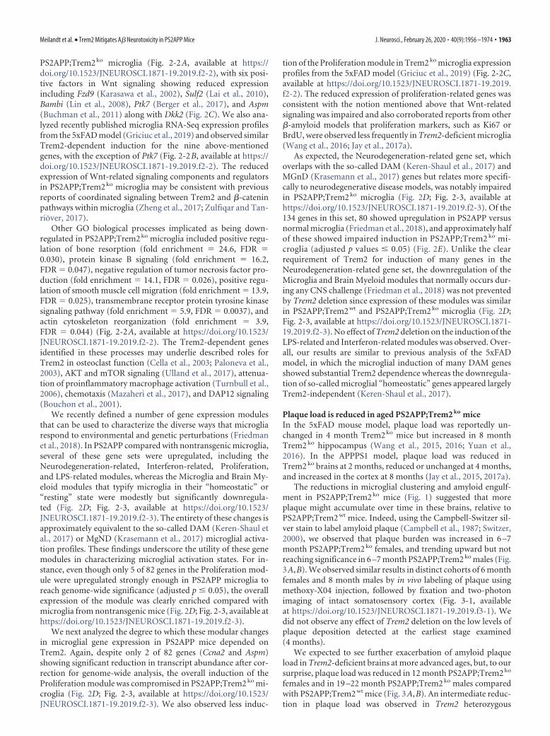
Figure 1. Trem2 deletion impairs microglial clustering, activation, and plaque uptake in PS2APP �-amyloidosis model. A, Immunohistochemical detection of microglial clustering in transgenicPS2APP mice or nontransgenic (Ntg) controls with WT (wt), heterozygous (het), or homozygous KO (ko) Trem2 alleles. For analysis of total, rather than clustered, Iba1 signal, see Figure 1-1 (availableat https://doi.org/10.1523/JNEUROSCI.1871-19.2019.f1-1). Quantification of the percent area covered by clusters of Iba1 � microglia was measured from coronal sections of female mice at 4, 6 –7,and 12 months of age, and from male mice at 6 –7 and 19 –22 months of age. Each data point represents the composite (average) histological score from several sections of an individual mouse. Barsand lines represent mean � SEM. Unpaired t test for most cohorts, or by ANOVA followed by Tukey’s multiple-comparison test for the 19 –22 month cohort with three genotypes: *p 0.05, **p 0.01, ***p 0.001, ****p 0.0001 versus PS2APP;Trem2 wt or as indicated. B, Representative low (left, scale bar, 400 �m) and high (right, scale bar, 100 �m) magnification images of Iba1 �
staining in 12 month female mice are shown. Iba1 � clusters (red arrowheads) showed reduced presence across Trem2 ko mice at all ages. C, D, Same as in A and B, except analyzing active microgliallysosomes as indicated by CD68 protein expression. E, Representative flow cytometry plots measuring CD45 immunoreactivity (low or high) of CD11b � brain-resident myeloid cells from 14 monthPS2APP mice with or without Trem2 and from Ntg mice. F, Percent of brain-resident myeloid cells with high CD45 expression measured from several mice of each genotype. Bars and lines representmean � SEM. G, H, Same as in E and F, except analyzing plaque content in brain CD11b � cells from �12 months mice injected with methoxy-X04 dye to stain amyloid material. Ntg mice are notplotted in H since they have zero methoxy labeling. n � 3 in Trem2 wt; n � 2 in Trem2 ko. For histological analysis of methoxy-X04 and Iba1 � microglia colocalization in sections, see Figure 1-1(available at https://doi.org/10.1523/JNEUROSCI.1871-19.2019.f1-1).
Meilandt et al. • Trem2 Mitigates A� Neurotoxicity in PS2APP Mice J. Neurosci., February 26, 2020 • 40(9):1956 –1974 • 1961

tivator of the pathway (Wu et al., 2000; Mao and Niehrs, 2003;Devotta et al., 2018) and since induction of regulators, includingWif1, can occur downstream of active �-catenin as negative feed-back (Diep et al., 2004; Boerboom et al., 2006). Supporting this
interpretation, the GO knowledge base identified “positive regu-lation of canonical Wnt signaling pathway” as a biological pro-cess overrepresented (fold enrichment � 10.3, FDR � 0.026)among the 144 transcripts with �2-fold reduced abundance in
Figure 2. Induction of Neurodegeneration-related and Proliferation gene modules and Wnt-related signaling components is impaired in Trem2 ko microglia. A, Heat map of differentiallyexpressed genes (DEGs) between Trem2 wt and Trem2 ko microglia from 14 month PS2APP mouse brains (fold change � 2, adjusted p � 0.05), cross-referenced against previously publisheddatasets from the PS2APP (GSE89482), APPswe/PS1de9 (GSE74615), and 5xFAD (GSE65067) models. Each row is a DEG, and each column is a microglial sort from a different mouse. z-score coloringrepresents a sample’s distance in SDs from the mean expression value for a given gene across samples within a dataset. Most of the downregulated DEGs in Trem2 ko microglia were typically inducedin models of �-amyloid pathology, and the majority also showed impaired microglial induction in Trem2 ko 5xFAD mice. For a complete list of genome-wide expression values in PS2APP;Trem2 wt
and PS2APP;Trem2 ko microglia, see Figure 2-1 (available at https://doi.org/10.1523/JNEUROSCI.1871-19.2019.f2-1). B, Induction of the Wnt/proliferation regulators Dkk2, Wif1, Ctnna3, and Asb11was completely impaired in Trem2 ko microglia of PS2APP mice. C, DEGs identified by GO analysis as “positive regulators of Wnt signaling” with reduced expression in PS2APP;Trem2 ko microgliaincluded Fzd9, Sulf2, Bambi, Ptk7, Aspm, and Dkk2. B, C, Bars and lines represent mean � SEM, with each data point representing microglial gene expression level from a given mouse. For genes inB and C, see also Figure 2-2 (available at https://doi.org/10.1523/JNEUROSCI.1871-19.2019.f2-2), showing microglial expression in the 5xFAD model with or without Trem2 (data from independentinvestigators). D, Top, Analysis of previously published expression profiles from the PS2APP model (GSE89482) indicated that six gene expression modules for brain myeloid cells (defined byFriedman et al., 2018) showed altered expression in microglia from brains with �-amyloid pathology. Bottom, Analysis of Trem2 ko and Trem2 wt microglia expression profiles from PS2APP miceshowed that the Neurodegeneration-related and Proliferation gene sets showed significant Trem2 dependence. For data from independent investigators showing enriched expression of Prolifer-ation module in 5xFAD model microglia and its dependence on Trem2 for full induction, see also Figure 2-2 (available at https://doi.org/10.1523/JNEUROSCI.1871-19.2019.f2-2). For heat mapdisplays of all individual genes in each module, see also Figure 2-3 (available at https://doi.org/10.1523/JNEUROSCI.1871-19.2019.f2-3). Each data point represents a gene set expression score formicroglia isolated from a given mouse. Lines indicate mean � SEM. Two-tailed t tests assuming unequal variance between groups: *p 0.05, **p 0.01, ***p 0.001, ****p 0.0001. E,Four-way plot of Neurodegeneration-related gene set, with each point representing a gene’s fold change in expression between PS2APP versus nontransgenic microglia on the x axis (red and bluepoints, adjusted p 0.05) and between PS2APP;Trem2 ko versus PS2APP;Trem2 wt microglia on the y axis (green and blue points, adjusted p 0.05). Blue points represent significant differentialexpression in both datasets. Tiny black points represent genes not differentially expressed in either dataset.
1962 • J. Neurosci., February 26, 2020 • 40(9):1956 –1974 Meilandt et al. • Trem2 Mitigates A� Neurotoxicity in PS2APP Mice
PS2APP;Trem2 ko microglia (Fig. 2-2A, available at https://doi.org/10.1523/JNEUROSCI.1871-19.2019.f2-2), with six posi-tive factors in Wnt signaling showing reduced expressionincluding Fzd9 (Karasawa et al., 2002), Sulf2 (Lai et al., 2010),Bambi (Lin et al., 2008), Ptk7 (Berger et al., 2017), and Aspm(Buchman et al., 2011) along with Dkk2 (Fig. 2C). We also ana-lyzed recently published microglia RNA-Seq expression profilesfrom the 5xFAD model (Griciuc et al., 2019) and observed similarTrem2-dependent induction for the nine above-mentionedgenes, with the exception of Ptk7 (Fig. 2-2B, available at https://doi.org/10.1523/JNEUROSCI.1871-19.2019.f2-2). The reducedexpression of Wnt-related signaling components and regulatorsin PS2APP;Trem2 ko microglia may be consistent with previousreports of coordinated signaling between Trem2 and �-cateninpathways within microglia (Zheng et al., 2017; Zulfiqar and Tan-riover, 2017).
Other GO biological processes implicated as being down-regulated in PS2APP;Trem2 ko microglia included positive regu-lation of bone resorption (fold enrichment � 24.6, FDR �0.030), protein kinase B signaling (fold enrichment � 16.2,FDR � 0.047), negative regulation of tumor necrosis factor pro-duction (fold enrichment � 14.1, FDR � 0.026), positive regu-lation of smooth muscle cell migration (fold enrichment � 13.9,FDR � 0.025), transmembrane receptor protein tyrosine kinasesignaling pathway (fold enrichment � 5.9, FDR � 0.0037), andactin cytoskeleton reorganization (fold enrichment � 3.9,FDR � 0.044) (Fig. 2-2A, available at https://doi.org/10.1523/JNEUROSCI.1871-19.2019.f2-2). The Trem2-dependent genesidentified in these processes may underlie described roles forTrem2 in osteoclast function (Cella et al., 2003; Paloneva et al.,2003), AKT and mTOR signaling (Ulland et al., 2017), attenua-tion of proinflammatory macrophage activation (Turnbull et al.,2006), chemotaxis (Mazaheri et al., 2017), and DAP12 signaling(Bouchon et al., 2001).
We recently defined a number of gene expression modulesthat can be used to characterize the diverse ways that microgliarespond to environmental and genetic perturbations (Friedmanet al., 2018). In PS2APP compared with nontransgenic microglia,several of these gene sets were upregulated, including theNeurodegeneration-related, Interferon-related, Proliferation,and LPS-related modules, whereas the Microglia and Brain My-eloid modules that typify microglia in their “homeostatic” or“resting” state were modestly but significantly downregula-ted (Fig. 2D; Fig. 2-3, available at https://doi.org/10.1523/JNEUROSCI.1871-19.2019.f2-3). The entirety of these changes isapproximately equivalent to the so-called DAM (Keren-Shaul etal., 2017) or MgND (Krasemann et al., 2017) microglial activa-tion profiles. These findings underscore the utility of these genemodules in characterizing microglial activation states. For in-stance, even though only 5 of 82 genes in the Proliferation mod-ule were upregulated strongly enough in PS2APP microglia toreach genome-wide significance (adjusted p � 0.05), the overallexpression of the module was clearly enriched compared withmicroglia from nontransgenic mice (Fig. 2D; Fig. 2-3, available athttps://doi.org/10.1523/JNEUROSCI.1871-19.2019.f2-3).
We next analyzed the degree to which these modular changesin microglial gene expression in PS2APP mice depended onTrem2. Again, despite only 2 of 82 genes (Ccna2 and Aspm)showing significant reduction in transcript abundance after cor-rection for genome-wide analysis, the overall induction of theProliferation module was compromised in PS2APP;Trem2 ko mi-croglia (Fig. 2D; Fig. 2-3, available at https://doi.org/10.1523/JNEUROSCI.1871-19.2019.f2-3). We also observed less induc-
tion of the Proliferation module in Trem2 ko microglia expressionprofiles from the 5xFAD model (Griciuc et al., 2019) (Fig. 2-2C,available at https://doi.org/10.1523/JNEUROSCI.1871-19.2019.f2-2). The reduced expression of proliferation-related genes wasconsistent with the notion mentioned above that Wnt-relatedsignaling was impaired and also corroborated reports from other�-amyloid models that proliferation markers, such as Ki67 orBrdU, were observed less frequently in Trem2-deficient microglia(Wang et al., 2016; Jay et al., 2017a).
As expected, the Neurodegeneration-related gene set, whichoverlaps with the so-called DAM (Keren-Shaul et al., 2017) andMGnD (Krasemann et al., 2017) genes but relates more specifi-cally to neurodegenerative disease models, was notably impairedin PS2APP;Trem2 ko microglia (Fig. 2D; Fig. 2-3, available athttps://doi.org/10.1523/JNEUROSCI.1871-19.2019.f2-3). Of the134 genes in this set, 80 showed upregulation in PS2APP versusnormal microglia (Friedman et al., 2018), and approximately halfof these showed impaired induction in PS2APP;Trem2 ko mi-croglia (adjusted p values � 0.05) (Fig. 2E). Unlike the clearrequirement of Trem2 for induction of many genes in theNeurodegeneration-related gene set, the downregulation of theMicroglia and Brain Myeloid modules that normally occurs dur-ing any CNS challenge (Friedman et al., 2018) was not preventedby Trem2 deletion since expression of these modules was similarin PS2APP;Trem2 wt and PS2APP;Trem2 ko microglia (Fig. 2D;Fig. 2-3, available at https://doi.org/10.1523/JNEUROSCI.1871-19.2019.f2-3). No effect of Trem2 deletion on the induction of theLPS-related and Interferon-related modules was observed. Over-all, our results are similar to previous analysis of the 5xFADmodel, in which the microglial induction of many DAM genesshowed substantial Trem2 dependence whereas the downregula-tion of so-called microglial “homeostatic” genes appeared largelyTrem2-independent (Keren-Shaul et al., 2017).
Plaque load is reduced in aged PS2APP;Trem2 ko miceIn the 5xFAD mouse model, plaque load was reportedly un-changed in 4 month Trem2 ko mice but increased in 8 monthTrem2 ko hippocampus (Wang et al., 2015, 2016; Yuan et al.,2016). In the APPPS1 model, plaque load was reduced inTrem2 ko brains at 2 months, reduced or unchanged at 4 months,and increased in the cortex at 8 months (Jay et al., 2015, 2017a).
The reductions in microglial clustering and amyloid engulf-ment in PS2APP;Trem2 ko mice (Fig. 1) suggested that moreplaque might accumulate over time in these brains, relative toPS2APP;Trem2 wt mice. Indeed, using the Campbell-Switzer sil-ver stain to label amyloid plaque (Campbell et al., 1987; Switzer,2000), we observed that plaque burden was increased in 6 –7month PS2APP;Trem2 ko females, and trending upward but notreaching significance in 6 –7 month PS2APP;Trem2 ko males (Fig.3A,B). We observed similar results in distinct cohorts of 6 monthfemales and 8 month males by in vivo labeling of plaque usingmethoxy-X04 injection, followed by fixation and two-photonimaging of intact somatosensory cortex (Fig. 3-1, availableat https://doi.org/10.1523/JNEUROSCI.1871-19.2019.f3-1). Wedid not observe any effect of Trem2 deletion on the low levels ofplaque deposition detected at the earliest stage examined(4 months).
We expected to see further exacerbation of amyloid plaqueload in Trem2-deficient brains at more advanced ages, but, to oursurprise, plaque load was reduced in 12 month PS2APP;Trem2 ko
females and in 19 –22 month PS2APP;Trem2 ko males comparedwith PS2APP;Trem2 wt mice (Fig. 3A,B). An intermediate reduc-tion in plaque load was observed in Trem2 heterozygous
Meilandt et al. • Trem2 Mitigates A� Neurotoxicity in PS2APP Mice J. Neurosci., February 26, 2020 • 40(9):1956 –1974 • 1963

(PS2APP;Trem2 het) mice at 19 –22 months (the only age whereheterozygous mice were analyzed) (Fig. 3A). At this age, theCampbell-Switzer stain also revealed a distinctive pattern of “am-ber core” amyloid staining in PS2APP brains (Fig. 3C), thoughtto represent a mature form of highly condensed plaque. Theseamber cores were reduced in PS2APP;Trem2 het and nearly absentin PS2APP;Trem2 ko brains, indicating that Trem2-dependentmicroglial activity is essential for the formation of these particu-lar amyloid structures.
Overall, our results are reminiscent of a recent study of APPPS1mice, which showed that Trem2 deletion produced increased seed-ing of amyloid plaques at early ages but slower rates of amyloidplaque accumulation at later ages (Parhizkar et al., 2019).
Reduced plaque consolidation, elevated neurotoxic A�species, and ApoE-laden microglia in Trem2-deficient brainsIn contrast to the effects of Trem2 deletion on total plaque burdenthat varied with age or sex, we observed consistent changes in
Figure 3. Trem2 deletion shows age- and sex-dependent effects on amyloid plaque pathology, with reduced plaque accumulation at later ages. A, B, Amyloid plaque was visualized using theCampbell-Switzer silver stain method in nontransgenic (Ntg) controls or transgenic PS2APP mice with WT (wt), heterozygous (het), or homozygous KO (ko) Trem2 alleles. Quantification of thepercent area covered by amyloid plaque was measured from coronal sections of indicated sex and age. Representative low (left, scale bar, 400 �m) and high (right, scale bar, 100 �m) magnificationimages of amyloid stains are shown. In the absence of Trem2, plaque loads were elevated in 6 –7 month females but reduced in 12 month females and in 19 –22 month males. For two-photonimaging of methoxy-X04-labeled plaque in somatosensory cortex of 6 month females and 8 month males, see also Figure 3-1 (available at https://doi.org/10.1523/JNEUROSCI.1871-19.2019.f3-1).C, The Campbell-Switzer silver stain turns highly mature plaque cores amber (red arrowheads). Quantification plot (left) and representative images (right, scale bar, 20 �m) of amber core frequencyin 19 –22 month male mice. Error bars indicate mean � SEM. Unpaired t test or ANOVA followed by Tukey’s multiple-comparison test: *p 0.05, **p 0.01, ****p 0.0001 versusPS2APP;Trem2 wt or as indicated.
1964 • J. Neurosci., February 26, 2020 • 40(9):1956 –1974 Meilandt et al. • Trem2 Mitigates A� Neurotoxicity in PS2APP Mice

plaque compaction and composition across ages and sexes. Weused the X-34 stain and confocal microscopy to visualize plaquemorphology. Although we were blinded to Trem2 genotype, therewas an obvious difference in plaque appearance betweenPS2APP;Trem2 wt and PS2APP;Trem2 ko brains, with X-34�
structures in PS2APP;Trem2 ko brains looking more splayed andless compact (Fig. 4A), similar to descriptions of Trem2-dependent plaque alterations in other �-amyloid models (Wanget al., 2016; Yuan et al., 2016). Using an algorithm based on X-34signal intensity to quantify the degree of plaque diffuseness, weobserved that plaque morphologies were significantly more dif-fuse in PS2APP;Trem2 ko brains in both sexes at all ages tested(Fig. 4B).
Because stains, such as Campbell-Switzer, methoxy-X04, andX-34, only label fibrillar amyloid structures, we also measured thetotal abundance of A�40 and A�42 peptides in soluble (TBS) andinsoluble (GuHCl) hippocampal fractions by ELISA. The abun-dance of A� peptides rose markedly with age while the A�42:A�40 ratio declined, particularly in the GuHCl fraction (Fig. 5).Notably, the A�42:A�40 ratio was higher in PS2APP;Trem2 ko
than in PS2APP;Trem2 wt brains, in both TBS and GuHCl frac-tions across ages (Fig. 5A,B). The elevated A�42:A�40 ratio inPS2APP;Trem2 ko brains resulted more from reduced abundanceof A�40 than from increased abundance of A�42 (Fig. 5-1, avail-able at https://doi.org/10.1523/JNEUROSCI.1871-19.2019.f5-1),although A�42 abundance was elevated in the 6 –7 month fe-males, coinciding with the increased plaque deposition we ob-served in that group (Fig. 3A). Together with our observation thattotal amyloid plaque is reduced in PS2APP;Trem2 ko brains at
later ages (Fig. 3A), these results suggest that the elevated A�42:A�40 ratio in Trem2-deficient brains may increase plaque seed-ing at younger ages (since A�42 is more prone to aggregate anddeposit than A�40) (Klein, 2002) while reducing the incorpora-tion and compaction of A� into existing plaques at older ages(since A�40 permeates dense core structures more readily thanA�42) (Condello et al., 2015). Thus, Trem2 may both restrict theinitial seeding of plaques and promote sequestration and com-paction of A� into existing plaques.
To determine whether the elevated A�42:A�40 ratio was ac-companied by altered abundance of soluble, fibrillar A� oligom-ers, we performed nondenaturing dot blots of hippocampal TBShomogenate supernatants from 12 month females. Using theconformation-specific OC antibody (Tomic et al., 2009), we de-tected significantly higher levels of soluble fibrillar A� oligomersin the soluble fraction from PS2APP;Trem2 ko mice (Fig. 5C,D),and we observed a positive correlation between the amount ofOC� fibrillar oligomers in this fraction and the A�42:A�40 ratio(Fig. 5E). In contrast, when we used the pan-reactive 6E10 A�antibody to detect total A� species in this fraction, the abundancetrended slightly downward in PS2APP;Trem2 ko mice and corre-lated negatively with A�42:A�40 ratio (Fig. 5F–H). Similar re-spective trends were also seen when staining with the prefibrillarA� oligomer antibody A11 and pan-reactive 4G8 A� antibody(data not shown). As a control, we spotted hippocampal TBShomogenate supernatant from a PS2APP;Bace1 ko animal (Mei-landt et al., 2019), in which soluble APP is still produced by�-cleavage while �-cleavage and thus A� production are pre-vented, and demonstrated that the OC antibody had minimal
Figure 4. Trem2 deletion impairs the consolidation of �-amyloid into dense plaque. A, �-amyloid plaques were visualized by X-34 staining and confocal z-stack imaging. Representativemaximum projection images of X-34 � plaques from 12 month PS2APP females of different Trem2 genotypes are shown (top). High-magnification images show a representative plaque from eachgenotype with the outlined masks used to delineate the compact core (green) and compact�diffuse (blue) areas of the plaque (bottom). Scale bars, 20 �m. B, The diffuseness index (see Materialsand Methods) of the X-34 � plaques in cohorts of the indicated age and sex was quantified. Each data point represents 1 animal’s plaque diffuseness index averaged from 3 to 4 FOV. Error barsindicate mean � SEM. Unpaired t test or ANOVA followed by Tukey’s multiple-comparison test: **p 0.01, ***p 0.001, ****p 0.0001 versus PS2APP;Trem2 wt.
Meilandt et al. • Trem2 Mitigates A� Neurotoxicity in PS2APP Mice J. Neurosci., February 26, 2020 • 40(9):1956 –1974 • 1965

Figure 5. Trem2 deletion increases the A�42:A�40 ratio and soluble fibrillar A� oligomers in PS2APP brains. A, B, Frozen hippocampal tissues were homogenized and processed for measuringthe abundance of A�40 and A�42 peptides by ELISA in the soluble (TBS) and insoluble (GuHCl) fractions. The ratio of A�42:A�40 in the TBS (A) and GuHCl (B) fractions is shown. Each data pointrepresents 1 animal’s A�42:A�40 ratio. For individual A�40 and A�42 peptide measurements, see Figure 5-1 (available at https://doi.org/10.1523/JNEUROSCI.1871-19.2019.f5-1). C, Image ofnondenaturing dot blot of hippocampal soluble TBS homogenates from 12 month female animals immunostained with the fibrillar oligomeric A� antibody OC (green) and control �-actin antibody(red). Squares outline PS2APP;Trem2 wt samples. Circles outline PS2APP;Trem2 ko samples. #, Control sample from a PS2APP;Bace1 ko mouse. D, Signal intensity ratios of OC antibody to actinantibody are plotted for the dot blot shown in C. Dotted line indicates the OC:actin ratio for a control sample from a PS2APP;Bace1 ko mouse. E, The A�42:A�40 ratio (from A) and normalized OC �
dot blot signal (from D) in the TBS soluble fraction showed a significant positive correlation (linear regression; df(1,28), F � 8.63, p 0.01). F, Same as in C, except immunostained with pan-A�antibody 6E10 (green) and control GAPDH antibody (red). G, Signal intensity ratios of A� 6E10 antibody to control GAPDH antibody are plotted for the dot blot shown in F. The PS2APP;Bace1 ko
control sample still has substantial 6E10 signal (see dotted line) since the N-terminal A� residues recognized by 6E10 are present in soluble APP when �-secretase is the responsible enzyme. H, TheA�42:A�40 ratio (from A) and normalized A� 6E10 dot blot signal (from G) in the TBS soluble fraction showed a significant negative correlation (linear regression; df(1,28), F � 4.98, p 0.05).Error bars indicate mean � SEM. Unpaired t test or ANOVA followed by Tukey’s multiple-comparison test: *p 0.05, **p 0.01, ***p 0.001, ****p 0.0001 versus PS2APP;Trem2 wt or asindicated.
1966 • J. Neurosci., February 26, 2020 • 40(9):1956 –1974 Meilandt et al. • Trem2 Mitigates A� Neurotoxicity in PS2APP Mice

detection, whereas 6E10 still had substantial signal (compare #symbols in Fig. 5C,F and dotted lines in Fig. 5D,G from aPS2APP;Bace1 ko mouse), consistent with the ability of 6E10 todetect both soluble APP and A� peptides. These results suggestthat, in the absence of Trem2, the increased A�42:A�40 ratioenhances the potential shift of soluble A� to a fibrillar oligomericform. Alternatively, the increased abundance of fibrillar A� oli-gomers in the TBS-soluble fraction could result from reducedincorporation into highly condensed plaques in brains withTrem2-deficient microglia.
Apoe is one of the most highly induced genes in mouse micro-glia in response to neurodegenerative stimuli (Deczkowska et al.,2018). A recent report in the APPPS1 model found that plaque-associated ApoE was reduced in Trem2-deficient mice (Parhizkaret al., 2019). To learn whether Trem2 deletion altered ApoE lo-calization in the PS2APP model and whether such alterationcorrelated with observed changes in plaque abundance, mor-phology, or composition, we costained for ApoE, plaques(methoxy-X04), and microglia (Iba1). In contrast to the findingsof Parhizkar et al. (2019), quantification of plaque-associatedApoE in whole-brain sections found a significant increase in fe-male PS2APP;Trem2 ko mice at 6 –7 and 12 months of age, but nosignificant changes in male cohorts at any age tested (Fig. 6A).The ApoE immunostaining pattern was especially prominent in,but not limited to, the hippocampal subiculum where plaquetends to first deposit (Fig. 6B). While the incongruous resultsbetween studies may be explained by differences between mousemodels, other variations in sampling and technical procedurescould also account for the differences. Parhizkar et al. (2019)examined 4 month males; we only examined females at that age,and our findings of elevated plaque-associated ApoE in Trem2-deficient females were only observed at older ages. The studiesused different ApoE monoclonal antibodies that likely interactwith distinct epitopes or configurations of ApoE (Kim et al.,2012) and whose binding may be differentially impacted by vari-ations in staining procedure. We immunostained for ApoE first,followed by plaque labeling, which involves treatments with eth-anol and NaOH. Parhizkar et al. (2019) did the plaque labelingfirst, followed by the ApoE immunostains, so the nature of theApoE at the time of immunostaining was somewhat differentbetween studies.
Parhizkar et al. (2019) also reported that colocalization ofApoE with Iba1� microglia was reduced in Trem2-deficientmice. To assess this finding in the PS2APP model, we performedconfocal imaging on the ApoE/Iba1/methoxy-X04 costains fromthe cortex of 12 month females and again observed a contrastingresult. Instead of ApoE labeling being diminished, we observedmicroglia in PS2APP;Trem2 ko mice to be markedly laden withApoE (Fig. 6C,D), suggesting that Trem2-deficient microglia ex-hibit deficits in lipid clearance. At the mRNA level, we did notdetect a difference in Apoe expression between PS2APP;Trem2 wt
and PS2APP;Trem2 ko microglia (Fig. 6E; see also Fig. 2E). Al-though two groups have reported reductions in microglial Apoeexpression in the APPPS1 model when Trem2 is deleted (Krase-mann et al., 2017; Parhizkar et al., 2019), this does not appear tobe a typical feature of Trem2 deficiency in �-amyloidosis modelssince previous analyses in the 5xFAD model observed Trem2-independent Apoe induction in either bulk microglia microarray(Fig. 6F) (Wang et al., 2015) or single-cell microglia RNA-Seqprofiles (Keren-Shaul et al., 2017). In a very recent dataset of bulkmicroglia RNA-Seq profiles from 5xFAD mice (Griciuc et al.,2019), we did see a twofold decrease in the extent of Apoe induc-tion in Trem2-deficient microglia, but the gene was still highly
induced relative to the expression level in microglia from non-transgenic mice (Fig. 6F). Together, our evidence indicates thatmicroglial Apoe expression is induced by A�-driven neuropa-thology in a largely Trem2-independent manner, and thatTrem2-deficient microglia accumulate disproportionately largeamounts of ApoE compared with the smaller ApoE puncta ob-served in normal PS2APP microglia (Fig. 6C).
Axonal dystrophy, dendritic spine loss, and CSF NfLdetection are exacerbated in PS2APP;Trem2 ko miceThe elevated A�42:A�40 ratio and fibrillar A� oligomers inPS2APP;Trem2 ko brains would seem to be detrimental for neu-ronal health since A�42 oligomers are commonly understood tobe the more toxic form of A� (Klein, 2002; Haass and Selkoe,2007). Alternatively, the reduced plaque load in aged Trem2 ko
brains suggested a possible benefit of Trem2 deficiency. There-fore, we turned to measures of neuronal dystrophy to better un-derstand the potential consequences of loss of Trem2 function.
First, we looked at neuritic dystrophy around plaque(D’Amore et al., 2003) by fluorescent costaining using methoxy-X04 to label plaque, anti-Iba1 to label microglia, and anti-Lamp1to label dystrophic axons (Gowrishankar et al., 2015) (Fig. 7A).The methoxy-X04/Iba1 costain showed that microglial associa-tion with plaque was severely compromised in PS2APP;Trem2 ko
brains at all examined ages (Fig. 7A,B), corroborating our earlierIba1 immunohistochemical stains that measured microglial clus-tering. The Lamp1 immunolabeling, which stains dilated dystro-phic axons, revealed two important findings. First, on a perplaque basis, axonal dystrophy was exacerbated from 7 monthsonward in PS2APP;Trem2 ko mice (Fig. 7C), similar to findings inother �-amyloid models (Wang et al., 2016; Yuan et al., 2016).This is consistent with the idea that the diffuse plaque structuresand elevated A�42:A�40 ratio in Trem2 ko brains are more dam-aging to surrounding axons than the more compacted, A�40-enriched plaques in PS2APP;Trem2 wt brains. Second, the totalLamp1� area was also increased from 7 months onward inPS2APP;Trem2 ko brain sections (Fig. 7D), indicating that totalaxonal damage was exacerbated at later ages despite the reducedplaque burden. These data strongly suggest that the Trem2-dependent clustering of microglia and their functions aroundplaque serve to mitigate the neurotoxic effects of A�.
Another method we used to visualize neuronal pathology wasan amino-cupric-silver stain or “disintegrative degeneration”stain that labels damaged or degenerating neurons (de Olmos etal., 1994; Switzer, 2000). Overall, the staining pattern appearedvery similar to the Lamp1 stain, with “bouquets” of argyrophilicstructures presumably surrounding plaques throughout the cor-tex and hippocampus. Staining was also observed in relevantwhite matter tracts, such as the corpus callosum, perforant path,and fornix, suggesting that degenerating axonal processes are notrestricted to dystrophic neurites around plaques. Degeneratingneurites detected by this stain were more abundant in PS2APP;Trem2 ko brains of the 12 month female and 19 –22 month malecohorts (Fig. 8A,B). Thus, again, axonal damage was exacerbatedin Trem2-deficient mice from older ages despite the fact thatplaque accumulation was reduced. The intermediate effect ofTrem2 heterozygosity on microglial clustering around plaque(Figs. 1A, 7B) and Campbell-Switzer plaque staining (Fig. 3A),but not on plaque diffuseness (Fig. 4B), A�42:A�40 ratio (Fig.5A), or neuritic dystrophy (Figs. 7C,D, 8A), suggested that theform rather than the amount of plaque correlates with neuronaldamage, and that sufficient microgliosis occurs in PS2APP;Trem2 het mice to enable plaque compaction and neuroprotection.
Meilandt et al. • Trem2 Mitigates A� Neurotoxicity in PS2APP Mice J. Neurosci., February 26, 2020 • 40(9):1956 –1974 • 1967

NfL measured in the CSF or plasma has recently emerged as apotential biomarker of neurodegeneration in human patientsand in mouse disease models (Bacioglu et al., 2016; Khalil et al.,2018). To determine whether Trem2 deficiency altered NfL levels,we collected plasma and CSF from a separate, mixed sex cohortof 12 month PS2APP;Trem2 wt and PS2APP;Trem2 ko mice.
PS2APP;Trem2 ko mice had significantly greater NfL levels in theCSF compared with PS2APP;Trem2 wt mice (Fig. 8C), consistentwith the measures of increased axonal dystrophy that were ob-served by histopathology in Trem2-deficient females at this age.Perhaps surprisingly, we did not observe a difference in plasmaNfL levels between genotypes (Fig. 8C), suggesting that CSF NfL
Figure 6. ApoE markedly accumulates on plaques and in microglia in PS2APP;Trem2 ko females. A, Plaque-associated ApoE signal was quantified for each animal in each cohort using 2 or 3sections per animal, with each section having a range of �50 – 800 plaques contributing to the analysis. Significant increases were observed in 6 –7 month and 12 month female (f) PS2APP;Trem2 ko mice, but not in males (m). B, Representative low-magnification images of the hippocampus (top row) and high-magnification images of the subiculum (enlarged from the boxed regions)from 12 month female brain sections stained with methoxy-X04 to label plaque (blue), anti-Iba1 to label microglia (green), and anti-ApoE (red). Arrows point to examples of plaques with intenseApoE staining, which are atypical in Trem2 wt females but typical in Trem2 ko females at this age. C, Representative 20� maximum projection of confocal z-stacks imaged from cortex, highlightingApoE (red) localization in microglia (Iba1, green) and plaque (blue). Microglia with small ApoE puncta (arrows) are typical in Trem2 wt, whereas microglia with enlarged ApoE structures (arrowheads)are frequent in Trem2 ko. Plaques with strong ApoE labeling are more common in Trem2 ko (asterisks). D, Analysis of the fraction of Iba1 signal that overlaps with ApoE staining (Mander’scolocalization coefficient) in 12 month PS2APP females revealed increased colocalization in Trem2 ko microglia. E, RNA-Seq profiles of microglia FACS-purified from brains of 14 month PS2APP miceshowed no difference in Apoe expression between Trem2 wt and Trem2 ko microglia (MG). F, Expression profiles of microglia FACS-purified from brains of 8 month nontransgenic or 5xFAD micerevealed strong Apoe induction by �-amyloid pathology in both Trem2 wt and Trem2 ko microglia, with induction in 5xFAD;Trem2 ko relative to 5xFAD;Trem2 wt microglia being twofold reduced inone dataset (GSE132508, RNA-Seq) and not significantly different in another (GSE65067, microarray). Error bars indicate mean � SEM. Unpaired t test or ANOVA followed by Tukey’s multiple-comparison test: *p 0.05, **p 0.01, ****p 0.0001 versus PS2APP;Trem2 wt or as indicated. n.s. - not significant.
1968 • J. Neurosci., February 26, 2020 • 40(9):1956 –1974 Meilandt et al. • Trem2 Mitigates A� Neurotoxicity in PS2APP Mice

measurements better represent ongoing neuronal damage or de-generation in the CNS than plasma measurements.
Finally, we looked at whether another feature of neuronalpathology observed in AD tissues and �-amyloid mouse models,reduced synaptic density, particularly near plaque (Spires andHyman, 2004; Tsai et al., 2004; Spires et al., 2005), was altered inTrem2-deficient mice. In �-amyloid models, the reduction insynapse number requires the presence of microglia since deplet-ing the microglial cell population largely prevents loss of synapticdensity (Olmos-Alonso et al., 2016; Spangenberg et al., 2016).
Therefore, we asked whether Trem2 deletion and the resultinglack of activated, plaque-associated microglia would prevent thedendritic spine loss from occurring or would worsen it. To an-swer this, we crossed the Thy1:GFP-M line, which labels a sparsepopulation of excitatory neurons, into the PS2APP model andanalyzed a cohort of 6 month female mice with different Trem2genotypes. Spine density loss in the proximity of plaques was notrescued by Trem2 deletion, but was actually further exacerbatedin PS2APP;Trem2 ko mice compared with PS2APP;Trem2 wt orPS2APP;Trem2 het groups (Fig. 9). Thus, Trem2 is not required
Figure 7. Trem2 deletion exacerbates plaque-proximal axonal dystrophy. A, Representative images from 12 month brain sections stained with methoxy-X04 to label plaque, anti-Iba1 to labelmicroglia, and anti-Lamp1 to label dystrophic axons around plaque. B, Microglial clustering around plaque is impaired in Trem2-deficient mice. Plaque-associated Iba1 signal was quantified for eachanimal in each cohort using 2 or 3 sections per animal, with each section having hundreds or thousands of plaques contributing to the analysis. C, Axonal dystrophy per plaque is exacerbated inTrem2-deficient mice. Plaque-associated Lamp1 signal was quantified for each animal in a similar manner as Iba1 signal in B. D, Total axonal dystrophy is exacerbated in Trem2-deficient mice. TotalLamp1 signal in each section was quantified, and each data point represents the average score from 2 or 3 sections per animal. Error bars indicate mean � SEM. Unpaired t test or ANOVA followedby Tukey’s multiple-comparison test: **p 0.01, ***p 0.001, ****p 0.0001 versus PS2APP;Trem2 wt or as indicated.
Meilandt et al. • Trem2 Mitigates A� Neurotoxicity in PS2APP Mice J. Neurosci., February 26, 2020 • 40(9):1956 –1974 • 1969

for microglia-mediated dendritic spine loss around plaque; andindeed, Trem2 seems to hedge against synapse loss. Overall, theexacerbated axonal dystrophy and dendritic spine loss observedaround plaque in PS2APP;Trem2 ko mice imply that Trem2-
dependent microglial activity is fundamentally neuroprotectivein �-amyloid-driven models of AD-like pathology.
DiscussionIn this study, we examined the role of Trem2 in microglial acti-vation, plaque accumulation, and neuronal dystrophy in thePS2APP model of �-amyloidosis. We observed both age- andsex-dependent effects of Trem2 deletion on plaque abundanceassessed using the Campbell-Switzer silver stain, with slightlymore plaque in PS2APP;Trem2 ko females at 6 –7 months of agebut markedly less plaque in both female and male PS2APP;Trem2 ko mice at later ages. Together with a recent similar report
Figure 8. Disintegrative degeneration staining and CSF NfL measurements reveal exacer-bated neuronal damage in Trem2-deficient mice at later ages. A, Quantification of sections fromnontransgenic (Ntg) and PS2APP mice with indicated Trem2 genotypes stained using an aminocupric silver staining method that labels degenerative neuronal processes. Each data pointrepresents the average percent area covered/section for �10 sections per animal. B, Represen-tative low (left, scale bar, 400 �m) and high (right, scale bar, 100 �m) magnification imagesfrom the 12 month female and 19 –22 month male cohorts are shown. Degenerative signal isapparent in plaque-accompanying foci (red arrowheads) throughout the cortex and hippocam-pus and in certain white matter tracts, including the corpus callosum (cc). C, Plasma (left) andCSF (right) NfL were measured from a separate, mixed sex cohort of 12 months PS2APP;Trem2 wt and PS2APP;Trem2 ko mice. Error bars indicate mean�SEM. Unpaired t test or ANOVAfollowed by Tukey’s multiple-comparison test: **p 0.01, ****p 0.0001 versus PS2APP;Trem2 wt or as indicated.
Figure 9. Trem2 deletion exacerbates dendritic spine loss near plaque. A, Intact brains from6 month female PS2APP mice with WT (wt), KO (ko), or heterozygous (het) Trem2 genotypesand carrying the Thy1::GFP-M transgene, which sparsely labels excitatory cortical neurons, wereimaged in the somatosensory cortex (SS Cx) using two-photon microscopy. Dendritic shaftsproximal to plaque (within 20 �m) or distally located (�100 �m from any plaque) wereimaged, with representative images shown. B, Each data point represents the average of fivespine density measurements from 1 animal. Spine density was �50% reduced on plaque-proximal dendrite segments relative to distal dendrite segments from the same animals. Spinedensity near plaque was lower in PS2APP;Trem2 ko mice than in PS2APP;Trem2 wt or PS2APP;Trem2 het mice. Error bars indicate mean�SEM. ANOVA: *p 0.05, **p 0.01. Spine volumewas unaffected by either plaque proximity or Trem2 genotype (data not shown).
1970 • J. Neurosci., February 26, 2020 • 40(9):1956 –1974 Meilandt et al. • Trem2 Mitigates A� Neurotoxicity in PS2APP Mice

(Parhizkar et al., 2019), these results suggest that Trem2-dependent microglial activity may both restrain the formation/seeding of plaques at an early stage of pathology, conceivablythrough uptake and degradation of soluble A� species, but alsoenhance the sequestration of A� into existing plaque structures,particularly at later stages of pathology.
At all ages examined, and in both sexes, microglial clusteringaround plaque and other measurements of microglial activationwere sharply reduced in PS2APP;Trem2 ko mice. Transcriptionalinduction of the neurodegeneration-related modules, the prolif-eration module, and certain genes related to Wnt regulation wereimpaired in PS2APP;Trem2 ko microglia. Presumably as a conse-quence of the impaired microglial response around plaque,plaques in PS2APP;Trem2 ko brains displayed a more diffusemorphology than in PS2APP;Trem2 wt brains. From 6 –7 monthsonward, axonal injury was magnified in PS2APP;Trem2 ko mice,even at later ages when the abundance of argyrophilic amyloidplaques was diminished. Our finding that plaque-proximal den-dritic spine loss was exacerbated in PS2APP;Trem2 ko micefurther underscored that the Trem2-dependent microglial acti-vation around plaque is protective for neurons and is distinctfrom microglia-mediated, complement-mediated activities thatcontribute to synapse loss in �-amyloid models (Fonseca et al.,2004; Hong et al., 2016; Shi et al., 2017; Wu et al., 2019). It will beimportant for future studies of microglial modulation in mousemodels to analyze plaque-associated neuritic dystrophy and notassume that decreases (or increases) in amyloid plaque burdenare evidence of beneficial (or detrimental) effects.
While the elevated A�42:A�40 ratio we observed in the solu-ble fraction of PS2APP;Trem2 ko brains could reflect direct defi-cits in the Trem2-mediated binding and clearance of oligomericA�42 by microglia (Yeh et al., 2016; Lessard et al., 2018; Zhao etal., 2018), it could also reflect enhanced clearance of A�40through other means, such as vascular efflux. The resulting ele-vation in soluble A�42:A�40 ratio may give rise to the increasedplaque formation we observed in 6 –7 month females, similar tothe increased plaque seeding activity observed by others at a sim-ilar age in another model when Trem2 was deleted (Parhizkar etal., 2019). Given that A�40 is more easily incorporated into ex-isting dense plaque structures than A�42 (Condello et al., 2015),our observation that the A�42:A�40 ratio declines as the miceage, particularly in the insoluble fraction, is consistent with theaccumulation of compact plaques in size and number over timein the brains of PS2APP mice. The reduction of this plaque accu-mulation activity in PS2APP;Trem2 ko brains may underlie theelevated levels of fibrillar oligomeric A� we detected in the solu-ble fraction, which may in turn be a source of increased neuronalinjury in these animals.
The weakness of correlation between plaque abundance andcognitive status in humans, along with several unsuccessful clin-ical trials aimed at preventing cognitive decline by reducing brainA� levels, has led some to question the relevance of A� in theetiology and progression of AD (Krstic and Knuesel, 2013; Morriset al., 2014; Itzhaki et al., 2016). Moreover, many have proposedthat chronic microglial activation is a key damaging agent thatcontributes to the neurotoxic environment in AD (Heneka et al.,2015; Park et al., 2018). Our results suggest the opposite, sincepreventing the microglial response to A� pathology via Trem2deletion reversed neither axonal dystrophy nor dendritic spineloss around plaque and indeed made both of these pathologiesworse. Trem2 deletion also increased the amount of NfL detectedin CSF. Evidence in humans and mice supports the mechanisticmodel that �-amyloid pathology accelerates the accumulation of
tau pathology or enhances its spreading (Pooler et al., 2015; He etal., 2018; Jack et al., 2018; Jacobs et al., 2018), and a new reportindicates that this effect is further magnified in Trem2-deficientmice (Leyns et al., 2019). Together, these findings suggest that theform of microglial activation brought on by A�-related patholo-gies (the DAM state, or at least the Trem2-dependent componentof it) protects neurons by limiting A�-induced neuronal injury.
Supporting the notion that microglial activation is primarilybeneficial in the context of AD pathology, elevated PET signal forligands of the “neuroinflammation marker” TSPO have pre-dicted better cognitive measures and slower AD progression inmice with �-amyloid pathology and human patients, respectively(Hamelin et al., 2016; Focke et al., 2019). The fact that plaques inTrem2-deficient mice are more injurious to adjacent neurites butshow weaker labeling with molecular probes (Thioflavin S,methoxy-X04 and X-34) related to those used in the clinic ( 11C-PiB, 18F-Florbetapir) helps explain why cognitive decline corre-lates better with tau pathology and synapse loss than with brainamyloid detection. The form of �-amyloid in the brain is morecritical than the amount, and Trem2-mediated microgliosis facil-itates the consolidation of �-amyloid into a highly compacted,less damaging form. Therefore, therapeutics that enhance thismicroglial activity may prevent AD or delay its progression whilesimultaneously (and perhaps counterintuitively) leading to in-creases in PET signals for amyloid content and microgliosis. Clin-ical biomarkers of neuronal degeneration and spreading ADpathology, such as neurofilament-L and tau, should be more in-formative indicators of whether a microglia-directed therapy isachieving efficacy.
ReferencesAndorfer C, Kress Y, Espinoza M, de Silva R, Tucker KL, Barde YA, Duff K,
Davies P (2003) Hyperphosphorylation and aggregation of tau in miceexpressing normal human tau isoforms. J Neurochem 86:582–590.
Bacioglu M, Maia LF, Preische O, Schelle J, Apel A, Kaeser SA, SchweighauserM, Eninger T, Lambert M, Pilotto A, Shimshek DR, Neumann U, KahlePJ, Staufenbiel M, Neumann M, Maetzler W, Kuhle J, Jucker M (2016)Neurofilament light chain in blood and CSF as marker of disease progres-sion in mouse models and in neurodegenerative diseases. Neuron 91:56 – 66.
Bemiller SM, McCray TJ, Allan K, Formica SV, Xu G, Wilson G, Kokiko-Cochran ON, Crish SD, Lasagna-Reeves CA, Ransohoff RM, LandrethGE, Lamb BT (2017) TREM2 deficiency exacerbates tau pathologythrough dysregulated kinase signaling in a mouse model of tauopathy.Mol Neurodegener 12:74.
Berger H, Wodarz A, Borchers A (2017) PTK7 faces the wnt in developmentand disease. Front Cell Dev Biol 5:31.
Boerboom D, White LD, Dalle S, Courty J, Richards JS (2006) Dominant-stable beta-catenin expression causes cell fate alterations and wnt signal-ing antagonist expression in a murine granulosa cell tumor model. CancerRes 66:1964 –1973.
Bouchon A, Hernandez-Munain C, Cella M, Colonna M (2001) A DAP12-mediated pathway regulates expression of CC chemokine receptor 7 andmaturation of human dendritic cells. J Exp Med 194:1111–1122.
Buchman JJ, Durak O, Tsai LH (2011) ASPM regulates wnt signaling path-way activity in the developing brain. Genes Dev 25:1909 –1914.
Busby V, Goossens S, Nowotny P, Hamilton G, Smemo S, Harold D, Turic D,Jehu L, Myers A, Womick M, Woo D, Compton D, Doil LM, Tacey KM,Lau KF, Al-Saraj S, Killick R, Pickering-Brown S, Moore P, HollingworthP, et al. (2004) Alpha-T-catenin is expressed in human brain and inter-acts with the wnt signaling pathway but is not responsible for linkage tochromosome 10 in Alzheimer’s disease. Neuromolecular Med 5:133–146.
Campbell SK, Switzer RC, Martin TL (1987) Alzheimer’s plaques and tan-gles: a controlled and enhanced silver staining method. Soc NeuroscienceAbstr 13:678.
Cella M, Buonsanti C, Strader C, Kondo T, Salmaggi A, Colonna M (2003)Impaired differentiation of osteoclasts in TREM-2-deficient individuals. JExp Med 198:645– 651.
Meilandt et al. • Trem2 Mitigates A� Neurotoxicity in PS2APP Mice J. Neurosci., February 26, 2020 • 40(9):1956 –1974 • 1971

Condello C, Yuan P, Schain A, Grutzendler J (2015) Microglia constitute abarrier that prevents neurotoxic protofibrillar Abeta42 hotspots aroundplaques. Nat Commun 6:6176.
D’Amore JD, Kajdasz ST, McLellan ME, Bacskai BJ, Stern EA, Hyman BT(2003) In vivo multiphoton imaging of a transgenic mouse model ofAlzheimer disease reveals marked thioflavine-S-associated alterations inneurite trajectories. J Neuropathol Exp Neurol 62:137–145.
Deczkowska A, Keren-Shaul H, Weiner A, Colonna M, Schwartz M, Amit I(2018) Disease-associated microglia: a universal immune sensor of neu-rodegeneration. Cell 173:1073–1081.
de Olmos JS, Beltramino CA, de Olmos de Lorenzo S (1994) Use of anamino-cupric-silver technique for the detection of early and semiacuteneuronal degeneration caused by neurotoxicants, hypoxia, and physicaltrauma. Neurotoxicol Teratol 16:545–561.
Devotta A, Hong CS, Saint-Jeannet JP (2018) Dkk2 promotes neural crestspecification by activating Wnt/beta-catenin signaling in a GSK3beta in-dependent manner. Elife 7:e34404.
Diep DB, Hoen N, Backman M, Machon O, Krauss S (2004) Characterisa-tion of the wnt antagonists and their response to conditionally activatedwnt signalling in the developing mouse forebrain. Brain Res Dev BrainRes 153:261–270.
Diks SH, Bink RJ, van de Water S, Joore J, van Rooijen C, Verbeek FJ, denHertog J, Peppelenbosch MP, Zivkovic D (2006) The novel gene asb11:a regulator of the size of the neural progenitor compartment. J Cell Biol174:581–592.
Dunn KW, Kamocka MM, McDonald JH (2011) A practical guide to eval-uating colocalization in biological microscopy. Am J Physiol Cell Physiol300:C723–C742.
Focke C, Blume T, Zott B, Shi Y, Deussing M, Peters F, Schmidt C, Klein-berger G, Lindner S, Gildehaus FJ, Beyer L, von Ungern-Sternberg B,Bartenstein P, Ozmen L, Baumann K, Dorostkar MM, Haass C, Adels-berger H, Herms J, Rominger A, et al. (2019) Early and longitudinalmicroglial activation but not amyloid accumulation predicts cognitiveoutcome in PS2APP mice. J Nucl Med 60:548 –554.
Fonseca MI, Zhou J, Botto M, Tenner AJ (2004) Absence of C1q leads to lessneuropathology in transgenic mouse models of Alzheimer’s disease.J Neurosci 24:6457– 6465.
Friedman BA, Srinivasan K, Ayalon G, Meilandt WJ, Lin H, Huntley MA, CaoY, Lee SH, Haddick PC, Ngu H, Modrusan Z, Larson JL, Kaminker JS, vander Brug MP, Hansen DV (2018) Diverse brain myeloid expression pro-files reveal distinct microglial activation states and aspects of Alzheimer’sdisease not evident in mouse models. Cell Rep 22:832– 847.
Gage PJ, Qian M, Wu D, Rosenberg KI (2008) The canonical wnt signalingantagonist DKK2 is an essential effector of PITX2 function during normaleye development. Dev Biol 317:310 –324.
Gowrishankar S, Yuan P, Wu Y, Schrag M, Paradise S, Grutzendler J, DeCamilli P, Ferguson SM (2015) Massive accumulation of luminalprotease-deficient axonal lysosomes at Alzheimer’s disease amyloidplaques. Proc Natl Acad Sci U S A 112:E3699 –E3708.
Gratuze M, Leyns CE, Holtzman DM (2018) New insights into the role ofTREM2 in Alzheimer’s disease. Mol Neurodegener 13:66.
Griciuc A, Patel S, Federico AN, Choi SH, Innes BJ, Oram MK, Cereghetti G,McGinty D, Anselmo A, Sadreyev RI, Hickman SE, El Khoury J, ColonnaM, Tanzi RE (2019) TREM2 acts downstream of CD33 in modulatingmicroglial pathology in Alzheimer’s disease. Neuron 103:820 – 835.e7.
Guerreiro R, Wojtas A, Bras J, Carrasquillo M, Rogaeva E, Majounie E, Cru-chaga C, Sassi C, Kauwe JS, Younkin S, Hazrati L, Collinge J, Pocock J,Lashley T, Williams J, Lambert JC, Amouyel P, Goate A, Rademakers R,Morgan K, et al. (2013) TREM2 variants in Alzheimer’s disease. N EnglJ Med 368:117–127.
Haass C, Selkoe DJ (2007) Soluble protein oligomers in neurodegeneration:lessons from the Alzheimer’s amyloid beta-peptide. Nat Rev Mol Cell Biol8:101–112.
Hamelin L, Lagarde J, Dorothee G, Leroy C, Labit M, Comley RA, de SouzaLC, Corne H, Dauphinot L, Bertoux M, Dubois B, Gervais P, Colliot O,Potier MC, Bottlaender M, Sarazin M (2016) Early and protective mi-croglial activation in Alzheimer’s disease: a prospective study using 18F-DPA-714 PET imaging. Brain 139:1252–1264.
Hansen DV, Hanson JE, Sheng M (2018) Microglia in Alzheimer’s disease.J Cell Biol 217:459 – 472.
He Z, Guo JL, McBride JD, Narasimhan S, Kim H, Changolkar L, Zhang B,Gathagan RJ, Yue C, Dengler C, Stieber A, Nitla M, Coulter DA, Abel T,
Brunden KR, Trojanowski JQ, Lee VM (2018) Amyloid-beta plaquesenhance Alzheimer’s brain tau-seeded pathologies by facilitating neuriticplaque tau aggregation. Nat Med 24:29 –38.
Heneka MT, Carson MJ, El Khoury J, Landreth GE, Brosseron F, FeinsteinDL, Jacobs AH, Wyss-Coray T, Vitorica J, Ransohoff RM, Herrup K,Frautschy SA, Finsen B, Brown GC, Verkhratsky A, Yamanaka K, Koisti-naho J, Latz E, Halle A, Petzold GC, et al. (2015) Neuroinflammation inAlzheimer’s disease. Lancet Neurol 14:388 – 405.
Hong S, Beja-Glasser VF, Nfonoyim BM, Frouin A, Li S, Ramakrishnan S,Merry KM, Shi Q, Rosenthal A, Barres BA, Lemere CA, Selkoe DJ, StevensB (2016) Complement and microglia mediate early synapse loss in Alz-heimer mouse models. Science 352:712–716.
Hsieh JC, Kodjabachian L, Rebbert ML, Rattner A, Smallwood PM, SamosCH, Nusse R, Dawid IB, Nathans J (1999) A new secreted protein thatbinds to wnt proteins and inhibits their activities. Nature 398:431– 436.
Itzhaki RF, Lathe R, Balin BJ, Ball MJ, Bearer EL, Braak H, Bullido MJ, CarterC, Clerici M, Cosby SL, Del Tredici K, Field H, Fulop T, Grassi C, GriffinWS, Haas J, Hudson AP, Kamer AR, Kell DB, Licastro F, et al. (2016)Microbes and Alzheimer’s disease. J Alzheimers Dis 51:979 –984.
Jack CR Jr, Wiste HJ, Schwarz CG, Lowe VJ, Senjem ML, Vemuri P, WeigandSD, Therneau TM, Knopman DS, Gunter JL, Jones DT, Graff-Radford J,Kantarci K, Roberts RO, Mielke MM, Machulda MM, Petersen RC(2018) Longitudinal tau PET in ageing and Alzheimer’s disease. Brain141:1517–1528.
Jacobs HI, Hedden T, Schultz AP, Sepulcre J, Perea RD, Amariglio RE, PappKV, Rentz DM, Sperling RA, Johnson KA (2018) Structural tract alter-ations predict downstream tau accumulation in amyloid-positive olderindividuals. Nat Neurosci 21:424 – 431.
Jay TR, Miller CM, Cheng PJ, Graham LC, Bemiller S, Broihier ML, Xu G,Margevicius D, Karlo JC, Sousa GL, Cotleur AC, Butovsky O, Bekris L,Staugaitis SM, Leverenz JB, Pimplikar SW, Landreth GE, Howell GR,Ransohoff RM, Lamb BT (2015) TREM2 deficiency eliminatesTREM2 � inflammatory macrophages and ameliorates pathology in Alz-heimer’s disease mouse models. J Exp Med 212:287–295.
Jay TR, Hirsch AM, Broihier ML, Miller CM, Neilson LE, Ransohoff RM,Lamb BT, Landreth GE (2017a) Disease progression-dependent effectsof TREM2 deficiency in a mouse model of Alzheimer’s disease. J Neurosci37:637– 647.
Jay TR, von Saucken VE, Landreth GE (2017b) TREM2 in neurodegenera-tive diseases. Mol Neurodegener 12:56.
Jonsson T, Stefansson H, Steinberg S, Jonsdottir I, Jonsson PV, Snaedal J,Bjornsson S, Huttenlocher J, Levey AI, Lah JJ, Rujescu D, Hampel H,Giegling I, Andreassen OA, Engedal K, Ulstein I, Djurovic S, Ibrahim-Verbaas C, Hofman A, Ikram MA, et al. (2013) Variant of TREM2 asso-ciated with the risk of Alzheimer’s disease. N Engl J Med 368:107–116.
Kallop DY, Meilandt WJ, Gogineni A, Easley-Neal C, Wu T, Jubb AM, Yay-laoglu M, Shamloo M, Tessier-Lavigne M, Scearce-Levie K, Weimer RM(2014) A death receptor 6-amyloid precursor protein pathway regulatessynapse density in the mature CNS but does not contribute to Alzheimer’sdisease-related pathophysiology in murine models. J Neurosci 34:6425– 6437.
Kang SS, Kurti A, Baker KE, Liu CC, Colonna M, Ulrich JD, Holtzman DM,Bu G, Fryer JD (2018) Behavioral and transcriptomic analysis of Trem2-null mice: not all knockout mice are created equal. Hum Mol Genet27:211–223.
Karasawa T, Yokokura H, Kitajewski J, Lombroso PJ (2002) Frizzled-9 isactivated by wnt-2 and functions in Wnt/beta-catenin signaling. J BiolChem 277:37479 –37486.
Keren-Shaul H, Spinrad A, Weiner A, Matcovitch-Natan O, Dvir-SzternfeldR, Ulland TK, David E, Baruch K, Lara-Astaiso D, Toth B, Itzkovitz S,Colonna M, Schwartz M, Amit I (2017) A unique microglia type associ-ated with restricting development of Alzheimer’s disease. Cell 169:1276 –1290.e17.
Khalil M, Teunissen CE, Otto M, Piehl F, Sormani MP, Gattringer T, Barro C,Kappos L, Comabella M, Fazekas F, Petzold A, Blennow K, Zetterberg H,Kuhle J (2018) Neurofilaments as biomarkers in neurological disorders.Nat Rev Neurol 14:577–589.
Kim J, Eltorai AE, Jiang H, Liao F, Verghese PB, Kim J, Stewart FR, Basak JM,Holtzman DM (2012) Anti-apoE immunotherapy inhibits amyloid ac-cumulation in a transgenic mouse model of abeta amyloidosis. J Exp Med209:2149 –2156.
1972 • J. Neurosci., February 26, 2020 • 40(9):1956 –1974 Meilandt et al. • Trem2 Mitigates A� Neurotoxicity in PS2APP Mice

Klein WL (2002) Abeta toxicity in Alzheimer’s disease: globular oligomers(ADDLs) as new vaccine and drug targets. Neurochem Int 41:345–352.
Klunk WE, Bacskai BJ, Mathis CA, Kajdasz ST, McLellan ME, Frosch MP,Debnath ML, Holt DP, Wang Y, Hyman BT (2002) Imaging abetaplaques in living transgenic mice with multiphoton microscopy andmethoxy-X04, a systemically administered Congo red derivative. J Neu-ropathol Exp Neurol 61:797– 805.
Krasemann S, Madore C, Cialic R, Baufeld C, Calcagno N, El Fatimy R,Beckers L, O’Loughlin E, Xu Y, Fanek Z, Greco DJ, Smith ST, Tweet G,Humulock Z, Zrzavy T, Conde-Sanroman P, Gacias M, Weng Z, Chen H,Tjon E, et al. (2017) The TREM2-APOE pathway drives the transcrip-tional phenotype of dysfunctional microglia in neurodegenerative dis-eases. Immunity 47:566 –581.e9.
Krstic D, Knuesel I (2013) The airbag problem—a potential culprit forbench-to-bedside translational efforts: relevance for Alzheimer’s disease.Acta Neuropathol Commun 1:62.
Lai JP, Oseini AM, Moser CD, Yu C, Elsawa SF, Hu C, Nakamura I, Han T,Aderca I, Isomoto H, Garrity-Park MM, Shire AM, Li J, Sanderson SO,Adjei AA, Fernandez-Zapico ME, Roberts LR (2010) The oncogenic ef-fect of sulfatase 2 in human hepatocellular carcinoma is mediated in partby glypican 3-dependent wnt activation. Hepatology 52:1680 –1689.
Law CW, Chen Y, Shi W, Smyth GK (2014) voom: precision weights unlocklinear model analysis tools for RNA-seq read counts. Genome Biol 15:R29.
Le Pichon CE, Dominguez SL, Solanoy H, Ngu H, Lewin-Koh N, Chen M,Eastham-Anderson J, Watts R, Scearce-Levie K (2013) EGFR inhibitorerlotinib delays disease progression but does not extend survival in theSOD1 mouse model of ALS. PLoS One 8:e62342.
Lessard CB, Malnik SL, Zhou Y, Ladd TB, Cruz PE, Ran Y, Mahan TE,Chakrabaty P, Holtzman DM, Ulrich JD, Colonna M, Golde TE (2018)High-affinity interactions and signal transduction between Abeta oligom-ers and TREM2. EMBO Mol Med 10:e9027.
Leyns CE, Ulrich JD, Finn MB, Stewart FR, Koscal LJ, Remolina Serrano J,Robinson GO, Anderson E, Colonna M, Holtzman DM (2017) TREM2deficiency attenuates neuroinflammation and protects against neurode-generation in a mouse model of tauopathy. Proc Natl Acad Sci U S A114:11524 –11529.
Leyns CE, Gratuze M, Narasimhan S, Jain N, Koscal LJ, Jiang H, Manis M,Colonna M, Lee VM, Ulrich JD, Holtzman DM (2019) TREM2 functionimpedes tau seeding in neuritic plaques. Nat Neurosci 22:1217–1222.
Lin Z, Gao C, Ning Y, He X, Wu W, Chen YG (2008) The pseudoreceptorBMP and activin membrane-bound inhibitor positively modulates Wnt/beta-catenin signaling. J Biol Chem 283:33053–33058.
Love MI, Huber W, Anders S (2014) Moderated estimation of fold changeand dispersion for RNA-seq data with DESeq2. Genome Biol 15:550.
Mao B, Niehrs C (2003) Kremen2 modulates Dickkopf2 activity duringWnt/LRP6 signaling. Gene 302:179 –183.
Mazaheri F, Snaidero N, Kleinberger G, Madore C, Daria A, Werner G, Krase-mann S, Capell A, Trumbach D, Wurst W, Brunner B, Bultmann S, Ta-hirovic S, Kerschensteiner M, Misgeld T, Butovsky O, Haass C (2017)TREM2 deficiency impairs chemotaxis and microglial responses to neu-ronal injury. EMBO Rep 18:1186 –1198.
Meilandt WJ, Maloney JA, Imperio J, Lalehzadeh G, Earr T, Crowell S, Bain-bridge TW, Lu Y, Ernst JA, Fuji RN, Atwal JK (2019) Characterization ofthe selective in vitro and in vivo binding properties of crenezumab tooligomeric A�. Alzheimers Res Ther 11:97.
Mi H, Muruganujan A, Ebert D, Huang X, Thomas PD (2019) PANTHERversion 14: more genomes, a new PANTHER GO-slim and improvementsin enrichment analysis tools. Nucleic Acids Res 47:D419 –D426.
Morris GP, Clark IA, Vissel B (2014) Inconsistencies and controversies sur-rounding the amyloid hypothesis of Alzheimer’s disease. Acta Neuro-pathol Commun 2:135.
Olmos-Alonso A, Schetters ST, Sri S, Askew K, Mancuso R, Vargas-CaballeroM, Holscher C, Perry VH, Gomez-Nicola D (2016) Pharmacologicaltargeting of CSF1R inhibits microglial proliferation and prevents the pro-gression of Alzheimer’s-like pathology. Brain 139:891–907.
Orre M, Kamphuis W, Osborn LM, Jansen AH, Kooijman L, Bossers K, HolEM (2014) Isolation of glia from Alzheimer’s mice reveals inflammationand dysfunction. Neurobiol Aging 35:2746 –2760.
Ozmen L, Albientz A, Czech C, Jacobsen H (2009) Expression of transgenicAPP mRNA is the key determinant for beta-amyloid deposition inPS2APP transgenic mice. Neurodegener Dis 6:29 –36.
Paloneva J, Mandelin J, Kiialainen A, Bohling T, Prudlo J, Hakola P, Haltia M,Konttinen YT, Peltonen L (2003) DAP12/TREM2 deficiency results inimpaired osteoclast differentiation and osteoporotic features. J Exp Med198:669 – 675.
Parhizkar S, Arzberger T, Brendel M, Kleinberger G, Deussing M, Focke C,Nuscher B, Xiong M, Ghasemigharagoz A, Katzmarski N, Krasemann S,Lichtenthaler SF, Muller SA, Colombo A, Monasor LS, Tahirovic S,Herms J, Willem M, Pettkus N, Butovsky O, et al. (2019) Loss of TREM2function increases amyloid seeding but reduces plaque-associated ApoE.Nat Neurosci 22:191–204.
Park J, Wetzel I, Marriott I, Dreau D, D’Avanzo C, Kim DY, Tanzi RE, Cho H(2018) A 3D human triculture system modeling neurodegeneration andneuroinflammation in Alzheimer’s disease. Nat Neurosci 21:941–951.
Poliani PL, Wang Y, Fontana E, Robinette ML, Yamanishi Y, Gilfillan S,Colonna M (2015) TREM2 sustains microglial expansion during agingand response to demyelination. J Clin Invest 125:2161–2170.
Pooler AM, Polydoro M, Maury EA, Nicholls SB, Reddy SM, Wegmann S,William C, Saqran L, Cagsal-Getkin O, Pitstick R, Beier DR, Carlson GA,Spires-Jones TL, Hyman BT (2015) Amyloid accelerates tau propaga-tion and toxicity in a model of early Alzheimer’s disease. Acta Neuro-pathol Commun 3:14.
Shi Q, Chowdhury S, Ma R, Le KX, Hong S, Caldarone BJ, Stevens B, LemereCA (2017) Complement C3 deficiency protects against neurodegenera-tion in aged plaque-rich APP/PS1 mice. Sci Transl Med 9:eaaf6295.
Spangenberg EE, Lee RJ, Najafi AR, Rice RA, Elmore MR, Blurton-Jones M,West BL, Green KN (2016) Eliminating microglia in Alzheimer’s miceprevents neuronal loss without modulating amyloid-beta pathology.Brain 139:1265–1281.
Spires TL, Hyman BT (2004) Neuronal structure is altered by amyloidplaques. Rev Neurosci 15:267–278.
Spires TL, Meyer-Luehmann M, Stern EA, McLean PJ, Skoch J, Nguyen PT,Bacskai BJ, Hyman BT (2005) Dendritic spine abnormalities in amyloidprecursor protein transgenic mice demonstrated by gene transfer andintravital multiphoton microscopy. J Neurosci 25:7278 –7287.
Srinivasan K, Friedman BA, Larson JL, Lauffer BE, Goldstein LD, Appling LL,Borneo J, Poon C, Ho T, Cai F, Steiner P, van der Brug MP, Modrusan Z,Kaminker JS, Hansen DV (2016) Untangling the brain’s neuroinflam-matory and neurodegenerative transcriptional responses. Nat Commun7:11295.
Switzer RC 3rd (2000) Application of silver degeneration stains for neuro-toxicity testing. Toxicol Pathol 28:70 – 83.
Switzer RC, Campbell SK, Murdock TM (1993) A histologic method forstaining Alzheimer pathology. U.S. Patent 5192688.
Tee JM, Sartori da Silva MA, Rygiel AM, Muncan V, Bink R, van den BrinkGR, van Tijn P, Zivkovic D, Kodach LL, Guardavaccaro D, Diks SH,Peppelenbosch MP (2012) asb11 is a regulator of embryonic and adultregenerative myogenesis. Stem Cells Dev 21:3091–3103.
Tomic JL, Pensalfini A, Head E, Glabe CG (2009) Soluble fibrillar oligomerlevels are elevated in Alzheimer’s disease brain and correlate with cogni-tive dysfunction. Neurobiol Dis 35:352–358.
Tsai J, Grutzendler J, Duff K, Gan WB (2004) Fibrillar amyloid depositionleads to local synaptic abnormalities and breakage of neuronal branches.Nat Neurosci 7:1181–1183.
Turnbull IR, Gilfillan S, Cella M, Aoshi T, Miller M, Piccio L, Hernandez M,Colonna M (2006) Cutting edge: TREM-2 attenuates macrophage acti-vation. J Immunol 177:3520 –3524.
Ulland TK, Colonna M (2018) TREM2: a key player in microglial biologyand Alzheimer disease. Nat Rev Neurol 14:667– 675.
Ulland TK, Song WM, Huang SC, Ulrich JD, Sergushichev A, Beatty WL,Loboda AA, Zhou Y, Cairns NJ, Kambal A, Loginicheva E, Gilfillan S,Cella M, Virgin HW, Unanue ER, Wang Y, Artyomov MN, HoltzmanDM, Colonna M (2017) TREM2 maintains microglial metabolic fitnessin Alzheimer’s disease. Cell 170:649 – 663.e13.
Ulrich JD, Ulland TK, Colonna M, Holtzman DM (2017) Elucidating therole of TREM2 in Alzheimer’s disease. Neuron 94:237–248.
Wang A, Das P, Switzer RC 3rd, Golde TE, Jankowsky JL (2011) Robustamyloid clearance in a mouse model of Alzheimer’s disease providesnovel insights into the mechanism of amyloid-beta immunotherapy.J Neurosci 31:4124 – 4136.
Wang Y, Cella M, Mallinson K, Ulrich JD, Young KL, Robinette ML, GilfillanS, Krishnan GM, Sudhakar S, Zinselmeyer BH, Holtzman DM, Cirrito JR,
Meilandt et al. • Trem2 Mitigates A� Neurotoxicity in PS2APP Mice J. Neurosci., February 26, 2020 • 40(9):1956 –1974 • 1973

Colonna M (2015) TREM2 lipid sensing sustains the microglial re-sponse in an Alzheimer’s disease model. Cell 160:1061–1071.
Wang Y, Ulland TK, Ulrich JD, Song W, Tzaferis JA, Hole JT, Yuan P, MahanTE, Shi Y, Gilfillan S, Cella M, Grutzendler J, DeMattos RB, Cirrito JR,Holtzman DM, Colonna M (2016) TREM2-mediated early microglialresponse limits diffusion and toxicity of amyloid plaques. J Exp Med213:667– 675.
Wu TD, Reeder J, Lawrence M, Becker G, Brauer MJ (2016) GMAP andGSNAP for genomic sequence alignment: enhancements to speed, accu-racy, and functionality. Methods Mol Biol 1418:283–334.
Wu T, Dejanovic B, Gandham VD, Gogineni A, Edmonds R, Schauer S,Srinivasan K, Huntley MA, Wang Y, Wang TM, Hedehus M, Barck KH,Stark M, Ngu H, Foreman O, Meilandt WJ, Elstrott J, Chang MC, HansenDV, Carano RA, et al. (2019) Complement C3 is activated in human ADbrain and is required for neurodegeneration in mouse models of amyloid-osis and tauopathy. Cell Rep 28:2111–2123.e6.
Wu W, Glinka A, Delius H, Niehrs C (2000) Mutual antagonism betweendickkopf1 and dickkopf2 regulates Wnt/beta-catenin signalling. Curr Biol10:1611–1614.
Yeh FL, Wang Y, Tom I, Gonzalez LC, Sheng M (2016) TREM2 binds to
apolipoproteins, including APOE and CLU/APOJ, and thereby facilitatesuptake of amyloid-beta by microglia. Neuron 91:328 –340.
Yeh FL, Hansen DV, Sheng M (2017) TREM2, microglia, and neurodegen-erative diseases. Trends Mol Med 23:512–533.
Yoshiyama Y, Higuchi M, Zhang B, Huang SM, Iwata N, Saido TC, Maeda J,Suhara T, Trojanowski JQ, Lee VM (2007) Synapse loss and microglialactivation precede tangles in a P301S tauopathy mouse model. Neuron53:337–351.
Yuan P, Condello C, Keene CD, Wang Y, Bird TD, Paul SM, Luo W, ColonnaM, Baddeley D, Grutzendler J (2016) TREM2 haplodeficiency in miceand humans impairs the microglia barrier function leading to decreasedamyloid compaction and severe axonal dystrophy. Neuron 92:252–264.
Zhao Y, Wu X, Li X, Jiang LL, Gui X, Liu Y, Sun Y, Zhu B, Pina-Crespo JC,Zhang M, Zhang N, Chen X, Bu G, An Z, Huang TY, Xu H (2018)TREM2 is a receptor for beta-amyloid that mediates microglial function.Neuron 97:1023–1031.e7.
Zheng H, Jia L, Liu CC, Rong Z, Zhong L, Yang L, Chen XF, Fryer JD, WangX, Zhang YW, Xu H, Bu G (2017) TREM2 promotes microglial survivalby activating Wnt/beta-catenin pathway. J Neurosci 37:1772–1784.
Zulfiqar S, Tanriover G (2017) �-Catenin pathway is involved in TREM2-mediated microglial survival. J Neurosci 37:7073–7075.
1974 • J. Neurosci., February 26, 2020 • 40(9):1956 –1974 Meilandt et al. • Trem2 Mitigates A� Neurotoxicity in PS2APP Mice