Embed Size (px)
Citation preview
Neurobiology of Disease
Dynamics of the Microglial/Amyloid Interaction Indicate aRole in Plaque Maintenance
Tristan Bolmont,1 Florent Haiss,1 Daniel Eicke,1 Rebecca Radde,1 Chester A. Mathis,2 William E. Klunk,3
Shinichi Kohsaka,4 Mathias Jucker,1 and Michael E. Calhoun1
1Department of Cellular Neurology, Hertie Institute for Clinical Brain Research, University of Tubingen, D-72076 Tubingen, Germany, Departments of2Radiology and 3Psychiatry, University of Pittsburgh School of Medicine, Pittsburgh, Pennsylvania 15213, and 4Department of Neurochemistry, NationalInstitute of Neuroscience, Kodaira, Tokyo 187-8502, Japan
Microglial cells aggregate around amyloid plaques in Alzheimer’s disease, but, despite their therapeutic potential, various aspects of theirreactive kinetics and role in plaque pathogenesis remain hypothetical. Through use of in vivo imaging and quantitative morphologicalmeasures in transgenic mice, we demonstrate that local resident microglia rapidly react to plaque formation by extending processes andsubsequently migrating toward plaques, in which individual transformed microglia somata remain spatially stable for weeks. Thenumber of plaque-associated microglia increased at a rate of almost three per plaque per month, independent of plaque volume. Largerplaques were surrounded by larger microglia, and a subset of plaques changed in size over time, with an increase or decrease related to thevolume of associated microglia. Far from adopting a more static role, plaque-associated microglia retained rapid process and membranemovement at the plaque/glia interface. Microglia internalized systemically injected amyloid-binding dye at a much higher rate in thevicinity of plaques. These results indicate a role for microglia in plaque maintenance and provide a model with multiple targets fortherapeutic intervention.
Key words: amyloid; presenilin; microglia; multiphoton imaging; Alzheimer’s disease; inflammation
IntroductionA defining feature of Alzheimer’s disease (AD) is the cerebralaccumulation of amyloid. Because a significant amyloid load isalready present at the time of earliest clinical symptoms (Loprestiet al., 2005), a therapeutic approach that addresses the deleteriouseffects of existing amyloid deposits is essential (Schenk, 2002).Although the production of �-amyloid (A�), the fibrillary com-ponent of amyloid plaques, is thought to be primarily neuronal(Goedert, 1987; Calhoun et al., 1999), it is the microglial cells thatreact to amyloid as to other brain injuries, and microglia havebeen shown to have the ability to phagocytose A�, at least inculture (Bard et al., 2000; Rogers et al., 2002; D’Andrea et al.,2004; Frackowiak et al., 2005). Indeed, the most successful of theamyloid clearing therapies, immunization, has been indirectly
linked to microglial activation (Hardy and Selkoe, 2002; Wilcocket al., 2004; Barger, 2005).
It has long been recognized that microglia are associated withcerebral amyloid deposits (Itagaki et al., 1989; Wegiel andWisniewski, 1990; Stalder et al., 1999), but other basic questionsabout their role in AD pathophysiology remain controversial(Morgan et al., 2005). Hypotheses with respect to the potentialrole of microglia in plaque turnover and clearance are paralleledby those focusing on the potential negative inflammatory effectsmediated by microglia (Monsonego and Weiner, 2003; Morganet al., 2005; Town et al., 2005; Wyss-Coray, 2006). In vivo imagingof amyloid over time has demonstrated long-term stability ofamyloid deposits (Christie et al., 2001) and existing amyloid de-posits remain even when overproduction of A� is interruptedusing a tet-off system (Jankowsky et al., 2005). Recent evidencehas also highlighted the role of infiltrating over resident cells,hypothesizing their importance for restricting plaque growth (Si-mard et al., 2006). Together, these results seem to indicate that,although microglia are capable of amyloid clearance, additionalfactors are necessary for induction of effective phagocytic activityin vivo and that the nature of their response is heterogeneous.
We have used repeated in vivo imaging with time delays vary-ing from minutes to weeks, to study the dynamic interaction ofmicroglia and amyloid. A double-transgenic mouse expressingmutant amyloid precursor protein (APP) and presenilin 1 (PS1)(Radde et al., 2006) was crossed with mice overexpressing greenfluorescent protein (GFP) in microglia via an Iba-1 promoter(Hirasawa et al., 2005). As shown previously (Davalos et al., 2005;
Received June 19, 2007; revised Feb. 20, 2008; accepted Feb. 29, 2008.This work was supported in part by the following: Deutsche Forschungsgemeinschaft Grant CA 477-1/2 and
Alzheimer Association Grant IIRG-05-13464 (M.E.C.); National Institutes of Health Grants AG020226 and AG0011039(W.E.K.); and German National Genome Network Grant NGFN2 and German Competence Network in DegenerativeDementias (Bundesministerium fur Bildung und Forschung Grant 01GI0705) (M.J.). Neuroptix holds a license agree-ment with the University of Pittsburgh based on the X-compound technology described in this manuscript. Drs.Mathis and Klunk have a financial interest in this license agreement. We thank Ana Fulgencio, Bettina Wegenast-Braun, Claudia Schafer, and Jorg Odenthal for experimental support, Matthias Staufenbiel and Paolo Paganetti forproviding the anti-A� antibodies, Carlos Portera-Cailliau and Thomas Deller for critical comments on this manu-script, and Cornelius Schwarz for surgical support and expertise.
Correspondence should be addressed to Michael Calhoun, Department of Cellular Neurology, Hertie Institute forClinical Brain Research, Otfried-Muller-Strasse 27, University of Tubingen, D-72076 Tubingen, Germany. E-mail:[email protected].
DOI:10.1523/JNEUROSCI.4814-07.2008Copyright © 2008 Society for Neuroscience 0270-6474/08/284283-10$15.00/0
The Journal of Neuroscience, April 16, 2008 • 28(16):4283– 4292 • 4283
Nimmerjahn et al., 2005), in control mice, microglia processesare remarkably motile, surveying a defined area of their environ-ment and expanding previous reports, also exhibit substantialmigration in baseline conditions. We compare this with plaque-bearing APPPS1 mice, show the time course of various alterationsin normal microglial function, and provide direct evidence forongoing uptake of A�. These data provide a model involvingmultiple previously hypothetical microglia-related pathways, il-lustrate the roles of resident microglia, and highlight potentialsites for therapeutic intervention.
Materials and MethodsMice. Hemizygous Iba-1–GFP mice (Hirasawa et al., 2005) were crossedwith hemizygous APPPS1 mice that express both the APP and PS1 trans-genes through coinjection of the constructs (Radde et al., 2006), and, forthis study, GFP � littermates were analyzed either with (APPPS1�) orwithout (APPPS1�) the double Alzheimer’s-associated mutations[Thy-1 promoter (Andra et al., 1996); APPKM670/671NL and PS1L166P].Both lines were generated on a C57BL/6 background, and both male andfemale mice were used in this study. Mice were group housed inpathogen-free conditions until surgery, after which they were singlyhoused. All procedures were in accordance with an animal protocol ap-proved by the University of Tubingen and the government ofBaden-Wurttemberg.
Surgery. One week before imaging, a rectangular cranial window wasinstalled under isoflurane anesthesia. Briefly, animals were secured on amodified stereotax while placed on a warming pad. The skin and theperiosteum were removed to expose the skull. Lines forming a rectangle(from the midline �2 mm and at approximately �0.5 and �4.5 mmfrom bregma) were gently drilled onto the skull surface. Using a pair ofangled forceps, the circumscribed portion of the skull was then gentlyremoved, being careful not to disturb or puncture the dura. A matching,custom-cut glass coverslip (thickness, 130 –170 �m) was held with amicromanipulator and slowly lowered until it was parallel to the edge ofthe skull. Once correctly positioned, the coverslip was sealed to the skullwith light-curing dental cement (Flowline; Heraeus Kulzer, Hanau, Ger-many). A 2-mm-high wall made of dental cement was built up aroundthe coverslip to create a chamber for water immersion. In addition, adental-cement screw receptacle was created to accept a metal rod forsecuring the mouse during imaging. The vast majority of mice so oper-ated had stable windows for at least several months, with very few in-stances of obscuring dural thickening.
Imaging. Based on pilot studies of the onset and decay of signal, 2 hbefore each imaging session, all mice for the quantitative studies (includ-ing APPPS1 �) were injected intraperitoneally with 10 mg/kg methoxy-X04 (Klunk et al., 2002) (5 mg/ml in 50% DMSO; 50% NaCl, pH 12).Although the rapidity of amyloid labeling is somewhat slower with intra-peritoneal versus intravenous injections, our preliminary work indicatedthat these injections led to reduced cardiovascular side effects and im-proved feasibility/consistency for multiple injections (10 per mouse inmany cases). To facilitate repeated imaging of the same sites in the quan-titative studies, Texas Red dextran (70,000 Da molecular weight; 12.5mg/ml in sterile PBS; Invitrogen, Carlsbad, CA) was injected intrave-nously immediately before imaging, providing a fluorescent angiogram(Spires et al., 2005). Before each imaging session, mice were anesthetizedwith isoflurane (induction at 5% and immediately reduced and heldconstant at 1%), and their skull was held stable within a custom-builtcircular stereotaxic apparatus via a tiltable and rotatable metal rodscrewed into the aforementioned screw receptacle. The window was thencleaned with EtOH, and the mouse/apparatus was secured under themicroscope within a custom-built bridge connected to a motorized x–ystage (Luigs & Neumann, Ratingen, Germany). Under the guidance of avideo camera (Pixelfly; pco imaging, Kelheim, Germany) using a 10�objective under ambient illumination, custom software recorded the lo-cation of two recognizable brain landmarks based on the window bordersand underlying blood vessels, allowing precise relocation of multipleregions across imaging sessions. All imagery was taken using a 40� waterimmersion lens (0.8 numerical aperture, U-V-I 0/D; Leica Microsystems,
Bensheim, Germany). A Leica DMLFS microscope attached to a SpectraPhysics (San Jose, CA) Mai-Tai laser (tunable 770 –990 nm) providedmultiphoton excitation at 910 nm, and detection was via two non-descanned detectors (R6357 P.M.T.; Hamamatsu, Bridgewater, NJ) lo-cated close to the objective lens. The two channels were detected simul-taneously, split by an FITC/tetramethylrhodamine isothiocyanate filter(reflection short pass, 560; bandpass, 525/50; bandpass, 610/75). Pilotstudies determined the minimal excitation necessary to detect microgliaand their processes, without causing detectable microglial activation,even after multiple xyzt scans per day. All subsequent imaging used aprotocol with substantially less excitation time. GFP, the Congo Redderivative methoxy-X04, and Texas Red were all detected in two imagingchannels, with the emission curve of methoxy-X04 leading to a 50/50split between channels. For the quantitative studies, imagery was subse-quently divided into three channels via a custom algorithm (as displayedin Fig. 1 A), the validity of which was tested by changing the excitationwavelength to 810 nm, where at the 910 nm optimized detection settings,no GFP was visible. As described in Figure 1, two distinct imaging pro-tocols were used in different mice to study microglial process dynamics(50 �m Z-stacks every 9 min, 10 sessions; n � 3, APPPS1 �; n � 4,APPPS1 �) and plaque development/microglial migration (100 �mZ-stacks, two rounds of five sessions over 3 d separated by 1 month; n �4, APPPS1�; n � 2, APPPS1�). An average of five different xyz locations(starting depth, 35–70 �m) could be consistently imaged across all sessionsfor each mouse. Additional mice were used for in vivo/histological mapping(n � 2) and as non-methoxy-injected controls (n � 2). In all subjects, theimaged location was selected in a semirandom manner (e.g., no bias attrib-utable to existing plaque size or other characteristics), with the only exclusioncriteria being the presence of a large vessel on the cortical surface. No micro-glial activation was observed because of imaging, as evidenced by subsequentoverview scans of the imaged area versus surrounding non-imaged area andby postmortem histology of the imaged regions.
Histology. Immediately after the last imaging session, mice were tran-scardially perfused with ice-cold 4% paraformaldehyde in PBS, and thebrains were removed, postfixed overnight in the same fixative, cryopro-tected in increasing concentrations of glycerol (10 –20%) in phosphatebuffer, frozen in 2-propanol (Merck, Darmstadt, Germany), and storedat �80°C until sectioning on a freezing-sliding microtome (40 �m sec-tions at a plane parallel to the window orientation). Sections were eitherobserved after washing without additional manipulation or were immu-nostained using antibodies to visualize microglia (rat polyclonal Iba-1;Waco, Richmond, VA), A� [mouse monoclonal �1 or rabbit polyclonalNT12 (Paganetti et al., 1996; Sturchler-Pierrat et al., 1997; Pfeifer et al.,2002); kindly provided by P. Paganetti and M. Staufenbiel, NovartisPharma, Basel, Switzerland], astrocytes (rabbit anti-GFAP; Dako, HighWycombe, UK), and/or lysosomes [mouse monoclonal lamp-1(lysosome-associated membrane protein 1) (BD Biosciences, San Diego,CA) and rat monoclonal lamp-1 (Abcam, Cambridge, UK)]. Visualiza-tion was via the appropriate fluorescent-coupled secondary antibodies(Alexa 568/594/633; Invitrogen) or via biotinylated antibodies, an avidin–biotin reaction and Vector SG Blue. Verification of the specificity of the A�and lysosomal labeling was done using Fab-fragment-specific secondary an-tibodies (Invitrogen) and/or by preblocking Fc receptors with Mouse BD FcBlock (rat monoclonal CD16/32; BD Biosciences PharMingen). Images ofentire serial sections were captured using the Mosaic X software feature ofAxiovision (Zeiss, Jena, Germany), and subsequent high-magnification im-aging was done with a Zeiss LSM 510 Meta confocal microscope, whichallowed measurement of emission spectra via lambda scanning and flexibledefinition of excitation parameters and detection wavelengths to avoidcrosstalk between fluorescent channels.
Data analysis. To ensure accurate analysis of slightly varying volumesacross imaging sessions, after deconvolution with AutoQuantX (Auto-Quant Imaging, Troy, NY), all data were registered using an automatedalgorithm within Amira software (Mercury Computer Systems, Berlin,Germany). Custom software then eliminated voxels that were notpresent in all imaging sessions. In practice, the change in imaged anglewas rarely �5°, and thus the vast majority of imaged voxels were presentin all datasets. The first of the five datasets from each month in thelong-term imaging protocol had higher GFP intensity and were not used
4284 • J. Neurosci., April 16, 2008 • 28(16):4283– 4292 Bolmont et al. • Microglia Dynamics and Amyloid Plaque Pathogenesis
for quantitative analyses. Several different cus-tom tools were developed using NIH ImageJ toanalyze changes in microglia and plaque sizeand location, as well as microglia process move-ment. The location of each microglia soma orplaque center was recorded in three dimensionsby a trained observer [and the three-dimensional (3D) change in this location overtime was used for migration calculation], andthe total number of voxels within each of thesestacks over a predetermined threshold was au-tomatically counted. The average plaque or mi-croglia volume was then calculated by dividingthe total number of voxels by the number ofcounted elements. Although visualization ofthe full extent of ramified processes was appar-ent in both APPPS1 � and APPPS1 � mice, wecannot exclude the possibility that the observedincrease in microglial volume is attributable inpart to an increased expression of GFP via theIba-1 promoter. Process movement and lengthwas measured by tracing processes in three di-mensions, which for movement was aided bycolor coding of the imagery according to sub-traction with the adjacent time points (see Fig.4). Polarity for the microglia near plaques wasmeasured by dividing processes into two cate-gories, split by a line through the center of thesoma, at an angle tangent to the nearest plaqueperimeter. Methoxy-X04 particles colocalizedwithin microglia were manually counted inthree dimensions. When differentiating micro-glia based on relation to amyloid, “on” weremicroglia with soma contacting the plaque sur-face (GFP-positive voxels directly neighboringor colocalized with methoxy-positive voxels),and “near” were those with soma within 50 �mfrom the plaque surface. Many of the figures,movies, and qualitative visualizations weredone with the assistance of Imaris software (Bit-plane, Zurich, Switzerland). The quantitativedata follow individual plaques, microglia, orimaged volumes over time, and thus the respec-tive statistics use these as independent observa-tions. This analysis accurately captures the de-sired variability in amyloid extent betweenheterogeneneous locations within an individualsubject (n � 4 – 6 locations per subject, withplaque-containing subvolumes ranging fromn � 3–18 per location). Total numbers aregiven for each comparison, and no additionalexclusion criteria were applied. Data reportedare mean � SEM, and reported significance isbased on ANOVA (in some cases repeated mea-sures), followed by Fisher’s PLSD for categori-cal variables, regression (linear/logarithmic) forcontinuous variables, or, in the case of multi-variate analyses of continuous variables, a stan-dard least squares regression model.
Ablood vesselsamyloid plaquesmicroglia
Iba1-GFP Methoxy-X04 Texas Red dextran
B
C Dt0 t1
0h12h
24h36h
48h
0h12h
24h36h
48h
3 months old
4 months old
1 month
3.5 months old
0 min9 min18 min
27 min36 min
45 min54 min
63 min72 min
81 min
E
Short-termn=4 APPPS-n=3 APPPS+
Long-termn=2 APPPS-n=4 APPPS+
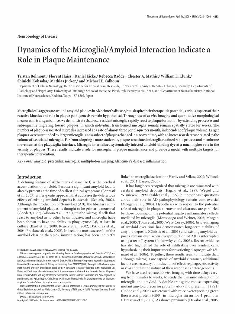
Figure 1. In vivo imaging methods and time course for quantification. A, Simultaneous imaging of three fluorescent signalswas performed: green, microglia (through Iba-1-driven GFP expression); red, amyloid deposits [after systemic injection ofmethoxy-X04 (Klunk et al., 2002)]; and blue, blood vessel labeling as a spatial reference (through systemic injection of TexasRed-coupled dextran). B, An overview image before channel separation of blood vessels (red), microglia (green), and amyloid(yellow) spanning 1.8 mm, which allowed for matching individual plaques and microglia with subsequent histological sections.The location of Z projections from 4 of the 10 high-magnification image stacks from this mouse are shown as insets. The rightmostlocation is shown with histological analysis in Figure 6 (flipped vertically). C, A coordinate system was devised for consistentimaging of the same volumes using ambient illumination of blood vessels on the brain surface. D, Volume rendering of microglia/plaque interaction over time, in this case illustrating microglial migration. E, Image stacks were taken in multiple locations over
4
time in each mouse (mean, 5 volumes per mouse for both AP-PPS1 � and APPPS1 �), using two different protocols: “shortterm” for study of the dynamics of individual microglia and“long term” for study of plaque development and microgliamigration/volume changes. Scale bar (in A), 50 �m. Imagesare from a maximum Z projection with a depth of 50 �m.
Bolmont et al. • Microglia Dynamics and Amyloid Plaque Pathogenesis J. Neurosci., April 16, 2008 • 28(16):4283– 4292 • 4285

ResultsVisualization of microglia and amyloid over timeSimultaneous imaging of microglia, amyloid, and blood vesselswas performed as described in Materials and Methods (Fig. 1).Imaging parameters were first optimized to ensure that theamount of laser power used did not result in any detectable mi-croglia reaction or photodynamic damage, even with multiplesessions of scanning the same volume. Mice used in the quanti-tative studies were divided into two groups imaged at differenttimescales. The first was imaged at brief intervals (minutes) tostudy process dynamics and subcellular events, and the other wasimaged over a longer time period (days to 1 month) to studyplaque formation and microglial migration, number, and size(Fig. 2A). A total of 39 amyloid-containing volumes from fourAPPPS1� mice and 33 volumes from two APPPS� mice were
imaged at 3 months of age and again at 4 months, over a total of10 sessions (“long-term” mice). A coordinate system was devisedwhereby multiple separate image stacks could be precisely locatedacross imaging sessions, and 3D alignment of the subsequentimage stacks based on blood vessels was performed to correct forslight changes in stack placement or orientation. Thus, imagesequences of disparate timescales could be reliably generated forquantitative analyses.
Microglia number and volume increaseMicroglia morphology was quantified over multiple sessions for aperiod of 1 month (Fig. 2). As a first analysis, imaged volumeswere categorized according to their plaque size (radius: small �10 �m � medium � 15 �m � large; n � 18, 9, 12 respectively),which revealed a change in the size of individual plaques over 1
Microglia #
0
510
1520
2530
3540
Volum
e(µ
m3x103)
Month3 4
0
2
4
6
8
10
12
14
#/v
olum
e
Month3 4
0
2
4
6
8
10Volum
e(µ
m3x103)
3 4Month
APPPS1 -Small Medium LargePlaque s ize:
Microglia # / unit volume
0
2
4
6
8
10
0 2 4 6 8 10 12 14 16 18
APPPS1 -APPPS1 +
Microglia
Volume
(µm
3x10³)
24681 01 2141618
0 10 20 30 40Plaque V olume ( µm3 x 1 0 3 )
Microglia
#/
unit
volume
A
B CPlaquevolume
Microgliavolume
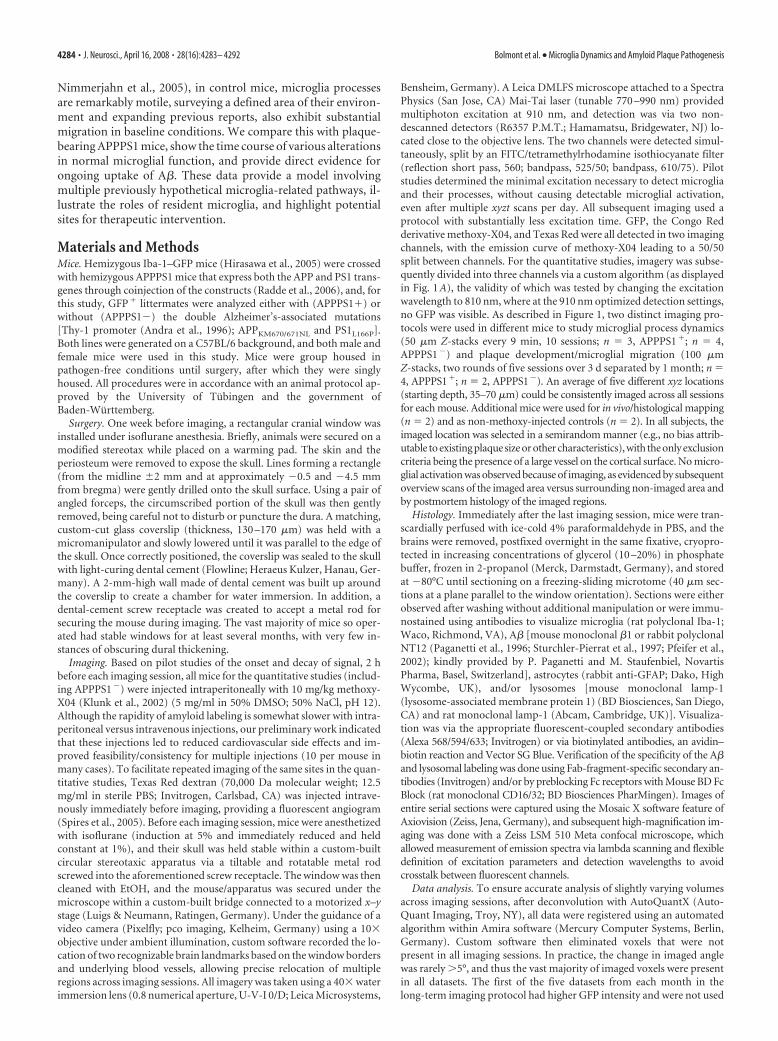
Figure 2. Existing amyloid deposits are stable over time despite the microglial response. A, Long-term imaging of the same volume is shown with microglia in green, amyloid in yellow, and bloodvessels in red (non-channel separated). Individual microglia surrounding amyloid remain stable over periods of 1 month (arrowheads), but new microglia are also evident (arrows). Microglialprocesses in each 12 h time interval have new spatial locations, with the exception of some of the largest processes, often also tied to amyloid. An example is also apparent of a resting microglia (openarrowhead) that first sends processes and subsequently migrates to contact a plaque. All images represent Z projections of 30 �m. Scale bar, 50 �m for all images. B, Quantification of mean plaquevolume and microglia number and volume over time. Overall plaque volume remained stable over 1 month but differed between size categories. The number of microglia in each volume increasedsignificantly over 1 month, independent of plaque volume. Although larger plaques were surrounded by larger microglia, the average microglia size did not increase over time. C, Size and volumeof microglia are inter-related, and the upper limit of the microglial number increase around plaques is compensated by an increase in individual microglial volume. In APPPS1 � mice (red diamonds),the number of microglia and the volume of individual microglia in any volume showed an inverse relationship (R 2 � 0.35; p � 0.001). The number of microglia surrounding plaques increased inrelation to plaque volume, reaching an apparent ceiling at a plaque size of �15,000 �m 3 (logarithmic regression, R 2 � 0.51; p � 0.0001). Values for each mouse represent the average of thelong-term imaging sessions of the second month (similar results were obtained regardless of which month’s values were used for this analysis).
4286 • J. Neurosci., April 16, 2008 • 28(16):4283– 4292 Bolmont et al. • Microglia Dynamics and Amyloid Plaque Pathogenesis
month (Fig. 2B) (repeated-measures ANOVA, size � month in-teraction, F(2,36) � 15.5; p � 0.001]. Small plaques exhibited anaverage size increase of 84% (F(1,17) � 19.1; p � 0.001), and largeplaques decreased in size by 12% (F(1,11) � 9.6; p � 0.05). From 3to 4 months of age, the number of microglia in volumes contain-ing plaques increased by an average of 2.7 microglia (Fig. 2B)(repeated-measures ANOVA; F(1,38) � 78.5; p � 0.001). Thisincrease was independent of the size of the amyloid deposit(repeated-measures ANOVA, month � plaque size, interaction,F(2,36) � 0.1; p � 0.95). Microglial number also increased slightlyin APPPS� mice but at a significantly lower rate than in theplaque-bearing APPPS1� mice (Fig. 2B) (repeated-measuresANOVA, month � transgene interaction, F(1,70) � 29.1; p �0.001). The number of microglia surrounding an individualplaque reached an upper limit in relation to plaque size (Fig.2B,C).
The average volume of plaque-associated microglia was sub-stantially higher than the volume of resting microglia in AP-
PPS1� mice (Fig. 2B) (221%; F(1,70) �12.1; p � 0.001). A strong positive rela-tionship was also found between plaquevolume and the average volume of associ-ated microglia (R 2 � 0.77; p � 0.001). Mi-croglia surrounding medium plaques wereonly 35% larger than those around smallplaques, but the large plaques had substan-tially larger microglia than the mediumplaques (225%). Together with the dataabove, microglia volume thus differenti-ates the large from medium plaques, whichhad equivalent numbers of microglia. In-terestingly, in the control mice, an inverserelationship was seen between the numberof microglia in any given volume and theaverage microglia volume (Fig. 2C), sug-gesting that regionally, the neuropil maybe surveyed by many smaller or fewerlarger microglia. Microglia volume wassimilar in fixed tissue immunostainedagainst Iba-1 (data not shown), indicatingthat increases were not simply a result ofincreased GFP expression.
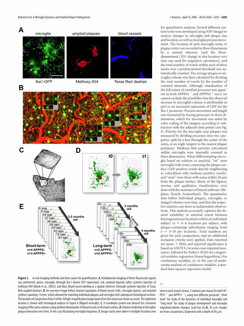
Microglial migrationExtending previous reports that studiedshorter time intervals (Davalos et al., 2005;Nimmerjahn et al., 2005), we were able toobserve substantial movement of micro-glia somata over time in both the AP-PPS1� and APPPS1� mice (Figs. 2A, 3).In APPPS� mice, approximately half ofthe microglia adjacent to plaques (within50 �m) subsequently migrated such thattheir somata were immediately adjacent tothe plaque periphery (over a period of24 – 48 h), whereas the somata of the otherhalf remained distant (10 –50 �m) fromthe plaque periphery with their processes,nevertheless maintaining a connection tothe amyloid surface. Previously resting mi-croglia with symmetrical ramified pro-cesses thus first became polarized and, onreaching the plaque, had enlarged somas
and reduced ramification (Figs. 2A, 3B). Despite microgliamovement toward the plaques, an overall decrease in microglialmigration was observed in APPPS1� mice compared with AP-PPS1� mice (Fig. 3C) (F(1,352) � 15.9; p � 0.001) primarily be-cause the vast majority of the more numerous microglia in theAPPS1� mice (Radde et al., 2006) remained spatially stablearound plaques. Plaque-associated microglial process extensionand migration left formerly surveyed areas of the neuropil with-out detectable microglial coverage, which, over time, in somecases, was followed by influx of new/surrounding microglial cellsand processes. Although we cannot completely exclude the pos-sibility of microglia cell death, disappearance of a plaque-associated microglia over the 1 month observation period wasrare and would also be explained by a microglia leaving the im-aged volume. In APPPS1� control mice, 48% of microglia mi-grated a distance �5 �m (up to 38 �m) over a 1 month period,whereas the percentage was only 29% in APPPS� mice (Fig. 3D).
+-0
4
8
1 2
05
1015
20253035
0 10 20 30 4005
1015
20253035
0 10 20 30 40Distance (µm) Distance (µm)
Dis
tanc
e (µ
m)
Perc
ent
Perc
ent
C D
B
A
APPPS1- APPPS1+
APPPS1- APPPS1+
*
Figure 3. Microglia migrate in control mice and also in plaque-bearing mice, unless indefinitely attached to amyloid. A, Zprojections (100 �m thick) of microglia in APPPS1 � or APPPS1 � mice with the movement history of individual microgliasuperimposed in colored “tracks” (actual distance moved was calculated via manual identification of the soma location in threedimensions at various time points). The displayed session is the fifth (first session from the second month; yellow marker). Thecenter of the amyloid deposit in the APPPS1 � mouse is indicated by an asterisk. Scale bar, 20 �m. B, A plaque-associatedmicroglia (arrow) first sends processes toward the plaque (12 h), and subsequently migrates until the soma is in contact with theamyloid (24 h). Scale bar is 50 �m and images represent Z projections of 25 �m. C, The average movement of microglial somaover 1 month is significantly decreased in the plaque mice (APPPS1 �, n � 248; APPPS1 �, n � 106). D, Shown is a histogram ofmigration for individual microglia in APPPS1 � mice (red) and APPPS1 � mice (blue). In both groups, the majority of microgliasoma remained spatially stable over 1 month, but a subpopulation exhibits significant migration, up to 40 �m.
Bolmont et al. • Microglia Dynamics and Amyloid Plaque Pathogenesis J. Neurosci., April 16, 2008 • 28(16):4283– 4292 • 4287
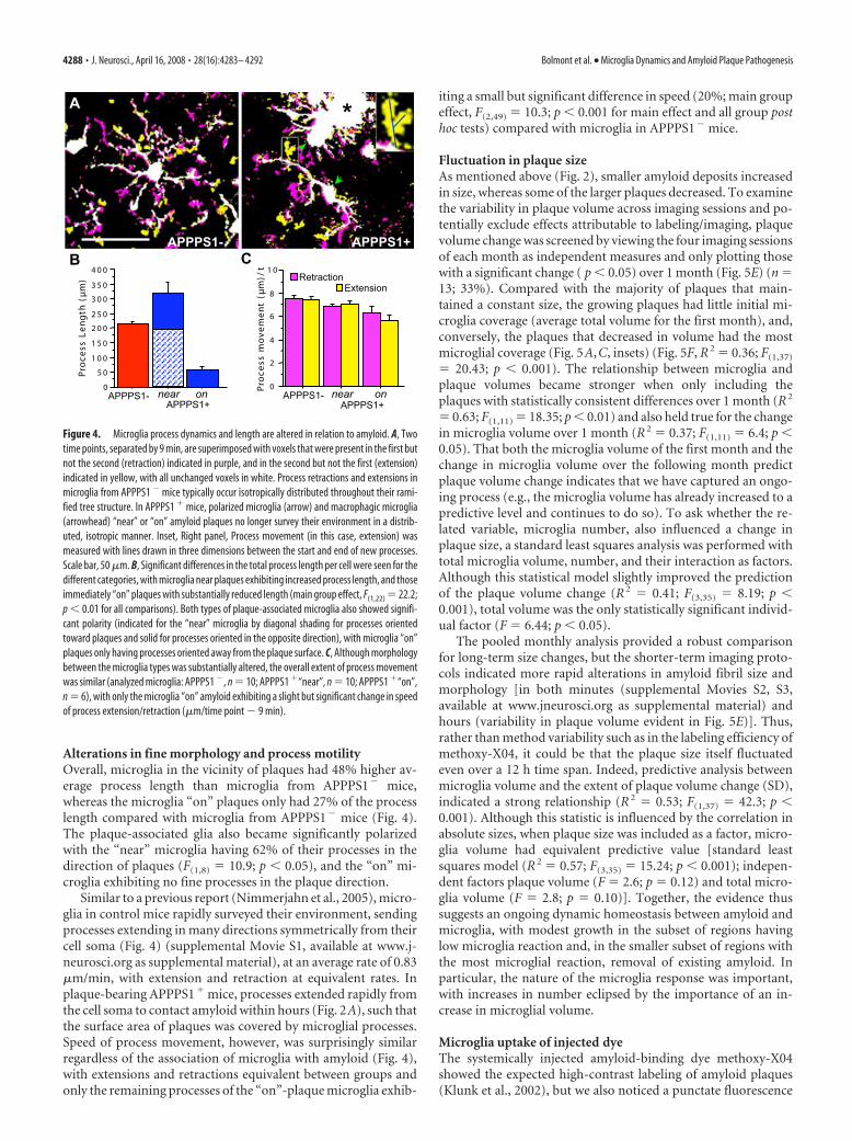
Alterations in fine morphology and process motilityOverall, microglia in the vicinity of plaques had 48% higher av-erage process length than microglia from APPPS1� mice,whereas the microglia “on” plaques only had 27% of the processlength compared with microglia from APPPS1� mice (Fig. 4).The plaque-associated glia also became significantly polarizedwith the “near” microglia having 62% of their processes in thedirection of plaques (F(1,8) � 10.9; p � 0.05), and the “on” mi-croglia exhibiting no fine processes in the plaque direction.
Similar to a previous report (Nimmerjahn et al., 2005), micro-glia in control mice rapidly surveyed their environment, sendingprocesses extending in many directions symmetrically from theircell soma (Fig. 4) (supplemental Movie S1, available at www.j-neurosci.org as supplemental material), at an average rate of 0.83�m/min, with extension and retraction at equivalent rates. Inplaque-bearing APPPS1� mice, processes extended rapidly fromthe cell soma to contact amyloid within hours (Fig. 2A), such thatthe surface area of plaques was covered by microglial processes.Speed of process movement, however, was surprisingly similarregardless of the association of microglia with amyloid (Fig. 4),with extensions and retractions equivalent between groups andonly the remaining processes of the “on”-plaque microglia exhib-
iting a small but significant difference in speed (20%; main groupeffect, F(2,49) � 10.3; p � 0.001 for main effect and all group posthoc tests) compared with microglia in APPPS1� mice.
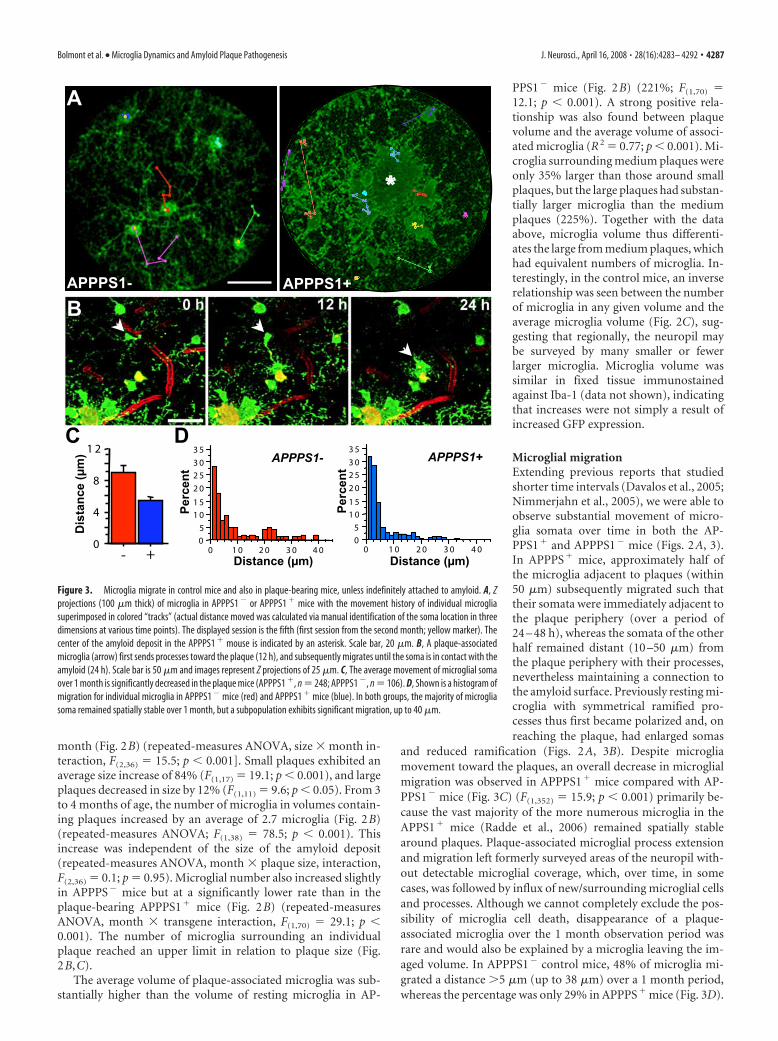
Fluctuation in plaque sizeAs mentioned above (Fig. 2), smaller amyloid deposits increasedin size, whereas some of the larger plaques decreased. To examinethe variability in plaque volume across imaging sessions and po-tentially exclude effects attributable to labeling/imaging, plaquevolume change was screened by viewing the four imaging sessionsof each month as independent measures and only plotting thosewith a significant change ( p � 0.05) over 1 month (Fig. 5E) (n �13; 33%). Compared with the majority of plaques that main-tained a constant size, the growing plaques had little initial mi-croglia coverage (average total volume for the first month), and,conversely, the plaques that decreased in volume had the mostmicroglial coverage (Fig. 5A,C, insets) (Fig. 5F, R 2 � 0.36; F(1,37)
� 20.43; p � 0.001). The relationship between microglia andplaque volumes became stronger when only including theplaques with statistically consistent differences over 1 month (R 2
� 0.63; F(1,11) � 18.35; p � 0.01) and also held true for the changein microglia volume over 1 month (R 2 � 0.37; F(1,11) � 6.4; p �0.05). That both the microglia volume of the first month and thechange in microglia volume over the following month predictplaque volume change indicates that we have captured an ongo-ing process (e.g., the microglia volume has already increased to apredictive level and continues to do so). To ask whether the re-lated variable, microglia number, also influenced a change inplaque size, a standard least squares analysis was performed withtotal microglia volume, number, and their interaction as factors.Although this statistical model slightly improved the predictionof the plaque volume change (R 2 � 0.41; F(3,35) � 8.19; p �0.001), total volume was the only statistically significant individ-ual factor (F � 6.44; p � 0.05).
The pooled monthly analysis provided a robust comparisonfor long-term size changes, but the shorter-term imaging proto-cols indicated more rapid alterations in amyloid fibril size andmorphology [in both minutes (supplemental Movies S2, S3,available at www.jneurosci.org as supplemental material) andhours (variability in plaque volume evident in Fig. 5E)]. Thus,rather than method variability such as in the labeling efficiency ofmethoxy-X04, it could be that the plaque size itself fluctuatedeven over a 12 h time span. Indeed, predictive analysis betweenmicroglia volume and the extent of plaque volume change (SD),indicated a strong relationship (R 2 � 0.53; F(1,37) � 42.3; p �0.001). Although this statistic is influenced by the correlation inabsolute sizes, when plaque size was included as a factor, micro-glia volume had equivalent predictive value [standard leastsquares model (R 2 � 0.57; F(3,35) � 15.24; p � 0.001); indepen-dent factors plaque volume (F � 2.6; p � 0.12) and total micro-glia volume (F � 2.8; p � 0.10)]. Together, the evidence thussuggests an ongoing dynamic homeostasis between amyloid andmicroglia, with modest growth in the subset of regions havinglow microglia reaction and, in the smaller subset of regions withthe most microglial reaction, removal of existing amyloid. Inparticular, the nature of the microglia response was important,with increases in number eclipsed by the importance of an in-crease in microglial volume.
Microglia uptake of injected dyeThe systemically injected amyloid-binding dye methoxy-X04showed the expected high-contrast labeling of amyloid plaques(Klunk et al., 2002), but we also noticed a punctate fluorescence
0
5 0
1 0 0
1 5 0
2 0 0
2 5 0
3 0 0
3 5 0
4 0 0
Proc
essLe
ngth
(µm)
0
2
4
6
8
1 0Proc
essmov
emen
t(µ
m)/t
near on near on
A
B C
*
APPPS1- APPPS1+
APPPS1- APPPS1-APPPS1+ APPPS1+
ExtensionRetraction
Figure 4. Microglia process dynamics and length are altered in relation to amyloid. A, Twotime points, separated by 9 min, are superimposed with voxels that were present in the first butnot the second (retraction) indicated in purple, and in the second but not the first (extension)indicated in yellow, with all unchanged voxels in white. Process retractions and extensions inmicroglia from APPPS1 � mice typically occur isotropically distributed throughout their rami-fied tree structure. In APPPS1 � mice, polarized microglia (arrow) and macrophagic microglia(arrowhead) “near” or “on” amyloid plaques no longer survey their environment in a distrib-uted, isotropic manner. Inset, Right panel, Process movement (in this case, extension) wasmeasured with lines drawn in three dimensions between the start and end of new processes.Scale bar, 50 �m. B, Significant differences in the total process length per cell were seen for thedifferent categories, with microglia near plaques exhibiting increased process length, and thoseimmediately “on” plaques with substantially reduced length (main group effect, F(1,22) � 22.2;p � 0.01 for all comparisons). Both types of plaque-associated microglia also showed signifi-cant polarity (indicated for the “near” microglia by diagonal shading for processes orientedtoward plaques and solid for processes oriented in the opposite direction), with microglia “on”plaques only having processes oriented away from the plaque surface. C, Although morphologybetween the microglia types was substantially altered, the overall extent of process movementwas similar (analyzed microglia: APPPS1 �, n � 10; APPPS1 �“near”, n � 10; APPPS1 �“on”,n � 6), with only the microglia “on” amyloid exhibiting a slight but significant change in speedof process extension/retraction (�m/time point � 9 min).
4288 • J. Neurosci., April 16, 2008 • 28(16):4283– 4292 Bolmont et al. • Microglia Dynamics and Amyloid Plaque Pathogenesis

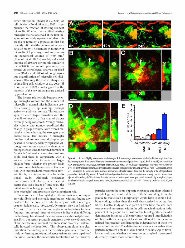
in the neuropil (Fig. 6A) that was not present in mice beforeinjection of the dye (Fig. 6E). The vast majority of these punctacolocalized with GFP, and additional 3D analysis indicated thatthe puncta were within GFP� microglia (Fig. 6B). Although thepuncta were also present within microglia in methoxy-injectedAPPPS1� mice, the density of puncta was markedly higher inAPPPS1� mice, particularly in the vicinity of amyloid plaques(Fig. 6E) (main group effect F(5,318) � 116.8; all post hoc tests forthe two plaque-associated groups, p � 0.001 compared with eachof the other groups). Subsequent high-magnification imaging ofindividual particles over time revealed both movement of theparticles within the cytoplasm and, less frequently, the disappear-ance of individual particles (Fig. 6A) (supplemental Movie S3,available at www.jneurosci.org as supplemental material).Higher-power imaging of the plaque– glial interface also revealedrapid membrane movement (supplemental Movies S2, S3, avail-able at www.jneurosci.org as supplemental material). To furtherstudy particle localization and whether they represent a nonspe-cific dye uptake or uptake of A�, we performed detailed postmor-tem morphological analyses.
To match individual locations, an overview volume was im-aged with overlapping tiles, followed by higher-resolution imag-ing of individual microglia and plaques within this volume (Fig.1B). Immediately after imaging, tissue was fixed, serial sectionedfor 3D reconstruction, and immunostained with antibodiesagainst A�. Subsequent confocal microscopy images revealedsimilar microglial morphology to that imaged in vivo and sur-rounding plaques, the spots of dye uptake imaged in vivo alsowere positive for A� (Fig. 6B–D). Examination of this colocal-ization throughout the tissue revealed much reduced A� labelingwithin microglia in areas distant from plaques and no such label-ing in APPPS1� mice. Although examples could be found inwhich fine amyloid fibrils emanated from the plaque core andwere interdigitated with microglial processes as shown previously(Stalder et al., 2001), the observed puncta had a round formmuch larger than, and not continuous with, amyloid fibrils. In-deed, they were most often found within the microglia somaopposite to the plaque– glia interface, moved within the cell somaindependently of the plaque itself (supplemental Movie S3, avail-able at www.jneurosci.org as supplemental material), and ap-peared and disappeared over time.
The internalized particles were then further characterized as
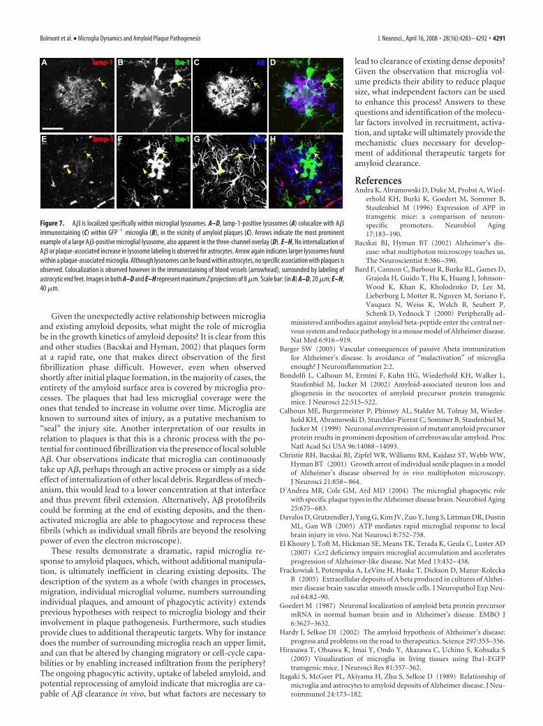
to subcellular compartment. Immuno-staining for the lysosomal marker lamp-1revealed small and large puncta throughoutthe neuropil and occasional loci of stainingnear blood vessels (Fig. 7). Larger and morefrequent lamp-1 immunostaining was ob-served in the vicinity of plaques, and manyof these puncta colocalized with GFP� mi-croglia (Fig. 7). This observation was spe-cific to microglia, because no such increaseor plaque-associated colocalization was ob-served in astrocytes (Fig. 7). Because wewere concerned about nonspecific labelingof lysosomes and microglia generally, par-allel tests to verify specificity were con-ducted with Fab-fragment secondary anti-bodies and/or a mouse Fc fragmentpreblock.
DiscussionMicroglial cells undoubtedly have multipleroles in the brain (Town et al., 2005), and
therefore their role in Alzheimer’s disease pathogenesis has re-mained enigmatic. The present study thus sought to satisfy sev-eral goals. The first was to study the basic biology and kinetics ofthe microglial response to amyloid. Although several previousstudies of amyloid-associated microglia have documented theirstatic morphology, until individual cells are followed over timewith quantitative measures, the origin, extent, and the timecourse of microglial transformation could not have been known.Second, previous reports have shown that plaque formation oc-curs at a very rapid rate and individual plaques subsequentlyremain relatively stable (Christie et al., 2001), but the role ofmicroglia in the “maintenance” of this process has not been stud-ied. By following individual plaques and microglia over varioustime intervals, our results support the idea that the initial rapidresident microglia reaction is followed by an ongoing heteroge-neous response, not only a case of microglia providing a staticphysical barrier but rather a dynamic process of homeostasis.That plaque-associated microglia remain very motile at theplaque– glial interface and exhibit signs of uptake of the A� pep-tide, provides evidence that they can inhibit additional fibrilliza-tion and plaque growth. New cells continue to be added overtime, and their volume predicts the fluctuation in plaque size.Finally, by comprehensively studying various aspects of the mi-croglial response, we sought to provide a benchmark for thera-peutic studies with simultaneous insight into mechanism andefficacy.
Similar to recent reports (Davalos et al., 2005; Nimmerjahn etal., 2005), we found that microglia in normal mice are rapidly andcontinuously surveying their environment. By imaging over alonger time course, we were also able to demonstrate that thepositioning of microglia continues to evolve in the adult brain,with a subset of glia migrating over relatively large distances.Although the potential role of invading cells has been highlightedrecently (Simard et al., 2006), our results indicate that the timecourse of this response (days) is inconsistent with a role in haltingthe initial rapid plaque growth and that process outgrowth byresident cells is a more likely candidate. By following the spatiallocation and various morphological measures over time, we werealso able to demonstrate that, once microglia become attached toamyloid, their somata remain spatially stable, that individual mi-croglia increase in volume, and that additional microglia [via
0
10
20
30
40
50
60
70
-16
-12
-8
-4
0
4
8
12
-20 20 60 1000 40 80
A B
C D
E F
Microglia volume (µm x 10 )
∆ P
laqu
e vo
lum
e (µ
m x
10
)Month 3Month 4
33
Pla
que
volu
me
(µm
x 1
0 )
33
Plaque growth/reduction3 3
a
c
Figure 5. Amyloid plaque volume changes over time. A–D, Although the majority of plaques did not change in size over 1month, examples of a reduction in plaque volume (A, B) and increase in plaque volume (C, D) were evident. Insets illustrate thecontrasting microglia coverage from the shrinking plaque (A) versus a growing plaque (B). E, The individual volume measure-ments for the 13 of 39 plaques that had consistent volume change over multiple imaging sessions. Measurements from the fourindependent imaging sessions at 3 months of age are shown as blue circles, and data from the following month are shown as reddiamonds. Only plaques with a significant difference between months are shown, and the plaques from A–D are indicated. F, Thechange in plaque volume over 1 month is related to microglia volume (total volume of all microglia/imaged volume, first month).Plaques that increased in volume over 1 month tended to have less microglia coverage, whereas those that decreased in volumehad the most microglia. Scale bar: (in A) A–D, 25 �m. Figures are maximum Z projections of 25 �m.
Bolmont et al. • Microglia Dynamics and Amyloid Plaque Pathogenesis J. Neurosci., April 16, 2008 • 28(16):4283– 4292 • 4289
either infiltration (Stalder et al., 2005) orcell division (Bondolfi et al., 2002)] sup-plement the reaction of existing residentmicroglia. Whether the ramified existingmicroglia that we observed at the first im-aging session truly represent resident mi-croglia or represent a population that hasrecently infiltrated the brain requires moredetailed study. The increase in number ofmicroglia [2.7 per imaged volume; assum-ing neocortical volume of �50 mm 3
(Bondolfi et al., 2002)], would yield a totalincrease of 250,000 per month, similar tothe 400,000 per month previously re-ported via stereological analysis on fixedtissue (Radde et al., 2006). Although rigor-ous quantification of microglia cell divi-sion is still lacking, the relative infrequencyof invading cells (Stalder et al., 2005; ElKhoury et al., 2007) would suggest that themajority of the new microglia are derivedby proliferation.
The inverse relationship between aver-age microglia volume and the number ofmicroglia in normal mice indicates a pro-cess ensuring neuropil coverage, perhapsactively via cell– cell contact. This was alsoapparent after plaque formation with theoverall volume or surface area of plaquecoverage being conserved. Average micro-glia volume and number predicted thechange in plaque volume, with overall mi-croglial volume having the strongest pre-dictive value. The increase in microglianumber and volume around plaques ap-peared to be independently regulated. Al-though we can only speculate about gov-erning mechanisms, the limited number of“available” microglia in any given volumecould lead these to compensate with agreater volumetric increase as largerplaques form. Whether this process leadsto a continued heightened state of activa-tion, with increased ability to remove amy-loid fibrils, is an important area for addi-tional study. Although molecularmechanisms will clearly be critical, itseems that basic issues of time (e.g., theinitial reaction being primarily the resi-dent microglia) and space also play a significant role.
Previous research has highlighted the intimate relationship ofamyloid fibrils and microglia membranes, without finding anyevidence for the presence of fibrillar amyloid within microgliaproper (Stalder et al., 1999, 2001). One might view our finding ofA�-positive particles within microglia as contradictory to thesefindings, but several lines of evidence indicate that differingmethodology has allowed visualization of an additional phenom-ena. First, our results primarily stem from the in vivo observationof an increase in an internalized fluorescent molecule (systemi-cally injected methoxy-X04). This observation alone is a clearindication that microglia in the vicinity of plaques are more ac-tively performing endo/pino/phagocytosis or are more capable ofthe same. Second, the subcellular localization of the observed
particles within the soma opposite the plaque and their spheroidmorphology are wholly different. Fibrils extending from theplaque to create such a morphology would have to exhibit bul-bous endings rather than the well characterized tapering finefibrils. Finally, study of these particles over time revealed bothturnover and movement within the cell soma, in directions inde-pendent of the plaque itself. Postmortem histological analysis diddemonstrate instances of the previously reported interdigitationof fibrils within microglia, at locations different from the inter-nalized fluorescence, confirming the independence of these twoobservations in vivo. The definitive answers as to whether theseparticles represent uptake of dyes bound to soluble A� or fibril-lary amyloid and whether methoxy-bound amyloid is processeddifferently require more detailed study.
01234567
#/m
icro
glia
near on
-
near on
-
no methoxy with methoxy
0h 12h 24h
A
B C
D E
Figure 6. Uptake of A� by plaque-associated microglia. A, A macrophagic plaque-associated cell exhibits many internalizedfluorescent particles that move within the cell soma over time (maximum Z projection, 35 �m). B–D, In vivo (B) and histological(C, D) analyses of the same plaque, microglia, and internalized puncta (B; red, blood vessels; green, microglia; yellow, methoxyX-04) confirm the localization and, via immunostaining, reveals colocalization with A� (C, D; red, A� via NT-12/Alexa 568, green,GFP � microglia). The same punctum is indicated by an arrow and can be visualized as within the microglia in the orthogonal x andy projections (indicated by a circle). E, Quantification of puncta colocalized with microglia in vivo in uninjected mice versus thoseinjected with methoxy-X-04 indicates a dramatic increase in the transgenic mice, particularly in the vicinity of amyloid plaques[number of microglia analyzed: no methoxy, 59/49/26; with methoxy, 62/57/71 (APPPS1 �/near/on)]. Scale bars: A–C, 10 �m;D, 5 �m.
4290 • J. Neurosci., April 16, 2008 • 28(16):4283– 4292 Bolmont et al. • Microglia Dynamics and Amyloid Plaque Pathogenesis
Given the unexpectedly active relationship between microgliaand existing amyloid deposits, what might the role of microgliabe in the growth kinetics of amyloid deposits? It is clear from thisand other studies (Bacskai and Hyman, 2002) that plaques format a rapid rate, one that makes direct observation of the firstfibrillization phase difficult. However, even when observedshortly after initial plaque formation, in the majority of cases, theentirety of the amyloid surface area is covered by microglia pro-cesses. The plaques that had less microglial coverage were theones that tended to increase in volume over time. Microglia areknown to surround sites of injury, as a putative mechanism to“seal” the injury site. Another interpretation of our results inrelation to plaques is that this is a chronic process with the po-tential for continued fibrillization via the presence of local solubleA�. Our observations indicate that microglia can continuouslytake up A�, perhaps through an active process or simply as a sideeffect of internalization of other local debris. Regardless of mech-anism, this would lead to a lower concentration at that interfaceand thus prevent fibril extension. Alternatively, A� protofibrilscould be forming at the end of existing deposits, and the then-activated microglia are able to phagocytose and reprocess thesefibrils (which as individual small fibrils are beyond the resolvingpower of even the electron microscope).
These results demonstrate a dramatic, rapid microglia re-sponse to amyloid plaques, which, without additional manipula-tion, is ultimately inefficient in clearing existing deposits. Thedescription of the system as a whole (with changes in processes,migration, individual microglial volume, numbers surroundingindividual plaques, and amount of phagocytic activity) extendsprevious hypotheses with respect to microglia biology and theirinvolvement in plaque pathogenesis. Furthermore, such studiesprovide clues to additional therapeutic targets. Why for instancedoes the number of surrounding microglia reach an upper limit,and can that be altered by changing migratory or cell-cycle capa-bilities or by enabling increased infiltration from the periphery?The ongoing phagocytic activity, uptake of labeled amyloid, andpotential reprocessing of amyloid indicate that microglia are ca-pable of A� clearance in vivo, but what factors are necessary to
lead to clearance of existing dense deposits?Given the observation that microglia vol-ume predicts their ability to reduce plaquesize, what independent factors can be usedto enhance this process? Answers to thesequestions and identification of the molecu-lar factors involved in recruitment, activa-tion, and uptake will ultimately provide themechanistic clues necessary for develop-ment of additional therapeutic targets foramyloid clearance.
ReferencesAndra K, Abramowski D, Duke M, Probst A, Wied-
erhold KH, Burki K, Goedert M, Sommer B,Staufenbiel M (1996) Expression of APP intransgenic mice: a comparison of neuron-specific promoters. Neurobiol Aging17:183–190.
Bacskai BJ, Hyman BT (2002) Alzheimer’s dis-ease: what multiphoton microscopy teaches us.The Neuroscientist 8:386 –390.
Bard F, Cannon C, Barbour R, Burke RL, Games D,Grajeda H, Guido T, Hu K, Huang J, Johnson-Wood K, Khan K, Kholodenko D, Lee M,Lieberburg I, Motter R, Nguyen M, Soriano F,Vasquez N, Weiss K, Welch B, Seubert P,Schenk D, Yednock T (2000) Peripherally ad-
ministered antibodies against amyloid beta-peptide enter the central ner-vous system and reduce pathology in a mouse model of Alzheimer disease.Nat Med 6:916 –919.
Barger SW (2005) Vascular consequences of passive Abeta immunizationfor Alzheimer’s disease. Is avoidance of “malactivation” of microgliaenough? J Neuroinflammation 2:2.
Bondolfi L, Calhoun M, Ermini F, Kuhn HG, Wiederhold KH, Walker L,Staufenbiel M, Jucker M (2002) Amyloid-associated neuron loss andgliogenesis in the neocortex of amyloid precursor protein transgenicmice. J Neurosci 22:515–522.
Calhoun ME, Burgermeister P, Phinney AL, Stalder M, Tolnay M, Wieder-hold KH, Abramowski D, Sturchler-Pierrat C, Sommer B, Staufenbiel M,Jucker M (1999) Neuronal overexpression of mutant amyloid precursorprotein results in prominent deposition of cerebrovascular amyloid. ProcNatl Acad Sci USA 96:14088 –14093.
Christie RH, Bacskai BJ, Zipfel WR, Williams RM, Kajdasz ST, Webb WW,Hyman BT (2001) Growth arrest of individual senile plaques in a modelof Alzheimer’s disease observed by in vivo multiphoton microscopy.J Neurosci 21:858 – 864.
D’Andrea MR, Cole GM, Ard MD (2004) The microglial phagocytic rolewith specific plaque types in the Alzheimer disease brain. Neurobiol Aging25:675– 683.
Davalos D, Grutzendler J, Yang G, Kim JV, Zuo Y, Jung S, Littman DR, DustinML, Gan WB (2005) ATP mediates rapid microglial response to localbrain injury in vivo. Nat Neurosci 8:752–758.
El Khoury J, Toft M, Hickman SE, Means TK, Terada K, Geula C, Luster AD(2007) Ccr2 deficiency impairs microglial accumulation and acceleratesprogression of Alzheimer-like disease. Nat Med 13:432– 438.
Frackowiak J, Potempska A, LeVine H, Haske T, Dickson D, Mazur-KoleckaB (2005) Extracellular deposits of A beta produced in cultures of Alzhei-mer disease brain vascular smooth muscle cells. J Neuropathol Exp Neu-rol 64:82–90.
Goedert M (1987) Neuronal localization of amyloid beta protein precursormRNA in normal human brain and in Alzheimer’s disease. EMBO J6:3627–3632.
Hardy J, Selkoe DJ (2002) The amyloid hypothesis of Alzheimer’s disease:progress and problems on the road to therapeutics. Science 297:353–356.
Hirasawa T, Ohsawa K, Imai Y, Ondo Y, Akazawa C, Uchino S, Kohsaka S(2005) Visualization of microglia in living tissues using Iba1-EGFPtransgenic mice. J Neurosci Res 81:357–362.
Itagaki S, McGeer PL, Akiyama H, Zhu S, Selkoe D (1989) Relationship ofmicroglia and astrocytes to amyloid deposits of Alzheimer disease. J Neu-roimmunol 24:173–182.
A B
E F
C D
G H
lamp-1lamp-1
lamp-1lamp-1
Iba-1Iba-1
Iba-1Iba-1
AßAß
GFAPGFAP
Figure 7. A� is localized specifically within microglial lysosomes. A–D, lamp-1-positive lysosomes (A) colocalize with A�immunostaining (C) within GFP � microglia (B), in the vicinity of amyloid plaques (C). Arrows indicate the most prominentexample of a large A�-positive microglial lysosome, also apparent in the three-channel overlay (D). E–H, No internalization ofA� or plaque-associated increase in lysosome labeling is observed for astrocytes. Arrow again indicates larger lysosomes foundwithin a plaque-associated microglia. Although lysosomes can be found within astrocytes, no specific association with plaques isobserved. Colocalization is observed however in the immunostaining of blood vessels (arrowhead), surrounded by labeling ofastrocytic end feet. Images in both A–D and E–H represent maximum Z projections of 8 �m. Scale bar: (in A) A–D, 20 �m; E–H,40 �m.
Bolmont et al. • Microglia Dynamics and Amyloid Plaque Pathogenesis J. Neurosci., April 16, 2008 • 28(16):4283– 4292 • 4291
Jankowsky JL, Slunt HH, Gonzales V, Savonenko AV, Wen JC, Jenkins NA,Copeland NG, Younkin LH, Lester HA, Younkin SG, Borchelt DR (2005)Persistent amyloidosis following suppression of Abeta production in atransgenic model of Alzheimer disease. PLoS Med 2:e355.
Klunk WE, Bacskai BJ, Mathis CA, Kajdasz ST, McLellan ME, Frosch MP,Debnath ML, Holt DP, Wang Y, Hyman BT (2002) Imaging Abetaplaques in living transgenic mice with multiphoton microscopy andmethoxy-X04, a systemically administered Congo red derivative. J Neu-ropathol Exp Neurol 61:797– 805.
Lopresti BJ, Klunk WE, Mathis CA, Hoge JA, Ziolko SK, Lu X, Meltzer CC,Schimmel K, Tsopelas ND, DeKosky ST, Price JC (2005) Simplifiedquantification of Pittsburgh Compound B amyloid imaging PET studies:a comparative analysis. J Nucl Med 46:1959 –1972.
Monsonego A, Weiner HL (2003) Immunotherapeutic approaches to Alz-heimer’s disease. Science 302:834 – 838.
Morgan D, Gordon MN, Tan J, Wilcock D, Rojiani AM (2005) Dynamiccomplexity of the microglial activation response in transgenic models ofamyloid deposition: implications for Alzheimer therapeutics. J Neuro-pathol Exp Neurol 64:743–753.
Nimmerjahn A, Kirchhoff F, Helmchen F (2005) Resting microglial cells arehighly dynamic surveillants of brain parenchyma in vivo. Science308:1314 –1318.
Paganetti PA, Lis M, Klafki HW, Staufenbiel M (1996) Amyloid precursorprotein truncated at any of the gamma-secretase sites is not cleaved tobeta-amyloid. J Neurosci Res 46:283–293.
Pfeifer M, Boncristiano S, Bondolfi L, Stalder A, Deller T, Staufenbiel M,Mathews PM, Jucker M (2002) Cerebral hemorrhage after passive anti-Abeta immunotherapy. Science 298:1379.
Radde R, Bolmont T, Kaser SA, Coomaraswamy J, Lindau D, Stoltze L, Cal-houn ME, Jaggi F, Wolburg H, Gengler S, Haass C, Ghetti B, Czech C,Holscher C, Mathews PM, Jucker M (2006) A�42-driven cerebral amy-loidosis in transgenic mice reveals early and robust pathology. EMBO Rep7:940 –946.
Rogers J, Strohmeyer R, Kovelowski CJ, Li R (2002) Microglia and inflam-matory mechanisms in the clearance of amyloid beta peptide. Glia40:260 –269.
Schenk D (2002) Amyloid-beta immunotherapy for Alzheimer’s disease:the end of the beginning. Nat Rev Neurosci 3:824 – 828.
Simard AR, Soulet D, Gowing G, Julien JP, Rivest S (2006) Bone marrow-derived microglia play a critical role in restricting senile plaque formationin Alzheimer’s disease. Neuron 49:489 –502.
Spires TL, Meyer-Luehmann M, Stern EA, McLean PJ, Skoch J, Nguyen PT,Bacskai BJ, Hyman BT (2005) Dendritic spine abnormalities in amyloidprecursor protein transgenic mice demonstrated by gene transfer andintravital multiphoton microscopy. J Neurosci 25:7278 –7287.
Stalder AK, Ermini F, Bondolfi L, Krenger W, Burbach GJ, Deller T, Cooma-raswamy J, Staufenbiel M, Landmann R, Jucker M (2005) Invasion ofhematopoietic cells into the brain of amyloid precursor protein transgenicmice. J Neurosci 25:11125–11132.
Stalder M, Phinney A, Probst A, Sommer B, Staufenbiel M, Jucker M (1999)Association of microglia with amyloid plaques in brains of APP23 trans-genic mice. Am J Pathol 154:1673–1684.
Stalder M, Deller T, Staufenbiel M, Jucker M (2001) 3D-Reconstruction ofmicroglia and amyloid in APP23 transgenic mice: no evidence of intracel-lular amyloid. Neurobiol Aging 22:427– 434.
Sturchler-Pierrat C, Abramowski D, Duke M, Wiederhold KH, Mistl C, Roth-acher S, Ledermann B, Burki K, Frey P, Paganetti PA, Waridel C, CalhounME, Jucker M, Probst A, Staufenbiel M, Sommer B (1997) Two amyloidprecursor protein transgenic mouse models with Alzheimer disease-likepathology. Proc Natl Acad Sci USA 94:13287–13292.
Town T, Nikolic V, Tan J (2005) The microglial “activation” continuum:from innate to adaptive responses. J Neuroinflammation 2:24.
Wegiel J, Wisniewski HM (1990) The complex of microglial cells and amy-loid star in three-dimensional reconstruction. Acta Neuropathol (Berl)81:116 –124.
Wilcock DM, Rojiani A, Rosenthal A, Levkowitz G, Subbarao S, Alamed J,Wilson D, Wilson N, Freeman MJ, Gordon MN, Morgan D (2004) Pas-sive amyloid immunotherapy clears amyloid and transiently activates mi-croglia in a transgenic mouse model of amyloid deposition. J Neurosci24:6144 – 6151.
Wyss-Coray T (2006) Inflammation in Alzheimer disease: driving force, by-stander or beneficial response? Nat Med 12:1005–1015.
4292 • J. Neurosci., April 16, 2008 • 28(16):4283– 4292 Bolmont et al. • Microglia Dynamics and Amyloid Plaque Pathogenesis