Embed Size (px)
Citation preview
Neurobiology of Disease
Amyloid Precursor Protein Dimerization and SynaptogenicFunction Depend on Copper Binding to the Growth Factor-Like Domain
Frederik Baumkotter,1 Nadine Schmidt,1 Carolyn Vargas,2 Sandra Schilling,1 Rebecca Weber,1 Katja Wagner,1
Sebastian Fiedler,2 Wilfried Klug,4 Jens Radzimanowski,5 Sebastian Nickolaus,3 Sandro Keller,2 Simone Eggert,1
Klemens Wild,4 and Stefan Kins1
1Division of Human Biology and Human Genetics, 2Molecular Biophysics and 3Division of Plant Physiology, University of Kaiserslautern, D-67663Kaiserslautern, Germany, 4Heidelberg University Biochemistry Center, University of Heidelberg, D-69120 Heidelberg, Germany, and 5Unit of Virus HostCell Interactions, International Coeducational Unit 3265, Joseph Fourier University–European Molecular Biology Laboratory–National Center of ScientificResearch, F-38042 Grenoble, France
Accumulating evidence suggests that the copper-binding amyloid precursor protein (APP) has an essential synaptic function. APPsynaptogenic function depends on trans-directed dimerization of the extracellular E1 domain encompassing a growth factor-like domain(GFLD) and a copper-binding domain (CuBD). Here we report the 1.75 Å crystal structure of the GFLD in complex with a copper ion boundwith high affinity to an extended hairpin loop at the dimerization interface. In coimmunoprecipitation assays copper binding promotesAPP interaction, whereas mutations in the copper-binding sites of either the GFLD or CuBD result in a drastic reduction in APP cis-orientated dimerization. We show that copper is essential and sufficient to induce trans-directed dimerization of purified APP. Further-more, a mixed culture assay of primary neurons with HEK293 cells expressing different APP mutants revealed that APP potentlypromotes synaptogenesis depending on copper binding to the GFLD. Together, these findings demonstrate that copper binding to theGFLD of APP is required for APP cis-/trans-directed dimerization and APP synaptogenic function. Thus, neuronal activity or disease-associated changes in copper homeostasis likely go along with altered APP synaptic function.
Key words: Alzheimer’s disease; amyloid precursor protein; copper; dimerization; metal homeostasis; synaptogenesis
IntroductionThe amyloid precursor protein (APP) was first identified as theprecursor of amyloid � (A�) peptides, which are derived fromsequential proteolysis by �- and �-secretases and aggregate in theform of amyloid plaques in the course of Alzheimer’s disease
(AD) (Masters and Beyreuther, 2006). Besides the central role ofAPP in AD pathogenesis, essential physiological functions of APPin synaptic processes have been identified, including trans-cellular synaptic adhesion (Muller and Zheng, 2012). Moreover,APP has been identified as an essential copper-binding proteinaltering metal homeostasis (White et al., 1999; Maynard et al.,2002). In turn, changes in copper homeostasis as observed for ADpatients have been shown to affect APP processing (Roberts et al.,2012).
Structurally, APP is a type I transmembrane protein with ashort cytoplasmic tail and a large ectodomain, including copper-binding sites in its E1 (Kong et al., 2008) and E2 (Dahms et al.,2012) domains. In addition, the A� peptide can bind copper onceit is cleaved from APP (Smith et al., 2007). The E1 domain can befurther subdivided into a growth factor-like domain (GFLD) anda copper-binding domain (CuBD), which has been reported toreduce Cu (II) to Cu (I) (Multhaup et al., 1996), a process possi-bly relevant for copper transport and the production of reactiveoxygen species. APP and its homologs amyloid precursors-likeprotein 1 (APLP1) and APLP2 are able to form homodimers andheterodimers. Besides a lateral interaction of APP in cis-orientation, cell-surface APP may also interact in a trans-cellularmanner (Baumkotter et al., 2012; Isbert et al., 2012; Kaden et al.,2012). cis-dimerization affects APP processing (Kaden et al.,
Received Jan. 15, 2014; revised June 18, 2014; accepted July 9, 2014.Author contributions: F.B., S.Ke., S.E., K.Wi., and S.Ki. designed research; F.B., N.S., C.V., S.S., R.W., K.Wa., S.F.,
W.K., J.R., and S.N. performed research; F.B., N.S., C.V., S.S., R.W., K.Wa., S.F., J.R., S.Ke., S.E., K.Wi., and S.Ki.analyzed data; F.B., S.Ke., K.Wi., and S.Ki. wrote the paper.
This work was supported by the Fritz Thyssen Foundation (S.Ki.) and the German Research Foundation, includingthe Research Group FOR1332 (S.Ki. and K.Wi.) and the Rheinland-Pfalz Foundation for Innovation (S.Ki. and S.Ke.).We thank L. Hanke for administrative support, D. Gross for excellent technical assistance, and J. Klement for help inpurifying APP fusion proteins. We thank C. Tischer (European Molecular Biology Laboratory, Heidelberg, Germany)for help with cell profiler and immunocytochemical quantification analysis, J. Kopp and C. Siegmann from thecrystallization platform of the Heidelberg University Biochemistry Center/Cluster of Excellence: CellNetworks forhelp in protein crystallization, and I. Sinning (Heidelberg University Biochemistry Center, Heidelberg, Germany) forgenerous support. We acknowledge the European Synchrotron Radiation Facility for providing synchrotron radiationand for assistance in using beam-lines ID23-2 and BM29. We thank A. Vest and P. Scheiffele for help with thehemisynapse assay and P. Scheiffele for providing the Neuroligin-1 cDNA.
The authors declare no competing financial interests.Correspondence should be addressed to either of the following: Stefan Kins, Division of Human Biology and
Human Genetics, University of Kaiserslautern, Erwin-Schrodinger-Straße 13, D-67663 Kaiserslautern, Germany,E-mail: [email protected]; or Klemens Wild, Heidelberg University Biochemistry Center, Im NeuenheimerFeld 328, University of Heidelberg, D-69120 Heidelberg, Germany. E-mail: [email protected].
DOI:10.1523/JNEUROSCI.0180-14.2014Copyright © 2014 the authors 0270-6474/14/3411159-14$15.00/0
The Journal of Neuroscience, August 13, 2014 • 34(33):11159 –11172 • 11159
2008; Eggert et al., 2009; Schmidt et al., 2012), whereas interac-tion in trans has been reported to promote cell adhesion (Soba etal., 2005). APP knock-out mice show cognitive deficits (Mullerand Zheng, 2012), and inactivation of APP on the APLP2 knock-out background in either the presynaptic or postsynapticcompartment caused defects in the neuromuscular synapse, sug-gesting that postsynaptic APP is required for presynaptic special-ization. Consistently, expression of APP in non-neuronal cellscocultured with neurons promotes presynaptic differentiation ofcontacting axons, similar to Neuroligin. Thus, APP has been clas-sified as a “synaptogenic” protein, which induce presynaptic orpostsynaptic differentiation when presented to axons or den-drites, respectively (Siddiqui and Craig, 2011). Remarkably, theconserved E1 domain has been identified as the major APP-interaction interface with possible contributions from the E2 do-main in the presence of heparin (Wang and Ha, 2004; Lee et al.,2011). Notably, soluble APP (sAPP) liberated after ectodomainshedding of APP as well as the purified E1 domain are mono-meric in solution and can dimerize in vitro only at very highconcentrations or in the presence of low-molecular-weight(LMW) heparin (Gralle et al., 2006; Kaden et al., 2008; Dahms etal., 2010). Heparin binding is mediated by a disulfide-bridgedhairpin loop in the GFLD [residues 95-109, in the following de-noted as ligand-binding loop (LBL); Rossjohn et al., 1999], as-sumed to constitute a major part of the dimerization interface(Dahms et al., 2010). Notably, the copper-binding site in theCuBD is close to the interface, but a possible metal bridge couldnot be confirmed in the crystallized E1 domain dimer (Dahms etal., 2010). Here, we report a novel high-affinity copper-bindingsite in the LBL of the GFLD and show that copper is essential andsufficient for APP dimerization in a cis- and trans-cellular man-ner. Moreover, we provide evidence by using a mixed cocultureassay with APP-expressing non-neuronal cells and primary neu-rons, that APP triggers specializations of contacting axons in acopper-dependent manner.
Materials and MethodsCrystallization, structure determination, and refinement of the GFLD.Crystals were grown by the sitting-drop method at 18°C by mixing equalvolumes of 10 mg/ml E1 and reservoir containing 6% (w/v) polyethyleneglycol 3350 and 100 mM HEPES, pH 7.5. Crystals appeared within 20 d.Diffraction data (� � 0.8726 Å) were measured on beam-line ID23-2under cryogenic conditions (100 K) at the European Synchrotron Radi-ation Facility (ESRF) in Grenoble, France. Data were processed withiMosflm (Battye et al., 2011) and Scala (Collaborative ComputationalProject Number 4, 1994). The crystal structure was determined by mo-lecular replacement using Phaser (McCoy et al., 2007) and the crystalstructure of the apo (without copper) GFLD [Protein Data Bank (PDB)identification number 1mwp] as a search model. Model building andrefinement were performed in Coot (Emsley et al., 2010) and PHENIX(Adams et al., 2010), respectively. Ramachandran statistics (Chen et al.,2010) show 94% of residues in the favored region and no outliers. Figureswere prepared with PyMOL [DeLano, The PyMOL molecular graphicssystem; http://www.pymol.org (2002)].
Purification of APP E1, APP GFLD, and CuBD. For crystallization,recombinant APP E1 (amino acids 18-189) was expressed as 6�His-tagged fusion protein in Pichia pastoris GS115 cells using a pBLHIS–SX-derived expression vector. Cells were grown in BMGY (1% yeast extract,2% peptone, 100 mM potassium phosphate, pH 6.0, 1.34% yeast nitrogenbase without amino acids, 0.0004% biotin, and 1% glycerol) at 30°Cunder vigorous shaking until the culture reached log phase growth. Cellswere harvested by centrifugation at 3000 � g for 10 min and resuspendedin BMMY (BMGY with 2% methanol, without glycerol). After 24 h, cellswere again sedimented, and the supernatant was loaded on a HisTrapFast Flow affinity column (GE Healthcare). The column was washed with
binding buffer (20 mM sodium phosphate, pH 7.2, 150 mM NaCl, and 10mM imidazole), and the protein was eluted in elution buffer (20 mM
sodium phosphate, pH 7.2, 150 mM NaCl, and 1 M imidazole). The pu-rified protein was concentrated and purified to homogeneity by gel per-meation chromatography (GPC) in 20 mM HEPES and 150 mM NaCl, pH7.2. Protein concentration was determined by absorbance measurementsat 280 nm, and purity of the protein was confirmed by SDS-PAGE.
For isothermal titration calorimetry (ITC) and small-angle x-ray scat-tering (SAXS) measurements, the APP E1 domain (amino acids 18-189)and the mutant APP E1 domain (APP E1 mut) were expressed as untaggedproteins in P. pastoris as described above. The supernatant was loaded ona Heparin High Performance affinity column (GE Healthcare) andwashed with binding buffer (20 mM HEPES, pH 7.2, and 150 mM NaCl),and the protein was eluted in elution buffer (20 mM HEPES, pH 7.2, and1 M NaCl). The purified protein was concentrated and subjected to GPCas described above. Protein purity was confirmed by SDS-PAGE.
Recombinant APP GFLD (amino acids 18-123) and CuBD (aminoacids 124-189) were expressed as GST-tagged fusion proteins in Esche-richia coli Origami (DE3) cells using a pGEX6P-1 derived expressionvector. Cells were grown at 18°C in 2 � YT (1.6% tryptone, 1.0% yeastextract, and 0.5% NaCl, pH 7.0). At 24 h after induction of proteinexpression by 1 mM isopropyl-�-D-thiogalactopyranoside, cells were har-vested by centrifugation at 6000 � g for 15 min. The cell pellet wasresuspended in cold PBS and incubated for 30 min at 4°C with 1 mg/mllysozyme. Cells were disrupted by sonication, and the lysate was clarifiedby centrifugation at 30,000 � g for 1 h at 4°C and loaded on a GST affinitycolumn (GE Healthcare). The column was washed with PBS, and theprotein was eluted in elution buffer (50 mM Tris, pH 8.0, and 10 mM
reduced glutathione). The purified protein was incubated overnight at4°C with 20 U/mg PreScission protease (GE Healthcare) and againloaded on a GST affinity column. The untagged protein was concentratedand purified to homogeneity by GPC in 20 mM HEPES, pH 7.2, and 150mM NaCl. Protein concentration was determined by absorbance mea-surements at 280 nm, and protein purity was confirmed by SDS-PAGE.
ITC. High-sensitivity ITC was performed at 25°C on a MicroCaliTC200 (GE Healthcare). The injection syringe either contained 1 mM
CuCl2 and 4 mM glycine, 1 mM ZnSO4, or 1 mM FeSO4. The proteinconcentration in the cell was 20 �M (APP E1), 43 �M (APP E1 mut), 14 �M
(GFLD), 38 �M (GFLD mut), or 13 �M (CuBD) as determined by absor-bance measurements using molar extinction coefficients, �, of 23.2mM
�1 cm �1 (APP E1 and APP E1 mut), 15.8 mM�1 cm �1 (APP GFLD
and APP GFLD mut), and 8.4 mM�1 cm �1 (APP CuBD) at 280 nm. Both
ligand and protein solutions were prepared in HEPES buffer (20 mM
HEPES and 150 mM NaCl, pH 7.2). Titrations of buffer or ligand solu-tions into buffer served as controls. Twenty injections of 2 �l each wereperformed at 4 min time intervals using a stirring speed of 1000 rpm. Thefirst injection was excluded from analysis. Automated baseline subtrac-tion and peak integration were accomplished with the public-domainprogram NITPIC (Keller et al., 2012). Thermodynamic parameters wereobtained by nonlinear least-squares data fitting using an in-house pro-gram (Kemmer and Keller, 2010) and SEDPHAT (Houtman et al., 2007).
We analyzed copper binding to APP E1, CuBD, and GFLD accordingto a thermodynamic model describing the competitive chelation of cop-per by protein and glycine (Hong et al., 2008). Briefly, copper ions werefirst chelated with glycine before titration into the protein solution toavoid hydroxo-complex formation of copper in aqueous solutions. Toextract the parameter values characterizing copper binding to the pro-tein, we accounted for all competing and coupled equilibria present inthis system. These include the following: (1) binding of free copper toglycine to form a 1:1 complex; (2) binding of this complex to a secondglycine molecule to form a 1:2 complex; and (3) binding of copper toprotein. Copper also binds to HEPES buffer, but this interaction has beenshown to be negligible (Hatcher et al., 2008). Literature values (Martell etal., 2004) were used to account for the glycine binding equilibria.
SAXS. SAXS was performed at the ESRF beam-line BM29 in Grenoble,France. APP E1 at concentrations of 0.25 mg/ml (12.6 �M), 0.5 mg/ml(25.3 �M), 1.0 mg/ml (50.5 �M), and 2.0 mg/ml (101 �M) dissolved inHEPES buffer was incubated either with or without a fivefold molarexcess of 1 mM CuCl2 with 4 mM glycine for 1 h at 4°C. A total of 50 �l of
11160 • J. Neurosci., August 13, 2014 • 34(33):11159 –11172 Baumkotter et al. • Copper-Induced APP cis- and trans-Synaptic Dimerization

protein solution for each sample (and buffer) was exposed to x-rays, andscattering data were collected. Ten individual frames of 1 s were collectedfor each exposure using a Pilatus 1M detector (Dectris). Individualframes were processed using the software BsxCUBE, yielding individualradially averaged curves of normalized intensity versus scattering angle,q � 4�sin�/�. Frames were combined, excluding any data points affectedby aggregation induced by radiation damage, to give the average scatter-ing curve for each measurement. Data were processed and analyzed usingthe ATSAS package, as described previously (Putnam et al., 2007). Thefinal merged scattering data was further evaluated using PRIMUS. The iso-tropic scattering intensity, I(q), was transformed to the distance distributionfunction P(r) using the program GNOM. For ab initio modeling of the SAXSdata, 20 sets of independent models were calculated using Dammin (Sver-gun, 1999) and then averaged and aligned using DAMAVER.
Circular dichroism spectroscopy. Thermal unfolding of the APP E1 do-main was monitored by circular dichroism (CD) spectroscopy. APP E1in HEPES buffer (20 mM HEPES and 150 mM NaCl, pH 7.2) either with-out or saturated with copper was heated from 20°C to 90°C, and theellipticity (�) at 222 nm was measured on a Chirascan-plus CD spec-trometer (Applied Photophysics). After the experiment, the sample wascooled back to 20°C, and the measurement was repeated to check forreversibility.
Coimmunoprecipitation. HEK293 cells were cultivated in DMEM sup-plemented with 10% FBS according to standard cell culture techniques.Cells were transiently cotransfected with HA-tagged full-length and myc-tagged full-length or mutated pcDNA3.1–APP constructs. As control,empty vector was cotransfected with myc-tagged APP constructs. Trans-fection with Lipofectamine 2000 (Invitrogen) was performed accordingto the protocol of the manufacturer. Coimmunoprecipitation (Co-IP)was performed as described previously (Soba et al., 2005). Briefly, trans-fected HEK293 cells were lysed in lysis buffer [50 mM Tris, pH 7.4, 150mM NaCl, 1% NP-40, and Complete Protease Inhibitor mix (Roche)] for20 min on ice. Lysates were clarified at 16,000 � g for 10 min, and 4% ofthe supernatant was denatured for the direct load. The remaining super-natant was incubated for 1 h at 4°C with 10 �l of protein A Sepharosebeads (GE Healthcare) to reduce unspecific binding. Beads were sedi-mented by centrifugation at 2000 � g for 1 min, and the supernatant wasincubated overnight at 4°C with 20 �l of anti-HA agarose beads (Roche).Beads were washed three times with lysis buffer and finally denatured(IP). Samples were separated on 8% Tris/glycine gels and probed byimmunoblotting for HA- and myc-tagged APP. Statistical analysis wasperformed with an unpaired Student’s t test (n 4; *p � 0.05; **p � 0.01;***p � 0.001). Results are presented as mean � SEM.
For metal ion treatment, cells were incubated with the indicatedamounts of glycine-complexed copper (10 mM CuCl2, 40 mM glycine in20 mM HEPES, and 150 mM NaCl, pH 7.2), zinc (10 mM ZnSO4 in 20 mM
HEPES and 150 mM NaCl, pH 7.2), or iron (10 mM FeSO4 in 20 mM
HEPES and 150 mM NaCl, pH 7.2) for 4 h before cell lysis. Afterward,Co-IP was performed as described above.
Immunocytochemistry. For immunocytochemistry, HeLa cells werecultivated and transfected as described above for HEK293 cells. At 20 hafter transfection with target cDNA, cells (20,000) were seeded on 14 mmglass coverslips and fixed with 4% (w/v) paraformaldehyde/saccharose inPBS and permeabilized with 0.1% (v/v) NP-40. Nonspecific sites wereblocked for 1 h with 5% (v/v) goat serum in PBS. Primary antibodydirected against c-myc (Santa Cruz Biotechnology) was used at 1:200dilution and detected using secondary IgG antibody conjugated to AlexaFluor 488 (green) fluorophore. Images were taken with an Axio ObserverZ.1 (Zeiss) confocal microscope.
To estimate the level of colocalization between APP and the markerpeptide for ER (KDEL-mRFP (Altan-Bonnet et al., 2006), kindly pro-vided by Bruce Snapp), the Pearson’s correlation coefficient was used.Pearson’s correlation coefficient (rp) is a statistical analysis of the rela-tionship between fluorescence intensities. An rp value of 0 indicates nocorrelation, whereas an rp value of 1 indicates complete colocalization(Bolte and Cordelieres, 2006). Statistical analysis was performed withone-way factorial ANOVA and Tukey’s HSD post hoc test (n � 3; *p �0.05; **p � 0.01; ***p � 0.001). Results are presented as mean � SEM.
Cell viability analysis. Cell viability was monitored by lactate dehydro-
genase (LDH) release as a marker for cell death. Briefly, HEK293 cellswere seeded at a density of 30,000 cells per well in a 96 multiwell plate andincubated for 4 h with the indicated amounts of glycine-complexed cop-per (10 mM CuCl2, 40 mM glycine in 20 mM HEPES, pH 7.2, and 150 mM
NaCl) in DMEM with 0.1% FCS. The release of LDH from cells wasdetermined in the medium with CytoTox-ONE Homogeneous Mem-brane Integrity Assay (Promega) according to the protocol of the manu-facturer. Maximal LDH release was determined in the medium after celllysis with 9% (w/v) Triton X-100. Results are presented as mean � SEMvalues from three independent experiments (n � 3).
Mixed co-culture assay. The preparation of primary cortical neuronsand coculture with HEK293 cells were performed essentially as describedpreviously (Biederer and Scheiffele, 2007). Briefly, primary cortical neu-rons were prepared at E14 from C57BL/6J mice. The dissected corticeswere suspended in 5 ml of trypsin–EDTA for 15 min at 37°C and subse-quently washed five times with 5 ml of HBSS supplemented with 10 mM
HEPES. To dissociate the cells, the cortices were resuspended in HBSSsupplemented with 10 mM HEPES and triturated with a fire-polishedglass pipette. Dissociated cells were resuspended in DB1 medium[DMEM with 10% (v/v) FCS, 0.79% (w/v) D-glucose, and 2 mM glu-tamine] and plated onto 14 mm coverslips pretreated with poly-L-lysine(20 �g/ml) at a density of 40,000 cells/cm 2. After 6 h incubation at 37°C,the DB1 medium was replaced by NM medium [Neurobasal mediumwith 2% (v/v) B-27 supplement and 2 mM glutamate].
At 6 d in vitro of the neuronal culture, HEK293 cells were transientlytransfected with GFP, HA-tagged Neuroligin, full-length APP (APP FL),APP �E1, APP H108/110A (GFLD mut), APP H147/151A (CuBD mut), ormyc-tagged APP �EC. After 24 h, transfected cells were detached andseeded at a density of 17,500 cells/cm 2 to the cortical culture. Twenty-four hours later, cultures were fixed in 4% (w/v) PFA supplemented with4% (w/v) sucrose and stained with an anti-Synaptophysin antibody (1:200; Sigma), anti-synaptic vesicle protein 2 (SV2) antibody (1:400; De-velopmental Studies Hybridoma Bank), or anti-Synapsin-1 antibody (1:5000; SYSY), all followed by an Alexa Fluor 594-conjugated secondaryantibody (1:400; Invitrogen). Cells were costained with an anti-microtubule-associated protein 2 (MAP2) antibody (1:300; Santa CruzBiotechnology), as well as anti-c-myc (1:200,; Serotec) and anti-HA (1:300; Santa Cruz Biotechnology) antibodies to detect dendrites and theheterologously expressed proteins, respectively. In addition, cells werecostained with an anti-Tau antibody (TAU-1 1:200; Millipore BioscienceResearch Reagents) to detect axons. Synapses were identified asSynaptophysin-positive puncta at sites of cell contacts between neuronsand HEK293 cells that do not colocalize with MAP2-positive dendriticstructures. Images were taken with an Axio Observer Z.1 (Zeiss) confocalmicroscope, and quantification was performed via NIH ImageJ analysis.Statistical analysis was performed with one-way factorial ANOVA andTukey’s HSD post hoc test (n 4; *p � 0.05; **p � 0.01; ***p � 0.001).Results are presented as mean � SEM.
Generation and purification of APPex-FC. COS7 cells stably transfectedwith the ectodomain of APP (APPex; amino acids 18-624) fused to thecrystallizable fragment (FC) of human IgG1 at the C terminus were cul-tivated at 37°C in growth medium (DMEM supplemented with 10%FCS, 1% penicillin/streptomycin, and 300 �g/ml hygromycin B) in ahumidified atmosphere with 5% CO2 according to standard cell culturetechniques. Three days after passaging, conditioned medium was col-lected and centrifuged at 3000 � g at room temperature for 5 min toclarify them from cell debris. The supernatant was filtered (0.22 �m) andloaded on a recombinant protein A Sepharose FF 20 ml affinity column(GE Healthcare). The column was washed with binding buffer (50 mM
Tris/HCl and 300 mM NaCl, pH 7.4) until UV absorption reached base-line. The protein was eluted with 100 mM glycine, pH 2.5. To restore thepH, 100 �l of 1 M Tris/HCl, pH 8.0, were added immediately to theprotein. The protein was concentrated and desalted with a PD-10 desalt-ing column (GE Healthcare). Afterward, the protein was loaded on aHiTrap Heparin HP 5 ml affinity column (GE Healthcare). The columnwas washed with binding buffer (20 mM HEPES and 150 NaCl, pH 7.2)until UV absorption reached baseline, and the protein was subsequentlyeluted with elution buffer (20 mM HEPES and 1 M NaCl, pH 7.2). Proteinconcentration was determined by absorbance measurements at 280 nm
Baumkotter et al. • Copper-Induced APP cis- and trans-Synaptic Dimerization J. Neurosci., August 13, 2014 • 34(33):11159 –11172 • 11161
on a V-630 UV-VIS spectrophotometer (Jasco). Aliquots were takenduring the purification process and analyzed by SDS-PAGE.
Bead aggregation assay. To analyze the dimerization properties of APP,10 �l of magnetic protein A polystyrene beads (Invitrogen) were incu-bated with 15 �g of either purified human IgG FC (Bethyl) or APPex-FC
in binding buffer (20 mM HEPES and 150 mM NaCl, pH 7.2) for 2 h at 4°Cwith end-over-end rotation. Afterward, the beads were washed threetimes with 500 �l of binding buffer and sonicated to singularize. Aggre-gation of the protein-coated beads was performed for 2 h at 4°C in 800 �lof binding buffer supplemented with 0.1% (w/v) BSA and the indicatedamounts of copper, zinc, iron, or heparin dodecasaccharide (dp12; Dex-tra Laboratories). For the measurement, 200 �l of each sample werediluted in 10 ml of Isotone II solution (Beckman Coulter) and inverted tomix. The number of aggregates �10 �m was counted with a CoulterCounter Z2 particle counter (Beckman Coulter; 100 �m aperture tube).The number of aggregates formed was normalized to APPex-FC. Errorbars represent mean � SEM values. Statistical analysis was performedwith unpaired Student’s t test (n 3; *p � 0.05, **p � 0.01, ***p � 0.001compared with APPex-FC in buffer; ##p � 0.01 compared with APPex-FC
in buffer supplemented with 100 �M CuCl2).GPC. For analytical GPC, either 35 �M APP GFLD or 25 �M APP
GFLD mut were incubated or not with either 10� molar excess ofheparin dodecasaccharide (dp12; Dextra Laboratories) or 50� molarexcess of CuCl2 for 30 min on ice. A total of 100 �l of the proteinsolution was loaded at a flow rate of 0.8 ml/min on a calibratedSuperdex 75 10/300 GL gel permeation column (GE Healthcare). Forcalibration of the column, a calibration curve [KAV vs. log molecularweight (MW)] was prepared with protein standards (conalbumin, Mr
� 75,000; carbonic anhydrase, Mr � 29,000; ribonuclease A, Mr �13,700; aprotinin, Mr � 6500) of the LMW gel filtration calibrationkit (GE Healthcare). The void volume (Vo) was determined by theelution volume (Ve) of Blue Dextran 2000.
Cellular copper uptake monitored by inductively coupled plasma-opticalemission spectrometry. To determine the amount of copper taken up bycells, HEK293 cells were cultivated in growth medium (DMEM with0.1% FCS and 1% penicillin/streptomycin) and incubated for 4 h withincreasing amounts (1-500 �M) of glycine-complexed copper (10 mM
CuCl2, 40 mM glycine in 20 mM HEPES, pH 7.2, and 150 mM NaCl).Cells were washed once with PBS and lysed in lysis buffer [50 mM Tris,pH 7.4, 150 mM NaCl, 1% NP-40, and Complete Protease Inhibitormix without EDTA (Roche)] for 20 min on ice. Lysates were clarifiedat 16,000 � g for 10 min and digested in 3 ml of 65% (v/v) HNO3 and1 ml of 30% (v/v) H2O2 in an MLS-Ethos microwave oven using atemperature step gradient with a maximum temperature of 210°C.Samples were diluted to a final volume of 7 ml with ddH2O andanalyzed by inductively coupled plasma-optical emission spectrome-try (ICP-OES) on an iCAP 6300 DUO spectrometer (Thermo FisherScientific). Copper was detected and quantified at 324.754 nm. Thedetection limit for copper was 0.5 �g/l. Protein concentration in celllysates was determined by BCA assay (Sigma). Error bars representmean � SEM values (n � 3).
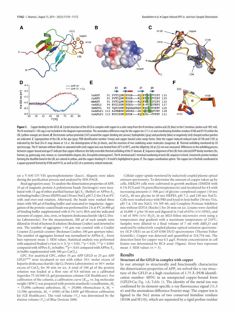
ResultsStructure of the GFLD in complex with copperIn our attempt to structurally and functionally characterizethe dimerization properties of APP, we solved the x-ray struc-ture of the GFLD at a high resolution of 1.75 Å (PDB identifi-cation number: 4JFN) in an unexpected copper-bound form(GFLD�Cu; Fig. 1A; Table 1). The identity of the metal ion wasconfirmed by its element-specific x-ray fluorescence signal (11.3) and the anomalous difference Fourier map. The copper ion isligated to the N�2 atoms of two conserved histidine residues(H108 and H110), which are separated by a rigid proline residue
Figure 1. Copper binding to the GFLD. A, Crystal structure of the GFLD in complex with copper in a color ramp from the N terminus (amino acid 28; blue) to the C terminus (amino acid 189; red).The N-terminal 6�His tag is not included in the diagram representation. The anomalous difference map for the copper ion (11.3 ) and coordinating histidine residues H108 and H110 within theLBL (yellow-orange) are shown. B, Electrostatic surface potential (3 kT) around the copper-binding site (arrow); hydrophobic (gray) and positively (blue) or negatively (red) charged surface patchesare indicated. C, Superposition of the LBL in the apo (gray; PDB identification number 1mwp) and copper-bound (color ramp) forms. Note the copper-induced reduced state of C98 and C105 asindicated by the final 2Fo-Fc map shown at 1.0 , the disintegration of the �-sheets, and the insertion of two stabilizing water molecules (magenta). D, Thermal unfolding monitored by CDspectroscopy. The E1 domain without (blue) or saturated with (red) copper ions was heated from 20°C to 90°C, and the ellipticity (�) at 222 nm was measured. Differences in the unfolding processbetween copper-bound and apo E1 indicate that copper influences the fully reversible thermal unfolding of the E1 domain. E, Sequence alignment of the LBL from selected APP family members (hs,human; cp, guinea pig; mm, mouse; ce, Caenorhabditis elegans; dm, Drosophila melanogaster). The N-terminal and C-terminal numbering of each LBL sequence is listed. Conserved cysteine residuesforming the disulfide bond in the LBL are colored in yellow, and the copper-binding H�H motif is highlighted in green. F, The copper coordination sphere. The copper ion is fivefold coordinated ina square pyramid formed by H108 and H110, as well as D23 of a symmetry-related molecule.
11162 • J. Neurosci., August 13, 2014 • 34(33):11159 –11172 Baumkotter et al. • Copper-Induced APP cis- and trans-Synaptic Dimerization
and are part of the LBL implicated in dimerization (Gralle et al.,2006; Kaden et al., 2008; Dahms et al., 2010). Copper bindingoccurs between conserved hydrophobic and basic surface patches(Fig. 1B), which are thought to be responsible for protein–pro-tein (Kong et al., 2008) and protein– heparin (Dahms et al., 2010)interactions, respectively. The structure of GFLD�Cu is overallhighly similar to those of the apo form (PDB identification num-ber 1mwp; rms deviation of 0.89 Å for 96 C�-atoms) and of theGFLD within the E1 domain (PDB identification number: 3ktmchain B; rms deviation of 0.93 Å for 95 C�-atoms), with oneprominent exception, which is the long LBL forming a �-hairpinbetween stands �5 and �8 (Fig. 1C). The copper-binding histi-dine H108 is bulged out of the LBL hairpin, and two watermolecules are placed within the bulge, thus stabilizing thecopper-binding ligand sphere (Fig. 1C). Temperature factorswithin the LBL increase up to twice the average toward the distalend and the very tip. Strikingly, copper binding coincides withthe reduction of the disulfide bridge (C98 –C105) in the LBL-integral �-sheet (�6 and �7; Fig. 1C), although purification andcrystallization were performed under nonreducing conditions.Opening of the disulfide bridge between C98 and C105 and theconformational changes within the LBL (Fig. 1C) go along withaltered thermal unfolding of the E1 domain, as shown by CDspectroscopy (Fig. 1D). Similarly, SAXS analyses of the APP E1domain in the presence and absence of copper revealed subtlestructural changes of the E1 domain during copper binding,which were obscured by the tendency for aggregation of the E1domain, probably because of clustering via intermolecular cop-per binding (data not shown).
Sequence alignment of the LBL from different species andAPP homologs reveals that the cysteines forming the disulfidebridge are conserved throughout the entire APP gene family,whereas the H�H arrangement of histidines is conserved inmammalian APP and APLP1 only (Fig. 1E). Besides the two his-tidines, the copper ion is coordinated by an aspartate (D23) of asymmetry-related molecule within the crystalline array, resultingin a square pyramidal ligand sphere (Fig. 1F). The five-coordinate geometry is reminiscent of Cu (II) binding to theCuBD, which is classified as a type-2 non-blue Cu (II) center
(Kong et al., 2008). This geometry is typical of redox activity ashas been implicated for the CuBD, in which Cu (II) is coordi-nated by three residues (H147, H151, and Y168) and two watermolecules (Kong et al., 2007b).
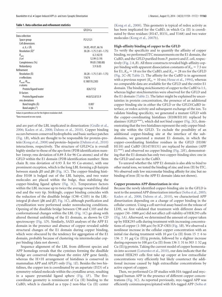
High-affinity binding of copper to the GFLDTo verify the specificity and to quantify the affinity of copperbinding, we performed ITC measurements on the E1 domain, theCuBD, and the GFLD purified from P. pastoris and E. coli, respec-tively (Fig. 2A,B). All three constructs revealed high-affinity cop-per binding with apparent dissociation constants of KD � 7.5 nM
for E1, KD � 18 nM for the CuBD, and KD � 28 nM for the GFLD(Fig. 2C–H; Table 2). The affinity for the CuBD is in agreementwith a previous report (KD � 10 nM; Hesse et al., 1994), whereasno comparable data are available for the GFLD and the entire E1domain. The binding stoichiometry of copper to the CuBD is 1:1,whereas higher stoichiometries were observed for the GFLD andthe E1 domain (Table 2). The latter might be explained by uncer-tainties in protein concentration, the presence of an additionalcopper-binding site in either the GFLD or the GFLD/CuBD in-terface, or redox activity and subsequent exchange of the ion. Tovalidate binding specificity, we generated a mutant GFLD withthe copper-coordinating histidines (H108/H110) replaced byalanines (GFLD mut), which did not bind copper (Fig. 2G), dem-onstrating that the two histidines represent the only copper bind-ing site within the GFLD. To exclude the possibility of anadditional copper-binding site at the interface of the sub-domains, we generated a mutant E1 domain, in which thecopper-coordinating histidine residues in the GFLD (H108/H110) and CuBD (H147/H151) are replaced by alanines (APPE1 mut) and observed no copper binding (Fig. 2H), demonstrat-ing that the E1 domain has only two copper-binding sites: one inthe GFLD and one in the CuBD.
To unravel whether the APP E1 domain is also able to bind toother metal ions, we tested the binding of zinc and iron (II) by ITC.We observed only low micromolar binding affinity for zinc but nobinding of iron (II) to the APP E1 domain (data not shown).
Copper promotes APP dimerization in vivoBecause the newly identified copper-binding site in the GFLD isnext to the assumed APP dimerization interface (Soba et al., 2005;Kaden et al., 2008; Dahms et al., 2010), we characterized APPdimerization depending on a change of copper binding in thecellular context. Using a cell survival assay based on the release ofLDH, we first validated that treatment with different doses ofcopper (50 –1000 �M) did not affect cell viability of HEK293 cells(Fig. 3A). Afterward, we determined the amount of copper takenup by HEK293 cells during treatment with increasing concentra-tions of copper (1-500 �M) by ICP-OES (Fig. 3B). We observed anonlinear increase in the cellular copper concentration with aninitial rise during treatment with 10 �M Cu (II) from 15 � 6 to136 � 31 �g Cu (II)/g protein, followed by a slighter increaseduring exposure to 100 �M Cu (II) from 136 � 31 to 303 � 52 �gCu (II)/g protein. Taking the current model of copper homeosta-sis into account (Lutsenko et al., 2010), our data indicate that thetreated HEK293 cells first take up copper at low extracellularconcentrations very efficiently but likely counteract the addi-tional increase caused by high extracellular concentrations byactive export of copper.
Then, we performed Co-IP studies with HA-tagged and myc-tagged human APP in the presence of different copper concen-trations (Fig. 3C). As reported previously, myc-tagged APP wasefficiently coimmunoprecipitated with HA-tagged APP (Soba et
Table 1. Data collection and refinement statistics
GFLD�Cu
Data collectionSpace group P212121Cell dimensions
a, b, c (Å) 34.05, 49.07, 66.16Resolution (Å)a 30.28 –1.75 (1.81–1.75)Rsym 6.3 (42.9)I/I 12.99 (3.19)Completeness (%) 99.85 (100.00)Redundancy 5.9 (6.0)
RefinementResolution (Å) 30.28 –1.75 (1.81–1.75)n reflections 11674Rwork /Rfree 0.1807/0.2303n atoms
Protein/ligand/water 826/7/52B-factors
Protein/ligand/water 44.8/52.8/53.9rms deviations
Bond lengths (Å) 0.007Bond angles (°) 1.16
Values in parentheses are for the highest resolution shell.aData measured on one crystal.
Baumkotter et al. • Copper-Induced APP cis- and trans-Synaptic Dimerization J. Neurosci., August 13, 2014 • 34(33):11159 –11172 • 11163
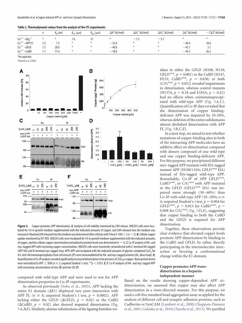
al., 2005). In the presence of 250 �M cop-per, we observed a twofold to threefoldincrease in APP dimer formation (n 3;unpaired Student’s t test, p � 0.005;Fig. 3D).
Because it has been previously re-ported that zinc and iron (II) bind to APP(Bush et al., 1993; Duce et al., 2010), wealso tested the influence of these metalions on APP dimer formation (Fig. 3E,F).In contrast to copper, we observed no in-crease in APP dimerization after additionof 50 or 250 �M zinc or iron (II) (Fig. 3D).Thus, we conclude that only copper is ableto specifically promote APP dimerization.
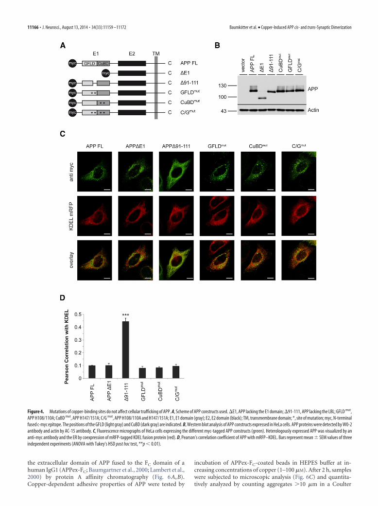
To validate that the copper-mediatedincrease in APP dimerization is attribut-able to the copper-binding sites in theGFLD and CuBD, we generated a set ofmutant human myc-tagged APP695 vari-ants lacking either the E1 domain (�E1)or the loop region (APP�91-111) or car-rying single amino acid exchanges of thecopper-binding histidine residues in theGFLD (GFLD mut) or CuBD (CuBD mut;Fig. 4A). Recently, it has been reportedthat some histidine-to-asparagine substi-tutions in the CuBD alter the maturationand subcellular localization of APP (Spo-erri et al., 2012). Therefore, we testedwhether the introduced mutations mightalso affect APP subcellular localization,which in turn could interfere with APPdimerization properties. Western blotanalysis confirmed the expected MWs ofthe different APP mutants (Fig. 4B). Inimmunocytochemical analyses, only mu-tant APP�91-111 showed a pronouncedER localization compared with wild-typeAPP (APP FL), as validated by costainingwith the ER marker mRFP–KDEL andPearson’s correlation analysis in HeLacells (n � 3; ANOVA with Tukey’s HSDpost hoc test, p � 0.0002; Fig. 4C,D). Thesedata indicate that deletion of the loop re-gion causes folding problems that retainAPP in the ER. Notably, identical resultswere also obtained with HEK293 cells(data not shown). Therefore, we excludedAPP�91-111 from additional analyses. Allother APP mutants showed no significantchanges in their subcellular localization
Figure 2. Copper binds to the GFLD of APP. A, Scheme of the APP subdomains used for ITC. The positions of the GFLD (light gray),CuBD (dark gray), and the N-terminal GST epitope (black circle) are indicated. The protease cleavage site is indicated by an arrow,and the sites of mutation are indicated by an asterisk. B, SDS-PAGE of the affinity-purified APP subdomains stained with Coomassieblue. C–H, ITC data recorded at 25°C. Titration of glycine-complexed CuCl2 into 20 �M APP E1 (C), 13 �M APP CuBD (D), 14 �M APPGFLD (E), HEPES buffer as control (F ), 38 �M APP GFLD H108/110A (GFLD mut) (G), or 43 �M APP E1 H108/110/
4
147/151A (APP E1 mut) (H). The panels in C (left) and D and E(top) show the differential heating power (�p) versus time (t)plot. C (right), D,E (bottom), Normalized heat of reaction (Q)versus molar copper/protein ratio (R). Copper binds with highaffinities to the APP E1 domain (KD � 7.5 nM), the CuBD (KD �18 nM), and the GFLD (KD � 28 nM). Binding to the APP GFLD orAPP E1 is completely abolished by substitution of the copper-coordinating histidines H108/H110 or H108/H110/H147/H151 with alanine residues, respectively.
11164 • J. Neurosci., August 13, 2014 • 34(33):11159 –11172 Baumkotter et al. • Copper-Induced APP cis- and trans-Synaptic Dimerization
compared with wild-type APP and were used to test for APPdimerization properties in Co-IP experiments.
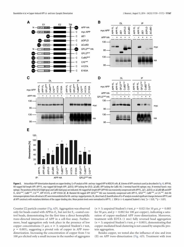
As observed previously (Soba et al., 2005), APP lacking theentire E1 domain (�E1) displayed very poor interaction withAPP FL (n 4; unpaired Student’s t test, p � 0.0002). APPlacking either the GFLD (�GFLD, p � 0.02) or the CuBD(�CuBD, p � 0.02) also showed impaired dimerization (Fig.5A,B,E). Similarly, alanine substitutions of the ligating histidine res-
idues in either the GFLD (H108, H110;GFLDmut, p � 0.001) or the CuBD (H147,H151; CuBDmut, p � 0.038) or both(C/Gmut, p � 0.012) revealed impairmentsin dimerization, whereas control mutants(H137A, p � 0.18 and E183A, p � 0.42)had no effects when coimmunoprecipi-tated with wild-type APP (Fig. 5A,C).Quantification of Co-IP data revealed thatthe dimerization of copper binding-deficient APP was impaired by 10-20%,whereas deletion of the entire subdomainsalmost abolished dimerization with APPFL (Fig. 5B,C,E).
In a next step, we aimed to test whethermutations of copper-binding sites in bothof the interacting APP molecules have anadditive effect on dimerization comparedwith dimers composed of one wild-typeand one copper binding-deficient APP.For this purpose, we precipitated differentmyc-tagged APP mutants with HA-taggedmutant APP (H108/110A; GFLD mut HA)instead of HA-tagged wild-type APP.Remarkably, Co-IP of APP GFLD mut,CuBD mut, or C/G mut with APP mutatedin the GFLD (GFLD mut HA) was im-paired more strongly (30 – 60%) thanCo-IP with wild-type APP (10 –20%; n 4; unpaired Student’s t test, p � 0.004 forGFLD mut, p � 0.013 for CuBD mut, p �0.008 for C/G mut; Fig. 5D,E), suggestingthat copper binding to both the CuBDand the GFLD is required for APPdimerization.
Together, these observations provideclear evidence that elevated copper levelspromote APP dimerization by binding tothe CuBD and GFLD, by either directlyparticipating in the intermolecular inter-action or favoring a conformationalchange within the E1 domain.
Copper promotes APP trans-dimerization in a heparin-independent manner
Based on the results showing copper-dependent APP cis-dimerization, we assumed that copper may also affect APPdimerization in a trans-directed manner. For this purpose, weused a cell-free standard bead aggregation assay as applied for theanalysis of different cell and synaptic adhesion proteins, such asCadherins or SynCAM (Lambert et al., 2000; Chappuis-Flamentet al., 2001; Galuska et al., 2010; Chavda et al., 2013). We purified
Table 2. Thermodynamic values from the analysis of the ITC experiments
n KD (nM) KD1 (�M) KD2 (�M) �H° (kJ/mol) �H1° (kJ/mol) �H2° (kJ/mol) �G° (kJ/mol) �T�S° (kJ/mol)
Cu 2–Gly2a * * 1.6 31 * �1.5 �5.7 * *
Cu 2–APP E1 3.5 7.5 * * �35.8 * * �46.4 10.6Cu 2–GFLD 1.5 28.0 * * �40.0 * * �43.1 3.1Cu 2–CuBD 1.1 17.6 * * �18.0 * * �44.3 26.2
*Not applicable.aMartell et al. (2004).
Figure 3. Copper promotes APP dimerization. A, Analysis of cell viability monitored by LDH release. HEK293 cells were incu-bated for 4 h in growth medium supplemented with the indicated amounts of copper, and LDH released into the medium wasmeasured. Maximal LDH released into the medium was determined after cell lysis with Triton X-100 () (n�3). B, Cellular copperuptake monitored by ICP-OES. HEK293 cells were incubated for 4 h in growth medium supplemented with the indicated amountsof copper, and the cellular copper concentration normalized to protein levels was determined (n � 3). C, Co-IP analysis of HA- andmyc-tagged APP with increasing copper concentrations. HEK293 cells were transiently cotransfected with C-terminal HA-tagged(APP HA) and N-terminal myc-tagged (myc APP) APP and incubated with the indicated amounts of glycine-complexed CuCl2 for4 h. Anti-HA immunoprecipitates from cell extracts (IP) were immunoblotted for HA- and myc-tagged proteins (DL, direct load). D,Quantification of Co-IP analysis revealed significantly increased dimerization in the presence of 250 �M copper. Mean protein levelswere normalized to APP � SEM (n 3; unpaired Student’s t test, **p � 0.01). E, F, Co-IP analyses of HA- and myc-tagged APPwith increasing concentrations of zinc (E) and iron (II) (F).
Baumkotter et al. • Copper-Induced APP cis- and trans-Synaptic Dimerization J. Neurosci., August 13, 2014 • 34(33):11159 –11172 • 11165
the extracellular domain of APP fused to the FC domain of ahuman IgG1 (APPex-FC; Baumgartner et al., 2000; Lambert et al.,2000) by protein A affinity chromatography (Fig. 6A,B).Copper-dependent adhesive properties of APP were tested by
incubation of APPex-FC-coated beads in HEPES buffer at in-creasing concentrations of copper (1–100 �M). After 2 h, sampleswere subjected to microscopic analysis (Fig. 6C) and quantita-tively analyzed by counting aggregates �10 �m in a Coulter
Figure 4. Mutations of copper-binding sites do not affect cellular trafficking of APP. A, Scheme of APP constructs used. �E1, APP lacking the E1 domain; �91-111, APP lacking the LBL; GFLD mut,APP H108/110A; CuBD mut, APP H147/151A; C/G mut, APP H108/110A and H147/151A; E1, E1 domain (gray); E2, E2 domain (black); TM, transmembrane domain; *, site of mutation; myc, N-terminalfused c-myc epitope. The positions of the GFLD (light gray) and CuBD (dark gray) are indicated. B, Western blot analysis of APP constructs expressed in HeLa cells. APP proteins were detected by W0-2antibody and actin by AC-15 antibody. C, Fluorescence micrographs of HeLa cells expressing the different myc-tagged APP constructs (green). Heterologously expressed APP was visualized by ananti-myc antibody and the ER by coexpression of mRFP-tagged KDEL fusion protein (red). D, Pearson’s correlation coefficient of APP with mRFP–KDEL. Bars represent mean � SEM values of threeindependent experiments (ANOVA with Tukey’s HSD post hoc test, **p � 0.01).
11166 • J. Neurosci., August 13, 2014 • 34(33):11159 –11172 Baumkotter et al. • Copper-Induced APP cis- and trans-Synaptic Dimerization
Counter Z2 particle counter (Fig. 6D). Aggregation was observedonly for beads coated with APPex-FC but not for FC-coated con-trol beads, demonstrating for the first time a direct homophilictrans-directed interaction of APP in a cell-free assay. Further-more, bead aggregation only took place in the presence of lowcopper concentrations (5 �M; n 3; unpaired Student’s t test,p � 0.005), suggesting a pivotal role of copper in APP trans-dimerization. Increasing the concentration of copper from 5 to100 �M elicited only a small increase in the number of aggregates
(n 3; unpaired Student’s t test, p � 0.021 for 10 �M, p � 0.005for 50 �M, and p � 0.002 for 100 �M copper), indicating a satu-ration of copper-mediated APP trans-dimerization. Moreover,treatment with EDTA (1 mM) fully reversed bead aggregation(n � 3; unpaired Student’s t test, p � 0.003), demonstrating thatcopper-mediated bead clustering is not caused by unspecific pro-tein aggregation.
Besides copper, we tested also the influence of zinc and iron(II) on APP trans-dimerization (Fig. 6D). Treatment with iron
Figure 5. Intracellular APP dimerization depends on copper binding. Co-IP analysis of HA- and myc-tagged APP in HEK293 cells. A, Scheme of APP constructs used (as described in Fig. 4). APP HA,HA-tagged full-length APP; APP FL, myc-tagged full-length APP; �GFLD, APP lacking the GFLD; �CuBD, APP lacking the CuBD; HA, C-terminal fused HA epitope; myc, N-terminal fused c-mycepitope. The positions of the GFLD (light gray) and CuBD (dark gray) are indicated. HA-tagged full-length APP (APP HA) was transiently coexpressed with APP FL, �E1, �GFLD, or �CuBD (B) and APPFL, GFLD mut, CuBD mut, C/G mut, APP H137A, or APP H183A (C). D, Mutated HA-tagged APP (GFLD mut HA) was transiently coexpressed with APP FL, GFLD mut, CuBD mut, or C/G mut. Anti-HAimmunoprecipitates from cell extracts (IP) were immunoblotted for HA- and myc-tagged proteins. DL, direct load. E, Quantification of Co-IP analysis revealed significant changes between APP FL andall APP constructs with mutations/deletions of the copper-binding sites. Mean protein levels were normalized to APP FL � SEM (n 4; unpaired Student’s t test, *p � 0.05, **p � 0.01).
Baumkotter et al. • Copper-Induced APP cis- and trans-Synaptic Dimerization J. Neurosci., August 13, 2014 • 34(33):11159 –11172 • 11167
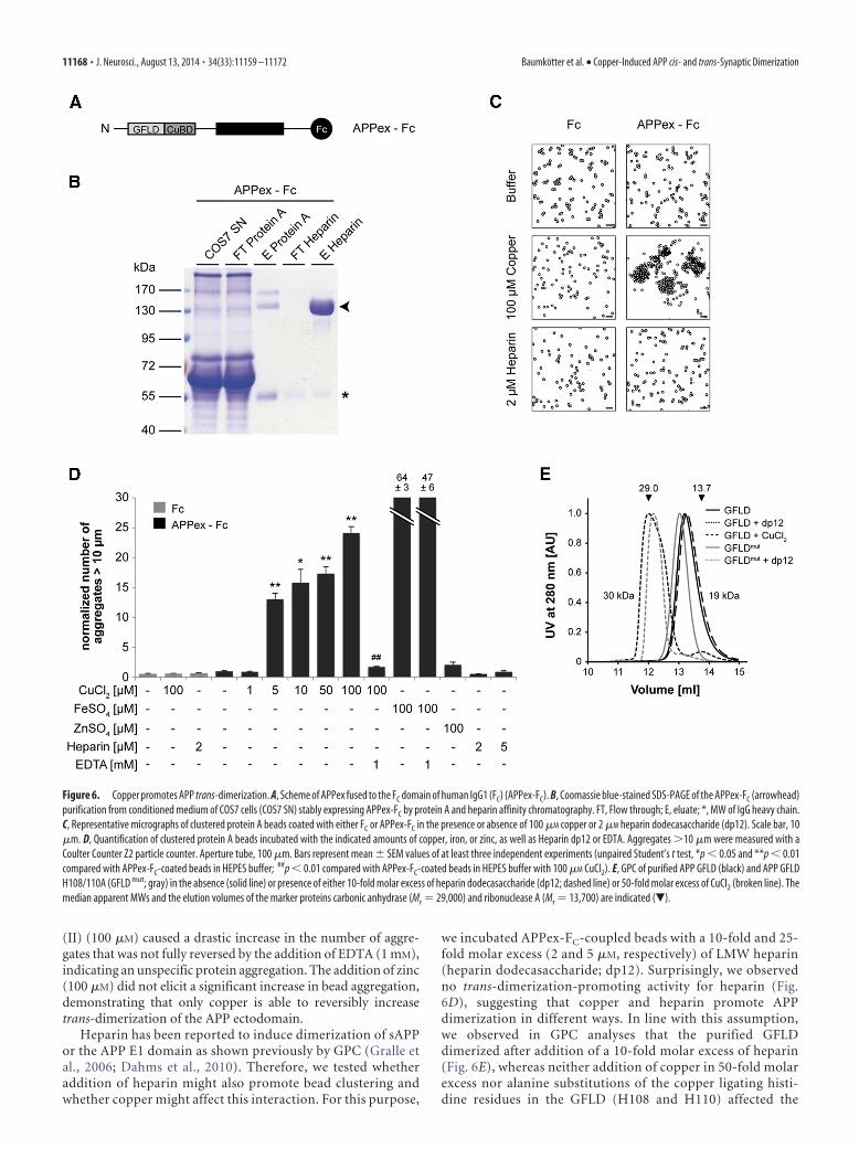
(II) (100 �M) caused a drastic increase in the number of aggre-gates that was not fully reversed by the addition of EDTA (1 mM),indicating an unspecific protein aggregation. The addition of zinc(100 �M) did not elicit a significant increase in bead aggregation,demonstrating that only copper is able to reversibly increasetrans-dimerization of the APP ectodomain.
Heparin has been reported to induce dimerization of sAPPor the APP E1 domain as shown previously by GPC (Gralle etal., 2006; Dahms et al., 2010). Therefore, we tested whetheraddition of heparin might also promote bead clustering andwhether copper might affect this interaction. For this purpose,
we incubated APPex-FC-coupled beads with a 10-fold and 25-fold molar excess (2 and 5 �M, respectively) of LMW heparin(heparin dodecasaccharide; dp12). Surprisingly, we observedno trans-dimerization-promoting activity for heparin (Fig.6D), suggesting that copper and heparin promote APPdimerization in different ways. In line with this assumption,we observed in GPC analyses that the purified GFLDdimerized after addition of a 10-fold molar excess of heparin(Fig. 6E), whereas neither addition of copper in 50-fold molarexcess nor alanine substitutions of the copper ligating histi-dine residues in the GFLD (H108 and H110) affected the
Figure 6. Copper promotes APP trans-dimerization. A, Scheme of APPex fused to the FC domain of human IgG1 (FC) (APPex-FC). B, Coomassie blue-stained SDS-PAGE of the APPex-FC (arrowhead)purification from conditioned medium of COS7 cells (COS7 SN) stably expressing APPex-FC by protein A and heparin affinity chromatography. FT, Flow through; E, eluate; *, MW of IgG heavy chain.C, Representative micrographs of clustered protein A beads coated with either FC or APPex-FC in the presence or absence of 100 �M copper or 2 �M heparin dodecasaccharide (dp12). Scale bar, 10�m. D, Quantification of clustered protein A beads incubated with the indicated amounts of copper, iron, or zinc, as well as Heparin dp12 or EDTA. Aggregates �10 �m were measured with aCoulter Counter Z2 particle counter. Aperture tube, 100 �m. Bars represent mean � SEM values of at least three independent experiments (unpaired Student’s t test, *p � 0.05 and **p � 0.01compared with APPex-FC-coated beads in HEPES buffer; ##p � 0.01 compared with APPex-FC-coated beads in HEPES buffer with 100 �M CuCl2). E, GPC of purified APP GFLD (black) and APP GFLDH108/110A (GFLD mut; gray) in the absence (solid line) or presence of either 10-fold molar excess of heparin dodecasaccharide (dp12; dashed line) or 50-fold molar excess of CuCl2 (broken line). Themedian apparent MWs and the elution volumes of the marker proteins carbonic anhydrase (Mr � 29,000) and ribonuclease A (Mr � 13,700) are indicated (�).
11168 • J. Neurosci., August 13, 2014 • 34(33):11159 –11172 Baumkotter et al. • Copper-Induced APP cis- and trans-Synaptic Dimerization
heparin-induced dimerization (Fig.6E). Notably, the mobility shift causedby addition of heparin show minor dif-ferences between the GFLD and theGFLD mut, which might be attributableto subtle alterations in heparin binding.
Together, our data from the bead ag-gregation assay and the GPC corroboratethe assumption that heparin- and copper-induced dimerization processes are inde-pendent from each other and thus arelikely underlying different molecularmechanisms.
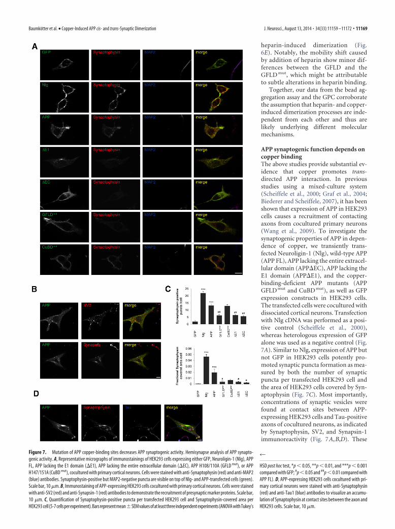
APP synaptogenic function depends oncopper bindingThe above studies provide substantial ev-idence that copper promotes trans-directed APP interaction. In previousstudies using a mixed-culture system(Scheiffele et al., 2000; Graf et al., 2004;Biederer and Scheiffele, 2007), it has beenshown that expression of APP in HEK293cells causes a recruitment of contactingaxons from cocultured primary neurons(Wang et al., 2009). To investigate thesynaptogenic properties of APP in depen-dence of copper, we transiently trans-fected Neuroligin-1 (Nlg), wild-type APP(APP FL), APP lacking the entire extracel-lular domain (APP�EC), APP lacking theE1 domain (APP�E1), and the copper-binding-deficient APP mutants (APPGFLD mut and CuBD mut), as well as GFPexpression constructs in HEK293 cells.The transfected cells were cocultured withdissociated cortical neurons. Transfectionwith Nlg cDNA was performed as a posi-tive control (Scheiffele et al., 2000),whereas heterologous expression of GFPalone was used as a negative control (Fig.7A). Similar to Nlg, expression of APP butnot GFP in HEK293 cells potently pro-moted synaptic puncta formation as mea-sured by both the number of synapticpuncta per transfected HEK293 cell andthe area of HEK293 cells covered by Syn-aptophysin (Fig. 7C). Most importantly,concentrations of synaptic vesicles werefound at contact sites between APP-expressing HEK293 cells and Tau-positiveaxons of cocultured neurons, as indicatedby Synaptophysin, SV2, and Synapsin-1immunoreactivity (Fig. 7A,B,D). These
Figure 7. Mutation of APP copper-binding sites decreases APP synaptogenic activity. Hemisynapse analysis of APP synapto-genic activity. A, Representative micrographs of immunostainings of HEK293 cells expressing either GFP, Neuroligin-1 (Nlg), APPFL, APP lacking the E1 domain (�E1), APP lacking the entire extracellular domain (�EC), APP H108/110A (GFLD mut), or APPH147/151A (CuBD mut), cocultured with primary cortical neurons. Cells were stained with anti-Synaptophysin (red) and anti-MAP2(blue) antibodies. Synaptophysin-positive but MAP2-negative puncta are visible on top of Nlg- and APP-transfected cells (green).Scale bar, 10 �m. B, Immunostaining of APP-expressing HEK293 cells cocultured with primary cortical neurons. Cells were stainedwith anti-SV2 (red) and anti-Synapsin-1 (red) antibodies to demonstrate the recruitment of presynaptic marker proteins. Scale bar,10 �m. C, Quantification of Synaptophysin-positive puncta per transfected HEK293 cell and Synaptophysin-covered area perHEK293 cell (5-7cellsperexperiment).Barsrepresentmean�SEMvaluesofat leastthreeindependentexperiments(ANOVAwithTukey’s
4
HSD post hoc test, *p � 0.05, **p � 0.01, and ***p � 0.001compared with GFP; #p � 0.05 and ##p � 0.01 compared withAPP FL). D, APP-expressing HEK293 cells cocultured with pri-mary cortical neurons were stained with anti-Synaptophysin(red) and anti-Tau1 (blue) antibodies to visualize an accumu-lation of Synaptophysin at contact sites between the axon andHEK293 cells. Scale bar, 10 �m.
Baumkotter et al. • Copper-Induced APP cis- and trans-Synaptic Dimerization J. Neurosci., August 13, 2014 • 34(33):11159 –11172 • 11169
data are consistent with the results ofWang et al. (2009). We found that APPlacking the entire extracellular domain orthe E1 domain or carrying mutations atthe copper-binding site in the GFLD(GFLD mut) showed an 50% loweredamount of Synaptophysin puncta and areduced cell area covered by Synapto-physin on the transfected HEK293 cells(Fig. 7 A, C). Remarkably, replacementof the copper coordinating histidineresidues in the CuBD (CuBD mut) didnot significantly reduce the amount ofSynaptophysin-positive puncta com-pared with APP (Fig. 7A,C). Additionally,the area covered by Synaptophysin, al-though smaller, was not significantlychanged. Together, these data corrobo-rate our hypothesis that APP synapto-genic activity mainly depends on copperbinding to the GFLD.
DiscussionHere, we show that copper binding toboth the CuBD and the newly identifiedbinding site in the GFLD promotes APPdimerization in both a cis- and trans-cellular manner, affecting APP physiolog-ical function at the synapse. Specific andhigh-affinity binding of copper to H108/H110 in the GFLD is validated by a crystalstructure of the GFLD in complex withcopper and by ITC measurements. Inter-estingly, the binding affinity for the GFLDis very similar to that for the CuBD (Hesseet al., 1994). Although binding might occur independently toeach of the two domains, a common binding of one copper ioncould be envisaged as the copper ligation geometries of the GFLD(Fig. 1) and the CuBD (Kong et al., 2007a) perfectly complementeach other by replacing the ligating symmetry or water moleculeswith the respective coordinating residues. Sequence alignment ofthe copper-binding site in the GFLD shows that the copper-binding histidines of APP are conserved in mammals and also inAPLP1 but not in APLP2 (Fig. 1). In contrast, the copper-bindingresidues in the CuBD are conserved in APP and APLP2 but not inAPLP1 (Kong et al., 2008). Thus, common binding of one copperion by the GFLD and the CuBD might be a unique feature of APP.
In contrast with copper binding to the CuBD (Kong et al.,2007b, 2008), comparison of the crystals of the GFLD � Cu crystalstructure with the apo structure (Rossjohn et al., 1999; Dahms etal., 2010) reveals clear structural changes involving disulfide re-duction in the LBL. This might contribute to the previouslydescribed redox activity of APP (Multhaup et al., 1996). Interest-ingly, these alterations are observed in the loop region (LBL),which forms the assumed APP dimerization interface (Kaden etal., 2008; Dahms et al., 2010). Thus, copper binding to the GFLDmight provoke a conformational change in the LBL favoring APPdimerization.
Consistently, exposure to increasing copper concentrations(up to 250 �M) causes an elevated APP dimerization in a cis- andtrans-directed manner (Fig. 3). In Co-IP studies, we observedthat single-point mutations of the copper-binding histidines inthe GFLD or CuBD had a similarly strong influence on APP
dimerization, as had complete deletions of the respective do-mains (Fig. 5). Together with our results from the bead aggrega-tion assay showing that copper is essential and sufficient to directAPP dimerization (Fig. 6), this strongly suggests a central role ofcopper in the regulation of APP cis- and trans-directed dimeriza-tion (Fig. 8). Notably, in previous S2 cell clustering experiments,we used a copper-induced expression system, in which expres-sion of the target proteins was induced by 500 �M copper (Soba etal., 2005). Thus, we most likely observed unintended copper-promoted cell clustering at that time. Although the precise mo-lecular mechanism remains enigmatic, the present data indicatethat the CuBD and GFLD are both directly involved in copper-mediated dimerization of two interacting APP molecules. In con-trast to this, heparin-induced APP dimerization seems to dependsolely on the GFLD, as evidenced by GPC analysis with the iso-lated subdomain (Fig. 6). These observations led to the conclu-sion that the heparin- and copper-induced dimerization isattributable to noncompetitive independent molecular mecha-nisms, which is further supported by the fact that alanine substi-tutions of the copper-binding histidine residues did not affectheparin-induced dimerization in GPC.
Under in vitro conditions, low micromolar concentrations ofcopper were sufficient to induce APP dimerization, whereas APPdimerization in the cellular context was achieved only at copperconcentrations of 100 �M. This difference is most likely ex-plained by the complex regulation of copper homeostasis in vivo,because copper taken up by cells is first bound to high-affinitycopper-binding components (Banci et al., 2010; Lutsenko et al.,
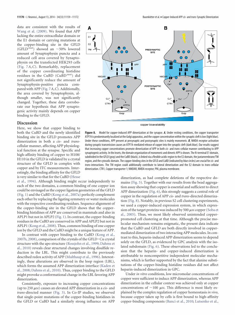
Figure 8. Model for copper-induced APP dimerization at the synapse. A, Under resting conditions, the copper transporterATP7A is predominantly localized at the Golgi apparatus, and the copper concentration within the synaptic cleft is low (light blue).Under these conditions, APP present at presynaptic and postsynaptic sites is mainly monomeric. B, NMDA receptor activationduring synaptic transmission causes an ATP7A-mediated release of copper into the synaptic cleft (dark blue). Our results suggestthat increasing copper concentrations promote dimerization of APP in both cis- and trans-cellular manner contributing to APPsynaptogenic activity. In the insets, the domain organization of monomeric and dimeric APP is shown. The N-terminal E1 domain,subdivided in the GFLD (gray) and the CuBD (black), is linked via a flexible acidic region to the E2 domain, the juxtamembrane/TMregion, and the cytosolic domain. The copper-binding sites in the GFLD and CuBD (indicated by blue circles) are crucial for cis- andtrans-interactions. The TM region could additionally contribute to lateral dimerization and the E2 domain to trans-cellulardimerization. CTR1, Copper transporter 1; NMDAR, NMDA receptor; PM, plasma membrane.
11170 • J. Neurosci., August 13, 2014 • 34(33):11159 –11172 Baumkotter et al. • Copper-Induced APP cis- and trans-Synaptic Dimerization
2010) and thus only available to APP at high intracellular copperlevels, when the pool of other high-affinity binding componentsis saturated. Under physiological conditions, the copper concen-tration in the human brain is 80 �M, with the highest concen-tration in the hippocampus (Lutsenko et al., 2010; Roberts et al.,2012; Gaier et al., 2013). Higher concentrations are most likelyachieved only for short time periods in locally restricted areas,e.g., during synaptic transmission, in which the temporal increaseof copper at the synaptic cleft has been reported to reach levelsthat are even above 250 �M (Kardos et al., 1989; Lutsenko et al.,2010; Gaier et al., 2013). These data support the idea that coppermight be a regulator of APP trans-dimerization at the synapse(Fig. 8).
In our studies, we further confirmed that expression of APP inHEK293 cells promotes synaptogenesis in contacting axons ofcocultured primary neurons (Fig. 7; Wang et al., 2009), similar tothe synaptogenic activity of Nlg (Scheiffele et al., 2000). Togetherwith recent data showing synaptic deficits in conditional APPknock-out mice and different APP knock-in mutants (Mullerand Zheng, 2012), APP was classified in the group of synaptic celladhesion molecules (Siddiqui and Craig, 2011). Interestingly,APP carrying mutations at the copper-binding sites in the GFLDshowed a dramatic reduction in promoting presynaptic special-ization, very similar to the deletion of the APP extracellular do-main or the E1 domain. This demonstrates that copper binding toAPP is not only essential for trans-dimerization but also requiredfor APP-promoted presynaptic specialization in mixed cocultureassays.
Moreover, accumulating evidence suggests important rolesfor endogenous copper in synaptic function or plasticity, such asa synaptic activity-dependent increase of copper levels at the syn-aptic cleft (Tamano and Takeda, 2011; Gaier et al., 2013). How-ever, because of the complex interconnected influences exertedby copper ions, additional in vivo studies will be required to de-cipher the different copper activities at the synapse, including themodulation of the postulated synaptogenic function of APP andprion protein, as well as GABAA receptors, NMDA receptors, andvoltage-gated calcium channels contributing to synaptic trans-mission (Gaier et al., 2013).
Although the current data suggest a correlation of APPdimerization, copper binding, and APP processing (Bayer et al.,2006; Kong et al., 2008; Eggert et al., 2009; Sato et al., 2009; Kadenet al., 2012; Spoerri et al., 2012; Noda et al., 2013), the inconsis-tency of results obtained with these different assay systems doesnot allow drawing a clear picture yet.
Together, our data provide strong evidence that altered cop-per homeostasis and/or synaptic activity-dependent changes inextracellular copper levels modulate APP cis- and trans-cellulardimerization, which in turn regulates the physiological functionof APP and has a severe affect on its pathogenicity in AD.
ReferencesAdams PD, Afonine PV, Bunkoczi G, Chen VB, Davis IW, Echols N, Headd JJ,
Hung LW, Kapral GJ, Grosse-Kunstleve RW, McCoy AJ, Moriarty NW,Oeffner R, Read RJ, Richardson DC, Richardson JS, Terwilliger TC, ZwartPH (2010) PHENIX: a comprehensive Python-based system for macro-molecular structure solution. Acta Crystallogr D Biol Crystallogr 66:213–221. CrossRef Medline
Altan-Bonnet N, Sougrat R, Liu W, Snapp EL, Ward T,Lippincott-Schwartz J(2006) Golgi inheritance in mammalian cells is mediated through endo-plasmic reticulum export activities. Mol Biol Cell 17:990-1005. CrossRefMedline
Banci L, Bertini I, Ciofi-Baffoni S, Kozyreva T, Zovo K, Palumaa P (2010)Affinity gradients drive copper to cellular destinations. Nature 465:645–648. CrossRef Medline
Battye TG, Kontogiannis L, Johnson O, Powell HR, Leslie AG (2011) iMOSFLM: anew graphical interface for diffraction-image processing with MOSFLM. ActaCrystallogr D Biol Crystallogr 67:271–281. CrossRef Medline
Baumgartner W, Hinterdorfer P, Ness W, Raab A, Vestweber D, Schindler H,Drenckhahn D (2000) Cadherin interaction probed by atomic force mi-croscopy. Proc Natl Acad Sci U S A 97:4005– 4010. CrossRef Medline
Baumkotter F, Wagner K, Eggert S, Wild K, Kins S (2012) Structural aspectsand physiological consequences of APP/APLP trans-dimerization. ExpBrain Res 217:389 –395. CrossRef Medline
Bayer TA, Schafer S, Breyhan H, Wirths O, Treiber C, Multhaup G (2006) Avicious circle: role of oxidative stress, intraneuronal Abeta and Cu inAlzheimer’s disease. Clin Neuropathol 25:163–171. Medline
Biederer T, Scheiffele P (2007) Mixed-culture assays for analyzing neuronalsynapse formation. Nat Protoc 2:670 – 676. CrossRef Medline
Bolte S, Cordelieres FP (2006) A guided tour into subcellular colocalizationanalysis in light microscopy. J Microsc 224:213–232. CrossRef Medline
Bush AI, Multhaup G, Moir RD, Williamson TG, Small DH, Rumble B,Pollwein P, Beyreuther K, Masters CL (1993) A novel zinc(II) bindingsite modulates the function of the beta A4 amyloid protein precursor ofAlzheimer’s disease. J Biol Chem 268:16109 –16112. Medline
Chappuis-Flament S, Wong E, Hicks LD, Kay CM, Gumbiner BM (2001)Multiple cadherin extracellular repeats mediate homophilic binding andadhesion. J Cell Biol 154:231–243. CrossRef Medline
Chavda AP, Prole DL, Taylor CW (2013) A bead aggregation assay for de-tection of low-affinity protein-protein interactions reveals interactionsbetween N-terminal domains of inositol 1,4,5-trisphosphate receptors.PLoS One 8:e60609. CrossRef Medline
Chen VB, Arendall WB 3rd, Headd JJ, Keedy DA, Immormino RM, KapralGJ, Murray LW, Richardson JS, Richardson DC (2010) MolProbity: all-atom structure validation for macromolecular crystallography. ActaCrystallogr D Biol Crystallogr 66:12–21. CrossRef Medline
Collaborative Computational Project, Number 4 (1994) The CCP4 suite:programs for protein crystallography. Acta Crystallogr D Biol Crystallogr50:760 –763. Medline
Dahms SO, Hoefgen S, Roeser D, Schlott B, Guhrs KH, Than ME (2010)Structure and biochemical analysis of the heparin-induced E1 dimer ofthe amyloid precursor protein. Proc Natl Acad Sci U S A 107:5381–5386.CrossRef Medline
Dahms SO, Konnig I, Roeser D, Guhrs KH, Mayer MC, Kaden D, MulthaupG, Than ME (2012) Metal binding dictates conformation and functionof the amyloid precursor protein (APP) E2 domain. J Mol Biol 416:438 –452. CrossRef Medline
Duce JA, Tsatsanis A, Cater MA, James SA, Robb E, Wikhe K, Leong SL, PerezK, Johanssen T, Greenough MA, Cho HH, Galatis D, Moir RD, MastersCL, McLean C, Tanzi RE, Cappai R, Barnham KJ, Ciccotosto GD, RogersJT, Bush AI (2010) Iron-export ferroxidase activity of beta-amyloid pre-cursor protein is inhibited by zinc in Alzheimer’s disease. Cell 142:857–867. CrossRef Medline
Eggert S, Midthune B, Cottrell B, Koo EH (2009) Induced dimerization ofthe amyloid precursor protein (APP) leads to decreased amyloid-betaprotein (Abeta) production. J Biol Chem 284:28943–28952. CrossRefMedline
Emsley P, Lohkamp B, Scott WG, Cowtan K (2010) Features and develop-ment of Coot. Acta Crystallogr D Biol Crystallogr 66:486 –501. CrossRefMedline
Gaier ED, Eipper BA, Mains RE (2013) Copper signaling in the mammaliannervous system: synaptic effects. J Neurosci Res 91:2–19. CrossRefMedline
Galuska SP, Rollenhagen M, Kaup M, Eggers K, OltmannNorden I, Schiff M,Hartmann M, Weinhold B, Hildebrandt H, Geyer R, Muhlenhoff M,Geyer H (2010) Synaptic cell adhesion molecule SynCAM 1 is a targetfor polysialylation in postnatal mouse brain. Proc Natl Acad Sci U S A107:10250 –10255. CrossRef Medline
Graf ER, Zhang X, Jin SX, Linhoff MW, Craig AM (2004) Neurexins inducedifferentiation of GABA and glutamate postsynaptic specializations vianeuroligins. Cell 119:1013–1026. CrossRef Medline
Gralle M, Oliveira CL, Guerreiro LH, McKinstry WJ, Galatis D, Masters CL,Cappai R, Parker MW, Ramos CH, Torriani I, Ferreira ST (2006) Solu-tion conformation and heparin-induced dimerization of the full-lengthextracellular domain of the human amyloid precursor protein. J Mol Biol357:493–508. CrossRef Medline
Hatcher LQ, Hong L, Bush WD, Carducci T, Simon JD (2008) Quantifica-
Baumkotter et al. • Copper-Induced APP cis- and trans-Synaptic Dimerization J. Neurosci., August 13, 2014 • 34(33):11159 –11172 • 11171
tion of the binding constant of copper(II) to the amyloid-beta peptide.J Phys Chem B 112:8160 – 8164. CrossRef Medline
Hesse L, Beher D, Masters CL, Multhaup G (1994) The beta A4 amyloidprecursor protein binding to copper. FEBS Lett 349:109 –116. CrossRefMedline
Hong L, Bush WD, Hatcher LQ, Simon J (2008) Determining thermody-namic parameters from isothermal calorimetric isotherms of the bindingof macromolecules to metal cations originally chelated by a weak ligand.J Phys Chem B 112:604 – 611. CrossRef Medline
Houtman JC, Brown PH, Bowden B, Yamaguchi H, Appella E, SamelsonLE, Schuck P (2007) Studying multisite binary and ternary proteininteractions by global analysis of isothermal titration calorimetry datain SEDPHAT: application to adaptor protein complexes in cell signal-ing. Protein Sci 16:30 – 42. CrossRef Medline
Isbert S, Wagner K, Eggert S, Schweitzer A, Multhaup G, Weggen S, Kins S,Pietrzik CU (2012) APP dimer formation is initiated in the endoplasmicreticulum and differs between APP isoforms. Cell Mol Life Sci 69:1353–1375. CrossRef Medline
Kaden D, Munter LM, Joshi M, Treiber C, Weise C, Bethge T, Voigt P,Schaefer M, Beyermann M, Reif B, Multhaup G (2008) Homophilic in-teractions of the amyloid precursor protein (APP) ectodomain are regu-lated by the loop region and affect beta-secretase cleavage of APP. J BiolChem 283:7271–7279. CrossRef Medline
Kaden D, Munter LM, Reif B, Multhaup G (2012) The amyloid precursorprotein and its homologues: structural and functional aspects of nativeand pathogenic oligomerization. Eur J Cell Biol 91:234 –239. CrossRefMedline
Kardos J, Kovacs I, Hajos F, Kalman M, Simonyi M (1989) Nerve endingsfrom rat brain tissue release copper upon depolarization. A possible rolein regulating neuronal excitability. Neurosci Lett 103:139 –144. CrossRefMedline
Keller S, Vargas C, Zhao H, Piszczek G, Brautigam CA, Schuck P (2012)High-precision isothermal titration calorimetry with automated peak-shape analysis. Anal Chem 84:5066 –5073. CrossRef Medline
Kemmer G, Keller S (2010) Nonlinear least-squares data fitting in Excelspreadsheets. Nat Protoc 5:267–281. CrossRef Medline
Kong GK, Adams JJ, Cappai R, Parker MW (2007a) Structure of Alzhei-mer’s disease amyloid precursor protein copper-binding domain atatomic resolution. Acta Crystallogr Sect F Struct Biol Cryst Commun63:819 – 824. CrossRef Medline
Kong GK, Adams JJ, Harris HH, Boas JF, Curtain CC, Galatis D, Masters CL,Barnham KJ, McKinstry WJ, Cappai R, Parker MW (2007b) Structuralstudies of the Alzheimer’s amyloid precursor protein copper-binding do-main reveal how it binds copper ions. J Mol Biol 367:148 –161. CrossRefMedline
Kong GK, Miles LA, Crespi GA, Morton CJ, Ng HL, Barnham KJ, McKinstryWJ, Cappai R, Parker MW (2008) Copper binding to the Alzheimer’sdisease amyloid precursor protein. Eur Biophys J 37:269 –279. CrossRefMedline
Lambert M, Padilla F, Mege RM (2000) Immobilized dimers ofN-cadherin-Fc chimera mimic cadherin-mediated cell contact forma-tion: contribution of both outside-in and inside-out signals. J Cell Sci113:2207–2219. Medline
Lee S, Xue Y, Hu J, Wang Y, Liu X, Demeler B, Ha Y (2011) The E2 domainsof APP and APLP1 share a conserved mode of dimerization. Biochemistry50:5453–5464. CrossRef Medline
Lutsenko S, Bhattacharjee A, Hubbard AL (2010) Copper handling machin-ery of the brain. Metallomics 2:596 – 608. CrossRef Medline
Martell A, RM Smith, Motekaitis R (2004) NIST critically selected stabilityconstants of metal complexes. In: NIST Standard Reference Database 46,version 80. College Station, TX; National Institute of Standards andTechnology.
Masters CL, Beyreuther K (2006) Alzheimer’s centennial legacy: prospectsfor rational therapeutic intervention targeting the Abeta amyloid path-way. Brain 129:2823–2839. CrossRef Medline
Maynard CJ, Cappai R, Volitakis I, Cherny RA, White AR, Beyreuther K,Masters CL, Bush AI, Li QX (2002) Overexpression of Alzheimer’s dis-
ease amyloid-beta opposes the age-dependent elevations of brain copperand iron. J Biol Chem 277:44670 – 44676. CrossRef Medline
McCoy AJ, Grosse-Kunstleve RW, Adams PD, Winn MD, Storoni LC, ReadRJ (2007) Phaser crystallographic software. J Appl Crystallogr 40:658 –674. CrossRef Medline
Muller UC, Zheng H (2012) Physiological functions of APP family proteins.Cold Spring Harb Perspect Med 2:a006288. CrossRef Medline
Multhaup G, Schlicksupp A, Hesse L, Beher D, Ruppert T, Masters CL,Beyreuther K (1996) The amyloid precursor protein of Alzheimer’s dis-ease in the reduction of copper(II) to copper(I). Science 271:1406 –1409.CrossRef Medline
Noda Y, Asada M, Kubota M, Maesako M, Watanabe K, Uemura M, Kihara T,Shimohama S, Takahashi R, Kinoshita A, Uemura K (2013) Copper en-hances APP dimerization and promotes Abeta production. Neurosci Lett547:10 –15. CrossRef Medline
Putnam CD, Hammel M, Hura GL, Tainer JA (2007) X-ray solution scat-tering (SAXS) combined with crystallography and computation: definingaccurate macromolecular structures, conformations and assemblies insolution. Q Rev Biophys 40:191–285. CrossRef Medline
Roberts BR, Ryan TM, Bush AI, Masters CL, Duce JA (2012) The role ofmetallobiology and amyloid-beta peptides in Alzheimer’s disease. J Neu-rochem 120 [Suppl 1]:149 –166. CrossRef
Rossjohn J, Cappai R, Feil SC, Henry A, McKinstry WJ, Galatis D, Hesse L,Multhaup G, Beyreuther K, Masters CL, Parker MW (1999) Crystalstructure of the N-terminal, growth factor-like domain of Alzheimer am-yloid precursor protein. Nat Struct Biol 6:327–331. CrossRef Medline
Sato T, Tang TC, Reubins G, Fei JZ, Fujimoto T, Kienlen-Campard P, Con-stantinescu SN, Octave JN, Aimoto S, Smith SO (2009) A helix-to-coiltransition at the epsilon-cut site in the transmembrane dimer of the am-yloid precursor protein is required for proteolysis. Proc Natl Acad SciU S A 106:1421–1426. CrossRef Medline
Scheiffele P, Fan J, Choih J, Fetter R, Serafini T (2000) Neuroligin expressedin nonneuronal cells triggers presynaptic development in contacting ax-ons. Cell 101:657– 669. CrossRef Medline
Schmidt V, Baum K, Lao A, Rateitschak K, Schmitz Y, Teichmann A, WiesnerB, Petersen CM, Nykjaer A, Wolf J, Wolkenhauer O, Willnow TE (2012)Quantitative modelling of amyloidogenic processing and its influence bySORLA in Alzheimer’s disease. EMBO J 31:187–200. CrossRef Medline
Siddiqui TJ, Craig AM (2011) Synaptic organizing complexes. Curr OpinNeurobiol 21:132–143. CrossRef Medline
Smith DG, Cappai R, Barnham KJ (2007) The redox chemistry of the Alz-heimer’s disease amyloid beta peptide. Biochim Biophys Acta 1768:1976 –1990. CrossRef Medline
Soba P, Eggert S, Wagner K, Zentgraf H, Siehl K, Kreger S, Lower A, Langer A,Merdes G, Paro R, Masters CL, Muller U, Kins S, Beyreuther K (2005)Homo- and heterodimerization of APP family members promotes inter-cellular adhesion. EMBO J 24:3624 –3634. CrossRef Medline
Spoerri L, Vella LJ, Pham CL, Barnham KJ, Cappai R (2012) The amyloidprecursor protein copper binding domain histidine residues 149 and 151mediate APP stability and metabolism. J Biol Chem 287:26840 –26853.CrossRef Medline
Svergun DI (1999) Restoring low resolution structure of biological macro-molecules from solution scattering using simulated annealing. Biophys J76:2879 –2886. CrossRef Medline
Tamano H, Takeda A (2011) Dynamic action of neurometals at the synapse.Metallomics 3:656 – 661. CrossRef Medline
Wang Y, Ha Y (2004) The X-ray structure of an antiparallel dimer of thehuman amyloid precursor protein E2 domain. Mol Cell 15:343–353.CrossRef Medline
Wang Z, Wang B, Yang L, Guo Q, Aithmitti N, Songyang Z, Zheng H (2009)Presynaptic and postsynaptic interaction of the amyloid precursor pro-tein promotes peripheral and central synaptogenesis. J Neurosci 29:10788 –10801. CrossRef Medline
White AR, Multhaup G, Maher F, Bellingham S, Camakaris J, Zheng H, BushAI, Beyreuther K, Masters CL, Cappai R (1999) The Alzheimer’s diseaseamyloid precursor protein modulates copper-induced toxicity and oxi-dative stress in primary neuronal cultures. J Neurosci 19:9170 –9179.Medline
11172 • J. Neurosci., August 13, 2014 • 34(33):11159 –11172 Baumkotter et al. • Copper-Induced APP cis- and trans-Synaptic Dimerization