Embed Size (px)
Citation preview
Neurobiology of Disease
Connecting TNF-� Signaling Pathways to iNOS Expressionin a Mouse Model of Alzheimer’s Disease: Relevancefor the Behavioral and Synaptic Deficits Induced byAmyloid � Protein
Rodrigo Medeiros,1* Rui D. S. Prediger,1* Giselle F. Passos,1 Pablo Pandolfo,1 Filipe S. Duarte,1 Jeferson L. Franco,2
Alcir L. Dafre,2 Gabriella Di Giunta,3 Claudia P. Figueiredo,3 Reinaldo N. Takahashi,1 Maria M. Campos,4 andJoao B. Calixto1
Departamentos de 1Farmacologia and 2Ciencias Fisiologicas, Centro de Ciencias Biologicas, and 3Departamento de Anatomia Patologica, HospitalUniversitario–Universidade Federal de Santa Catarina, 88049-900, Florianopolis, Santa Catarina, Brazil, and 4Faculdade de Odontologia, PontifıciaUniversidade Catolica do Rio Grande do Sul 90619-900, Rio Grande do Sul, Brazil
Increased brain deposition of amyloid � protein (A�) and cognitive deficits are classical signals of Alzheimer’s disease (AD) that havebeen highly associated with inflammatory alterations. The present work was designed to determine the correlation between tumornecrosis factor-� (TNF-�)-related signaling pathways and inducible nitric oxide synthase (iNOS) expression in a mouse model of AD, bymeans of both in vivo and in vitro approaches. The intracerebroventricular injection of A�1– 40 in mice resulted in marked deficits oflearning and memory, according to assessment in the water maze paradigm. This cognition impairment seems to be related to synapsedysfunction and glial cell activation. The pharmacological blockage of either TNF-� or iNOS reduced the cognitive deficit evoked byA�1– 40 in mice. Similar results were obtained in TNF-� receptor 1 and iNOS knock-out mice. A�1– 40 administration induced an increasein TNF-� expression and oxidative alterations in prefrontal cortex and hippocampus. Likewise, A�1– 40 led to activation of both JNK(c-Jun-NH2-terminal kinase)/c-Jun and nuclear factor-�B, resulting in iNOS upregulation in both brain structures. The anti-TNF-�antibody reduced all of the molecular and biochemical alterations promoted by A�1– 40. These results provide new insights in mousemodels of AD, revealing TNF-� and iNOS as central mediators of A� action. These pathways might be targeted for AD drug development.
Key words: Alzheimer’s disease; amyloid �; inflammation; TNF-�; iNOS; cognitive deficits
IntroductionAlzheimer’s disease (AD) is a progressive neurodegenerative dis-order associated with memory loss, spatial disorientation, andweakness of intellectual capacity. Histopathological studies of ADbrain have revealed the presence of classical hallmarks includingneurofibrillary tangles and senile plaques. The deposition ofamyloid � protein (A�) in brain areas involved in cognitive func-tions is assumed to initiate a pathological cascade that results insynaptic dysfunction, synaptic loss, and neuronal death (Walshand Selkoe, 2004).
Recent evidence suggests an inflammatory component in AD,which is characterized by astrogliosis, microgliosis, cytokine ele-
vation, and changes in acute phase proteins (Walsh and Selkoe,2004; Wyss-Coray, 2006). Tumor necrosis factor-� (TNF-�) is acytokine thought to play a central role in the self-propagation ofneuroinflammation (Perry et al., 2001). TNF-� regulates manycellular processes, including inflammation, differentiation, andcell death through activation of TNF receptor 1 (TNFR1) orTNFR2 (Wajant et al., 2003). The transduction pathways acti-vated by TNF-� include mitogen-activated protein kinases(MAPKs) and I�B kinase, which control gene expression throughtranscriptional factors such as activator protein-1 (AP-1) andnuclear factor-�B (NF-�B) (Wajant et al., 2003). Regarding theCNS, microglia and astrocytes are believed to be the primarysources of TNF-�. Evidence indicates the presence of increasedlevels of TNF-� in the brain and plasma of AD patients and anupregulation of TNFR1 have been detected in the AD brain (Fillitet al., 1991; Li et al., 2004). In addition, TNF-� has been impli-cated recently as a critical mediator of long-term potentiation(LTP) reduction by A� (Wang et al., 2005). However, the mech-anisms through which TNF-� promotes its pathological actionsin AD are poorly understood.
Brain oxidative stress seems to exert an important role in cog-nitive impairment observed in AD. Inducible nitric oxide syn-
Received Nov. 21, 2006; revised April 14, 2007; accepted April 17, 2007.This work was supported by grants from the Conselho Nacional de Desenvolvimento Cientıfico e Tecnologico, the
Coordenacao de Aperfeicoamento de Pessoal de Nıvel Superior, the Programa de Apoio aos Nucleos de Excelencia,and the Fundacao de Apoio a Pesquisa do Estado de Santa Catarina, all from Brazil.
*R.M. and R.D.S.P. contributed equally to this work.Correspondence should be addressed to Dr. Joao B. Calixto, Departamento de Farmacologia, Universidade Fed-
eral de Santa Catarina, Campus Universitario, Trindade, Bloco D, Caixa Postal 476, 88049-900, Florianopolis, SantaCatarina, Brazil. E-mail: [email protected] or [email protected].
DOI:10.1523/JNEUROSCI.5047-06.2007Copyright © 2007 Society for Neuroscience 0270-6474/07/275394-11$15.00/0
5394 • The Journal of Neuroscience, May 16, 2007 • 27(20):5394 –5404
thase (iNOS) generates nitric oxide (NO) and NO-derived reac-tive nitrogen species such as peroxynitrite. Accumulation ofhighly reactive molecules induces lipid peroxidation, tyrosine ni-trosylation, DNA oxidative damage, and neuronal disruption,which are common characteristics of the AD brain (Butterfield etal., 2001). Interestingly, these events seem to be modulated byTNF-�. Also, A� has been shown to interact in a synergistic man-ner with cytokines to induce neuronal damage via reactive oxy-gen species (ROS)- and NO-dependent pathways (Goodwin etal., 1995; Meda et al., 1995).
Numerous animal models have been used to evaluate the roleof inflammation in the course of AD. An experimental model thatmimics the progression of AD was developed using an intracere-broventricular injection of A� in mice (for review, see Van Damand De Deyn, 2006). Using this model, we assessed the role ofTNF-� and iNOS in A�-induced early impairment of learningand memory. Our data indicate that TNF-� production is one ofthe earliest events induced by A�1– 40, representing an importantsignal for iNOS expression. The cross talk between TNF-� andiNOS is probably mediated by activation of two major intracel-lular pathways: c-Jun-NH2-terminal kinase (JNK)/c-Jun and NF-�B. The present results implicate TNF-� and iNOS as importantmediators of A�-induced cognitive impairment.
Materials and MethodsSubjects. Experiments were conducted using male Swiss, C57BL/6,TNFR1 knock-out, and iNOS knock-out mice (20 –30 g) kept in a con-trolled room temperature (22 � 2°C) and humidity (60 – 80%) under a12 h light/dark cycle (lights on 6:00 A.M.). TNFR1 (Rothe et al., 1993)and iNOS (MacMicking et al., 1995) knock-out mice are on the C57BL/6background, constructed as described previously. All procedures used inthe present study followed the “Principles of Laboratory Animal Care”from National Institutes of Health (NIH) publication number 85-23 andwere approved by the Animal Ethics Committee of the UniversidadeFederal de Santa Catarina.
Drug treatment protocol. Human A�1– 40 (Tocris, Ellisville, MO) andA�40 –1 (Bachem, Torrance, CA) were dissolved in sterile PBS, pH 7.4, at1 mg/ml and were incubated at 37°C for 4 d, as described previously (ElKhoury et al., 1996). Control mice received sterile PBS (vehicle). Theaggregated form of amyloid fragments (400 pmol per mice) and vehiclesolution were administered intracerebroventricularly as described previ-ously (Laursen and Belknap, 1986). As a positive control for molecularstudies, some animals received an intracerebroventricular injection of2.5 �g of lipopolysaccharide (LPS) from Escherichia coli (serotype 0111:B4; Sigma-Aldrich, Sao Paulo, Brazil). Briefly, each mouse was given aninjection at bregma with a 5 �l Hamilton microsyringe fitted with a 26gauge needle that was inserted to a depth of 2.4 mm. The injection vol-ume was 3 �l. Mice exhibited normal behavior within 1 min after injec-tion. Accurate placement of the injection or needle track was verified atthe time of dissection. The present A�1– 40 dose was selected according toprevious literature data (Yan et al., 2001).
Some animals were treated with anti-TNF-� antibody (AbTNF-�; 10�g, i.c.v., per mouse; R & D Systems, Minneapolis, MN) 15 min beforeA�1– 40 injection. The AbTNF-� dose was determined in pilot experi-ments (results not shown). The iNOS inhibitor aminoguanidine (AG;100 mg/kg, i.p.; Sigma-Aldrich) was administered 1 h before an intrace-rebroventricular injection of A�1– 40 and throughout consecutive daysuntil the day of the experiment. In addition, some animals were pre-treated with the selective inhibitor of JNK, SP600126 (anthra[1-9-cd]pyrazol-6(2H)-one; 50 mg/kg; i.p.; 1 h before; Tocris), or the NF-�Bblocker pyrrolidine dithiocarbamate (PDTC; 100 mg/kg, i.p., 1 h before;Sigma-Aldrich).
Water maze test–memory reference test. The Morris water maze test wasperformed as described previously (Morris et al., 1982). The experimen-tal apparatus consisted of a circular water tank (diameter, 97 cm; height,60 cm) containing water at 23 � 2°C. The target platform (10 � 10 cm)
was submerged 1 cm below the water surface and placed at the midpointof one quadrant. The platform was located in a fixed position, equidistantfrom the center and the wall of the pool. The pool was located in a testroom containing various prominent visual cues. Mice were submitted toa spatial reference memory version of the water maze as described previ-ously (Prediger et al., 2007). The acquisition training session was per-formed 7 d after A�1– 40 injection and consisted of 10 consecutive trialsduring which the animals were left in the tank facing the wall and allowedto swim freely to the escape platform. If an animal did not find theplatform within a period of 60 s, it was gently guided to it. The animal wasallowed to remain on the platform for 10 s after escaping to it, and it wasthen removed from the tank for 20 s before being placed at the nextstarting point in the tank. This procedure was repeated 10 times, with thestarting points varying in a pseudo-randomized manner. The test sessionwas performed 24 h after the training session (on day 8 after injection).The test session consisted of a single probe trial in which the platform wasremoved from the pool and each mouse was allowed to swim for 60 s inthe maze. The time spent in the correct quadrant (i.e., where the platformwas located on the training session) was recorded, and the percentage ofthe total time was calculated.
Open-field test. To discard the possible effects on A�1– 40 in locomotoractivity, the animals were tested in the open-field paradigm. The appa-ratus, made of wood covered with impermeable Formica, had a blackfloor of 30 � 30 cm (divided by white lines into nine squares of 10 � 10cm) and transparent walls, 15 cm high. Each mouse was placed in thecenter of the open field, and the total number of squares crossed with thefour paws and the rearing behavior were registered for 5 min.
Histology. Eight days after intracerebroventricular injection of aggre-gated A�1– 40, mice were perfused transcardially with PBS solution con-taining 4% paraformaldeyde (w/v). Brain was removed and kept over-night in the same solution. Serial sections (3 �M) were selected to includethe lateral ventricle areas. Sections were stained with 0.5% cresyl violetreagent (Sigma-Aldrich) according to standard procedures. Stained cellswere determined on visual inspection in the hippocampal CA1, CA2, andCA3 subfields and cortical layer with a microscope (Eclipse 50I; Nikon,Melville, NY) using a counting grid at 400� magnification. The light-microscopic characteristics of the A�1– 40 aggregates were studied usingthe alkaline Congo red technique according to standard procedures.
Immunohistochemistry. Immunohistochemistry detection of apopto-tic cell death, immunoreactivity of the astrocyte marker glial fibrillaryacidic protein (GFAP), and synaptic changes were assessed on paraffintissue sections 1 and 8 d after A�1– 40 intracerebroventricular injection,using the polyclonal rabbit anti-caspase-3 (1:200; Cell Signaling Tech-nology, Beverly, MA), monoclonal mouse anti-GFAP (1:300; Dako Cy-tomation, Carpinteria, CA), and monoclonal mouse anti-synaptophysin(1:400; Novocastra, Newcastle, UK), respectively. High-temperature an-tigen retrieval was performed by immersion of the slides in a water bathat 95–98°C in 10 mM trisodium citrate buffer, pH 6.0, for 45 min. Thenonspecific binding was blocked by incubating sections for 1 h with goatnormal serum diluted in PBS. After overnight incubation at 4°C withprimary antibodies, the slides were washed with PBS and incubated withthe secondary antibody Envision plus (Dako Cytomation), ready to use,for 1 h at room temperature. The sections were washed in PBS, and thevisualization was completed by using 3,3�-diaminobenzidine (Dako Cy-tomation) in chromogen solution and counterstained lightly with Har-ris’s hematoxylin solution. Images were taken with a microscope (Eclipse50i; Nikon) and digital sight camera (DS-5M-L1; Nikon). Control andexperimental tissues were placed on the same slide and processed underthe same conditions. Settings for image acquisition were identical forcontrol and experimental tissues. For each mouse, we obtained fourimages (one per section) of the parietal cortex and three images (one persection) of the hippocampal CA1, CA2, and CA3 subregions. Digitized,8-bit images were transferred to a computer, and the average pixel inten-sity of synaptophysisn staining was calculated for each image using NIHImageJ 1.36b imaging software (NIH, Bethesda, MD). For each mouse,the values obtained for the parietal cortex or the hippocampal subregionswere averaged. This approach for the assessment of synaptic degenera-tion has been validated in various experimental models of neurodegen-eration (Buttini et al., 1999) and in diseased human brains (Masliah et al.,
Medeiros et al. • Role of TNF-� and iNOS on A� Cognitive Deficit J. Neurosci., May 16, 2007 • 27(20):5394 –5404 • 5395
1992). GFAP- and caspase-3-positive cells weredetermined on visual inspection in the samebrain areas using a counting grid at 400�magnification.
Enzymatic studies. Mice were anesthetizedwith pentobarbital (50 mg/kg, i.p.) and per-fused transcardially with ice-cold 0.9% NaCl(10 ml/10 g body weight) to remove the freeradical-scavenging and radical-generatingsources in the brain. The prefrontal cortex andhippocampus were carefully excised and storedat �70°C. Tissues were homogenized in 20 mM
HEPES, pH 7.0, and centrifuged at 20,000 � gfor 30 min at 4°C. The enzyme activities weredetermined in the supernatant in a Varian(Palo Alto, CA) Cary 50 spectrophotometer.Glutathione reductase (GR) and glutathioneperoxidase (GPx) activity was determined asdescribed previously (Prediger et al., 2007).Briefly, GR reduces oxidized glutathione(GSSG) to glutathione (GSH) in expendingNADPH, the disappearance of which can beaccompanied at 340 nm. GPx activity was mea-sured indirectly by the NADPH consumption at 340 nm. The GPx usesGSH to reduce the tert-butylhydroperoxide-producing GSSG, which isreadily reduced to GSH by excess GR, consuming NADPH. The disap-pearance of NADPH in this reaction reflects the GPx activity. The en-zyme activities were expressed in milliunits per milligram of total proteincontent, which was quantified using the Bio-Rad (Hercules, CA) proteinassay kit.
Determination of total GSH level. The prefrontal cortex and hippocam-pus were isolated and homogenized in cooled 0.5 M perchloric acid. Thehomogenates were centrifuged at 15,000 � g for 2 min, and the superna-tant was separated, neutralized in potassium phosphate buffer (0.1 M, pH7.4), and subsequently submitted to the assay. Total GSH, comprising thetotal of reduced (GSH) and oxidized (GSSG) forms, was determined bythe GR–5�,5�-dithio-bis(2-nitrobenzoic acid) recycling assay as de-scribed previously (Prediger et al., 2007).
Preparation of cytosolic and nuclear extracts. Tissues were homogenizedin ice-cold 10 mM HEPES, pH 7.4, containing 1.5 mM MgCl2, 10 mM KCl,1 mM phenylmethylsulphonyl fluoride, 5 �g/ml leupeptin, 5 �g/ml pep-statin A, 10 �g/ml aprotinin, 1 mM sodium orthovanadate, 10 mM
�-glycerophosphate, 50 mM sodium fluoride, and 0.5 mM dithiothreitol(all from Sigma-Aldrich). The homogenates were chilled on ice for 15min and vigorously shaken for 15 min in the presence of 0.1% TritonX-100. The nuclear fraction was precipitated by centrifugation at10,000 � g for 30 min. The supernatant containing the cytosolic fractionwas stored at �70°C until use. The nuclear pellet was resuspended in 500�l of high-salt extraction buffer (20 mM HEPES, pH 7.4, 420 mM NaCl,1.5 mM MgCl2, 0.2 mM EDTA, 25% v/v glycerol, 1 mM phenylmethylsul-phonyl fluoride, 5 �g/ml pepstatin A, 5 �g/ml leupeptin, 10 �g/ml apro-tinin, and 0.5 mM dithiothreitol) and incubated under continuous shak-ing at 4°C for 30 min. The nuclear extract was then centrifuged for 30 minat 10,000 � g, and the supernatant was aliquoted and stored at �70°C.Protein concentration was determined using the Bio-Rad protein assaykit.
Western blot analysis. Equal protein amounts were separated on anSDS-PAGE and transferred to a polyvinylidene fluoride membrane (Im-mobilon P; Millipore, Bedford, MA). The membranes were saturated byincubation with 10% nonfat dry milk solution and incubated overnightwith one of the following antibodies: p65 NF-�B (sc-372, 1:1000), c-jun(sc-45, 1:1000), iNOS (sc-7271, 1:200), �-actin (sc-1615, 1:2000), JNK1(sc-571, 1:1000), phosphorylated JNK (sc-6254, 1:1000) (all from SantaCruz Biotechnology, Santa Cruz, CA), or lamin A/C (catalog #2032; CellSignaling Technology). After washing, the membranes were incubatedwith adjusted secondary antibodies coupled to horseradish peroxidase oralkaline phosphatase. The immunocomplexes were visualized using theECL chemiluminescence detection system (GE Healthcare, Sao Paulo,Brazil) or 5-bromo-4-chloro-3-indolyl phosphate/nitroblue tetrazolium
color development substrate (Promega, Madison, WI). Band densitymeasurements were made using the Scion (Frederick, MD) Image soft-ware package.
RNA preparation and reverse transcription-PCR. Total RNA was ex-tracted from �100 mg of prefrontal cortex and hippocampus usingTrizol reagent (Invitrogen, Sao Paulo, Brazil) according to the manufac-turer’s instructions. The concentration of total RNA was determined bymeasuring the absorbance at 260 nm in a spectrophotometer. The qualityof the preparations was checked using gel electrophoresis. One micro-gram of total RNA was reverse transcribed using oligo(dT) and 200 U ofreverse transcriptase (Invitrogen) in 20 �l of PCR buffer containing (inmM) 0.5 dNTP, 10 DTT, 2.5 MgCl2, 50 KCl, and 20 Tris-HCl, pH 8.4. Thesamples were incubated for 5 min at 70°C, 5 min at 4°C, 1 h at 37°C, 5 minat 70°C, and 5 min at 4°C. Specific primers (Invitrogen) were used forTNF-� (sense, TCTCATCAGTTCTATGGCCC; antisense, GGGAGTA-GACAAGGTACAAC), iNOS (sense, CAGAAGCAGAATGTGACCATC;antisense, CTTCTGGTCGATGTCATGA), and �-actin (sense, TCCT-TCGTTGCCGGTCCACA; antisense, CGTCTCCGGAGTCCATCACA).�-Actin cDNA was used for standardization of the amount of RNA. Fivemicroliters of reverse transcription (RT) aliquots were mixed in a 20 mM
Tris-HCl buffer, pH 8.4, containing 1.5 mM MgCl2, 300 �M dNTP, 2 �g/mlof each primer, and 50 U/ml Taq polymerase (Invitrogen) in a final volumeof 100 �l. The following cycling protocol was used: initial denaturation, 95°Cfor 4 min; cycling: 95°C for 30 s, 53°C for 30 s (TNF-� and iNOS) or 62°C for30 s (�-actin), and 72°C for 1 min; and a final extension period at 72°C for 5min. Optimal amplification was achieved at 29 cycles for TNF-�, iNOS, and�-actin. Aliquots of 25 �l were analyzed by PAGE and stained with silversalts. Band density measurements for TNF-�, iNOS, and �-actin mRNAswere made using the Scion Image software package.
Software. All Western blot and RT-PCR experiments were scanned toacquire digital images using a Genius ColorPage Scanner (KYE Systems,Taipei, Taiwan). Digital images were processed with the Photoshop soft-
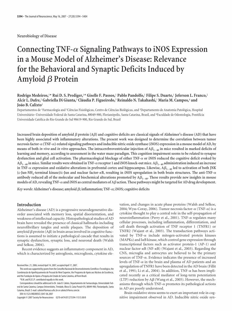
Figure 1. Intracerebroventricular injection of A�1– 40 disrupts learning and memory in mice. The spatial reference memoryversion of the Morris water maze test in Swiss mice (n � 9 –10 mice per group) was used as a measure of cognition. A, Trainingtrials were performed on day 7 after a single intracerebroventricular administration of aggregated A�1– 40 (400 pmol per mouse),inverse peptide A�40 –1 (400 pmol per mouse) (used as a negative control), or vehicle (PBS). Treatment with A�1– 40 significantlyincreased the escape latencies to find the submerged platform during the training trials (F(2,26) � 104.32; p � 0.0001). B, Theprobe test session was performed 24 h after the training trials. Treatment with A�1– 40 reduced the frequency of time spent in thecorrect quadrant (F(2,26) � 19.91; p � 0.0001). The values represent the mean � SEM. *p � 0.05 compared with the vehicle-treated group (Newman–Keuls test).
Table 1. Intracerebroventricular (i.c.v.) injection of A�1– 40 did not affect motorbehavior in mice
Treatment (i.c.v.) Squares crossing Rearing
Vehicle 78.0 � 4.6 32.5 � 2.9A�1– 40 78.6 � 4.3 34.3 � 3.2A�40 –1 75.7 � 2.4 27.3 � 1.9
Data are expressed as the mean � SEM of the total squares crossed and rearing (n � 9 –10 animals in each group).A�1– 40 treatment did not significantly alter the number of squares crossed (F(2,26) � 0.14; p � 0.86) and rearing(F
(2,26)� 1.49; p � 0.24) in the open-field arena. Total squares crossed and rearing by Swiss mice in the open-field
arena (for 5 min) was used as a measure of locomotor activity. Experiments were performed on day 8 after a singleintracerebroventricular administration of aggregated A�1– 40 (400 pmol per mouse), the inverse peptide A�40 –1
(400 pmol per mouse; used as a negative control), or vehicle (PBS).
5396 • J. Neurosci., May 16, 2007 • 27(20):5394 –5404 Medeiros et al. • Role of TNF-� and iNOS on A� Cognitive Deficit
ware package (Adobe Systems, Mountain View, CA), complying withstrict standards (Rossner and Yamada, 2004).
Statistical analysis. All values are expressed as means � SEM (n is thenumber of mice included in each analysis). Differences between groups
in total GSH levels and GR and GPx activitieswere analyzed using an unpaired Student’s ttest. Statistical analysis for the rest of the datawere performed using two- or three-wayANOVA with strain (or pretreatment), treat-ment, or the number of trials (repeated mea-sure) as independent variables. After signifi-cant ANOVAs, multiple post hoc comparisonswere performed using the Newman–Keuls test.The accepted level of significance for the tests wasp � 0.05. All tests were performed using the Sta-tistica software package (StatSoft, Tulsa, OK).
ResultsThe role of TNF-� and iNOS in A�-induced learning andmemory impairmentLearning and memory functions are vul-nerable to several pathological processes,including AD (Budson and Price, 2005).To investigate some of the mechanisms in-volved in A�-induced cognitive decline,the Morris water maze task was used. Wetested the ability of mice to acquire (train-ing session) and retrieve (test session) spa-tial information as indicative of learningand memory functions. As expected, in-tracerebroventricular injection of A�1– 40
in Swiss mice resulted in a significant de-cline in both learning and memory, as in-dicated by longer latencies (Fig. 1A) andreduced target quadrant preference (Fig.1B) during the probe trial. No cognitivedeficits were observed in Swiss micetreated with the inverse A�40 –1 (Fig. 1).The effects of A�1– 40 administration onwater maze performance are not directlyrelated to motor impairment, becauseno alterations of the swimming speed inthe water maze (data not shown), or thetotal squares crossed and rearing behav-ior in the open-field arena, were ob-served (Table 1).
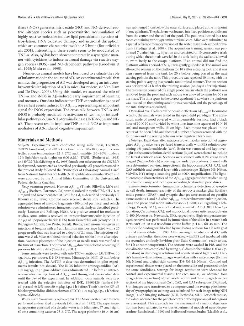
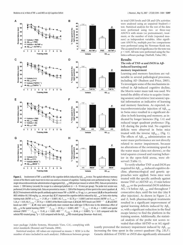
To verify whether TNF-� and iNOS arerequired for A�1– 40-induced cognitive de-cline, pharmacological and genetic ap-proaches were applied. Swiss mice weretreated with the specific antibody againstmouse TNF-� (AbTNF-�), 15 min beforeA�1– 40, or the preferential iNOS inhibitorAG, 1 h before A�1– 40, and throughout 7consecutive days, until the test day (day 8after injection). As shown in Figure 2, Aand E, both pharmacological treatmentsresulted in a significant improvement ofspatial learning deficits induced by A�1–
40, with a significant reduction in the finalescape latency to find the platform in thetraining session. Additionally, the statisti-cal analysis of the probe test scores re-vealed that either AbTNF-� or AG signif-
icantly prevented the memory impairment induced by A�1– 40,increasing the time spent in the correct quadrant (Fig. 2B,F).Genetic deletion of TNFR1 or iNOS also significantly attenuated
Figure 2. Involvement of TNF-� and iNOS in the cognitive deficits induced by A�1– 40 in mice. The spatial reference memoryversion of the Morris water maze test in mice was used as a measure of cognition. Training trials were performed on day 7 after asingle intracerebroventricular administration of aggregated A�1– 40 (400 pmol per mouse) or vehicle (PBS). Data are presented asmeans � SEM latency (seconds) for escape to a submerged platform (n � 8 –10 mice per group). The probe test session wasperformed 24 h after training trials. Data are presented as means � SEM of the frequency of time spent in the correct quadrant. A,B, E, F, Pretreatment with the specific antibody against mouse TNF-� (AbTNF-�; 10 �g, i.c.v., per mouse) (A, B) or the preferentialiNOS inhibitor AG (100 mg/kg, i.p., once per day) (E, F ) improved the cognitive deficits induced by A�1– 40 in Swiss mice duringtraining trials (AbTNF-�: F(1,27) � 21.89, p � 0.0001; AG: F(1,33) � 82.78, p � 0.0001) and test sessions (AbTNF-�: F(1,27) �5.26, p � 0.05; AG: F(1,33) � 29.14, p � 0.0001) of the Morris water maze. C, D, G, H, TNFR1 knock-out (TNFR1�/�; C, D) and iNOSknock-out (iNOS�/�; G, H ) mice were significantly more resistant than wild-type C57BL/6 mice to the deleterious effect ofA�1– 40 in the spatial learning (TNFR1�/�: F(1,30) � 29.58, p � 0.0001; iNOS�/�: F(1,26) � 81.46, p � 0.0001) and spatialretrieval (TNFR1�/�: F(1,30) � 15.45, p � 0.001; iNOS�/�: F(1,26) � 28.46, p � 0.0001). *p � 0.05 compared with thevehicle/PBS-treated group; #p � 0.05 compared with the A�1– 40/PBS-treated group (Newman–Keuls test).
Medeiros et al. • Role of TNF-� and iNOS on A� Cognitive Deficit J. Neurosci., May 16, 2007 • 27(20):5394 –5404 • 5397
the spatial learning deficits induced by A�1–40, as indicated by areduction in the final escape latency to find the platform when com-pared with wild-type (C57BL/6) A�-treated mice (Fig. 2C,G). In thetest session, TNFR1�/� and iNOS�/� mice given injections ofA�1– 40 presented higher swimming scores in the correct quad-rant compared with wild-type A�1– 40-treated mice, indicating adiminished sensitivity to memory deficits (Fig. 2D,H).
Prevention of synaptic dysfunction by TNF-� andiNOS inhibitionTo evaluate the possible amyloid deposits and/or neuronal dam-age after A�1– 40 intracerebroventricular injection, we performedhistological and immunohistochemistry analysis. Histologicalexamination was performed on cresyl violet- and on Congo red-stained sections of mouse brain, 8 d after the intracerebroventric-ular administrations. Cresyl violet-stained sections indicated thatinjection by itself failed to produce any significant neuronal dam-age in control animals, at or distant from the injection site. Theadministration of A�1– 40 peptide also failed to produce any neu-ronal loss in the hippocampus or cortex of treated mice (data notshown). Using Congo red staining, control brain sections orbrain sections of animals treated with A�1– 40 showed no evidenceof amyloid deposits (data not shown).
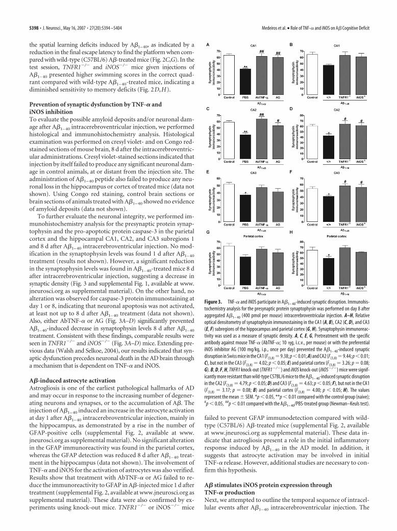
To further evaluate the neuronal integrity, we performed im-munohistochemistry analysis for the presynaptic protein synap-tophysin and the pro-apoptotic protein caspase-3 in the parietalcortex and the hippocampal CA1, CA2, and CA3 subregions 1and 8 d after A�1– 40 intracerebroventricular injection. No mod-ification in the synaptophysin levels was found 1 d after A�1– 40
treatment (results not shown). However, a significant reductionin the synaptophysin levels was found in A�1– 40-treated mice 8 dafter intracerebroventricular injection, suggesting a decrease insynaptic density (Fig. 3 and supplemental Fig. 1, available at www.jneurosci.org as supplemental material). On the other hand, noalteration was observed for caspase-3 protein immunostaining atday 1 or 8, indicating that neuronal apoptosis was not activated,at least not up to 8 d after A�1– 40 treatment (data not shown).Also, either AbTNF-� or AG (Fig. 3A–D) significantly preventedA�1–40-induced decrease in synaptophysin levels 8 d after A�1–40
treatment. Consistent with these findings, comparable results wereseen in TNFR1�/� and iNOS�/� (Fig. 3A–D) mice. Extending pre-vious data (Walsh and Selkoe, 2004), our results indicated that syn-aptic dysfunction precedes neuronal death in the AD brain througha mechanism that is dependent on TNF-� and iNOS.
A�-induced astrocyte activationAstrogliosis is one of the earliest pathological hallmarks of ADand may occur in response to the increasing number of degener-ating neurons and synapses, or to the accumulation of A�. Theinjection of A�1– 40 induced an increase in the astrocyte activationat day 1 after A�1– 40 intracerebroventricular injection, mainly inthe hippocampus, as demonstrated by a rise in the number ofGFAP-positive cells (supplemental Fig. 2, available at www.jneurosci.org as supplemental material). No significant alterationin the GFAP immunoreactivity was found in the parietal cortex,whereas the GFAP detection was reduced 8 d after A�1– 40 treat-ment in the hippocampus (data not shown). The involvement ofTNF-� and iNOS for the activation of astrocytes was also verified.Results show that treatment with AbTNF-� or AG failed to re-duce the immunoreactivity to GFAP in A�-injected mice 1 d aftertreatment (supplemental Fig. 2, available at www.jneurosci.org assupplemental material). These data were also confirmed by ex-periments using knock-out mice. TNFR1�/� or iNOS�/� mice
failed to prevent GFAP immunodetection compared with wild-type (C57BL/6) A�-treated mice (supplemental Fig. 2, availableat www.jneurosci.org as supplemental material). These data in-dicate that astrogliosis present a role in the initial inflammatoryresponse induced by A�1– 40 in the AD model. In addition, itsuggests that astrocyte activation may be involved in initialTNF-� release. However, additional studies are necessary to con-firm this hypothesis.
A� stimulates iNOS protein expression throughTNF-� productionNext, we attempted to outline the temporal sequence of intracel-lular events after A�1– 40 intracerebroventricular injection. The
Figure 3. TNF-� and iNOS participate in A�1– 40-induced synaptic disruption. Immunohis-tochemistry analysis for the presynaptic protein synaptophysin was performed on day 8 afteraggregated A�1– 40 (400 pmol per mouse) intracerebroventricular injection. A–H, Relativeoptical densitometry of synaptophysin immunostaining in the CA1 (A, B), CA2 (C, D), and CA3(E, F) subregions of the hippocampus and parietal cortex (G, H). Synaptophysin immunoreac-tivity was used as a measure of synaptic density. A, C, E, G, Pretreatment with the specificantibody against mouse TNF-� (AbTNF-�; 10 �g, i.c.v., per mouse) or with the preferentialiNOS inhibitor AG (100 mg/kg, i.p., once per day) prevented the A�1– 40-induced synapticdisruption in Swiss mice in the CA1 (F(3,8) �9.38; p�0.01; A) and CA2 (F(3,8) �9.44; p�0.01;C), but not in the CA3 (F(3,8) � 4.02; p � 0.05; E) and parietal cortex (F(3,8) � 3.26; p � 0.08;G). B, D, F, H, TNFR1 knock-out (TNFR1�/�) and iNOS knock-out (iNOS�/�) mice were signif-icantly more resistant than wild-type C57BL/6 mice to the A�1– 40-induced synaptic disruptionin the CA2 (F(3,8) � 4.79; p � 0.05; D) and CA3 (F(3,8) � 4.63; p � 0.05; F), but not in the CA1(F(3,8) � 3.17; p � 0.08; B) and parietal cortex (F(3,8) � 4.00; p � 0.05; H). The valuesrepresent the mean � SEM. *p � 0.05, **p � 0.01 compared with the control group (naive);#p � 0.05, ##p � 0.01 compared with the A�1– 40/PBS-treated group (Newman–Keuls test).
5398 • J. Neurosci., May 16, 2007 • 27(20):5394 –5404 Medeiros et al. • Role of TNF-� and iNOS on A� Cognitive Deficit
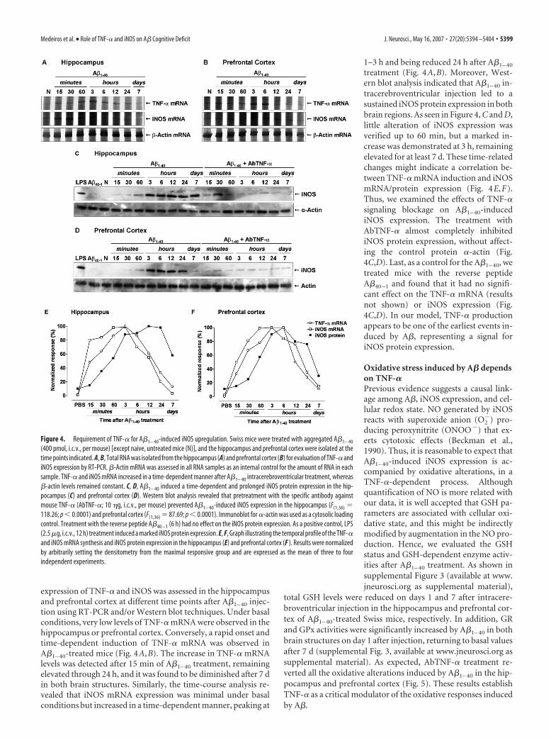
expression of TNF-� and iNOS was assessed in the hippocampusand prefrontal cortex at different time points after A�1– 40 injec-tion using RT-PCR and/or Western blot techniques. Under basalconditions, very low levels of TNF-� mRNA were observed in thehippocampus or prefrontal cortex. Conversely, a rapid onset andtime-dependent induction of TNF-� mRNA was observed inA�1– 40-treated mice (Fig. 4A,B). The increase in TNF-� mRNAlevels was detected after 15 min of A�1– 40 treatment, remainingelevated through 24 h, and it was found to be diminished after 7 din both brain structures. Similarly, the time-course analysis re-vealed that iNOS mRNA expression was minimal under basalconditions but increased in a time-dependent manner, peaking at
1–3 h and being reduced 24 h after A�1– 40
treatment (Fig. 4A,B). Moreover, West-ern blot analysis indicated that A�1– 40 in-tracerebroventricular injection led to asustained iNOS protein expression in bothbrain regions. As seen in Figure 4, C and D,little alteration of iNOS expression wasverified up to 60 min, but a marked in-crease was demonstrated at 3 h, remainingelevated for at least 7 d. These time-relatedchanges might indicate a correlation be-tween TNF-� mRNA induction and iNOSmRNA/protein expression (Fig. 4E,F).Thus, we examined the effects of TNF-�signaling blockage on A�1– 40-inducediNOS expression. The treatment withAbTNF-� almost completely inhibitediNOS protein expression, without affect-ing the control protein �-actin (Fig.4C,D). Last, as a control for the A�1– 40, wetreated mice with the reverse peptideA�40 –1 and found that it had no signifi-cant effect on the TNF-� mRNA (resultsnot shown) or iNOS expression (Fig.4C,D). In our model, TNF-� productionappears to be one of the earliest events in-duced by A�, representing a signal foriNOS protein expression.
Oxidative stress induced by A� dependson TNF-�Previous evidence suggests a causal link-age among A�, iNOS expression, and cel-lular redox state. NO generated by iNOSreacts with superoxide anion (O2
�) pro-ducing peroxynitrite (ONOO�) that ex-erts cytotoxic effects (Beckman et al.,1990). Thus, it is reasonable to expect thatA�1– 40-induced iNOS expression is ac-companied by oxidative alterations, in aTNF-�-dependent process. Althoughquantification of NO is more related withour data, it is well accepted that GSH pa-rameters are associated with cellular oxi-dative state, and this might be indirectlymodified by augmentation in the NO pro-duction. Hence, we evaluated the GSHstatus and GSH-dependent enzyme activ-ities after A�1– 40 treatment. As shown insupplemental Figure 3 (available at www.jneurosci.org as supplemental material),
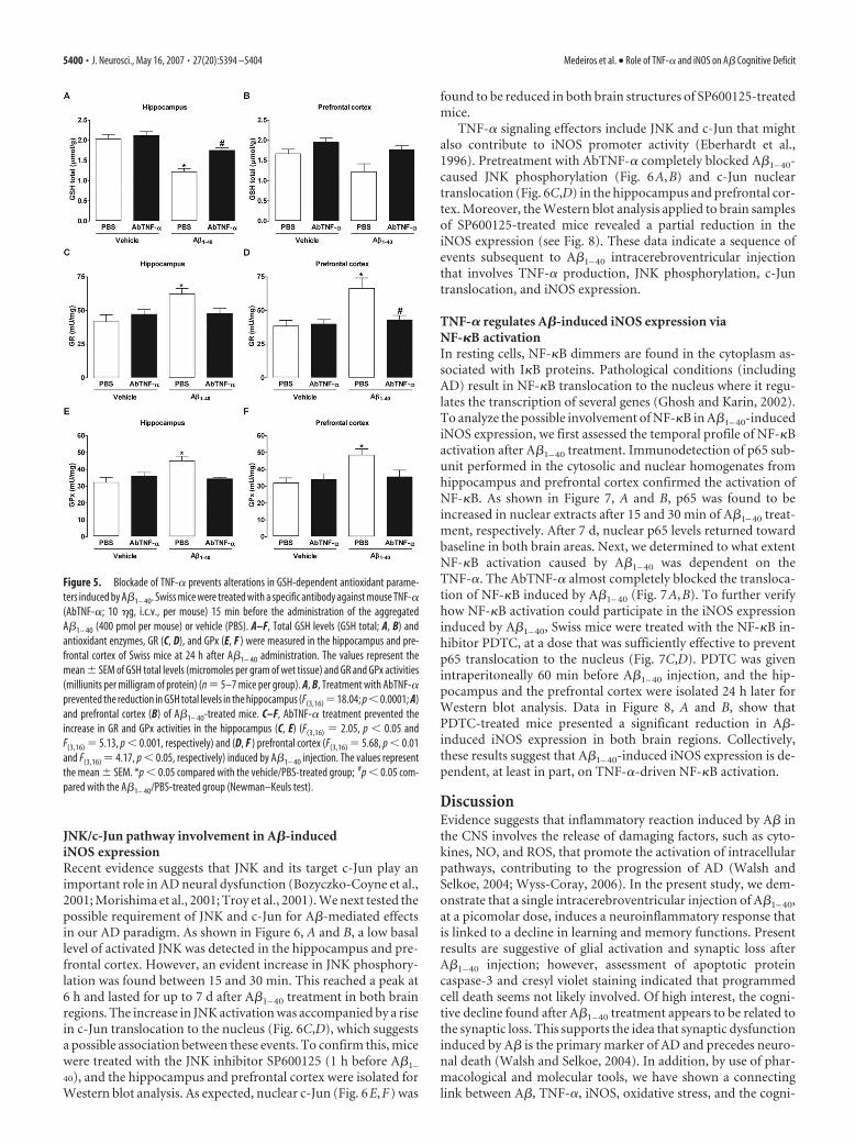
total GSH levels were reduced on days 1 and 7 after intracere-broventricular injection in the hippocampus and prefrontal cor-tex of A�1– 40-treated Swiss mice, respectively. In addition, GRand GPx activities were significantly increased by A�1– 40 in bothbrain structures on day 1 after injection, returning to basal valuesafter 7 d (supplemental Fig. 3, available at www.jneurosci.org assupplemental material). As expected, AbTNF-� treatment re-verted all the oxidative alterations induced by A�1– 40 in the hip-pocampus and prefrontal cortex (Fig. 5). These results establishTNF-� as a critical modulator of the oxidative responses inducedby A�.
Figure 4. Requirement of TNF-� for A�1– 40-induced iNOS upregulation. Swiss mice were treated with aggregated A�1– 40
(400 pmol, i.c.v., per mouse) [except naive, untreated mice (N)], and the hippocampus and prefrontal cortex were isolated at thetime points indicated. A, B, Total RNA was isolated from the hippocampus (A) and prefrontal cortex (B) for evaluation of TNF-� andiNOS expression by RT-PCR. �-Actin mRNA was assessed in all RNA samples as an internal control for the amount of RNA in eachsample. TNF-� and iNOS mRNA increased in a time-dependent manner after A�1– 40 intracerebroventricular treatment, whereas�-actin levels remained constant. C, D, A�1– 40 induced a time-dependent and prolonged iNOS protein expression in the hip-pocampus (C) and prefrontal cortex (D). Western blot analysis revealed that pretreatment with the specific antibody againstmouse TNF-� (AbTNF-�; 10 �g, i.c.v., per mouse) prevented A�1– 40-induced iNOS expression in the hippocampus (F(1,36) �118.26; p � 0.0001) and prefrontal cortex (F(1,36) � 87.69; p � 0.0001). Immunoblot for �-actin was used as a cytosolic loadingcontrol. Treatment with the reverse peptide A�40 –1 (6 h) had no effect on the iNOS protein expression. As a positive control, LPS(2.5 �g, i.c.v., 12 h) treatment induced a marked iNOS protein expression. E, F, Graph illustrating the temporal profile of the TNF-�and iNOS mRNA synthesis and iNOS protein expression in the hippocampus (E) and prefrontal cortex (F ). Results were normalizedby arbitrarily setting the densitometry from the maximal responsive group and are expressed as the mean of three to fourindependent experiments.
Medeiros et al. • Role of TNF-� and iNOS on A� Cognitive Deficit J. Neurosci., May 16, 2007 • 27(20):5394 –5404 • 5399
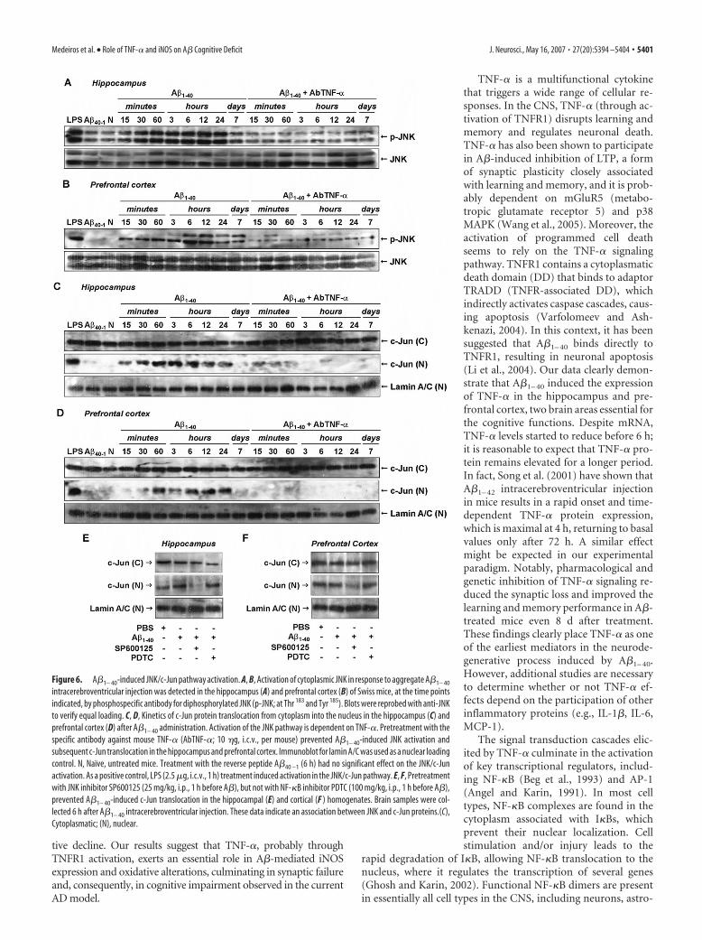
JNK/c-Jun pathway involvement in A�-inducediNOS expressionRecent evidence suggests that JNK and its target c-Jun play animportant role in AD neural dysfunction (Bozyczko-Coyne et al.,2001; Morishima et al., 2001; Troy et al., 2001). We next tested thepossible requirement of JNK and c-Jun for A�-mediated effectsin our AD paradigm. As shown in Figure 6, A and B, a low basallevel of activated JNK was detected in the hippocampus and pre-frontal cortex. However, an evident increase in JNK phosphory-lation was found between 15 and 30 min. This reached a peak at6 h and lasted for up to 7 d after A�1– 40 treatment in both brainregions. The increase in JNK activation was accompanied by a risein c-Jun translocation to the nucleus (Fig. 6C,D), which suggestsa possible association between these events. To confirm this, micewere treated with the JNK inhibitor SP600125 (1 h before A�1–
40), and the hippocampus and prefrontal cortex were isolated forWestern blot analysis. As expected, nuclear c-Jun (Fig. 6E,F) was
found to be reduced in both brain structures of SP600125-treatedmice.
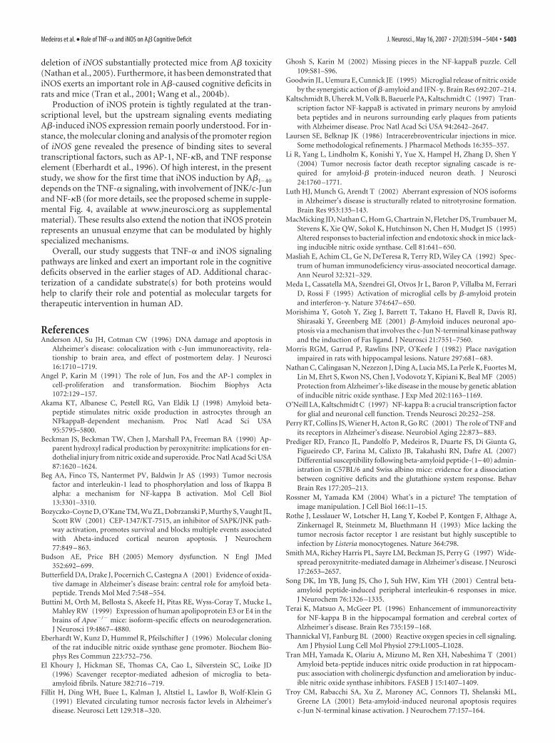
TNF-� signaling effectors include JNK and c-Jun that mightalso contribute to iNOS promoter activity (Eberhardt et al.,1996). Pretreatment with AbTNF-� completely blocked A�1– 40-caused JNK phosphorylation (Fig. 6A,B) and c-Jun nucleartranslocation (Fig. 6C,D) in the hippocampus and prefrontal cor-tex. Moreover, the Western blot analysis applied to brain samplesof SP600125-treated mice revealed a partial reduction in theiNOS expression (see Fig. 8). These data indicate a sequence ofevents subsequent to A�1– 40 intracerebroventricular injectionthat involves TNF-� production, JNK phosphorylation, c-Juntranslocation, and iNOS expression.
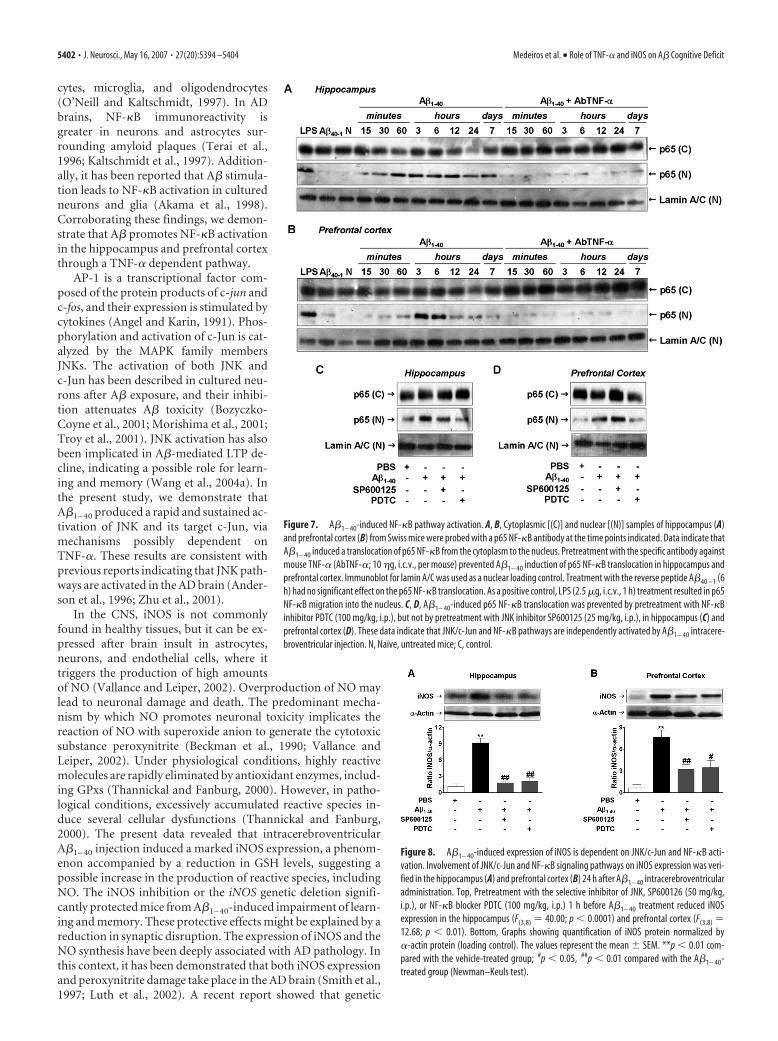
TNF-� regulates A�-induced iNOS expression viaNF-�B activationIn resting cells, NF-�B dimmers are found in the cytoplasm as-sociated with I�B proteins. Pathological conditions (includingAD) result in NF-�B translocation to the nucleus where it regu-lates the transcription of several genes (Ghosh and Karin, 2002).To analyze the possible involvement of NF-�B in A�1– 40-inducediNOS expression, we first assessed the temporal profile of NF-�Bactivation after A�1– 40 treatment. Immunodetection of p65 sub-unit performed in the cytosolic and nuclear homogenates fromhippocampus and prefrontal cortex confirmed the activation ofNF-�B. As shown in Figure 7, A and B, p65 was found to beincreased in nuclear extracts after 15 and 30 min of A�1– 40 treat-ment, respectively. After 7 d, nuclear p65 levels returned towardbaseline in both brain areas. Next, we determined to what extentNF-�B activation caused by A�1– 40 was dependent on theTNF-�. The AbTNF-� almost completely blocked the transloca-tion of NF-�B induced by A�1– 40 (Fig. 7A,B). To further verifyhow NF-�B activation could participate in the iNOS expressioninduced by A�1– 40, Swiss mice were treated with the NF-�B in-hibitor PDTC, at a dose that was sufficiently effective to preventp65 translocation to the nucleus (Fig. 7C,D). PDTC was givenintraperitoneally 60 min before A�1– 40 injection, and the hip-pocampus and the prefrontal cortex were isolated 24 h later forWestern blot analysis. Data in Figure 8, A and B, show thatPDTC-treated mice presented a significant reduction in A�-induced iNOS expression in both brain regions. Collectively,these results suggest that A�1– 40-induced iNOS expression is de-pendent, at least in part, on TNF-�-driven NF-�B activation.
DiscussionEvidence suggests that inflammatory reaction induced by A� inthe CNS involves the release of damaging factors, such as cyto-kines, NO, and ROS, that promote the activation of intracellularpathways, contributing to the progression of AD (Walsh andSelkoe, 2004; Wyss-Coray, 2006). In the present study, we dem-onstrate that a single intracerebroventricular injection of A�1– 40,at a picomolar dose, induces a neuroinflammatory response thatis linked to a decline in learning and memory functions. Presentresults are suggestive of glial activation and synaptic loss afterA�1– 40 injection; however, assessment of apoptotic proteincaspase-3 and cresyl violet staining indicated that programmedcell death seems not likely involved. Of high interest, the cogni-tive decline found after A�1– 40 treatment appears to be related tothe synaptic loss. This supports the idea that synaptic dysfunctioninduced by A� is the primary marker of AD and precedes neuro-nal death (Walsh and Selkoe, 2004). In addition, by use of phar-macological and molecular tools, we have shown a connectinglink between A�, TNF-�, iNOS, oxidative stress, and the cogni-
Figure 5. Blockade of TNF-� prevents alterations in GSH-dependent antioxidant parame-ters induced by A�1– 40. Swiss mice were treated with a specific antibody against mouse TNF-�(AbTNF-�; 10 �g, i.c.v., per mouse) 15 min before the administration of the aggregatedA�1– 40 (400 pmol per mouse) or vehicle (PBS). A–F, Total GSH levels (GSH total; A, B) andantioxidant enzymes, GR (C, D), and GPx (E, F ) were measured in the hippocampus and pre-frontal cortex of Swiss mice at 24 h after A�1– 40 administration. The values represent themean � SEM of GSH total levels (micromoles per gram of wet tissue) and GR and GPx activities(milliunits per milligram of protein) (n � 5–7 mice per group). A, B, Treatment with AbTNF-�prevented the reduction in GSH total levels in the hippocampus (F(3,16) � 18.04; p � 0.0001; A)and prefrontal cortex (B) of A�1– 40-treated mice. C–F, AbTNF-� treatment prevented theincrease in GR and GPx activities in the hippocampus (C, E) (F(3,16) � 2.05, p � 0.05 andF(3,16) � 5.13, p � 0.001, respectively) and (D, F ) prefrontal cortex (F(3,16) � 5.68, p � 0.01and F(3,16) � 4.17, p � 0.05, respectively) induced by A�1– 40 injection. The values representthe mean � SEM. *p � 0.05 compared with the vehicle/PBS-treated group; #p � 0.05 com-pared with the A�1– 40/PBS-treated group (Newman–Keuls test).
5400 • J. Neurosci., May 16, 2007 • 27(20):5394 –5404 Medeiros et al. • Role of TNF-� and iNOS on A� Cognitive Deficit
tive decline. Our results suggest that TNF-�, probably throughTNFR1 activation, exerts an essential role in A�-mediated iNOSexpression and oxidative alterations, culminating in synaptic failureand, consequently, in cognitive impairment observed in the currentAD model.
TNF-� is a multifunctional cytokinethat triggers a wide range of cellular re-sponses. In the CNS, TNF-� (through ac-tivation of TNFR1) disrupts learning andmemory and regulates neuronal death.TNF-� has also been shown to participatein A�-induced inhibition of LTP, a formof synaptic plasticity closely associatedwith learning and memory, and it is prob-ably dependent on mGluR5 (metabo-tropic glutamate receptor 5) and p38MAPK (Wang et al., 2005). Moreover, theactivation of programmed cell deathseems to rely on the TNF-� signalingpathway. TNFR1 contains a cytoplasmaticdeath domain (DD) that binds to adaptorTRADD (TNFR-associated DD), whichindirectly activates caspase cascades, caus-ing apoptosis (Varfolomeev and Ash-kenazi, 2004). In this context, it has beensuggested that A�1– 40 binds directly toTNFR1, resulting in neuronal apoptosis(Li et al., 2004). Our data clearly demon-strate that A�1– 40 induced the expressionof TNF-� in the hippocampus and pre-frontal cortex, two brain areas essential forthe cognitive functions. Despite mRNA,TNF-� levels started to reduce before 6 h;it is reasonable to expect that TNF-� pro-tein remains elevated for a longer period.In fact, Song et al. (2001) have shown thatA�1– 42 intracerebroventricular injectionin mice results in a rapid onset and time-dependent TNF-� protein expression,which is maximal at 4 h, returning to basalvalues only after 72 h. A similar effectmight be expected in our experimentalparadigm. Notably, pharmacological andgenetic inhibition of TNF-� signaling re-duced the synaptic loss and improved thelearning and memory performance in A�-treated mice even 8 d after treatment.These findings clearly place TNF-� as oneof the earliest mediators in the neurode-generative process induced by A�1– 40.However, additional studies are necessaryto determine whether or not TNF-� ef-fects depend on the participation of otherinflammatory proteins (e.g., IL-1�, IL-6,MCP-1).
The signal transduction cascades elic-ited by TNF-� culminate in the activationof key transcriptional regulators, includ-ing NF-�B (Beg et al., 1993) and AP-1(Angel and Karin, 1991). In most celltypes, NF-�B complexes are found in thecytoplasm associated with I�Bs, whichprevent their nuclear localization. Cellstimulation and/or injury leads to the
rapid degradation of I�B, allowing NF-�B translocation to thenucleus, where it regulates the transcription of several genes(Ghosh and Karin, 2002). Functional NF-�B dimers are presentin essentially all cell types in the CNS, including neurons, astro-
Figure 6. A�1– 40-induced JNK/c-Jun pathway activation. A, B, Activation of cytoplasmic JNK in response to aggregate A�1– 40
intracerebroventricular injection was detected in the hippocampus (A) and prefrontal cortex (B) of Swiss mice, at the time pointsindicated, by phosphospecific antibody for diphosphorylated JNK (p-JNK; at Thr 183 and Tyr 185). Blots were reprobed with anti-JNKto verify equal loading. C, D, Kinetics of c-Jun protein translocation from cytoplasm into the nucleus in the hippocampus (C) andprefrontal cortex (D) after A�1– 40 administration. Activation of the JNK pathway is dependent on TNF-�. Pretreatment with thespecific antibody against mouse TNF-� (AbTNF-�; 10 �g, i.c.v., per mouse) prevented A�1– 40-induced JNK activation andsubsequent c-Jun translocation in the hippocampus and prefrontal cortex. Immunoblot for lamin A/C was used as a nuclear loadingcontrol. N, Naıve, untreated mice. Treatment with the reverse peptide A�40 –1 (6 h) had no significant effect on the JNK/c-Junactivation. As a positive control, LPS (2.5 �g, i.c.v., 1 h) treatment induced activation in the JNK/c-Jun pathway. E, F, Pretreatmentwith JNK inhibitor SP600125 (25 mg/kg, i.p., 1 h before A�), but not with NF-�B inhibitor PDTC (100 mg/kg, i.p., 1 h before A�),prevented A�1– 40-induced c-Jun translocation in the hippocampal (E) and cortical (F ) homogenates. Brain samples were col-lected 6 h after A�1– 40 intracerebroventricular injection. These data indicate an association between JNK and c-Jun proteins.(C),Cytoplasmatic; (N), nuclear.
Medeiros et al. • Role of TNF-� and iNOS on A� Cognitive Deficit J. Neurosci., May 16, 2007 • 27(20):5394 –5404 • 5401
cytes, microglia, and oligodendrocytes(O’Neill and Kaltschmidt, 1997). In ADbrains, NF-�B immunoreactivity isgreater in neurons and astrocytes sur-rounding amyloid plaques (Terai et al.,1996; Kaltschmidt et al., 1997). Addition-ally, it has been reported that A� stimula-tion leads to NF-�B activation in culturedneurons and glia (Akama et al., 1998).Corroborating these findings, we demon-strate that A� promotes NF-�B activationin the hippocampus and prefrontal cortexthrough a TNF-� dependent pathway.
AP-1 is a transcriptional factor com-posed of the protein products of c-jun andc-fos, and their expression is stimulated bycytokines (Angel and Karin, 1991). Phos-phorylation and activation of c-Jun is cat-alyzed by the MAPK family membersJNKs. The activation of both JNK andc-Jun has been described in cultured neu-rons after A� exposure, and their inhibi-tion attenuates A� toxicity (Bozyczko-Coyne et al., 2001; Morishima et al., 2001;Troy et al., 2001). JNK activation has alsobeen implicated in A�-mediated LTP de-cline, indicating a possible role for learn-ing and memory (Wang et al., 2004a). Inthe present study, we demonstrate thatA�1– 40 produced a rapid and sustained ac-tivation of JNK and its target c-Jun, viamechanisms possibly dependent onTNF-�. These results are consistent withprevious reports indicating that JNK path-ways are activated in the AD brain (Ander-son et al., 1996; Zhu et al., 2001).
In the CNS, iNOS is not commonlyfound in healthy tissues, but it can be ex-pressed after brain insult in astrocytes,neurons, and endothelial cells, where ittriggers the production of high amountsof NO (Vallance and Leiper, 2002). Overproduction of NO maylead to neuronal damage and death. The predominant mecha-nism by which NO promotes neuronal toxicity implicates thereaction of NO with superoxide anion to generate the cytotoxicsubstance peroxynitrite (Beckman et al., 1990; Vallance andLeiper, 2002). Under physiological conditions, highly reactivemolecules are rapidly eliminated by antioxidant enzymes, includ-ing GPxs (Thannickal and Fanburg, 2000). However, in patho-logical conditions, excessively accumulated reactive species in-duce several cellular dysfunctions (Thannickal and Fanburg,2000). The present data revealed that intracerebroventricularA�1– 40 injection induced a marked iNOS expression, a phenom-enon accompanied by a reduction in GSH levels, suggesting apossible increase in the production of reactive species, includingNO. The iNOS inhibition or the iNOS genetic deletion signifi-cantly protected mice from A�1– 40-induced impairment of learn-ing and memory. These protective effects might be explained by areduction in synaptic disruption. The expression of iNOS and theNO synthesis have been deeply associated with AD pathology. Inthis context, it has been demonstrated that both iNOS expressionand peroxynitrite damage take place in the AD brain (Smith et al.,1997; Luth et al., 2002). A recent report showed that genetic
Figure 7. A�1– 40-induced NF-�B pathway activation. A, B, Cytoplasmic [(C)] and nuclear [(N)] samples of hippocampus (A)and prefrontal cortex (B) from Swiss mice were probed with a p65 NF-�B antibody at the time points indicated. Data indicate thatA�1– 40 induced a translocation of p65 NF-�B from the cytoplasm to the nucleus. Pretreatment with the specific antibody againstmouse TNF-� (AbTNF-�; 10 �g, i.c.v., per mouse) prevented A�1– 40 induction of p65 NF-�B translocation in hippocampus andprefrontal cortex. Immunoblot for lamin A/C was used as a nuclear loading control. Treatment with the reverse peptide A�40 –1 (6h) had no significant effect on the p65 NF-�B translocation. As a positive control, LPS (2.5 �g, i.c.v., 1 h) treatment resulted in p65NF-�B migration into the nucleus. C, D, A�1– 40-induced p65 NF-�B translocation was prevented by pretreatment with NF-�Binhibitor PDTC (100 mg/kg, i.p.), but not by pretreatment with JNK inhibitor SP600125 (25 mg/kg, i.p.), in hippocampus (C) andprefrontal cortex (D). These data indicate that JNK/c-Jun and NF-�B pathways are independently activated by A�1– 40 intracere-broventricular injection. N, Naıve, untreated mice; C, control.
Figure 8. A�1– 40-induced expression of iNOS is dependent on JNK/c-Jun and NF-�B acti-vation. Involvement of JNK/c-Jun and NF-�B signaling pathways on iNOS expression was veri-fied in the hippocampus (A) and prefrontal cortex (B) 24 h after A�1– 40 intracerebroventricularadministration. Top, Pretreatment with the selective inhibitor of JNK, SP600126 (50 mg/kg,i.p.), or NF-�B blocker PDTC (100 mg/kg, i.p.) 1 h before A�1– 40 treatment reduced iNOSexpression in the hippocampus (F(3,8) � 40.00; p � 0.0001) and prefrontal cortex (F(3,8) �12.68; p � 0.01). Bottom, Graphs showing quantification of iNOS protein normalized by�-actin protein (loading control). The values represent the mean � SEM. **p � 0.01 com-pared with the vehicle-treated group; #p � 0.05, ##p � 0.01 compared with the A�1– 40-treated group (Newman–Keuls test).
5402 • J. Neurosci., May 16, 2007 • 27(20):5394 –5404 Medeiros et al. • Role of TNF-� and iNOS on A� Cognitive Deficit
deletion of iNOS substantially protected mice from A� toxicity(Nathan et al., 2005). Furthermore, it has been demonstrated thatiNOS exerts an important role in A�-caused cognitive deficits inrats and mice (Tran et al., 2001; Wang et al., 2004b).
Production of iNOS protein is tightly regulated at the tran-scriptional level, but the upstream signaling events mediatingA�-induced iNOS expression remain poorly understood. For in-stance, the molecular cloning and analysis of the promoter regionof iNOS gene revealed the presence of binding sites to severaltranscriptional factors, such as AP-1, NF-�B, and TNF responseelement (Eberhardt et al., 1996). Of high interest, in the presentstudy, we show for the first time that iNOS induction by A�1– 40
depends on the TNF-� signaling, with involvement of JNK/c-Junand NF-�B (for more details, see the proposed scheme in supple-mental Fig. 4, available at www.jneurosci.org as supplementalmaterial). These results also extend the notion that iNOS proteinrepresents an unusual enzyme that can be modulated by highlyspecialized mechanisms.
Overall, our study suggests that TNF-� and iNOS signalingpathways are linked and exert an important role in the cognitivedeficits observed in the earlier stages of AD. Additional charac-terization of a candidate substrate(s) for both proteins wouldhelp to clarify their role and potential as molecular targets fortherapeutic intervention in human AD.
ReferencesAnderson AJ, Su JH, Cotman CW (1996) DNA damage and apoptosis in
Alzheimer’s disease: colocalization with c-Jun immunoreactivity, rela-tionship to brain area, and effect of postmortem delay. J Neurosci16:1710 –1719.
Angel P, Karin M (1991) The role of Jun, Fos and the AP-1 complex incell-proliferation and transformation. Biochim Biophys Acta1072:129 –157.
Akama KT, Albanese C, Pestell RG, Van Eldik LJ (1998) Amyloid beta-peptide stimulates nitric oxide production in astrocytes through anNFkappaB-dependent mechanism. Proc Natl Acad Sci USA95:5795–5800.
Beckman JS, Beckman TW, Chen J, Marshall PA, Freeman BA (1990) Ap-parent hydroxyl radical production by peroxynitrite: implications for en-dothelial injury from nitric oxide and superoxide. Proc Natl Acad Sci USA87:1620 –1624.
Beg AA, Finco TS, Nantermet PV, Baldwin Jr AS (1993) Tumor necrosisfactor and interleukin-1 lead to phosphorylation and loss of Ikappa Balpha: a mechanism for NF-kappa B activation. Mol Cell Biol13:3301–3310.
Bozyczko-Coyne D, O’Kane TM, Wu ZL, Dobrzanski P, Murthy S, Vaught JL,Scott RW (2001) CEP-1347/KT-7515, an inhibitor of SAPK/JNK path-way activation, promotes survival and blocks multiple events associatedwith Abeta-induced cortical neuron apoptosis. J Neurochem77:849 – 863.
Budson AE, Price BH (2005) Memory dysfunction. N Engl JMed352:692– 699.
Butterfield DA, Drake J, Pocernich C, Castegna A (2001) Evidence of oxida-tive damage in Alzheimer’s disease brain: central role for amyloid beta-peptide. Trends Mol Med 7:548 –554.
Buttini M, Orth M, Bellosta S, Akeefe H, Pitas RE, Wyss-Coray T, Mucke L,Mahley RW (1999) Expression of human apolipoprotein E3 or E4 in thebrains of Apoe�/� mice: isoform-specific effects on neurodegeneration.J Neurosci 19:4867– 4880.
Eberhardt W, Kunz D, Hummel R, Pfeilschifter J (1996) Molecular cloningof the rat inducible nitric oxide synthase gene promoter. Biochem Bio-phys Res Commun 223:752–756.
El Khoury J, Hickman SE, Thomas CA, Cao L, Silverstein SC, Loike JD(1996) Scavenger receptor-mediated adhesion of microglia to beta-amyloid fibrils. Nature 382:716 –719.
Fillit H, Ding WH, Buee L, Kalman J, Altstiel L, Lawlor B, Wolf-Klein G(1991) Elevated circulating tumor necrosis factor levels in Alzheimer’sdisease. Neurosci Lett 129:318 –320.
Ghosh S, Karin M (2002) Missing pieces in the NF-kappaB puzzle. Cell109:S81–S96.
Goodwin JL, Uemura E, Cunnick JE (1995) Microglial release of nitric oxideby the synergistic action of �-amyloid and IFN-�. Brain Res 692:207–214.
Kaltschmidt B, Uherek M, Volk B, Baeuerle PA, Kaltschmidt C (1997) Tran-scription factor NF-kappaB is activated in primary neurons by amyloidbeta peptides and in neurons surrounding early plaques from patientswith Alzheimer disease. Proc Natl Acad Sci USA 94:2642–2647.
Laursen SE, Belknap JK (1986) Intracerebroventricular injections in mice.Some methodological refinements. J Pharmacol Methods 16:355–357.
Li R, Yang L, Lindholm K, Konishi Y, Yue X, Hampel H, Zhang D, Shen Y(2004) Tumor necrosis factor death receptor signaling cascade is re-quired for amyloid-� protein-induced neuron death. J Neurosci24:1760 –1771.
Luth HJ, Munch G, Arendt T (2002) Aberrant expression of NOS isoformsin Alzheimer’s disease is structurally related to nitrotyrosine formation.Brain Res 953:135–143.
MacMicking JD, Nathan C, Hom G, Chartrain N, Fletcher DS, Trumbauer M,Stevens K, Xie QW, Sokol K, Hutchinson N, Chen H, Mudget JS (1995)Altered responses to bacterial infection and endotoxic shock in mice lack-ing inducible nitric oxide synthase. Cell 81:641– 650.
Masliah E, Achim CL, Ge N, DeTeresa R, Terry RD, Wiley CA (1992) Spec-trum of human immunodeficiency virus-associated neocortical damage.Ann Neurol 32:321–329.
Meda L, Cassatella MA, Szendrei GI, Otvos Jr L, Baron P, Villalba M, FerrariD, Rossi F (1995) Activation of microglial cells by �-amyloid proteinand interferon-�. Nature 374:647– 650.
Morishima Y, Gotoh Y, Zieg J, Barrett T, Takano H, Flavell R, Davis RJ,Shirasaki Y, Greenberg ME (2001) �-Amyloid induces neuronal apo-ptosis via a mechanism that involves the c-Jun N-terminal kinase pathwayand the induction of Fas ligand. J Neurosci 21:7551–7560.
Morris RGM, Garrud P, Rawlins JNP, O’Keefe J (1982) Place navigationimpaired in rats with hippocampal lesions. Nature 297:681– 683.
Nathan C, Calingasan N, Nezezon J, Ding A, Lucia MS, La Perle K, Fuortes M,Lin M, Ehrt S, Kwon NS, Chen J, Vodovotz Y, Kipiani K, Beal MF (2005)Protection from Alzheimer’s-like disease in the mouse by genetic ablationof inducible nitric oxide synthase. J Exp Med 202:1163–1169.
O’Neill LA, Kaltschmidt C (1997) NF-kappa B: a crucial transcription factorfor glial and neuronal cell function. Trends Neurosci 20:252–258.
Perry RT, Collins JS, Wiener H, Acton R, Go RC (2001) The role of TNF andits receptors in Alzheimer’s disease. Neurobiol Aging 22:873– 883.
Prediger RD, Franco JL, Pandolfo P, Medeiros R, Duarte FS, Di Giunta G,Figueiredo CP, Farina M, Calixto JB, Takahashi RN, Dafre AL (2007)Differential susceptibility following beta-amyloid peptide-(1– 40) admin-istration in C57BL/6 and Swiss albino mice: evidence for a dissociationbetween cognitive deficits and the glutathione system response. BehavBrain Res 177:205–213.
Rossner M, Yamada KM (2004) What’s in a picture? The temptation ofimage manipulation. J Cell Biol 166:11–15.
Rothe J, Lesslauer W, Lotscher H, Lang Y, Koebel P, Kontgen F, Althage A,Zinkernagel R, Steinmetz M, Bluethmann H (1993) Mice lacking thetumor necrosis factor receptor 1 are resistant but highly susceptible toinfection by Listeria monocytogenes. Nature 364:798.
Smith MA, Richey Harris PL, Sayre LM, Beckman JS, Perry G (1997) Wide-spread peroxynitrite-mediated damage in Alzheimer’s disease. J Neurosci17:2653–2657.
Song DK, Im YB, Jung JS, Cho J, Suh HW, Kim YH (2001) Central beta-amyloid peptide-induced peripheral interleukin-6 responses in mice.J Neurochem 76:1326 –1335.
Terai K, Matsuo A, McGeer PL (1996) Enhancement of immunoreactivityfor NF-kappa B in the hippocampal formation and cerebral cortex ofAlzheimer’s disease. Brain Res 735:159 –168.
Thannickal VJ, Fanburg BL (2000) Reactive oxygen species in cell signaling.Am J Physiol Lung Cell Mol Physiol 279:L1005–L1028.
Tran MH, Yamada K, Olariu A, Mizuno M, Ren XH, Nabeshima T (2001)Amyloid beta-peptide induces nitric oxide production in rat hippocam-pus: association with cholinergic dysfunction and amelioration by induc-ible nitric oxide synthase inhibitors. FASEB J 15:1407–1409.
Troy CM, Rabacchi SA, Xu Z, Maroney AC, Connors TJ, Shelanski ML,Greene LA (2001) Beta-amyloid-induced neuronal apoptosis requiresc-Jun N-terminal kinase activation. J Neurochem 77:157–164.
Medeiros et al. • Role of TNF-� and iNOS on A� Cognitive Deficit J. Neurosci., May 16, 2007 • 27(20):5394 –5404 • 5403
Vallance P, Leiper J (2002) Blocking NO synthesis: how, where and why?Nat Rev Drug Discov 1:939 –950.
Van Dam D, De Deyn PP (2006) Drug discovery in dementia: the role ofrodent models. Nat Rev Drug Discov 5:956 –970.
Varfolomeev EE, Ashkenazi A (2004) Tumor necrosis factor: an apoptosisJuNKie? Cell 116:491– 497.
Wajant H, Pfizenmaier K, Scheurich P (2003) Tumor necrosis factor signal-ing. Cell Death Differ 10:45– 65.
Walsh DM, Selkoe DJ (2004) Deciphering the molecular basis of memoryfailure in Alzheimer’s disease. Neuron 44:181–193.
Wang Q, Walsh DM, Rowan MJ, Selkoe DJ, Anwyl R (2004a) Block of long-term potentiation by naturally secreted and synthetic amyloid �-peptidein hippocampal slices is mediated via activation of the kinases c-JunN-terminal kinase, cyclin-dependent kinase 5, and p38 mitogen-activatedprotein kinase as well as metabotropic glutamate receptor type 5. J Neu-rosci 24:3370 –3378.
Wang Q, Rowan MJ, Anwyl R (2004b) �-Amyloid-mediated inhibition ofNMDA receptor-dependent long-term potentiation induction involvesactivation of microglia and stimulation of inducible nitric oxide synthaseand superoxide. J Neurosci 24:6049 – 6056.
Wang Q, Wu J, Rowan MJ, Anwyl R (2005) Beta-amyloid inhibition oflong-term potentiation is mediated via tumor necrosis factor. Eur J Neu-rosci 22:2827–2832.
Wyss-Coray T (2006) Inflammation in Alzheimer disease: driving force, by-stander or beneficial response? Nat Med 12:1005–1015.
Yan JJ, Cho JY, Kim HS, Kim KL, Jung JS, Huh SO, Suh HW, Kim YH, SongDK (2001) Protection against beta-amyloid peptide toxicity in vivo withlong-term administration of ferulic acid. Br J Pharmacol 133:89 –96.
Zhu X, Raina AK, Rottkamp CA, Aliev G, Perry G, Boux H, Smith MA (2001)Activation and redistribution of c-jun N-terminal kinase/stress activatedprotein kinase in degenerating neurons in Alzheimer’s disease. J Neuro-chem 76:435– 441.
5404 • J. Neurosci., May 16, 2007 • 27(20):5394 –5404 Medeiros et al. • Role of TNF-� and iNOS on A� Cognitive Deficit