Embed Size (px)
Citation preview

Nervous SystemNervous System
NeurophysiologyNeurophysiology

Neurons are highly irritableNeurons are highly irritable Action potentials, or nerve impulses, Action potentials, or nerve impulses,
are:are: Electrical impulses carried along the Electrical impulses carried along the
length of axonslength of axons Always the same regardless of stimulusAlways the same regardless of stimulus The underlying functional feature of the The underlying functional feature of the
nervous systemnervous system
NeurophysiologyNeurophysiology

Voltage (V) – measure of potential energy Voltage (V) – measure of potential energy generated by separated chargegenerated by separated charge
Potential difference – voltage measured Potential difference – voltage measured between two pointsbetween two points
Current (I) – the flow of electrical charge Current (I) – the flow of electrical charge between two pointsbetween two points
Resistance (R) – hindrance to charge flowResistance (R) – hindrance to charge flow Insulator – substance with high electrical Insulator – substance with high electrical
resistanceresistance Conductor – substance with low electrical Conductor – substance with low electrical
resistanceresistance
Electricity DefinitionsElectricity Definitions

Reflects the flow of ions rather than Reflects the flow of ions rather than electronselectrons
There is a potential on either side of There is a potential on either side of membranes when:membranes when: The number of ions is different across The number of ions is different across
the membranethe membrane The membrane provides a resistance to The membrane provides a resistance to
ion flowion flow
Electrical Current and Electrical Current and the Bodythe Body

Types of plasma membrane ion channels:Types of plasma membrane ion channels: Passive, or leakage, channels – always openPassive, or leakage, channels – always open Chemically gated channels – open with Chemically gated channels – open with
binding of a specific neurotransmitterbinding of a specific neurotransmitter Voltage-gated channels – open and close in Voltage-gated channels – open and close in
response to membrane potentialresponse to membrane potential Mechanically gated channels – open and Mechanically gated channels – open and
close in response to physical deformation of close in response to physical deformation of receptorsreceptors
Role of Ion ChannelsRole of Ion Channels

Example: NaExample: Na++-K-K++ gated channel gated channel Closed when a neurotransmitter is not Closed when a neurotransmitter is not
bound to the extracellular receptorbound to the extracellular receptor NaNa++ cannot enter the cell and K cannot enter the cell and K++ cannot cannot
exit the cellexit the cell Open when a neurotransmitter is Open when a neurotransmitter is
attached to the receptorattached to the receptor NaNa++ enters the cell and K enters the cell and K++ exits the cell exits the cell
Operation of a Gated Operation of a Gated ChannelChannel

Operation of a Gated ChannelOperation of a Gated Channel
Figure 11.6a

Example: NaExample: Na++ channel channel Closed when the intracellular Closed when the intracellular
environment is negative environment is negative NaNa+ + cannot enter the cellcannot enter the cell
Open when the intracellular Open when the intracellular environment is positive environment is positive NaNa+ + can enter the cellcan enter the cell
Operation of a Voltage-Operation of a Voltage-Gated ChannelGated Channel
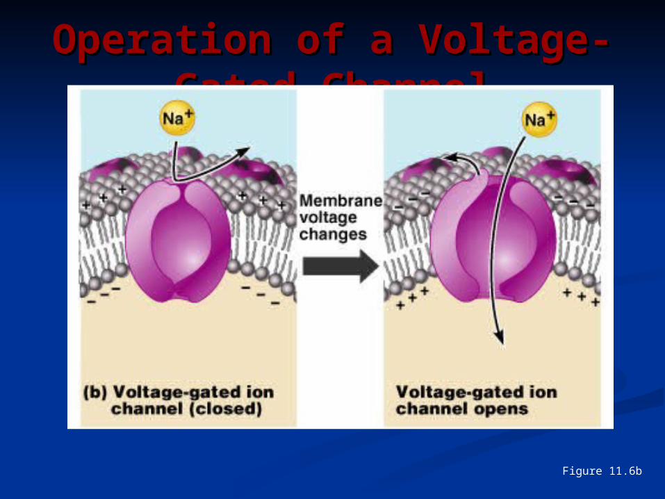
Operation of a Voltage-Operation of a Voltage-Gated ChannelGated Channel
Figure 11.6b
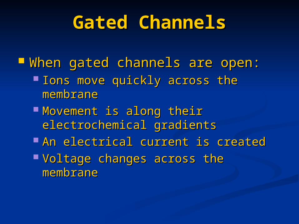
When gated channels are open: When gated channels are open: Ions move quickly across the membrane Ions move quickly across the membrane Movement is along their electrochemical Movement is along their electrochemical
gradientsgradients An electrical current is createdAn electrical current is created Voltage changes across the membraneVoltage changes across the membrane
Gated ChannelsGated Channels

Ions flow along their chemical gradient when Ions flow along their chemical gradient when they move from an area of high they move from an area of high concentration to an area of low concentrationconcentration to an area of low concentration
Ions flow along their electrical gradient when Ions flow along their electrical gradient when they move toward an area of opposite chargethey move toward an area of opposite charge
Electrochemical gradient – the electrical and Electrochemical gradient – the electrical and chemical gradients taken togetherchemical gradients taken together
Electrochemical GradientElectrochemical Gradient

The potential difference (–70 mV) across The potential difference (–70 mV) across the membrane of a resting neuronthe membrane of a resting neuron
It is generated by different concentrations It is generated by different concentrations of Naof Na++, K, K++, Cl, Cl, and protein anions (A, and protein anions (A))
Ionic differences are the consequence of:Ionic differences are the consequence of: Differential permeability of the neurilemma to Differential permeability of the neurilemma to
NaNa++ and K and K++
Operation of the sodium-potassium pumpOperation of the sodium-potassium pump
Resting Membrane Potential Resting Membrane Potential (V(Vrr))

Resting Membrane Resting Membrane Potential (VPotential (Vrr))
Figure 11.8

Used to integrate, send, and receive Used to integrate, send, and receive informationinformation
Membrane potential changes are Membrane potential changes are produced by:produced by: Changes in membrane permeability to ionsChanges in membrane permeability to ions Alterations of ion concentrations across the Alterations of ion concentrations across the
membranemembrane Types of signals – graded potentials Types of signals – graded potentials
and action potentialsand action potentials
Membrane Potentials: Membrane Potentials: SignalsSignals

Changes are caused by three eventsChanges are caused by three events Depolarization – the inside of the Depolarization – the inside of the
membrane becomes less negative membrane becomes less negative Repolarization – the membrane returns Repolarization – the membrane returns
to its resting membrane potentialto its resting membrane potential Hyperpolarization – the inside of the Hyperpolarization – the inside of the
membrane becomes more negative than membrane becomes more negative than the resting potentialthe resting potential
Changes in Membrane Changes in Membrane PotentialPotential

Changes in Membrane Changes in Membrane PotentialPotential
Figure 11.9

Short-lived, local changes in Short-lived, local changes in membrane potentialmembrane potential
Decrease in intensity with distanceDecrease in intensity with distance Their magnitude varies directly with Their magnitude varies directly with
the strength of the stimulusthe strength of the stimulus Sufficiently strong graded potentials Sufficiently strong graded potentials
can initiate action potentialscan initiate action potentials
Graded PotentialsGraded Potentials
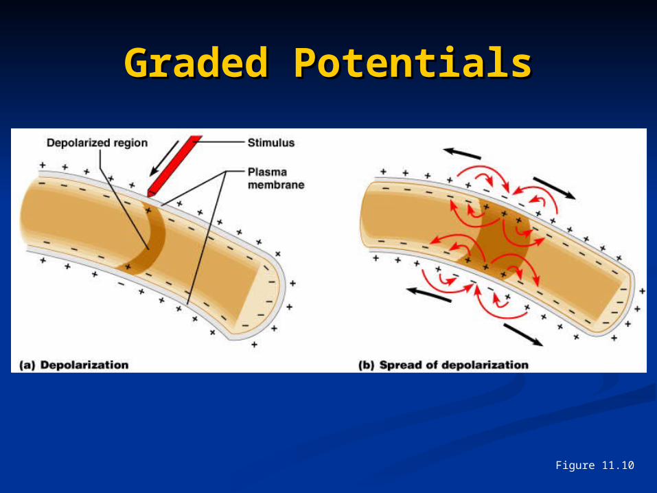
Graded PotentialsGraded Potentials
Figure 11.10

Graded PotentialsGraded Potentials
Voltage changes in graded potentials Voltage changes in graded potentials are decrementalare decremental
Current is quickly dissipated due to Current is quickly dissipated due to the leaky plasma membranethe leaky plasma membrane
Can only travel over short distancesCan only travel over short distances

Graded PotentialsGraded Potentials
Figure 11.11

A brief reversal of membrane potential A brief reversal of membrane potential with a total amplitude of 100 mVwith a total amplitude of 100 mV
Action potentials are only generated by Action potentials are only generated by muscle cells and neuronsmuscle cells and neurons
They do not decrease in strength over They do not decrease in strength over distancedistance
They are the principal means of neural They are the principal means of neural communicationcommunication
An action potential in the axon of a neuron An action potential in the axon of a neuron is a nerve impulseis a nerve impulse
Action Potentials (APs)Action Potentials (APs)
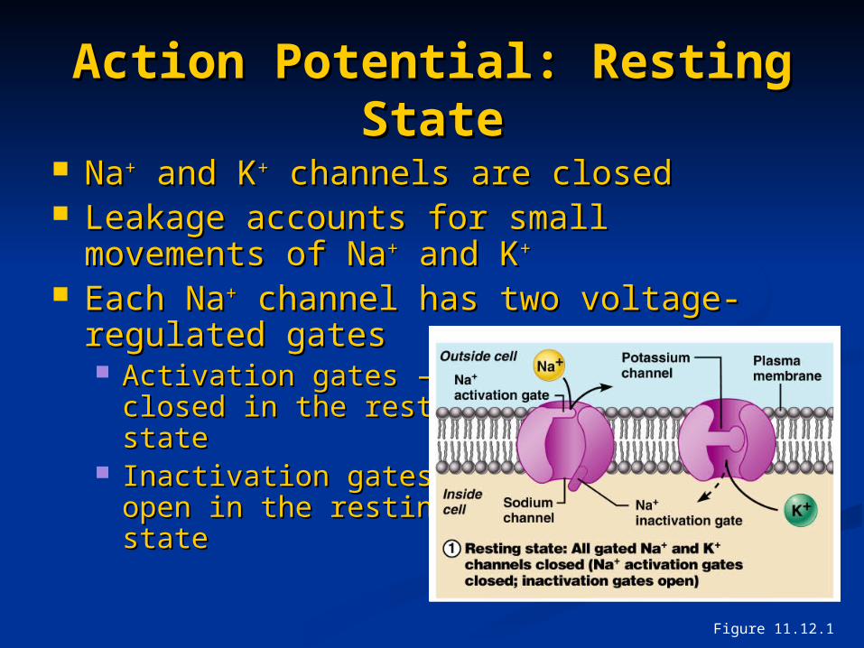
NaNa++ and K and K++ channels are closed channels are closed Leakage accounts for small movements of Leakage accounts for small movements of
NaNa++ and K and K++
Each NaEach Na++ channel has two voltage- channel has two voltage-regulated gates regulated gates Activation gates – Activation gates –
closed in the resting closed in the resting state state
Inactivation gates – Inactivation gates – open in the resting open in the resting statestate
Action Potential: Resting Action Potential: Resting StateState
Figure 11.12.1

NaNa++ permeability increases; membrane permeability increases; membrane potential reversespotential reverses
NaNa++ gates are opened; K gates are opened; K++ gates are closed gates are closed Threshold – a critical level of Threshold – a critical level of
depolarization depolarization (-55 to -50 mV)(-55 to -50 mV)
At threshold, At threshold, depolarization depolarization becomes becomes self-generatingself-generating
Action Potential: Action Potential: Depolarization PhaseDepolarization Phase
Figure 11.12.2

Sodium inactivation gates closeSodium inactivation gates close Membrane permeability to NaMembrane permeability to Na++ declines to declines to
resting levelsresting levels As sodium gates close, voltage-sensitive As sodium gates close, voltage-sensitive
KK++ gates open gates open KK+ + exits the cell and exits the cell and
internal negativity internal negativity of the resting neuron of the resting neuron is restoredis restored
Action Potential: Action Potential: Repolarization PhaseRepolarization Phase
Figure 11.12.3
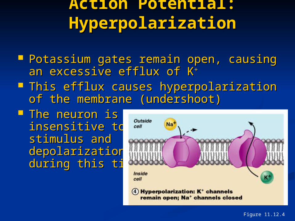
Action Potential: Action Potential: HyperpolarizationHyperpolarization
Potassium gates remain open, causing an Potassium gates remain open, causing an excessive efflux of Kexcessive efflux of K++
This efflux causes hyperpolarization of the This efflux causes hyperpolarization of the membrane (undershoot)membrane (undershoot)
The neuron is The neuron is insensitive to insensitive to stimulus and stimulus and depolarization depolarization during this timeduring this time
Figure 11.12.4

Repolarization Repolarization Restores the resting electrical conditions Restores the resting electrical conditions
of the neuronof the neuron Does not restore the resting ionic Does not restore the resting ionic
conditionsconditions Ionic redistribution back to resting Ionic redistribution back to resting
conditions is restored by the sodium-conditions is restored by the sodium-potassium pumppotassium pump
Action Potential: Action Potential: Role of the Sodium-Potassium Role of the Sodium-Potassium
PumpPump

Phases of the Action Phases of the Action PotentialPotential
1 1 – resting state– resting state
2 – depolarization2 – depolarization
3 – repolarization3 – repolarization
4 – 4 – hyperpolarizationhyperpolarization
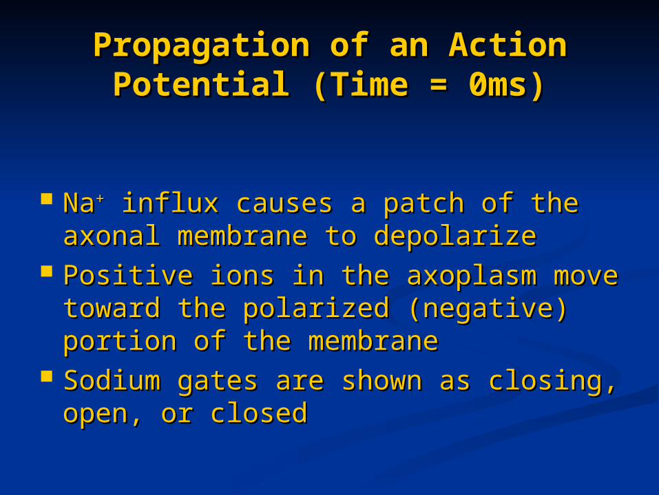
NaNa++ influx causes a patch of the influx causes a patch of the axonal membrane to depolarizeaxonal membrane to depolarize
Positive ions in the axoplasm move Positive ions in the axoplasm move toward the polarized (negative) toward the polarized (negative) portion of the membraneportion of the membrane
Sodium gates are shown as closing, Sodium gates are shown as closing, open, or closedopen, or closed
Propagation of an Action Propagation of an Action Potential (Time = 0ms)Potential (Time = 0ms)

Propagation of an Action Propagation of an Action Potential (Time = 0ms)Potential (Time = 0ms)
Figure 11.13a
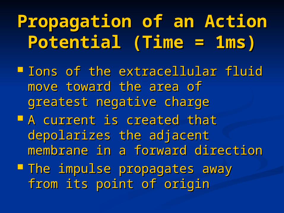
Ions of the extracellular fluid move Ions of the extracellular fluid move toward the area of greatest negative toward the area of greatest negative chargecharge
A current is created that depolarizes A current is created that depolarizes the adjacent membrane in a forward the adjacent membrane in a forward directiondirection
The impulse propagates away from The impulse propagates away from its point of originits point of origin
Propagation of an Action Propagation of an Action Potential (Time = 1ms)Potential (Time = 1ms)

Propagation of an Action Propagation of an Action Potential (Time = 1ms)Potential (Time = 1ms)
Figure 11.13b

The action potential moves away The action potential moves away from the stimulusfrom the stimulus
Where sodium gates are closing, Where sodium gates are closing, potassium gates are open and create potassium gates are open and create a current flowa current flow
Propagation of an Action Propagation of an Action Potential (Time = 2ms)Potential (Time = 2ms)

Propagation of an Action Potential Propagation of an Action Potential (Time = 2ms)(Time = 2ms)
Figure 11.13c

Threshold – membrane is depolarized by Threshold – membrane is depolarized by 15 to 20 mV15 to 20 mV
Established by the total amount of current Established by the total amount of current flowing through the membrane flowing through the membrane
Weak (subthreshold) stimuli are not Weak (subthreshold) stimuli are not relayed into action potentialsrelayed into action potentials
Strong (threshold) stimuli are relayed into Strong (threshold) stimuli are relayed into action potentialsaction potentials
All-or-none phenomenon – action All-or-none phenomenon – action potentials either happen completely, or potentials either happen completely, or not at allnot at all
Threshold and Action Threshold and Action PotentialsPotentials

Absolute Refractory PeriodAbsolute Refractory Period
Figure 11.15

The interval following the absolute The interval following the absolute refractory period when:refractory period when: Sodium gates are closedSodium gates are closed Potassium gates are openPotassium gates are open Repolarization is occurringRepolarization is occurring
The threshold level is elevated, The threshold level is elevated, allowing strong stimuli to increase allowing strong stimuli to increase the frequency of action potential the frequency of action potential eventsevents
Relative Refractory Relative Refractory PeriodPeriod

Conduction velocities vary widely Conduction velocities vary widely among neuronsamong neurons
Rate of impulse propagation is Rate of impulse propagation is determined by:determined by: Axon diameter – the larger the diameter, Axon diameter – the larger the diameter,
the faster the impulsethe faster the impulse Presence of a myelin sheath – Presence of a myelin sheath –
myelination dramatically increases myelination dramatically increases impulse speedimpulse speed
Conduction Velocities of Conduction Velocities of AxonsAxons

Current passes through a myelinated Current passes through a myelinated axon only at the nodes of Ranvieraxon only at the nodes of Ranvier
Voltage-gated NaVoltage-gated Na++ channels are channels are concentrated at these nodesconcentrated at these nodes
Action potentials are triggered only at Action potentials are triggered only at the nodes and jump from one node to the nodes and jump from one node to the nextthe next
Much faster than conduction along Much faster than conduction along unmyelinated axonsunmyelinated axons
Saltatory ConductionSaltatory Conduction

Saltatory ConductionSaltatory Conduction
Figure 11.16


![arXiv:cond-mat/9911472v1 [cond-mat.dis-nn] 29 Nov … potentials over some suitable intervals. ... on activities of transducer neurons such as motor and relay neurons, ... CdV ¯ j(t)/dt](https://img.dokumen.tips/doc/110x75/5b07120e7f8b9ac33f8db981/arxivcond-mat9911472v1-cond-matdis-nn-29-nov-potentials-over-some-suitable.jpg)


