Embed Size (px)
Citation preview

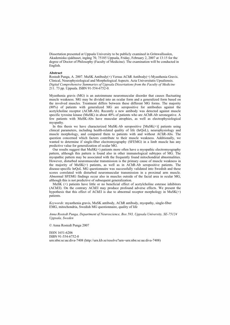
ACTAUNIVERSITATISUPSALIENSISUPPSALA2007
Digital Comprehensive Summaries of Uppsala Dissertationsfrom the Faculty of Medicine 211
MuSK Antibody(+) Versus AChRAntibody(+) Myasthenia Gravis
Clinical, Neurophysiological and MorphologicalAspects
ANNA ROSTEDT PUNGA
ISSN 1651-6206ISBN 91-554-6752-0urn:nbn:se:uu:diva-7408

“ I can respect, but never accept, living with myasthenia”
To my mother Margaretha


List of papers
This thesis is based on the following papers. They will be referred to in thetext by their Roman numerals:
I Anna Rostedt, Linda L Sanders, Lloyd J Edwards, Janice MMassey, Donald B Sanders, Erik V Stålberg. Predictive valueof single-fiber electromyography in the extensor digitorumcommunis muscle of patients with ocular myasthenia gravis: aretrospective study. J Clinical Neuromuscular Disease 2000;2:6-9.
II Anna Rostedt, Luca Padua, Erik V Stålberg. Validation ofthe Swedish version of the disease-specific myasthenia gravisquestionnaire. Neurol Sci. 2006; 27:91-96.
III Anna Rostedt, Luca Padua, Erik V Stålberg. Correlationbetween a patient-derived functional questionnaire and ab-normal neuromuscular transmission in myasthenia gravis pa-tients. Clinical Neurophysiology 2005; 116: 2058-2064.
IV Anna Rostedt Punga, Kati Ahlqvist, Emanuela Bartoccioni,Flavia Scuderi, Mariapaola Marino, Anu Suomalainen, HannuKalimo, Erik V Stålberg. Neurophysiological and mitochon-drial abnormalities in MuSK antibody seropositive myasthe-nia gravis compared to other immunological subtypes. Clini-cal Neurophysiology 2006; 117:1434-1443
V Anna Rostedt Punga, Roland Flink, Håkan Askmark, Erik VStålberg. Cholinergic neuromuscular hyperactivity in patientswith myasthenia gravis seropositive for MuSK antibody.Muscle Nerve 2006; 34:111-115.
I Lippincott Williams&Wilkins. II Springer Science and Business Me-dia. III and IV Elsevier. V Wiley Interscience.
Reprints were made with the kind permission of the publishers and the Inter-national Federation of Clinical Neurophysiology (III and IV).

Contents
Introduction...................................................................................................13Myasthenia Gravis (MG) .........................................................................14
History .................................................................................................14Epidemiology.......................................................................................15Pathophysiology ..................................................................................15Clinical picture of MG.........................................................................17Types of myasthenia gravis and myasthenic syndromes .....................17Diagnosis .............................................................................................18Quality of life assessment....................................................................23
General aim of the study...............................................................................24Specific aims ............................................................................................24
Study I..................................................................................................24Study II ................................................................................................24Study III ...............................................................................................24Study IV...............................................................................................25Study V................................................................................................25
Patients and methods ....................................................................................26Patients .....................................................................................................26Methods....................................................................................................27
Clinical neurological examination.......................................................27Myasthenia Gravis Questionnaire........................................................29SF-36 ...................................................................................................29Single Fiber Electromyography...........................................................30Repetitive nerve stimulation................................................................30Analysis of AChR antibodies ..............................................................30Analysis of MuSK antibodies..............................................................31Analysis of titin and ryanodine receptor antibodies ............................31Muscle biopsy......................................................................................31Statistical analysis................................................................................33
Results...........................................................................................................35Predictive value of SFEMG in a limb muscle for generalization of MG(Study I)....................................................................................................35
Time to generalization .........................................................................35
Presence and degree of abnormal SFEMG findings ............................35Other possible predictive tests .............................................................35
Health-related quality of life findings in Swedish MG patients andvalidation of the Swedish MG Questionnaire (Study II)..........................36
Translation and cultural adaptation .....................................................36Descriptive of the Italian sample .........................................................36Scores of the Swedish sample..............................................................36Correlation of MGQ and SF-36 scores ................................................37Evaluation capacity of Swedish MGQ ................................................37
Correlation between patient-oriented findings and abnormalneuromuscular transmission in MG (Study III)........................................38
Outcome measures...............................................................................38Correlation between MGQ and neurophysiology................................38Correlation between SF-36 and neurophysiology ...............................38
Comparison of neurophysiological, muscle biopsy and health-relatedquality of life parameters in MuSK(+), AChR(+) and AChR(-) MGpatients (Study IV) ...................................................................................40
MuSK-Ab presence .............................................................................40Clinical findings ..................................................................................41Neurophysiological findings................................................................41Correlation of decrement, mean MCD and percentage of blockings...42Comparison of myopathic pattern .......................................................43Morphological and mitochondrial findings .........................................44Mitochondrial DNA and POLG1 findings...........................................45
Extra discharges due to AChEI (Study V) ...............................................48
Discussion.....................................................................................................49Disturbed neuromuscular transmission in the EDC in OMG...................49Health-related quality of life in Swedish MG patients.............................50Correlation between disturbed neuromuscular transmission and health-related quality of life ................................................................................51MuSK antibodies and AChR antibodies may coexist ..............................51Primary factors contributing to the muscular weakness and fatigue inMuSK(+) patients.....................................................................................52
Neuromuscular transmission ...............................................................52Health-related quality of life in MuSK(+) patients..............................53Therapy considerations in MuSK(+) patients......................................53Myopathy in MuSK(+) patients...........................................................54Pathogenic role of MuSK antibodies ...................................................54Muscle pathology in MuSK(+) patients and mitochondrial defects inMG.......................................................................................................55Adverse effects of pyridostigmine treatment in MuSK(+) MG:neurophysiological indications of AChEI side effects ........................56

Conclusions...................................................................................................57
Summary in Swedish- sammanfattning på Svenska .....................................58Studie I .....................................................................................................59Studie II ....................................................................................................59Studie III...................................................................................................59Studie IV ..................................................................................................59Studie V....................................................................................................60
Acknowledgements.......................................................................................61
References.....................................................................................................64


Abbreviations
ACh AcetylcholineAChEI Acetylcholine esterase inhibitorsAChR Acetylcholine receptorAChR(+) Acetylcholine receptor antibody se-
ropositiveAChR(-) Acetylcholine receptor antibody se-
ronegativeAChR-Ab Acetylcholine receptor antibodyBD Bulbar domainBP Bodily painCOX Cytochrome C oxidaseCMAP Compound muscle action potentialEDC Extensor digitorum communis mus-
cleEMG ElectromyographyFD Fiber densityGD Generalized domainGH General healthGMG Generalized myasthenia gravisHrQoL Health-related quality of lifeIgG Immunoglobulin type GLEMS Lambert Eaton myasthenic syndromeMCD Mean of consecutive interpotential
interval differencesMCS Mental composite scoreMG Myasthenia gravisMGFA Myasthenia Gravis Foundation of
AmericaMGQ Myasthenia gravis questionnaireMH Mental healthMtDNA Mitochondrial DNAMuSK Muscle specific tyrosine kinaseMuSK(+) Muscle specific tyrosine kinase anti-
body seropositiveMuSK(-) Muscle specific tyrosine kinase anti-
body seronegative

MuSK-Ab Muscle specific tyrosine kinase anti-body
MUP Motor unit potentialNADH-TR NADH-tetrazolium reductaseOD Ocular domainOMG Ocular myasthenia gravisPCS Physical composite scorePF Physical functioningPOLG Polymerase γQEMG Quantitative electromyographyRE Role emotionalRNS Repetitive nerve stimulationRP Role physicalSD Standard deviationSDH Succinate dehydrogenaseSFEMG Single fiber electromyographySF Social functioningSF-36 36-item Short-Form health survey
13
Introduction
People have asked me why I chose to study myasthenia gravis, which is arare neuromuscular disorder, when there are so many other diseases in theworld. The main reason is that my mother got myasthenia when I was quiteyoung, so I have basically lived close to it for a long time. I have seen itsconstant presence, although fluctuating in severity. Therefore, through itschronic nature, I can understand how it directly and indirectly affects qualityof life.
Myasthenia Gravis (MG) causes fluctuating muscle weakness, which of-ten involves droopy eyelids, swallowing difficulties and generalized musclefatigue. In the literature MG is often said to be the best understood autoim-mune disorder, but how come then that we do not know what triggers it orhow to cure it? Further we can not answer why some muscles are moreprone to be affected than others.
Recently, a new antibody was detected against the muscle specific tyro-sine kinase (MuSK) located at the neuromuscular junction. A few patientswith MuSK antibodies have, in addition to myasthenic symptoms, prominentmuscular atrophies, as well as myopathic findings on electromyography. Theexact role of these antibodies remains unclear to date.
The focus of this thesis are the main factors contributing to the muscularweakness and fatigue in MuSK-Ab seropositive [MuSK(+)] patients, com-pared to other major immunological subtypes of MG. These aspects includeclinical parameters, e.g. health-related quality of life, neurophysiology andmuscle morphology. Additionally, we wanted to elucidate if disturbed neu-romuscular transmission in a limb muscle in ocular MG predicts the risk ofgeneralization.

14
Myasthenia Gravis
HistoryHistorical reviews start with the description from the English physicianThomas Willis in 1672 (Guthrie, 1903) but it was not until 1895 that theGerman neurologist Friedrich Jolly proposed naming this newly discoveredneurological disorder myasthenia gravis (Jolly, 1895). Patients were reportedto present with abnormal muscle fatigability, the most prominent signs beingthe inability to swallow and to articulate. He suggested the full name“Myasthenia gravis pseudo-paralytica” and this name was accepted after aconsensus meeting of the Berlin Society of Psychiatry and Neurology in1899 (Viets, 1953).
The cause underlying MG has been an area of debate. In 1960 a Scottishneurologist, Simpson, suspected that MG might be caused by an autoim-mune mechanism. This suspicion was based on the relatively high incidenceof concomitant autoimmune diseases (such as rheumatoid arthritis and sys-temic lupus erytematosus) among the patients (Simpson, 1960). Thymusabnormalities were discovered, as well as the presence of lymphorrages inmuscles, which further supported the autoimmune hypothesis (Miller, 1961).Further, a humoral factor was implicated in the development of MG, after itwas found that approximately 20% of babies whose mothers were diagnosedwith MG were reported to develop transient neonatal MG (Strickroot, 1942).
In 1934, patients were found to be hyper-reactive to d-tubocurarine (Ben-net and Cash, 1942). Subsequently, it was demonstrated that the musclecontraction of MG patients declined with repetitive nerve stimulation of theafferent nerve (Harvey and Masland, 1941). Further, both miniature andregular end plate potentials were found to be reduced when measured usingmicro electrophysiological techniques (Elmqvist et al., 1964), which local-ized the defect to the neuromuscular junction. In the 1970s, when the immu-nization of animals with acetylcholine receptor (AChR) was reported to in-duce an autoimmune response to the AChR of mammalian skeletal muscles,the autoimmune hypothesis was accepted as a theory (Heilbronn et al., 1975;Heinemann et al., 1977; Patrick and Lindstrom, 1973). This finding indi-cated that the target in MG was located more precisely at the postsynapticmembrane. Analysis of AChR antibodies (AChR-Ab) became an importantdiagnostic tool since MG patients often had elevated titers of this receptorantibody (Lindstrom et al., 1976). Further, it was reported that maternal

15
AChR-Ab titer correlated with the frequency and severity of transient neo-natal MG present in the infant (Eymard et al., 1989).
Patients who did not possess elevated AChR-Ab were called seronegative[AChR(-)]. There were indicators that seronegative MG also had an autoim-mune etiology. These indicators included: 1) neonatal transient MG in babiesof women with seronegative MG (Mier and Havard, 1985), 2) improvementwith immunotherapy and 3) disruption of neuromuscular transmission whenmice were exposed to patient sera (Mossman et al., 1986). In 2001, 70% ofAChR(-) MG patients were reported to have serum antibodies againstMuSK at the postsynaptic membrane (Hoch et al., 2001). Studies of the re-maining seronegative MG patients have shown that their plasma reducedAChR function (Plested et al., 1998), although the exact target has not beendetermined. This target, as well as the triggering factor of MG, has yet to beestablished.
EpidemiologyFor the past 50+ years, population-based studies on the epidemiology of
MG have shown a clear trend toward an increase in the reported prevalenceof the disease. This increase is most likely due to more sensitive diagnosticmethods, as well as to higher survival rate, due to better treatment regimens.The prevalence of MG is twice as high in women, compared to men. Typi-cally, affected women are 20-30 years old, whereas men are 60-80 years old(Osserman and Genkins, 1971). The annual incidence of MG has been re-ported to be about 3-4 cases per million and the prevalence about 60 casesper million (Araki et al., 1987; Oosterhuis, 1981; Storm-Mathisen, 1984);however, higher rates have been reported. For example, in the USA, currentestimates indicate a prevalence as high as about 20 per 100,000 persons(Phillips, 2003). Further, the prevalence of MG was recently estimated to be14 cases per 100,000 in Stockholm, Sweden (Kalb et al., 2002).
Pathophysiology
Types of antibodies
Acetylcholine receptor antibodiesAbout 80% of patients with generalized MG and 55% of patients with ocularMG have AChR-Ab (Lindstrom et al., 1976; Sanders, 2003; Vincent andNewsom-Davis, 1985). These antibodies impair AChR function by threemain mechanisms: 1) blocking of the ACh-binding site (Lefvert et al., 1981)

16
2) cross-linking of the AChRs on the postsynaptic membrane, which resultsin a functional blockade of the receptors as well as accelerated degradationof the AChRs (Drachman et al., 1981) and 3) complement activation, re-sulting in destruction of the postsynaptic muscle membrane (Engel et al.,1977). The consequence of these events is less efficient neuromusculartransmission.
Muscle specific tyrosine kinase antibodiesQuite recently it was discovered that among the 20% of patients who are
AChR(-), 40-70% are seropositive for antibodies directed against MuSK,which is located at the neuromuscular junction (Bartoccioni et al., 2003;Hoch et al., 2001; Sanders et al., 2003; Scuderi et al., 2002; Zhou et al.,2004). MuSK is assumed to be essential in the early development of the neu-romuscular junction as well as in maintaining the postsynaptic apparatus(Liyanage et al., 2002). During development of the neuromuscular junction,neural agrin activates MuSK, and this induces clustering of AChRs (Hopfand Hoch, 1998). These clusters are necessary for a normal neuromusculartransmission. It has been questioned whether MuSK-Abs are responsible forthe weakness found in AChR(-) MG patients (Selcen et al., 2004; Vincent etal., 2005). Nevertheless, their pathogenic role has recently been confirmed.Rabbits that were immunized with MuSK ectodomain protein manifestedMG-like muscle weakness. Additionally, a decremental response was elic-ited in the facial nerve and there was a significant reduction of AChR clus-tering at the neuromuscular junctions. The produced MuSK-Abs specificallyinhibited in vitro AChR clustering (Shigemoto et al., 2006). In animal ex-periments, MuSK knockout mice fail to cluster AChRs and to differentiatepostsynaptic regions, therefore dying perinatally (DeChiara et al., 1996).
Titin and ryanodine receptor antibodiesMG patients with a thymoma may have antibodies not only directed to theAChR but also to other components of striated muscle. Two of these compo-nents strongly associated with a thymoma are the sarcomeric cytoskeletalprotein titin (Aarli et al., 1990) and the Ca2+ release channel of the sarco-plasmic reticulum, the ryanodine receptor (Mygland et al., 1992). On thebasis of their cross-striational pattern by immunofluorescent staining (Peerset al., 1977), they have been named antistriational antibodies.

17
Rapsyn antibodies
A fifth antigen is the small intracellular AChR-associated protein, rapsyn,which is located in the muscle end plate. Antibodies directed against rapsynhave been detected in about 15% of MG patients (Agius et al., 1998), both inAChR-Ab seropositive [AChR(+)] and AChR(-) patients. Rapsyn is pre-cisely colocalized with AChR from the earliest stages of synapse formation(Hall and Sanes, 1993). Analysis of rapsyn knockout mice proved thatrapsyn is, as well as MuSK, necessary for clustering AChRs (Gautam et al.,1995).
Clinical picture of MGMG is characterized by an increased fatigability of skeletal muscles, pre-dominantly affecting the proximal muscles. Muscle weakness fluctuatesduring the day, becoming more pronounced in the afternoon/evening andwith physical activity. When extraocular muscles are involved, conditionssuch as ptosis and/or diplopia occur. Additionally, bulbar muscle involve-ment leads to dysphagia and dysarthria. Proximal arm and leg muscles aswell as neck muscles are also involved in most patients. Muscle wasting isuncommon in MG, but has been reported in a few MuSK-Ab seropositiveMuSK(+) patients (Evoli et al., 2003) as well as in about 10% of AChR(+)patients (Oosterhuis and Oosterhuis, 1997).
Types of myasthenia gravis and myasthenic syndromes1. Acquired (autoimmune) myasthenia gravis (MG) is the most common anddevelops at any age after birth. This type of MG may be further divided intothe following subtypes:
• Pure ocular MG (OMG): Fatigue is limited to the extraocularmuscles, i.e., the levator palpebrae muscle and the eye mov-ing muscles. This form may develop at any age. Generaliza-tion may occur within two years from onset.
• Early onset generalized MG: Develops before the age of 40 years.
• Late onset generalized MG: Starting after the age of 40years.
• MG with thymoma: May develop at any age.2. Neonatal myasthenia gravis affects newborn babies of myasthenic moth-
ers and is caused by circulating antibodies from the mother via the pla-centa.
3. Congenital myasthenia includes syndromes that are due to deficiency ofstructures pre-synaptic (e.g. choline acetyltransferase), intra-synaptic(e.g. end plate ACh esterase) or postsynaptic (e.g. rapsyn).

18
4. Lambert Eaton myasthenic syndrome (LEMS) is an acquired autoim-mune disorder, which causes muscle fatigue, loss of tendon reflexes atrest and autonomic dysfunction. LEMS can occur in a paraneoplasticform usually associated with small cell lung cancer or in a non-paraneoplastic form. In the majority (90%) of patients, antibodies tovoltage-gated calcium channels (of P-/Q-type) are detected (Lang et al.,1998).
Diagnosis
Serum analysis of autoantibodies
The presence of autoantibodies in MG is of great importance for guidance tothe correct therapy in individual patients. The two types of antibodiespresented in this section have been of most importance to this study.
Anti-AChR antibodies
AChR-Abs are pathogenic and highly specific for MG. They are present inabout 85% of patients with clinical features of myasthenia gravis (Vincentand Newsom-Davis, 1985). Therefore, this analysis adds important diagnos-tic information. The binding of AChR-Abs (IgG type) is measured using astandard radioimmunoassay in which the antigen consists of AChR fromhuman muscle labeled with [125I]α-bungarotoxin (Lefvert et al., 1978; Lind-strom, 1977).
Anti-MuSK antibodies
The presence of MuSK-Abs is important for the diagnosis of MG in patientswhere AChR-Abs are absent, since some of these patients present a clinicalpicture of severe bulbar symptoms (Evoli et al., 2003), which calls for ag-gressive immunosuppressive treatment. The existence of MuSK-Ab (IgGtype) can be determined by the immunoprecipitation of native MuSK ex-tracted from TE671 plasma membranes (Scuderi et al., 2002). Alternatively,MuSK-Ab presence can be ascertained by immunoprecipitation of recombi-nant 125 I-MuSK extracellular domains (McConville et al., 2004).
Neurophysiology
At the neurophysiological level in the diagnostic process, tests that showimpaired neuromuscular transmission are essential for diagnosis.
Repetitive nerve stimulation
In MG, a progressive decline in the compound muscle action potential(CMAP) amplitude is found using 3 Hz repetitive nerve stimulation (RNS),
19
which is usually most pronounced at the 4th or 5th response (Desmedt, 1973).This decrement is due to the “run down” of the amplitude of individual endplate potentials. In MG, a certain proportion of potentials is reduced to asubthreshold level and therefore is insufficient to give a depolarization of themuscle fiber. Since the CMAP constitutes the sum of activated muscle fi-bers, its amplitude is successively reduced with an increasing block of indi-vidual muscle fibers. If the drop in amplitude, or decrement, exceeds a cer-tain limit, e.g. 5%, the finding is considered pathological. RNS is first per-formed at rest and then after 20 seconds of maximal voluntary activity,which investigates post-exercise facilitation. New tests after 1, 3 and 5 min-utes explore post-exercise exhaustion.
Single-Fiber ElectromyographySingle-fiber electromyography (SFEMG) can be performed during voluntarymuscle contraction or when the muscle is electrically stimulated. SFEMG,has become the standard diagnostic tool of MG; (Ekstedt and Stålberg, 1963;Ekstedt and Stålberg, 1975; Stålberg, 1980; Stålberg and Trontelj, 1994).This method detects subclinical defects in neuromuscular transmission andcan be used to quantify the degree of neuromuscular disturbance. SFEMGjitter reflects the safety factor of the motor end plate (Stålberg and Trontelj,1994). When jitter measurements are made in voluntarily activated muscle,activity from two muscle fibers innervated by the same axon is recorded andone action potential used as a time reference. The jitter then results fromvariations in the difference in conduction times taken by impulses from thenerve branching point via the motor end plates along each muscle fiber to therecording site. Most of the jitter results from problems at the neuromuscularjunction. Additionally, by counting the number of single muscle fibers fromone motor unit within the uptake area of the electrode at many electrodepositions, a measure of the mean fiber density within a motor unit can becalculated.
Quantitative electromyography
For motor unit potential (MUP) analysis and for the analysis of the electro-myography (EMG) pattern at strong contraction, quantitative methods havebeen developed and are a part of the EMG routine investigation (Stålberg etal., 2003). Quantitative electromyography (QEMG) is performed with a con-centric needle electrode in the muscle at rest, slight activation and strongcontraction. If a differential diagnosis is suspected in MG, such as myo-pathy, QEMG is performed.
Other neurophysiological techniques
While there are established neurophysiological methods for diagnosing MG,some other techniques have been suggested.

20
Some departments perform intracellular recordings, where microelec-trodes are inserted into individual muscle fibers to record the membranepotential and/or to pass current. Synaptic currents can thereby be recordedusing a two-electrode patch-clamp configuration (Slater et al., 1992).
Infrared reflection oculography is sometimes undertaken to study eyemovement recordings of saccades when ocular MG is suspected. The diag-nostic yield of this examination is however not as high as the other neuro-physiological techniques (Oey et al., 1993).
The stapedius reflex decay, in response to a one-minute sound stimulus of500 Hz, is analogous to the decremental response of muscle action potentialsto rapid nerve stimulation. This represents another test for the diagnosis ofMG (Warren et al., 1977).
Muscle morphology
Muscle biopsyA muscle biopsy is occasionally taken in MG to rule out other disorders suchas mitochondrial or inflammatory myopathies. In MG there are few specificalterations to detect at the light microscopic level, such as some atrophictype II fibers and the appearance of minor lymphocyte infiltrates due to animmunological reaction (Kalimo and Stålberg, 2003).
In motor point biopsies, the mean number of AChRs at the neuromuscularjunction is significantly lower in MG patients compared to healthy controls(Pestronk et al., 1985). Electron microscopy has visualized the deposition ofthe membrane attack complex on the motor end plates, where the normalfolding of the junctional muscle membrane is markedly reduced and AChRsare lost (Engel et al., 1976). However, motor point biopsies are too difficultto be used in routine diagnostic examination of MG patients.
The differential diagnosis of MG vs. other neuromuscular disorders inmuscle biopsies relies on the presence of structural alterations that are absentor uncommon in MG. For example, in mitochondrial myopathies one fre-quent finding is the presence of cytochrome C oxidase (COX) negative fi-bers, which indicates impaired function of complex IV in the mitochondrialrespiratory chain. Ragged red fibers, i.e. fibers − usually COX negative −with increased numbers of abnormal mitochondria, are commonly seen indiseases due to translation defects, especially in large-scale deletions of themitochondrial DNA which impinge on the three genes coding for subunits ofCOX. On the other hand, even though there may be lymphocyte infiltrates inMG, necrotic and/or degenerative alterations of myositis are absent.

21
Clinical neurological examinationThe clinical examination of MG patients is important and should include
an assessment of muscle strength at rest and after repeated exercises, to de-termine fatigability. Specific muscle groups to be examined include thelimbs, neck, face and bulbar region. Muscle fatigue is provoked by per-forming repetitive muscle strength testing or prolonged tonic contraction.This fatigue is quantified, e.g., as time until onset and/or worsening of ptosisduring upward gaze. Almost all patients have weakness of ocular musclesevident upon close examination (Sanders, 2003). Palatal weakness producesnasal speech. Weakness of the laryngeal muscles results in impaired speechand weakness of the jaw muscles may cause difficulties holding with jawclosure. The deltoid muscle is most often involved in the upper limbs andmay be evaluated by arm abduction and repeated straight leg raises can testthigh/hip muscles. Finally, neck flexor and neck extensor muscles are evalu-ated.
To measure the degree of impairment, a quantitative MG score is calcu-lated (Barohn et al., 1998).
Edrophonium (Tensilon) testEdrophonium is a fast- and short lasting drug that inhibits acetylcholinester-ase, allowing for increased availability of acetylcholine (ACh) at the neuro-muscular junction, and so enhances neuromuscular transmission. Most MGpatients respond to edrophonium within two minutes, which results in anincrease in muscle strength. The test is considered positive if strength im-proves in muscles that can be objectively assessed such as the ocular andbulbar muscles. The effects of edrophonium have usually dissipated withinfive minutes. Possible adverse reactions include epigastric distress, sweatingand muscle fasciculations. The edrophonium test is positive in 60-95% ofocular myasthenia patients and in 72-95% with generalized MG (Pascuzzi,2003). However, improvement after edrophonium has also been found inother conditions such as congenital myasthenic syndromes, LEMS, intracra-nial aneurysms, brainstem lesions, tumors located in the cavernous sinus andsphenoid ridge, end-stage renal disease and in other muscle diseases affect-ing the ocular muscles including mitochondrial myopathy (Ajtai et al., 2004;Ben Yaou et al., 2006; Dirr et al., 1989; Moorthy et al., 1989).
22
Treatment of MG
Treatment of MG can be divided into symptomatic and disease-modulating(immunosuppressive) treatment.
Symptomatic treatmentAcetylcholine esterase inhibitors (AChEI) improve neuromuscular trans-
mission, especially at MG onset, providing short-term symptomatic relief ofmuscle weakness. This medication has been used since 1935, when Walkerreported the beneficial effect from Prostigmin (Walker, 1935). The mostcommon form is pyridostigmine bromide (Mestinon). In some patients, typi-cally those with purely ocular weakness, this may be sufficient. The usualstarting dose of pyridostigmine bromide is 60 mg every 4 to 6 hours. Therecommended maximum dose is 120 mg every 3 hours. Additionally, neo-stigmine bromide is a longer-acting agent for intramuscular administration,although not very commonly used.
Thymectomy and immunosuppressive treatmentIn patients with disabling ocular symptoms and/or generalized weakness,
thymectomy should be considered before the age of 60. Plasma exchange,intravenous immunoglobulins or high-dose prednisone administration isoften performed when there is a crisis involving oropharyngeal or respiratorymuscles, especially before thymectomy is performed. Other long-term im-munosuppressive medications include azathioprin (Imurel), cyclosporine(Sandimmun) and the most recent medicine, mycophenolate mofetil (Cell-cept). Being able to discontinue AChEI in immunosuppressed patients is acriterion of adequate immunosuppression (Sanders, 2003).
Acetylcholinesterase inhibitors and neurophysiology
After an injection of edrophonium normal jitter remains unchanged. In anabnormal potential pair, the degree of impulse blocking decreases or disap-pears and the jitter tends to return towards the normal value. If the patient isalready on AChEI treatment the reverse edrophonium effect, namely in-creased jitter and more frequent blockings, can be observed in a muscle,which otherwise responds positively to the drug (Stålberg and Trontelj,1994).
Extra discharges following the CMAP in a patient receiving treatmentwith AChEI, may signify an upcoming cholinergic crisis (van Dijk et al.,1996). Two possible mechanisms for their generation are: 1) prolonged con-tact of ACh with its postsynaptic receptor, which results in repetitive musclemembrane excitation or 2) backfiring of nerve action potentials due to the

23
stimulation of pre-synaptic ACh receptors (Besser et al., 1989; Okamoto andRiker, 1969).
Quality of life assessmentThe patient’s self-assessment of his/her condition is becoming increasinglyimportant, e.g., for the monitoring of specific treatments (Ware and Sher-bourne, 1992). The Myasthenia Gravis Foundation of America (MGFA) hasstated that in research and clinical trials on MG, in addition to clinical ex-amination, a health-related quality of life (hrQoL) questionnaire has to beincluded (Jaretzki et al., 2000). To validate a questionnaire is to determine towhich degree the self-assessment scores reflect a particular clinical question,such as general physical and mental health and disability related to a specificdisease. The scores are required to correlate with particular clinical meas-ures, such as physical examination and effect of treatment.
SF-36The Short-Form 36-item questionnaire (SF-36) is the most commonly usedgeneric patient oriented tool to assess a patient’s general health. It compriseseight separate question domains that together define physical and mentalhealth clusters (Ware and Sherbourne, 1992). The SF-36 is accessible inmany languages and has been validated in numerous countries. For the pur-pose of this study, the Swedish version of SF-36 was used (Sullivan et al.,1995).
Myasthenia Gravis questionnaire (MGQ)A disease specific hrQoL questionnaire for MG patients was developed byPadua et al in Rome, Italy, to address each patient’s hrQoL (Padua et al.,2002). The MG questionnaire (MGQ) assesses how the degree of myasthenicsymptoms affects patients’ every day life. This was initially validated inItalian; hence, the questionnaire had to undergo validation in Swedish inorder to be applied to the Swedish patients.

24
General aim of the study
The first objective of this study was to determine the presence and degree ofdisturbed neuromuscular transmission in a limb muscle in the ocular form ofMG to see whether this is predictive of generalization. We also wanted toelucidate whether disturbed neuromuscular transmission in a limb musclecorrelates to the patient-oriented findings.
The second objective was to characterize the main factors contributing tothe muscular weakness and fatigue in MuSK(+) patients, compared to othermajor immunological types of MG. The parameters included were neuro-physiological findings in proximal muscles, muscle pathology and health-related quality of life.
Specific aims
Study ITo determine if the jitter in the extensor digitorum (EDC) muscle in patientswith purely ocular MG (OMG) predicts the subsequent development of gen-eralized myasthenic weakness.
Study IITo translate and validate the disease-specific patient-derived MG question-naire (MGQ), enabling its use among Swedish MG patients.
Study IIITo correlate the self-assessed physical function score, using the MGQ andSF-36 questionnaires, with the degree of abnormal neuromuscular transmis-sion, as measured by the most disease sensitive neurophysiological methods,i.e., SFEMG and RNS.

25
Study IVTo compare the electrophysiological and histopathological features, i.e.,
mitochondrial characteristics, as well as to assess the genetic background ofthe histopathological mitochondrial alterations, in different immunologicalsubtypes of MG.
Study VTo describe the extra discharges occurring after the CMAP on motor nervestimulation in a MuSK(+) patient with moderate dose of pyridostigminebromide.
26
Patients and methods
PatientsAll patients included in the study had a diagnosis of MG. The MG diagnosiswas based on standard diagnostic criteria, including symptoms of fluctuatingmuscular weakness, supported by neurophysiological findings of disturbedneuromuscular transmission (for which no other cause could be found, suchas reinnervation). Their MG status was classified into ocular, generalizedand bulbar forms, according to the Myasthenia Gravis Foundation of Amer-ica (MGFA) protocol for clinical classification (Jaretzki et al., 2000).
The ethical committee of the Duke University Medical Center approved thestudy I and the ethical committee of Uppsala University Hospital approvedstudies II-IV. All patients in study II gave their informed consent, whichincluded approval for the storage of biological specimens.
The first study group (study I) comprised 50 patients (14 women, 36 men,mean age 45, range 29-66 years) at the Duke University Medical Center MGClinic, Durham, NC, USA. All patients fulfilled the following entry criteria:ocular MG and no treatment other than AChEI at the time of the initialSFEMG examination. These criteria excluded patients with any weaknesspresent in oropharyngeal, limb or trunk muscles. The included patients werefollowed for ≥2 years after MG onset. No patients were known to haveMuSK-Ab.
The second group of patients (study II-IV) consisted of 50 MG individualswho were recruited from medical records at the Department of Neurology,Uppsala University Hospital. MG patients who repeatedly tested negative forAChR-Ab [AChR(-) patients; N = 14] were initially included. Additionally,36 age-, sex-, and disease-matched AChR(+) patients were included forcomparison. Altogether, 50 (45 women, 5 men, mean age 55.5, range 29-78years) participated in the study. The duration of symptoms ranged from 1 to57 years (mean 22.2 years). Two AChR(-) patients with a strong clinicalsuspicion of MG were included in the group of AChR(-) MG, although theyinitially did not have abnormal neurophysiological findings (including

27
SFEMG in the orbicularis oculi and extensor digitorum communis muscle).Forty-eight patients (45 women, 3 men), who chose to answer the patient-oriented questionnaires, were included in study II. Forty-five patients (42women, 3 men) who had undergone SFEMG and RNS examination wereincluded in study III in order to correlate patient-oriented measures withneurophysiological results. In study IV all 50 patients were included andeach patient was subsequently examined on the same day. Evaluation of allpatients in study IV was completed within a period of 3 months.
In study V one of the MuSK(+) patients (man, 75 years) was examined infurther detail, since neurophysiological examination displayed extra dis-charges after the CMAP at low frequency stimulation, following the intakeof AChEI.
Methods
Clinical neurological examinationClinical examination was performed to assess the degree of weakness and
fatigue of ocular, bulbar, neck and limb muscles (see table 1). Additionally,the quantitative score for disease severity, which ranges from 0 (absence ofimpairment) to 39 (maximal impairment) was calculated (Barohn et al.,1998). Clinical MG classification was performed according to the MGFA(Jaretzki et al., 2000) (see table 2). Subclass (a) indicates predominantlylimb or axial muscle involvement, whereas subclass (b) indicates predomi-nantly bulbar muscle involvement. Remission is further subdivided intocomplete stable remission (no medication) and pharmacological remission(immunosuppressive medication).
28
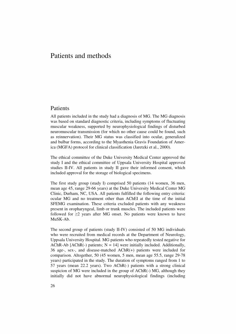
Muscle groups Test items Test limit
Ocular Upward gaze 120 sec
Bulbar Speech or counting aloud
Swallowing water
90 sec
½ cup
Arms Abduction 90° 90 sec
Hands Alternating flexion and extension of fingers 70 times
Neck Head lifts 45°(supine) 30 times
Legs Repetitive leg lifts 70° (supine) 35 times
Trunk Sitting upright from supine position
Table 1. Clinical neurological examination of MG patients (adapted from the origi-nal form used at the MG clinics in Stockholm, by Drs Matell and in Uppsala, by DrOsterman). Each muscle group is tested initially for weakness and then exercise testsare performed in order to provoke fatigue. The test limit for each muscle group isdisplayed in column the column to the right. Each muscle group, except for trunk, isgraded 0 (no impairment) to 4 (maximum impairment).
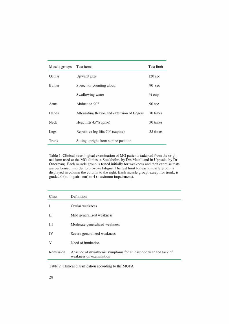
Class Definition
I Ocular weakness
II Mild generalized weakness
III Moderate generalized weakness
IV Severe generalized weakness
V Need of intubation
Remission Absence of myasthenic symptoms for at least one year and lack ofweakness on examination
Table 2. Clinical classification according to the MGFA.

29
Myasthenia Gravis QuestionnaireThe disease specific MGQ consists of 25 items (Padua et al., 2002). Theglobal MGQ score is obtained from the sum of these items; hence 0 formaximal impairment and 50 for absence of impairment. Some items, e.g.,1,3-5,16, do not assess regional muscle involvement, but are focused on suchitems as the timing of daily activity, fluctuations etc. Most items on theMGQ scale have 3 possible responses (0 = definite inability; 1 = partial in-ability; and 2 = ability). Other items evaluate the presence or absence of adeficit as a dichotomous categorical score with a yes or no answer, scored 2for absence of deficit and 0 for deficit. Items are further divided into threesymptom domains: (1) generalized domain (GD; including items 2a-h, 6, 8-12); (2) bulbar domain (BD; including items 7 and 15) and (3) ocular domain(OD; including items 13,14). Each domain score of the MGQ is obtainedfrom the average of the involved items, hence 0 for maximum impairmentand 2 for absence of impairment (Padua et al., 2005). For specifics, see ap-pendix 1 in paper II.
SF-36We used the SF-36 Standard Swedish Version 1.0 - 3/94 questionnaire(IQOLA, QualityMetric, Lincoln, USA). This form includes 36 questionsand one multi-item scale measuring eight health concepts:1) physical functioning (PF), i.e., the ability to perform vigorous, moderate
or light physical activities2) role limitations due to impaired physical health (RP), i.e., accomplishing
less or cutting down time spent on work or leisure activities3) bodily pain (BP), i.e., interference of pain with normal activities4) social functioning (SF), i.e., the extent that health problems interfere
with normal social activities5) general mental health (MH), ranging from happy and peaceful to sad
and “down in the dumps”6) role-emotional (RE), i.e., limitations in work or other activities due to
impaired mental health7) vitality, i.e.,including subjective energy level and tiredness8) general health (GH), i.e., rating the overall health in the range of poor to
excellent.The scores from these domains are summarized in two main scores: thephysical composite score (PCS) and mental composite score (MCS). ThePCS is mainly comprised of PF, RP, BP and GH whereas the MCS incorpo-rates mainly vitality, SF, RE and MH. Very low scores for PCS indicate
30
severe physical dysfunction, distressing bodily pain, frequent tiredness andan unfavorable health status, while low scores for MCS indicate frequentpsychological distress and severe social and role disability due to emotionalproblems.
Single-Fiber ElectromyographySFEMG was performed during voluntary muscle activation in 47 patients.Jitter was calculated as the mean of consecutive interpotential interval dif-ferences (MCD) (Stålberg and Trontelj, 1994). A study was considered ab-normal if either of the following criteria were met: (1) the mean MCD ex-ceeded the normal limit for the respective muscle (frontalis > 32 µs, EDC >34 µs, orbicularis oculi > 40 µs, deltoid > 33 µs) or (2) more than 10% offiber pairs had MCD greater than the upper limit for individual fiber pairs(frontalis > 45 µs, EDC > 55 µs, orbicularis oculi > 55 µs, deltoid > 45 µs)(Gilchrist, 1992). Fiber density (FD) was also measured. An increased FDusually reflects neurogenic fiber type grouping, but is also seen as a result ofa myopathic reorganization of the motor unit. Normal FD for orbicularisoculi muscle is < 1.7 and for the deltoid muscle <1.6
Repetitive nerve stimulationRNS was performed on the axillary nerve (recording from the deltoid mus-cle) and on the accessory nerve (recording from the trapezius muscle) in 48consenting patients. Stimulation (3 Hz) and recordings were made with sur-face electrodes according to standard protocols, with the patient sitting re-laxed and the stimulus strength being 25% above the giving maximal re-sponse (supramaximal). RNS was considered positive if the decrement (1st to4th response) of the CMAP was > 5%.
Analysis of AChR antibodiesAll assays of binding AChR-Ab levels were performed by Dr. Lefvert at theKarolinska Institute Immunology research laboratory using a standard ra-dioimmunoassay (Lefvert et al., 1978). This method has been used for thefirst diagnostic evaluation of MG, prior to drug treatment and thymectomy.Patients with AChR-Ab titers below the normal reference value of 0.2nmol/L were regarded as AChR(-). The previously verified seronegativityfor AChR-Ab in the 14 AChR(-) patients was ascertained with a new analy-sis. Furthermore, the seropositivity for AChR-Ab was verified in patients

31
who were also seropositive for MuSK-Ab. The analysis was not repeated forpatients who had previously tested positive for AChR-Ab.
Analysis of MuSK antibodiesPresence of MuSK-Ab was determined by immunoprecipitation of nativeMuSK, extracted from TE671 plasma membranes, as previously described(Scuderi et al., 2002). Sera from 50 Swedish patients were assayed and ana-lyzed. The test tubes were coded so that the testing laboratory was blinded tothe patient´s clinical status. The results were expressed as positive (“+” forslight, “++” for moderate, and "+++" for strong positivity) or negative forMuSK-Ab [MuSK(-)].
Analysis of titin and ryanodine receptor antibodiesAll patients who were found to be MuSK(+) and MuSK(-)/AChR(-) weretested for concomitant antibodies directed against titin and the ryanodinereceptor. This analysis was performed in Bergen, Norway, as previouslydescribed (Mygland et al., 1992; Skeie et al., 1995).
Muscle biopsy
TechniqueThe deltoid muscle was selected for biopsy since the neurophysiologicalexaminations targeted this muscle and it is often affected in MG. For exam-ple, significantly reduced acetylcholine receptors of the deltoid muscle havebeen demonstrated in AChR(+) MG patients, even when this muscle is clini-cally unaffected (Pestronk et al., 1985). Open biopsy was considered an un-comfortably invasive procedure; thus, the conchotome biopsy technique wasthe method of choice. The biopsy was performed under local anesthesia(Xylocain, Astra, 10 mg/ml). After a 1 cm incision had been made, the bi-opsy was taken by means of Weil Blakesley forceps (Henriksson, 1979).Biopsy was performed in 41 consenting patients immediately after neuro-physiological examinations in an adjacent part of the muscle, to avoid EMGneedle artifacts in the specimen. The biopsied tissue was frozen in isopen-tane chilled with liquid nitrogen and stored at -80oC until further processed.

32
Histochemical stainings and cytochrome C oxidase negativityFrozen cross-sections were stained with hematoxylin and eosin and modifiedGomori´s trichrome. The sections were also stained for myofibrillar ATPasewith preincubations at pH 10.4 and 4.3, counterstained using the Herovicimethod and additionally stained for NADH-tetrazolium reductase (NADH-TR), as well as double reacted for COX and succinate dehydrogenase (SDH)(Dubowitz, 1985). The percentage of COX-negative/SDH positive fiberswas counted. The presence of any COX negative fibers in patients under theage of 40 years can be regarded as abnormal (Pesce et al., 2001). Even as apossible aging phenomenon, COX negative fibers are not usually seen untilafter the age of 50 years. Therefore, COX negative fibers were considered asan indication for further molecular studies. Muscle specimens with less than0.1 % COX negative fibers were interpreted as normal variants, since thisfrequency of COX negative fibers can occasionally be seen in middle-agedsubjects (Kalimo, unpublished observations).
Size distribution of muscle fibersThe size distribution of type I and II myofibers was measured using a com-puterized muscle biopsy analyzer (Muscle Biopsy Surveyor®; PIT Oy,Turku, Finland) according to the “lesser diameter” principle (Brooke andEngel, 1969). The atrophy and hypertrophy factors were calculated for bothfiber types. The diameters of at least 800 fibers were measured in each bi-opsy. We used the reference values reported for normal adult male and fe-male biceps brachii muscle as the upper limits of atrophy and hypertrophyfactors. The upper limits for the atrophy factors for type I/type II fibers were150/150 (male) and 100/150 (female), whereas the upper limits for the hy-pertrophy factors for type I/type II fibers were 300/500 (male) and 200/150(female) (Brooke and Engel, 1969).
Mitochondrial DNA analysisMitochondrial DNA (mtDNA) analysis was performed altogether on biopsysamples from 25 patients [3 MuSK(+)/AChR(+), 2 MuSK(+)/AChR(-), 8MuSK(-)/AChR(-), 12 MuSK(-)/AChR(+)]. Twenty-three of these samplescontained COX negative/SDH positive fibers. Total muscle DNA was ex-tracted using the standard phenol-chloroform method. MtDNA deletionanalysis was performed using the long-PCR method as previously described(Luoma et al., 2005) with certain modifications (see paper IV).
The presence of multiple mitochondrial DNA (mtDNA) deletions was evalu-ated visually using agarose gel electrophoresis. Since mutations in mito-chondrial polymerase γ (POLG), encoded by the nuclear gene POLG1, are a
33
common cause of multiple mtDNA deletions, the coding sequence ofPOLG1 and its intron/exon boundaries were amplified by PCR and se-quenced using primers and conditions previously described (Luoma et al.,2005; Van Goethem et al., 2001). The positive control consisted of muscleDNA with multiple mtDNA deletions, from a patient who was previouslydiagnosed with autosomal dominant progressive external opthalmoplegia. Asa negative control, muscle DNA from a healthy 50-year-old female wasused.
Statistical analysis
Comparisons between the ocular MG outcome group and the patients whodeveloped generalized MG were made using the Wilcoxon rank sum test, ifthe variable of interest was continuous. Fisher´s exact test was applied if thevariable was categorical. With the Spearman rank correlation, the relation-ship between the percentage of fiber pairs with increased jitter in the EDCmuscle and time from MG onset to generalization was characterized. Whenordinal and nominal scales, e.g., SF-36, were measured, the non-parametricSpearman´s rank correlation also assessed the correlation between the hrQoLscore and neurophysiological findings. It was also used in determining thecorrelation of the subjective assessments of SF-36 and MGQ as well as cor-relation between different SFEMG parameters. Mean values of SF-36 scoresfrom MG patients and normative Swedish data were compared with one-sample-T test. A p-value < 0.05 was considered significant.
The impact of several potential predictors on generalization was exam-ined with a univariate logistic regression analysis. A p-value < 0.05 wasconsidered significant. The internal consistency of the MGQ, i.e., theequivalence of responses within the same test from a single administration,was assessed using Cronbach coefficient alpha. Reproducibility was testedwith Spearman-Brown reliability coefficient. Comparing the MGQ scoreswith the SF-36 and the clinical score using a Mann-Whitney test tested theconstruct validity. A p-value < 0.05 was considered significant.
In order to compare neurophysiological abnormality (as measured bySFEMG in the orbicularis oculi muscle) and patient perspective (as meas-ured by ocular domain score) in the same regional muscles, a 2x2 table Chisquare test was applied. The Chi square test was also used to determine ifmyopathy was present to a higher degree in MuSK(+) patients than inMuSK(-)/AChR(+) patients. A p-value < 0.05 was considered significant.The significance of co-occurrence of COX activity and mtDNA deletionstatus was calculated with Fisher’s exact test.

34
The following hypothesis was generated in order to draw conclusionsfrom the validation of the Swedish MGQ: patients with more severe fatigue(higher quantitative MG score and higher MGFA class) would score lower.
35
Results
Predictive value of SFEMG in a limb muscle forgeneralization of MG (Study I)
Time to generalizationOf the 50 OMG patients, 26 developed generalized MG (GMG). The mediantime from OMG onset to new generalized symptoms was 1.6 years (range 3months to 16 years) and from SFEMG examination to GMG development 10months (range 0.5 months to 9 years).
Presence and degree of abnormal SFEMG findingsThe frequency or degree of abnormal SFEMG results did not differ betweenthe patients who remained ocular and those who became generalized. Themean MCD ranged from 24 to 74 µsec (median 38.5 µsec) in the OMGgroup and from 22 to 73 µsec (median 40.0 µsec) in the patients who lateracquired generalized MG.
Other possible predictive testsElevated AChR-Abs were found more often in the patients who had GMG.Furthermore, an odds ratio of 1.24 (p = 0.59) indicated no predictive valuefor the generalization of MG according to the sum of positive diagnostictests (edrophonium test, AChR-Ab analysis, SFEMG in the EDC muscle,SFEMG in the frontalis muscle).

36
Health-related quality of life findings in Swedish MGpatients and validation of the Swedish MGQuestionnaire (Study II)
Translation and cultural adaptationTranslation of the Italian MGQ to Swedish was successful and the back-translation to Italian corresponded well with the original version. The itemsof the Swedish MGQ were concluded to have relevance to everyday life inSweden. Only one minor cultural adaptation was undertaken; “taking a bath”was modified to “taking a shower”, which is common in Sweden. Further-more “bocchia”, which is a common sport in Italy, was substituted with“walking in the forest or gardening” which is a common leisure activity inSweden and also a part of the Swedish SF-36.
Descriptive of the Italian sampleThe Italian sample of 41 patients consisted of 19 women and 22 men,whereas the Swedish sample of 48 patients included 45 women and 3 men.The two samples did not significantly differ in the parameters age, MGFAclass or global MGQ score.
Scores of the Swedish sampleMean quantitative disease score for the Swedish population ranged from 0 to14 (mean 3.8; SD 3.9), indicating overall mild to moderate disease. Meanvalues for the global MGQ score was 37.8 (SD 10.1), for GD score 1.61 (SD0.4), for BD score 1.55 (SD 0.6) and for OD score 1.63 (SD 0.6).
The scores of the SF-36 domains compared to healthy Swedish popula-tion are displayed in table 3. Ratings provided by the MG patients werelower, on average, than the normative data on six of the eight domains(higher score indicate better functioning). The SF-36 parameters that weregraded significantly lower by MG patients compared to control subjectswere physical functioning (p = 0.001) as well as role physical, general healthand vitality (p < 0.05). Mean ratings on the mental health domain and thebodily pain domain were almost identical between the two groups. Meanscore for PCS was 45.1 (SD 10.1) and for MCS 52.8 (SD 9.9).

37
Correlation of MGQ and SF-36 scoresThe global MGQ score correlated with the PCS of the SF-36 (R= 0.83; p
< 0.001), but not with the MCS of SF-36. Further, the global MGQ scorecorrelated with the clinical score (R= –0.59; p < 0.001). The BD score cor-related with both the clinical score and the scores of MGQ, whereas OD didnot correlate with any of the other parameters.
Evaluation capacity of Swedish MGQThe internal consistency of the MGQ was excellent (Cronbach´s alpha of0.91), i.e. individuals responded consistently to the items within the MGQ.Also the reproducibility, measured with a Spearman-Brown test-retest analy-sis, was very good (correlation coefficient of 0.98), i.e. there was a goodability of the test to be accurately reproduced. Therefore, we conclude thatthe disease specific MGQ has an equivalent evaluation capacity in the twocountries.
SF-36 domain MG patients
Mean score (SD)
Normative Swedish data φ
Mean score (SD)
PF (physical function) 76.0∗ (23.4) 87.9 (19.6)
RP (role physical) 67.6∗ (42.6) 83.2 (31.8)
BP (bodily pain) 75.4 (25.6) 74.8 (26.1)
GH (general health) 66.0∗ (21.6) 75.8 (22.2)
Vitality 60.9∗ (19.4) 68.8 (22.8)
SF (social) 81.8 (22.2) 88.6 (20.3)
RE (role emotional) 81.6 (35.3) 85.7 (29.2)
MH (mental health) 80.8 (16.6) 80.9 (18.9)
Table 3. Mean score of the eight different domains of SF-36 for the Swedish MGpatients. ∗Significant difference of mean values (p< 0.05); φ possible range 0-100.(Sullivan et al., 1995)
38
Correlation between patient-oriented findings andabnormal neuromuscular transmission in MG (Study III)
Outcome measuresThe SFEMG abnormality in the deltoid muscle increased with disease se-verity, as measured by the MGFA clinical class and quantitative diseasescore. Patients in remission had high MGQ scores, with a mean of 43.7.Some of these patients had persistent abnormal jitter; however, to a lesserdegree than patients with clinical fatigue. The degree of bulbar impairmentwas more accurately evaluated with the BD score than with neurophysi-ological examination.
Correlation between MGQ and neurophysiologyThe percentage of abnormal jitter, as well as the mean MCD in the deltoidmuscle, significantly correlated with the global MGQ and generalized do-main scores (p < 0.01, Spearman R= -0.4). However, there was no correla-tion between global MGQ or generalized domain scores and SFEMG find-ings in the orbicularis oculi or RNS in the deltoid muscle. The OD score didnot correlate with SFEMG findings in the orbicularis oculi muscle or withthe neurophysiological abnormalities in the deltoid. SFEMG findings in theorbicularis oculi muscle were more abnormal than the corresponding reportof ocular impairment from the patient in the OD (p < 0.01). The BD scoredid not correlate with the neurophysiological abnormality in the deltoid or inthe orbicularis oculi muscle.
Correlation between SF-36 and neurophysiologyThe percentage of abnormal jitter in the deltoid muscle correlated signifi-cantly with the physical composite score (p < 0.05; Spearman R = -0.3), thephysical function domain (PF) (p < 0.01; Spearman R = -0.4) and the rolephysical function (RP) (p < 0.01; Spearman R = -0.4). PF also correlatedwith mean MCD in the deltoid.
39
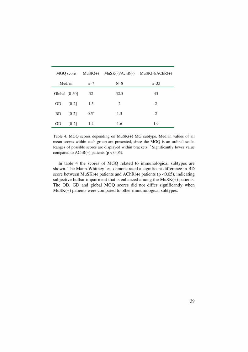
MGQ score
Median
MuSK(+)
n=7
MuSK(-)/AchR(-)
N=8
MuSK(-)/AChR(+)
n=33
Global [0-50] 32 32.5 43
OD [0-2] 1.5 2 2
BD [0-2] 0.5∗ 1.5 2
GD [0-2] 1.4 1.6 1.9
Table 4. MGQ scores depending on MuSK(+) MG subtype. Median values of allmean scores within each group are presented, since the MGQ is an ordinal scale.Ranges of possible scores are displayed within brackets. ∗ Significantly lower valuecompared to AChR(+) patients (p < 0.05).
In table 4 the scores of MGQ related to immunological subtypes areshown. The Mann-Whitney test demonstrated a significant difference in BDscore between MuSK(+) patients and AChR(+) patients (p <0.05), indicatingsubjective bulbar impairment that is enhanced among the MuSK(+) patients.The OD, GD and global MGQ scores did not differ significantly whenMuSK(+) patients were compared to other immunological subtypes.

40
Comparison of neurophysiological, muscle biopsy andhealth-related quality of life parameters in MuSK(+),AChR(+) and AChR(-) MG patients (Study IV)
MuSK-Ab presenceFive of the 14 AChR-Ab seronegative [AChR(-)] patients wereMuSK(+)/AChR(-) and five out of the 36 AChR-Ab seropositive [AChR(+)]patients were MuSK(+)/AChR(+). Patients 2, 8 and 9 who were initiallyAChR(+) had at the present analysis no detectable AChR-Ab, but were stillconsidered to belong to the AChR(+) subtype. None of the analyzedMuSK(+) and MuSK(-)/AChR(-) sera had detectable antibodies directedagainst titin or the ryanodine receptor.
Characteristic MuSK(+)(n=10)
MuSK(-)/AChR(-)(n=9)
MuSK(-)/AChR(+)(n=31)
Gender: female/male 7 / 3 8 / 1 29 / 2
Age (yrs) mean [range] 63.5 [46-78] 57.9 [40-84] 53.9 [30-73]
Disease duration (yrs)Mean [range]
16.6 [0.5-55] 16.7 [2-33] 24.8 [1-57]
Post-thymectomy (# of pat)
Thymoma
Hyperplasia
Normal thymus
Thymus inflammation
6
1
1
4
0
6
0
2
2
2
27
5
12
7
3
Pyridostigmine treatment(# of pat)
5 4 15
Immunosuppressive medi-cation (# of pat)
1 2 6
Table 5. Patient subtype characteristics. Immunosuppressive medication includedcortisone, azathioprine and sendoxan.

41
Clinical findings
MuSK(+)/AChR(+) patients (n=5)The severity of symptoms ranged from clinical remission (n=1) to severearm muscle atrophy (n=1). There was no correlation between disease gradeand level of MuSK-Ab or AChR-Ab. Patient 4 was AChR(+) at both occa-sions the analysis was performed, whereas the rest of the patients wereAChR(+) at only one examination. Four patients had undergone thymec-tomy, one had hyperplasia and three had a normal thymus. Patient 4 hadreceived immunosuppressive treatment (azathioprin) and patient 8 had cur-rent medication with azathioprin.
MuSK(+)/AChR(-) patients (n=5)Symptoms ranged from clinical remission (n=1) to severe bulbar involve-ment (n=1). Also in this group, the overall MuSK-Ab levels did not correlatewith the degree of symptoms. Out of the two patients in whom thymectomywas performed, one had a thymoma and one had a normal thymus. No pa-tient received any immunosuppressive treatment.
MuSK(-)/AChR(-) patients (n=9)The MGFA class ranged from pharmacological remission to mild general-ized weakness. Six patients were thymectomized, see table 5 for furthertreatment information.
MuSK(-)/AChR(+) patients (n=31)Disease severity ranged from remission (10 patients) to moderate general-ized disease. Twenty-seven patients (87%) had undergone thymectomy, seetable 5 for further details regarding treatment.
Neurophysiological findingsTable 6 displays the individual neurophysiological findings of MuSK(+)patients. For individual data of MuSK(+)/AChR(-) and MuSK(-)/AChR(+)patients, see table 1 in paper IV.
Three of five MuSK(+)/AChR(+) patients consented to neurophysiologi-cal examination. All had an abnormal decrement on RNS in the deltoid mus-cle and abnormal SFEMG findings in at least one of the examined muscles.Two patients had myopathic changes on QEMG, one severe and one slightchanges.
Four of the five MuSK(+)/AChR(-) patients consented to neurophysi-ological examination. Abnormal RNS was not detected, whereas all exam-

42
ined patients had abnormal SFEMG findings in either the deltoid or orbicu-laris oculi muscle. QEMG displayed slight to moderate myopathic pattern inthree patients (6, 7, 10; Fig. 1).
RNS was normal in all nine MuSK(-)/AChR(-) patients, whereas in sevenpatients SFEMG was abnormal. Slight to moderate myopathic pattern wasobserved in three patients.
Thirteen MuSK(-)/AChR(+) patients (42%) had abnormal decrement inthe deltoid and/or trapezius muscle. Overall, 24 patients (77%) in this sub-group had abnormal SFEMG in either muscle. Slight to moderate myopathicpattern was found in seven (23%) patients, four of which had abnormal neu-romuscular transmission.
Correlation of decrement, mean MCD and percentage ofblockings
Among the MuSK(+) patients, there was no correlation between mean MCDand percentage of blockings in the orbicularis oculi muscle (Fig.2). On thecontrary, mean MCD correlated to percentage of blockings both in MuSK(-)/AChR(-) patients (Spearman R=0.69; p < 0.05) and in MuSK(-)/AChR(+)patients (Spearman R= 0.85; p < 0.01).
In the deltoid muscle, the percentage of blockings correlated with meanMCD (Spearman R= 0.77; p < 0.05) in MuSK(+) patients, but not with thedecrement, i.e. in some cases where the decrement was normal, the SFEMGwas pathological. A similar picture was seen among the MuSK(-)/AChR(-)patients and a strong correlation between decrement, mean MCD and thepercentage of blockings (p < 0.01) was seen in MuSK(-)/AChR(+) patients.

43
Comparison of myopathic patternMyopathic EMG pattern was present in five (71%) examined MuSK(+) pa-tients, three (33%) MuSK(-)/AChR(-) patients and seven (23%) MuSK(-)/AChR(+) patients. In MuSK(+) patients, there was a significantly higherfrequency of myopathic EMG pattern, compared with MuSK(-)/AChR(+)patients (χ2 = 6.3; p=0.01). In two MuSK(+) patients (4 and 7) myopathywas accompanied by reduced objective muscle strength before exercise testin that particular muscle. In the two other patients, there was fatigue but noinitial weakness. Among the MuSK(-)/AChR(-) patients, there was fatigue inone patient and normal objective findings in the myopathic muscle. Amongthe MuSK(-)/AChR(+) patients, fatigue was found in the myopathic muscleof three patients, as detemined by QEMG.
Figure 1. QEMG showing moderate myopathy of the splenius capitis muscle inMuSK(+) patient 7. Size index –0.13. The interference pattern displayed early re-cruitment of MUPs and low envelope amplitude.

44
A) B) C)
Figure 2. SFEMG recording in the orbicularis oculi muscle in one MuSK(+) patient9. A) The trigger is set on the second potential. Mean MCD for the first potential is91 µs and for the third potential 107 µs. B) 10 consecutive traces are shown in rastermode. C) The ten traces superimposed.
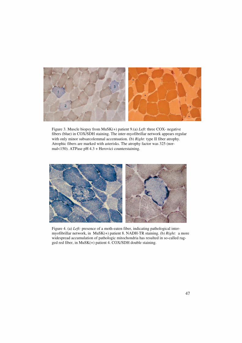
Morphological and mitochondrial findingsType II fiber atrophy was seen in four out of seven MuSK(+) patients (Fig.3b). In patient 4 there was marked type I fiber hypertrophy, presence of rag-ged red fibers (Fig. 4b) and almost no type II fibers at all. A pathologicalinter-myofibrillar network and several COX negative fibers were present inall MuSK(+) patients (Fig. 3a & 4a). No signs of inflammation were foundin any biopsies from MuSK(+) patients.
Among the nine MuSK(-)/AChR(-) patients, a pathological inter-myofibrillar network was seen in two patients, with COX negative fibersfound in all patients, although in three patients < 0.1 % (considered withinnormal limits), and ragged red fibers in three patients. Signs of inflammationwere observed in one patient.

45
Seven out of 26 MuSK(-)/AChR(+) patients had a pathological inter-myofibrillar network. COX negative fibers were present in 22 patients, threeof whom had ragged red fibers. Inflammatory cell infiltrates were detected inseven patients.
For individual data regarding all immunological subtypes, please see table1 in paper IV.
Mitochondrial DNA and POLG1 findingsMultiple mtDNA deletions (see article IV Fig. 2B) were found in 16 out of25 assayed muscle specimens (64 %). The frequency of COX negative fiberscorrelated with the presence of mtDNA deletions. The percentage of COXnegative fibers and multiple mtDNA deletions correlated significantly(p < 0.01). The mean age of patients with mtDNA deletions was 64 years(range 42 – 84 years) and 46 years (range 30 – 62 years) for patients withoutdeletions.
Multiple mtDNA deletions were detected in all tested serotypes, most fre-quently in MuSK(-)/AChR(-) patients, among whom six of eight (75 %)patients had deletions. MtDNA deletions were seen in three of five (60 %)MuSK(+)/AChR(-) patients and in seven of 12 (58 %) MuSK(-)/AChR(+)patients. The coding sequence of POLG1, which was analyzed in 15 patientswith mtDNA deletions, did not reveal pathogenic mutations. Nevertheless,specific polymorphisms were detected in a few of these patients.

46
Pat Age/Sex
MuSK-Ab
Max.AChR-Ab
(nM)
MGFAClass
DiseaseDuration
(yrs)
QEMGMyopathy(yes/no)
SFEMGOrb.oc/delt
COXneg
fibres( %)**
Musclebiopsyat/ ht
N= noat/ht
MtDNAdeletions
1 78F + 0 IIa 55 No N/Abn 1.7 Type II at Yes
2 62M + 4.0 0 1 n.p n.p n.p n.p n.p
3 69F + 0 0 15 n.p n.p n.p n.p n.p
4 67F + 75 IIIa 43 Yes N/Abn 1.0** Type I htType II ht
n.p
5 60F + 1.0 I 15 n.p n.p n.p n.p n.p
6 56F ++ 0 IIb 3 Yes N/Abn 0.3 Type II ht Yes
7 75M ++ 0 I 18 Yes Abn/N 0.9** Type I atType II at
Yes
8 47F +++ 40 IIa 11 N Abn/Abn 0.5 N No
9 46F +++ 3.0 IIa 4 Yes Abn/Abn 0.6 Type II at No
10 75M +++ 0 IVb 0.5 Yes Abn/Abn 1.1 Type II at n.p
Table 6. Comparison of clinical classification, neurophysiological and morphologi-cal features among MuSK(+) patients. Pat= patient; F= female; M= male; nM=nanomol/liter (normal<0.2 nM/L); MGFA= Myasthenia Gravis foundation ofAmerica; N= normal; Abn= abnormal; Orb.oc= orbicularis oculi muscle; delt= del-toid muscle; at= atrophy; ht= hypertrophy; MtDNA= mitochondrial DNA; n.p.=notperformed. ** Presence of ragged red fibers.
47
Figure 3. Muscle biopsy from MuSK(+) patient 9.(a) Left: three COX- negativefibers (blue) in COX/SDH staining. The inter-myofibrillar network appears regularwith only minor subsarcolemmal accentuation. (b) Right: type II fiber atrophy.Atrophic fibers are marked with asterisks. The atrophy factor was 325 (nor-mal<150). ATPase pH 4.3 + Herovici counterstaining.
Figure 4. (a) Left: presence of a moth-eaten fiber, indicating pathological inter-myofibrillar network, in MuSK(+) patient 8. NADH-TR staining. (b) Right: a morewidespread accumulation of pathologic mitochondria has resulted in so-called rag-ged red fiber, in MuSK(+) patient 4. COX/SDH double staining.

48
Extra discharges due to AChEI (Study V)A patient with severe oculobulbar weakness, MGFA class IVB, had a
high titer of MuSK-Ab (+++), and was initially treated with a moderate doseof pyridostigmine bromide (Mestinon) 120 mg three times daily. Duringthe initial motor neurography, extra discharges were observed after the mo-tor response at low-frequency stimulation. In some nerves the dischargesformed slow wave components with increasing intervals from 4 ms to 12 ms,with a duration of at least 50 ms, and with an amplitude of about one third ofthe CMAP. In other nerves there were irregular discharges of low amplitude,i.e., less than 0.5 mV, with durations of up to 100 ms. Both types of extradischarges disappeared at the second stimulus in a train of 3Hz stimulation.RNS showed a slight decrement of 13% in the anconeus muscle, but in theother examined muscles there was no decrement. SFEMG was abnormal inthe EDC and orbicularis oculi muscles.
After a methylprednisolone infusion, the patient developed a respiratorycrisis, resulting in the need for respiratory support and neck muscle support.When the patient recovered, a second neurophysiological examination wascarried out before and after the injection of 10 mg edrophonium intrave-nously. The last pyridostigmine dose was taken 6 hours prior to examination.After the injection, extra discharges followed the CMAP and there was dete-rioration with weakness in the neck and bulbar muscles. Approximately nineweeks and five days after the subsequent discontinuation of pyridostigminebromide, no extra discharges were seen following the CMAP and the patientdemonstrated improvement to MGFA class IIB.

49
Discussion
Disturbed neuromuscular transmission in the EDC inOMGThe first main objective of this study was to characterize the presence anddegree of disturbed neuromuscular transmission in a limb muscle in patientswith purely ocular myasthenic weakness. The first aim determined that thepresence of abnormal SFEMG in a limb muscle did not predict the subse-quent generalization of MG. This proved true even for clinically unaffectedmuscles, such as the EDC, which had a decreased safety margin of neuro-muscular transmission, e.g., abnormal jitter findings in the majority (75%) ofOMG patients. Neither the presence nor the degree of this abnormality wasrelated to a shift from ocular to generalized MG in patients with purely ocu-lar involvement. Not only was the degree of jitter comparable between thetwo outcome groups, there was also no difference in the degree of impulseblockings.
It is intriguing that patients with generalized weakness as well as patientswith only ocular weakness have the same amount of blockings in an armmuscle, since the degree of blocking determines whether clinical fatigueoccurs. One possible explanation is that a lower degree of blocking in EDCin the ocular group gives merely subclinical limb fatigability. Thus, an ab-normal electrophysiological finding in a muscle outside the facial area is notpredictive of later clinical generalization and therefore clinical classificationmust be re-evaluated each time the MG patient is examined in the neurologyclinic.
Further, while SFEMG abnormalities are common in the orbicularis oculimuscle and useful for diagnosis, they did not correlate to the degree of ocu-lar dysfunction, nor did they reflect the general severity of MG. This factwas recognized earlier by the MGFA Task Force, resulting in the possibilityto include patients with only slight ocular dysfunction in the remission clas-sification (Jaretzki et al., 2000). Early recognition of those patients who willsubsequently acquire generalized MG would be important from a therapeuti-

50
cal point of view. However, the current knowledge for this group of patientsis insufficient to allow for this characterization by SFEMG.
Health-related quality of life in Swedish MG patientsThe hrQoL may be negatively influenced in MG patients by the persistent
and fluctuating symptoms of muscle weakness. MG is a chronic disorder andalthough there are effective medications, most patients do not reach the sameprior to MG level of function (Ochs et al., 1998). To achieve patient-orientedevaluations of MG patients in Sweden, it is essential to have a validated dis-ease-specific questionnaire on a national basis. While a number of hrQoLinstruments, such as the SF-36, have been developed for the general popula-tion, they are unlikely to detect small, clinically important changes in a par-ticular disorder (Guyatt et al., 1986). Therefore, the MGQ will provide animportant measure of the effects of a specific treatment on hrQoL.
The observed correlation in Swedish MG patients between the MGQ andSF-36, as well as clinical assessment, was comparable to the Italian version.The MGQ obtained an excellent internal consistency of 0.91 (Cronbach´salpha). Suggested levels of reliability are 0.70 or greater for scales used ingroup-level analyses and 0.90 or greater for scales used in decisions at theindividual level (Nunnally JC, 1994). When compared to normative datafrom the Swedish population (Sullivan et al., 1995), physical aspects ofhrQoL is affected. However, the patient sample included mostly patientswith mild MG and an extended study including more patients with moderateand severe MG should be conducted to cover the entire severity of the dis-ease. For Swedish MG patients this brings the opportunity to participate ininternational clinical trials.
There tends to be a close link between a person’s perception of theirmental and physical energy and their general feeling of well being (Wood,1990). The significant correlation in study II of vitality as measured by SF-36 with global MGQ score, general domain score and bulbar domain scoreindicate that the subjective energy level increases when generalized and bul-bar weakness decrease. This supports one earlier study where a notable inter-ference in perceived energy, as measured by the vitality domain of SF-36,was detected in patients with generalized myasthenic weakness (Paul et al.,2000). Fatigue increases over the day when energy levels for most peoplefall and many MG patients state that their fatigue worsens during stressfulperiods; thus, these two measures seem to be linked. However, patients withMG are not affected by greater mood disturbances than are healthy controlsubjects.
51
Correlation between disturbed neuromusculartransmission and health-related quality of lifeSecondly, we found there was a correlation between disturbed neuromuscu-lar transmission and hrQoL. A proximal muscle, such as the deltoid muscle,seems important to examine with SFEMG in MG patients, since the degreeof abnormal neuromuscular transmission correlated well with the patient-oriented global MGQ and generalized domain scores. Changes in jitter in asentinel muscle also correlated with the changes in disease severity for mostpatients (Sanders, 2002); therefore, SFEMG in the deltoid may serve as atool for therapy monitoring, as well as for diagnostic purposes. However,testing of a single muscle is not enough for diagnosis and follow-up of MG.A multidimensional evaluation of MG patients is important in everydayclinical practice, as well as in future clinical trials.
MuSK antibodies and AChR antibodies may coexistMuSK-Abs were present in 36% of the examined AChR(-) patients,
which is consistent with previous studies (Evoli et al., 2003)(Hoch et al.,2001)(Sanders et al., 2003). However, the discovery of the presence ofMuSK-Abs in 14% of the examined AChR(+) patients is noteworthy. Whilea "MuSK-Ab" had been reported in AChR(+) MG by Ohta (Ohta et al.,2004), this antibody was subsequently found to be directed against alkalinephosphatase (Ohta et al., 2005). Thus, our report is the first of its kind toshow that MuSK-Ab and AChR-Ab do coexist in a group of MG patients.Additionally, MuSK-Abs were detected in OMG, which supports a previ-ously reported case study (Caress et al., 2005). Patients 2, 8 and 9 who wereAChR(+) at the initial diagnostic antibody analysis proved to beMuSK(+)/AChR(-) at the previous analysis. One possible explanation forthis is epitope-spreading, i.e. that the immunological attack against AChRhas been expanded to include MuSK. Epitope spreading is an establishedphenomenon in animal models of mediated autoimmune diseases (Vander-lugt and Miller, 2002). In experimental autoimmune MG in rabbit models,the autoimmune attack is initially directed against the main immunogenicregion of the AChR. However, through the course of the immune response, asignificant attack is also mounted against less immunogenic epitopes (Vin-cent et al., 1998). There is also information from human MG suggesting thepresence of intermolecular epitope spreading. Screening of sera fromAChR(+) patients has demonstrated rapsyn antibodies in 15% of patients(Agius et al., 1998),

52
Primary factors contributing to the muscular weaknessand fatigue in MuSK(+) patients
Neuromuscular transmissionThe second principal objective was to characterize the main factors con-
tributing to the muscular weakness and fatigue in MuSK(+) patients. Thisincluded a spectrum of features from the detailed neurophysiological picture,as well as the microscopic view, which focused on the histopathology of themuscle fibers, as detected in muscle biopsies, to the patient´s hrQoL. Theseparameters in MuSK(+) patients were then compared to the other major im-munological types of MG including AChR(+) and AChR(-) patients.
Regarding neurophysiological findings in MuSK(+) patients, our data sug-gested that MuSK(+) patients have the same degree of neuromuscular trans-mission defect as AChR(+) patients in proximal muscles, although withsomewhat different distribution. The EDC muscle has been reported to re-veal less abnormality in MuSK(+) patients than in AChR(+) patients (Farru-gia et al., 2006a; Nemoto et al., 2005; Stickler et al., 2005). Thus, diagnosticexaminations of patients with MuSK-Ab should be performed in affectedmuscles, since muscle weakness may be focal. The normal RNS findings inall of the MuSK(+)/AChR(-) patients are intriguing. Decrement is frequentlynormal in patients with OMG and in generalized MG with mild weakness oflimited distribution (Sanders, 2002). Thus, one explanation is that these pa-tients mainly had weakness restricted to the ocular or bulbar muscles, whichare not readily tested with RNS. Nevertheless, we cannot exclude that RNSexamination would have been abnormal in the facial muscles, regarding thepredominant involvement of these muscles. The orbicularis oculi muscle wasrecently shown to have abnormal decrement in 86% of MuSK Ab-positivepatients (Oh et al., 2006).
The normal SFEMG findings in about 23% of MuSK(-)/AChR(+) patientsmay depend on the fact that many patients in this subgroup were in remis-sion and had immunosuppressive treatment since many years. It is knownthat when MG improves, jitter typically continues to fall toward normal(Howard and Sanders, 1981). If patients in remission or with minimal mani-festations were excluded, then jitter was abnormal in 92% of these patients.Furthermore, overall the deltoid displayed the most pronounced decrement.

53
Health-related quality of life in MuSK(+) patientsConcerning hrQoL, MuSK(+) patients indicated significantly more im-
pairment in the MGQ bulbar domain compared with MuSK(-)/AChR(+)patients. This finding reflects the previously reported fact that MuSK(+)patients tend to suffer from bulbar impairment to a larger extent thanAChR(+) patients (Evoli et al., 2003). Nevertheless, a global MGQ scoredid not differ significantly between MuSK(+) and MuSK(-)/AChR(+) pa-tients. One explanation for the overall higher global MGQ score in theMuSK(-)/AChR(+) patients may be that a higher number in this group wasin clinical remission and thus should not indicate as much impairment.
Therapy considerations in MuSK(+) patientsThere were differences in therapy between immunological subgroups. In
the group of MuSK(+)/AChR(-) patients, no patient received immunosup-pressive treatment, although two patients had undergone thymectomy. Over-all, thymectomy was performed in 78% of patients, whereas immunosup-pressive therapy was only applied in 18% (present) and 42% (previous) ofpatients. The high thymectomy rate may reflect that most patients in thisstudy were women and that removal of the thymus is the general recom-mended therapy for women with early onset MG. One MuSK(+)/AChR(-)patient was found to have a thymoma and improved after thymectomy. Nocase of thymoma in MuSK(+) MG has previously been described. On thecontrary, the absence of hyperplastic changes to the thymus and the apparentlack of benefit of thymectomy has been reported in these patients (Lauriolaet al., 2005). This may be the reason for the absence of the thymoma associ-ated titin- and ryanodine receptor antibodies in these patients. MuSK-Ablevels have been shown to often decrease after immunosuppression but notafter thymectomy (Bartoccioni et al., 2006). This latter finding is in contrastto the known AChR-Ab reduction after thymectomy in AChR(+) patients, inwhom the hyperplastic thymus is considered the possible site of immuniza-tion against AChR and a major source of specific Abs (Marx et al., 1997;Vincent, 2002).
MuSK-Ab levels have been shown to correlate with disease status in mostpatients (Bartoccioni et al., 2006). In contrast to these findings, there was noobvious correlation between overall antibody concentration and disease se-verity in our series of MuSK(+) patients, albeit the population is small. Themost probable explanation for this is that the Swedish patients were quitestable in their MG and more fluctuations in Ab-levels have been noticed inpatients with a stable MG status (Bartoccioni et al., 2006).

54
Myopathy in MuSK(+) patientsA myopathic EMG pattern was found in 32% of all examined MG patients,comparable to 19% of patients in an earlier MUP analysis of MG patients(Somnier and Trojaborg, 1993). QEMG myopathy was significantly morecommon in MuSK(+) patients than in MuSK(-)/AChR(+) patients. Never-theless, since a myopathic pattern is present in other immunological subtypesof MG as well, it is not specific for the MuSK(+) subtype.
The myopathic pattern is not likely to be due only to neuromuscularblocking since the presence of blockings does not necessarily give rise to amyopathic pattern. Also, in some of the patients with myopathy diagnosedusing QEMG, the degree of blockings was not sufficiently high to signify amyopathy-like EMG. It does not seem likely that the myopathic picture de-pends on major structural damage of the muscle fibers. For example, musclepathology did not reveal any muscle fiber necrosis and we noticed only sometype II fiber atrophy. Further, the diameter of the muscle fibers was normalwith minimal variation, and there were no signs of muscle fiber grouping.No denervation activity (fibrillations or positive sharp waves) was foundusing EMG and signs of myositis were absent from muscle biopsy. In thoseMuSK(+) patients with QEMG myopathy in a proximal muscle, there wasconcomitant weakness in two patients and concomitant fatigue in the twoother patients. However, excluding patient 4, there was not a distinct corre-lation between the degree of weakness and the degree of myopathy.
A few patients in both MuSK(+) and AChR(+) groups have been reportedto present with visible tongue atrophy although magnetic resonance imagingevidence of atrophy of individual facial muscles is only significant inMuSK(+) patients (Farrugia et al., 2006b). In that report, however, muscleatrophy correlated with long duration of steroid treatment (> 40 mg AD).Thus, on the basis on the current results as well as previous reports, the dif-ferences in myopathy and muscle atrophy between MuSK(+) and AChR(+)patients regarding atrophy appear to be mainly quantitative in some of thepatients.
Pathogenic role of MuSK antibodiesIt has been questioned whether MuSK-Abs are responsible for the weak-
ness found in AChR(-) MG patients (Selcen et al., 2004; Vincent et al.,2005). Nevertheless, MuSK-Abs were recently shown to significantly reduceAChR clustering at the neuromuscular junctions (Shigemoto et al., 2006).MuSK(+) sera from patients with severe MG also induce inhibition of prolif-
55
eration in TE671 muscle cells. The accompanied down regulation of thepostsynaptic AChR subunits as well as rapsyn, could be relevant to the de-fective neuromuscular transmission (Boneva et al., 2006).
It remains unknown, though, why oculobulbar muscles are most affectedin MuSK(+) MG patients. One reason may be the substantial variability be-tween muscles in the shape of the neuromuscular junctions and the differ-ence of muscle fibers in respect to physiology, metabolism and patterns ofgene expression (Burke, 1998). At the molecular level, developing and adultskeletal muscles can be subdivided into two distinct categories: Fast Syn-apsing, e.g., intercostal muscles, and Delayed Synapsing muscles, e.g., ster-nomastoid and diaphragm muscles. The two classes of muscles differ in thefocal clustering of AChRs and in the alignment of the pre-synaptic nervewith focal AChR clusters (Pun et al., 2002). These properties are believed toplay a prominent role in defining the progress of neuromuscular junctionformation during synapse development and in the maintenance of adult mus-cle. Although no data is available, it may be speculated that neck/bulbarmuscles may be more susceptible to defective nerve-muscle signaling due tothe above-proposed differences, since they may be Delayed Synapsing mus-cles.
Muscle pathology in MuSK(+) patients and mitochondrialdefects in MG
Muscle pathology displayed type II fiber atrophy, histological presence ofmitochondrial dysfunction, such as moth-eaten and ragged red fibers, andCOX negative fibers. No inflammation or dystrophic changes were seen inMuSK(+) patients. Type II fiber atrophy is an unspecific finding, since itmay be associated with conditions such as the disuse of muscles and aging.In some MG patients, individual muscle fibers may be totally blocked, andtherefore inactive, giving rise to disuse atrophy. The presence of COX nega-tive fibers raises the question whether the myopathy is (1) a primary effect ofMuSK-Ab, (2) secondary to the effect of MuSK-Ab on the motor end plateor (3) a concomitant phenomenon to the production of MuSK-Ab with an-other underlying disorder.
The possible co-existence of a mitochondrial myopathy and disturbedneuromuscular transmission is intriguing, since muscle weakness and fatiga-bility similar to MG occur in certain mitochondrial myopathies. One exam-ple is late-onset mitochondrial myopathy (Johnston et al., 1995) in which thenumber of COX negative and/or ragged-red myofibers is increased similarly

56
as in our patients. The absence of COX activity in myofibers is usuallycaused by deletions in mtDNA, which remove loci that encode for threesubunits of COX, i.e. the complex IV of the respiratory chain. Mutations inthe nuclear POLG1 gene (which encodes polymerase gamma 1, the enzymewhich participates in mtDNA processing) is a common cause of multipledeletions in mtDNA.
In our study, mitochondrial defects were not associated with any particu-lar antibody, since the abnormalities were observed in all immunologicalMG subtypes. In 64% of our investigated samples, multiple mtDNA dele-tions were present. Progressive external ophthalmoplegia and muscle weak-ness with exercise intolerance are the most common clinical pictures associ-ated with such deletions (Suomalainen and Kaukonen, 2001). MtDNA dele-tions become more frequent with aging, although not as prominent as in ourMG patients. The cause of the multiple mtDNA deletions in our patients,remains unknown since POLG1 mutations were not found in these musclebiopsies (Kajander et al., 2000).
Adverse effects of pyridostigmine treatment in MuSK(+) MG:neurophysiological indications of AChEI side effects
The effect of pyridostigmine treatment in MuSK(+) patients may rangefrom mild benefit to ineffectiveness and side effects including cramps, my-algia (Rostedt and Stalberg, 2004), muscle fasciculations and other choliner-gic side effects (Evoli et al., 2003). Our hypothesis was that the neuromus-cular transmission defect found in MuSK(+) patients is due to abnormalmorphology of the receptor area, rather than reduced receptor sensitivity, asin AChR(+) MG. Based on this assumption, AChEI should not have a bene-ficial effect on neuromuscular transmission. Extra discharges are usuallyseen after an overdose of acetylcholinesterase inhibitors in MG. The pres-ence of extra discharges in MuSK(+) patients may be a useful neurophysi-ological indicator of adverse effects of AChEI treatment.

57
Conclusions
In conclusion, our results suggest the following:
1) Disturbed neuromuscular transmission in a muscle outside of the fa-cial area in OMG is not predictive of subsequent generalization.
2) The presence of neuromuscular transmission defects in MG does notalways correlate with the weakness and fatigability found in the ex-amined muscles.
3) The correlation of SFEMG findings in a proximal muscle and the pa-tient-oriented findings adds new impact to the neurophysiologicalevaluation of patients with MG.
4) A myopathic EMG pattern in MG is significantly more common inMuSK(+) patients, although it is not a feature found exclusively inthis immunological subtype. Further, the degree of weakness/fatiguedoes not correlate with the degree of myopathy overall. The fre-quently detected myopathic pattern on EMG in MuSK(+) may be as-sociated with the concomitant mitochondrial abnormalities.
5) A neuromuscular transmission defect is the primary cause of muscleweakness in the majority of MuSK(+) patients, as well as in AChR(+)MG.
6) Since AChEI do not have a noticeable beneficial effect in MuSK(+)patients, an abnormal receptor morphology which cannot be restoredby AChEI is suggested. This abnormality is proposed to cause the un-derlying defect of neuromuscular transmission.

58
Summary in Swedish- sammanfattning påSvenska
Myasthenia Gravis (MG) är en autoimmun sjukdom som beror på antikrop-par riktade mot olika komponenter på motorändplattan. Resultatet blir stördneuromuskulär transmission. Symptomen utgörs av en onormal uttröttbarhetav skelettmuskler i armar, ben, ansikte och nacke. Vissa patienter har symp-tom endast i ögonmuskler. Denna form kan generaliseras till att innefattaäven andra muskler. Behandlingen är delvis symptomatisk med acetylcholi-nesterashämmare (pyridostigmin), vilket förbättrar signaleringen mellan nervoch muskel genom att öka överlevnaden av signalsubstansen acetylcholin.Bromsande behandling består av olika immunosuppressiva mediciner såsomcortison och andra terapier såsom tymektomi, plasmaferes och intravenösaimmunogloblulier.
Den vanligaste formen av MG, som drabbar ca 80%, orsakas av an-tikroppar mot acetylcholin receptorn (AChR). Av de ca 20% som saknardessa antikroppar har antikroppar identifierats mot muskelspecifikt tyrosin-kinas (MuSK) hos ca 40%. Patienter med MuSK-antikroppar har rapporte-rats ha mer uttalade symptom kring ansikte och nacke och fokal distributionav störd neuromuskulär transmission. Hos dessa patienter har även myopatirapporterats. Andra muskelantikroppar, riktade mot rapsyn, ryanodinrecep-torn och titin, existerar också, framförallt om patienten har ett samtidigt ty-mom.
Diagnosen vid MG görs med hjälp av klinisk neurologisk undersökninginkluderande uttröttbarhetstester, serumanalys för antikroppar och neurofy-siologisk undersökning. Singel fiber elektromyografi (SFEMG) är den käns-ligaste diagnostiska undersökningen och visar hos MG patienter en sänktsäkerhetströskel för neuromuskulär transmission, sk jitter. Vid högre grad avtransmissionstörning uppkommer impulsblockeringar, vilket ger klinisksvaghet. Vid sidan av SFEMG utförs också repetitiv nervstimulering vid 3Hzstimulering för att se en nedgång i amplitud hos muskelsvaret som är typiskvid MG.

59
Studie IFrågeställningen gällde huruvida ökat jitter i en underarmsmuskel (EDC) hospatienter med enbart okulär MG kan förebåda en senare klinisk generalise-ring av myastenin. Det vore av stort kliniskt intresse att hitta en sådan pre-diktor för att kunna sätta in aggressiv immunosuppressiv behandling i etttidigt skede. Fynden var att SFEMG i EDC ej kan användas för att förutbe-stämma vilka patienter som utvecklar generaliserad sjukdom trots att pati-enter med enbart ögonsymptom ofta visade störd neuromuskulär transmis-sion i extremitetsmuskulatur.
Studie IIDetta arbete syftade till att översätta och adaptera det för MG sjukdomsspe-cifika italienska livskvalitéformuläret Myasthenia Gravis Questionnaire(MGQ) till svenska förhållanden. Den internationellt accepterade metodenför validering användes och vi lyckades ta fram ett svenskt formulär som nuframöver kan användas för att utvärdera behandling vid MG och för att kun-na inkludera även svenska patienter i kliniska studier, där den subjektivalivskvalitéskattningen idag är ett krav.
Studie IIISyftet med denna studie var att korrelera patientens subjektiva välbefinnande(poäng på livskvalitéformulären SF-36 och MGQ) med subklinisk sjuk-domsgrad (mätt som störd neuromuskulär transmisison med metodernaSFEMG och RNS). Resultaten visade att graden av störd neuromuskulärtransmission i överarmsmuskulatur, som ofta är involverad vid MG, korrele-rar väl med generellt välbefinnande angivet som poäng i MGQ. Detta med-för en ny dimension för neurofysiologiska undersökningar vid MG; inte baraför diagnosen utan även som uppföljningsverktyg.
Studie IVMålet var att karakterisera de elektrofysiologiska fynden avseende myopa-ti/neuropati samt muskelbiopsifynden, framförallt mitokondriefunktionen,hos olika antikroppsubtyper av MG. Detta gjordes för att besvara frågan:varför är patienter med MuSK-antikroppar svaga? Vi fann förekomst av

60
både MuSK-antikroppar och AChR-antikroppar hos 14% av de undersöktaMG patienterna, något som tidigare inte rapporterats. Myopatimönster påEMG undersökning av proximal muskulatur var överrepresenterat hosMuSK-patienter jämfört med de patienter som saknade dessa antikroppar,men patienter från alla immunologiska subtyper hade mild myopati samt oftaförekommande mitokondriella avvikelser. MuSK-positiva patienter hade likamycket jitter som AChR-positiva patienter, talande för att svagheten hosMuSK-patienter beror på störd neuromuskulär transmissison.
Studie VDetta delarbete beskriver i detalj de extraurladdningar i motoriska nerver videlektrisk stimulering som noterades hos en patient med MuSK-antikropparoch mestinonbehandling. Detta stödjer den hypotes som finns om att MuSK-patienter är mer känsliga för medicinering med acetylcholinesterashämmareoch oftare utvecklar biverkningar.
61
Acknowledgements
This work was carried out at the Department of Clinical Neurophysiology,Uppsala University Hospital, Uppsala, Sweden. Collaborations were per-formed at the MG Clinic of Duke University Medical Center, Durham, USAand the Institute of Neurology, Catholic University, Rome, Italy. I wouldlike to thank the staff of the department and university, colleagues andfriends in Sweden, Italy and in the USA, for their support and contribution.I wish to express my sincere gratitude especially to:
Erik Stålberg- my supervisor. Thank you for everything! If it wasn´t foryou I would never have gone into neurophysiology and not been the kind ofdoctor I am today. I can never thank you enough for all the things you havetaught me about SFEMG, research and life. I always learn something newfrom you and I look forward to our future projects. You are an amazinglyenthusiastic person and a good friend, who ALWAYS have time and energyto answer questions and provide great ideas for every occasion- whereveryou are! We haven’t solved the entire mystery about MG yet…
Hannu Kalimo- my co-supervisor. Thank you for all the time and effort youput on interpretation of the muscle biopsies and for teaching me the musclebiopsy surveyor. Your great sense of humor enlightened these moments andtaught me some valuable Finnish jokes.
Colleagues at the department of Clinical Neurophysiology:Roland Flink, Arne Sandberg, Tomas Winkler, Karin Edebol EegOlofsson, Hans Axelson, Lars Larsson and Pirkko Hynninen - for yourgenuine support, interesting discussions and important advice.Margareta Grindlund and Lena Eriksson: thank you for your great helpwith the repetitive nerve stimulation as well as all “practicalities” and yoursense of humour. You really lighted up the long hard days!Susanne Östgren “Sussie”: thank you so much for your helpfulness andalways positive attitude during the muscle biopsies. I could not have donethem without you!!Peo Fällmar- your technical support has made the environment in the lab alot easier for a “non-technical” person…☺David Ullström- thanks for all your help with uncooperative computers, thefront picture, mp3 files and an outstanding knowledge about all zip, jpg, pdf,rtf etc etc.
62
Catarina Färnstrand- for positive thinking and always being so supportive.Olga Manriquez- for enlightening the sometimes late evenings.The administrative staff and secretaries of the department and institution:Karin Mineur, Gunilla Thornander, Gun Westerlund, Lena Petterssonand Anki Gustafsson- for administrative help.
Italian colleagues and friends.Luca Padua- Grazie per tempo a Roma! Thanks for the lessons about qual-ity of life, mixed with Friday research discussions with dinner, wine andcigars.Pietro Caliandro- Thank you for arranging a wonderful stay in Rome andfor explaining the Roman lifestyle. “When in Rome, do as the Romansdo…” Yes, I still mix spaghetti with meatballs and ketchup☺Irene Aprile, Constanza Pazagglia and everyone in the Rome EMG lab forproviding great atmosphere in the 6 square feet office and teaching me howto make REAL pasta à la carbonara.Emanuela Bartoccioni, Flavia Scuderi and Mariapaola Marino- “theMuSK-gang” - thank you for helping with the MuSK antibody analysis andfor great Italian pizza.
American collaborators and colleagues:Donald B Sanders and Janice Massey. Thank you for teaching me how toexamine and treat MG patients in the best way. I admire your clinical skillsand your hospitality during the stay in Durham.Charlotte, Kevin, Bernadette and everyone in the Duke MG Clinic for agreat time in Durham.
Technicians at the department of Neuropathology:Maude Olofsson and Monica Sternesjö - for great help with musclespecimens and for many coffee breaks with cookies.
Colleagues and nurses at the Neurology Department in Uppsala, especially:Håkan Askmark- for relevant clinical advice.Inga-Lena, Eva-Lena, Lillemor, Gertrud and Susanne at the departmentof neurology- for great help with the blood samples for MuSK antibodyanalysis and for running with them to the famous freezer ☺
Collaborators in Helsinki, Finland:Kati Ahlqvist and Anu Vartiovaara- for cooperation on the mitochondrialDNA analysis.
Friends:Holly Norman-for always having time to translate, being SO positive and atrue friend. You are an amazing person.
63
Sara and Karolina-thanks for many happy times and your constant supportduring these years and all through the LÄFO period.The “PAX-3 gang”: Josefine, Alina, Shaman and Anna-Karin. Thanks forfun discussions, dinners and great advice about the dress☺.The “Eriksberg-gang”: Bec and Pernilla- for long walks, fika, running and“baby talk”…
My family:Ida- for sharing many wonderful moments through the years, being a loyalsister and never giving up on teaching me how to cook…Margaretha- Thank you for always believing in me and being my source ofinspiration! Without you I would not have been who or where I am today.Your never ending support and encouragement has meant everything to me.Karl Erik-Thanks for teaching me how to always be optimistic and to getthe true “fighting spirit” both on and off the tennis court. It has helped me alot in the hard academic world ☺Gertrud- thanks for always being there for me and Ida during our childhoodand for the summers in Bordsjö.Kristiina, Aime and Eduard- thanks for embracing me into your familyand for great times both in Estonia and Uppsala.
Tanel- I am truely grateful for all your patience and support, through goodand bad times! Thank you for compromising, for working evenings andnights in order for me to finish this, and for putting up with my energeticlife-style!☺… You make me a calmer and happier person and I love youendlessly!Olivia-Your smiles bright up the day and you have taught me what the “bigthings” are in life. Being a mother to you is the greatest thing I could everwish for!
64
References
Aarli, J.A., Stefansson, K., Marton, L.S., Wollmann, R.L., 1990. Pa-tients with myasthenia gravis and thymoma have in their seraIgG autoantibodies against titin. Clin Exp Immunol 82, 284-288.
Agius, M.A., Zhu, S., Kirvan, C.A., Schafer, A.L., Lin, M.Y., Fair-clough, R.H., Oger, J.J., Aziz, T., Aarli, J.A., 1998. Rapsynantibodies in myasthenia gravis. Ann N Y Acad Sci 841, 516-521.
Ajtai, B., Fine, E.J., Lincoff, N., 2004. Pupil-sparing, painless com-pression of the oculomotor nerve by expanding basilar arteryaneurysm: a case of ocular pseudomyasthenia. Arch Neurol 61,1448-1450.
Araki, S., Uchino, M., Kumamoto, T., 1987. Prevalence studies ofmultiple sclerosis, myasthenia gravis, and myopathies in Ku-mamoto district, Japan. Neuroepidemiology 6, 120-129.
Barohn, R.J., McIntire, D., Herbelin, L., Wolfe, G.I., Nations, S.,Bryan, W.W., 1998. Reliability testing of the quantitative my-asthenia gravis score. Ann.N.Y.Acad.Sci. 841, 769-772.
Bartoccioni, E., Marino, M., Evoli, A., Ruegg, M.A., Scuderi, F., Pro-venzano, C., 2003. Identification of disease-specific autoanti-bodies in seronegative myasthenia gravis. Ann.N.Y.Acad.Sci.998, 356-358.
Bartoccioni, E., Scuderi, F., Minicuci, G.M., Marino, M., Ciaraffa, F.,Evoli, A., 2006. Anti-MuSK antibodies: correlation with my-asthenia gravis severity. Neurology 67, 505-507.
Ben Yaou, R., Laforet, P., Becane, H.M., Jardel, C., Sternberg, D.,Lombes, A., Eymard, B., 2006. [Misdiagnosis of mitochon-drial myopathies: a study of 12 thymectomized patients]. RevNeurol (Paris) 162, 339-346.
Bennet, A.E., Cash, P.T., 1942. Curare as a diagnostic test test formyasthenia gravis. Curarization an etiologic clue in the dis-ease. Trans Am Neurol Assoc, 102-106.
65
Besser, R., Gutmann, L., Dillmann, U., Weilemann, L.S., Hopf, H.C.,1989. End plate dysfunction in acute organophosphate intoxi-cation. Neurology 39, 561-567.
Boneva, N., Frenkian-Cuvelier, M., Bidault, J., Brenner, T., Berrih-Aknin, S., 2006. Major pathogenic effects of anti-MuSK anti-bodies in myasthenia gravis. J Neuroimmunol 177, 119-131.
Brooke, M.H., Engel, W.K., 1969. The histographic analysis of humanmuscle biopsies with regard to fiber types. Neurology 19, 221-233.
Burke, R.E., 1998. Motor units: anatomy, physiology, and functionalorganization, Handbook of physiology - The nervous systemII, pp. 345-350.
Caress, J.B., Hunt, C.H., Batish, S.D., 2005. Anti-MuSK myastheniagravis presenting with purely ocular findings. Arch.Neurol 62,1002-1003.
DeChiara, T.M., Bowen, D.C., Valenzuela, D.M., Simmons, M.V.,Poueymirou, W.T., Thomas, S., Kinetz, E., Compton, D.L.,Rojas, E., Park, J.S., Smith, C., DiStefano, P.S., Glass, D.J.,Burden, S.J., Yancopoulos, G.D., 1996. The receptor tyrosinekinase MuSK is required for neuromuscular junction formationin vivo. Cell 85, 501-512.
Desmedt, J.E., 1973. The neuromuscular disorder in myasthenia gra-vis. I. Electrical and mechanical responses to nerve stimulationin hand muscles. Karger,Basel. 1st, 241-304.
Dirr, L.Y., Donofrio, P.D., Patton, J.F., Troost, B.T., 1989. A false-positive edrophonium test in a patient with a brainstem glioma.Neurology 39, 865-867.
Drachman, D.B., Adams, R.N., Josifek, L.F., Pestronk, A., Stanley,E.F., 1981. Antibody-mediated mechanisms of ACh receptorloss in myasthenia gravis: clinical relevance. Ann N Y AcadSci 377, 175-188.
Dubowitz, V., 1985. Muscle biopsy, a practical approach.Baiilire.Tindall,London.
Ekstedt, J., Stålberg, E., 1963. A method of recording extra-cellularaction potentials of single muscle fibres and measuring theirpropagation velocity in voluntarily activated human muscle.Bull Am Assoc EMG 10, 16-16.
Ekstedt, J., Stålberg, E., 1975. Single muscle fibre electromyographyin myasthenia gravis. Studies in Neuromuscular diseases, 157-161.

66
Elmqvist, D., Hofmann, W.W., Kugelberg, J., Quastel, D.M.J., 1964.An electrophysiological investigation of neuromuscular trans-mission in myasthenia gravis. J Physiology (Lond) 174, 417-434.
Engel, A.G., Lambert, E.H., Howard, F.M., 1977. Immune complexes(IgG and C3) at the motor end-plate in myasthenia gravis: ul-trastructural and light microscopic localization and electro-physiologic correlations. Mayo Clin Proc 52, 267-280.
Engel, A.G., Tsuyihata, M., Lindstrom, J.M., Lennon, V.A., 1976.The motor end-plate in myasthenia gravis and in experimentalautoimmune myasthenia gravis. A quantitative ultrastructuralstudy. Ann N Y Acad Sci 274, 60-79.
Evoli, A., Tonali, P.A., Padua, L., Monaco, M.L., Scuderi, F., Batoc-chi, A.P., Marino, M., Bartoccioni, E., 2003. Clinical corre-lates with anti-MuSK antibodies in generalized seronegativemyasthenia gravis. Brain 126, 2304-2311.
Eymard, B., Morel, E., Dulac, O., Moutard-Codou, M.L., Jeannot, E.,Harpey, J.P., Rondot, P., Bach, J.F., 1989. [Myasthenia andpregnancy: a clinical and immunologic study of 42 cases (21neonatal myasthenia cases)]. Rev Neurol (Paris) 145, 696-701.
Farrugia, M.E., Kennett, R.P., Newsom-Davis, J., Hilton-Jones, D.,Vincent, A., 2006a. Single-fiber electromyography in limb andfacial muscles in muscle-specific kinase antibody and acetyl-choline receptor antibody myasthenia gravis. Muscle Nerve33, 568-570.
Farrugia, M.E., Robson, M.D., Clover, L., Anslow, P., Newsom-Davis, J., Kennett, R., Hilton-Jones, D., Matthews, P.M., Vin-cent, A., 2006b. MRI and clinical studies of facial and bulbarmuscle involvement in MuSK antibody-associated myastheniagravis. Brain 129, 1481-1492.
Gautam, M., Noakes, P.G., Mudd, J., Nichol, M., Chu, G.C., Sanes,J.R., Merlie, J.P., 1995. Failure of postsynaptic specializationto develop at neuromuscular junctions of rapsyn-deficientmice. Nature 377, 232-236.
Gilchrist, J., 1992. Single fiber EMG reference values: a collaborativeeffort. Muscle and Nerve 15, 151-161.
Guthrie, L., 1903. Myasthenia gravis in the seventeenth century. .Lancet 1, 330-331.
Guyatt, G.H., Bombardier, C., Tugwell, P.X., 1986. Measuring dis-ease-specific quality of life in clinical trials. CMAJ. 134, 889-895.

67
Hall, Z.W., Sanes, J.R., 1993. Synaptic structure and development: theneuromuscular junction. Cell 72 Suppl, 99-121.
Harvey, A.M., Masland, R.L., 1941. The electromyogram in myasthe-nia gravis. Bull Johns Hopkins Hosp 48, 1-13.
Heilbronn, E., Mattsson, C., Stålberg, E., Hilton-Brown, P., 1975.Neurophysiological signs of myasthenia gravis in rabbits afterreceptor antibody development. J Neurological Sciences 24,59-64.
Heinemann, S., Bevan, S., Kullberg, R., Lindstrom, J., Rice, J., 1977.Modulation of acetylcholine receptor by antibody against thereceptor. Proc Natl Acad Sci 74, 3090-3094.
Henriksson, K.G., 1979. "Semi-open" muscle biopsy technique. Asimple outpatient procedure. Acta Neurol Scand 59, 317-323.
Hoch, W., McConville, J., Helms, S., Newsom-Davis, J., Melms, A.,Vincent, A., 2001. Auto-antibodies to the receptor tyrosine ki-nase MuSK in patients with myasthenia gravis without acetyl-choline receptor antibodies. Nat.Med. 7, 365-368.
Hopf, C., Hoch, W., 1998. Tyrosine phosphorylation of the muscle-specific kinase is exclusively induced by acetylcholine recep-tor-aggregating agrin fragments. Eur.J.Biochem. 253, 382-389.
Howard, J.F., Sanders, D.B., 1981. Serial single-fiber EMG studies inmyastheniac patients treated with corticosteroids and plasmaexchange therapy. Muscle and Nerve 4, 254-254.
Jaretzki, A., III, Barohn, R.J., Ernstoff, R.M., Kaminski, H.J., Keesey,J.C., Penn, A.S., Sanders, D.B., 2000. Myasthenia gravis: rec-ommendations for clinical research standards. Task Force ofthe Medical Scientific Advisory Board of the Myasthenia Gra-vis Foundation of America. Neurology 55, 16-23.
Johnston, W., Karpati, G., Carpenter, S., Arnold, D., Shoubridge,E.A., 1995. Late-onset mitochondrial myopathy. Ann.Neurol.37, 16-23.
Jolly, F., 1895. Ueber Myasthenia gravis pseudoparalytica. BerlinerKlinische Wochenschrift, 1-7.
Kajander, O.A., Rovio, A.T., Majamaa, K., Poulton, J., Spelbrink,J.N., Holt, I.J., Karhunen, P.J., Jacobs, H.T., 2000. HumanmtDNA sublimons resemble rearranged mitochondrial genomsfound in pathological states. Hum.Mol.Genet. 9, 2821-2835.
Kalb, B., Matell, G., Pirskanen, R., Lambe, M., 2002. Epidemiologyof myasthenia gravis: a population-based study in Stockholm,Sweden. Neuroepidemiology 21, 221-225.
68
Kalimo, H., Stålberg, E., 2003. Pathology and pathogenesis och mus-cle diseases, Clinical neurophysiology of disorders of muscleand neuromuscular junction, including fatigue, pp. 67-98.
Lang, B., Waterman, S., Pinto, A., Jones, D., Moss, F., Boot, J., Brust,P., Williams, M., Stauderman, K., Harpold, M., Motomura,M., Moll, J.W., Vincent, A., Newsom-Davis, J., 1998. The roleof autoantibodies in Lambert-Eaton myasthenic syndrome.Ann N Y Acad Sci 841, 596-605.
Lauriola, L., Ranelletti, F., Maggiano, N., Guerriero, M., Punzi, C.,Marsili, F., Bartoccioni, E., Evoli, A., 2005. Thymus changesin anti-MuSK-positive and -negative myasthenia gravis. Neu-rology 64, 536-538.
Lefvert, A.K., Bergström, K., Matell, G., Osterman, P.O., Pirskanen,R., 1978. Determination of acetylcholine receptor antibody inmyasthenia gravis: clinical usefulness and pathogenetic impli-cations. J Neurol Neurosurg Psychiatry 41, 394-403.
Lefvert, A.K., Cuenoud, S., Fulpius, B.W., 1981. Binding propertiesand subclass distribution of anti-acetylcholine receptor anti-bodies in myasthenia gravis. J Neuroimmunol 1, 125-135.
Lindstrom, J., 1977. An assay for antibodies to human acetylcholinereceptor in serum from patients with myasthenia gravis.Clin.Immunol.Immunopathol. 7, 36-43.
Lindstrom, J.M., Seybold, M.E., Lennon, V.A., Whittingham, S.,Duane, D.D., 1976. Antibody to acetylcholine receptor in my-asthenia gravis. Prevalence, clinical correlates, and diagnosticvalue. Neurology 26, 1054-1059.
Liyanage, Y., Hoch, W., Beeson, D., Vincent, A., 2002. The ag-rin/muscle-specific kinase pathway: new targets for autoim-mune and genetic disorders at the neuromuscular junction.Muscle and Nerve 25, 4-16.
Luoma, P.T., Luo, N., Loscher, W.N., Farr, C.L., Horvath, R., Wan-schitz, J., Kiechl, S., Kaguni, L.S., Suomalainen, A., 2005.Functional defects due to spacer-region mutations of humanmitochondrial DNA polymerase in a family with an ataxia-myopathy syndrome. Hum Mol Genet 14, 1907-1920.
Marx, A., Wilisch, A., Schultz, A., Gattenlohner, S., Nenninger, R.,Muller-Hermelink, H.K., 1997. Pathogenesis of myastheniagravis. Virchows Arch 430, 355-364.
McConville, J., Farrugia, M.E., Beeson, D., Kishore, U., Metcalfe, R.,Newsom-Davis, J., Vincent, A., 2004. Detection and charac-

69
terization of MuSK antibodies in seronegative myasthenia gra-vis. Annals of Neurology 55, 580-584.
Mier, A.K., Havard, C.W., 1985. Diaphragmatic myasthenia in motherand child. Postgrad Med J 61, 725-727.
Miller, J.F.A.P., 1961. Immunologic function of the thymus. Lancet,748.
Moorthy, G., Behrens, M.M., Drachman, D.B., Kirkham, T.H., Knox,D.L., Miller, N.R., Slamovitz, T.L., Zinreich, S.J., 1989. Ocu-lar pseudomyasthenia or ocular myasthenia "plus". Neurology39, 1150-1154.
Mossman, S., Vincent, A., Newsom-Davis, J., 1986. Myasthenia gra-vis without acetylcholine-receptor antibody: a distinct diseaseentity. Lancet 1, 116-119.
Mygland, A., Tysnes, O.B., Matre, R., Volpe, P., Aarli, J.A., Gilhus,N.E., 1992. Ryanodine receptor autoantibodies in myastheniagravis patients with a thymoma. Ann Neurol 32, 589-591.
Nemoto, Y., Kuwabara, S., Misawa, S., Kawaguchi, N., Hattori, T.,Takamori, M., Vincent, A., 2005. Patterns and severity of neu-romuscular transmission failure in seronegative myastheniagravis. J Neurol Neurosurg Psychiatry 76, 714-718.
Nunnally JC, B.I., 1994. Psychometric theory 2nd edition. New York:McGraw-Hill.
Ochs, C.W., Bradley, R.J., Katholi, C.R., Byl, N.N., Brown, V.M.,Jones, L.L., Shohet, J.S., 1998. Symptoms of patients withmyasthenia gravis receiving treatment. J Med 29, 1-12.
Oey, P.L., Wieneke, G.H., Hoogenraad, T.U., 1993. Ocular myasthe-nia gravis: the diagnostic yield of repetitive nerve stimulationand stimulated single fiber EMG of orbicularis oculi muscleand infrared oculography. Muscle and Nerve 16, 142-149.
Oh, S.J., Hatanaka, Y., Hemmi, S., Young, A.M., Scheufele, M.L.,Nations, S.P., Lu, L., Claussen, G.C., Wolfe, G.I., 2006. Re-petitive nerve stimulation of facial muscles in MuSK antibody-positive myasthenia gravis. Muscle Nerve 33, 500-504.
Ohta, K., Shigemoto, K., Kubo, S., Maruyama, N., Abe, Y., Ueda, N.,Fujinami, A., Ohta, M., 2005. MuSK Ab described in sero-positive MG sera found to be Ab to alkaline phosphatase. Neu-rology 65, 1988.
Ohta, K., Shigemoto, K., Kubo, S., Maruyama, N., Abe, Y., Ueda, N.,Ohta, M., 2004. MuSK antibodies in AChR Ab-seropositiveMG vs AChR Ab-seronegative MG. Neurology 62, 2132-2133.
70
Okamoto, M., Riker, W.F., 1969. Motor nerve terminals as the site ofinitial functional changes after denervation. J Gen Physiol 53,70-80.
Oosterhuis, H.J.G.H., 1981. Myasthenia gravis. Clin Neurol Neuro-surg 83-3, 105-135.
Oosterhuis, H.J.G.H., Oosterhuis, H.J.G.H., 1997. Myasthenia Gravis.Groningen Neurological Press, Groningen.
Osserman, K.E., Genkins, G., 1971. Studies in myasthenia gravis:review of a twenty-year experience in over 1200 patients.Mt.Sinai J.Med. 38, 497-537.
Padua, L., Evoli, A., Aprile, I., Caliandro, P., Batocchi, A.P., Punzi,C., Mazza, S., Padua, R., Tonali, P., 2002. Myasthenia gravisoutcome measure: development and validation of a disease-specific self-administered questionnaire. Neurol.Sci. 23, 59-68.
Padua, L., Galassi, G., Ariatti, A., Aprile, I., Caliandro, P., Evoli, A.,Pazzaglia, C., Tonali, P., 2005. Myasthenia gravis self-administered questionnaire: development of regional domains.Neurol.Sci. 25, 331-336.
Pascuzzi, R.M., 2003. The edrophonium test. Semin Neurol 23, 83-88.Patrick, J., Lindstrom, J., 1973. Autoimmune response to acetylcho-
line receptor. Science 180, 871-872.Paul, R.H., Cohen, R.A., Goldstein, J.M., Gilchrist, J.M., 2000. Fa-
tigue and its impact on patients with myasthenia gravis. Mus-cle Nerve 23, 1402-1406.
Peers, J., McDonald, B.L., Dawkins, R.L., 1977. The reactivity of theantistriational antibodies associated with thymoma and myas-thenia gravis. Clin Exp Immunol 27, 66-73.
Pesce, V., Cormio, A., Fracasso, F., Vecchiet, J., Felzani, G., Lezza,A.M., Cantatore, P., Gadaleta, M.N., 2001. Age-related mito-chondrial genotypic and phenotypic alterations in humanskeletal muscle. Free Radic Biol Med 30, 1223-1233.
Pestronk, A., Drachman, D.B., Self, S.G., 1985. Measurement ofjunctional acetylcholine receptors in myasthenia gravis: clini-cal correlates. Muscle and Nerve 8, 245-251.
Phillips, L.H., 2003. The epidemiology of myasthenia gravis.Ann.N.Y.Acad.Sci. 998, 407-412.
Plested, C.P., Newsom-Davis, J., Vincent, A., 1998. Seronegativemyasthenia plasmas and non-IgG fractions transiently inhibitnAChR function. Ann N Y Acad Sci 841, 501-504.
Pun, S., Sigrist, M., Santos, A.F., Ruegg, M.A., Sanes, J.R., Jessell,T.M., Arber, S., Caroni, P., 2002. An intrinsic distinction in
71
neuromuscular junction assembly and maintenance in differentskeletal muscles. Neuron 34, 357-370.
Rostedt, A., Stålberg, E., 2004. Joint pain and hyperalgesia due topyridostigmine bromide in a patient with myasthenia gravis.Neurology 62, 835-836.
Sanders, D., 2003. Neuromuscular junction disorders. Elsevier., pp.675.
Sanders, D.B., 2002. Clinical impact of single-fiber electromyogra-phy. Muscle and Nerve Supplement 11, S15-S20.
Sanders, D.B., El Salem, K., Massey, J.M., McConville, J., Vincent,A., 2003. Clinical aspects of MuSK antibody positive sero-negative MG. Neurology 60, 1978-1980.
Scuderi, F., Marino, M., Colonna, L., Mannella, F., Evoli, A., Proven-zano, C., Bartoccioni, E., 2002. Anti-p110 autoantibodiesidentify a subtype of "seronegative" myasthenia gravis withprominent oculobulbar involvement. Lab Invest 82, 1139-1146.
Selcen, D., Fukuda, T., Shen, X.M., Engel, A.G., 2004. Are MuSKantibodies the primary cause of myasthenic symptoms? Neu-rology 62, 1945-1950.
Shigemoto, K., Kubo, S., Maruyama, N., Hato, N., Yamada, H., Jie,C., Kobayashi, N., Mominoki, K., Abe, Y., Ueda, N., Matsuda,S., 2006. Induction of myasthenia by immunization againstmuscle-specific kinase. J Clin Invest 116, 1016-1024.
Simpson, J.A., 1960. Myasthenia gravis, a new hypothesis. ScottishMedical Journal, 419-436.
Skeie, G.O., Mygland, A., Aarli, J.A., Gilhus, N.E., 1995. Titin anti-bodies in patients with late onset myasthenia gravis: clinicalcorrelations. Autoimmunity 20, 99-104.
Slater, C.R., Lyons, P.R., Walls, T.J., Fawcett, P.R.W., Young, C.,1992. Structure and function of neuromuscular junctions in thevastus lateralis in man - a motor point biopsy study. Brain 115,451-478.
Somnier, F.E., Trojaborg, W., 1993. Neurophysiological evaluation inmyasthenia gravis. A comprehensive study of a complete pa-tient population. Electroencephalogr Clin Neurophysiol 89, 73-87.
Stålberg, E., 1980. Clinical electrophysiology in myasthenia gravis. JNeurol Neurosurg Psychiatry 43, 622-633.
Stålberg, E., Daube, J.R., St†lberg, E., 2003. Electromyographicmethods. In: Daube, J.R., Mauguiere, F. (Eds.), Clinical Neu-
72
rophysiology of Disorders of Muscle and NeuromuscularJunction, Including Fatigue, Handbook of Clinical Neuropysi-ology, Elsevier, Amsterdam, pp. 147-185.
Stålberg, E., Trontelj, J.V., 1994. Single Fiber Electromyography inHealthy and Diseased Muscle. Raven Press, New York, pp. 1-291.
Stickler, D.E., Massey, J.M., Sanders, D.B., 2005. MuSK-antibodypositive myasthenia gravis: clinical and electrodiagnostic pat-terns. Clin Neurophysiol 116, 2065-2068.
Storm-Mathisen, A., 1984. Epidemiology of myasthenia gravis inNorway. Acta Neurol.Scand. 70, 274-284.
Strickroot, F., Schaeffer, RL and Bergo, HE., 1942. Myasthenia gravisoccuring in an infant born of a myasthenic mother. JAMA 120,1207-1209.
Sullivan, M., Karlsson, J., Ware, J.E., Jr., 1995. The Swedish SF-36Health Survey--I. Evaluation of data quality, scaling assump-tions, reliability and construct validity across general popula-tions in Sweden. Soc.Sci.Med. 41, 1349-1358.
Suomalainen, A., Kaukonen, J., 2001. Diseases caused by nucleargenes affecting mtDNA stability. Am J.Med.Genet. 106, 53-61.
Walker, M., 1935. Case showing the effect of prostigmine on myas-thenia gravis. Proceedings of the Royal Society of Medicine28, 33-35.
van Dijk, J.G., Lammers, G.J., Wintzen, A.R., Molenaar, P.C., 1996.Repetitive CMAPs: mechanisms of neural and synaptic gene-sis. Muscle Nerve 19, 1127-1133.
Van Goethem, G., Dermaut, B., Lofgren, A., Martin, J.J., Van Bro-eckhoven, C., 2001. Mutation of POLG is associated with pro-gressive external ophthalmoplegia characterized by mtDNAdeletions. Nat Genet 28, 211-212.
Vanderlugt, C.L., Miller, S.D., 2002. Epitope spreading in immune-mediated diseases: implications for immunotherapy. Nat RevImmunol 2, 85-95.
Ware, J.E., Jr., Sherbourne, C.D., 1992. The MOS 36-item short-formhealth survey (SF-36). I. Conceptual framework and item se-lection. Med.Care 30, 473-483.
Warren, W.R., Gutmann, L., Cody, R.C., Flowers, P., Segal, A.T.,1977. Stapedial reflex decay in myasthenia gravis. Archives ofNeurology 34, 496-467.
73
Viets, H.R., 1953. A historical review of myasthenia gravis from 1672to 1900. Jama-Journal of the American Medical Association,1273-1280.
Vincent, A., 2002. Unravelling the pathogenesis of myasthenia gravis.Nat Rev Immunol 2, 797-804.
Vincent, A., Newsom-Davis, J., 1985. Acetylcholine receptor anti-body as a diagnostic test for myasthenia gravis: results in 153validated cases and 2967 diagnostic assays.J.Neurol.Neurosurg.Psychiatry 48, 1246-1252.
Vincent, A., Willcox, N., Hill, M., Curnow, J., MacLennan, C., Bee-son, D., 1998. Determinant spreading and immune responsesto acetylcholine receptors in myasthenia gravis. Immunol Rev164, 157-168.
Vincent, A.C., McConville, J., Newsom-Davis, J., 2005. Is "seronega-tive" MG explained by autoantibodies to MuSK? Neurology64, 399.
Wood, C., 1990. Measuring vitality. Nursing times 86, 26-28.Zhou, L., McConville, J., Chaudhry, V., Adams, R.N., Skolasky, R.L.,
Vincent, A., Drachman, D.B., 2004. Clinical comparison ofmuscle-specific tyrosine kinase (MuSK) antibody-positive and-negative myasthenic patients. Muscle and Nerve 30, 55-60.
Acta Universitatis UpsaliensisDigital Comprehensive Summaries of Uppsala Dissertationsfrom the Faculty of Medicine 211
Editor: The Dean of the Faculty of Medicine
A doctoral dissertation from the Faculty of Medicine, UppsalaUniversity, is usually a summary of a number of papers. A fewcopies of the complete dissertation are kept at major Swedishresearch libraries, while the summary alone is distributedinternationally through the series Digital ComprehensiveSummaries of Uppsala Dissertations from the Faculty ofMedicine. (Prior to January, 2005, the series was publishedunder the title “Comprehensive Summaries of UppsalaDissertations from the Faculty of Medicine”.)
Distribution: publications.uu.seurn:nbn:se:uu:diva-7408
ACTAUNIVERSITATISUPSALIENSISUPPSALA2007