Embed Size (px)
Citation preview
Sains Malaysiana 42(10)(2013): 1449–1453
Morphology, Anatomy and Cytology of Critically Endangered Endemic Minuartia nifensis from West Anatolia, Turkey
(Morfologi, Anatomi dan Sitologi bagi Minuartia nifensis yang Endemik dan Dalam Bahaya Secara Kritikal dari Anatolia Barat, Turki)
SALIH GÜCEL*
ABSTRACT
Minuartia nifensis Mc Neill belongs to Caryophyllaceae family. It is distributed only on Nif Mountain. In order to prepare the basis for the ex-situ and in-situ protection principles, ecological data was collected as well as population size and distributon areas were recorded in an earlier study. Present study investigates the M. nifensis anatomically, morphologically and cytologically, with the aim of improving the description of this endemic species and establishing the basic information for future biosystematic studies.
Keywords: Anatomy; karyology; morphology; pollen; seed
ABSTRAK
Minuartia nifensis Mc Neill tergolong dalam famili Caryophyllaceae. Ia hanya tertabur di Gunung Nif. Untuk menyediakan asas bagi prinsip perlindungan ex-situ dan in-situ, maklumat ekologi telah dikumpul bersama saiz populasi dan kawasan taburan telah direkodkan dalam kajian terdahulu. Kajian ini meneliti anatomi, morfologi dan sitologi M. nifensis dengan tujuan memperbaiki huraian spesies endemik ini dan menubuhkan maklumat asas bagi kajian biosistematik pada masa depan.
Kata kunci: Anatomi; biji benih; debunga; kariologi; morfologi
INTRODUCTION
M. nifensis belongs to the Xeralsine subsection of the Minuartia section of Caryophyllaceae family. M. nifensis was discovered by Reino Alava in 1966 and identified by Mc Neill in 1969 as a distinct endemic taxon which has only distribution from the type gathering from Turkey. John Mc Neill (1969) indicates that the taxonomy of this subsection is extremely complicated and there is a need for evolutionary research in order to solve the taxonomical problems within this subsection. The present study has therefore investigated M. nifensis anatomically, morphologically and cytologically, laying the basis for future biosystematic studies.
MATERIALS AND METHODS
Minuartia nifensis McNeill is an endemic perennial herb which grows around the peak of Nif Mountain, near Izmir in Turkey.
ANATOMICAL STUDİES
The samples were collected and fixed in 70% alcohol. These were prepared for anatomical studies using paraffin method (Algan 1981). The cross-sections were taken by hand using razor blade. Photographs of all sections were taken and the best ones included here. General views of
the seeds are presented in microphotographs taken with an Olympus camera and VM binocular stereo microscope, at magnifications of 10 × 0.6 to 10 × 4. The microphotographs were used to determine seed morphology according to Stearn (1996).
PALYNOLOGICAL STUDIES
Mature pollen grains were processed according to Erdtman (1960) method and dried in centrifuge tubes for one month. Following this procedure, pollen polar diameter (P), equatorial diameter (E), exine thickness, colpus length (Clg), pore width and colpus width (Clt) were measured in 50 samples by means of light microscopy. Arithmetic averages and standard deviations were calculated. LM Photomicrographs showing the optical section, ornamentation, equatorial view and colpus and general view were taken. Terminology was used according to Moore et al. (1997) and Punt et al. (1994).
CYTOLOGICAL STUDIES
Mature seeds were collected in the field, from at least 5 plants and stored in envelopes. Herbarium specimens are at the Ege University Herbarium, Izmir, Turkey. The seeds were set to germinate by placing them on filter paper in petri dishes. At least 20 cell divisions were observed in each preparation. Root tips reaching a length of 0.5 mm-1 cm
1450
were removed and pretreated with 0.5% colchicine for 1 to 5 h. The aceto-orcein squash method (Elçi 1994) was then applied. Photomicrographs of the samples were obtained with an Olympus triocular microscope with D-plan 100-1.25 160/0.17 oil immersion objective and NFK X 3.3 LD 125 lens. Based on the photomicrographs, the somatic chromosome images were redrawn by hand on tracing paper. Chromosome numbers were evaluated according to Darlington and Wylie (1955); Davis (1988); Federov (1974); Güner et al. (2000); Löve (1978(a), 1978(b)); Löve and Löve (1961) and Moore (1982).
RESULTS
MORPHOLOGICAL STUDIES
Minuartia nifensis is a perennial, forming dense aggregations up to 8 cm. Flowers and sterile branches emerge from the base or the stem. The whole plant is covered with glandular hairs and this is the distinctive character of this species. Sepal’s have a single vein. Petal’s are white and the same size as the sepal’s. Different types of flowers are found in this species in different individuals, in terms of sex: hermaphrodite flowers have 10 pcs stamen, the female flowers have 5 short and 5 long sterile stamens (Figure 1). Stigmas on both flowers have 3-lobes, lobes of the hermaphrodite flower are clavate and plumose in female flowers (Figure 1), ovary is superior consisting of many ovules.
SEED STUDIES
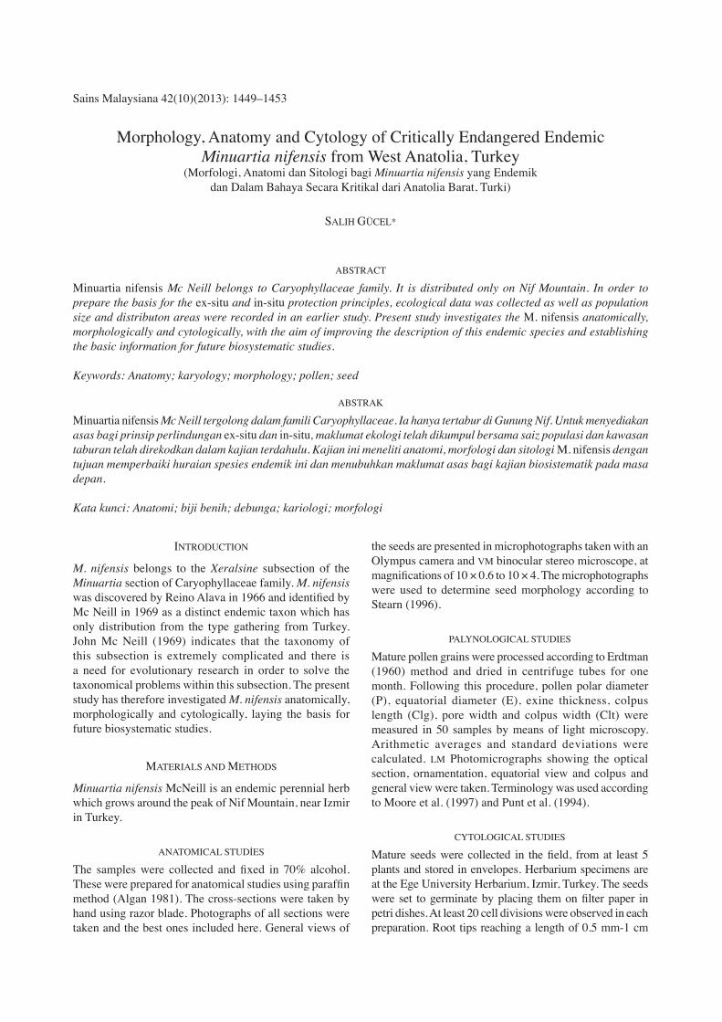
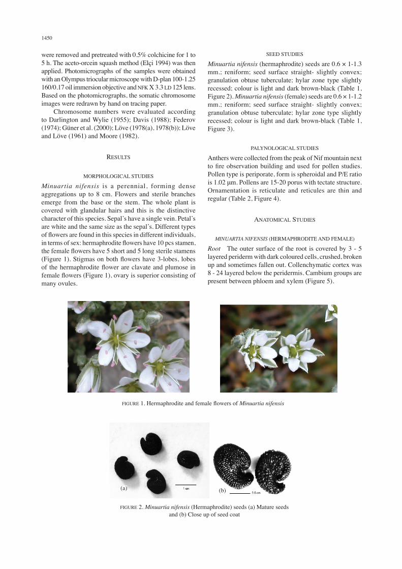
Minuartia nifensis (hermaphrodite) seeds are 0.6 × 1-1.3 mm.; reniform; seed surface straight- slightly convex; granulation obtuse tuberculate; hylar zone type slightly recessed; colour is light and dark brown-black (Table 1, Figure 2). Minuartia nifensis (female) seeds are 0.6 × 1-1.2 mm.; reniform; seed surface straight- slightly convex; granulation obtuse tuberculate; hylar zone type slightly recessed; colour is light and dark brown-black (Table 1, Figure 3).
PALYNOLOGICAL STUDIES
Anthers were collected from the peak of Nif mountain next to fire observation building and used for pollen studies. Pollen type is periporate, form is spheroidal and P/E ratio is 1.02 μm. Pollens are 15-20 porus with tectate structure. Ornamentation is reticulate and reticules are thin and regular (Table 2, Figure 4).
ANATOMICAL STUDIES
MINUARTIA NIFENSIS (HERMAPHRODITE AND FEMALE)
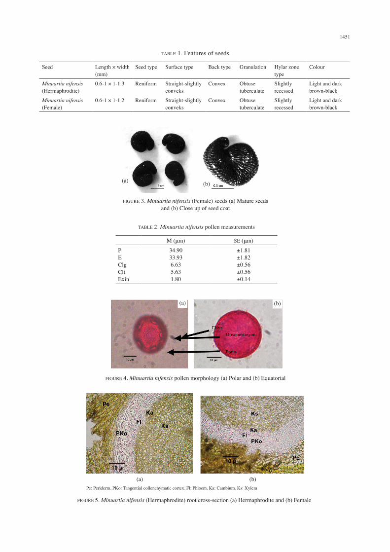
Root The outer surface of the root is covered by 3 - 5 layered periderm with dark coloured cells, crushed, broken up and sometimes fallen out. Collenchymatic cortex was 8 - 24 layered below the peridermis. Cambium groups are present between phloem and xylem (Figure 5).
FIGURE 1. Hermaphrodite and female flowers of Minuartia nifensis
FIGURE 2. Minuartia nifensis (Hermaphrodite) seeds (a) Mature seeds and (b) Close up of seed coat
(a) (b)
1451
TABLE 1. Features of seeds
Seed Length × width (mm)
Seed type Surface type Back type Granulation Hylar zone type
Colour
Minuartia nifensis (Hermaphrodite)
0.6-1 × 1-1.3 Reniform Straight-slightly conveks
Convex Obtuse tuberculate
Slightly recessed
Light and dark brown-black
Minuartia nifensis (Female)
0.6-1 × 1-1.2 Reniform Straight-slightly conveks
Convex Obtuse tuberculate
Slightly recessed
Light and dark brown-black
TABLE 2. Minuartia nifensis pollen measurements
M (μm) SE (μm)PEClgCltExin
34.9033.936.635.631.80
±1.81±1.82±0.56±0.56±0.14
FIGURE 3. Minuartia nifensis (Female) seeds (a) Mature seeds and (b) Close up of seed coat
(a) (b)
FIGURE 4. Minuartia nifensis pollen morphology (a) Polar and (b) Equatorial
(a) (b)
FIGURE 5. Minuartia nifensis (Hermaphrodite) root cross-section (a) Hermaphrodite and (b) Female
Pe: Periderm, PKo: Tangential collenchymatic cortex, Fl: Phloem, Ka: Cambium, Ks: Xylem
(a) (b)
1452
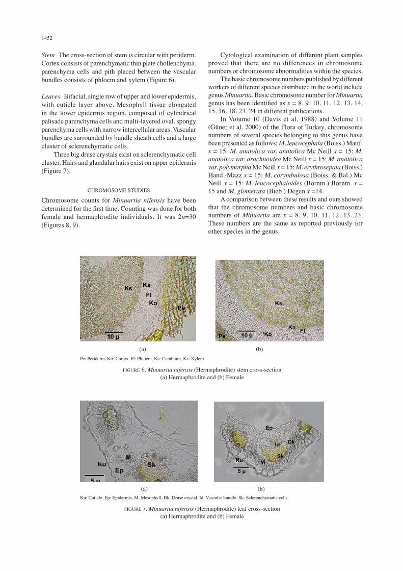
Stem The cross-section of stem is circular with periderm. Cortex consists of parenchymatic thin plate chollenchyma, parenchyma cells and pith placed between the vascular bundles consists of phloem and xylem (Figure 6).
Leaves Bifacial, single row of upper and lower epidermis, with cuticle layer above. Mesophyll tissue elongated in the lower epidermis region, composed of cylindrical palisade parenchyma cells and multi-layered oval, spongy parenchyma cells with narrow intercellular areas. Vascular bundles are surrounded by bundle sheath cells and a large cluster of sclerenchymatic cells. Three big druse crystals exist on sclerenchymatic cell cluster. Hairs and glandular hairs exist on upper epidermis (Figure 7).
CHROMOSOME STUDIES
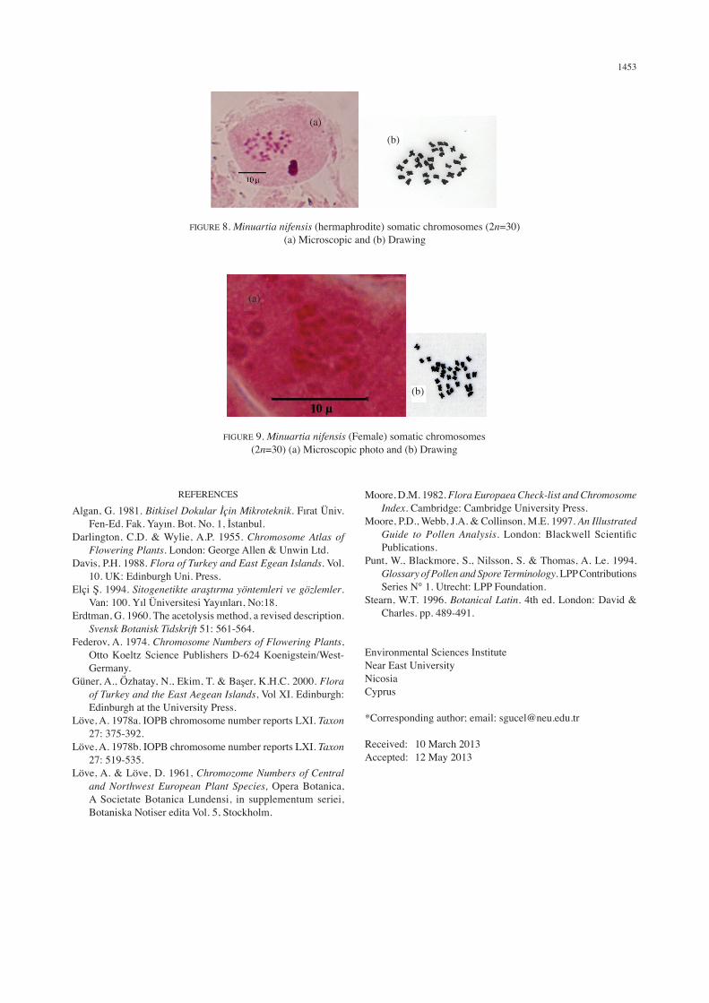
Chromosome counts for Minuartia nifensis have been determined for the first time. Counting was done for both female and hermaphrodite individuals. It was 2n=30 (Figures 8, 9).
Cytological examination of different plant samples proved that there are no differences in chromosome numbers or chromosome abnormalities within the species. The basic chromosome numbers published by different workers of different species distributed in the world include genus Minuartia. Basic chromosome number for Minuartia genus has been identified as x = 8, 9, 10, 11, 12, 13, 14, 15, 16, 18, 23, 24 in different publications. In Volume 10 (Davis et al. 1988) and Volume 11 (Güner et al. 2000) of the Flora of Turkey, chromosome numbers of several species belonging to this genus have been presented as follows: M. leucocephala (Boiss.) Mattf. x = 15; M. anatolica var. anatolica Mc Neill x = 15; M. anatolica var. arachnoidea Mc Neill x = 15; M. anatolica var. polymorpha Mc Neill x = 15; M. erythrosepala (Boiss.) Hand.-Mazz x = 15; M. corymbulosa (Boiss. & Bal.) Mc Neill x = 15; M. leucocephaloides (Bornm.) Bornm. x = 15 and M. glomerata (Bieb.) Degen x =14. A comparison between these results and ours showed that the chromosome numbers and basic chromosome numbers of Minuartia are x = 8, 9, 10, 11, 12, 13, 23. These numbers are the same as reported previously for other species in the genus.
Pe: Periderm, Ko: Cortex, Fl: Phloem, Ka: Cambium, Ks: Xylem
FIGURE 6. Minuartia nifensis (Hermaphrodite) stem cross-section (a) Hermaphrodite and (b) Female
(a) (b)
Ku: Cuticle, Ep: Epidermis, M: Mesophyll, Dk: Druse crystal, Id: Vascular bundle, Sk: Sclerenchymatic cells
FIGURE 7. Minuartia nifensis (Hermaphrodite) leaf cross-section (a) Hermaphrodite and (b) Female
(a) (b)
1453
REFERENCES
Algan, G. 1981. Bitkisel Dokular İçin Mikroteknik. Fırat Üniv. Fen-Ed. Fak. Yayın. Bot. No. 1, İstanbul.
Darlington, C.D. & Wylie, A.P. 1955. Chromosome Atlas of Flowering Plants. London: George Allen & Unwin Ltd.
Davis, P.H. 1988. Flora of Turkey and East Egean Islands. Vol. 10. UK: Edinburgh Uni. Press.
Elçi Ş. 1994. Sitogenetikte araştırma yöntemleri ve gözlemler. Van: 100. Yıl Üniversitesi Yayınları, No:18.
Erdtman, G. 1960. The acetolysis method, a revised description. Svensk Botanisk Tidskrift 51: 561-564.
Federov, A. 1974. Chromosome Numbers of Flowering Plants, Otto Koeltz Science Publishers D-624 Koenigstein/West-Germany.
Güner, A., Özhatay, N., Ekim, T. & Başer, K.H.C. 2000. Flora of Turkey and the East Aegean Islands, Vol XI. Edinburgh: Edinburgh at the University Press.
Löve, A. 1978a. IOPB chromosome number reports LXI. Taxon 27: 375-392.
Löve, A. 1978b. IOPB chromosome number reports LXI. Taxon 27: 519-535.
Löve, A. & Löve, D. 1961, Chromozome Numbers of Central and Northwest European Plant Species, Opera Botanica, A Societate Botanica Lundensi, in supplementum seriei, Botaniska Notiser edita Vol. 5, Stockholm.
Moore, D.M. 1982. Flora Europaea Check-list and Chromosome Index. Cambridge: Cambridge University Press.
Moore, P.D., Webb, J.A. & Collinson, M.E. 1997. An Illustrated Guide to Pollen Analysis. London: Blackwell Scientific Publications.
Punt, W., Blackmore, S., Nilsson, S. & Thomas, A. Le. 1994. Glossary of Pollen and Spore Terminology. LPP Contributions Series N° 1. Utrecht: LPP Foundation.
Stearn, W.T. 1996. Botanical Latin. 4th ed. London: David & Charles. pp. 489-491.
Environmental Sciences InstituteNear East University Nicosia Cyprus
*Corresponding author; email: [email protected]
Received: 10 March 2013Accepted: 12 May 2013
FIGURE 8. Minuartia nifensis (hermaphrodite) somatic chromosomes (2n=30) (a) Microscopic and (b) Drawing
(b)(a)
FIGURE 9. Minuartia nifensis (Female) somatic chromosomes (2n=30) (a) Microscopic photo and (b) Drawing
(a)
(b)