Embed Size (px)
Citation preview
8/6/2019 Model of the ECD of Human Alpha7 Based on Crystal Structure of the Mouse Alpha 1
http://slidepdf.com/reader/full/model-of-the-ecd-of-human-alpha7-based-on-crystal-structure-of-the-mouse-alpha 1/5
Model of the extracellular domain of the human a 7 nAChR based onthe crystal structure of the mouse a 1 nAChR extracellular domain
Maria Konstantakaki a , Socrates J. Tzartos b ,c , Konstantinos Poulas b ,*, Elias Eliopoulos a ,**a Department of Agricultural Biotechnology, Agricultural University of Athens, 75, Iera Odos, Votanikos, GR11855, Athens, Greece
b Department of Pharmacy, University of Patras, Rio-Patras, GR26504, Greecec Department of Biochemistry, Hellenic Pasteur Institute, 127 Vas. Soas Ave., GR11521, Athens, Greece
Received 15 September 2007; received in revised form 17 January 2008; accepted 23 January 2008Available online 2 February 2008
Abstract
Neuronal nicotinic acetylcholine receptors (nAChRs) are important therapeutic targets for various diseases, including Alzheimer’s disease,Parkinson’s disease, and schizophrenia, as well as for cessation of smoking. Based on the recently determined crystal structure of the extracellulardomain (ECD) of the mouse nAChR a 1 subunit complexed with a -bungarotoxin at 1.94 A resolution, we have constructed three-dimensionalmodels of the ECD of the monomer, homodimer, and homopentamer of the human a 7 nAChR and investigated in detail the interface between thetwo a 7 subunits. The docking of the agonist in the ligand-binding pocket of the human a 7 dimer was also performed and found consistent withresults from labeling and mutagenesis experiments. Since the nAChR ligand-binding site is a useful target for mutagenesis studies and the rationaldesign of drugs against diseases, these models provide useful information for future work.# 2008 Elsevier Inc. All rights reserved.
Keywords: a 7 nAChR; a 1 nAChR; Homology model; Structure; Extracellular domain; Ligand-binding domain; Rational drug design
1. Introduction
Nicotinic acetylcholine receptors (nAChRs), integral mem-brane proteins that respond to the binding of acetylcholine(ACh), are composed of ve subunits organized around acentral pore perpendicular to the membrane, and consist of twogroups: (a) the muscle type, found in sh electric organs and invertebrate skeletal muscles, where they mediate neuromusculartransmission at the neuromuscular junction, and (b) theneuronal type, found mainly throughout the peripheral andcentral nervous system, but also in non-neuronal tissues. The
muscle-type nAChR consists of ve homologous subunits inthe stoichiometry ( a 1)2 b 1gd (embryonic form) or ( a 1)2 b 1e d(adult form) with two ACh-binding sites per molecule.Neuronal nAChRs consist of a variety of subunits in different
combinations. To date, nine a (a 2–a 10) and three b (b 2–b 4)neuronal subunit genes have been cloned. The a 7 neuronal-typesubunit can form homopentamer of ve identical a 7 subunitswith ve ACh-binding sites per molecule [1]. nAChRs exist indifferent states, closed, open or desensitized, while the bindingof nicotinic ligands, agonists, or competitive antagonists,affects the equilibrium between the various states.
Extensive studies on nAChRs from various species haveshown that each subunit consists of: (a) an N-terminalextracellular domain (ECD) of approximately 210–220 aminoacids, of which a high proportion forms b strands, and bearing
the domain that binds agonists and competitive antagonists[2,3], (b) four small (15–20 residues) hydrophobic transmem-brane domains (M1–M4) and two small hydrophobic loops,linking segments M1–M2 and M2–M3, (c) a larger loop, whichvaries in size (100–150 residues) and sequence betweensubunits, and which lies between M3 and M4 and bearsphosphorylation sites [4], and (d) the C-terminal region of eachsubunit, which consists of a small (4–28 residues) hydrophilicextracellular segment [1].
It is well documented that brain nAChRs, including a 7,participate in complex functions, such as attention, memory,
www.elsevier.com/locate/JMGM
Available online at www.sciencedirect.com
Journal of Molecular Graphics and Modelling 26 (2008) 1333–1337
Abbreviations: nAChR, nicotinic acetylcholine receptor; ECD, extracellu-lar domain; ACh, acetylcholine.
* Corresponding author. Tel.: +30 2610 969953; fax: +30 2610 969954.** Corresponding author. Tel.: +30 210 5294223; fax: +30 210 5294322.
E-mail addresses: [email protected] (K. Poulas), [email protected](E. Eliopoulos).
1093-3263/$ – see front matter # 2008 Elsevier Inc. All rights reserved.
doi:10.1016/j.jmgm.2008.01.004
8/6/2019 Model of the ECD of Human Alpha7 Based on Crystal Structure of the Mouse Alpha 1
http://slidepdf.com/reader/full/model-of-the-ecd-of-human-alpha7-based-on-crystal-structure-of-the-mouse-alpha 2/5
and cognition, while clinical data suggest their involvement inthe pathogenesis of several disorders (Alzheimer’s disease,Parkinson’s disease, schizophrenia, and depression) [1].Smoking is a major public health problem, and the a 7 nAChRsubtype is closely associated with nicotine addiction andnicotine-induced behavior [5]. a 7 nAChRs are overexpressedin small cell lung carcinoma in smokers [6]. In order to treatthese diseases, it would be useful to design drugs that canselectively activate a 7 nAChRs and for this purpose it isimportant to have a detailed knowledge of the a 7 ligand-binding site.
Early biochemical studies on muscle-type nAChR indicatedthat two separate parts of the nAChRligand-binding domain areinvolvedin the formationof the agonist/competitive antagonist-binding site [7–9] . The ACh-binding pocket of the nAChR isformed between loops A, B, and C of the a subunit and strandsb 5 and b 6 of the b -sandwich core of the adjacent g or d subunit[10]. The residues in the loops known to be involved in theformation of the ACh-binding site are Tyr190, Cys192, and
Tyr198 in the C loop, which is incorporated in the b 9–b 10hairpin, and Trp149 in the B loop.
Detailed information on the atomic structure of an ACh-binding domain rst became available following the elucida-tion of the crystal structure at 2.7 A resolution of theacetylcholine binding protein (AChBP) from the glial cellsof the mollusc, Lymnaea stagnalis [11] . This protein, a water-soluble homopentamer of a 210 amino acid subunit, is afunctional homologue of the ECDs of nAChRs subunits andhas been extensively used as a model for their ligand-bindingdomain.
Based on the crystal structure of the AChBP, the atomic
model of the Torpedo muscle-type nAChR at 4 A˚
resolutionallowed a detailed description of the whole receptor in itsclosed-channel form [10]. More recently, the co-crystal-lization of various mollusk AChBPs with several agonists andantagonists [12–16] has revealed details of the atomicinteractions between ligands and specic residues in theligand-binding site.
A further breakthrough in the investigation of the structureof the nAChRs was the recent crystal structure determinationof the mouse muscle nAChR a 1 subunit ECD [17]. Structuralcomparison of the ECDs of the a 1 subunit of the mousenAChR, the Torpedo nAChR, and the AChBP bound to eithercarbamylcholine [12] or a -cobratoxin [16] indicated a highoverall similarity of main chain folding, with most sidechains adopting a similar conformation [17]. Specically, thepredicted pentamer assembly interfaces of the mouse a 1subunit resemble the corresponding interfaces in the TorpedonAChR and AChBP. On the minus surface, there is structuralsimilarity both in main chain and side chain conformation.On the plus surface, only minor differences are observed.The side chains of Thr150, Tyr151, Asp152, and Ala155(mutated from Val in the mouse a 1 subunit) in loop B of mouse a 1, are oriented as in the Torpedo nAChR asubunit with a slight offset, which is not seen in thecomparison with the structures of AChBP bound to agonist or
antagonist [17].
The structure of the ECD of the a 1 subunit of the mousenAChR is a good starting point for the modeling of the ECD of the human neuronal a 7 receptor.
2. Methodology
2.1. Sequence alignment
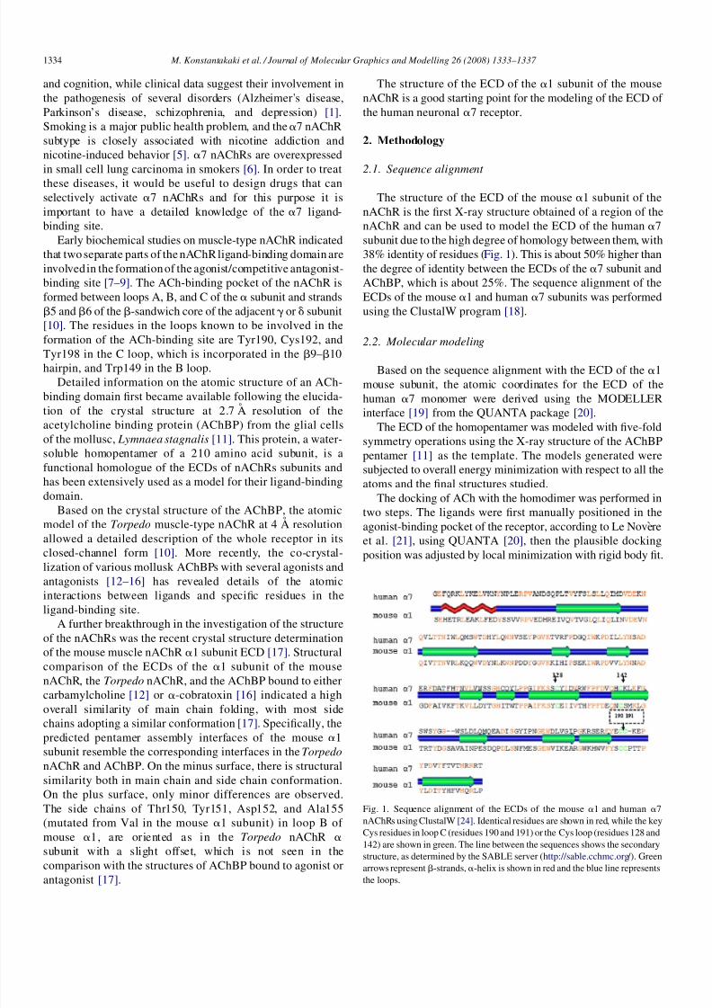
The structure of the ECD of the mouse a 1 subunit of thenAChR is the rst X-ray structure obtained of a region of thenAChR and can be used to model the ECD of the human a 7subunit due to the high degree of homology between them, with38% identity of residues ( Fig. 1). This is about 50% higher thanthe degree of identity between the ECDs of the a 7 subunit andAChBP, which is about 25%. The sequence alignment of theECDs of the mouse a 1 and human a 7 subunits was performedusing the ClustalW program [18].
2.2. Molecular modeling
Based on the sequence alignment with the ECD of the a 1mouse subunit, the atomic coordinates for the ECD of thehuman a 7 monomer were derived using the MODELLERinterface [19] from the QUANTA package [20].
The ECD of the homopentamer was modeled with ve-foldsymmetry operations using the X-ray structure of the AChBPpentamer [11] as the template. The models generated weresubjected to overall energy minimization with respect to all theatoms and the nal structures studied.
The docking of ACh with the homodimer was performed intwo steps. The ligands were rst manually positioned in the
agonist-binding pocket of the receptor, according to Le Nove `reet al. [21] , using QUANTA [20], then the plausible dockingposition was adjusted by local minimization with rigid body t.
Fig. 1. Sequence alignment of the ECDs of the mouse a 1 and human a 7nAChRs using ClustalW [24]. Identical residues are shown in red, while the keyCys residues in loop C (residues 190 and 191) or the Cys loop (residues 128 and142) are shown in green. The line between the sequences shows the secondarystructure, as determined by the SABLE server ( http://sable.cchmc.org /). Greenarrows represent b -strands, a -helix is shown in red and the blue line represents
the loops.
M. Konstantakaki et al. / Journal of Molecular Graphics and Modelling 26 (2008) 1333–1337 1334
8/6/2019 Model of the ECD of Human Alpha7 Based on Crystal Structure of the Mouse Alpha 1
http://slidepdf.com/reader/full/model-of-the-ecd-of-human-alpha7-based-on-crystal-structure-of-the-mouse-alpha 3/5
3. Results
3.1. a 7 nAChR: from monomer to pentamer
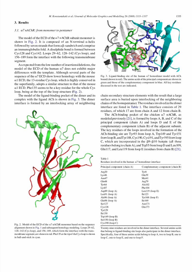
The model of the ECD of the a 7 nAChR subunit monomer isshown in Fig. 2. It is composed of an N-terminal a -helixfollowed by seven strands that form a b -sandwich and comprisean immunoglobulin fold. A disulphide bond is formed betweenCys128 and Cys142. Loops 29–62, 128–142 (Cys loop), and156–189 form the interface with the following transmembranesegment.
As expected from the low number of insertions/deletions, themodel of the ECD of the human a 7 does not exhibit majordifferences with the template. Although several parts of thesequence of the a 7 ECD show lower homology with the mousea 1 ECD, the 13-residue Cys loop, which is highly conserved inthe superfamily, adopts a similar structure to that of the mousea 1 ECD. Phe135 seems to be a key residue for the whole Cysloop, being at the top of the loop structure ( Fig. 2).
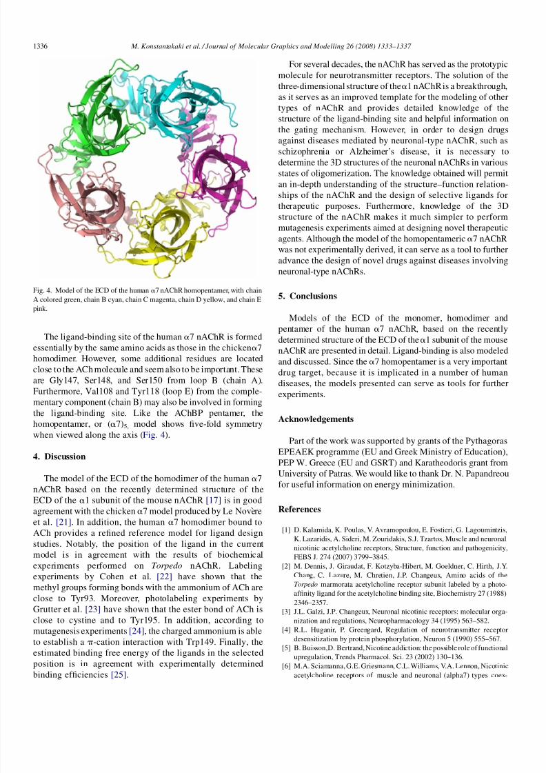
The model of the ligand-binding pocket of the dimer and itscomplex with the ligand ACh is shown in Fig. 3. The dimerinterface is formed by an interlocking array of neighboring
chain secondary structure elements with the result that a largesurface area is buried upon interlocking of the neighboringchains of the homopentamer. The residues involved in thedimerinterface are listed in Table 1 . The interface consists of 29residues, of which 17 are from chain A and 12 from chain B.
The ACh-binding pocket of the chicken a 7 nAChR, asmodeled previously [21] , is formed by loops A, B, and C of theprincipal component (chain A) and loops D and E of thecomplementary component (chain B) of the adjacent subunit.The key residues of the loops involved in the formation of theACh-binding site are Tyr93 from loop A, Trp149 and Tyr151from loop B,andTyr188,Cys190,Cys191, andTyr195 from loop
C, which are incorporated in the b 9–b 10 hairpin (all theseresidues belong to chain A), and Trp55 fromloop D and Leu109,Gln117, and Leu119 from loop E (residues from chain B) [21] .
Fig. 2. Model of the ECD of the a 7 nAChR monomer based on the sequencealignment shown in Fig. 1 and subsequent homology modeling. Loops 29–62,128–142 (Cys loop), and 156–189, which form the interface with the trans-membrane segment,are shown in red. Phe135 at the tipof theCys loop is shown
in ball-and-stick in cyan.
Fig. 3. Ligand-binding site of the human a 7 homodimer model with AChbound (shown in red). The amino acids of the principal component are shown ingreen and those of the complementary component in blue. All key residuesdiscussed in the text are indicated.
Table 1Residues involved in the human a 7 homodimer interface
Principal component (chain A) Complementary component (chain B)
Arg20 Tyr8Lys46 Gln39Asn47 Met41Gln48 Arg79Tyr64 Ala102Lys87 Phe104Asp89 (loop A) Leu119 (loop E)Leu91 (loop A) Ile123Ala96 (loop A) Tyr168 (loop F)Glu98 (loop A) Ile169Ser127 Asn171Cys128 Glu173Tyr129Ile130Trp149 (loop B)Ser150 (loop B)Cys190 (loop C)
Twenty-nine residues are involved in the dimer interface. Several amino acidsthat belong to ligand-binding site loops also participate in the dimer interface.Specically, four of these amino acids belong to loop A, two to loop B, one to
loop C, one to loop E, and one to loop F.
M. Konstantakaki et al. / Journal of Molecular Graphics and Modelling 26 (2008) 1333–1337 1335
8/6/2019 Model of the ECD of Human Alpha7 Based on Crystal Structure of the Mouse Alpha 1
http://slidepdf.com/reader/full/model-of-the-ecd-of-human-alpha7-based-on-crystal-structure-of-the-mouse-alpha 4/5
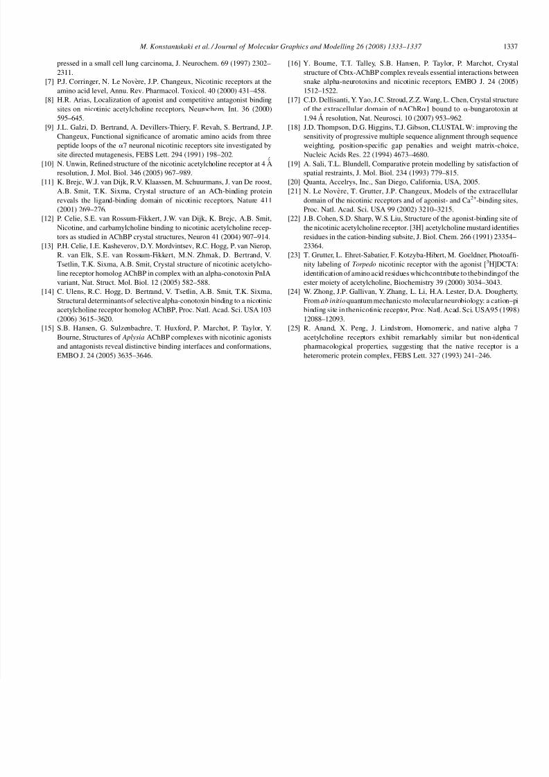
The ligand-binding site of the human a 7 nAChR is formedessentially by the same amino acids as those in the chicken a 7homodimer. However, some additional residues are locatedclose to the ACh molecule and seem also to be important. Theseare Gly147, Ser148, and Ser150 from loop B (chain A).Furthermore, Val108 and Tyr118 (loop E) from the comple-
mentary component (chain B) may also be involved in formingthe ligand-binding site. Like the AChBP pentamer, thehomopentamer, or ( a 7)5, model shows ve-fold symmetrywhen viewed along the axis ( Fig. 4).
4. Discussion
The model of the ECD of the homodimer of the human a 7nAChR based on the recently determined structure of theECD of the a 1 subunit of the mouse nAChR [17] is in goodagreement with the chicken a 7 model produced by Le Nove reet al. [21]. In addition, the human a 7 homodimer bound toACh provides a rened reference model for ligand designstudies. Notably, the position of the ligand in the currentmodel is in agreement with the results of biochemicalexperiments performed on Torpedo nAChR. Labelingexperiments by Cohen et al. [22] have shown that themethyl groups forming bonds with the ammonium of ACh areclose to Tyr93. Moreover, photolabeling experiments byGrutter et al. [23] have shown that the ester bond of ACh isclose to cystine and to Tyr195. In addition, according tomutagenesis experiments [24], the charged ammonium is ableto establish a p -cation interaction with Trp149. Finally, theestimated binding free energy of the ligands in the selectedposition is in agreement with experimentally determined
binding efciencies [25] .
For several decades, the nAChR has served as the prototypicmolecule for neurotransmitter receptors. The solution of thethree-dimensional structure of the a 1 nAChRis a breakthrough,as it serves as an improved template for the modeling of othertypes of nAChR and provides detailed knowledge of thestructure of the ligand-binding site and helpful information onthe gating mechanism. However, in order to design drugsagainst diseases mediated by neuronal-type nAChR, such asschizophrenia or Alzheimer’s disease, it is necessary todetermine the 3D structures of the neuronal nAChRs in variousstates of oligomerization. The knowledge obtained will permitan in-depth understanding of the structure–function relation-ships of the nAChR and the design of selective ligands fortherapeutic purposes. Furthermore, knowledge of the 3Dstructure of the nAChR makes it much simpler to performmutagenesis experiments aimed at designing novel therapeuticagents. Although the model of the homopentameric a 7 nAChRwas not experimentally derived, it can serve as a tool to furtheradvance the design of novel drugs against diseases involving
neuronal-type nAChRs.
5. Conclusions
Models of the ECD of the monomer, homodimer andpentamer of the human a 7 nAChR, based on the recentlydetermined structure of the ECD of the a 1 subunit of the mousenAChR are presented in detail. Ligand-binding is also modeledand discussed. Since the a 7 homopentamer is a very importantdrug target, because it is implicated in a number of humandiseases, the models presented can serve as tools for furtherexperiments.
Acknowledgements
Part of the work was supported by grants of the PythagorasEPEAEK programme (EU and Greek Ministry of Education),PEP W. Greece (EU and GSRT) and Karatheodoris grant fromUniversity of Patras. We would like to thank Dr. N. Papandreoufor useful information on energy minimization.
References
[1] D. Kalamida, K. Poulas, V. Avramopoulou, E. Fostieri, G. Lagoumintzis,
K. Lazaridis, A. Sideri, M. Zouridakis, S.J. Tzartos, Muscle and neuronalnicotinic acetylcholine receptors, Structure, function and pathogenicity,FEBS J. 274 (2007) 3799–3845.
[2] M. Dennis, J. Giraudat, F. Kotzyba-Hibert, M. Goeldner, C. Hirth, J.Y.Chang, C. Lazure, M. Chretien, J.P. Changeux, Amino acids of theTorpedo marmorata acetylcholine receptor subunit labeled by a photo-afnity ligand for the acetylcholine binding site, Biochemistry 27 (1988)2346–2357.
[3] J.L. Galzi, J.P. Changeux, Neuronal nicotinic receptors: molecular orga-nization and regulations, Neuropharmacology 34 (1995) 563–582.
[4] R.L. Huganir, P. Greengard, Regulation of neurotransmitter receptordesensitization by protein phosphorylation, Neuron 5 (1990) 555–567.
[5] B. Buisson,D. Bertrand,Nicotine addiction: the possible role of functionalupregulation, Trends Pharmacol. Sci. 23 (2002) 130–136.
[6] M.A. Sciamanna,G.E.Griesmann, C.L. Williams, V.A. Lennon, Nicotinic
acetylcholine receptors of muscle and neuronal (alpha7) types coex-
Fig. 4. Model of the ECD of the human a 7 nAChR homopentamer, with chainA colored green, chain B cyan, chain C magenta, chain D yellow, and chain Epink.
M. Konstantakaki et al. / Journal of Molecular Graphics and Modelling 26 (2008) 1333–1337 1336
8/6/2019 Model of the ECD of Human Alpha7 Based on Crystal Structure of the Mouse Alpha 1
http://slidepdf.com/reader/full/model-of-the-ecd-of-human-alpha7-based-on-crystal-structure-of-the-mouse-alpha 5/5
pressed in a small cell lung carcinoma, J. Neurochem. 69 (1997) 2302–2311.
[7] P.J. Corringer, N. Le Nove`re, J.P. Changeux, Nicotinic receptors at theamino acid level, Annu. Rev. Pharmacol. Toxicol. 40 (2000) 431–458.
[8] H.R. Arias, Localization of agonist and competitive antagonist bindingsites on nicotinic acetylcholine receptors, Neurochem. Int. 36 (2000)595–645.
[9] J.L. Galzi, D. Bertrand, A. Devillers-Thiery, F. Revah, S. Bertrand, J.P.
Changeux, Functional signicance of aromatic amino acids from threepeptide loops of the a 7 neuronal nicotinic receptors site investigated bysite directed mutagenesis, FEBS Lett. 294 (1991) 198–202.
[10] N. Unwin, Rened structure of the nicotinic acetylcholine receptor at 4 A ˚resolution, J. Mol. Biol. 346 (2005) 967–989.
[11] K. Brejc, W.J. van Dijk, R.V. Klaassen, M. Schuurmans, J. van De roost,A.B. Smit, T.K. Sixma, Crystal structure of an ACh-binding proteinreveals the ligand-binding domain of nicotinic receptors, Nature 411(2001) 269–276.
[12] P. Celie, S.E. van Rossum-Fikkert, J.W. van Dijk, K. Brejc, A.B. Smit,Nicotine, and carbamylcholine binding to nicotinic acetylcholine recep-tors as studied in AChBP crystal structures, Neuron 41 (2004) 907–914.
[13] P.H. Celie, I .E. Kasheverov, D.Y. Mordvintsev, R.C. Hogg, P. van Nierop,R. van Elk, S.E. van Rossum-Fikkert, M.N. Zhmak, D. Bertrand, V.Tsetlin, T.K. Sixma, A.B. Smit, Crystal structure of nicotinic acetylcho-line receptor homolog AChBP in complex with an alpha-conotoxin PnIAvariant, Nat. Struct. Mol. Biol. 12 (2005) 582–588.
[14] C. Ulens, R.C. Hogg, D. Bertrand, V. Tsetlin, A.B. Smit, T.K. Sixma,Structural determinants of selective alpha-conotoxin binding to a nicotinicacetylcholine receptor homolog AChBP, Proc. Natl. Acad. Sci. USA 103(2006) 3615–3620.
[15] S.B. Hansen, G. Sulzenbachre, T. Huxford, P. Marchot, P. Taylor, Y.Bourne, Structures of Aplysia AChBP complexes with nicotinic agonistsand antagonists reveal distinctive binding interfaces and conformations,EMBO J. 24 (2005) 3635–3646.
[16] Y. Bourne, T.T. Talley, S.B. Hansen, P. Taylor, P. Marchot, Crystalstructure of Cbtx-AChBP complex reveals essential interactions betweensnake alpha-neurotoxins and nicotinic receptors, EMBO J. 24 (2005)1512–1522.
[17] C.D. Dellisanti, Y. Yao, J.C. Stroud, Z.Z. Wang, L. Chen, Crystal structureof the extracellular domain of nAChR a 1 bound to a -bungarotoxin at1.94 A resolution, Nat. Neurosci. 10 (2007) 953–962.
[18] J.D. Thompson, D.G. Higgins, T.J. Gibson, CLUSTAL W: improving the
sensitivity of progressive multiple sequence alignment through sequenceweighting, position-specic gap penalties and weight matrix-choice,Nucleic Acids Res. 22 (1994) 4673–4680.
[19] A. Sali, T.L. Blundell, Comparative protein modelling by satisfaction of spatial restraints, J. Mol. Biol. 234 (1993) 779–815.
[20] Quanta, Accelrys, Inc., San Diego, California, USA, 2005.[21] N. Le Nove re, T. Grutter, J.P. Changeux, Models of the extracellular
domain of the nicotinic receptors and of agonist- and Ca 2+ -binding sites,Proc. Natl. Acad. Sci. USA 99 (2002) 3210–3215.
[22] J.B. Cohen, S.D. Sharp, W.S. Liu, Structure of the agonist-binding site of the nicotinic acetylcholine receptor. [3H] acetylcholine mustard identiesresidues in the cation-binding subsite, J. Biol. Chem. 266 (1991) 23354–23364.
[23] T. Grutter, L. Ehret-Sabatier, F. Kotzyba-Hibert, M. Goeldner, Photoaf-nity labeling of Torpedo nicotinic receptor with the agonist [ 3 H]DCTA:identication of amino acid residues whichcontribute to thebindingof theester moiety of acetylcholine, Biochemistry 39 (2000) 3034–3043.
[24] W. Zhong, J.P. Gallivan, Y. Zhang, L. Li, H.A. Lester, D.A. Dougherty,From ab initio quantummechanicsto molecularneurobiology: a cation–pibinding site in thenicotinic receptor, Proc. Natl. Acad. Sci. USA95 (1998)12088–12093.
[25] R. Anand, X. Peng, J. Lindstrom, Homomeric, and native alpha 7acetylcholine receptors exhibit remarkably similar but non-identicalpharmacological properties, suggesting that the native receptor is aheteromeric protein complex, FEBS Lett. 327 (1993) 241–246.
M. Konstantakaki et al. / Journal of Molecular Graphics and Modelling 26 (2008) 1333–1337 1337