Embed Size (px)
Citation preview
STÉPHANE BARETE, MD, PHD
UNIT OF DERMATOLOGY
PITIÉ -SALPÊTRIÈRE HOSPITAL
PARIS
Erdheim-Chester disease and Skin issues
• Erdheim-Chester (ECD) is an orphan disease included in the spectrum of systemic non-langerhans cellhistiocytosis with frequent recurrent BRAFV600Emutation
• Recent data have been released for skin manifestations of ECD
• As many molecular targeted therapies (MTT) are currently used for patients with BRAF mutation, one might expect MTT toxicities including skin manifestations
Introduction
•ECD skin manifestations
•ECD and MTT skin toxicities
Skin issues
• Recently described≃ 5 years
• Prevalence 19-28 % of ECD patients
• Various clinical manifestations
• First Series n=40 pts• Chasset F, Barete S, Charlotte F et al JAAD. 2016;74:513-20
Arnaud L et al Blood 2011, Haroche J et al Blood 2012, Haroche J et al Rheum Dis Clin North Am. 2013.
ECD and skin manifestations
Patients and methods
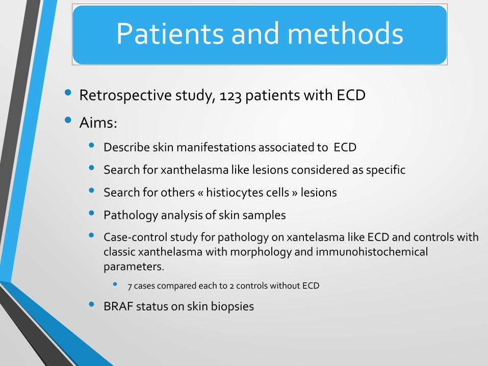
• Retrospective study, 123 patients with ECD
• Aims:• Describe skin manifestations associated to ECD
• Search for xanthelasma like lesions considered as specific
• Search for others « histiocytes cells » lesions
• Pathology analysis of skin samples
• Case-control study for pathology on xantelasma like ECD and controls withclassic xanthelasma with morphology and immunohistochemicalparameters.
• 7 cases compared each to 2 controls without ECD
• BRAF status on skin biopsies
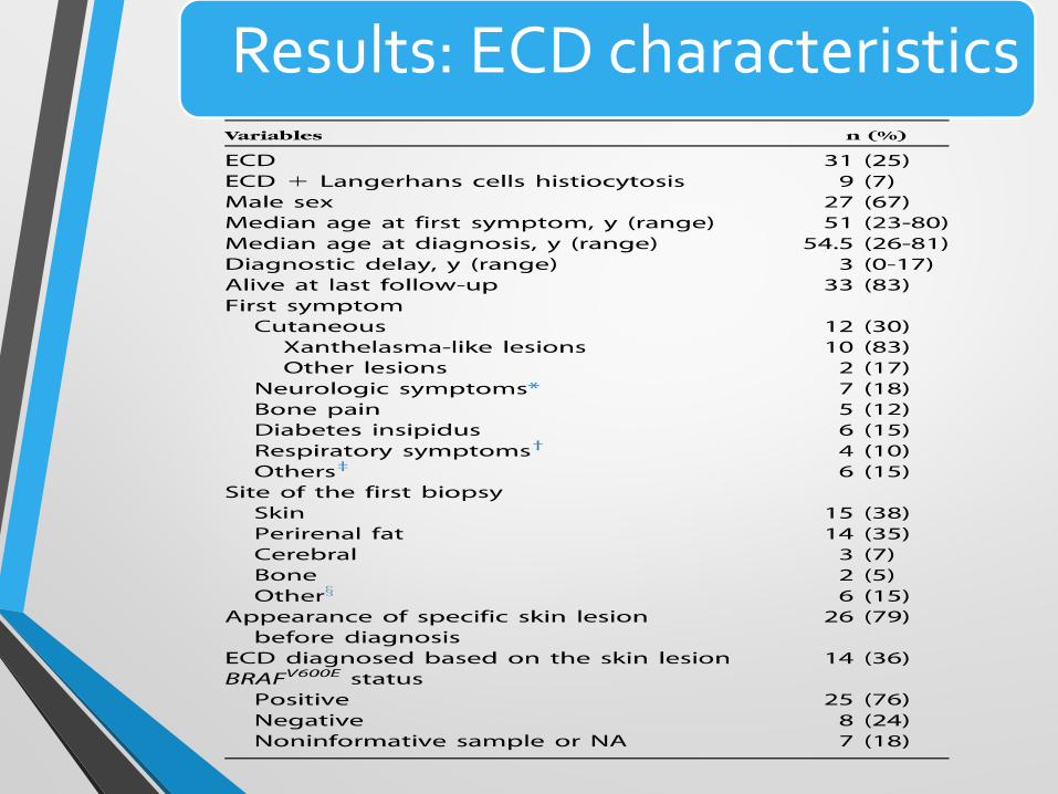
Results: ECD characteristics
Xanthelasma-like lesions
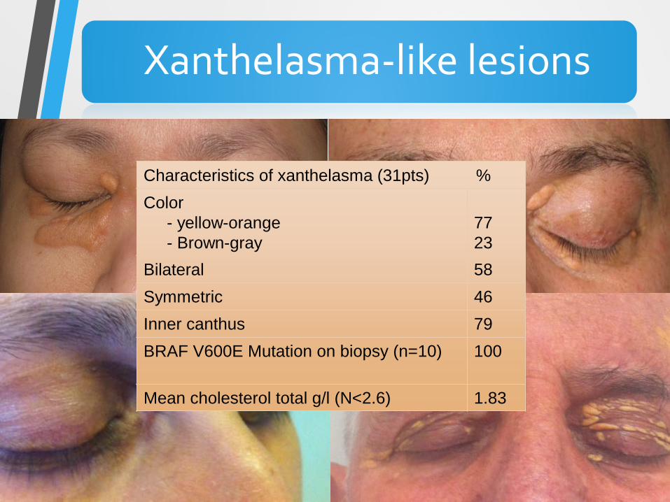
Characteristics of xanthelasma (31pts) %Color
- yellow-orange- Brown-gray
7723
Bilateral 58Symmetric 46Inner canthus 79BRAF V600E Mutation on biopsy (n=10) 100
Mean cholesterol total g/l (N<2.6) 1.83
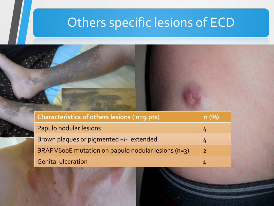
Others specific lesions of ECD
Characteristics of others lesions ( n=9 pts) n (%)
Papulo nodular lesions 4
Brown plaques or pigmented +/- extended 4
BRAF V600E mutation on papulo nodular lesions (n=3) 2
Genital ulceration 1
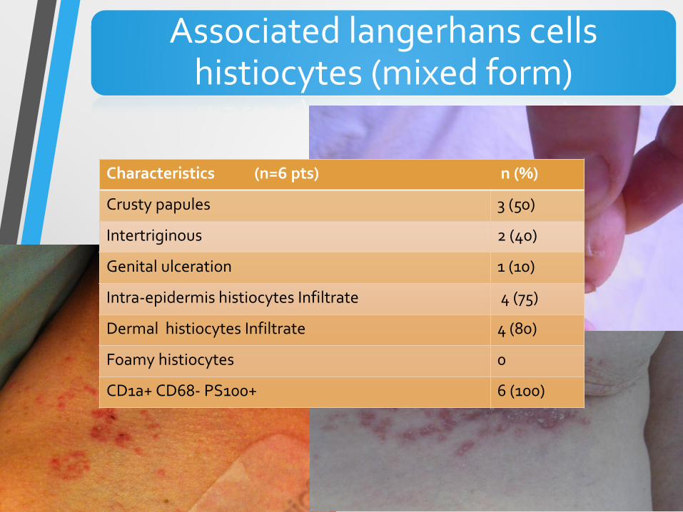
Associated langerhans cellshistiocytes (mixed form)
Characteristics (n=6 pts) n (%)
Crusty papules 3 (50)
Intertriginous 2 (40)
Genital ulceration 1 (10)
Intra-epidermis histiocytes Infiltrate 4 (75)
Dermal histiocytes Infiltrate 4 (80)
Foamy histiocytes 0
CD1a+ CD68- PS100+ 6 (100)
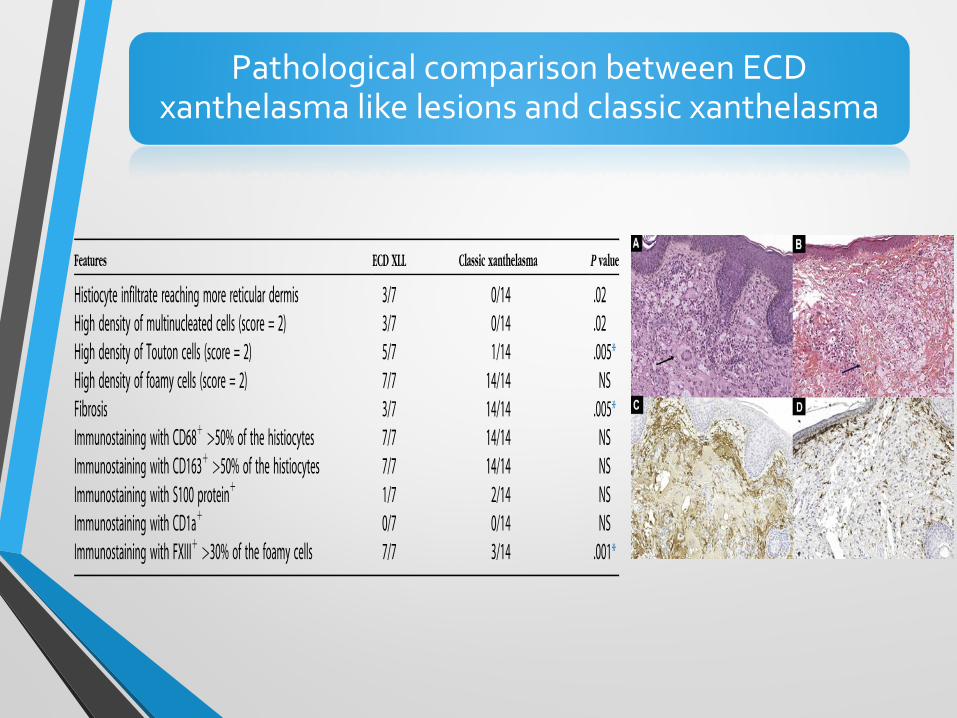
Pathological comparison between ECDxanthelasma like lesions and classic xanthelasma
Take away messages• First description of a series of ECD skin lesions: XLL is the
most prevalent
• Pathology of XLL: interest of morphology and density of histiocytes cells infiltrate and touton cells for ECD diagnosis
• XLL biopsy is easy and usefull for mutational status(BRAF status) in a context of ECD
ECD and MTT skin toxicities
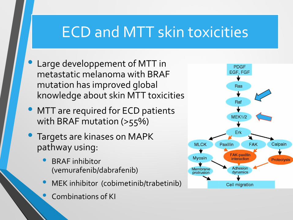
• Large developpement of MTT in metastatic melanoma with BRAF mutation has improved global knowledge about skin MTT toxicities
• MTT are required for ECD patients with BRAF mutation (>55%)
• Targets are kinases on MAPK pathway using:• BRAF inhibitor
(vemurafenib/dabrafenib)
• MEK inhibitor (cobimetinib/trabetinib)
• Combinations of KI
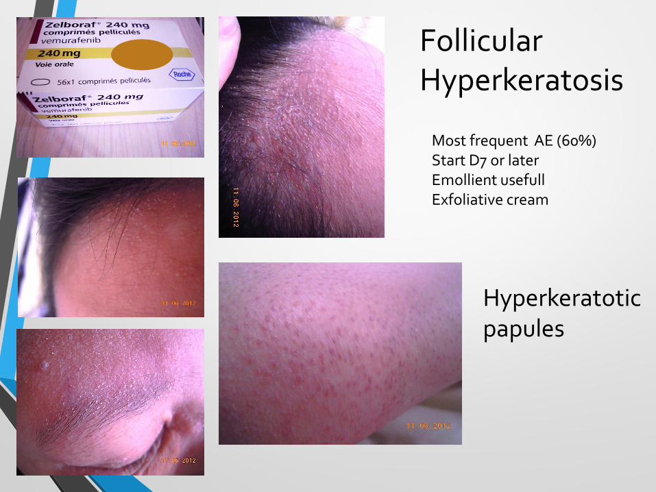
FollicularHyperkeratosis
Most frequent AE (60%)Start D7 or laterEmollient usefullExfoliative cream
Hyperkeratoticpapules
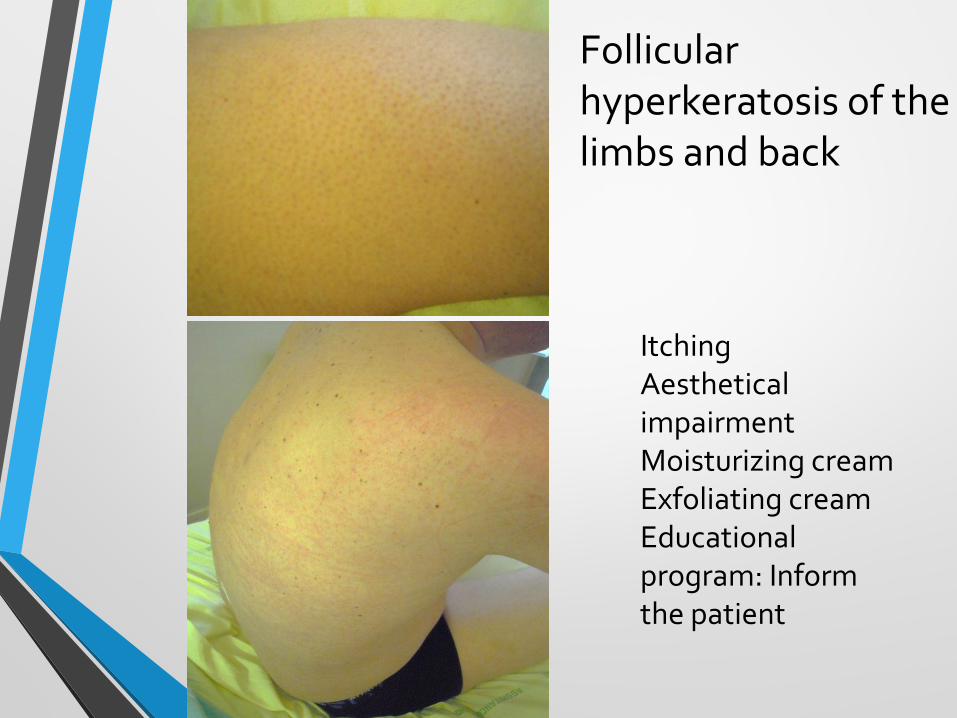
Follicularhyperkeratosis of the limbs and back
ItchingAestheticalimpairmentMoisturizing creamExfoliating creamEducationalprogram: Informthe patient
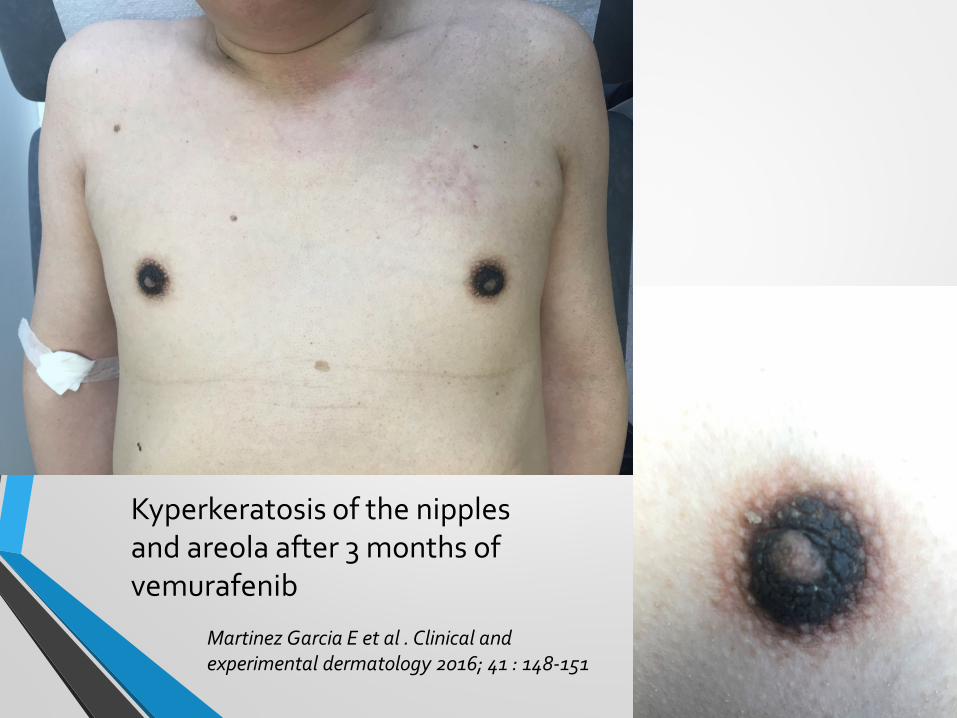
Kyperkeratosis of the nipplesand areola after 3 months of vemurafenib
Martinez Garcia E et al . Clinical and experimental dermatology 2016; 41 : 148-151
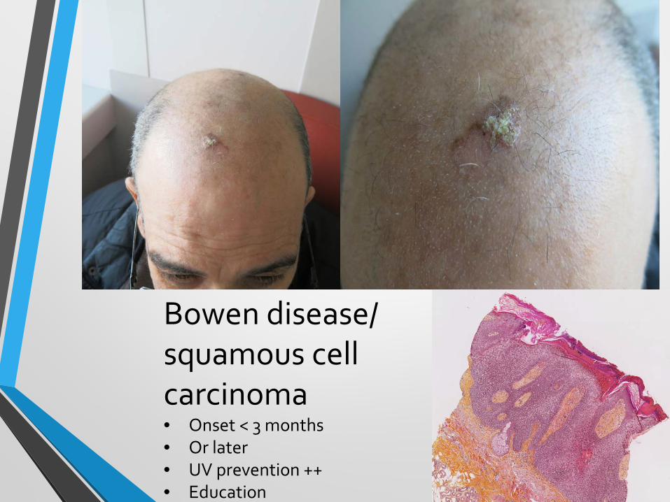
Bowen disease/ squamous cellcarcinoma• Onset < 3 months• Or later• UV prevention ++• Education
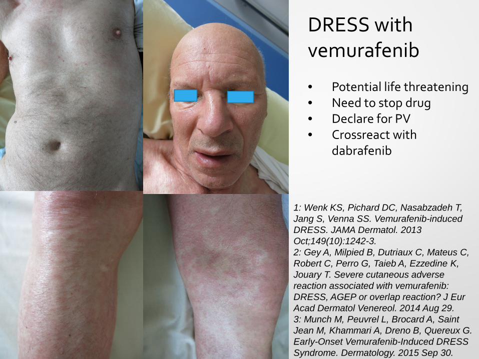
DRESS withvemurafenib
• Potential life threatening• Need to stop drug• Declare for PV• Crossreact with
dabrafenib
1: Wenk KS, Pichard DC, Nasabzadeh T, Jang S, Venna SS. Vemurafenib-inducedDRESS. JAMA Dermatol. 2013 Oct;149(10):1242-3.2: Gey A, Milpied B, Dutriaux C, Mateus C, Robert C, Perro G, Taieb A, Ezzedine K, Jouary T. Severe cutaneous adversereaction associated with vemurafenib:DRESS, AGEP or overlap reaction? J Eur Acad Dermatol Venereol. 2014 Aug 29.3: Munch M, Peuvrel L, Brocard A, Saint Jean M, Khammari A, Dreno B, Quereux G.Early-Onset Vemurafenib-Induced DRESS Syndrome. Dermatology. 2015 Sep 30.
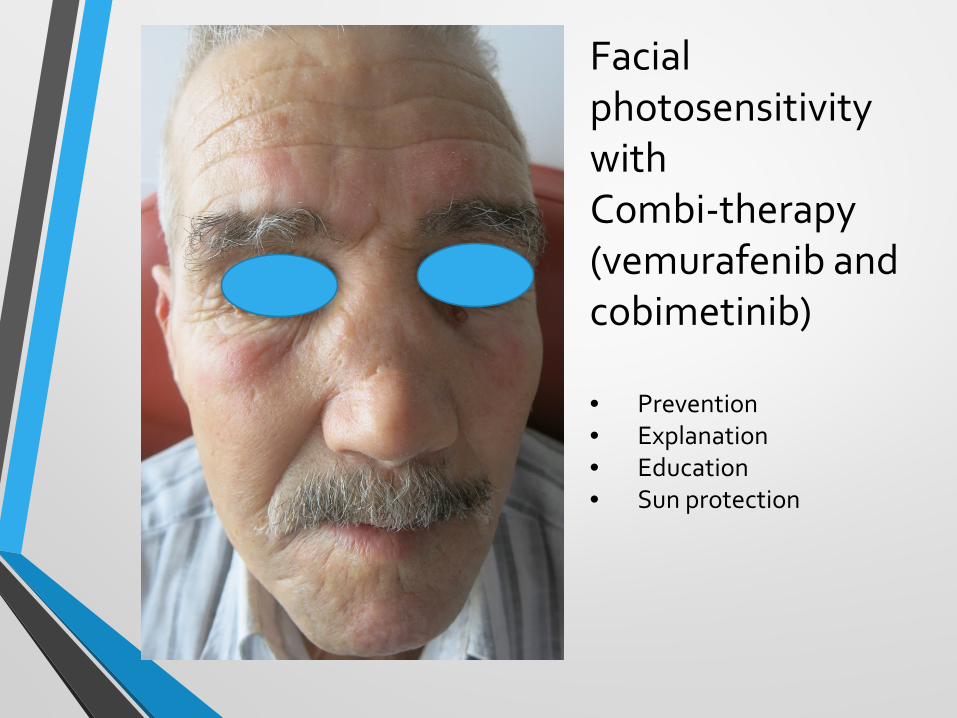
Facial photosensitivitywithCombi-therapy(vemurafenib and cobimetinib)
• Prevention• Explanation• Education• Sun protection
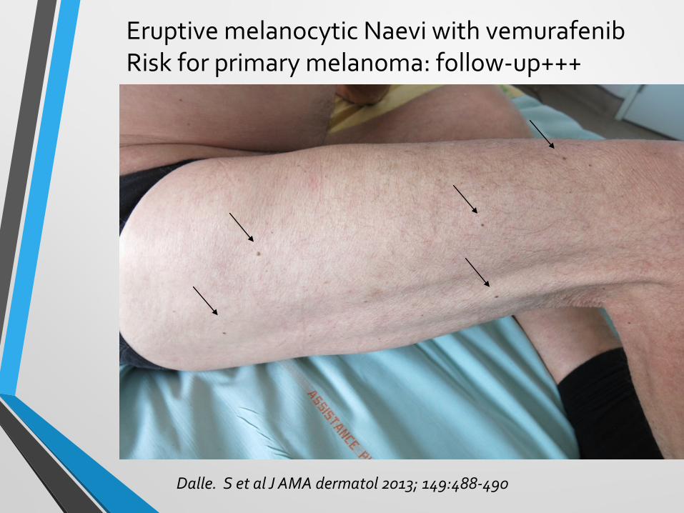
Eruptive melanocytic Naevi with vemurafenibRisk for primary melanoma: follow-up+++
Dalle. S et al J AMA dermatol 2013; 149:488-490
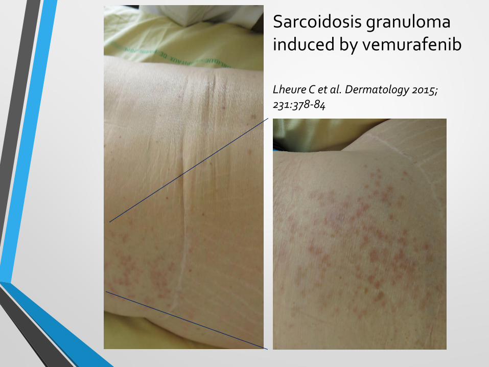
Sarcoidosis granulomainduced by vemurafenib
Lheure C et al. Dermatology 2015; 231:378-84
• Avoid sun exposure with protective sun cream with SPF 50 and protective dressing
• Educate patient to auto-screening of skin lesions to report to practitioner/dermatologist
• Talk with patients about frequency and severity of AEs
• Check skin with regular evaluation and referral once a month for 3 months then each 3 months
• Include patients in observational study like ACséVému(Unicancer) for best detection and screening of AEs withMTT
My practice guidelines
Conclusion
• Skin issues in ECD
• Diagnosis: XLL++
• BRAF mutational status: easy, safe and usefull from skin
• Skin MTT toxicities
• Increasing with emerging therapies (signalling pathways)
• Mostly manageable, sometime stop MTT
• Need to be checked regularly and evaluated
• Learn about physiopathology of the disease whatevermutational status of BRAF
• Need for gathering cohorts of ECD patients for skin follow-up under MTT
Acknowledgements for working acrossspecialities
• Clinical care team of Internal Medicine: J Haroche, F Cohen, Z Amoura
• Dermatological team: F Chasset, F Herms, A Galezowski
• MaRIH referal center for rare diseases
• Patients…


![Ecd 2017[1892]](https://img.dokumen.tips/doc/110x75/58eec42c1a28ab81718b4593/ecd-20171892.jpg)