Embed Size (px)
Citation preview
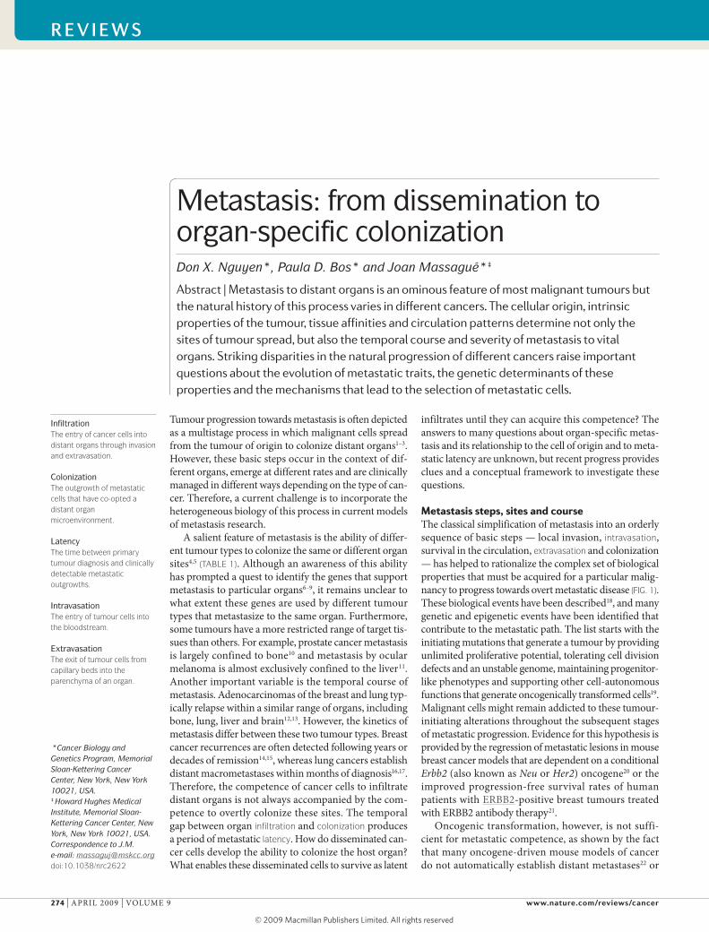
Tumour progression towards metastasis is often depicted as a multistage process in which malignant cells spread from the tumour of origin to colonize distant organs1–3. However, these basic steps occur in the context of dif-ferent organs, emerge at different rates and are clinically managed in different ways depending on the type of can-cer. Therefore, a current challenge is to incorporate the heterogeneous biology of this process in current models of metastasis research.
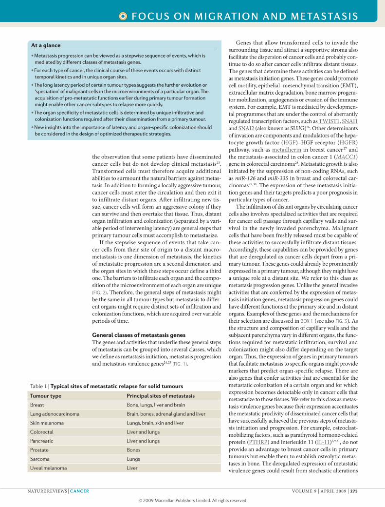
A salient feature of metastasis is the ability of differ-ent tumour types to colonize the same or different organ sites4,5 (TABLE 1). Although an awareness of this ability has prompted a quest to identify the genes that support metastasis to particular organs6–9, it remains unclear to what extent these genes are used by different tumour types that metastasize to the same organ. Furthermore, some tumours have a more restricted range of target tis-sues than others. For example, prostate cancer metastasis is largely confined to bone10 and metastasis by ocular melanoma is almost exclusively confined to the liver11. Another important variable is the temporal course of metastasis. Adenocarcinomas of the breast and lung typ-ically relapse within a similar range of organs, including bone, lung, liver and brain12,13. However, the kinetics of metastasis differ between these two tumour types. Breast cancer recurrences are often detected following years or decades of remission14,15, whereas lung cancers establish distant macrometastases within months of diagnosis16,17. Therefore, the competence of cancer cells to infiltrate distant organs is not always accompanied by the com-petence to overtly colonize these sites. The temporal gap between organ infiltration and colonization produces a period of metastatic latency. How do disseminated can-cer cells develop the ability to colonize the host organ? What enables these disseminated cells to survive as latent
infiltrates until they can acquire this competence? The answers to many questions about organ-specific metas-tasis and its relationship to the cell of origin and to meta-static latency are unknown, but recent progress provides clues and a conceptual framework to investigate these questions.
Metastasis steps, sites and courseThe classical simplification of metastasis into an orderly sequence of basic steps — local invasion, intravasation, survival in the circulation, extravasation and colonization — has helped to rationalize the complex set of biological properties that must be acquired for a particular malig-nancy to progress towards overt metastatic disease (FIG. 1). These biological events have been described18, and many genetic and epigenetic events have been identified that contribute to the metastatic path. The list starts with the initiating mutations that generate a tumour by providing unlimited proliferative potential, tolerating cell division defects and an unstable genome, maintaining progenitor-like phenotypes and supporting other cell-autonomous functions that generate oncogenically transformed cells19. Malignant cells might remain addicted to these tumour-initiating alterations throughout the subsequent stages of metastatic progression. Evidence for this hypothesis is provided by the regression of metastatic lesions in mouse breast cancer models that are dependent on a conditional Erbb2 (also known as Neu or Her2) oncogene20 or the improved progression-free survival rates of human patients with ERBB2-positive breast tumours treated with ERBB2 antibody therapy21.
Oncogenic transformation, however, is not suffi-cient for metastatic competence, as shown by the fact that many oncogene-driven mouse models of cancer do not automatically establish distant metastases22 or
*Cancer Biology and Genetics Program, Memorial Sloan-Kettering Cancer Center, New York, New York 10021, USA.‡ Howard Hughes Medical Institute, Memorial Sloan-Kettering Cancer Center, New York, New York 10021, USA.Correspondence to J.M. e-mail: [email protected]:10.1038/nrc2622
InfiltrationThe entry of cancer cells into distant organs through invasion and extravasation.
ColonizationThe outgrowth of metastatic cells that have co-opted a distant organ microenvironment.
LatencyThe time between primary tumour diagnosis and clinically detectable metastatic outgrowths.
IntravasationThe entry of tumour cells into the bloodstream.
ExtravasationThe exit of tumour cells from capillary beds into the parenchyma of an organ.
Metastasis: from dissemination to organ-specific colonizationDon X. Nguyen*, Paula D. Bos* and Joan Massagué*‡
Abstract | Metastasis to distant organs is an ominous feature of most malignant tumours but the natural history of this process varies in different cancers. The cellular origin, intrinsic properties of the tumour, tissue affinities and circulation patterns determine not only the sites of tumour spread, but also the temporal course and severity of metastasis to vital organs. Striking disparities in the natural progression of different cancers raise important questions about the evolution of metastatic traits, the genetic determinants of these properties and the mechanisms that lead to the selection of metastatic cells.
R E V I E W S
274 | ApRil 2009 | VOluME 9 www.nature.com/reviews/cancer
R E V I E W S
© 2009 Macmillan Publishers Limited. All rights reserved
the observation that some patients have disseminated cancer cells but do not develop clinical metastasis23. Transformed cells must therefore acquire additional abilities to surmount the natural barriers against metas-tasis. in addition to forming a locally aggressive tumour, cancer cells must enter the circulation and then exit it to infiltrate distant organs. After infiltrating new tis-sue, cancer cells will form an aggressive colony if they can survive and then overtake that tissue. Thus, distant organ infiltration and colonization (separated by a vari-able period of intervening latency) are general steps that primary tumour cells must accomplish to metastasize.
if the stepwise sequence of events that take can-cer cells from their site of origin to a distant macro-metastasis is one dimension of metastasis, the kinetics of metastatic progression are a second dimension and the organ sites in which these steps occur define a third one. The barriers to infiltrate each organ and the compo-sition of the microenvironment of each organ are unique (FIG. 2). Therefore, the general steps of metastasis might be the same in all tumour types but metastasis to differ-ent organs might require distinct sets of infiltration and colonization functions, which are acquired over variable periods of time.
General classes of metastasis genesThe genes and activities that underlie these general steps of metastasis can be grouped into several classes, which we define as metastasis initiation, metastasis progression and metastasis virulence genes24,25 (FIG. 1).
Genes that allow transformed cells to invade the surrounding tissue and attract a supportive stroma also facilitate the dispersion of cancer cells and probably con-tinue to do so after cancer cells infiltrate distant tissues. The genes that determine these activities can be defined as metastasis initiation genes. These genes could promote cell motility, epithelial–mesenchymal transition (EMT), extracellular matrix degradation, bone marrow progeni-tor mobilization, angiogenesis or evasion of the immune system. For example, EMT is mediated by developmen-tal programmes that are under the control of aberrantly regulated transcription factors, such as TWiST1, SNAi1 and SNAi2 (also known as SluG)26. Other determinants of invasion are components and modulators of the hepa-tocyte growth factor (HGF)–HGF receptor (HGFR) pathway, such as metadherin in breast cancer27 and the metastasis-associated in colon cancer 1 (MACC1) gene in colorectal carcinoma28. Metastatic growth is also initiated by the suppression of non-coding RNAs, such as miR‑126 and miR‑335 in breast and colorectal car-cinomas29,30. The expression of these metastasis initia-tion genes and their targets predicts a poor prognosis in particular types of cancer.
The infiltration of distant organs by circulating cancer cells also involves specialized activities that are required for cancer cell passage through capillary walls and sur-vival in the newly invaded parenchyma. Malignant cells that have been freshly released must be capable of these activities to successfully infiltrate distant tissues. Accordingly, these capabilities can be provided by genes that are deregulated as cancer cells depart from a pri-mary tumour. These genes could already be prominently expressed in a primary tumour, although they might have a unique role at a distant site. We refer to this class as metastasis progression genes. unlike the general invasive activities that are conferred by the expression of metas-tasis initiation genes, metastasis progression genes could have different functions at the primary site and in distant organs. Examples of these genes and the mechanisms for their selection are discussed in BOX 1 (see also FIG. 3). As the structure and composition of capillary walls and the subjacent parenchyma vary in different organs, the func-tions required for metastatic infiltration, survival and colonization might also differ depending on the target organ. Thus, the expression of genes in primary tumours that facilitate metastasis to specific organs might provide markers that predict organ-specific relapse. There are also genes that confer activities that are essential for the metastatic colonization of a certain organ and for which expression becomes detectable only in cancer cells that metastasize to those tissues. We refer to this class as metas-tasis virulence genes because their expression accentuates the metastatic proclivity of disseminated cancer cells that have successfully achieved the previous steps of metasta-sis initiation and progression. For example, osteoclast-mobilizing factors, such as parathyroid hormone-related protein (pTHRp) and interleukin 11 (il-11)6,9,31, do not provide an advantage to breast cancer cells in primary tumours but enable them to establish osteolytic metas-tases in bone. The deregulated expression of metastatic virulence genes could result from stochastic alterations
At a glance
•Metastasisprogressioncanbeviewedasastepwisesequenceofevents,whichismediatedbydifferentclassesofmetastasisgenes.
•Foreachtypeofcancer,theclinicalcourseoftheseeventsoccurswithdistincttemporalkineticsandinuniqueorgansites.
•Thelonglatencyperiodofcertaintumourtypessuggeststhefurtherevolutionor‘speciation’ofmalignantcellsinthemicroenvironmentsofaparticularorgan.Theacquisitionofpro-metastaticfunctionsearlierduringprimarytumourformationmightenableothercancersubtypestorelapsemorequickly.
•Theorganspecificityofmetastaticcellsisdeterminedbyuniqueinfiltrativeandcolonizationfunctionsrequiredaftertheirdisseminationfromaprimarytumour.
•Newinsightsintotheimportanceoflatencyandorgan-specificcolonizationshouldbeconsideredinthedesignofoptimizedtherapeuticstrategies.
Table 1 | Typical sites of metastatic relapse for solid tumours
Tumour type Principal sites of metastasis
Breast Bone, lungs, liver and brain
Lung adenocarcinoma Brain, bones, adrenal gland and liver
Skin melanoma Lungs, brain, skin and liver
Colorectal Liver and lungs
Pancreatic Liver and lungs
Prostate Bones
Sarcoma Lungs
Uveal melanoma Liver
R E V I E W S
NATuRE REViEWS | CanCer VOluME 9 | ApRil 2009 | 275
f o c u S o n m I g R at I o n a n d m E ta S ta S I S
© 2009 Macmillan Publishers Limited. All rights reserved
Metastasis virulence functions: organ-specific colonization PTHRP, IL11, CSF2RB (GM-CSF), IL6, TNFα
Nature Reviews | Cancer
Metastasis initiation functions: invasion, angiogenesis, marrow mobilization and circulation Gain of TWIST1, SNAI1, SNAI2, MET, ID1, Loss of KISS1, miR-126, miR-335, DARC, GPR56
Tumour initiation functions: growth, survival, progenitor-like state and genomic instability Oncogenes: ERBB2, CTNNB1 (β-catenin), KRAS, PI3K, EGFR, MYCTumour suppressors: APC, TP53, PTEN, BRCA1, BRCA2
ColonizationInfiltration (Latency)
Distant metastasisPrimary tumour
Carcinomain situ
Invasivecarcinoma Circulation
Metastasis virulePTHRP, IL11, CS
ation functions: invasion, angiogenesisST1, SNAI1, SNAI2, MET, TT ID1,
miR-126, miR-335, DARC, GPR56
Metastasis progression functions: extravasation, survival and reinitiation PTGS2, EREG, MMP1, LOX, ANGPTL4, CCL5 targets
in the context of genomic instability, and subsequently their expression could become stabilized as it provides a selective advantage to malignant cells in a particular microenvironment. These genes would not contribute to the expression signatures that are predictive of metastasis in primary tumours.
The temporal course of metastasisThe diverse temporal courses of metastasis in different types of cancer and patient populations are evident from clinical observations. As the kinetics of disease progres-sion and distinct physiological barriers can dictate the latency between the infiltrating and colonizing steps of metastasis, each clinical course has different implica-tions for the organ-selective evolution of metastatic cell populations (FIG. 4). in oestrogen receptor-positive breast cancer, prostate cancer and ocular melanoma, metasta-sis might become manifest decades after the removal of even a small primary malignancy11,32,33. The absence of immediate clinical relapse implies that these tumour cells are not fully competent to overtake organs imme-diately after infiltration. A protracted period of latency might ensue during which further malignant evolution of the disseminated cell population, of their microenviron-ment or of both must occur for colonization to proceed.
in other types of cancer, however, metastasis follows a swift course with rapid expansion in multiple organs that leaves little margin for speciation of the metastatic cell
population. For example, in lung cancers and pancreatic adenocarcinomas, malignant cells might rapidly acquire activities that confer both infiltration and colonization competence, as implied by the short time between pri-mary tumour diagnosis and metastatic relapse in these diseases16,34. in tumours with a rapid course of metastasis, the acquisition of robust metastatic traits in the primary tumour would obviate the need for extensive adaptation on dissemination to distant organs.Colorectal carcinoma is a defined paradigm of malignant progression and most metastatic traits seem to be acquired during local progression in the primary site. The transition from one stage to the next — from colorectal hyperpla-sia to adenoma to invasive carcinoma — is characterized by the acquisition of specific genetic alterations over a protracted period of up to three decades35. Colorectal tumours are initiated by the activation of the canonical Wnt pathway, through either mutational inactivation of the tumour suppressor adenomatous polyposis coli (APC) or activation of the pathway co-activator β-catenin36. The transition to carcinoma occurs with mutational activation of KRAS37, followed by oncogenic activation of the pi3K pathway38, inactivation of TP53 (REF. 39) and loss of the transforming growth factor-β (ΤGFβ) tumour suppressor pathway40. Once a colon tumour invades the underlying colonic wall, metastatic progression can proceed without latency. Colorectal tumours predominantly spread along the mesenteric circulation to the liver in 80% of patients
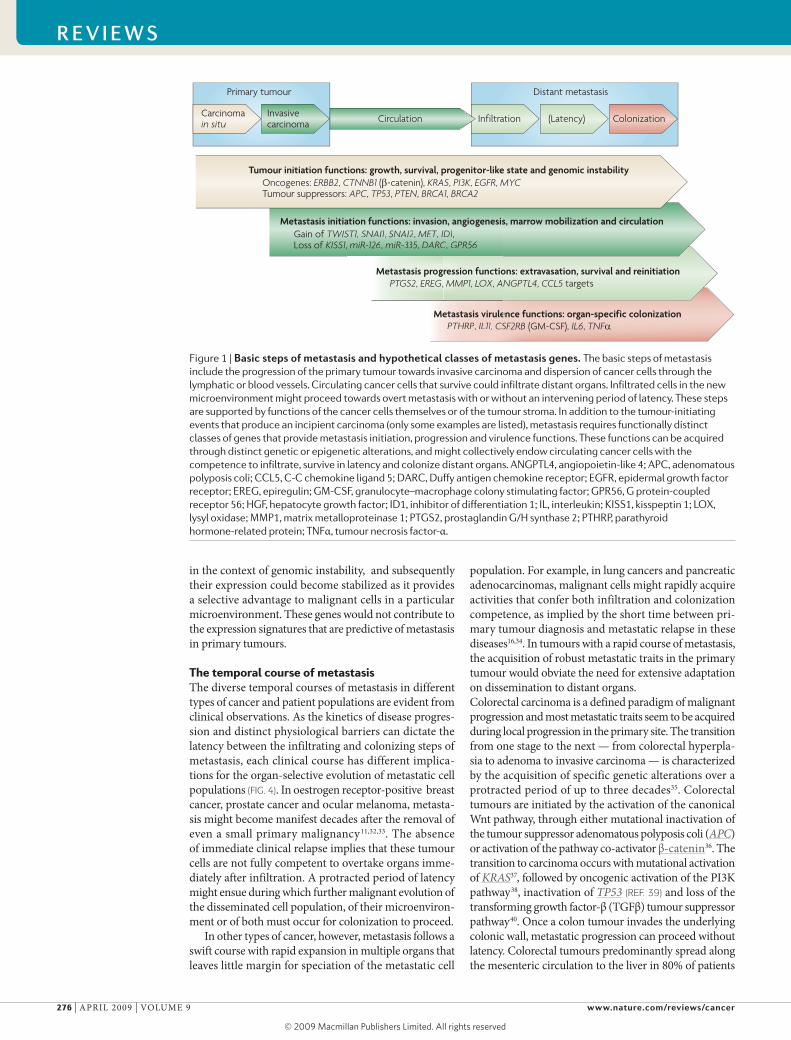
Figure 1 | Basic steps of metastasis and hypothetical classes of metastasis genes. The basic steps of metastasis include the progression of the primary tumour towards invasive carcinoma and dispersion of cancer cells through the lymphatic or blood vessels. Circulating cancer cells that survive could infiltrate distant organs. Infiltrated cells in the new microenvironment might proceed towards overt metastasis with or without an intervening period of latency. These steps are supported by functions of the cancer cells themselves or of the tumour stroma. In addition to the tumour-initiating events that produce an incipient carcinoma (only some examples are listed), metastasis requires functionally distinct classes of genes that provide metastasis initiation, progression and virulence functions. These functions can be acquired through distinct genetic or epigenetic alterations, and might collectively endow circulating cancer cells with the competence to infiltrate, survive in latency and colonize distant organs. ANGPTL4, angiopoietin-like 4; APC, adenomatous polyposis coli; CCL5, C-C chemokine ligand 5; DARC, Duffy antigen chemokine receptor; EGFR, epidermal growth factor receptor; EREG, epiregulin; GM-CSF, granulocyte–macrophage colony stimulating factor; GPR56, G protein-coupled receptor 56; HGF, hepatocyte growth factor; ID1, inhibitor of differentiation 1; IL, interleukin; KISS1, kisspeptin 1; LOX, lysyl oxidase; MMP1, matrix metalloproteinase 1; PTGS2, prostaglandin G/H synthase 2; PTHRP, parathyroid hormone-related protein; TNFα, tumour necrosis factor-α.
R E V I E W S
276 | ApRil 2009 | VOluME 9 www.nature.com/reviews/cancer
R E V I E W S
© 2009 Macmillan Publishers Limited. All rights reserved
Bone marrow sinusoid
Haematopoietic cell
Cancer cell
Fenestration
Sinus cell
Alveolar cell
Endothelial cell
Basementmembrane
Cancer cell
Disseminatedbreast tumour cell
Brain infiltration
Brain colonization
Bone infiltration
Bone colonization
Lung colonization
Nature Reviews | Cancer
Blood–brain barrier
Lung capillary
Basementmembrane
Endothelial cell
Astrocyte
Pericyte
Cancer cell
with recurrent disease12. it is estimated that most genetic alterations for metastasis are acquired during progression to the invasive carcinoma stage, and few, if any, additional genetic events are required for the formation of distant liver metastases41. Therefore, colorectal cancers progress slowly to invasive carcinomas but progress rapidly from this stage to the metastatic phase (FIG. 4).
General versus organ-specific infiltrationTo enter the circulation and infiltrate distant organs, aggressive cancer cells must invade the surrounding tissues. Various mechanisms that confer invasiveness, such as cellular motility and basement membrane deg-radation, have been proposed to mediate cancer cell entry into the circulation42,43. Deregulated cytoskel-etal modifiers such as RHOC can specifically enhance metastatic dissemination44. The aberrant expression of developmental transcription factors might trigger EMT, which is associated with cellular plasticity and invasion26.
The capacity to disseminate could be intrinsic to certain pre-malignant cell lineages. it has long been recognized that many normal cell types are involved in complex migratory and invasive behaviours during development and adulthood. The traffic and homing to peripheral tissues of bone marrow-derived progeni-tors of myeloid, endothelial and mesenchymal lineages have been characterized. Normal epithelial cells are also motile. in the mammary gland, the invasive and migratory mechanisms that underlie the branching morphogenesis of normal epithelial cells also regu-late the formation of mammary hyperplasia45. A sub-set of luminal progenitors in early breast carcinomas might use these invasive and migratory mechanisms to disseminate to the lungs46. Depending on their cel-lular origin, epithelial stem cells or progenitors that leave their original niche might have intrinsic invasive capabilities that are independent of malignant trans-formation. indeed, a proportion of normal murine pre-malignant mammary cells can infiltrate the lungs when
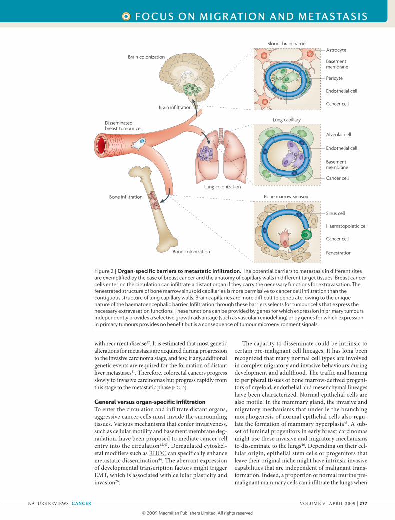
Figure 2 | Organ-specific barriers to metastatic infiltration. The potential barriers to metastasis in different sites are exemplified by the case of breast cancer and the anatomy of capillary walls in different target tissues. Breast cancer cells entering the circulation can infiltrate a distant organ if they carry the necessary functions for extravasation. The fenestrated structure of bone marrow sinusoid capillaries is more permissive to cancer cell infiltration than the contiguous structure of lung capillary walls. Brain capillaries are more difficult to penetrate, owing to the unique nature of the haematoencephalic barrier. Infiltration through these barriers selects for tumour cells that express the necessary extravasation functions. These functions can be provided by genes for which expression in primary tumours independently provides a selective growth advantage (such as vascular remodelling) or by genes for which expression in primary tumours provides no benefit but is a consequence of tumour microenvironment signals.
R E V I E W S
NATuRE REViEWS | CanCer VOluME 9 | ApRil 2009 | 277
f o c u S o n m I g R at I o n a n d m E ta S ta S I S
© 2009 Macmillan Publishers Limited. All rights reserved
injected into the circulation47. Moreover, some human mammary epithelial cell types are more metastatic than others following experimental transformation with defined oncogenes48. The potential for dissemina-tion of tumour cells can be further enhanced by signals that induce EMT, which also augment the fraction of progenitor-like cancer cells49.
Certain aspects of the vasculature have been pro-posed to contribute to dissemination. For example, the metastasis suppressor CD82 (also known as KAi1) nor-mally anchors tumour cells to the endothelium through its interaction with the Duffy antigen chemokine recep-tor (DARC), inducing the senescence of bound epithe-lial cells50. loss of CD82 therefore facilitates metastatic spread. Tumour cell–platelet interactions could also enable dissemination by forming cell aggregates that protect tumour cells from immune surveillance51,52 or by collaborating during extravasation53,54.
Although some general mechanisms of dissemination enable tumour cells to abandon the primary tumour and reach distal organs, more specialized mechanisms might be necessary for the infiltration of specific organs. Four of the most common sites of secondary relapse include the bone marrow, lungs, liver and brain. infiltration into these organs is influenced in part by circulation patterns. in colorectal carcinoma, the mesenteric circulation from the bowels and the permissiveness of the liver capil-lary sinusoids are thought to favour liver metastasis55,56. Following blood flow patterns from the liver or from pri-mary tumours on the descending colon, the second most frequent sites of metastasis are the lungs12. However, in addition to the influence of haematogenous dynamics, colon carcinoma cells preferentially adhere to the liver and lung endothelia, suggesting the existence of specific molecular interactions that favour the retention of tumour cells in these organs57. The role of unique endothelial sur-face molecules as target sites for compatible disseminat-ing cancer cells has been shown in breast cancer cell lines overexpressing the cell adhesion molecule metadherin. in a mouse model, metadherin specifically bound to the pulmonary vasculature and enhanced lung metastasis58.
in addition to the possible role of organ-specific endothelial adhesive interactions, we must consider how the structural features of capillary walls in differ-ent organs can affect infiltration (FIG. 2). The capillaries in the bone marrow, called sinusoids, are lined with fenes-trated endothelia to facilitate the traffic of haematopoietic cells59. Therefore, the bone marrow sinusoids are likely to be more permissive to circulating tumour cells. The capil-laries of the liver are also fenestrated and readily traversed by tumour cells compared with other organs55,56. By con-trast, lung capillaries are lined with endothelial cells that are surrounded by a basement membrane (the fusion of two basal laminae) and adjacent alveolar cells. The base-ment membrane is an obstacle that circulating tumour cells can bypass only by expressing specific mediators of transendothelial migration60–63. Additional contacts between the tumour cells and an exposed basement membrane might facilitate the infiltration of the target organ, as exemplified by the interactions between α3β1 integrin on breast cancer cells and laminin 5 on lung cap-illary basement membranes64. The blood–brain barrier, with its tight layer of endothelial cells and astrocyte foot processes, represents more of an obstacle65. Consequently, the infiltration of circulating cancer cells into the brain parenchyma could require highly specialized functions, many of which remain to be characterized.
Several mediators of pulmonary extravasation have been recently identified and are upregulated in the pri-mary tumours of breast cancer patients that relapse to the lungs61,66. These include epiregulin, prostaglandin G/H synthase 2 (pTGS2; also known as COX2), matrix metallo-proteinase 1 (MMp1) and MMp2, which support not only vascular remodelling in primary tumours, but also lung extravasation61. Another specific mediator of lung extrava-sation is the cytokine angiopoietin-like 4 (ANGpTl4), which enhances the infiltration of tumour cells into the lungs by inducing the dissociation of endothelial cell–cell junctions63. However, the absence of a robust association
Box 1 | Mediators of metastasis in the primary tumour
Theexpressioninprimarytumoursofgenesthatmediatemetastaticactivitiesmightseemparadoxical.Howcouldagenethathasaspecificfunctioninadistantorganbeselectedforbeforeprimarytumourcellsbecomeexposedtotheselectivepressuresofthatuniquemicroenvironment?Variousanswersaresuggestedbyrecentprogress.Theexpressionofmetastasisinitiationgenesinprimarytumoursisdrivenbytheneedforcellmobility,invasiveness,angiogenesisandimmuneevasionduringtheoutgrowthofprimarytumours,aswellasthesubsequentoutgrowthofdistantmetastases.However,theprominentexpressionofmetastasisprogressiongenesinprimarytumourshasamorecomplexbasis.ExpressionofprostaglandinG/Hsynthase2(PTGS2;alsoknownasCOX2),matrixmetalloproteinase1andtheepidermalgrowthfactorreceptorligandepiregulininbreastcancercellspromotesangiogenesisinexperimentalmammarytumours.Whenexpressedincancercellsenteringthecirculation,thissetofgenesalsoincreasestheabilityofthedisseminatedcellstoextravasateintothepulmonaryparenchyma61.Thesemetastasisprogressiongenesareprominentlyexpressedinaprimarytumourbecausetheysupporttumourgrowththroughoneparticulareffect,whereastheyenhancedistantmetastasisthroughanothereffect.Theboundariesbetweenmetastasisinitiationandmetastasisprogressiongenesare
notrigid.Mediatorsofmetastasisthatwerepreviouslythoughttoregulateoneactivitymightalsoconferactivitiesthatparticipateinotherprocesses.Forexample,thehypoxia-regulatedgenelysyloxidaseispredictiveofrelapseinhumanbreasttumoursandwasinitiallyfoundtoenhancecancercellinvasion106.RecentstudiessuggestthatsystemicsecretionoflysyloxidaseintothelungandlivermightfacilitatethehomingofdisseminatedcancercellstotheseorgansthrougheffectsontheextracellularmatrixthathelptorecruitCD11b+myeloidcells,formingapro-metastaticmicroenvironment107.Inothersituations,theexpressionofpro-metastaticgenesinaprimarytumourmight
beoneofmanybystandereventsthatdonotcontributetoprimarytumorigenesis.Forexample,theexpressionofthesegenescouldbepartofaglobalresponsetocytokinesinthetumourmicroenvironment(FIG. 3).Bonemarrowprogenitors108,endothelialcells109,macrophagesandothermyeloidcells110,111,aswellasmesenchymalprogenitorcells62,arestromalcomponentsthatreleaseparacrinefactorsinresponsetomalignancy.Althoughsomeofthesesignalsareco-optedbythetumourforgrowth,othersareneutraltoprimarytumourdevelopmentbutmightprimecancercellsfordistantmetastasis.CCL5isreleasedbybonemarrow-derivedmesenchymalprogenitorcellsinfiltratingintomammarytumoursandstimulatescancercellstometastasizetothelungwithoutaffectingtumorigenesis62.Transforminggrowthfactor-β (TGFβ)isaprominentcytokineinthetumourmicroenvironment112.TGFβinducestheexpressionofalargesetofgenesinbreasttumourcells,amongwhichangiopoietin-like 4(ANGPTL4)disruptsendothelialcell–cellcontactswithoutprovidinganydiscernablebenefitinmammarytumours.However,cancercellsdepartingfromTGFβ-richprimarytumoursandexpressingANGPTL4haveaninfiltrationadvantageastheyreachthelungcapillaries63.Metastasisprogressiongenesinothercancermodelsawaitfurthercharacterization.
R E V I E W S
278 | ApRil 2009 | VOluME 9 www.nature.com/reviews/cancer
R E V I E W S
© 2009 Macmillan Publishers Limited. All rights reserved
Nature Reviews | Cancer
Breast tumour
Prostaglandins
Epiregulin
LOXTGFβ
Lung capillary
ANGPTL4Prostaglandins
Epiregulin
PTGS2EREG
EREGPTGS2
ANGPTL4
LOX
LOX
Basal breast cancerA more aggressive subtype of breast cancer with characteristics of mammary basal cells, and that typically lacks oestrogen and progesterone receptors.
Luminal breast cancerA subtype of breast cancer with characteristics of cells that originate from the normal lumen or ducts of the mammary gland.
between any primary tumour gene expression event and bone metastasis might reflect the more permissive nature of bone marrow sinusoids and hence less of a requirement for extravasation functions in the departing breast cancer cells that enter the bone marrow7,67.
Metastasis without intervening latencyThe duration of metastatic latency and the sites in which it occurs have implications for the development of organ-specific metastasis functions. in some types of cancer, aggressive macrometastases frequently develop soon after cancer cells infiltrate distant organs; exam-ples include lung and pancreatic adenocarcinomas, which are two highly prevalent types of cancer with high relapse and mortality rates following initial diagnosis. The relapse rate is substantial even following the detec-tion of early-stage tumours. For example, the 5-year recurrence-free rate in stage i lung adenocarcinoma patients is 60–70% (REFS 17,68). By contrast, patients diagnosed with stage i breast cancers have a 98% 5-year recurrence-free survival rate69, and differences in diag-nostic modalities alone do not account for this differ-ence. patients with limited-stage small-cell lung cancer are even more likely to have metastatic disease at the time of diagnosis70. Moreover, lung adenocarcinoma relapses to brain, bone and the contralateral lung, and metastases to these various sites frequently occur concomitantly12. Malignant skin melanoma can also relapse swiftly,
spreading to cutaneous tissues, lungs, liver, brain and bone. Recurrence in melanoma usually occurs within 2 years of diagnosis, with few relapses after 5 years71. There are also differences between different tumour sub-types; for example, basal breast cancers classified by gene signatures relapse earlier than luminal breast cancers72.
The short latency of metastatic relapse in aggres-sive diseases implies that potent multi-organ metastatic competence either exists in the pre-malignant cells or is acquired during the early stages of malignant trans-formation. The early acquisition of robust multi-organ metastatic competence might obviate the need for exten-sive adaptation by cancer cells to the microenvironment of different affected organs. The determinants of this competence remain unknown and are a topic of intense investigation. The cell and tissue of origin might provide a partial explanation for such rapid metastasis. During melanoma progression, for example, the melanocyte lineage specification programme predisposes to trans-formation by lineage-determining oncogenes such as microphthalmia-associated transcription factor (MiTF), a key transcription factor for melanocyte lineage sur-vival73. it has also been proposed that melanocytes retain some embryonic plasticity owing to their neural crest ori-gin. The transcription factor SNAi2 is required for neural crest cell migration and its expression in melanoma cells drives metastasis to multiple organ sites in mice74.
The influence of the epigenetic state of progenitor cells on their metastatic competence might extend to other cancers. indeed, different subsets of solid tumours that express transcriptional modules that are unique to embryonic stem cells have a higher probability of general recurrence75. The embryonic-like plasticity of aggressive cancer cells might reflect or phenocopy the plasticity that is inherent to stem and early progenitor cells, which is maintained in part by global epigenetic regulators. Some of these regulators have been linked to metastasis, including polycomb chromatin remodelling complexes76,77 and microRNAs30,78–80. The selection for activated developmental pathways might also enhance metastatic competence to multiple organs by enforcing this plasticity and providing strong invasive and adap-tation functions to cancer cells. identifying the mecha-nisms that promote metastatic progression without latent speciation might indicate crucial therapeutic targets for early intervention.
Metastatic latency and its host sitesThe counterpoint to tumour types that rapidly colonize distant organs with a short disease-free interval on ini-tial diagnosis are tumours that can efficiently infiltrate distant organs at early stages but are unable to promptly grow as macrometastases. in breast cancer, disseminated tumour cells (DTCs) enter a state of metastatic latency, which is defined as the time between primary tumour diagnosis and clinically detectable metastatic relapse15,81. Malignant cells from breast tumours that disseminate early can reside as single cells or as micrometastatic clus-ters, as shown in studies of bone marrow samples from patients without overt metastatic disease14,82,83. These DTCs either lack the ability to colonize or are prevented
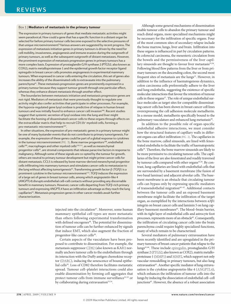
Figure 3 | Metastasis progression genes expressed in the primary tumour. Mediators of metastasis might have dual functions that provide both a local advantage for malignant progression in the primary tumour and a distal advantage for infiltration of a particular organ, such as the lung in a breast cancer patient in this example. The expression of genes such as epiregulin (EREG) and prostaglandin G/H synthase 2 (PTGS2) promotes capillary assembly from endothelial and smooth muscle cells in mammary tumours. However, these genes also increase the ability of breast cancer cells to pass through endothelial barriers, a function that increases cancer cell extravasation in the lungs. Lysyl oxidase (LOX) is induced in primary tumours that respond to hypoxic signals to enhance cancer cell invasion. However, systemic secretion of LOX leads to its accumulation in the lung, where it has been suggested to act on extracellular matrix proteins to establish a permissive niche for infiltrating cancer cells. In the case of the cytokine angiopoietin-like 4 (ANGPTL4), expression in mammary tumour cells is driven not by a selective growth advantage, but by the action of tumour-derived transforming growth factor-β (TGFβ), which also stimulates the expression of many other genes. By itself, ANGPTL4 does not provide any discernable advantage in the primary tumour, but its induction by TGFβ in departing tumour cells primes these cells for infiltration of the lungs. ANGPTL4 dissociates vascular endothelial cell–cell junctions, an effect that in lung capillaries increases the infiltration of ANGPTL4-secreting cancer cells into the lung parenchyma.
R E V I E W S
NATuRE REViEWS | CanCer VOluME 9 | ApRil 2009 | 279
f o c u S o n m I g R at I o n a n d m E ta S ta S I S
© 2009 Macmillan Publishers Limited. All rights reserved
Nature Reviews | Cancer
Breast carcinoma
Bone metastasis
Lung metastasis
Brainmetastasis
Latency(years to decades)
Competenceto infiltrate
Competenceto colonize
Lung adenocarcinoma
Bone metastasis
Lung metastasis
Brain metastasis
Latency(months)
Latency(months)
Competenceto infiltrateand to colonize
Invasive carcinoma
Livermetastasis
Lungmetastasis
Competenceto infiltrateand to colonize
Colorectal carcinoma
Progression(years to decades)
Adenoma
a
b
c
DormancyA state of cellular quiescence in the G0 phase of the cell cycle. When referring to a tumour cell mass, dormancy describes a balanced state of proliferation and apoptosis.
Angiogenic switchThe transition between a non-angiogenic state of the tumour cell mass and a neovascularized state that enables tumour oxygenation and growth.
from displaying colonization by the environment. As a result, DTCs can enter a state of proliferative dormancy by exiting the proliferative cycle for an indef-inite period. Alternatively, DTCs grow indolently as micrometastatic colonies owing to a rate of cell death that counterbalances the rate of proliferation, which gives rise to ‘tumour mass dormancy’. The two general forms of latency are not mutually exclusive and could coexist in the DTC population of a particular cancer patient (FIG. 5).
Both the organ microenvironment and the oncoge-netic background might play important parts in forc-ing metastatic latency. in a polyoma middle T antigen (pyMT) mouse model, tumour cells lacking β1 integrins fail to sense fibronectin as an environmental cue, result-ing in growth arrest84. Stress signals stemming from
the foreign microenvironment have been proposed to induce dormancy by modulating the ratio of Erk and p38 MApKs in DTCs85,86. interestingly, DTCs obtained from transgenic tumour models and transplanted into the marrow of wild-type recipients can expand in the recipient marrow, suggesting that the dormant state can be rapidly discontinued by changes in the micro-environment83. The expression of active metastasis suppressor genes could also contribute to metastatic latency, as exemplified by kisspeptin 1 (KiSS1), which prevents metastatic cells from reinitiating growth on infiltration of distant organs87. Another metastasis suppressor is the G protein-coupled receptor GpR56, which interacts with tissue transglutaminase from the extracellular matrix. loss of GpR56 expression in meta-static melanoma cells promotes tumour outgrowth88. Furthermore, host polymorphisms can modulate the efficiency of tumour metastases, as exemplified by the Sipa1 polymorphism that has been described in mice89. The failure of micrometastatic lesions to trigger the angiogenic switch owing to local anti-angiogenic fac-tors such as thrombospondin has also been associated with dormant metastasis90–92.
Although most breast cancer cells that enter the cir-culation and infiltrate distant organs die owing to restric-tive forces in the host microenvironment93, other factors could provide unique advantages to infiltrating cancer cells that are equipped to exploit survival signals. The bone marrow is a permissive niche for the traffic and residence of haematopoietic stem cells59 and seems to be a protective environment for disseminated tumour cells in patients undergoing chemotherapy94. These observa-tions suggest that the bone marrow might provide sur-vival signals that sustain the viability of DTCs. C-X-C chemokine receptor 4 (CXCR4) is the receptor for the cell survival chemokine stromal cell-derived factor 1 (SDF1; also known as CXCl12), which is produced by mesenchymal cells in the bone marrow. Notably, CXCR4 expression in breast cancer cells is a marker and media-tor of bone metastasis in breast cancer9,95. Therefore, SDF1 and CXCR4 are candidate mediators of latent DTC survival in the bone marrow (FIG. 5).
To be compatible with eventual macrometastatic outgrowth, latent DTCs require not only the ability to survive during latency, but also the capacity to reinitiate a tumour when conditions are favourable. The develop-ment of macrometastases in patients who have isolated DTCs is a manifestation of the ‘tumour-propagating phe-notype’ — also referred to as the ‘cancer stem cell pheno-type’ — which DTCs require for the eventual reinitiation of aggressive tumour growth. The expression of inhibi-tor of differentiation 1 (ID1) and ID3 supports the abil-ity of human breast cancer cells to bypass senescence and reinitiate growth on extravasation into the lungs of mice. Furthermore, the expression of ID1 and ID3 in cell clusters of basal or triple-negative subtype breast tumours is associated with lung metastasis96,97. These examples suggest that the ability to reinitiate growth at the secondary site can be stochastic owing to newly established interactions between the tumour cell and the target microenvironment or can be already encoded
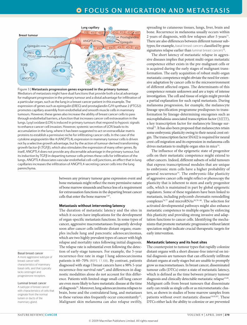
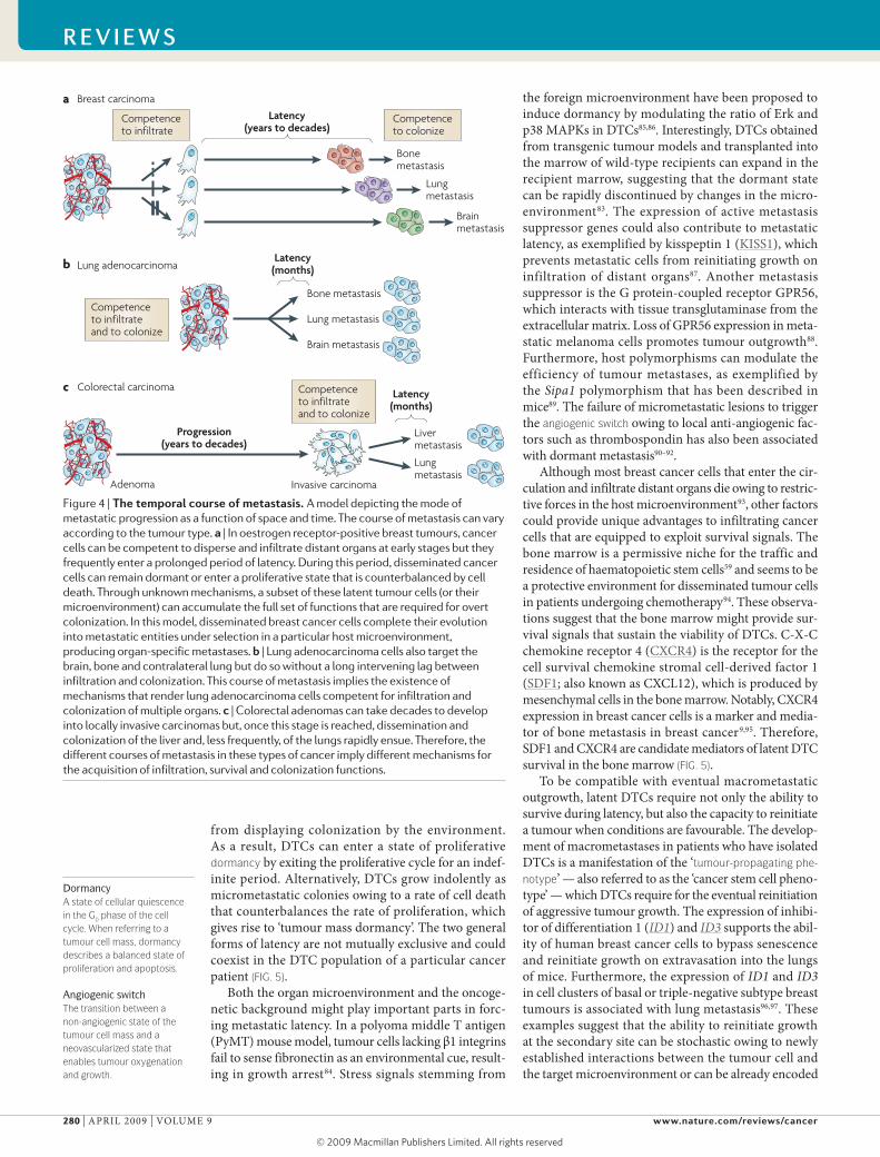
Figure 4 | The temporal course of metastasis. A model depicting the mode of metastatic progression as a function of space and time. The course of metastasis can vary according to the tumour type. a | In oestrogen receptor-positive breast tumours, cancer cells can be competent to disperse and infiltrate distant organs at early stages but they frequently enter a prolonged period of latency. During this period, disseminated cancer cells can remain dormant or enter a proliferative state that is counterbalanced by cell death. Through unknown mechanisms, a subset of these latent tumour cells (or their microenvironment) can accumulate the full set of functions that are required for overt colonization. In this model, disseminated breast cancer cells complete their evolution into metastatic entities under selection in a particular host microenvironment, producing organ-specific metastases. b | Lung adenocarcinoma cells also target the brain, bone and contralateral lung but do so without a long intervening lag between infiltration and colonization. This course of metastasis implies the existence of mechanisms that render lung adenocarcinoma cells competent for infiltration and colonization of multiple organs. c | Colorectal adenomas can take decades to develop into locally invasive carcinomas but, once this stage is reached, dissemination and colonization of the liver and, less frequently, of the lungs rapidly ensue. Therefore, the different courses of metastasis in these types of cancer imply different mechanisms for the acquisition of infiltration, survival and colonization functions.
R E V I E W S
280 | ApRil 2009 | VOluME 9 www.nature.com/reviews/cancer
R E V I E W S
© 2009 Macmillan Publishers Limited. All rights reserved
Nature Reviews | Cancer
PTHRP, IL-6IL-11 and TNFα
SDF1
Stromalniche
RANKL
TGFβ,IGF1 andBMPs
Osteoclasts
Osteoblasts
OPG
Circulatingcancer cells
G0 (dormant)
Micrometastasis(indolent growth)
Growthfactors
Deathsignals
Osteolyticmacrometastasis
Bone matrixBone marrowInfiltration and latency
Bonelysis
Colonization competence (years to decades)
in the arriving tumour cell. identifying the balance of signals that affect DTC turnover and the properties required for these cells to maintain a viable state despite latency should provide valuable clues for therapeutic intervention against minimal residual disease.
Organ-selective metastatic speciationin malignancies of the breast and prostate, in which relapses occur after a prolonged latency period, the acquisition of competency for colonization is likely to occur during the residence of DTCs in a particular organ microenvironment. The bone marrow, lung, brain and liver parenchyma impose different selective pressures for the establishment of metastatic colonies. Therefore, the eventual colonization of these organs by temporar-ily latent DTCs could involve the acquisition of specific functions. This process would be predicted to yield organ-specific metastatic cells. indeed, for a patient with breast cancer, tumour recurrence frequently occurs in one particular organ before it occurs in others. Strikingly, in prostate cancer, bone metastasis is frequently the only site of distant relapse, implying that metastatic prostate cancer cells are not competent to aggressively colonize other organs.
Genetic or epigenetic fluctuation of a DTC population, systemic or local changes in the microenvironment, or a combination of these factors might eventually endow surviving DTCs with full competence for aggressive colo-nization. under the selective pressure of the host microen-vironment, these events can produce different metastatic cells that are specifically adapted to grow in one particular organ, yielding different ‘species’ of metastasis in differ-ent organs of the same patient. Metastatic cells that are released from distant organs in patients with advanced metastatic disease can commingle in the circulation and other fluids, providing a demographic cross section of the extant species of metastasis in that patient. indeed, malig-nant cells and cell lines isolated from the pleural fluid of patients with breast cancer produce subpopulations with distinct organ-specific tropisms when inoculated in mice7. Notably, these organ-specific metastatic phenotypes are stable ex vivo, suggesting that they evolved through the accumulation of genetic or epigenetic alterations that became fixed in the metastatic population. According to this hypothesis, metastasis speciation results from the pro-tracted evolution of latent DTCs towards full metastatic competence at secondary sites (BOX 2).
Organ-specific colonization functions have been well documented in bone metastasis. The ability of breast cancer cells to form typical osteolytic metastases requires the production of osteoclast-activating factors, such as pTHRp, il-11, il-6, tumour necrosis factor-α (TNFα) and granulocyte–macrophage colony stimulating factor (GM-CSF)6,9,31,98. pTHRp, il-6, il-11 and TNFα act on osteoclasts to promote the secretion of receptor activa-tor of nuclear factor-κB ligand (RANKl), which induces osteoclast formation. GM-CSF directly promotes osteo-clastogenesis. Expression of these secreted factors would be unlikely to provide a selective advantage in another metastatic site or in the primary tumour, yet they are essential for the development of osteolytic lesions (FIG. 5). The identity of specific molecular mediators of coloni-zation in other organ microenvironments, such as the brain or the liver, remains unknown. However, the range of unique cell types that comprise the brain parenchyma and their anatomical organization65 raise the possibility that brain metastasis involves an active crosstalk between
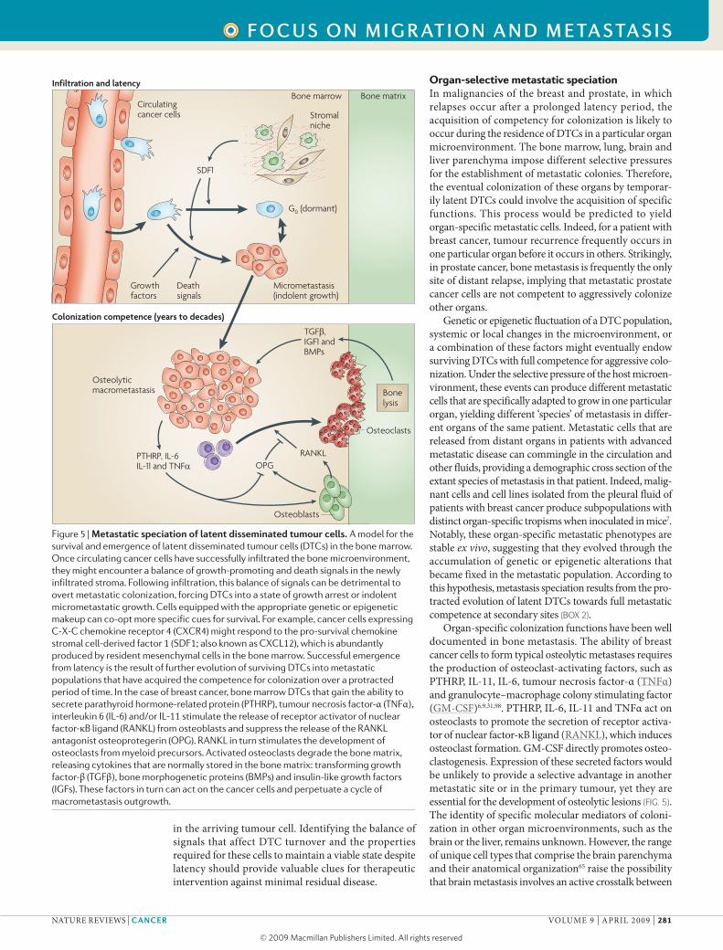
Figure 5 | Metastatic speciation of latent disseminated tumour cells. A model for the survival and emergence of latent disseminated tumour cells (DTCs) in the bone marrow. Once circulating cancer cells have successfully infiltrated the bone microenvironment, they might encounter a balance of growth-promoting and death signals in the newly infiltrated stroma. Following infiltration, this balance of signals can be detrimental to overt metastatic colonization, forcing DTCs into a state of growth arrest or indolent micrometastatic growth. Cells equipped with the appropriate genetic or epigenetic makeup can co-opt more specific cues for survival. For example, cancer cells expressing C-X-C chemokine receptor 4 (CXCR4) might respond to the pro-survival chemokine stromal cell-derived factor 1 (SDF1; also known as CXCL12), which is abundantly produced by resident mesenchymal cells in the bone marrow. Successful emergence from latency is the result of further evolution of surviving DTCs into metastatic populations that have acquired the competence for colonization over a protracted period of time. In the case of breast cancer, bone marrow DTCs that gain the ability to secrete parathyroid hormone-related protein (PTHRP), tumour necrosis factor-α (TNFα), interleukin 6 (IL-6) and/or IL-11 stimulate the release of receptor activator of nuclear factor-κB ligand (RANKL) from osteoblasts and suppress the release of the RANKL antagonist osteoprotegerin (OPG). RANKL in turn stimulates the development of osteoclasts from myeloid precursors. Activated osteoclasts degrade the bone matrix, releasing cytokines that are normally stored in the bone matrix: transforming growth factor-β (TGFβ), bone morphogenetic proteins (BMPs) and insulin-like growth factors (IGFs). These factors in turn can act on the cancer cells and perpetuate a cycle of macrometastasis outgrowth.
R E V I E W S
NATuRE REViEWS | CanCer VOluME 9 | ApRil 2009 | 281
f o c u S o n m I g R at I o n a n d m E ta S ta S I S
© 2009 Macmillan Publishers Limited. All rights reserved
Tumour-propagating phenotypeThe ability of the infiltrated tumour cells to reinitiate growth at the secondary site. This is referred to by some investigators as the ‘cancer stem cell phenotype’.
Metastatic speciationAn evolutionary process by which new metastatic populations arise, owing to the various selective pressures that act on the heterogeneous cancer cells escaping the primary tumour.
GliosisStimulation of astrocytes in injured areas of the brain.
cancer and stromal cells. Consistent with this hypothesis, strong gliosis has been observed in clinical samples of human metastasis, and in vitro evidence suggests that glial cells support the growth of metastatic cells99.
Influence of therapy on metastatic courseAs an end-stage malignant disease, metastatic relapse is often associated with resistance to therapy. Relapse following systemic treatments might be due to cell-intrinsic mechanisms such as genetic alterations that confer drug resistance following a period of therapeu-tic response. lung adenocarcinomas with epidermal growth factor receptor (EGFR) mutations respond to EGFR kinase inhibitors but frequently relapse owing to secondary EGFR mutations that confer resistance100. Certain mechanisms of drug resistance might simul-taneously render the tumour more competent for metastasis. For example, a subset of EGFR-mutant lung adenocarcinomas becomes insensitive to the EGFR kinase inhibitors gefinitib and erlotinib owing to the amplification of MET, which encodes the tyrosine kinase receptor HGFR. HGFR activation can heter-ologously increase EGFR signalling, thus promoting the survival of tumours that are addicted to this path-way101,102. However, HGF has a direct role in develop-mental and pathogenic cell migration101,102. Moreover, MET expression is also regulated by metadherin, which
confers resistance to chemotherapy27 and mediates lung metastasis58. Consequently, resistance to therapy is cou-pled with the potential acquisition of pro-metastatic functions in tumours in which the HGF–HGFR pathway is activated.
Alternatively, therapies can indirectly influence the course and pattern of metastasis by delaying systemic disease and favouring the emergence of recurrences in specific organs; this is exemplified by the rising incidence of brain metastasis in ERBB2-positive breast cancer patients treated with the ERBB2 antibody trastuzumab (Herceptin)65,103. The causes of preferential relapse in the brain following systemic therapy are intriguing and have been attributed to the ‘sanctuary’ nature of the brain parenchyma. Cancer cells growing in the central nervous system could be shielded by the blood–brain barrier from drug delivery or protected by survival sig-nals from the host microenvironment104. The partial effectiveness of adjuvant intervention might increase the incidence of more latent brain metastases, in which DTCs are forced to adapt and acquire specific genetic alterations that favour macrometastatic outgrowth in the central nervous system.
Other specific organ niches could similarly pro-vide microenvironments protective against therapeutic intervention. it has been suggested that DTCs remain quiescent in the bone marrow, providing an explana-tion for the failure of cytotoxic therapies to treat patients with breast cancer, of whom 15–20% can have residual tumour cells after completing adjuvant cytotoxic and endocrine therapy93,105. in this regard, the identification of tissue-specific prognostic signatures might provide more tailored clinical options. Although these examples show how therapies might select for specific metastatic traits, aggressive metastatic cells could also emerge inde-pendently of intervention and be intrinsically resistant to subsequent treatment. This might be particularly rel-evant in rapidly progressing tumour types, such as lung cancer and melanoma, for which there are few effective treatment modalities other than surgical intervention.
PerspectivesAs clinical oncology progresses towards personalized cancer medicine, the need to understand the biology of metastasis becomes increasingly acute. in the past few years, we have witnessed an invigoration of this field, accompanied by technological developments that are enhancing our understanding of how metastasis deve-lops and how it might be amenable to therapy. Three important needs could be addressed at this point. The first is the incorporation of clinical knowledge of the steps, sites and temporal course of metastasis into experimental models of disease. The second is the dis-section of metastasis into clinically relevant cellular and molecular components that drive this process in organ-specific patterns. The third is the translation of this information into a better classification of tumours on the basis of molecular markers of metastatic potential and of therapeutic intervention against latent and active metastatic disease. We are optimistic that progress will be made towards these goals in the coming years.
Box 2 | Metastatic speciation from disseminated tumour cells
Thepresenceofdisseminatedtumourcells(DTCs)inpatientswhoseprimarytumourshavebeenremovedcorrelateswithmetastaticrelapse,suggestingthatthesecellsareasourceoffuturerecurrence113.DTCshavebeendetectedprimarilyinthebonemarrowbutalsointheperipheralbloodandlymphnodes.ThelackofspecificmarkersandthedifficultyofisolatingDTCsfromotherorgansprecludeusfromknowingwhethertheywidelydisseminateorthebonemarrowpreferentiallyactsasaninitialreservoirofDTCs114,115.Ifthebonemarrowactsassuchareservoir,DTCscouldevolvefromindolentdiseaseintoafullyfledgedlocalbonemetastasisor,alternatively,evolveuntiltheyarecompetenttoseedsecondaryorgans,suchasthelungandthebrain,inwhichfurtherorgan-specificevolutionmightoccur.Irrespectiveofthedistantorganlocation,DTCswillencounterdifferentselective
pressuresfromthoseattheprimarysite.DTCsmightbeunabletosurviveowingtotheirfailuretoestablishproductiveinteractionswiththisnewlyinfiltratedenvironment.Alternatively,DTCscanuseexistingcomponentsoftheircellularmachinerytoderiveanadvantagefromnewlyencounteredsurvivalcues.DependingonhowDTCsrespondtolocalsignals,apopulationofDTCscancontinuegrowingorenteraphaseofbalancedproliferationandapoptosisuntilsufficientrandomgeneticandepigeneticvariationaccumulatesformetastaticexpansionofclonesthatareoptimallyadaptedtothehostmicroenvironment.Wecallthisprocessmetastatic speciation.DTCsobtainedfromthebonemarrow,lymphnodesandbloodofindividualcancer
patientsexistinadiversegenomicstate116.Usingsingle-cellcomparativegenomichybridization,DTCsisolatedfromthebonemarrowafterprimarytumourresectionwerefoundtohavefeweraberrationsthantheprimarytumourtheywerederivedfrom.Theearlydisseminationofbreastcancercellsandtheirgeneticdivergenceimplythatmetastaticlesionsandtheoriginaltumourcouldevolveindependently14,117.However,itremainsunclearwhethermetastaticoutgrowthpreferentiallyoccursfromtheseearliestlatentDTCsorinitiatesfromalaterseedingofcancercellsthathadalreadybecomemoreaggressiveinthecontextoftheexpandingprimarytumour.Indeed,severalstudiesshowthatovertmetastasesandmostaggressiveprimarytumourssharesimilargeneexpressionpatterns,implyingthatatleastsomemetastatictraitsarecommonbetweenmetastasesandtheirprimarytumouroforiginatsomestage118,119.ProgressinunderstandingtheoriginandfateofDTCs,andhowtheysuccessfullyproducesecondarytumours,willoffernewinsightsintothisintriguingprocess.
R E V I E W S
282 | ApRil 2009 | VOluME 9 www.nature.com/reviews/cancer
R E V I E W S
© 2009 Macmillan Publishers Limited. All rights reserved
1. Christofori, G. New signals from the invasive front. Nature 441, 444–450 (2006).
2. Gupta, G. P. & Massague, J. Cancer metastasis: building a framework. Cell 127, 679–695 (2006).
3. Steeg, P. S. Tumor metastasis: mechanistic insights and clinical challenges. Nature Med. 12, 895–904 (2006).
4. Fidler, I. J. The pathogenesis of cancer metastasis: the ‘seed and soil’ hypothesis revisited. Nature Rev. Cancer 3, 453–458 (2003).
5. Paget, S. The distribution of secondary growths in cancer of the breast. 1889. Cancer Metastasis Rev. 8, 98–101 (1989).
6. Yin, J. J. et al. TGF-β signaling blockade inhibits PTHrP secretion by breast cancer cells and bone metastases development. J. Clin. Invest. 103, 197–206 (1999).
7. Minn, A. J. et al. Distinct organ-specific metastatic potential of individual breast cancer cells and primary tumors. J. Clin. Invest. 115, 44–55 (2005).
This paper shows that pleural effusion-derived metastatic cell populations are heterogeneous in their ability to colonize different organs, supporting the notion that various target organs impose different requirements on arriving tumour cells.
8. Minn, A. J. et al. Genes that mediate breast cancer metastasis to lung. Nature 436, 518–524 (2005).
9. Kang, Y. et al. A multigenic program mediating breast cancer metastasis to bone. Cancer Cell 3, 537–549 (2003).
This paper shows that in vivo selection enriches for bone metastatic ability and identifies genetic mediators of bone metastatic colonization.
10. Edlund, M., Sung, S. Y. & Chung, L. W. Modulation of prostate cancer growth in bone microenvironments. J. Cell. Biochem. 91, 686–705 (2004).
11. Triozzi, P. L., Eng, C. & Singh, A. D. Targeted therapy for uveal melanoma. Cancer Treat. Rev. 34, 247–258 (2008).
12. Hess, K. R. et al. Metastatic patterns in adenocarcinoma. Cancer 106, 1624–1633 (2006).
A recent clinical study that reports the frequency of organ-specific relapse in 11 different types of adenocarcinomas from over 4,000 patients.
13. Patanaphan, V., Salazar, O. M. & Risco, R. Breast cancer: metastatic patterns and their prognosis. South. Med. J. 81, 1109–1112 (1988).
14. Schmidt-Kittler, O. et al. From latent disseminated cells to overt metastasis: genetic analysis of systemic breast cancer progression. Proc. Natl Acad. Sci. USA 100, 7737–7742 (2003).
This paper shows that disseminated tumour cells have different and fewer aberrations than their matched primary tumours, suggesting that dissemination is an early event during cancer development.
15. Karrison, T. G., Ferguson, D. J. & Meier, P. Dormancy of mammary carcinoma after mastectomy. J. Natl Cancer Inst. 91, 80–85 (1999).
16. Feld, R., Rubinstein, L. V. & Weisenberger, T. H. Sites of recurrence in resected stage I non-small-cell lung cancer: a guide for future studies. J. Clin. Oncol. 2, 1352–1358 (1984).
17. Hoffman, P. C., Mauer, A. M. & Vokes, E. E. Lung cancer. Lancet 355, 479–485 (2000).
18. Chambers, A. F., Groom, A. C. & MacDonald, I. C. Dissemination and growth of cancer cells in metastatic sites. Nature Rev. Cancer 2, 563–572 (2002).
19. Hanahan, D. & Weinberg, R. A. The hallmarks of cancer. Cell 100, 57–70 (2000).
20. Moody, S. E. et al. Conditional activation of Neu in the mammary epithelium of transgenic mice results in reversible pulmonary metastasis. Cancer Cell 2, 451–461 (2002).
21. Slamon, D. J. et al. Use of chemotherapy plus a monoclonal antibody against HER2 for metastatic breast cancer that overexpresses HER2. N. Engl. J. Med. 344, 783–792 (2001).
22. Minna, J. D., Kurie, J. M. & Jacks, T. A big step in the study of small cell lung cancer. Cancer Cell 4, 163–166 (2003).
23. Klein, C. A. The systemic progression of human cancer: a focus on the individual disseminated cancer cell—the unit of selection. Adv. Cancer Res. 89, 35–67 (2003).
24. Chiang, A. C. & Massagué, J. Molecular basis of metastasis. N. Engl. J. Med. 359, 2814–2823 (2008).
25. Nguyen, D. X. & Massagué, J. Genetic determinants of cancer metastasis. Nature Rev. Genet. 8, 341–352 (2007).
26. Yang, J. & Weinberg, R. A. Epithelial–mesenchymal transition: at the crossroads of development and tumor metastasis. Dev. Cell 14, 818–829 (2008).
27. Hu, G. et al. MTDH activation by 8q22 genomic gain promotes chemoresistance and metastasis of poor-prognosis breast cancer. Cancer Cell 15, 9–20 (2009).
A study that mechanistically links the pro-metastatic gene metadherin with resistance to chemotherapy.
28. Stein, U. et al. MACC1, a newly identified key regulator of HGF–MET signaling, predicts colon cancer metastasis. Nature Med. 15, 59–67 (2009).
29. Guo, C. et al. The noncoding RNA, miR-126, suppresses the growth of neoplastic cells by targeting phosphatidylinositol 3-kinase signaling and is frequently lost in colon cancers. Genes Chromosomes Cancer 47, 939–946 (2008).
30. Tavazoie, S. F. et al. Endogenous human microRNAs that suppress breast cancer metastasis. Nature 451, 147–152 (2008).
31. Mundy, G. R. Metastasis to bone: causes, consequences and therapeutic opportunities. Nature Rev. Cancer 2, 584–593 (2002).
32. Lee, Y. T. Patterns of metastasis and natural courses of breast carcinoma. Cancer Metastasis Rev. 4, 153–172 (1985).
33. Johansson, J. E. et al. Natural history of early, localized prostate cancer. JAMA 291, 2713–2719 (2004).
34. Nieto, J., Grossbard, M. L. & Kozuch, P. Metastatic pancreatic cancer 2008: is the glass less empty? Oncologist 13, 562–576 (2008).
35. Fearon, E. R. & Vogelstein, B. A genetic model for colorectal tumorigenesis. Cell 61, 759–767 (1990).
36. Kinzler, K. W. & Vogelstein, B. Lessons from hereditary colorectal cancer. Cell 87, 159–170 (1996).
37. Vogelstein, B. et al. Genetic alterations during colorectal-tumor development. N. Engl. J. Med. 319, 525–532 (1988).
38. Samuels, Y. et al. High frequency of mutations of the PIK3CA gene in human cancers. Science 304, 554 (2004).
39. Baker, S. J. et al. Chromosome 17 deletions and p53 gene mutations in colorectal carcinomas. Science 244, 217–221 (1989).
40. Markowitz, S. et al. Inactivation of the type II TGF-β receptor in colon cancer cells with microsatellite instability. Science 268, 1336–1338 (1995).
41. Jones, S. et al. Comparative lesion sequencing provides insights into tumor evolution. Proc. Natl Acad. Sci. USA 105, 4283–4288 (2008).
A study that evaluated the frequency and timing of somatic mutations to estimate the clinical course of colorectal metastatic progression.
42. Kedrin, D., van Rheenen, J., Hernandez, L., Condeelis, J. & Segall, J. E. Cell motility and cytoskeletal regulation in invasion and metastasis. J. Mammary Gland Biol. Neoplasia 12, 143–152 (2007).
43. Weber, G. F. Molecular mechanisms of metastasis. Cancer Lett. 270, 181–190 (2008).
44. Clark, E. A., Golub, T. R., Lander, E. S. & Hynes, R. O. Genomic analysis of metastasis reveals an essential role for RhoC. Nature 406, 532–535 (2000).
45. Ewald, A. J., Brenot, A., Duong, M., Chan, B. S. & Werb, Z. Collective epithelial migration and cell rearrangements drive mammary branching morphogenesis. Dev. Cell 14, 570–581 (2008).
46. Kouros-Mehr, H. et al. GATA-3 links tumor differentiation and dissemination in a luminal breast cancer model. Cancer Cell 13, 141–152 (2008).
47. Podsypanina, K. et al. Seeding and propagation of untransformed mouse mammary cells in the lung. Science 321, 1841–1844 (2008).
This paper showed that phenotypically normal mouse mammary cells introduced into the mouse circulation can infiltrate the lungs and survive, leading to tumour initiation.
48. Ince, T. A. et al. Transformation of different human breast epithelial cell types leads to distinct tumor phenotypes. Cancer Cell 12, 160–170 (2007).
This study showed that there are intrinsic differences in the tumorigenic and metastatic capabilities of different mammary cell types.
49. Mani, S. A. et al. The epithelial–mesenchymal transition generates cells with properties of stem cells. Cell 133, 704–715 (2008).
50. Bandyopadhyay, S. et al. Interaction of KAI1 on tumor cells with DARC on vascular endothelium leads to metastasis suppression. Nature Med. 12, 933–938 (2006).
51. Karpatkin, S. & Pearlstein, E. Role of platelets in tumor cell metastases. Ann. Intern. Med. 95, 636–641 (1981).
52. Nieswandt, B., Hafner, M., Echtenacher, B. & Mannel, D. N. Lysis of tumor cells by natural killer cells in mice is impeded by platelets. Cancer Res. 59, 1295–1300 (1999).
53. Im, J. H. et al. Coagulation facilitates tumor cell spreading in the pulmonary vasculature during early metastatic colony formation. Cancer Res. 64, 8613–8619 (2004).
54. Jain, S. et al. Platelet glycoprotein Ibα supports experimental lung metastasis. Proc. Natl Acad. Sci. USA 104, 9024–9028 (2007).
55. Paku, S., Dome, B., Toth, R. & Timar, J. Organ-specificity of the extravasation process: an ultrastructural study. Clin. Exp. Metastasis 18, 481–492 (2000).
56. Lalor, P. F., Lai, W. K., Curbishley, S. M., Shetty, S. & Adams, D. H. Human hepatic sinusoidal endothelial cells can be distinguished by expression of phenotypic markers related to their specialised functions in vivo. World J. Gastroenterol. 12, 5429–5439 (2006).
57. Schluter, K. et al. Organ-specific metastatic tumor cell adhesion and extravasation of colon carcinoma cells with different metastatic potential. Am. J. Pathol. 169, 1064–1073 (2006).
58. Brown, D. M. & Ruoslahti, E. Metadherin, a cell surface protein in breast tumors that mediates lung metastasis. Cancer Cell 5, 365–374 (2004).
Using phage-display libraries — a technology that had previously allowed this group to identify tissue-specific vasculature differences (or ‘zipcodes’) — this paper identifies metadherin as a lung-specific homing molecule.
59. Kopp, H. G., Avecilla, S. T., Hooper, A. T. & Rafii, S. The bone marrow vascular niche: home of HSC differentiation and mobilization. Physiology 20, 349–356 (2005).
60. Weis, S., Cui, J., Barnes, L. & Cheresh, D. Endothelial barrier disruption by VEGF-mediated Src activity potentiates tumor cell extravasation and metastasis. J. Cell Biol. 167, 223–229 (2004).
61. Gupta, G. P. et al. Mediators of vascular remodelling co-opted for sequential steps in lung metastasis. Nature 446, 765–770 (2007).
62. Karnoub, A. E. et al. Mesenchymal stem cells within tumour stroma promote breast cancer metastasis. Nature 449, 557–563 (2007).
63. Padua, D. et al. TGFβ primes breast tumors for lung metastasis seeding through angiopoietin-like 4. Cell 133, 66–77 (2008).
References 62 and 63 exemplify how paracrine signals from the stroma of a primary tumour can stimulate departing cancer cells to extravasate into the lung without affecting primary tumorigenesis.
64. Wang, H. et al. Tumor cell α3β1 integrin and vascular laminin-5 mediate pulmonary arrest and metastasis. J. Cell Biol. 164, 935–941 (2004).
65. Weil, R. J., Palmieri, D. C., Bronder, J. L., Stark, A. M. & Steeg, P. S. Breast cancer metastasis to the central nervous system. Am. J. Pathol. 167, 913–920 (2005).
66. Minn, A. J. et al. Lung metastasis genes couple breast tumor size and metastatic spread. Proc. Natl Acad. Sci. USA 104, 6740–6745 (2007).
67. Smid, M. et al. Genes associated with breast cancer metastatic to bone. J. Clin. Oncol. 24, 2261–2267 (2006).
68. Nesbitt, J. C., Putnam, J. B. Jr, Walsh, G. L., Roth, J. A. & Mountain, C. F. Survival in early-stage non-small cell lung cancer. Ann. Thorac. Surg. 60, 466–472 (1995).
69. Ries, L. A. G. et al. SEER Cancer Statistics Review, 1975–2005 National Cancer Institute [online] http://seer.cancer.gov/csr/1975_2005/index.html (2008)
70. Janne, P. A. et al. Twenty-five years of clinical research for patients with limited-stage small cell lung carcinoma in North America. Cancer 95, 1528–1538 (2002).
71. Briele, H. A. & Das Gupta, T. K. Natural history of cutaneous malignant melanoma. World J. Surg. 3, 255–270 (1979).
72. Sorlie, T. et al. Gene expression patterns of breast carcinomas distinguish tumor subclasses with clinical implications. Proc. Natl Acad. Sci. USA 98, 10869–10874 (2001).
73. Garraway, L. A. et al. Integrative genomic analyses identify MITF as a lineage survival oncogene amplified in malignant melanoma. Nature 436, 117–122 (2005).
74. Gupta, P. B. et al. The melanocyte differentiation program predisposes to metastasis after neoplastic transformation. Nature Genet. 37, 1047–1054 (2005).
R E V I E W S
NATuRE REViEWS | CanCer VOluME 9 | ApRil 2009 | 283
f o c u S o n m I g R at I o n a n d m E ta S ta S I S
© 2009 Macmillan Publishers Limited. All rights reserved
75. Wong, D. J. et al. Module map of stem cell genes guides creation of epithelial cancer stem cells. Cell Stem Cell 2, 333–344 (2008).
This study shows an association between the expression of embryonic stem cell gene modules in primary tumours and increased metastatic potential.
76. Varambally, S. et al. The polycomb group protein EZH2 is involved in progression of prostate cancer. Nature 419, 624–629 (2002).
77. Kleer, C. G. et al. EZH2 is a marker of aggressive breast cancer and promotes neoplastic transformation of breast epithelial cells. Proc. Natl Acad. Sci. USA 100, 11606–11611 (2003).
78. Varambally, S. et al. Genomic loss of microRNA-101 leads to overexpression of histone methyltransferase EZH2 in cancer. Science 322, 1695–1699 (2008).
79. Gregory, P. A. et al. The miR-200 family and miR-205 regulate epithelial to mesenchymal transition by targeting ZEB1 and SIP1. Nature Cell Biol. 10, 593–601 (2008).
80. Ma, L., Teruya-Feldstein, J. & Weinberg, R. A. Tumour invasion and metastasis initiated by microRNA-10b in breast cancer. Nature 449, 682–688 (2007).
81. Demicheli, R. Tumour dormancy: findings and hypotheses from clinical research on breast cancer. Semin. Cancer Biol. 11, 297–306 (2001).
82. Braun, S. et al. Cytokeratin-positive cells in the bone marrow and survival of patients with stage I, II, or III breast cancer. N. Engl. J. Med. 342, 525–533 (2000).
83. Husemann, Y. et al. Systemic spread is an early step in breast cancer. Cancer Cell 13, 58–68 (2008).
This paper shows that dissemination of tumour cells can occur at early stages of primary tumour development in ERBB2 and PyMT mouse models. Moreover, transplantation of pre-malignant DTCs into recipient bone marrow releases these cells from dormancy.
84. White, D. E. et al. Targeted disruption of β1-integrin in a transgenic mouse model of human breast cancer reveals an essential role in mammary tumor induction. Cancer Cell 6, 159–170 (2004).
85. Aguirre-Ghiso, J. A., Estrada, Y., Liu, D. & Ossowski, L. ERKMAPK activity as a determinant of tumor growth and dormancy; regulation by p38SAPK. Cancer Res. 63, 1684–1695 (2003).
86. Aguirre-Ghiso, J. A., Ossowski, L. & Rosenbaum, S. K. Green fluorescent protein tagging of extracellular signal-regulated kinase and p38 pathways reveals novel dynamics of pathway activation during primary and metastatic growth. Cancer Res. 64, 7336–7345 (2004).
87. Nash, K. T. et al. Requirement of KISS1 secretion for multiple organ metastasis suppression and maintenance of tumor dormancy. J. Natl Cancer Inst. 99, 309–321 (2007).
88. Xu, L., Begum, S., Hearn, J. D. & Hynes, R. O. GPR56, an atypical G protein-coupled receptor, binds tissue transglutaminase, TG2, and inhibits melanoma tumor growth and metastasis. Proc. Natl Acad. Sci. USA 103, 9023–9028 (2006).
89. Park, Y. G. et al. Sipa1 is a candidate for underlying the metastasis efficiency modifier locus Mtes1. Nature Genet. 37, 1055–1062 (2005).
90. Holmgren, L., O’Reilly, M. S. & Folkman, J. Dormancy of micrometastases: balanced proliferation and apoptosis in the presence of angiogenesis suppression. Nature Med. 1, 149–153 (1995).
91. Almog, N. et al. Transcriptional switch of dormant tumors to fast-growing angiogenic phenotype. Cancer Res. 69, 836–844 (2009).
92. Naumov, G. N., Akslen, L. A. & Folkman, J. Role of angiogenesis in human tumor dormancy: animal models of the angiogenic switch. Cell Cycle 5, 1779–1787 (2006).
93. Luzzi, K. J. et al. Multistep nature of metastatic inefficiency: dormancy of solitary cells after successful extravasation and limited survival of early micrometastases. Am. J. Pathol. 153, 865–873 (1998).
94. Becker, S., Becker-Pergola, G., Wallwiener, D., Solomayer, E. F. & Fehm, T. Detection of cytokeratin-positive cells in the bone marrow of breast cancer patients undergoing adjuvant therapy. Breast Cancer Res. Treat. 97, 91–96 (2006).
95. Ling, L. J. et al. A novel mouse model of human breast cancer stem-like cells with high CD44+CD24-/lower phenotype metastasis to human bone. Chin. Med. J. 121, 1980–1986 (2008).
96. Gupta, G. P. et al. ID genes mediate tumor reinitiation during breast cancer lung metastasis. Proc. Natl Acad. Sci. USA 104, 19506–19511 (2007).
97. Swarbrick, A., Roy, E., Allen, T. & Bishop, J. M. Id1 cooperates with oncogenic Ras to induce metastatic mammary carcinoma by subversion of the cellular senescence response. Proc. Natl Acad. Sci. USA 105, 5402–5407 (2008).
98. Park, B. K. et al. NF-κB in breast cancer cells promotes osteolytic bone metastasis by inducing osteoclastogenesis via GM-CSF. Nature Med. 13, 62–69 (2007).
99. Fitzgerald, D. P. et al. Reactive glia are recruited by highly proliferative brain metastases of breast cancer and promote tumor cell colonization. Clin. Exp. Metastasis 25, 799–810 (2008).
100. Sharma, S. V., Bell, D. W., Settleman, J. & Haber, D. A. Epidermal growth factor receptor mutations in lung cancer. Nature Rev. Cancer 7, 169–181 (2007).
101. Engelman, J. A. et al. MET amplification leads to gefitinib resistance in lung cancer by activating ERBB3 signaling. Science 316, 1039–1043 (2007).
102. Bean, J. et al. MET amplification occurs with or without T790M mutations in EGFR mutant lung tumors with acquired resistance to gefitinib or erlotinib. Proc. Natl Acad. Sci. USA 104, 20932–20937 (2007).
103. Aragon-Ching, J. B. & Zujewski, J. A. CNS metastasis: an old problem in a new guise. Clin. Cancer Res. 13, 1644–1647 (2007).
104. Fidler, I. J., Yano, S., Zhang, R. D., Fujimaki, T. & Bucana, C. D. The seed and soil hypothesis: vascularisation and brain metastases. Lancet Oncol. 3, 53–57 (2002).
105. Wiedswang, G. et al. Isolated tumor cells in bone marrow three years after diagnosis in disease-free breast cancer patients predict unfavorable clinical outcome. Clin. Cancer Res. 10, 5342–5348 (2004).
106. Erler, J. T. et al. Lysyl oxidase is essential for hypoxia-induced metastasis. Nature 440, 1222–1226 (2006).
107. Erler, J. T. et al. Hypoxia-induced lysyl oxidase is a critical mediator of bone marrow cell recruitment to form the premetastatic niche. Cancer Cell 15, 35–44 (2009).
References 106 and 107 describe the metastasis progression gene LOX and how it can be induced in the primary tumour to mediate one function at the primary site and a different function in secondary organs, both of which are required for metastasis.
108. Kaplan, R. N. et al. VEGFR1-positive haematopoietic bone marrow progenitors initiate the pre-metastatic niche. Nature 438, 820–827 (2005).
109. Hiratsuka, S. et al. The S100A8–serum amyloid A3–TLR4 paracrine cascade establishes a pre-metastatic phase. Nature Cell Biol. 10, 1349–1355 (2008).
110. Condeelis, J. & Pollard, J. W. Macrophages: obligate partners for tumor cell migration, invasion, and metastasis. Cell 124, 263–266 (2006).
111. Kim, S. et al. Carcinoma-produced factors activate myeloid cells through TLR2 to stimulate metastasis. Nature 457, 102–106 (2009).
112. Bierie, B. & Moses, H. L. Tumour microenvironment: TGFβ: the molecular Jekyll and Hyde of cancer. Nature Rev. Cancer 6, 506–520 (2006).
113. Braun, S. et al. A pooled analysis of bone marrow micrometastasis in breast cancer. N. Engl. J. Med. 353, 793–802 (2005).
114. Wikman, H., Vessella, R. & Pantel, K. Cancer micrometastasis and tumour dormancy. APMIS 116, 754–770 (2008).
115. Pantel, K. & Brakenhoff, R. H. Dissecting the metastatic cascade. Nature Rev. Cancer 4, 448–456 (2004).
116. Klein, C. A. et al. Genetic heterogeneity of single disseminated tumour cells in minimal residual cancer. Lancet 360, 683–689 (2002).
117. Schardt, J. A. et al. Genomic analysis of single cytokeratin-positive cells from bone marrow reveals early mutational events in breast cancer. Cancer Cell 8, 227–239 (2005).
118. Ramaswamy, S., Ross, K. N., Lander, E. S. & Golub, T. R. A molecular signature of metastasis in primary solid tumors. Nature Genet. 33, 49–54 (2003).
119. Weigelt, B. et al. Gene expression profiles of primary breast tumors maintained in distant metastases. Proc. Natl Acad. Sci. USA 100, 15901–15905 (2003).
AcknowledgementsWe thank members of the Massagué lab for insightful discus-sions. The primary research for the topic of this review is sup-ported by grants from the National Institutes of Health, the Hearst Foundation and the Kleberg Foundation. D.X.N was a postdoctoral fellow of the Damon Runyon Cancer Research Foundation. J.M. is an Investigator of the Howard Hughes Medical Institute.
DATABASESEntrez gene: http://www.ncbi.nlm.nih.gov/entrez/query.fcgi?db=geneAPC | KRAS | ID1 | ID3 | MACC1 | MET | TP53uniProtKB: http://www.uniprot.org β-catenin | ANGPTL4 | CD82 | CXCR4 | DARC | EGFR | epiregulin | ERBB2 | GM-CSF | HGF | HGFR | IL-11 | KISS1 | metadherin | MITF | MMP1 | MMP2 | PTGS2 | PTHRP | RANKL | RHOC | SDF1 | SNAI1 | SNAI2 | TNFα | TWIST1
FURTHER INFORMATIONJoan massagué’s homepage: http://www.mskcc.org/mskcc/html/10614.cfm
all links are aCTive in The Online Pdf
R E V I E W S
284 | ApRil 2009 | VOluME 9 www.nature.com/reviews/cancer
R E V I E W S
© 2009 Macmillan Publishers Limited. All rights reserved
Author Biographies
Don X. Nguyen received his ph.D. in 2004 from the university of Rochester, New York, uSA, in the Department of Microbiology and immunology. He was a postdoctoral fellow of the Damon Runyon Cancer Research Foundation, and is currently working in the labo-ratory of Joan Massagué. His scientific interests are identifying the determinants of lung cancer metastasis.
paula D. Bos received her M.Sc. in genetics from the National university of Misiones, Argentina. She is currently a graduate student in the laboratory of Joan Massagué, where she focuses on the molecu-lar mechanisms of metastasis, particularly breast cancer metastasis to the brain.
Joan Massagué is chairman of the Cancer Biology and Genetics program at the Memorial Sloan-Kettering Cancer Center, New York, uSA, and an investigator of the Howard Hughes Medical institute, New York, uSA. He received his ph.D. in biochemistry from the university of Barcelona, Spain, in 1978. in 1982, he joined the faculty at the university of Massachusetts Medical School, Massachusetts, uSA. He assumed his current positions in 1989. He is interested in how growth factors, signalling pathways and gene expression pro-grammes control normal cell behaviour and cancer cell metastasis.
000 metastasis: from dissemination to organ-specific colonizationDon X. Nguyen, Paula D. Bos and Joan MassaguéThe natural history of metastasis — which appears to be cancer-type specific — varies by target organ, latency and severity. This Review discusses how organ speciation and the competence to colonize might develop.
o n l I n E o n ly
© 2009 Macmillan Publishers Limited. All rights reserved