Embed Size (px)
Citation preview

This presentation is the intellectual property of the author. Contact them for permission to reprint and/or distribute.
Medial and Lateral Collateral Ligament
InjuriesJohn C. Pearce, MD
Medial Collateral Ligament
Most commonly injured Incidence is probably higher 50% chance of meniscal injury ACL most commonly associate
Lateral Collateral Ligament
Incidence not known Isolated tear rare More functional knee disabilities
Medial Collateral Ligament

This presentation is the intellectual property of the author. Contact them for permission to reprint and/or distribute.
Anatomy - MCL
Static– Superficial medial collateral ligament Medial femoral epicondyle to anteromedial tibia Anterior fibers are constant tension throughout
flexion Posterior fibers are sack in flexion
– Posterior oblique ligament Triangular capsular ligament Tight in extension - slack in flexion Dynamized by semimebranosus
– Deep medial collateral ligament Capsular ligament Meniscotibial and meniscofemoral fibers Dynamic
– Semimebranosus– Pes Anserine Sartorius Semitendenosis Gracilis
– Vastus medialis
Biomechanics
Resist valgus and external rotation of tibia Superficial medial collateral ligament
5-7 mm increase in laxity 200-300% increase in rotational laxity
Clinical Evaluation
HistoryWhen ability to returnActivity previous injuryMechanism since injuryPain initial treatmentSwelling

This presentation is the intellectual property of the author. Contact them for permission to reprint and/or distribute.
Physical Exam
Observation edmaGait ecchymosisEffusion deformity
MechanicalPalpationNevrovascularROMAbduction stress testOther ligament and structures
Abduction stress test at different degrees of flexion
grade I 1-4 mmgrade II 5-9 mmgrade III 10-15 mm
Diagnostic Testing
Radiographs– Fractures– Loose bodies– Physical injuries
MRI– Location of tear– Degree of tear– Associated injuries
Treatment
Non-operative – Grade I and grade II injuries criteria Stable in extension No more than 10 mm of valgus opening at 30
degree flexion No rotational instability Localized tenderness Minimal effusion Normal radiographs
This presentation is the intellectual property of the author. Contact them for permission to reprint and/or distribute.
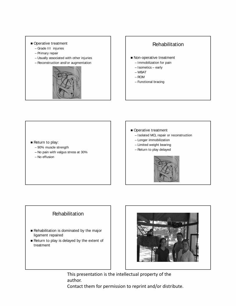
Operative treatment– Grade III injuries– Primary repair– Usually associated with other injuries– Reconstruction and/or augmentation
Rehabilitation
Non-operative treatment– Immobilization for pain– Isometics – early– WBAT– ROM– Functional bracing
Return to play:– 90% muscle strength– No pain with valgus stress at 30%– No effusion
Operative treatment– Isolated MCL repair or reconstruction– Longer immobilization– Limited weight bearing– Return to play delayed
Rehabilitation
Rehabilitation is dominated by the major ligament repaired
Return to play is delayed by the extent of treatment

This presentation is the intellectual property of the author. Contact them for permission to reprint and/or distribute.
Anatomy: LCL and PLC
Arcuate complex– Lateral collateral ligament Static restraint to varus Static restraint to external rotation of the tibia
Arcuate ligament – static– Variable– Reinforces posterolateral capsule
Popliteus muscle – dynamic – Reinforces posterior lateral capsule – Internally rotates tibia
Popliteofibular ligament– Variable– Static resistance to external rotation of the
tibia Biceps femoris tendon and ilitibial band
– Dynamic stability
Biomechanics
Lateral and posterolateral structures – Variable– Stronger and more substantial – Subject to greater forces– Primary resistance Varus rotation External tibial rotation Posterior tibial translation
Clinical Evaluation
HistoryWhen ability to returnActivity previous injuryMechanism since injuryPain initial treatmentSwelling

This presentation is the intellectual property of the author. Contact them for permission to reprint and/or distribute.
Physical Exam
Observation gait ecchymosiseffusion deformityedma
Mechanical– Palpation– Neurovascular Peroneal nerve injury 15-30%
– ROM– Adduction stress test– Increased external rotation of tibia at 30
degrees and 90 degrees of flexion
– Dial test– Posterior tibial translation at 30 degrees not
at 90 degrees– External rotation recurvatum – Reverse pivot shift test
Diagnostic Testing
Radiographs– Fractures– Loose bodies– Physeal injuries
MRI– Location of tear– Degree of tear– Associated injuries

This presentation is the intellectual property of the author. Contact them for permission to reprint and/or distribute.
Treatment
Non-operative– Grade I and grade II– 2-4 weeks of protected weight bearing– Progressive rehabilitation
Operative – Generally grade III injuries– Combination injuries– Primary repair – augmentation – Acute injuries much easier
Rehabilitation
Depends upon repair and/or augmentation– Limited weight bearing– Immobilization or combination of ROM and
immoblization– Slow progression back to play
Adolesants
Consider physeal injuries Knee pain – think hip
Thank YouJohn C. Pearce