Embed Size (px)
Citation preview

Posterior Cruciate Ligament Injuries

Anatomy
• Origin: Lateral border of the medial femoral condyle
• Insertion: 1.5 cm below the top of the tibia in the PCL facet

Anatomy
• 2 bundles – Anterolateral
• Larger • Tightens in flexion
– Posteromedial • Smaller • Tightens in extension
– 38 mm long – 13 mm wide
AL PM

Biomechanics of the native PCL
• Primary restraint to posterior tibial translation at 30 and 90 degrees* – 90-95% of tibial translation force – LCL, popliteus and MCL are secondary
posterior restraints • Secondary restraint to IR, varus-valgus
instability forces – PLC, MCL
*Butler and Noyes, JBJS, 1990

Mechanism of Injury
Direct blow to Anterior tibia
Hyperextension injury
Dashboard injury
Fall onto a flexed knee with foot In plantarflexion

Mechanism
Schulz, et al. Arch Orthop Trauma Surg. 2003.

Epidemiology
• Major Trauma
• Sporting Injuries

Epidemiology
Fanelli, et al. Arthroscopy 1995 – 222 patients with acute hemarthrosis in ER
• 38% (85 of 222) had PCL injuries • 55% from trauma • 33% from sports • 95% (82/85) had multiple ligament injury

Epidemiology
Schulz, et al. Arch Orthop Trauma Surg. 2003.

Epidemiology
Associated Injuries • More common in MVA than in sports
• PLC corner injury is most common combined injury
• Knee dislocation, N/V damage should be ruled out

Clinical Evaluation
• History – Traumatic knee event, but often cannot
exactly recall injury – No clear ‘pop’ as with ACL injuries – Often continue to play sports after injury – Mild complaints of effusion – Instability complaints not as common

Clinical Evaluation
• Delay to diagnosis is common
Schulz, et al. Arch Orthop Trauma Surg. 2003.

Clinical Evaluation
• Physical Exam – Gait and alignment
• Tibia Vara, genu recurvatum – Inspection and palpation
• Anterior tibial bruising • Effusion (usually mild) • ROM (lack terminal extension)
– Assume multiple ligament injury in all cases of acute knee injury
Clinical Evaluation
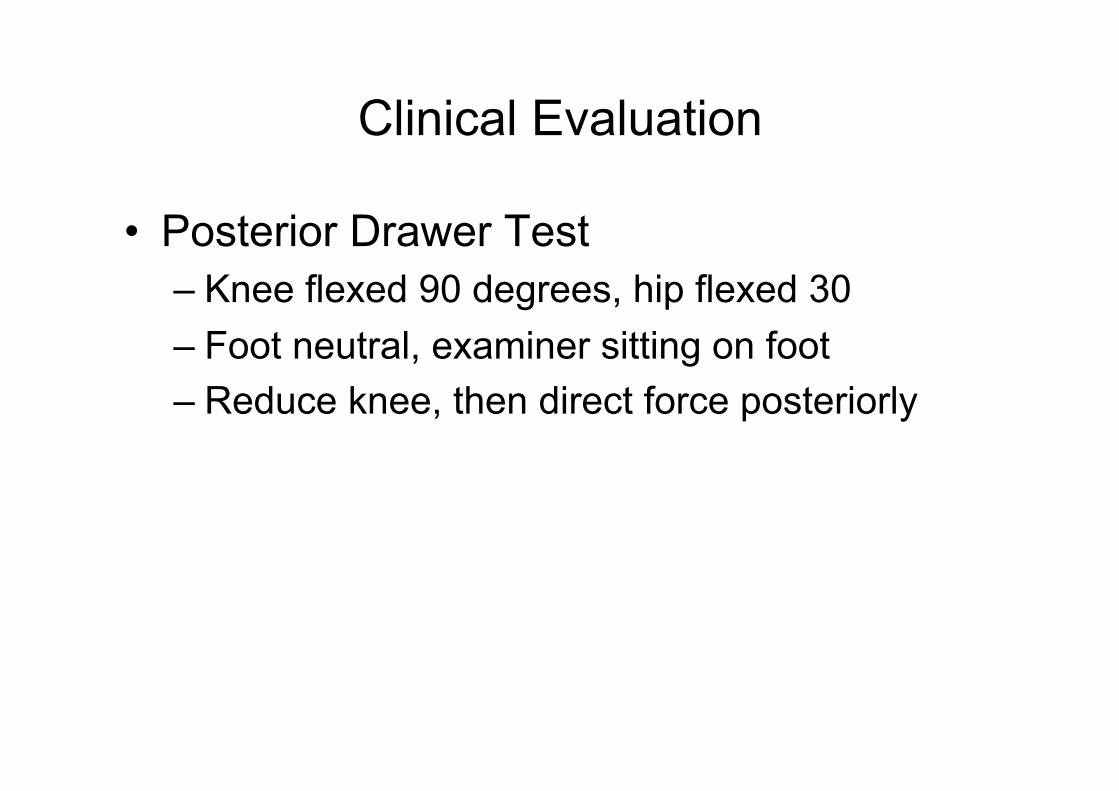
• Posterior Drawer Test – Knee flexed 90 degrees, hip flexed 30 – Foot neutral, examiner sitting on foot – Reduce knee, then direct force posteriorly

Clinical Evaluation
• Posterior Drawer Test

Clinical Evaluation
• Posterior Drawer Test
Grade Position of Tibial Plateau vs Medial Femoral Condyle
Translation (mm)
I Anterior 0-5
II Flush 6-10
III Posterior >10

Clinical Evaluation
• Quadriceps Active Test – Knee flexed 90 degrees, examiner holds foot – Active quad contraction will shift tibia
anteriorly more than 2 mm in PCL deficient knee
– 54% sensitive, 97% specific

Clinical Evaluation
• Quadriceps Active Test

Clinical Evaluation
• Posterior Sag Test – Hip flexed 45 degrees, knee 90 degrees – Tibia will sag with a disrupted PCL – 79% sensitive, 100% specific

Clinical Evaluation
• Posterior Sag Test – Hip flexed 45 degrees, knee 90 degrees – Tibia will sag with a disrupted PCL – 79% sensitive, 100% specific

Clinical Evaluation
• Posterior Sag Test – Hip flexed 45 degrees, knee 90 degrees – Tibia will sag with a disrupted PCL – 79% sensitive, 100% specific

Clinical Evaluation
• PLC injuries – Best evaluated with Dial test
• Patient prone, knees flexed to 30 • Increase of more than 10 is abnormal • Repeat at 90 degrees
– Positive suggests PLC and PCL injury

Clinical Evaluation
Rubenstein, et al. AJSM 1994 • 39 patients, 18 with PCL, 9 with ACL, 12
normal knees – Accuracy: 96%, 90% sensitivity 99% specificity – Better for grade II and III – 80% agreement on grade – Posterior drawer test was best overall

Radiographic Evaluation • Posterior sag of the
tibia on femur
Normal Knee PCL deficient
Normal PCL deficient

Radiographic Evaluation
• PCL avulsion

Radiographic Evaluation
LCL avulsion injury Pelligrini-Steida lesion

Radiographic Evaluation
• Posterior tibial stress test

Radiographic Evaluation
Axial T2 fat suppressed image Axial T1 image

Radiographic Evaluation
dashboard injury showing bruising to anterior tibia

Radiographic Evaluation
Accuracy of MRI for PCL tears • Fischer, et al. JBJS 1991
– RCT of 1014 patients • 99% for PCL • 93% for ACL • 89% for medial meniscus • 88% for lateral meniscus

Radiographic Evaluation
Chronic PCL Tears • Servant, et al. Knee 2004
– MRI was performed on 10 knees with a clinical and arthroscopic diagnosis of a PCL injury sustained at least 6 months previously.
– Seven experienced musculoskeletal radiologists
– accuracy in diagnosing a PCL injury was 57% (40-80%).

Radiographic Evaluation
8 year old tear Grade III laxity 4 of 7 correct

Radiographic Evaluation
14 year old tear Grade II laxity 0 of 7 correct

Radiographic Evaluation
Chronic PCL Tear -elongated, posterior sag
Normal PCL -Short, thick -No posterior sag

Treatment
• Non operative
• Operative

Rational for non-operative management
• Often found as incidental finding – MRI often return to ‘normal’
• Most athletes return to normal function • Good patient satisfaction • PCL surgery does not restore laxity
Rationale for non-operative management
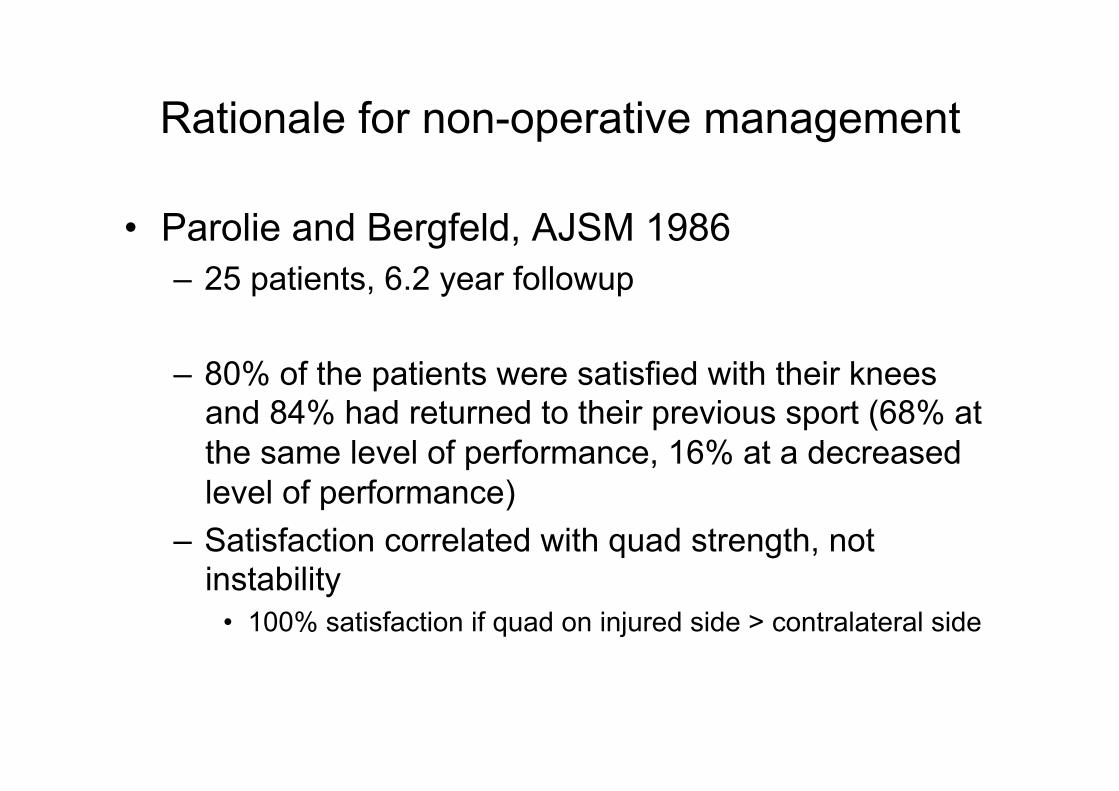
• Parolie and Bergfeld, AJSM 1986 – 25 patients, 6.2 year followup
– 80% of the patients were satisfied with their knees and 84% had returned to their previous sport (68% at the same level of performance, 16% at a decreased level of performance)
– Satisfaction correlated with quad strength, not instability
• 100% satisfaction if quad on injured side > contralateral side

Rational for non-operative management
• Shelbourne et al. Arthroscopy 2005 – Prospective cohort study, 271 pts – 100 grade I, 43 grade 1.5, 128 grade II
• No grade III injuries
– 7.8 year follow-up – Subjective outcomes

Rational for non-operative management
• Shelbourne et al. Arthroscopy 2005 • subjective outcomes independent of laxity • Good/excellent/improving in 56% patients • 12% decreasing function

Shelbourne, et al. Arthroscopy, 2005
– 50% return to sports at same level – 30% return to sport at lower level

Non operative rehab protocol
• Knee immobilizer for comfort – Wear until active quad function
• ROM • Quad strengthening
– Add Hamstrings when full ROM achieved • Full activity at 8 weeks • Yearly XR to eval for changes

Rationale for PCL reconstruction
• Few non-operative studies on Grade III PCL injuries
• Increased knee pain and PF arthritis • Abnormal kinematics and contact
pressures in a PCL-deficient knee – Does current surgical technique prevent this?

Rationale for operative management
• Skyhar, et al. JBJS 1993 – Cadaveric study sectioning PCL and PLC in
10 knees – Elevated PF and medial compartment
pressures

Rationale for reconstruction
• Gill, et al. AJSM 2004 – 8 cadaveric knees – Tibial tunnel technique – Measurement of PF forces

Gill, et al. AJSM 2004
• Increased PF forces with deficient and reconstructed knee • Incorrect (medial) tunnel placement resulting in high PF
forces

Rationale for Reconstruction
• Raminiraka, et al. Clin Biomech 2005 – Finite element analysis to compare
• Native • Resected • single bundle • double bundle

Raminiraka, et al. Clin Biomech 2005
• High forces medially and in PF joint • May lead to early arthritis
Medial tibial forces Patellofemoral forces

Rationale for operative management
• Strobel, et al. Arthroscopy 2003 – 181 patients with knee a/s after PCL injury
• Increased MFC and PF OA • 40% MFC lesions at 1 year • 77% had MFC lesion at 5 years • 47% patella lesions

Rationale for operative management
• Strobel, et al. Arthroscopy 2003
MFC Chondomalacia

Rationale for operative management
• Strobel, et al. Arthroscopy 2003
Patella chondromalacia

Operative Management of PCL Injuries
• Indications – Multiple ligament injury – Grade III laxity – Symptomatic instability – Failure of conservative management

Outcomes following PCL reconstruction
• Cooper, et al. AJSM 2004 – Single bundle, inlay technique, prospective
• 41 patients, most were combined procedure (85%)
• PD examination: 0 (normal) in 9 patients, 1+ in 25 patients, 2+ in 7 patients, and none >2+
• Stress XR: side-to-side difference of 4.11 mm (-2 to 10 mm)
• Better knee scores with allograft

Outcomes following PCL reconstruction
• Seikya, et al. Arthroscopy 2006 – 21 patients, single bundle, transtibial
• Better outcomes with subacute vs. chronic • 57% of the patients had normal/near normal knee function • 62% had a normal/near normal activity level • 62% had less than a 3-mm 31% had a 3- to 5-mm laxity.
• 75% normal/near normal XR

Controversies in PCL reconstruction
• Why does PCL reconstruction not restore normal AP laxity?
– What is the ideal graft type? – What operative technique is best? – Is one bundle better than two? – How should the graft be tensioned? – Where should the tunnels be placed?

Controversies • Inlay vs. Transtibial? • Single vs. Double Bundle?

Inlay vs. Transtibial technique
• Bergfeld, et al. AJSM 2001 – 6 pairs of cadaveric knees
• 6 inlay, 6 transtibial
– mechanical degradation in the tunnel group but not in the inlay group
– Less AP laxity in the inlay group vs. tunnel group from 30 - 90º

Inlay vs. Transtibial technique
• Bergfeld, et al. AJSM 2001
Tunnel graft Inlay graft
Effect of cyclic loading

Inlay vs. Transtibial Technique
• Markolf, et al. JBJS 2002 – 62 knees, 31 inlay, 31 transtibial – 2000 cycles of tensile force of 50 to 300 N with the
angle of pull at 45° • 10/31 transtibial grafts failed vs. 0/31 • 40% reduction of thickness at ‘killer corner’ • 3.9 mm increase in graft length with transtibial vs. inlay
– “inlay technique…was superior with respect to graft failure, graft thinning, and permanent increase in graft length.”

Inlay vs. Transtibial technique
• Seon, et al. Arthroscopy 2006 – 21 transtibial, 20 inlay – 2 year follow up
– Good subjective results with both techniques, no significant difference in laxities post op (3.3 vs. 3.7mm)
Inlay vs. Transtibial technique
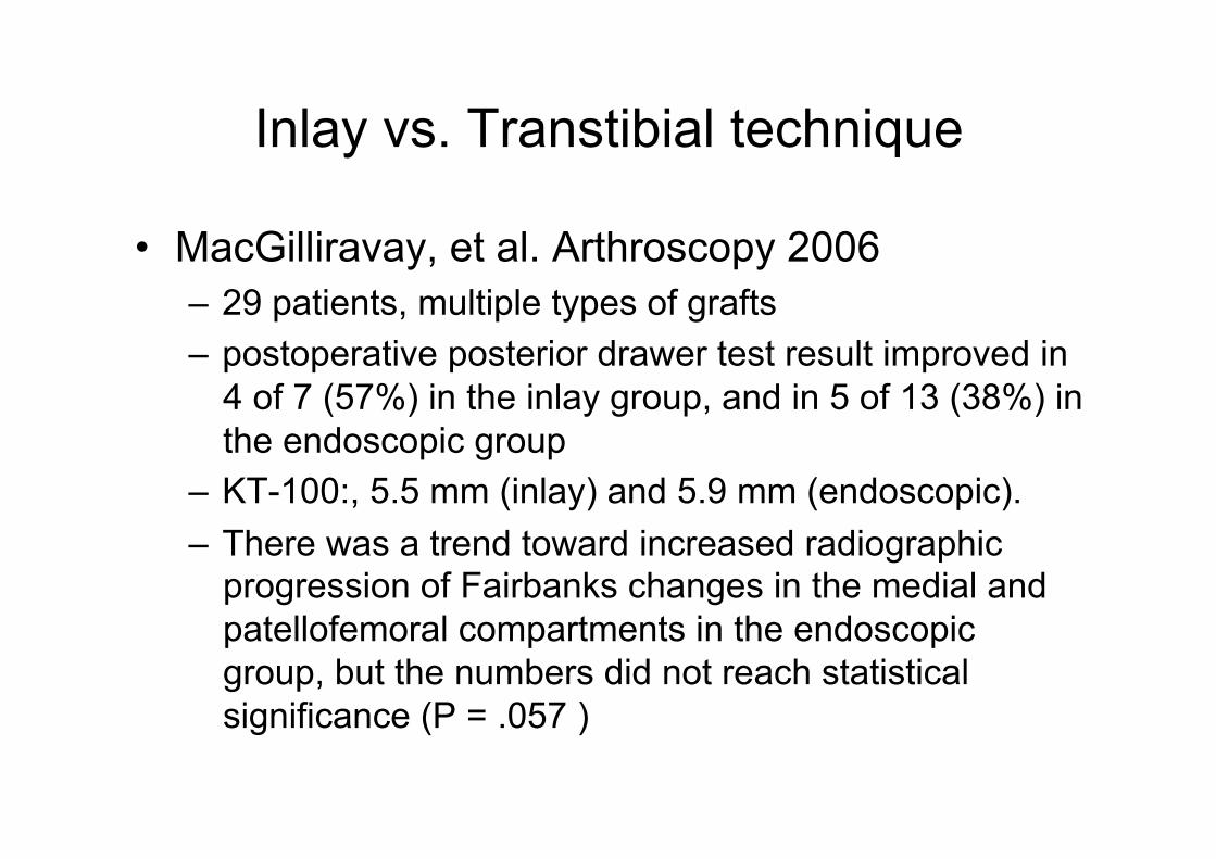
• MacGilliravay, et al. Arthroscopy 2006 – 29 patients, multiple types of grafts – postoperative posterior drawer test result improved in
4 of 7 (57%) in the inlay group, and in 5 of 13 (38%) in the endoscopic group
– KT-100:, 5.5 mm (inlay) and 5.9 mm (endoscopic). – There was a trend toward increased radiographic
progression of Fairbanks changes in the medial and patellofemoral compartments in the endoscopic group, but the numbers did not reach statistical significance (P = .057 )

Single vs. Double Bundle
– A dual-bundle reconstruction more closely replicates PCL anatomy, and should therefore better restore normal knee biomechanics. • Harner, et al. AJSM (2000) • Race and Amis, JBJS-B (1997)
• PM may increase stability in extension • Methodology of studies—low pretension may affect
results

AL graft
PM graft
AL
PM
Single vs. Double Bundle: our experience

Single vs. Double Bundle: our experience
0 10 30 45 70 90
Knee Flexion Angle (Degrees)

Single vs. Double Bundle: our experience

Single vs. Double Bundle: our experience

Single vs. Double Bundle
• Clinical Studies – Noyes, et al. JBJS (2005)
• Q-PT autograft double bundle reconstruction • 19 patients • Excellent subjective outcomes (18/19) • 14 knees <5 mm posterior translation • 5 knees >5 mm posterior translation
– Wang, et al. Injury (2004) • Double blind comparison of single vs. double bundle • No difference in subjective or objective outcomes

Conclusions
• Always look for combined ligament injuries • Conservative treatment for Grade I and II
(?) injuries • Single bundle inlay for Grade III injuries
• Multicenter RCT needed to determine best treatment for PCL injuries

The End