Embed Size (px)
Citation preview
Korr and Denslow: Stretch Receptors in Facilitation and Recent Research
Mark A.W. Andrews, Ph.D., FNAOME
Professor and Director of Physiology
Lecturer in OPP
LECOM at Seton Hill, Greensburg, PA

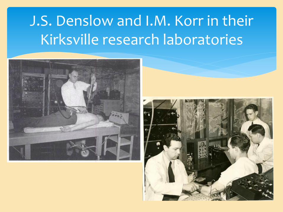
J.S. Denslow and I.M. Korr in their Kirksville research laboratories
“To the osteopathic physician the osteopathic lesion is a demonstrable entity. [They] perceives it with [their] sense of touch and often with [their] muscle sense as [they move] the parts …” J.S. Denslow
3
“In synaptic and myoneural transmission, lie the answers to some of the most important theoretical and practical osteopathic problems.” I.M. Korr
4

5 Historical Perspective
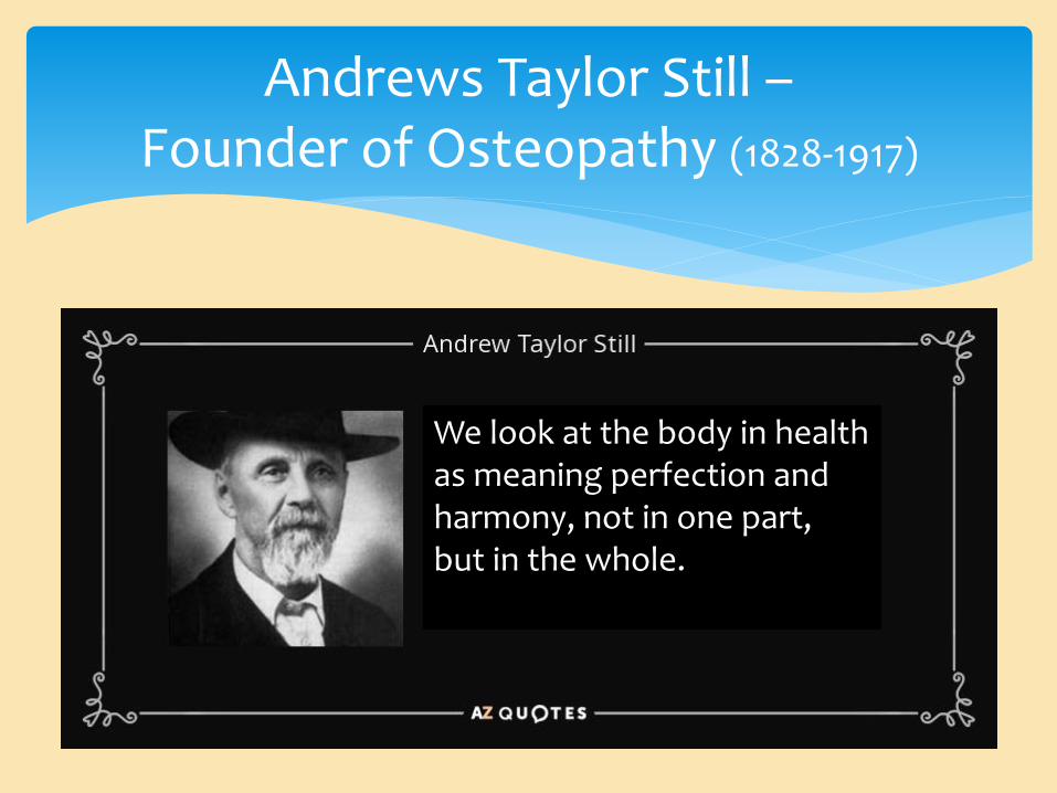
Andrews Taylor Still – Founder of Osteopathy (1828-1917)
We look at the body in health as meaning perfection and harmony, not in one part, but in the whole.

Andrews Taylor Still – Founder of Osteopathy (1828-1917)
Still had a mechanistic view of the body, proposing that once mechanical disturbances were eliminated, the body would be able to function harmoniously.
Still referred to these disturbances as lesions.
The harmonious interaction which must happen included neural, cardiovascular, lymphatic and all other flows occurring unhindered.

Claude Bernard – Founder of Modern Physiology (1813-78)
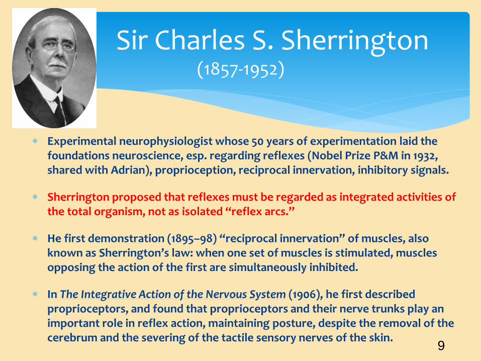
Experimental neurophysiologist whose 50 years of experimentation laid the foundations neuroscience, esp. regarding reflexes (Nobel Prize P&M in 1932, shared with Adrian), proprioception, reciprocal innervation, inhibitory signals.
Sherrington proposed that reflexes must be regarded as integrated activities of the total organism, not as isolated “reflex arcs.”
He first demonstration (1895–98) “reciprocal innervation” of muscles, also known as Sherrington’s law: when one set of muscles is stimulated, muscles opposing the action of the first are simultaneously inhibited.
In The Integrative Action of the Nervous System (1906), he first described proprioceptors, and found that proprioceptors and their nerve trunks play an important role in reflex action, maintaining posture, despite the removal of the cerebrum and the severing of the tactile sensory nerves of the skin.
Sir Charles S. Sherrington (1857-1952)
9

This landmark series of three research publications showed the response of a single-sensory receptors to a defined stimulus (Nobel Prize P&M in 1932, shared with Sherrington), and the all-or-nothing firing of APs.
Afferent rate of APs was seen to vary with the strength of the input: the first indication of neural signaling by means of a frequency coding.
The receptor afferents studied were from frog muscle spindles.
They recognized that, with maybe the exception of the eye and ear, mammalian muscle spindles are the most complex sensory organ.
These receptors were later found to receive efferent motor innervation, allowing adjustment of the sensory response.
Adrian and Zotterman (1926) Muscle Spindle Receptors
10
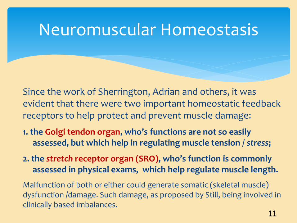
Since the work of Sherrington, Adrian and others, it was evident that there were two important homeostatic feedback receptors to help protect and prevent muscle damage:
1. the Golgi tendon organ, who’s functions are not so easily assessed, but which help in regulating muscle tension / stress;
2. the stretch receptor organ (SRO), who’s function is commonly assessed in physical exams, which help regulate muscle length.
Malfunction of both or either could generate somatic (skeletal muscle) dysfunction /damage. Such damage, as proposed by Still, being involved in clinically based imbalances.
Neuromuscular Homeostasis
11
J.S. Denslow and I.M. Korr in their Kirksville research laboratories
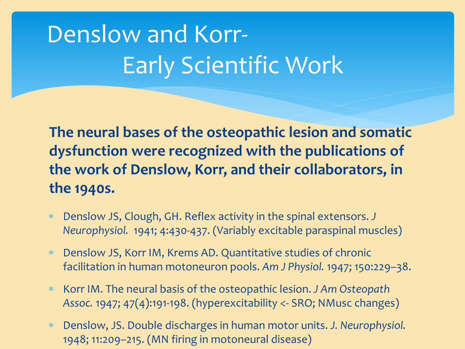
The neural bases of the osteopathic lesion and somatic dysfunction were recognized with the publications of the work of Denslow, Korr, and their collaborators, in the 1940s.
Denslow JS, Clough, GH. Reflex activity in the spinal extensors. J Neurophysiol. 1941; 4:430-437. (Variably excitable paraspinal muscles)
Denslow JS, Korr IM, Krems AD. Quantitative studies of chronic facilitation in human motoneuron pools. Am J Physiol. 1947; 150:229–38.
Korr IM. The neural basis of the osteopathic lesion. J Am Osteopath Assoc. 1947; 47(4):191-198. (hyperexcitability <- SRO; NMusc changes)
Denslow, JS. Double discharges in human motor units. J. Neurophysiol. 1948; 11:209–215. (MN firing in motoneural disease)
Denslow and Korr- Early Scientific Work

Korr, IM, Wilkinson, PN, Chornock, FW. Axonal Delivery of Neuroplasmic Components to Muscle Cells. Science 1967: 155:342-345.
Denslow JS. Neural basis of the somatic component in health and disease and its clinical management. J Am Osteopath Assoc. 1972; 72(2):149-156.
Korr, IM. Proprioceptors and somatic dysfunction. J Am Osteopath Assoc. 1975; 74:638–50. (gamma-loop hypothesis; 1978)
Korr, IM. The spinal cord as organizer of disease processes: IV. Axonal transport and neurotrophic function in relation to somatic dysfunction. J Am Osteopath Assoc. 1981; 80:451-459.
Korr, IM. Somatic dysfunction, osteopathic manipulative treatment, and the nervous system: a few facts, some theories, many questions. J Am Osteopath Assoc 1986; 86(2):109.
Korr and Denslow then continued to refine the understanding of the physiology of the
Osteopathic lesion.
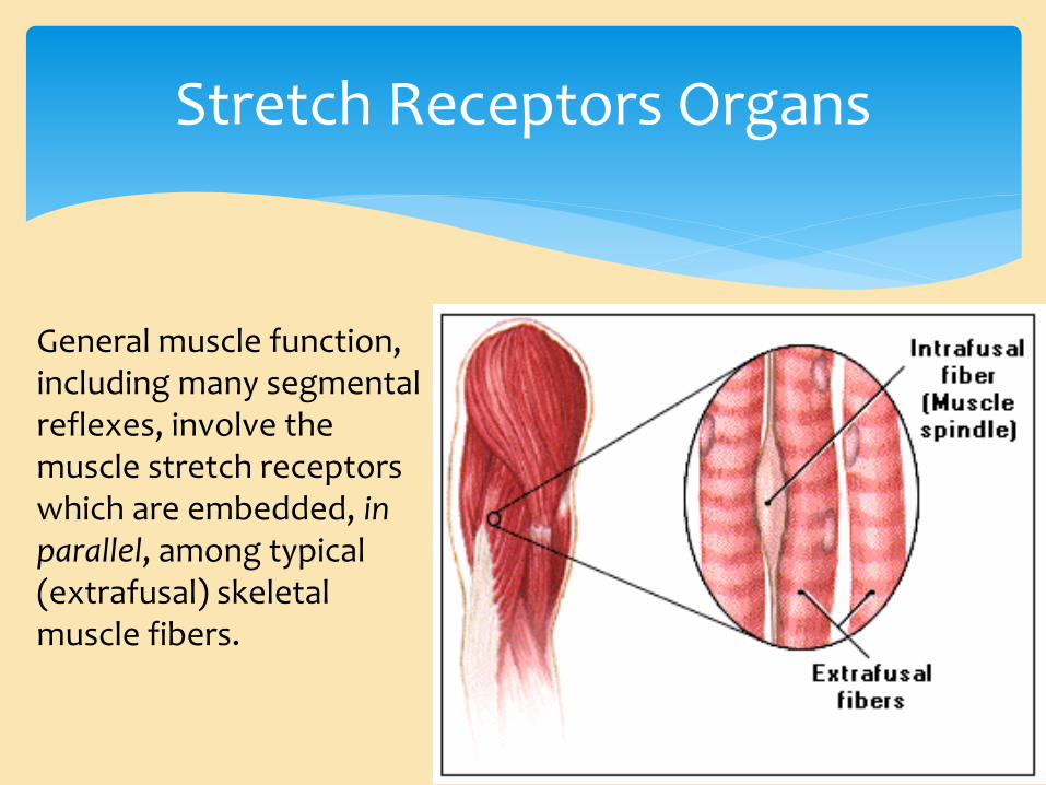
General muscle function, including many segmental reflexes, involve the muscle stretch receptors which are embedded, in parallel, among typical (extrafusal) skeletal muscle fibers.
Stretch Receptors Organs

Spinal facilitation (Glossary of Osteopathic Terminology, of AACOM)
1. The maintenance of a pool of neurons (…in one or more segments of the spinal cord) in a state of partial or subthreshold excitation; in this state, less afferent stimulation is required to trigger the discharge of impulses.
2. A theory regarding the neurophysiological mechanisms underlying the neuronal activity associated with somatic dysfunction.
3. Facilitation may be due to sustained increase in afferent input, aberrant patterns of afferent input, or changes within the affected neurons themselves or their chemical environment. Once established, facilitation can be sustained by normal central nervous system (CNS) activity.
Spinal Facilitation- The Facilitated Segment
16
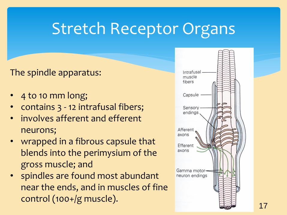
Stretch Receptor Organs
The spindle apparatus: • 4 to 10 mm long; • contains 3 - 12 intrafusal fibers; • involves afferent and efferent
neurons; • wrapped in a fibrous capsule that
blends into the perimysium of the gross muscle; and
• spindles are found most abundant near the ends, and in muscles of fine control (100+/g muscle).
17

Stretch Receptor Organs
Two types of intrafusal fibers are found in the SRO: 1) Nuclear chain fibers, which are thin and narrow, inform the CNS of absolute muscle length; 2) Nuclear bag fibers, which are spindle shaped, primarily inform the CNS of dynamic changes in length (bag1). There are different sub- types (bag2) which mainly inform of absolute length. 18
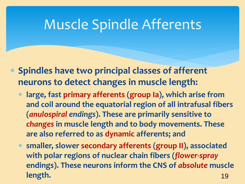
Spindles have two principal classes of afferent neurons to detect changes in muscle length:
large, fast primary afferents (group Ia), which arise from and coil around the equatorial region of all intrafusal fibers (anulospiral endings). These are primarily sensitive to changes in muscle length and to body movements. These are also referred to as dynamic afferents; and
smaller, slower secondary afferents (group II), associated with polar regions of nuclear chain fibers (flower-spray endings). These neurons inform the CNS of absolute muscle length.
Muscle Spindle Afferents
19

Muscle Spindle Innervation
20
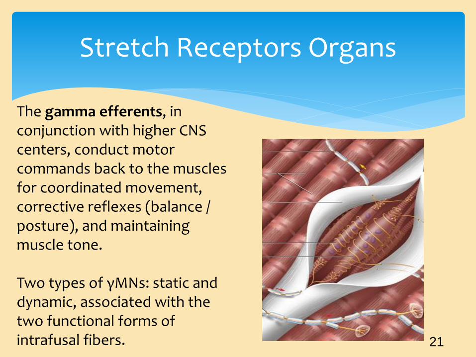
The gamma efferents, in conjunction with higher CNS centers, conduct motor commands back to the muscles for coordinated movement, corrective reflexes (balance / posture), and maintaining muscle tone. Two types of γMNs: static and dynamic, associated with the two functional forms of intrafusal fibers.
Stretch Receptors Organs
21
22
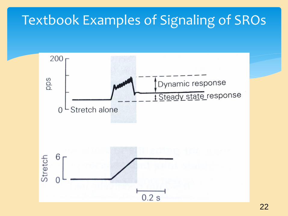
Textbook Examples of Signaling of SROs

23
The Reality of Signaling is More Complex
Hunt, et al., J Gen Physiol 71:683–698. 1978
SROs are not directly linked to the extrafusals in a syncytial arrangement.
Thus, extrafusal fibers and the SROs are do not work in “lock-step,” or in any way directly communicating, except through the CNS.
As a result, it is most likely that there is miscommunication, in agreement with Korr’s somatic dysfunction and facilitated segment hypothesis.
Basic Concepts
Alterations in intrafusal fiber length have been noted experimentally as long-lasting mechanical after-effects, having to be removed by a conditioning pre-stretch or oscillation of the experimental muscle.
Banks, et al., J Physiol. 1997; 498:177-199.
Such results indicate that if intrafusal fibers in vitro are not returned to their original state, with a periodic stretches, there may be phase advance and distortion in the afferent output, which would then affect the efferent response.
Consequent SRO findings
That such a situation may occur in vivo is rather likely, with OMT functioning as a possible means to reset the SRO.
In fact, in a recent study involving simulated spinal manipulation in cats, the unloading of the SROs silenced their afferent firing for 1.3 +/- 0.6 seconds (range, 0.1-4.3 seconds), a physiologically relevant duration which would be more than sufficient to reset SROs.
Haftel, et a., J Neurophysiol. 2004 May;91(5):2164-71.
Recent SRO findings
gamma-Motoneuron firing is more complex than responding with a simple alpha-gamma coactivation.
Instead, the gamma-static and gamma-dynamic neurons have been recorded firing at various rates seemingly dependent, not only on feedback from the SRO, but also from input from the CNS, and what is expected to be sensed.
Ellaway, et al., J Anat. 2015; 227(2):157-66.
Recent SRO findings

Stretch Receptor Section
Bewick and Banks, Pflugers Arch - Eur J Physiol (2015) 467:175–190
Of significant interest, and a caveat when discussing research on SROs, is the fact that most SRO research has been accomplished in distal limb muscles.
With better techniques and surgeries, recent work has indicated that spinal muscle SROs differ functionally from those found in the limbs, with a high proportion of “b2c.”
Durbaba, et al., J Physiol. 2006; 571(Pt 2): 489–498.
Recent SRO findings
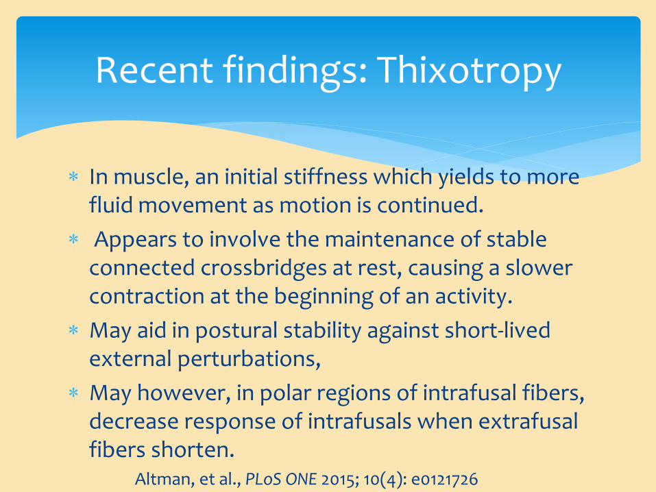
In muscle, an initial stiffness which yields to more fluid movement as motion is continued.
Appears to involve the maintenance of stable connected crossbridges at rest, causing a slower contraction at the beginning of an activity.
May aid in postural stability against short-lived external perturbations,
May however, in polar regions of intrafusal fibers, decrease response of intrafusals when extrafusal fibers shorten.
Altman, et al., PLoS ONE 2015; 10(4): e0121726
Recent findings: Thixotropy
Recent research using immunohistochemical staining has indicated evidence of direct sympathetic innervation of intrafusal fibers. Indicating that sympathetic innervation is not restricted to the blood vessels supplying spindles.
Radovanovic, et al., J. Anat. 2015. 226:542-5
Recent findings: SNS innervation
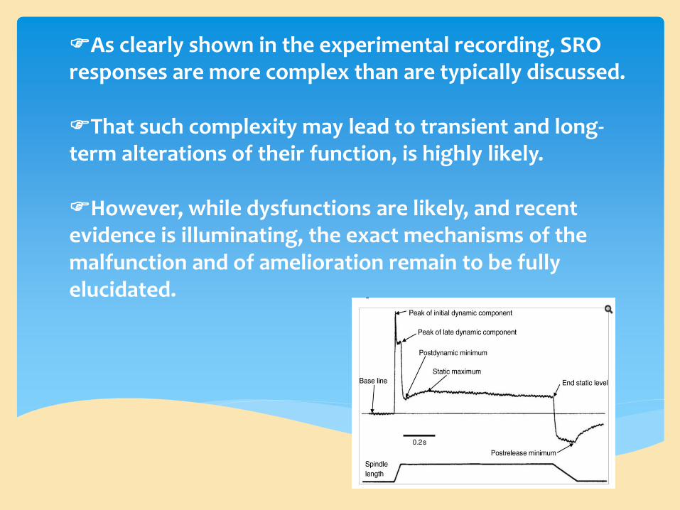
As clearly shown in the experimental recording, SRO responses are more complex than are typically discussed. That such complexity may lead to transient and long-term alterations of their function, is highly likely. However, while dysfunctions are likely, and recent evidence is illuminating, the exact mechanisms of the malfunction and of amelioration remain to be fully elucidated.