Embed Size (px)
Citation preview

This is a repository copy of Management of nystagmus in children : a review of the literature and current practice in UK specialist services.
White Rose Research Online URL for this paper:http://eprints.whiterose.ac.uk/155626/
Version: Published Version
Article:
Self, J.E., Dunn, M.J., Erichsen, J.T. et al. (10 more authors) (2020) Management of nystagmus in children : a review of the literature and current practice in UK specialist services. Eye. ISSN 0950-222X
https://doi.org/10.1038/s41433-019-0741-3
[email protected]://eprints.whiterose.ac.uk/
Reuse
This article is distributed under the terms of the Creative Commons Attribution (CC BY) licence. This licence allows you to distribute, remix, tweak, and build upon the work, even commercially, as long as you credit the authors for the original work. More information and the full terms of the licence here: https://creativecommons.org/licenses/
Takedown
If you consider content in White Rose Research Online to be in breach of UK law, please notify us by emailing [email protected] including the URL of the record and the reason for the withdrawal request.
brought to you by COREView metadata, citation and similar papers at core.ac.uk
provided by White Rose Research Online

Eye
https://doi.org/10.1038/s41433-019-0741-3
REVIEW ARTICLE
Management of nystagmus in children: a review of the literatureand current practice in UK specialist services
J. E. Self 1,2● M. J. Dunn 3
● J. T. Erichsen3● I. Gottlob4
● H. J. Griffiths5 ● C. Harris6 ● H. Lee 1,2● J. Owen6 ●
J. Sanders7 ● F. Shawkat1 ● M. Theodorou8,9● J. P. Whittle10 ● Nystagmus UK Eye research group (NUKE)
Received: 18 October 2019 / Accepted: 24 November 2019
© The Author(s) 2020. This article is published with open access
Abstract
Nystagmus is an eye movement disorder characterised by abnormal, involuntary rhythmic oscillations of one or both eyes,initiated by a slow phase. It is not uncommon in the UK and regularly seen in paediatric ophthalmology and adult general/strabismus clinics. In some cases, it occurs in isolation, and in others, it occurs as part of a multisystem disorder, severevisual impairment or neurological disorder. Similarly, in some cases, visual acuity can be normal and in others can beseverely degraded. Furthermore, the impact on vision goes well beyond static acuity alone, is rarely measured and may varyon a minute-to-minute, day-to-day or month-to-month basis. For these reasons, management of children with nystagmus inthe UK is varied, and patients report hugely different experiences and investigations. In this review, we hope to shine a lighton the current management of children with nystagmus across five specialist centres in the UK in order to present, for thefirst time, a consensus on investigation and clinical management.
Introduction and demographics
The estimated prevalence of nystagmus in the UK is 24 per10,000 [1]. It can be broadly grouped into Infantile Nys-tagmus Syndrome (INS) and acquired nystagmus. Theonset of INS is usually within the first 6 months of life(average age= 1.9 months) [2]. INS can be idiopathic,associated with albinism, retinal diseases such as achroma-topsia, congenital stationary night blindness (CSNB) or early-onset retinal degenerations, low vision in infancy and avariety of other syndromes and developmental diseases (foran exhaustive list, see Leigh and Zee) [3]. The most commonforms of INS are idiopathic INS and INS associated withalbinism or retinal diseases. The most common form of non-INS nystagmus in childhood is fusional maldevelopmentnystagmus syndrome (FMNS, previously Manifest latentnystagmus, MLN).
Children with nystagmus can be severely visuallyimpaired or can have almost normal visual acuity (VA),depending on the underlying disease. However, VA is not aglobal measure of visual function and nystagmus of anytype can associated with significant visual loss [4] beyondthat of acuity alone, and accordingly, both nystagmus andalbinism are cited as key priorities in the area of childhood-onset disorders, as determined by the Sight Loss and VisionPriority Setting Partnership in 2013.
Despite much research into nystagmus over many years,there are still many unanswered questions about diagnosis,treatment and broad management. Consequently, many clin-icians are less comfortable managing children with nystagmusthan other conditions and management varies widely. In thisreview, we seek to clarify the current state-of-play regardingdiagnosis, management, treatment options and the use of var-ious investigations in managing this complex group of children.
Basic clinical assessment
Patient history and examination
When assessing the infant/child with nystagmus, althoughINS (with/without associated ocular disorders) is morecommon, it must be borne in mind that some infants and
Members of the Nystagmus UK Eye research group (NUKE) are listedat the end of the paper.
* J. E. [email protected]
Extended author information available on the last page of the article.
Published online: 09 January 2020
1234
5678
90();,:
1234567890();,:

young children will have ‘acquired’ nystagmus with anunderlying neurological cause. Indeed, some older childrenand/or adults will have previously undiagnosed INS ornystagmus associated with early lack of fusion (FMNS) or,less commonly, nystagmus secondary to severe acquiredvisual loss. History and clinical examination are bothimportant in tailoring the management pathway to allowappropriate investigations and/or treatment. Since INS isassociated with a wide range of underlying disorders, thepresence of nystagmus in an infant/child should stimulate acomprehensive search for a cause. Table 1 summarisessome of the important questions to be included in a thor-ough history.
The clinical examination begins as the child enters theroom, particularly observing for signs of: photophobia, eyerubbing for retinal stimulation, head postures (variable/alternating/consistent) and/or head shaking (both often, butnot always, after 1 year of age), skin/hair tone particularly inrelation to other family members present, as well as signs ofassociated systemic and neurological features. The exam-ination can then be split into two parts: eye movements andocular (+/−systemic) examination.
Ocular and systemic examination
An ocular examination should be performed with the bestage-appropriate equipment available, specifically lookingfor ocular signs commonly associated with nystagmus,which may include:
Cornea
Size (e.g. microcornea associated with coloboma, orbuphthalmic eye associated with glaucoma), epitheliopathy(e.g. associated with PAX6 gene disorders).
Anterior chamber
Structure (e.g. anterior segment dysgenesis).
Iris
Iris structure (e.g. aniridia, coloboma), iris transillumination(e.g. albinism or some PAX6 related disorders).
Lens
Cataract, aphakia (congenital/acquired), intraocular lensimplant following previous surgery.
Vitreous
Clarity (e.g. vitreous haemorrhage).
Retina and optic nerve
Structure, e.g. coloboma, disc anomalies (e.g. papilloedema,hypoplasia, coloboma or small cup seen in albinism), retinalhypo/hyperpigmentation and or pigment, foveal structure(e.g. hypoplasia, atrophy from congenital infection).
Ocular disorders commonly associated with INS aresummarized in Table 2.
Where there is any concern that there is an associatedsystemic or neurological disorder based on either the eyeexamination or the overall assessment of the child, theinfant/child should also be reviewed by a paediatrician orpaediatric neurologist.
Red flag signs
Red flag signs are features in the history and examinationwhich should alert the clinician to acquired pathologythat requires further systemic investigations such asneuroimaging.
Red flag signs
• Later onset nystagmus (in the absence of signs in keepingwith an ocular disorder).
• Constant oscillopsia in older children.
• Dysconjugate/gaze evoked/seesaw/convergence-retractionnystagmus.
• Horizontal nystagmus becoming vertical in vertical gaze.
• Vertical or torsional nystagmus (in the absence of retinalpathology (e.g. achromatopsia).
• Any associated neurological signs and/or a systemicallyunwell child.
Family history
Taking an accurate family history is an important part of theinitial evaluation for all children with nystagmus. If a clearfamily history of nystagmus is noted, identifying thestructure of the pedigree (family tree), in addition to infor-mation about the clinical characteristics of those affected, iskey. Sometimes it will become apparent that the nystagmusin older relatives seems to be isolated and, in others, isassociated with other visual disorders (such as retinal dys-trophies or aniridia) or systemic disorders (such as ataxia inthe case of spino-cerebellar ataxia syndromes). Asking thedegree of visual disability and treatment history for thoseaffected can help to differentiate these groups of disorders.In other cases, the history may include apparently non-ocular disorders (such as relatives with strikingly pale skinand hair in contrast to the family context in albinism
J. E. Self et al.

disorders) or ocular disorders without nystagmus (such asunexplained low vision from a young age in older relativesor night blindness). It is therefore important to ask aboutany medical disorders in relatives, whether they seem to berelated to nystagmus or not, and in all cases to draw afamily pedigree in order to narrow the search for potentialhereditary causes (and reduce the number of investigationsneeded in many cases). In many cases, especially where fullcooperation is difficult in a young child, examining parentscan yield diagnostic information (such as iris transillumi-nation in parents as a clue to albinism as an underlyingcause). Figure 1 shows how to draw a pedigree diagram andincludes a key to remind clinicians how such diagrams areconstructed.
Clinical tip
When drawing a pedigree diagram, start with the proband(the presenting patient) and work horizontally before ver-tically where possible. Also include names and dates ofbirth when available (and according to local data protectionpolicy) and older siblings to the left and younger siblings tothe right.
Orthoptic examination
Orthoptic examination of all children presenting with nys-tagmus is essential, not only for the comprehensiveassessment of visual function and VA throughout the cri-tical period of visual development, but also for investigationof ocular alignment and binocular vision [5]. This isrequired due to increased prevalence of strabismus in thepresence of childhood nystagmus, reported as between 16and 52% [6, 7]. Children with idiopathic INS are less likelyto develop strabismus, whereas those with congenital retinaldystrophies or albinism are at intermediate risk, and thosewith bilateral optic nerve hypoplasia are at particularly highrisk [6].
Clinical recommendations for the orthoptic assessment,additional to those discussed in other sections of this paper,are summarised in Table 3; the specific investigation in eachcase will depend on the age and cooperation of the patient.Additional clinical investigations may be required depend-ing on findings and clinical judgement.
Giving time and relaxing the child as much as possible sothat they are comfortable during the orthoptic assessmentmay give the best performance and improve the responses
Table 1 History taking in an infant/child presenting with nystagmus.
Question Clinical relevance
Pregnancy, maternal medication/drug use and birth history Maternal drug exposure and prematurity have been associated with nystagmus.
Family history of eye/neurological disease/systemic disease Many eye movement disorders have a hereditary component with differentinheritance patterns indicating which genes may be involved.
Pigmentation in skin and hair compared with rest of family Albinism is a common underlying associated disorder, but sometimes without adefinite family history.
Specific questions about visual behaviours—e.g. nyctalopiaor photophobia
Photophobia and nystagmus are common findings in disorders of cone functionand albinism. High-frequency low amplitude nystagmus with photophobia is morecommon in cone dysfunction. Nyctalopia is a common symptom in roddysfunction.
Open questioning about other visual behaviours Parents will often report a very detailed description of visual behaviours, which candirect clinical examination such as a child with chin depression and vertically‘wobbly eyes’ (commonly seen in downbeat nystagmus), or pushing/rubbing eyesfirmly for retinal stimulation in blind babies/children.
Does the child experience oscillopsia? Lack of oscillopsia in the presence of involuntary eye movements such asnystagmus in an older child suggests early onset. Infrequent episodes of oscillopsiadespite a constant nystagmus is also seen in early-onset nystagmus. Constantoscillopsia suggests an acquired disorder.
If oscillopsia is reported, is it when stationary or whenmoving?
Oscillopsia, which is only present during head movement, implies a vestibularpathology.
Are there associated speech or swallowing problems? Possible brainstem pathology or myasthenia gravis.
Are there associated coordination problems? Possible cerebellar pathology.
Is there associated hearing loss or tinnitus? Possible peripheral vestibular pathology.
Is the patient on any medications? Many medications can cause abnormalities of eye movement, most commonlyanti-epileptic medication.
Are there any concerns about any other aspect of the child’sdevelopment or health besides their eyes?
Eye movement abnormalities form a part of many multisystem syndromes and canbe the presenting feature.
At what age did the parent/carer notice the nystagmus? INS is typically noticed in the first 4–6 months of life but it’s typical onset (whenseeking it in at risk patients) is 1.9 months [2].
Management of nystagmus in children: a review of the literature and current practice in UK specialist. . .

recorded, as both anecdotal reports from patients andexperimental studies have reported that the nystagmusintensity increases with increased effort to fixate anddecreases when relaxed [8, 9].
In the presence of nystagmus, the cover test willbe more difficult to perform as small movements to takeup fixation can be impossible to distinguish. Observationfor asymmetrical corneal reflections may thereforebe relied upon. Caution should be taken as a significantassociation that has been found between a positive anglekappa and clinical signs of albinism in patients withINS [10].
Clinical tip
The involuntary head nodding often seen in INS can bedistinguished from rhythmic head movement due to a moresinister cause; if the child can voluntarily stop the headmovement when asked, it is caused by the nystagmus.
Nystagmus examination
A simple, methodical clinical assessment of a child’s nys-tagmus can provide key information in order to direct fur-ther investigations. It can sometimes identify the type of
Table 2 Classic presentation of common ophthalmic conditions associated with INS: key points.
Idiopathic INS Sometimes family history (typically X-linked). Conjugate, typically horizontal nystagmus, may dampen onconvergence, +/− head shake, +/− one/alternating null point, in an otherwise well infant child with normaleyes and systemic examination.
Oculo-cutaneous/ocular albinism Often family history, typically less pigmentation in hair and skin, hypopigmented iris pigment epithelium(often transillumination) and retinal pigment epithelium, tilted discs and foveal hypoplasia. Evidence ofchiasmal misrouting.
PAX6 gene disorders Often family history, corneal epitheliopathy, early-onset lens opacity, varying degrees of iris hypoplasia (nearnormal to aniridic) and foveal hypoplasia. No evidence of chiasmal misrouting.
Achromatopsia Often family history, photophobia, reduced/absent colour vision, nystagmus typically ‘fine or shimmering’grossly normal macula appearance (may have retinal pigment epithelium (RPE) changes/atrophy).
Fig. 1 How to draw a pedigree diagram whilst taking a family history. This pedigree is consistent with X-linked inheritance with variablepenetrance in females (typical in FRMD7 gene related INS).
J. E. Self et al.

nystagmus, but it can also rule out, or at least reduce thelikelihood of, some nystagmus aetiologies. Even if thenystagmus type cannot be identified, it is important todocument its features. There are different diagrammaticschemes for describing the nystagmus in the medical notes,but consistency is important. Using words, whilst verbose,does avoid confusion.
The initial and crucial task is to look for nystagmus in allgaze directions, not just primary position (usually the ninecardinal points). There may be nystagmus in far eccentricgaze, which can be easily overlooked in an uncooperativechild, so perseverance is required. The axis of oscillation,whether it is horizontal, vertical, torsional, circumrotatory(i.e. circular or elliptical) or a mixture, should be noted ateach cardinal point. Does the nystagmus appear similar ineach eye (i.e. conjugate), or is there an asymmetry? If thenystagmus appears jerky, document the direction of the fastphase, otherwise note that it appears pendular. Note thefrequency (how fast) and amplitude (how big).
It is also important to examine the nystagmus duringmonocular viewing to look for FMNS, which is relatively
common either as the sole nystagmus or in conjunction withother types of nystagmus (usually INS). In sole FMNS, thenystagmus is conjugate, horizontal and in primary position,beats in the direction of the viewing eye. That is, the nys-tagmus reverses with alternate occlusion. The nystagmus alsointensifies with increased abduction of the viewing eye anddampens (sometimes completely) in full adduction. Thus,FMNS is usually best identified by alternating occlusion withthe eyes in far lateral gaze, as this will bring out the biggestchange in intensity. Patching may sometimes be preferable toan occluder in the young uncooperative patient.
Typically, INS is horizontal and remains so in elevationand depression. The nystagmus often has a null region(a direction of gaze in which the nystagmus dampens) andincreases in intensity, becoming jerkier farther from the null.
Gaze-evoked nystagmus is the most common acquirednystagmus. It is usually caused by cerebellar lesions/mal-formations or drug toxicities (esp. anticonvulsants). Thenystagmus is evoked on lateral gaze but absent in primaryposition. It beats in the direction of gaze, similar to FMNSbut is unaffected by monocular occlusion. There may (or
Table 3 Orthoptic assessment inchildren with nystagmus.
Clinical test Description
Head posture • Presence and degree of any anomalous head posture (AHP) should berecorded—including a description for both near and distance fixation, withand ideally without any refractive correction.
• Any change with visual demand should be noted.• Presence or absence of any involuntary head nodding should be recorded withactivity in which this occurs.
VA • With refractive correction—both eyes open and monocularly.• A note taken as to whether measured with or without head posture.• Near using preferred reading distance and distance.• Record method of occlusion, e.g. opaque occluder/high plus lens.
Cover test • With and without refractive correction.• With and without AHP.• Near and distance fixation.• Note presence or absence of nystagmus, any change in amplitude and/ordirection of nystagmus on covering one eye.
Binocular single vision • With refractive correction and any AHP.• Sensory, motor fusion for near and distance fixation.• Presence or absence of stereopsis—stereoacuity when possible.• Note AHP adopted to achieve binocular responses.
Ocular movements • Testing of ductions and versions in nine positions of gaze for near fixation,with description of nystagmus in primary and secondary positions.
• VOR (vertical and horizontal)—presence/absence.• Optokinetic nystagmus (OKN)—presence/absence/abnormal (expected orinverted response).
• Smooth pursuit—horizontal and vertical.• (For detail of methods of testing, see Osborne et al. [5]).
Convergence • With refractive correction with AHP—noting ability to convergence andchange in amplitude and/or frequency of nystagmus.
Measurement of deviation • If possible, using alternating prism cover test—with refractive correction,with and without AHP.
• Near and at distance—primary position, secondary positions if indicated todocument change from primary position or to confirm concomitance.
• Individual reading position if different from above.
Management of nystagmus in children: a review of the literature and current practice in UK specialist. . .

may not) be downbeat nystagmus in lateral gaze ordepression. In elevation, there may (or may not) be unsteadygaze or upbeat nystagmus. Horizontal smooth pursuit isalmost always quite saccadic, which is one way to differ-entiate it from end point nystagmus.
If the nystagmus is downbeat, upbeat or asymmetric,then a neurological cause should be considered, althoughINS cannot be excluded. Periodic Alternating Nystagmus(PAN) describes a horizontal jerk nystagmus that reversesdirection every few minutes. PAN can occur as an acquiredneurological nystagmus or as an aspect of INS (often raisingsuspicion of albinism as the underlying aetiology). To testfor PAN, the nystagmus should be examined for a reversalin direction for at least 5 min. It is important to keep thegaze in primary position, otherwise a spurious reversalcould occur due to a gaze-evoked null shift. Such prolongedobservations can be difficult for young or non-compliantpatients. If the nystagmus beat direction (or anomalous headpostures (AHP)) is different than indicated in previous notesor reports by carers, then PAN should be suspected. Whenassociated with INS, PAN has no sinister implications butmay be a contraindication for standard AHP surgery as itimplies spontaneous null shifting.
Clinical tip
For a more detailed, practical description of how to examinenystagmus and other supranuclear eye movements in chil-dren (and interpret findings), see [5].
Specialised clinical assessment
Optical coherence tomography (OCT) in INS
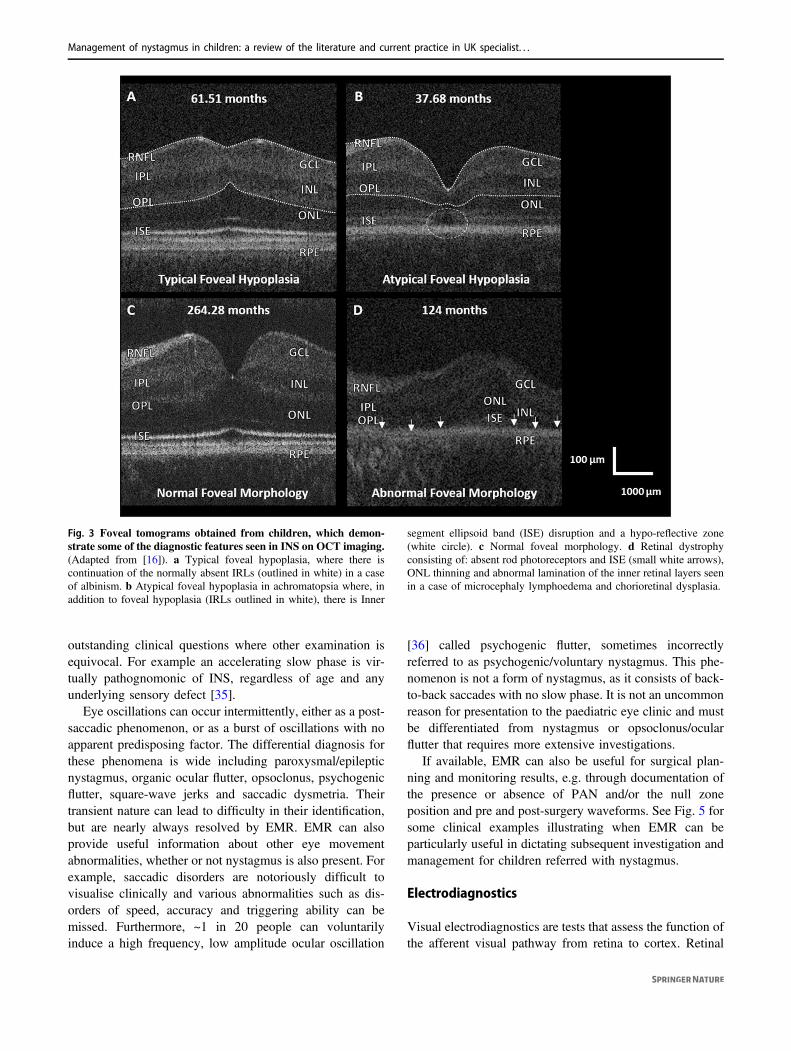
OCT imaging has been established as a tool that canstreamline diagnosis of the aetiology of INS [11–31]. Forinfants and young children who cannot cooperate withstandard table-top OCTs, a hand-held spectral-domain OCTimaging device can be used, which has been shown to bereliable in the presence of nystagmus [32]. By identifyingthe presence or absence of typical or atypical fovealhypoplasia (continuation of the normally absent inner ret-inal layers (IRLs) across the fovea) and the presence ofother abnormal morphological features, it is possible todivide INS into four diagnostic categories: (1) typical fovealhypoplasia; (2) atypical foveal hypoplasia; (3) abnormalfoveal morphology; and (4) normal foveal morphology(Fig. 2) [31]. In this way, conditions such as albinism andPAX6 mutations, which are usually associated with typicalfoveal hypoplasia (Fig. 3a), can be distinguished from otherconditions such as achromatopsia, which is characterised byatypical foveal hypoplasia (Fig. 3b), or retinal dystrophies,which are typically associated with abnormal foveal
morphology (Fig. 3d). Furthermore, the severity of fovealhypoplasia can be graded (Fig. 4), and this can potentiallybe used as a visual prognostic indicator [33].
Clinical tip
There are frequently movement artefacts in macular OCTsfrom patients with INS. When attempting to diagnose fovealhypoplasia, obtain a macular volume scan, ensuring that (1)the optic nerve is visible as a landmark and (2) there is aminimum of five uninterrupted B scans (i.e. withoutrefixations or blinks on either side of the central fovealB scan).
Eye movement recordings (EMRs) in INS
EMR, if available, provides a means for objectivelyvisualising the details of oculomotor phenomena that arenot visible to the naked eye or occur transiently. EMR canalso provide a permanent quantitative record for long-itudinal comparisons to monitor disease progression orremission. EMR is particularly valuable for patients withoscillatory eye movements as it can reveal the underlyingnystagmus waveform that distinguishes various types ofnystagmus and saccadic oscillations. EMR recording canalso detect abnormal smooth pursuit, saccades, OKN, andvestibular responses depending on the type of equipment athand [34].
Modern eye trackers are mostly video based and providenon-intrusive accurate recordings with high resolution forhorizontal and vertical eye movements. Hence, they arecapable of providing objective evidence for the presenceand type of nystagmus, but also for the absence of nys-tagmus. Note that the majority of EMR systems do notrecord torsional eye movements; for this, highly specialisedequipment is needed.
In a busy clinical setting, it is usually not possible toperform a standardised battery of EMRs with each patient.Instead, and where available, EMRs can help address
Fig. 2 Diagnostic use of OCT in INS. An algorithm adapted from‘potential of hand-held optical coherence tomography to determinecause of INS in children by using foveal morphology’ [31].
J. E. Self et al.
outstanding clinical questions where other examination isequivocal. For example an accelerating slow phase is vir-tually pathognomonic of INS, regardless of age and anyunderlying sensory defect [35].
Eye oscillations can occur intermittently, either as a post-saccadic phenomenon, or as a burst of oscillations with noapparent predisposing factor. The differential diagnosis forthese phenomena is wide including paroxysmal/epilepticnystagmus, organic ocular flutter, opsoclonus, psychogenicflutter, square-wave jerks and saccadic dysmetria. Theirtransient nature can lead to difficulty in their identification,but are nearly always resolved by EMR. EMR can alsoprovide useful information about other eye movementabnormalities, whether or not nystagmus is also present. Forexample, saccadic disorders are notoriously difficult tovisualise clinically and various abnormalities such as dis-orders of speed, accuracy and triggering ability can bemissed. Furthermore, ~1 in 20 people can voluntarilyinduce a high frequency, low amplitude ocular oscillation
[36] called psychogenic flutter, sometimes incorrectlyreferred to as psychogenic/voluntary nystagmus. This phe-nomenon is not a form of nystagmus, as it consists of back-to-back saccades with no slow phase. It is not an uncommonreason for presentation to the paediatric eye clinic and mustbe differentiated from nystagmus or opsoclonus/ocularflutter that requires more extensive investigations.
If available, EMR can also be useful for surgical plan-ning and monitoring results, e.g. through documentation ofthe presence or absence of PAN and/or the null zoneposition and pre and post-surgery waveforms. See Fig. 5 forsome clinical examples illustrating when EMR can beparticularly useful in dictating subsequent investigation andmanagement for children referred with nystagmus.
Electrodiagnostics
Visual electrodiagnostics are tests that assess the function ofthe afferent visual pathway from retina to cortex. Retinal
Fig. 3 Foveal tomograms obtained from children, which demon-
strate some of the diagnostic features seen in INS on OCT imaging.
(Adapted from [16]). a Typical foveal hypoplasia, where there iscontinuation of the normally absent IRLs (outlined in white) in a caseof albinism. b Atypical foveal hypoplasia in achromatopsia where, inaddition to foveal hypoplasia (IRLs outlined in white), there is Inner
segment ellipsoid band (ISE) disruption and a hypo-reflective zone(white circle). c Normal foveal morphology. d Retinal dystrophyconsisting of: absent rod photoreceptors and ISE (small white arrows),ONL thinning and abnormal lamination of the inner retinal layers seenin a case of microcephaly lymphoedema and chorioretinal dysplasia.
Management of nystagmus in children: a review of the literature and current practice in UK specialist. . .

function is assessed using the electroretinogram (ERG) andthe post retinal pathway using visual evoked potentials(VEP). Paediatric ERGs and VEPs are non-invasive,objective tests that do not require any anaesthesia, sedationor mydriasis. The tests are relatively quick to perform(30–40 min) with immediate access to results and are per-formed according to a modified, combined paediatric pro-tocol [37]. Adults with nystagmus should be investigatedusing the standards and recommendations of the Interna-tional Society for Clinical Electrophysiology of Vision [38].
A common cause for nystagmus in children is retinaldystrophy, and in many cases, a fleeting posterior segmentexamination is normal. These patients need ERGs and VEPsto assess retinal integrity and isolate cone and rod function:severe retinal dystrophy, such as Leber’s amaurosis, resultsin all ERGs being attenuated, whereas in achromatopsia,cone-mediated ERGs are attenuated but rod ERGs arenormal. Alteration in the ERG waveform that results in anegative configuration (better preserved ERG ‘a’ wave than‘b’ wave) is commonly seen in an X-linked CSNB and X-linked retinoschisis. Pigmentary retinopathies can also be
associated with a number of systemic and neurometabolicconditions that may be present with nystagmus: ERGs aredegraded with rod responses often more severely affectedinitially with later involvement of the cones.
The concurrent recording of pattern VEPs determines theextent of macula involvement and provides an estimate of thelevel of vision. A patient could have extinguished flash ERGsbut preserved pattern VEPs, indicating retinal dysfunctionprimarily involving the extra-macular areas, whereas patternVEPs are degraded in patients with cone dysfunction. VEPsshould be recorded to a range of different size patterns: blackand white checkerboard pattern that is alternating (patternreversal) or appearing and disappearing (onset/offset VEPs).Patients with nystagmus have degraded pattern VEPs corre-sponding to their decreased vision. However, if ERGs arenormal but VEPs are degraded to all pattern sizes, then a post-retinal problem needs to be excluded. Neurological conditionssuch as optic nerve hypoplasia, glioma, craniopharyngiomaand achiasmia may present with nystagmus, and the VEPs inthese patients can be abnormal not only in waveform, but alsoin distribution across the occiput. Midline and lateral scalp
Fig. 4 Grading foveal
hypoplasia. An algorithm forgrading foveal hypoplasia on thebasis of OCT findings. Adaptedfrom [33].
J. E. Self et al.

electrodes are used to enable the recording of the contribu-tions of each occipital hemisphere to the VEP. If the occipitaldistribution shows an asymmetry that is similar for the twoeyes, (uncrossed asymmetry) then hemispheric dysfunction isindicated; if the asymmetry for stimulation of one eyereverses when the other eye is stimulated (crossed asym-metry), then a chiasmal anomaly is indicated.
Albinism and its associated excessive decussation ofchiasmal fibres may lead to a crossed VEP asymmetry. Theasymmetry is more conspicuous on monocular flash VEPtesting in infants and becomes less conspicuous in olderchildren and adults, where it is better seen using patternreversal and onset stimulation. Previously, a crossed asym-metry was believed to be a prerequisite for the diagnosis ofalbinism. However, it is common to see children withgenetically confirmed albinism but no crossed asymmetry. Itseems that, for children with more severe albinism pheno-types (typical foveal hypoplasia, iris transillumination, skinand hair signs), crossed asymmetry is common and clear (and
arguably unnecessary for the clinical diagnosis anyway),however for those with hypomorphic (less obvious) albinismphenotypes such as OCA1b, crossing is far less reliable and assuch its role in albinism diagnosis is often limited.
Young infants with INS can present with large amplitudependular nystagmus that results in the infant appearing tohave roving eyes. Electrodiagnostics are essential to establishwhether there is a visual pathway problem as well as to gainan insight into the level of vision. Vertical and torsionalnystagmus, as well as nystagmus that is asymmetric whencomparing the two eyes, is strongly associated with neuro-logical disease. However, such atypical nystagmus is seen inretinal dystrophies, CSNB, albinism and idiopathic INS [39],indicating that although neuroimaging can be necessary insuch cases, non-invasive, electrodiagnostic studies remainimportant and should be carried out in all such children.
It is important to note that the role of electrodiagnosticsin children with nystagmus goes beyond that of initialdiagnosis alone (see Fig. 6).
Fig. 5 Examples of the use of eye tracking in clinical cases.
a Schematic of idealised horizontal jerk nystagmus waveformsshowing (top row) accelerating slow phases (ASP’s) that are almostpathognomonic for infantile nystagmus (INS), and bottom rowdecelerating slow phases (DSP’s), which are typically seen in FMNSand acquired gaze-evoked nystagmus. b Example from a 6-year-oldboy referred with apparent recent onset of gaze-evoked nystagmus.Urgent brain MRI was normal and there were no other neurologicalsigns. EOM recording revealed a conjugate horizontal jerk nystagmusin lateral gaze with clear ASPs (top panel). In primary position, nys-tagmus was not evident clinically, but recordings showed a very finenystagmus with frequent ASPs (bottom panel). Conclusion was INS
since infancy that had been undetected due to broad null around pri-mary position, and MRI was had not been necessary. c A 15-year-oldfemale presented with spasms of oscillopsia and blurred vision thatwere correlated with clinically visible flutter-like episodes. EOMrecording showed sporadic bursts of back-to-back saccadic oscillationsthat were predominantly horizontal. Episodes were not post-saccadicoscillations, as typically seen in ocular flutter, but were associated withspontaneous convergence and conjugate depression. Upon question-ing, patient demonstrated ability to generate voluntary nystagmus withconvergence at will. Precautionary brain MRI and chest X-ray werenormal. Conclusion was ‘involuntary’ voluntary nystagmus or ‘eyemovement tics’ [4].
Management of nystagmus in children: a review of the literature and current practice in UK specialist. . .

Diagnostic workflow
When seeking a diagnosis for children with nystagmus, it isimportant to recognise the limitations and inconsistent accessto clinical equipment. For example, a 4-month-old infant whohas nystagmus, but for whom no other clinical information isavailable, may have profound visual loss, a significant neu-rological disorder, albinism or many other disorders. Thedegree of phenotyping (clinical assessments/descriptions)dictates the ability to narrow the search for these groupsof disorders. Some clinical diagnostic tools are freely avail-able (such as direct anterior and posterior segment examina-tion) and some are scarcer (such as hand-held OCT orelectrodiagnostics). Most diagnostic workflows used in prac-tice have the aim of streamlining the diagnostic process for asmany children as possible by relying on the most freelyavailable diagnostic tests and seeking the most urgent diag-noses as a priority. However, the inconsistency in approachhas led to significant variations in clinical practice, sometimesincluding unnecessary invasive tests (such as MagneticResonance (MR) neuroimaging often requiring generalanaesthesia) and a lack of resulting diagnosis for manyinfants/children. In practice, most diagnostic workflows seekto identify which of seven common patient groups childrenreferred with nystagmus fall into as they broadly guide sub-sequent management or further investigation (see Table 4).
As discussed in Table 4, most patients following detailedclinical workup will fall into one of seven patient cate-gories. However, the process of prioritising the investiga-tions and the order in which they are completed variessignificantly, partly due to the availability of clinicalresources. In Fig. 7, we propose a diagnostic workflow thatforms the basis of our clinical practice across a number of
specialist paediatric nystagmus services in the UK. It isimportant to note that this workflow focusses on the initialroute to diagnosis only, and in many cases, additional testswill be required to support clinical management, forexample VEP testing in order to quantify visual pathwaylesions and visual prognosis in most cases.
Genetic testing
As detailed above, genetic testing forms a part of diagnosisfor many children with nystagmus. Indeed, the role ofgenetic testing has changed significantly with the recentadvent of multi-gene testing panels for clinical use. Cur-rently, a variety of gene panels are relevant to children withnystagmus [40, 41]. Clinical phenotyping is a necessaryprerequisite in order to select the appropriate panel (e.g. aretinal dystrophy identified clinically might advocate a ret-inal, rather than albinism, gene panel). NHS England iscurrently in a process of standardising these panels, and thegenes that comprise them in addition to widening access totesting and centralising funding. Future approaches mightdiffer from the current model, such as implementing muchbroader gene panel testing (such as those including all genesknown to cause any eye disease) and using these as the firststeps towards diagnosis with subsequent phenotypingemployed to prove or disprove putative genetic diagnoses.These differing approaches are currently the topic of muchdebate, and clinicians will increasingly be required tounderstand the limitations of genetic testing along with itschanging role in diagnostics, in many cases through closercollaboration with clinical genetics colleagues. Futuredirections are likely to be dictated by the cost and speed ofgenetic testing but will always require detailed clinical
Fig. 6 The use of visual electrodiagnostics in diagnosing nystagmus. A schema summarising the role of paediatric visual electrodiagnostic foraiding diagnosis in infants and children presenting with nystagmus. It is imporant to note that visual electrodiagnostic testing has other rolesbesides diagnosis such as evaluating potential for vision which is discussed elsewhere.
J. E. Self et al.

assessment in order to confirm or refute putative geneticdiagnosis (which can be numerous when many genes aretested at once) [42] and direct future clinical care.
Treatments and ongoing medical care
Refractive treatment
Refractive correction is a priority when managing childrenwith nystagmus. Contact lenses may be superior to glassesin improving visual function, due to a combination ofoptimal optical correction in a constantly moving eye, aswell as an additional proprioceptive effect (see Table 5).Contact lenses can be helpful for patients with a significantrefractive error and a significant AHP (as such patientsotherwise tend to be limited by the frame of their glasses).Both rigid gas permeable (RGP) and soft contact lenses(SCL) are able to correct very large refractive errors and
significant astigmatism (up to 15.00 DC with soft), althoughtraditionally RGPs have been used in INS.
Prisms are an alternative form of refractive correction.By moving images, prisms can be used to exploit a con-vergence null, or null point/zone with a small associatedcompensatory head posture, either as short term pre-operative assessment or (far less commonly in the UK) aslonger term management.
To date, there are only two published randomised trialslooking at refractive correction in INS, and a further 12 casereports/series (summarised in Table 5): eight looking at con-tact lens wear, three at prism therapy and one port-holetreatment.
Jayaramachandran et al. [43] reported a randomised, con-trolled cross-over trial with an intention-to-treat design com-paring spectacles, SCL and RGP wear. There was a total of 24participants (12 idiopathic, 12 with albinism). There were nosignificant differences in nystagmus intensity or any nystagmusparameter with either contact lens wear compared with the
Table 4 Seven of the most common patient cohorts into which most children presenting with nystagmus fall.
Patient cohort Description
Idiopathic infantile nystagmus syndrome (IINS) Seen in patients with no apparent cause for nystagmus either systemically or after detailedocular examination. Typical clinical features include onset between 4 and 6 months of age,horizontal nystagmus, staying horizontal in vertical gaze, beating in the direction of gaze,dampening on convergence and associated with null zones and head postures. Typically,further investigation would include electrodiagnostics, OCT and genetic testing but notMRI brain imaging.
Nystagmus due to inherited retinal dystrophy Clinical features may include photophobia, nyctalopia and very low VA. The nystagmuscan be multiplanar and often high intensity (fast and small amplitude). Typically, furtherinvestigation would include electrodiagnostics, OCT and either retinal gene panel testingor additional retinal phenotyping but not MRI brain imaging.
Nystagmus with abnormal ocular findings (notretinal dystrophy)
Often subtle signs suggesting a group of underlying disorders such as iris transilluminationor foveal hypoplasia suggesting hypomorphic forms of PAX6 gene disease, mutations inSLC38A8 or albinism spectrum disorders. Further investigation would typically includeadditional ocular or non-ocular phenotyping, VEP and OCT or bespoke genetic testing andnot MRI brain imaging.
Fusion maldevelopment nystagmus syndrome(FMNS, previously MLN)
Caused by early loss of binocularity and seen very commonly in strabismus, congenitalcataract and any cause of early visual loss. Typical clinical features include horizontalnystagmus that beats in the direction of the viewing eye with monocular occlusion and thatdampens in adduction of the viewing eye. Typically, no further investigation is required.
Acquired nystagmus or those with significantoscillopsia
As these cases are rarely caused by true congenital genetic disorders, most warrantsystemic, investigation in the first instance. Clinical features may include an older patient(or child older than 6 months) with recent onset nystagmus, not beating in the direction ofgaze and associated with oscillopsia. Typically, MR neuroimaging would form an earlypart in further investigation in addition to electrodiagnostics.
Nystagmus in a patient with very poor vision frominfancy (not retinal dystrophy)
As any cause of poor vision in infants can cause a stimulus deprivation nystagmus (such ascongenital cataract or optic nerve hypoplasia). Clinical features may include obviousclinical signs and very poor vision with nystagmus of varying forms. Further investigationwould be directed by the findings and typically include electrodiagnostics to assess post-retinal and chiasmal integrity as well as levels of vision.
Non-nystagmus eye movement disorders Such as abnormal square-wave jerks, psychogenic flutter, opsoclonus or ocular flutter.These disorders can be misdiagnosed as nystagmus but have different aetiologies andinvestigation pathways according to findings.
These cohorts broadly dictate the next line of investigation of management, and clinical investigation workflows are designed in order to arrive atone of these broad diagnostic categories for most patients
Management of nystagmus in children: a review of the literature and current practice in UK specialist. . .

Fig. 7 A diagnostic workflow that forms the basis of our clinical
practice across a number of specialist paediatric nystagmus ser-
vices in the UK. It is important to note that most cases will require
additional evaluation for visual prognosis and/or monitoring (e.g.electrodiagnostics in the case with optic nerve hypoplasia) and thispathway is meant as a guide to seeking an initial diagnosis only.
J. E. Self et al.

Table 5 A summary of the literature on the use of contact lenses in adults and children with nystagmus.
Title Author, Year Study and intervention Results
Contact lens application in four cases of congenitalnystagmus
Hale, 1962 Retrospective case series: four patients with INSIntervention: contact lens
Prism exploitation of gaze and fusional null angles incongenital nystagmus
Dell’Osso, 1976 Study design: case series (four patients 27–41-year old)Interventions: refraction correction with spectaclesand prism
All four adults had prisms prescribed to provide a shiftin the null zone, with an improvement in binocularSnellen VA.
Role of contact lenses in the management of congenitalnystagmus
Allen, 1983 Retrospective case series over 7 years. Eight patients(10–43-year old) with INS (three associated albinism). Norandomisation or maskingInterventions: contact lenses: some initially soft, all patientsended with hard contact lenses
13 eyes: ≥1 line improvement, five eyes ≥ 3 linesimprovement.
The port-hole method in the treatment of congenitalnystagmus
Sasso, 1986 Case series: 38 childrenIntervention: port-hole treatment (peripheral occlusion) for5 years
Improved VA and recordings.
The application of hard contact lenses in patients withcongenital nystagmus
Golubovic, 1989 Retrospective case series: 112 patients with nystagmus witheither myopia or mixed form of astigmatismIntervention: hard contact lens wear
210 contact lenses were fitted in 112 patients. VAimproved significantly in the 79% who with correctionof refractive error with CLs. Well tolerated in all.
Intermittent oscillopsia in a case of congenitalnystagmus, dependence upon waveform
Abel, 1991 Case report (one patient with INS, 14 years old), two visits2 weeks apartInterventions: contact lenses and anaesthesia
Stable image with contact lenses (with/withoutanaesthesia). Drift velocity was <4°/s and foveationduration was >100 ms.
Congenital nystagmus: rebound phenomenon followingremoval of contact lenses
Saffran, 1992 Case report (one patient, 20 years old) with INSInterventions: contact lens wear (90 min trial)
Patient experienced transient dizziness attributed tooscillopsia following intervention.
The use of contact lenses to treat visually symptomaticcongenital nystagmus
Biousse, 2004 Prospective case series. Four participant patients(18–64 years old) with INS (two associated albinism)Interventions: SCL wear (versus spectacle wear)
Improvement VA (mean BCVA 20/ 64 to 20/40),contrast sensitivity and VFQ-25 scores. Severalparameters of nystagmus showed no change in twopatients participants, worsening in one patient andimprovement in one patient.
Soft contact lenses to improve motor and sensoryfunction in congenital nystagmus
Rutner, 2005 Case report (one patient, 18 years old). IN associated withalbinismInterventions: SCL (CooperVision Preference Toric)Intervention: spectacles v SCL v SCL with anaesthetic(1 week)
Results: improvement in Snellen and Bailey-Lovie VA1 week post SCL wear. Reduced amplitude andfrequency with SCL (increased with anaesthetic)—persistent reduction in amplitude after 1 week.
Combined gaze-angle and vergence variation in INS:two therapies that improve the high-visual-acuity fieldand methods to measure it
Serra, 2006 Case report (two patients, only one optical intervention)Intervention: base out prisms (convergence null)
Improved NAFX at null, and broadened null region.
Preliminary observation on the effect of pressing tripleprism in correcting residual compensatory head postureafter congenital nystagmus surgery
Tang, 2013 Case series (28 children, 4–20 years old, residual AHP postsurgery)Intervention: pressing triple prism
1/28 lost to follow up. Improvement in VA (notstatistically significant), and AHP (statisticallysignificant) in 26/27–18/27 had resolution (<5°).
Effect of rigid gas permeable contact lenses onnystagmus and visual function in hyperopic patientswith INS
Bagheri, 2017 Prospective interventional case series: 16 participants withINS and hyperopia more than/equal to +0.50 D andastigmatism more than −1.00 DIntervention: RGP for 3 months
RGPs fitted in 16 participants. Improvement in VA,contrast sensitivity and motor indices of nystagmus.
Managem
entofnystag
musin
children
:areview
oftheliteratu
reandcurren
tpractice
inUKspecialist...

baseline of spectacle wear. In fact, there was a worsening ofnear vision in both groups, despite a reduction in intensity.
Theodorou et al. [44] reported a pilot randomised controltrial comparing fully corrective SCL with plano SCL in agroup of 38 adult idiopaths. Despite the small effect size,there was an improvement in best corrected VA (BCVA) inboth groups along with an improvement in some of thewaveform parameters, in keeping with published data.However, due to the contrasting results, a larger randomisedcontrol trial is required to confirm/dispute the use of contactlenses as a safe evidence-based option for treatment inpeople of all ages, particularly in young children withgreater plasticity in the visual cortex.
In the authors’ experience, some children and youngadults report good outcomes from contact lenses, particu-larly where high refractive errors are present and, as such,contact lenses should be considered, particularly in olderchildren, but the possible benefit should be weighed againstrisks of CL related complications such as infection.
Medical treatment
Baclofen, gabapentin, cannabis, memantine, aminopyr-idines and several other drugs have been used in acquirednystagmus [45–47]. Baclofen has been shown to be usefulin infantile PAN. Gabapentin (up to 2400 mg in divideddoses) and memantine (up to 40 mg in divided doses) havebeen found to be useful in reducing nystagmus intensity inINS and in some patients to increase VA [48]. Both areusually well tolerated if the dose is increased slowly.Pregabalin has also been used in some cases, and brinzo-lamide eye drops are gaining popularity worldwide but withlimited supportive evidence. Most systemic drugs used totreat nystagmus, whether acquired or infantile, have sig-nificant side-effect profiles, and it is not clear which patientsrespond best, and at what age, and clearly larger clinicaltrials are needed before most are included in routine clinicalpractice. It is also worth noting that the aim of treatmentvaries widely between patients, for example those with INSand no oscillopsia and an adult with acute onset nystagmusand disabling oscillopsia. Most medical treatments havebeen used in patients over 16 years of age.
Surgical treatment
Current evidence suggests that the Kestenbaum–Andersonprocedure is an effective treatment to correct AHP occurringsecondary to an eccentric null zone in INS. This is achievedby creating a gaze palsy in the preferred direction of gaze[49–51]. The classical procedure was modified by Parks[52] to the well-known ‘5, 6, 7, 8’ procedure. Subsequently,it was recommended that the classical surgical amounts be
augmented by 40% for AHPs up to 30° and 60% when anAHP exceeds 45° [53–58], or that a greater amount ofsymmetric surgery be performed [59–61]. With thesemodifications, 72–100% of patients achieve an AHP of 15°or less postoperatively [53, 56, 58, 62–65]. Smaller surgicalamounts are required to correct a vertical AHP [66]. Werecommend the simpler approach of performing largeamounts of symmetric surgery, where possible (Table 6).Up to 43% of patients achieve increases in BCVA and up toa 60% reduction in recorded recognition times[58, 62, 67, 68] in some studies, but debate still existsparticularly regarding improvements in VA. Up to 40% ofpatients may require additional surgery after a mean intervalof 6 years for under-corrections [69].
Preoperatively, an AHP should be evaluated by testing bothwith and without glasses at both distance and near, monocularand binocularly, noting the direction and degree of AHPadopted when maximal visual effort is exerted. Of particularimportance is the identification of PAN, FMNS or co-existentstrabismus, in which the recommended surgical approachneeds to be altered. A standardised method for the objectivemeasurement of the size of the AHP remains to be established,with many surgeons relying on photos and subjective esti-mates [70]. Objective methods that can be used for measuringAHP, including using a cervical range of motion device [71],orthopaedic goniometer [72], torticollometer, Harms’ wall[73, 74] and other devices. More recently developed smart-phone applications may also have a role in the evaluation ofAHP [75] but are not in routine use currently.
AHP surgery during the pre-school years to optimisevisual function and alleviate the cosmetic defect prior to thiscritical developmental period should be considered inchildren with significant torticollis (greater than 20°),which can be robustly measured and is consistent acrossseveral clinical visits [76].
Clinical tips
As part of the pre-operative assessment of horizontal AHPs,all patients should be observed for at least 5 min to becertain that one is not dealing with PAN. In addition, theauthors would generally advocate more than one repeatorthoptic examination and careful history taking regardshead posture direction. Where the possibility of PAN arisesfrom either, EMRs should be considered for confirmation.PAN has been noted to be common in albinism.
In INS, the use of a dynamic target, such as a video, canelicit AHPs that may not be evident on testing of static VA.
In the authors experience, significant duction deficits arerarely seen using this surgical paradigm and avoiding a non-anchored hang-back technique in favour of either directscleral suture or anchored hang-back techniques.
J. E. Self et al.

Ongoing management
Due to the often-complex medical needs of children withnystagmus, continued ophthalmic care is important; particularlyfor those with structural eye disorders, such as retinal dystro-phies or anterior segment dysgenesis, who may need intrao-cular pressure checks or retinal therapies in addition tomanagement of the nystagmus. Orthoptic follow-up of childrenwith nystagmus is needed to provide timely identification andtreatment of any associated amblyopia and management ofstrabismus. Coexisting strabismus and amblyopia should betreated conventionally, as appropriate for each individual,without specific changes to account for the nystagmus [77, 78].Referral to paediatric low vision services is recommended at anearly stage, so that low vision aids can be introduced withtraining to use them. Ongoing access to low vision andorthoptic services provides an opportunity to support childrenand parents as they face new challenges, such as starting
nursery and full-time school, in both the provision of infor-mation and updated management options depending on thechild’s needs.
A particularly difficult time for parents and older childrenis at discharge from hospital eye services to the general opticalservice. Lack of information transferring with the child oftenleads, at least, to difficult consultations for optometrists, butmore concerningly, to patients feeling that community-basedeye-care practitioners do not understand their condition, los-ing their trust in the care received. In some instances, the lackof information transitioning with the patient leads to inap-propriate re-referral and reinvestigation due to concerning‘new’ findings that are long-standing but unknown to thereferrer. Clear discharge information given to the patient isadvised, including: a description of the nystagmus and anyassociated head posture, full diagnosis of the type of nys-tagmus and associated conditions, results of specific investi-gations carried out (e.g. genetic testing, EDTs, summary of
Table 6 Simplified guide toAHP surgery in INS.
Horizontal AHP Abducting eye Adducting eye
A minimum dosage (in mm) on each eye of 2/3 the AHP (in degrees) is recommended [81]
Mild 24°–30° LRc(−) and MRs(+) 8.0–10.0 mm MRc(−) and LRs(+) 8.0–10.0 mm
Moderate 30°–36° LRc(−) and MRs(+) 10.0–12.0 mm MRc(−) and LRs(+) 10.0–12.0 mm
Severe >36° LRc(−) and MRs(+) 12.0–14.0 mm MRc(−) and LRs(+) 12.0–14.0 mm
Graded Anderson Lateral rectus recession Medial rectus recession
Minimal 15° 10.0 mm 7.0 mm
Mild 20°– 25° 11.0 mm 8.0 mm
Moderate 30° 12.0 mm 9.0 mm
Moderate to severe 35°(30°–40°)
10.0–17.0 mm 13.0–15.0 mm
In the presence of FMNS or a tropia, surgery should be performed on the fixing eye, with surgery for anyresidual heterotropia performed on the non-fixing eye [54, 82]
Vertical AHP Superior recti BE Inferior recti BE
A minimum dosage (in mm) on each eye of ~1/4 of the amount of head elevation/depression (in degrees) isrecommended [81]
Chin-up 32°–40° IRc(−) 8.0–10.0 mm*
Chin-down 32°–40° SRc(−) 8.0–10.0 mm
Chin-up >40° IRc(−) 10.0–12.0 mm*
Chin-down >40° SRc(−) 10.0–12.0 mm
Bilateral inferior rectus recessions may cause A-pattern deviation because of weakened adduction in downgaze. The inferior rectus may be transposed nasally to avoid creating an A pattern
Torsional AHP Ipsilateral eye to tilt Contralateral eye to tilt
Torsion 15° or less Induce excyclotorsionInfraplacement of MR andsupraplacement of LR ½ TW
Induce incyclotorsionInfraplacement of LR andsupraplacement of MR ½ TW
Torsion 15° or greater Induce excyclotorsionInfraplacement of MR andsupraplacement of LR 1 TW
Induce incyclotorsionInfraplacement of LR andsupraplacement of MR 1 TW
PAN [83–85] Right eye Left eye
LRc(−) and MRc(−) 10.0–12.0 mm LRc(−) and MRc(−) 10.0–12.0 mm
MRc(−) medial rectus recession, LRc(−) lateral rectus recession, MRs(+) medial rectus resection, LRs(+)lateral rectus resection, SRc(−) superior rectus recession, IRc(−) inferior rectus recession, BE both eyes, TWtendon width
Management of nystagmus in children: a review of the literature and current practice in UK specialist. . .

treatments given, refractive correction and BCVA) and anycertification completed (severely sight impaired/sightimpaired). Patient and GP combined ownership of this dis-charge report with clear support mechanisms for the future arevital in allowing patients to have positive experiences insubsequent participation in society and future eye care.
Patient support and resources
Information
It is hard to exaggerate the value of information aboutnystagmus and associated condition(s) to patients and theirfamilies [79]. Generally, patients’ questions fall into threecategories: why do they or their child(ren) have nystagmus(cause), how will it affect them (impact) and what can bedone about it (solution)?
Knowing the cause of the nystagmus (specific diagnosis)has many benefits for patients and clinicians alike. Evenwhen no specific treatment is identified, an accurate diag-nosis empowers families to talk about nystagmus, under-stand the context and what the future may hold and toadvocate for themselves and/or their children. In addition,the wider family often wants to know the probability ofothers being born with nystagmus. Moreover, an accuratediagnosis will allow families to assess how future treat-ments may or may not benefit them, particularly as treat-ments are becoming more tailored to specific underlyingaetiologies.
Parents especially want to understand the impact nys-tagmus has on their child. Their concerns typically includeeducation, employment, driving and relationships (seeTable 7). A realistic assessment of how nystagmus mayaffect each child is essential. Without it, parents willstruggle to understand their needs and to make the neces-sary adaptations. Parents also have a vital role in explainingthe impact of nystagmus to teachers and others involved in achild’s education and development.
Over time, most patients accept that surgery and phar-maceuticals cannot cure nystagmus, although they may helpin some cases. Nonetheless, it is important that patients fullyunderstand the options available, including refraction andlow vision aids, what will help, what will not help and why.
Clinicians may sometimes feel unable or lack theexperience or time to answer some of these questionssatisfactorily. It is important to recognise that national andlocal organisations exist to provide this support and reducethe strain on health services (see Table 8).
‘Following the initial shock and panic, we did as mostparents would do in this situation, and went against medicaladvice and consulted the internet for information. Afterscaring ourselves silly, we eventually came across the
Nystagmus Network…. We have found their support andadvice invaluable in helping us to accept and understand thediagnosis better’. (Comment to NN helpline published inNN’s 2013 annual report).
The role of Certificate of Vision Impairment (CVI)registration
Based on anecdotal evidence to the authors over 30 years,issuing a child or an adult who has nystagmus with a CVI isin most cases a great help. Although the concrete benefitsmay appear limited, in practice, having a CVI enables
Table 7 A single year of enquiries to the patient support charity,Nystagmus Network (NN), in 2015.
Enquiries (phone, email and social media) to NystagmusNetwork helpline, 2015
Number
UK general public enquiries 495
Overseas enquiries 127
Enquiries from UK professionals 271
Administration, fundraising, volunteering 293
Total 1186
UK general public enquiries by category Number
General support and information 233
Research and treatment 70
Education 64
Benefits and discrimination 47
Acquired nystagmus 29
Employment 18
Driving and transport 18
Other 16
Total 495
Source: data prepared for NN annual report 2015 and presented at NNAnnual General Meeting, Birmingham, 7th May 2016
Table 8 Examples of national support groups.
Name Region Website
Albinism Fellowship National http://www.albinism.org.uk/
Aniridia Network National https://aniridia.org.uk/
CVI Society (cerebral visualimpairment)
National https://cvisociety.org.uk/
LOOK National http://www.look-uk.org/
Nystagmus Network National https://nystagmusnetwork.org/
RNIB National https://www.rnib.org.uk/
VICTA National https://www.victa.org.uk/
It is important to note that many local support groups are also anexcellent source of information and support for children withnystagmus and their families
J. E. Self et al.

patients to access support they may otherwise be denied.For instance, it is now difficult for a child with nystagmus toget an Education Health and Care Plan without firsthaving a CVI.
A CVI also helps in accessing sports and entertainmentvenues, obtaining travel concessions and using theDepartment of Work and Pensions Access to Work scheme.Very few patients find CVIs a stigma. Patients can chooseto revoke their CVI, for example if vision is good enough toapply for a driving licence at age 17.
‘Having been spurred on by your advice I mentioned thevarious difficulties to my optician the next time I went. Shereferred me to the local hospital where I saw an ophthal-mologist who immediately said I should be registeredvisually impaired. The process was much easier than Iimagined’. (Comment to NN helpline published in NN’s2014 annual report).
Patient perspective
Ideally, when patients are discharged from hospital theyshould have a realistic understanding of the impact nys-tagmus will have on them or their child. This should extendbeyond VA and include the null zone, the difficulties causedby clutter/crowding and movement, the additional timeneeded to see (slow-to-see phenomenon), the variability ofvision and whether or not the nystagmus is part of a pro-gressive or largely static condition. Often simple things canhave a great impact such as sitting children in class in aposition in which they can utilise, rather than be penalised,by their null zone.
Patients should also be aware of the potential socialimpacts of nystagmus. For instance, research [80] suggeststhat the cosmetic consequences of nystagmus (abnormalhead posture, flickering eyes and difficulty making eyecontact) are underestimated and contribute to feelings ofisolation, low self-esteem and depression.
The challenge for clinicians, at a time when we are stilllearning about the impacts of nystagmus and how to mea-sure them, is knowing where to strike the correct balance.On the one hand, patients should not be discharged thinking‘nystagmus is the end of the world’. On the other hand, theyshould not leave a hospital eye department thinking nys-tagmus will have no impact at all.
Summary
Children with nystagmus are not uncommon in paediatricophthalmic practice. Despite this, investigation and clinicalmanagement can vary widely across the UK and beyond. Itseems likely that this is due to a potent combination ofclinical concern regarding urgent underlying causes,
subtlety to the clinical examination, variability of clinicalpicture and limited understanding of the mechanismsinvolved in causality. Herein, we hope to provide someinformation to clinicians on how children with nystagmusare currently managed in specialist centres in the UK andhighlight the view of patients and their families. We hopethat this will help us move towards improved health equityacross UK centres for children with nystagmus anddemystify what is often a relatively straight-forward,methodical approach.
Nystagmus UK Eye research group (NUKE) G. E. Arblaster10, A .Bjerre10, M. J. Dunn3, J. T. Erichsen3, I . Gottlob4, H. J. Griffiths5, C.Harris6, H. Lee1,2, L. McIlreavy3, J. Owen6, J. Sanders7, J. E. Self1,2, F.Shawkat1, M. Theodorou8,9, J. P. Whittle10, D. Osborne1,2, M. Ran-ger1,2, C. Norman2, K. MacKenzie8, N. Venturi8, TailorV8, FrankProudlock4, Rebecca McLean4, Mervyn Thomas4, Viral Sheth4, PerryCarter1
Compliance with ethical standards
Conflict of interest The authors declare that they have no conflict ofinterest.
Publisher’s note Springer Nature remains neutral with regard tojurisdictional claims in published maps and institutional affiliations.
Open Access This article is licensed under a Creative CommonsAttribution 4.0 International License, which permits use, sharing,adaptation, distribution and reproduction in any medium or format, aslong as you give appropriate credit to the original author(s) and thesource, provide a link to the Creative Commons license, and indicate ifchanges were made. The images or other third party material in thisarticle are included in the article’s Creative Commons license, unlessindicated otherwise in a credit line to the material. If material is notincluded in the article’s Creative Commons license and your intendeduse is not permitted by statutory regulation or exceeds the permitteduse, you will need to obtain permission directly from the copyrightholder. To view a copy of this license, visit http://creativecommons.org/licenses/by/4.0/.
References
1. Sarvananthan N, Surendran M, Roberts EO, Jain S, Thomas S,Shah N, et al. The prevalence of nystagmus: the Leicestershirenystagmus survey. Investig Ophthalmol Vis Sci. 2009;50:5201–6.
2. Gottlob I, Zubcov A, Catalano RA, Reinecke RD, Koller HP,Calhoun JH, et al. Signs distinguishing spasmus nutans (with andwithout central nervous system lesions) from infantile nystagmus.Ophthalmology. 1990;97:1166–75.
3. Leigh RJ, Zee DS. The neurology of eye movements. New York:Oxford University Press; 2006.
4. Casteels I, Harris CM, Shawkat F, Taylor D. Nystagmus ininfancy. Br J Ophthalmol. 1992;76:434–7.
5. Osborne D, Theodorou M, Lee H, Ranger M, Hedley-Lewis M,Shawkat F, et al. Supranuclear eye movements and nystagmus inchildren: a review of the literature and guide to clinical exam-ination, interpretation of findings and age-appropriate norms. Eye.2019;33:261–73.
Management of nystagmus in children: a review of the literature and current practice in UK specialist. . .

6. Brodsky MC, Fray KJ. The prevalence of strabismus in congenitalnystagmus: the influence of anterior visual pathway disease.JAAPOS. 1997;1:16–9.
7. Dell'Osso LF. Congenital, latent and manifest latent nystagmus-similarities, differences and relation to strabismus. Jpn J Oph-thalmol. 1985;29:351–68.
8. Abadi RV, Dickinson CM. Waveform characteristics in congenitalnystagmus. Doc Ophthalmol. 1986;64:153–67.
9. Dell'Osso LF, Flynn JT, Daroff RB. Hereditary congenital nys-tagmus. An intrafamilial study. Arch Ophthalmol. 1974;92:366–74.
10. Brodsky MC, Fray KJ. Positive angle kappa: a sign of albinism inpatients with congenital nystagmus. Am J Ophthalmol.2004;137:625–9.
11. Hove MN, Kilic-Biyik KZ, Trotter A, Grønskov K, Sander B, LarsenM, et al. Clinical characteristics, mutation spectrum, and prevalenceof Åland eye disease/incomplete congenital stationary night blind-ness in Denmark. Investig Ophthalmol Vis Sci. 2016;57:6861–9.
12. Benouaich X, Mahieu L, Matonti F, Soler V. Persistence of fovealcapillary plexi in a case of fovea plana evident on OCT angio-graphy. J Fr Ophtalmol. 2017;40:4–7.
13. Bowl W, Andrassi-Darida M, Holve K, Schweinfurth S, KnoblochR, Lorenz B. [Handheld optical coherence tomography in pae-diatric ophthalmology: experience of the Department of Oph-thalmology in Giessen]. Klin Monbl Augenheilkd. 2016;233:1142–8.
14. Sánchez-Vicente JL, Contreras-Díaz M, Llerena-Manzorro L,Rueda T, López-Herrero F, Molina-Socola FE, et al. Fovealhypoplasia: diagnosis using optical coherence tomographyangiography. Retin Cases Brief Rep. 2018;12:122–6.
15. Langlo CS, Patterson EJ, Higgins BP, Summerfelt P, Razeen MM,Erker LR, et al. Residual foveal cone structure in CNGB3-associatedachromatopsia. Investig Ophthalmol Vis Sci. 2016;57:3984–95.
16. Lee H, Proudlock FA, Gottlob I. Pediatric optical coherencetomography in clinical practice-recent progress. Investig Oph-thalmol Vis Sci. 2016;57:69–79.
17. Kumar V, Molla K, Chandra P, Kumar A. Dome-shaped maculain oculocutaneous albinism. BMJ Case Rep. 2016;2016. https://doi.org/10.1136/bcr-2016-215368.
18. Al Oreany AA, Al Hadlaq A, Schatz P. Congenital stationarynight blindness with hypoplastic discs, negative electroretinogramand thinning of the inner nuclear layer. Graefes Arch Clin ExpOphthalmol. 2016;254:1951–6.
19. Bouraoui R, Bouladi M, Nefaa F, Limaiem R, El Matri L. Role ofSD-OCT in the diagnosis and prognosis of macular hypoplasia innystagmus patients. J Fr Ophtalmol. 2016;39:272–6.
20. Matalia J, Rajput VK, Chillal GJ, Shetty BK. Upbeat nystagmusin a 3.5-year-old boy. J Aapos. 2016;20:88–90.
21. Hull S, Arno G, Holder GE, Plagnol V, Gomez K, Liesner R, et al.The ophthalmic presentation of Hermansky-Pudlak syndrome 6.Br J Ophthalmol. 2016;100:1521–4.
22. Mallipatna A, Vinekar A, Jayadev C, Dabir S, Sivakumar M,Krishnan N, et al. The use of handheld spectral domain opticalcoherence tomography in pediatric ophthalmology practice: ourexperience of 975 infants and children. Indian J Ophthalmol.2015;63:586–93.
23. Mohammad S, Gottlob I, Sheth V, Pilat A, Lee H, Pollheimer E,et al. Characterization of Abnormal optic nerve head morphologyin albinism using optical coherence tomography. Investig Oph-thalmol Vis Sci. 2015;56:4611–8.
24. Han R, Wang X, Wang D, Wang L, Yuan Z, Ying M, et al.GPR143 gene mutations in five Chinese families with X-linkedcongenital nystagmus. Sci Rep. 2015;5:12031.
25. McCafferty BK, Wilk MA, McAllister JT, Stepien KE, DubisAM, Brilliant MH, et al. Clinical insights into foveal morphologyin albinism. J Pediatr Ophthalmol Strabismus. 2015;52:167–72.
26. Putnam CM, Bland PJ. Macular pigment optical density spatialdistribution measured in a subject with oculocutaneous albinism. JOptom. 2014;7:241–5.
27. Cornish KS, Reddy AR, McBain VA. Concentric macular ringssign in patients with foveal hypoplasia. JAMA Ophthalmol.2014;132:1084–8.
28. Cai CY, Zhu H, Shi W, Su L, Shi O, Cai CQ, et al. A novelsplicing site mutation of the GPR143 gene in a Chinese X-linkedocular albinism pedigree. Genet Mol Res. 2013;12:5673–9.
29. Thomas S, Thomas MG, Andrews C, Chan WM, Proudlock FA,McLean RJ, et al. Autosomal-dominant nystagmus, fovealhypoplasia and presenile cataract associated with a novel PAX6mutation. Eur J Hum Genet. 2014;22:344–9.
30. Lee H, Purohit R, Sheth V, McLean RJ, Kohl S, Leroy BP, et al.Retinal development in infants and young children with achro-matopsia. Ophthalmology. 2015;122:2145–7.
31. Lee H, Sheth V, Bibi M, Maconachie G, Patel A, McLean RJ,et al. Potential of handheld optical coherence tomography todetermine cause of infantile nystagmus in children by using fovealmorphology. Ophthalmology. 2013;120:2714–24.
32. Lee H, Proudlock F, Gottlob I. Is handheld optical coherencetomography reliable in infants and young children with and withoutnystagmus? Investig Ophthalmol Vis Sci. 2013;54:8152–9.
33. Thomas MG, Kumar A, Mohammad S, Proudlock FA, Engle EC,Andrews C, et al. Structural grading of foveal hypoplasia usingspectral-domain optical coherence tomography a predictor ofvisual acuity? Ophthalmology. 2011;118:1653–60.
34. Clark R, Blundell J, Dunn MJ, Erichsen JT, Giardini ME, GottlobI, et al. The potential and value of objective eye tracking in theophthalmology clinic. Eye. 2019;33:1200–2.
35. Dell'Osso L, Gauthier G, Liberman G, Stark L. Eye movementrecordings as a diagnostic tool in a case of congenital nystagmus.Am J Optom Arch Am Acad Optom. 1972;49:3–13.
36. Ramat S, Leigh RJ, Zee DS, Shaikh AG, Optican LM. Applyingsaccade models to account for oscillations. Prog Brain Res.2008;171:123–30.
37. Kriss A, Russell-Eggitt I. Electrophysiological assessment ofvisual pathway function in infants. Eye. 1992;6:145–53.
38. McCulloch DL, Marmor MF, Brigell MG, Hamilton R, HolderGE, Tzekov R, et al. Erratum to: ISCEV standard for full-fieldclinical electroretinography (2015 update). Doc Ophthalmol.2015;131:81–3.
39. Shawkat FS, Kriss A, Thompson D, Russell-Eggitt I, Taylor D,Harris C. Vertical or asymmetric nystagmus need not implyneurological disease. Br J Ophthalmol. 2000;84:175–80.
40. O'Gorman L, Norman CS, Michaels L, Newall T, Crosby AH,Mattocks C, et al. A small gene sequencing panel realises a highdiagnostic rate in patients with congenital nystagmus followingbasic phenotyping. Sci Rep. 2019;9:13229.
41. Thomas MG, Maconachie G, Sheth V, McLean RJ, Gottlob I.Development and clinical utility of a novel diagnostic nystagmusgene panel using targeted next-generation sequencing. Eur J HumGenet. 2017;25:725–34.
42. Norman CS, O'Gorman L, Gibson J, Pengelly RJ, Baralle D,Ratnayaka JA, et al. Identification of a functionally significant tri-allelic genotype in the Tyrosinase gene (TYR) causing hypo-morphic oculocutaneous albinism (OCA1B). Sci Rep. 2017;7:4415.
43. Jayaramachandran P, Proudlock FA, Odedra N, Gottlob I,McLean RJ. A randomized controlled trial comparing soft contactlens and rigid gas-permeable lens wearing in infantile nystagmus.Ophthalmology. 2014;121:1827–36.
44. Theodorou M, Quartilho A, Xing W, Bunce C, Rubin G, AdamsG, et al. Soft contact lenses to optimize vision in adults withidiopathic infantile nystagmus: a pilot parallel randomized con-trolled trial. Strabismus. 2018;26:11–21.
J. E. Self et al.

45. Mehta AR, Kennard C. The pharmacological treatment ofacquired nystagmus. Pr Neurol. 2012;12:147–53.
46. Kalla R, Strupp M. Aminopyridines and acetyl-DL-leucine: newtherapies in cerebellar disorders. Curr Neuropharmacol.2019;17:7–13.
47. McLean RJ, Gottlob I. The pharmacological treatment ofnystagmus: a review. Expert Opin Pharmacother. 2009;10:1805–16.
48. McLean R, Proudlock F, Thomas S, Degg C, Gottlob I. Con-genital nystagmus: randomized, controlled, double-masked trial ofmemantine/gabapentin. Ann Neurol. 2007;61:130–8.
49. ANDERSON JR. Causes and treatment of congenital eccentricnystagmus. Br J Ophthalmol. 1953;37:267–81.
50. Goto N. A study opt nystagmus electro-oculogram.1954;58:851–65.
51. Kestenbaum A. Novell operation du nystagmus. 1954;2:851–65.52. Parks MM. Symposium: nystagmus. Congenital nystagmus sur-
gery. Am Orthopt J. 1973;23:35–9.53. Nelson LB, Ervin-Mulvey LD, Calhoun JH, Harley RD, Keisler
MS. Surgical management for abnormal head position in nys-tagmus: the augmented modified Kestenbaum procedure. Br JOphthalmol. 1984;68:796–800.
54. Calhoun JH, Harley RD. Surgery for abnormal head position incongenital nystagmus. Trans Am Ophthalmol Soc. 1973;71:70–83.Discussion 4–7.
55. Kang NY, Isenberg SJ. Kestenbaum procedure with posteriorfixation suture for anomalous head posture in infantile nystagmus.Graefes Arch Clin Exp Ophthalmol. 2009;247:981–7.
56. Lee IS, Lee JB, Kim HS, Lew H, Han SH. Modified Kestenbaumsurgery for correction of abnormal head posture in infantile nys-tagmus: outcome in 63 patients with graded augmentaton. BinoculVis Strabismus Q. 2000;15:53–8.
57. Taylor JN, Jesse K. Surgical management of congenital nys-tagmus. Aust N Z J Ophthalmol. 1987;15:25–34.
58. Scott WE, Kraft SP. Surgical treatment of compensatory headposition in congenital nystagmus. J Pediatr Ophthalmol Stra-bismus. 1984;21:85–95.
59. Schild AM, Thoenes J, Fricke J, Neugebauer A. Kestenbaumprocedure with combined muscle resection and tucking fornystagmus-related head turn. Graefes Arch Clin Exp Ophthalmol.2013;251:2803–9.
60. Kommerell G. Surgical management of altered head posture inpatients with congenital nystagmus (author's transl). KlinischeMonatsblatter fur Augenheilkd. 1974;164:172–91.
61. Pratt-Johnson JA. Results of surgery to modify the null-zone posi-tion in congenital nystagmus. Can J Ophthalmol. 1991;26:219–23.
62. Sandall GS. Surgical treatment of congenital nystagmus inpatients with singular binocular vision. Ann Ophthalmol. 1976;8:227–38.
63. Kraft SP, O'Donoghue EP, Roarty JD. Improvement of compen-satory head postures after strabismus surgery. Ophthalmology.1992;99:1301–8.
64. Chang YH, Chang JH, Han SH, Lee JB. Outcome study of twostandard and graduated augmented modified Kestenbaum surgeryprotocols for abnormal head postures in infantile nystagmus.Binocul Vis strabismus Q. 2007;22:235–41.
65. Biglan AW, Hiles DA, Ying-Fen Z, Kortvelesy JS, Pettapiece MC
66. Spielmann A. Clinical rationale for manifest congenital nystagmussurgery. J Aapos. 2000;4:67–74.
67. Kumar A, Shetty S, Vijayalakshmi P, Hertle RW. Improvement invisual acuity following surgery for correction of head posture ininfantile nystagmus syndrome. J Pediatr Ophthalmol Strabismus.2011;48:341–6.
68. ElKamshoushy A, Shawky D, ElMassry A, ElBaha S, AbdelWahab MM, Sprunger D. Improved visual acuity and recognitiontime in nystagmus patients following four-muscle recession orKestenbaum-Anderson procedures. J Aapos. 2012;16:36–40.
69. Biglan AW, Hiles DA, Ying-Fen Z, Kortvelesy JS, PettapieceMC. Results after surgery for null point nystagmus with abnormalhead position. Am Orthop J. 1989;39:134–42.
70. Lee J. Surgical management of nystagmus. J R Soc Med.2002;95:238–41.
71. Kushner BJ. The usefulness of the cervical range of motion devicein the ocular motility examination. Arch Ophthalmol.2000;118:946–50.
72. Mitchell PR, Wheeler MB, Parks MM. Kestenbaum surgicalprocedure for torticollis secondary to congenital nystagmus. JPediatr Ophthalmol Strabismus. 1987;24:87–93.
73. Tyedmers M, Roper-Hall G. The harms tangent screen test. AmOrthopt J. 2006;56:175–9.
74. Harms H. Ueber die Untersuchung der Augenmuskellahmungen.Graefes Arch Ophthalmol. 1941;144:129.
75. Pourahmadi MR, Bagheri R, Taghipour M, Takamjani IE,Sarrafzadeh J, Mohseni-Bandpei MA. A new iPhone applicationfor measuring active craniocervical range of motion in patientswith non-specific neck pain: a reliability and validity study. SpineJ. 2018;18:447–57.
76. Dell'Osso LF, Flynn JT. Congenital nystagmus surgery. A quanti-tative evaluation of the effects. Arch Ophthalmol. 1979;97:462–9.
77. von Noorden GK, Avilla C, Sidikaro Y, LaRoche R. Latentnystagmus and strabismic amblyopia. Am J Ophthalmol.1987;103:87–9.
78. Thurtell MJ, Leigh RJ. Therapy for nystagmus. J Neu-roophthalmol. 2010;30:361–71.
79. Bjerre A, Arblaster GE, Nye A, Griffiths HJ. The provision ofpatient information about nystagmus. Br Ir Orthopt J. 2018;14:25–9.
80. McLean RJ, Windridge KC, Gottlob I. Living with nystagmus: aqualitative study. Br J Ophthalmol. 2012;96:981–6.
81. Gräf M, Droutsas K, Kaufmann H. Congenital nystagmus: indi-cation, results and dosage of Kestenbaum surgery in 34 patients.Klinische Monatsblatter fur Augenheilkd. 2000;217:334–9.
82. Wang P, Lou L, Song L. Design and efficacy of surgery for hor-izontal idiopathic nystagmus with abnormal head posture and stra-bismus. J Huazhong Univ Sci Technol Med Sci. 2011;31:678–81.
83. von Noorden GK, Sprunger DT. Large rectus muscle recessionsfor the treatment of congenital nystagmus. Arch Ophthalmol.1991;109:221–4.
84. Arruga A. Posterior fixation of recti in nystagmus with retinaldetachment. Mod Probl Ophthalmol. 1975;15:304–6.
85. Mühlendyck H, Linnen HJ. The operative treatment ofnystagmus-caused variable squint angles with Cüppers "Fadeno-peration" (author's transl). Klinische Monatsblatter fur Augen-heilkd. 1975;167:273–90.
Affiliations
J. E. Self 1,2● M. J. Dunn 3
● J. T. Erichsen3● I. Gottlob4
● H. J. Griffiths5 ● C. Harris6 ● H. Lee 1,2● J. Owen6 ●
J. Sanders7 ● F. Shawkat1 ● M. Theodorou8,9● J. P. Whittle10 ● Nystagmus UK Eye research group (NUKE)
Management of nystagmus in children: a review of the literature and current practice in UK specialist. . .

1 University Hospital Southampton, Southampton, UK
2 Clinical and Experimental Sciences, School of Medicine,University of Southampton, Southampton, UK
3 School of Optometry and Vision Sciences, Cardiff University,Cardiff, UK
4 Ulverscroft Eye Unit, Department of Neuroscience, Psychologyand Behaviour, University of Leicester, Leicester, UK
5 Division of Ophthalmology and Orthoptics, Health SciencesSchool, University of Sheffield, Sheffield, UK
6 Royal Eye Infirmary, Derriford Hospital, Plymouth, UK
7 Patient Representative, Plymouth, UK
8 Paediatric Ophthalmology and Strabismus, Moorfields EyeHospital, London, UK
9 National Institute for Health Research Biomedical Research Centreat Moorfields Eye Hospital, London, UK
10 Eye Department, Sheffield Children’s Hospital, Sheffield, UK
J. E. Self et al.