Embed Size (px)
Citation preview

8. HORMONES
Medical Biochemistry
Molecular Principles of Structural Organization of Cells

The endocrine system includes special glands whose cells
function is to secrete chemical regulators, commonly
referred to as hormones, into the internal media of the
organism (blood, lymph).
Hormones are produced in the gland cells, secreted into the
blood or lymph and exercise control over metabolism and
development of the organism.
General biological characters:
• Remote action
• Strict specificity of biological action (no hormone can be
entirely replaced by another one)
• High biological acitivity (small amounts are sufficient for
the vital activity of the organism)
Hormone-secreting glands are:
• Central glands
• Peripheral glands

CENTRAL GLANDS
Hypotalamus Neuropeptides
Releasing hormones (liberins)
Inhibitory hormones (statins)
Vasopressin,
Oxytocin
Control of the secretion of the tropic hypophyseal hormones
Control of the metabolism and function of the peripheral tissues and organs
Pituitary gland
Thyrotropin
Corticotropin
Gonadotropin
Follitropin
Lutropin
Prolactin (lactotropin)
Somatotropin
Melanotropin
α and β lipotropins
Vasopresin, oxytocin supplied from hypotalamus
Control of the formation and secretion of hormones in the peripheral endocrine glands,
Partial involvement in direct metabolism in peripheral tissues and organs
Epiphysis Melatonin
Adrenoglomerulotropin
Control of production of hypophyseal gonadotropin
Control of aldosteron secretion in adrenal cortex
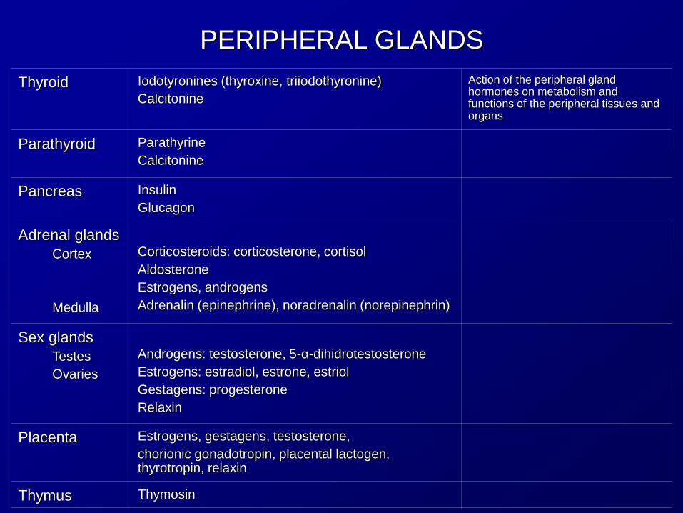
PERIPHERAL GLANDS
Thyroid Iodotyronines (thyroxine, triiodothyronine)
Calcitonine
Action of the peripheral gland hormones on metabolism and functions of the peripheral tissues and organs
Parathyroid
Parathyrine
Calcitonine
Pancreas Insulin
Glucagon
Adrenal glands
Cortex
Medulla
Corticosteroids: corticosterone, cortisol
Aldosterone
Estrogens, androgens
Adrenalin (epinephrine), noradrenalin (norepinephrin)
Sex glands
Testes
Ovaries
Androgens: testosterone, 5-α-dihidrotestosterone
Estrogens: estradiol, estrone, estriol
Gestagens: progesterone
Relaxin
Placenta Estrogens, gestagens, testosterone,
chorionic gonadotropin, placental lactogen, thyrotropin, relaxin
Thymus Thymosin

Hormone-like compounds
Endocrine functions are also exercised by other secreting biologically active compounds whose properties resemble those of hormones: hormone-like compounds or hormonoids or local hormones or parahormones.
Their action is at the site they are produced.
They are produced by cells dispersed in different tissues: – Cells of the gastrointestinal tract (gastrine, secretine)
– Intestinal chromaffin cells ( serotonin – regulator of the intetinal function)
– Most cells of the connective tissue (heparin, histamine)
– Cells of kidneys, seminal vesicles (prostaglandins

CHEMICAL STRUCTURE
Protein-peptide hormones produced by
Hypothalamus: regulatory hormones
Pituitary gland: tropic hormones
Thyroid: calcitonin
Parathyroid: parathyrine, calcitonin
Pancreas: insulin, glucagon
Aminoacid derivatives:
Adrenalin derived from phenylalanine and tyrosine
Iodothyronines derived from tyrosine
Melatonin derived from tryptophan
Steroids:
Sex hormones: androgens, estrogens, gestagens
Glucocorticoids
Mineralocorticoid: aldosterone

NEUROENDOCRINE RELATIONSHIP
Neural input
Hypothalamus
Anterior pituitary gland Posterior
pituitary gland Oxytocin
Vasopressin
Regulatory
hormones (R, I)
Adrenal
cortex Thyroid Testes / ovaries
PRL GH
Mamary
glands
TSH ACTH FSH LH
Muscles, liver,
other tissues
Sex accesory
tissues Bones
MSH
thyroxine cortico-
steroids testosterone
estrogens
gestagens
Primary
target
Secondary
target
Final
target Skin
Tropic
hormones

MUTUALLY EXCLUSSIVE RELATIONSHIP
OF ENDOCRINE SYSTEM Nervous impulse
Hypothalamus
Releasing hormones
(liberins)
Pituitary gland
Pituitary gland
hormones
(tropic hormones)
Peripheral
glands
Specific
hormones
Peripheral
organs/
cells
Short feed-back
Long feed-back
Metabolites:
•Glucose
•Aminoacids
•Fatty acids, cholesterol
•Nucleotides, nucleosides
•Ca2+, Na+, K+, Cl-

HORMONAL CONTROL
Extracellular regulators, including hormones, act as first
messengers.
Types of action:
• Membrane, local action
• Membrane intracellular, indirect action
• Cytosolic, direct action
1. MEMBRANE TYPE OF ACTION
The hormone, at the site of its binding with the cell membrane,
acts as an allosteric effector for membrane transport system and
renders the membrane permeable to glucose, aminoacids, certain
ions.
The glucose and amino acids influence the biochemical cellular
processes, while a change in ion partition on both sides of the
membrane affects the electric potentioal and function of the cell.
E.g. insulin

2. MEMBRANE-INTRACELLULAR TYPE OF ACTION
The first messengers are not able to enter in the cell and cannot
influence the intracellular processes directly. They act through a second
messenger, intracellular, which triggers a chain of successive
biochemical reactions leading to a modification of cellular functions.
First messenger (hormone) reaches the receptor on the outer side of the
cell membrane.
The hormone-receptor complex acts on a protein (membrane transducer)
The transducer transmits the signal to an enzyme (chemical amplifier)
acting as a catalyst for the production of a second messenger inside the
cell.
The second messenger binds to a special protein (internal efector) which
exerts an influence in the activity of a definite enzyme or on the
properties of non-enzyme proteins (changes of the chemical rates,
permeability, contractility, activation of genes)
E.g. cAMP, cGMP, diacylglycerides, inositol-triphosphate, Ca2+, peptides

3. CYTOSOLIC MECHANISM OF ACTION
Is typical for the compounds that can penetrate through the lipid layer of cell membrane, for example steroid hormones, vitamin D
The hormone forms a complex with a cytosolic or nuclear receptor
By selectively affecting the gene activity of nuclear chromosomes and exerting influence on the metabolism and function of cell, the hormone-receptor complex controls the enzyme concentration in the cell
E.g. iodothyronine have a combined type of action, both intracellular-membrane and cytosolic

PROHORMONES
Polypeptide hormones are synthesized as inactive
prohormones (hormonogens).
They become active after the extracellular activation by the
peptidases
Prohormone Source
Proinsulin pancreas
Proparathyroid hormone parathyroid
Angiotensinogen liver
Progastrin stomach

HORMONES OF
HYPOTHALAMUS-HYPOPHYSEAL SYSTEM
HORMONES OF HYPOTHALAMUS
The hypothalamus, a specialized portion of the brain, coordinates the endocrine system, receiving and integrating messages from the central nervous system.
It produces regulatory hormones that act on the anterior lobe of pituitary gland (adenohypophysis) controlling its function:
• stimulating its production (releasing factors, liberins) or
• inhibiting the production (inhibiting factors)
Hypothalamus also produces neurohormones: oxytocin, vasopressin that are stored in the posterior lobe of pituitary gland (neurohypophysis)

HORMONES OF
HYPOTHALAMUS-HYPOPHYSEAL SYSTEM
HORMONES OF PITUITARY GLAND
In the anterior lobe of pituitary gland (adenohypophysis) tropic hormones (tropins) are produced
From the posterior lobe (neurohypophysis) neurohormones (oxytocin, vasopressin) are released
Structure:
thyrotropin, follitropin, lutropin – glycoproteins
vasopressin, oxytocin – cyclic octapeptides

HORMONES OF HYPOTHALAMUS-HYPOPHYSEAL SYSTEM
Hypothalamic hormones Tropic hormones
Releasing factors Inhibiting factors
Thyreoliberin – thyrotropin regulatory hormone (TRH)
Thyrotropin (TSH)
Corticoliberin – corticotropin regulatory hormone
Corticotropin (ACTH)
Folliliberin – follicle stimulating h. regulatory hormone (FSH-RH)
Follitropin (FSH)
Luliberin – luteinizing regulatory hormone (LH-RH)
Lutropin (LH)
Prolactoliberin – prolactin regulatory hormone (PRH)
Prolactostatin – prolactin inhibitory hormone (PIH)
Prolactin (PRL)
Melanoliberin - melanocyte regulatory hormone (MSH-RH)
Melanostatin - melanocyte inhibitory hormone (MSH-IH)
Melanotropin (MSH)
Somatoliberin – growth hormone regulatory hormone (GH-RH)
Somatostatin - growth h. inhibitory hormone (GH-RH)
Somatotropin (STH) Growth h (GH)

MECHANISM OF ACTION OF HYPOPHYSEAL HORMONES
Tropic hormones exert their function
• on the peripheral glands or
• directly on the peripheral tissues
by binding on the membrane receptors and activating
adenylate cyclase.
The resulting cAMP determines the effects in the target cells:
• Control of biosynthesis and hormonal secretion by
peripheral glands (thyrotropin, corticotropin, follitropin,
lutropin, prolactin, somatotropin)
• Control of sex cell production (follitropin)
• Control of effector tissues (corticotropin, lutropin, follitropin,
prolactin, somatotropin, melanotropin, oxytocin,
vasopressin)
• Control of the nervous system (corticotropin)

DIRECT EFFECT ON PERIPHERAL TISSUES
Thyroid stimulating hormone = thyrotropin (TSH)
Adrenocorticotropin (ACTH):
• adenylate cyclase activation; cAMP activates the lipase
release of fatty acids and glycerol (direct action on fat
tissue by stimulating the glucose absorption and fat
mobilizing);
• action on melanin production
and lipotropins:
• fat mobilizing action (cAMP mechanism)
Gonadotropins (follicle stimulating h= FSH, luteinizing h= LH)
• Fat mobilizing (cAMP)
Prolactin: protein and lactose synthesis by mamary gland
epithelium

Melanotropin:
• Production of melanin in the skin, iris, epithelial pigment
in retina
• Fat-mobilizing (cAMP)
Somatotropin/ growth hormone (STH, GH):
• Only hormone with species-specific effect
• Stimulates cartilage cell division, growth of bones in
length, internal organs, soft tissue of face and oral cavity
• Stimulates secretion of glucagon more than insulin
• Defficiency – dwarfism proportionate constitution, no
mental retardation
• Hypersecretion – giantism or acromegalia

Vasopressin or antidiuretic hormone (ADH):
• Fat mobilizing action
• Selective control of water reabsorption in the distal tubes and
collecting ducts of the kidneys and activates adenylate cyclase;
cAMP activates protein kinases that phosphorylate the proteins in
the membranes to increase the permeability for water; reduces
diuresis, density and Na+ and Cl- concentration in urine.
contraction of muscles in arterioles and capillaries and determine
moderate in blood pressure
• Deficiency: diabetes insipidus ( large discharge of urine (4-10L/day),
low density, polydipsia
Oxytocin:
contraction of uterus muscles, Ca2+ intracellular, cAMP,
synthesis of protein in mammary glands during lactation
the release of milk – contractility of myoepithelium of mammary
ducts
• Insulin-like effect on fat tissue ( G consumption and TG synthesis)

THYROID HORMONES Iodothyronines:
– Triiodothyronine (T3)
– Tetraiodothyronine = Thyroxine (T4)
Function:
– control the energy metabolism
– exert influence on cell division and differentiation

HO O CH2-CH-COOH
NH2
HO O CH2-CH-COOH
NH2
HO O CH2-CH-COOH
NH2
HO O CH2-CH-COOH
NH2
I I
3' 3
HO O CH2-CH-COOH
NH2
HO O CH2-CH-COOH
NH2
HO O CH2-CH-COOH
NH2
I I
3' 3
I
5'
HO O CH2-CH-COOH
NH2
HO O CH2-CH-COOH
NH2
HO O CH2-CH-COOH
NH2
I I
3' 3
I
5
HO O CH2-CH-COOH
NH2
HO O CH2-CH-COOH
NH2
HO O CH2-CH-COOH
NH2
I I
3' 3
I
5
I
5'
4-(4-hydroxiphenoxi)phenylalaninetyronine
3,3'-diiodotyronine, T2
reduced hormonal activity
3,3',5'-triiodotyronine, T3'
reduced hormonal activity
3,5,3'-triiodotyronine, T3
active
3,5,3',5'-tetraiodotyronine, T4Thyroxine
active

THYROID HORMONES Calcitonin - polipeptide MW 30,000
Function: control of calcium-phosphorus metabolism
NH2-Cys-Ser-Asp-Leu-Ser-Tre-Cys-Val-Leu-Ser-Ala-Tyr-Trp-Arg-Asp-Leu- 1 2 3 4 5 6 7 8 9 10 11 12 13 14 15 16 Asp-Asp-Phe-His-Arg-Phe-Ser-Gly-Met-Gly-Phe-Gly-Pro-Glu-Tre-Pro-CO-NH2 17 18 19 20 21 22 23 24 25 26 27 28 29 30 31 32
SS

Hyperfunction = Hyperthyroidism
Thyrotoxicosis = Graves-Basedow’s disease
T3 is predominant
Accelerated catabolism of carbohydrate, triacylglycerides, proteins.
Increased basal metabolism
Elevated body temperature
Loss of body weight
Tachycardia
Hyperexcitability
Exophthalmia (protrusion of the eyeballs)

Hypofunction = Hypothyrosis
In child: infantile myxedema, cretinism
= Ineffective action of the hormones on cell division and cell differentiation – Physical retardation with disproportionate constitution due to
improper growth of bone tissue,
– Extreme mental retardation due to impaired differentiation of the neurons
– Basal metabolism reduced, body temperature below normal
In adult: myxedema manifested in – Reduced basal metabolism, lowered body temperature
– Less retentive memory
– Impaired renewal of dermal epithelium (dry skin)
– Deposition of mucoid materials in subcutaneous fat

PARATHYROID GLANDS
Calcitonine (also secreted by thyroid gland) – protein of 32 aa
Parathyrine (parathormone, PTH) – 84 aa
Function: control the balance of calcium and organic phosphate
NH2-Cys-Ser-Asp-Leu-Ser-Tre-Cys-Val-Leu-Ser-Ala-Tyr-Trp-Arg-Asp-Leu- 1 2 3 4 5 6 7 8 9 10 11 12 13 14 15 16 Asp-Asp-Phe-His-Arg-Phe-Ser-Gly-Met-Gly-Phe-Gly-Pro-Glu-Tre-Pro-CO-NH2 17 18 19 20 21 22 23 24 25 26 27 28 29 30 31 32
SS

Dysfunction of parathyroids
Hypofunction = hypoparathyrosis = determine reduced Ca2+ concentration in the blood and extracellular fluid, that facilitates the Na+ flow into the cell, increasing the excitability of nerve and mucle cells = hyperexcitability of the neuromuscular system (convulsive contraction of muscles)
Hyperparathyrosis =
– mobilization of endogenic calcium from bones (risk of fracture);
– calcemia is increased,
– phosphate lower;
– calcium is deposited in the internal organs (calcification of blood vessels, kidney, gastrointestinal tract, liver)

PANCREAS HORMONES
Cells of Langerhans islands
– A-type (α-cells) secrete glucagon
– B-type (β-cells) secrete insulin
– D-type secrete somatostatin
– PP-type (F-cells) secrete pancreatic polypeptide (that is produced in the acinous tissue, too)

Glucagon
• A single-chain polypeptide with MW 3485,
composed of 29 aa residues
• Produced by the α-cells as pre-proglucagon and
proglucagon (37aa) which is hydrolyzed by
proteases to generate the active glucagon
• Secretion is
increased by Ca2+ and arginine
inhibited by glucose and somatostatin
NH2-His-Ser-Glu-Gly-Thre-Phe-Thre-Ser-Asp-Tyr-Ser-Lys-Tyr-Leu-Asp- 1 2 3 4 5 6 7 8 9 10 11 12 13 14 15 -Ser-Arg-Arg-Ala-Gln-Asp-Phe-Val-Gln-Try-Leu-Met-Asn-Thre-COOH 16 17 18 19 20 21 22 23 24 25 26 27 28 29

Glucagon Mechanism of Action Targets: liver, fat tissue, muscle
Binds to the membrane receptors, activates the adenylate cyclase, increase the cAMP that stimulates
– the mobilization of glycogen in the liver and muscles and
– triglycerides in the fat tissue.
Thus the concentration of glucose↑, glycerol↑, fatty acids ↑ The catabolism of FA produce a large amount of acetyl-CoA and ketone bodies (ketonemia, ketonuria)
In the liver it inhibits the protein synthesis and facilitates the protein breakdown. The aa ↑ are used in
– urea production and
– gluconeogenesis → glucose ↑

Insulin
Secreted by β-cells as preproinsulin a single-chain polypeptide
this is hydrolyzed and generates the proinsulin (84 aa);
this is cleaved into peptide-C (33 aa) and insulin (51 aa) with MW about 6000
The secretion is increased by glucose and Ca2+, asparagine and leucine

NH2 Gli Asn (CO-NH 2)
NH2 Phe Ala COOH
S
S S
SS
S1 6 7 11 20 21
1 7 19 30
A
B
Composed of 2 polypeptide chains
A-chain of 21 aa, that presents
a disulphide bond (-S-S-) between Cys in position 6 and
Cys in position 11 and
C-terminal asparagine, essential for the biological activity
B-chain of 30 aa
linked through disulphide (-S-S-) bridges between:
Cys in position 7 on A-chain and 7 on B-chain
Cys in position 20 on A-chain and 19 on B-chain

Insulin Mechanism of Action Insulin exists as:
– free insulin - influences all the insulin-sensitive tissues (muscles, connective tissue, including fat tissue) and
– bound to plasma proteins – influences only fat tissue;
– less sensitive is the liver; not sensitive is the nervous tissue
Insulin binds to membrane receptor (a glycoprotein)
The insulin-receptor complex changes the permeability of the membrane for the glucose, aminoacids, Ca2+, K+, Na+, accelerating their transport into the cell.
Peptide second messenger(s) activate cAMP-phospho-diesterase, decreasing cAMP; this inhibits the glycogenolysis, gluconeogenesis, lipolysis, ketogenesis
A lower cAMP/cGMP ratio facilitates the glycogenogenesis, lipogenesis, protein synthesis
Through cGMP and Ca2+, accelerates the DNA synthesis (replication) and RNA (transcription), favoring the proliferation, growth and differentiation of cells
The result is an anabolic action with a positive nitrogen balance:
– In blood: Glucose↓, FA↓, glycerol↓, aminoacids↓, K+↓
– In urine: aminoacids↓, K+↓,

DISTURBANCES OF ENDOCRINE PANCREAS
Excessive insulin in insulinoma (tumors with β-cells) or in overdose in insulin therapy → Hypoglycemia → syncopal states, convulsions, fatal outcome
Deficient insulin → diabetes mellitus:
– Hyperglycemia (G↑), glycosuria
– FA, glycerol, cholesterol↑ in blood
– Aminoacids ↑ in blood and urine
– Ketone bodies ↑ in blood and urine → acidosis → fatal outcome

Practical application of insulin:
Treatment of diabetes mellitus
Anabolic stimulators in dystrophy of organs, malnutrition, starvation
Restoration of metabolism after heavy muscular work

Somatostatin
Polypeptide hormone
Inhibits the secretion of insulin and glucagon
It is secreted also by the hypothalamus and certain intestinal cells

HORMONES OF ADRENAL GLANDS
ADRENAL MEDULLA produces and stores into chromaffin cells
– Adrenalin / epinephrin
– Noradrenalin / norepinephrin
Adrenalin secretion is influenced by
– hypoglycemia
– stress (physiologic activity of the organism increases faster than the adaptive responses)
Effect on adrenoreceptors
– α → stimulates the guanidine cyclase → cGMP
– β → stimulates the adenylate cyclase → cAMP
cAMP has a similar effect as glucagon on the liver, muscle, fat tissue
Affects the function of cardiovascular system (amplitude and frequency of heart contraction ↑, blood pressure ↑) relaxes smooth muscles of the intestine, bronchi, uterus.

Adrenaline Noradrenaline
(epinephrine) (norepinephrine)
Metanephrine Normetanephrine 3-methoxi-4-hydroximandelic acid
vanylmandelic acid
OH
OH
CH OH
CH2 NH
CH3
OH
O-CH3
CH OH
CH2 NH
CH3
OH
O-CH3
CH OH
CH2 NH2
OH
O-CH3
CH OH
COOH

HORMONES OF ADRENAL GLANDS
ADRENAL CORTEX produces steroid hormones (corticosteroids) subdivided in:
Glucocorticosteroids – affecting the carbohydrate metabolism
• hydrocortisone
• corticosterone
Mineralocorticosteroids - affecting the mineral metabolism
• aldosterone
Sex hormones (androgens, estrogens) in small amounts

Glucocorticoids: hydrocortisone, corticosterone Controlled by corticotropin released from the pituitary gland as a response to stress; it is bound to the adrenocortical cell membrane, stimulates the production of cAMP, triggering the delivery of cholesterol esters for the synthesis of glucocorticoids; they inhibit the corticotropin (negative feed-back mechanism)
Mechanism of action:
– Transported in the plasma by transcortin (protein)
– Targets: liver, kidney, lymphoid tissue (spleen, lymph nodes, lymphoid plaques in the intestin, lymphocytes, thymus), connective tissue (bones, subcutaneous tissue, adipose tissue) muscle
Result:
– In the blood: glucose, fatty acids, glycerol, aminoacids, ketone bodies↑
– In urine: glucose, aminoacids, ketone bodies↑
– In the kidneys: ↑ Na+ reabsorption, K+ excretion;
– Na and H2O are retained in extracellular space (edema)
– In bones: ↓ protein synthesis, deossification, Ca and P →blood →urine

O
CH 3
CH 3
C O
H 2 C OH
34
5
1 7
O
CH 3
CH 3
C O
H 2 C OH
34
5
1 7H O
4 - p r e g n e n d i o l - 1 1 - , 2 1 - d i o n ac o r t i c o s t e r o n a
O
CH3
CH3
C O
H2C OH
HOOH
4-pregnentriol-11 ,17 ,21-diona-(3,20)17 -hidrocorticosteronahidrocortizon, cortisol
O
CH3
CH3
C O
H2C OH
OOH
4-pregnendiol-17 ,21-triona-(3,11,20)11-dehidro- -hidroxi-corticosteronacortizon
O
C H3
C H3
C O
H2C OH
O
4-pregnenol-21-triona-(3,11,20)11-dehidrocorticosterona
O
CH3
CH3
C O
H2C OH
4-pregnenol-21-diona-(3,20)11-dezoxicorticosteron
O
CH3
CH3
C O
H2C OH
OH
4-pregnendiol-17 ,21-diona-(3,20)11-dezoxi-17 -hidroxicorticosteroncortexon, DOC
Glucocorticosteroids

Mineralocorticoids Aldosterone secretion is controled by
• Na+ and K+ (stimulated by low Na+ and
• high K+ concentration)
It is believed that the epiphysis produces
• a tropic hormone = adrenoglomerulotropin that stimulates the secretion
Mechanism of action:
– Transported in the blood → tissue using plasma albumins
– Target: epithelial cells of the distal tubules of the kidney
– Bound to receptor, the complex penetrates the nucleus activating the transcription of the genes that carry information referring to a protein involved in the transport of Na+ across the membrane of tubular epithelium:
↑ reabsorption of Na+ , Cl- and water from the urine to the intercellular fluid and to the blood and
↑ excretion of K+ in the urine
A B
CD
3
1117
C=O
H2C OH
CH=O
HO
O
4-pregnendiol-11 diona-(3,20)-al-(18)aldosteron

DISTURBANCES OF ADRENAL GLANDS
Hyperfunction = hypercorticoidism
– Cushing's disease (hypersecretion of corticotropin) “steroid” diabetes,
atrophy of subcutaneous connective tissue
osteoporosis
hypertension (due to secondary increase of adrenalin and noradrenalin)
– Hyperaldosteronism (Konn's disease) Edema, high blood pressure, myocardial hyperexcitability
Hypofunction = hypocorticoidism = Addison's disease – Glucocorticoid deficiency: reduced resistance to emotional stress and
infections, chemical, mechanical factors; it determines hypoglycemia
– Aldosterone deficiency: disturbed water-salt imbalance - loss of Na+, H2O and accumulation of K+ → hypotension, myastenia, progressive fatigability, low muscular excitability

Practical use of corticosteroids
Treatment of allergic and autoimmune diseases: rheumatism, collagenoses, nonspecific arthrites, bronchial asthma, dermatoses
Desensitizing
Antiinflammatory
Immunodepressive agents (prophylaxis of rejection of transplanted organs)

SEX HORMONES Sex glands: in male testes or in female ovaries produce
• sex cells: spermatozoa and respectively ova • sex hormones: androgens, estrogens and gestagens
In mature male • FSH controls spermatogenesis • LH controls the production of androgens (testosteron) In female • Follicle cells secrete estrogens (estradiol, estrone, estriol) • Corpus luteus produces gestagens (progesterone) Placenta, formed during pregnancy, produces: • chorionic gonadotropin (lutropin activity), • placental lactogen (somatomammotropin), • thyrotropin, • sex hormones.

FEMALE SEX HORMONES Estrogens ensure the normal
• development of genital organs
• formation of secondary sex characteristics in puberty (hair growth, laringeal cartilage, vocal apparatus, mamary glands, skeleton)
• regulation of endometrial proliferation, contraction of fallopian tubes and uterus in follicular phase of the ovarian cycle
• formation of sexual instinct and physic status of female
• gestation, lactation, development of mammary glands in pregnancy and parturition

FEMALE SEX HORMONES
Progesterone acts during the luteal phase of ovarian cycle:
• contractile inhibition of uterus and fallopian tubes
• pre-pregnancy changes of endometrium during sex cycles and implantation of fertilized ovum
• growth of milk ducts and lactation
• reduction of excitability of the hippocampus and heat center and sexual reactivity

Female sex hormones deficiency
In prepuberal period deficiency of estrogens determine • retardation of development of genital organs • delayed formation of secondary sex characteristics • late ossification of epiphyseal cartilage • disturbance of sex cycle • negative nitrogen balance, loss of calcium phosphate,
hyperlipidemia In adult the deficient progesterone determines: - disturbances of ovarian cycle, - habitual abortion

Practical application of female sex hormones
Restoration of sex cycles disturbances
Treatment of ovary insufficiency
Pregnancy preservation (progesterone)

Male sex hormones
Anabolic action
Development of skeleton, muscles,
Development of male genitals and accessory sex glands (prostate, seminal vesicles)
Development of secondary male sex characteristics (hair, larynx, vocal apparatus) during puberty
Activate spermatogenesis (with follitropin)
Cerebral development, behavior, male character

Deficiency of male sex hormones
Genital underdevelopment
Failure to develop secondary sex characters
Absence of libido
Retardation of ossification, atrophy of muscles
Excessive fat tissue
Disturbed cortical inhibitory processes

Practical applications of male sex hormones
Treatment of
Seminal hypofunction, disturbed sex differentiation, functional sexual disturbances
Dystrophies, diabetes mellitus, thyrotoxicosis, steroid diabetes
Stimulate growth and physical development in children
Stimulate consolidation of fractured bone


![Construction of amylolytic yeasts secreting xylanase and phytase …170-176]KJM20-007.pdf · 2020. 6. 30. · Amylolytic baker’s yeasts secreting xylanase and phytase∙ 171 Korean](https://img.dokumen.tips/doc/110x75/5fe5f83ed7c59c3789287938/construction-of-amylolytic-yeasts-secreting-xylanase-and-phytase-170-176kjm20-007pdf.jpg)