Embed Size (px)
Citation preview
CASE REPORT Open Access
A challenging TSH/GH co-secretingpituitary adenoma with concomitantthyroid cancer; a case report and literaturereviewJee Hee Yoon1, Wonsuk Choi1, Ji Yong Park1, A Ram Hong1, Sung Sun Kim2, Hee Kyung Kim1 andHo-Cheol Kang1*
Abstract
Background: Thyroid stimulating hormone (TSH) secreting pituitary adenoma (TSHoma) with coexisting thyroidcancer is extremely rare, and proper treatment of both diseases may pose a unique clinical challenge. WhenTSHoma has plurihormonality, particularly involving the co-secretion of growth hormone (GH), management can bemore complicated. Herein, we present a difficult-to-manage case of papillary thyroid cancer with an incurable TSH/GH-secreting pituitary adenoma.
Case presentation: A 59-year-old man was referred to our hospital due to memory impairment and inappropriateTSH level. Sella magnetic resonance imaging revealed a huge pituitary mass extending to the suprasellar area.Clinical diagnosis of TSH/GH co-secreting pituitary adenoma was made based on elevated free T4, total T3, serumα-subunit, insulin-like growth factor-1 levels and non-suppressible GH levels after oral glucose loading. Rectal cancerand multifocal papillary thyroid microcarcinoma (PTMC) were diagnosed during initial screening for internalmalignancy; lower anterior resection was performed and close observation was planned for PTMC. Long-actingoctreotide therapy was commenced, which resulted in a dramatic reduction in TSHoma size and facilitated controlof hormonal excess. Total thyroidectomy and radioactive iodine (RAI) therapy were needed during follow up due tothe growth of PTMC. After the surgery, the pituitary adenoma represented resistance to somatostatin analoguetherapy and the tumor size gradually increased despite the addition of dopamine agonist therapy. Furthermore,TSH suppressive therapy with levothyroxine was impossible and an adequate TSH level for RAI therapy wasunmountable. Late debulking pituitary surgery was ineffective, and the patient gradually deteriorated and lost tofollow up.
Conclusion: We report the first aggravated case of TSH/GH co-secreting pituitary tumor after total thyroidectomyfor concomitant multifocal PTMC. Deferring of thyroid surgery until the TSHoma is well controlled may be theoptimal therapeutic strategy in patients with TSHoma and coexistent thyroid cancer; ablative thyroid surgery mayresult in catastrophic pituitary tumor growth.
© The Author(s). 2021 Open Access This article is licensed under a Creative Commons Attribution 4.0 International License,which permits use, sharing, adaptation, distribution and reproduction in any medium or format, as long as you giveappropriate credit to the original author(s) and the source, provide a link to the Creative Commons licence, and indicate ifchanges were made. The images or other third party material in this article are included in the article's Creative Commonslicence, unless indicated otherwise in a credit line to the material. If material is not included in the article's Creative Commonslicence and your intended use is not permitted by statutory regulation or exceeds the permitted use, you will need to obtainpermission directly from the copyright holder. To view a copy of this licence, visit http://creativecommons.org/licenses/by/4.0/.The Creative Commons Public Domain Dedication waiver (http://creativecommons.org/publicdomain/zero/1.0/) applies to thedata made available in this article, unless otherwise stated in a credit line to the data.
* Correspondence: [email protected] of Internal Medicine, Chonnam National University MedicalSchool, 160, Baekseo-ro, Dong-gu, 61469 Gwangju, South KoreaFull list of author information is available at the end of the article
Yoon et al. BMC Endocrine Disorders (2021) 21:177 https://doi.org/10.1186/s12902-021-00839-x
Keywords: Thyroid stimulating hormone (TSH) secreting pituitary adenoma (TSHoma), Acromegaly, Thyroid cancer,Coexistence, Complications
BackgroundThyroid stimulating hormone (TSH)-secreting pituitaryadenomas (TSHomas) are less than 1% of pituitary aden-omas, however it has been reported more frequently inrecent decades due to increased screening tests and clin-ical awareness [1]. Up to 30% of TSHomas representplurihormonality and the most common co-secretinghormone is growth hormone (GH) [2]. The diagnosticapproach and management of TSHoma with plurihor-monality are challenging. Firstly, clinical features of hor-monal excess could overlap or be hidden, which makesmisdiagnosis or delayed diagnosis. Additionally, pluri-hormonal pituitary adenomas reveal greater tumor re-currence and local invasion than do mono-secretingpituitary adenomas [3]. Lastly, increased rates of variouscancers and cardiovascular complications are predicted,which is exaggerated if co-secreting pituitary hormone isGH [4–6]. Plurihormonal TSHoma with concomitantthyroid cancer developed by chronic hypersecretion ofTSH is extremely rare, and its management is morecomplicated [7, 8]. Herein, we report our experience intreating a case of papillary thyroid cancer (PTC) with in-curable TSH/GH-secreting pituitary adenoma and dis-cuss the therapeutic strategy and follow-up.
Case presentationA 59-year-old man was referred to our hospital for fur-ther evaluation of an inappropriate TSH level that hadbeen detected at local clinic during a workup for demen-tia due to recurrent memory loss at local clinic. He com-plained of chronic fatigue, recently decreased visualacuity, and loss of libido. The thyroid function test re-vealed an increase in serum TSH level to 5.41 μIU/mL(reference range: 0.4–4.8), despite elevated level of freethyroxine (FT4) 3.21 ng/dL (reference range: 0.8–1.71ng/dL) and total triiodothyronine (T3) 289 ng/dL (refer-ence range: 6o–160 ng/dL) levels. Thyroid autoantibodytests, including of the anti-thyroid peroxidase antibody,anti-thyroglobulin antibody, and TSH-binding inhibitoryimmunoglobulin, were negative. He had no signs orsymptoms of thyrotoxicosis, and a physical examinationrevealed no goiter. He had no significant medical history,including thyroid disease or head and neck irradiation.His elder brother had thyroid cancer, but thyroid func-tion was normal before the surgery.Additional hormonal and imaging studies were per-
formed to distinguish between TSH-secreting pituitaryadenoma and thyroid hormone resistance syndrome.The serum α-subunit level was 1.88 mIU/mL (reference
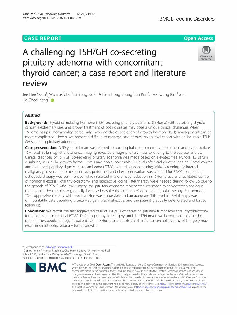
range: 0–0.8 mIU/mL) and sellar magnetic resonanceimaging (MRI) revealed a 7.0 cm, multi-lobulated hetero-geneously enhanced pituitary mass with invasion of thecavernous sinuses and supra-sellar extension (Fig. 1).Thyroid hormone receptor-beta gene mutation was notdetected. A pituitary basal hormone study showed an el-evated level of insulin-like growth factor-1 (IGF-1)478.61 ng/mL (age-matched reference range: 71–263 ng/mL); the other pituitary hormones were within normallimits (Supplementary Table 1). A GH suppression testwith 75 g oral glucose revealed no suppression of GH(GH nadir, 1.3 ng/mL). He was diagnosed with a TSH/GH-secreting pituitary adenoma. An ophthalmologicalexamination revealed bitemporal hemianopsia (Supple-mentary Fig. 1). Surgical resection was considered thebest treatment option; however, curative pituitary sur-gery, which the patient refused, was impossible due toinvasion. We decided to start long-acting octreotide tocontrol the abnormal TSH and GH secretions.Positron emission tomography-computed tomography
(PET-CT) was performed to screen for malignancy asso-ciated with the overproduction of TSH and GH; itshowed benign hepatic cysts up to 3.5 cm and 18F-fluor-odexoyglucose (FDG) avid lesions on the thyroid glandand sigmoid colon. Thyroid ultrasonography (US) re-vealed a 0.5 cm spiculated hypoechoic nodule withmicrocalcification, and fine needle aspiration revealedPTC. We decided to closely observe the thyroid cancerwithout surgery due to the small sized tumor (< 1 cm),no suspicious lymph nodes on US and staging neck CT,and our concerns for potential rapid growth of the pitu-itary tumor after surgical removal of the thyroid gland.Follow-up thyroid US at 11 months revealed an increasein tumor size from 0.3 × 0.5 × 0.4 cm to 0.7 × 0.5 × 0.5 cmso, surgery was considered. The patient underwent totalthyroidectomy with central lymph node dissection;BRAF V600E-positive multifocal PTCs and metastaticcervical lymph nodes (LNs; 7 of the 11 resected LNs)were confirmed pathologically. A metastatic paratrachealLN measuring 8 mm in the largest diameter had extra-nodal extension. The final TNM stage was pT1aN1aM0(stage II). After surgery, it was impossible to suppressthe TSH sufficiently, despite 400 μg levothyroxine, toachieve a goal of the middle to upper half of the refer-ence range for free T4 in a short period immediatelyafter surgery. Furthermore, the TSH level increased in-sufficiently after levothyroxine withdrawal in preparationfor RAI therapy (TSH, 12.22 μIU/mL). On RAI therapywith dose of 180 mCi, off-Tg level was less than 1 ng/ml,
Yoon et al. BMC Endocrine Disorders (2021) 21:177 Page 2 of 8
and post-treatment scan showed strong RAI uptake inanterior neck, both thyroid beds, and right upper medi-astinum. After RAI therapy, levothyroxine (200–250 μg/day) replacement was continued and there was no PTCrecurrence during the next 47 months.Sellar MRI, performed at the 6-month follow-up after
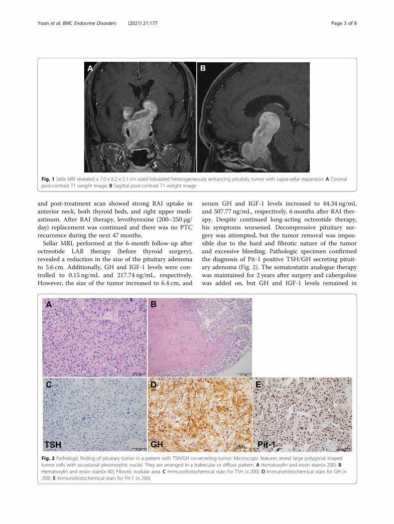
octreotide LAR therapy (before thyroid surgery),revealed a reduction in the size of the pituitary adenomato 5.6 cm. Additionally, GH and IGF-1 levels were con-trolled to 0.15 ng/mL and 217.74 ng/mL, respectively.However, the size of the tumor increased to 6.4 cm, and
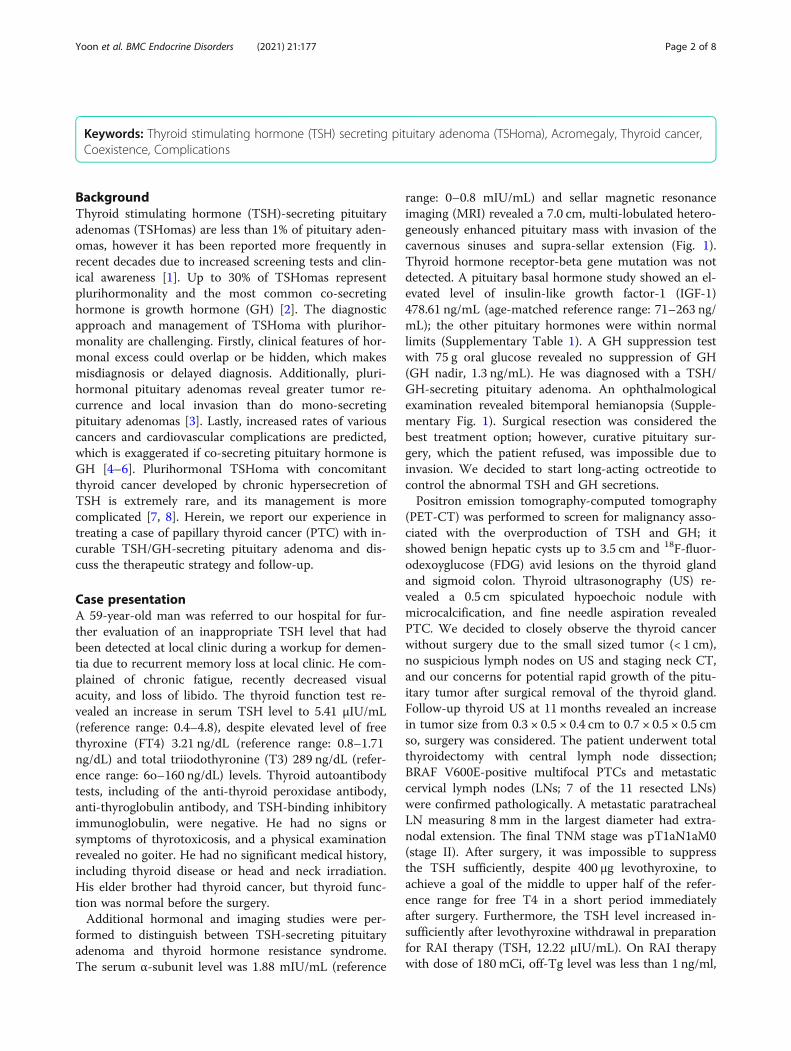
serum GH and IGF-1 levels increased to 44.34 ng/mLand 507.77 ng/mL, respectively, 6 months after RAI ther-apy. Despite continued long-acting octreotide therapy,his symptoms worsened. Decompressive pituitary sur-gery was attempted, but the tumor removal was impos-sible due to the hard and fibrotic nature of the tumorand excessive bleeding. Pathologic specimen confirmedthe diagnosis of Pit-1 positive TSH/GH secreting pituit-ary adenoma (Fig. 2). The somatostatin analogue therapywas maintained for 2 years after surgery and cabergolinewas added on, but GH and IGF-1 levels remained in
Fig. 1 Sella MRI revealed a 7.0 × 6.2 × 5.1 cm sized lobulated heterogeneously enhancing pituitary tumor with supra-sellar expansion. A Coronalpost-contrast T1 weight image; B Sagittal post-contrast T1 weight image
Fig. 2 Pathologic finding of pituitary tumor in a patient with TSH/GH co-secreting tumor: Microscopic features reveal large polygonal shapedtumor cells with occasional pleomorphic nuclei. They are arranged in a trabecular or diffuse pattern. A Hematoxylin and eosin stain(× 200). BHematoxylin and eosin stain(× 40), Fibrotic nodular area. C Immunohistochemical stain for TSH (× 200). D Immunohistochemical stain for GH (×200). E Immunohistochemical stain for Pit-1 (× 200)
Yoon et al. BMC Endocrine Disorders (2021) 21:177 Page 3 of 8
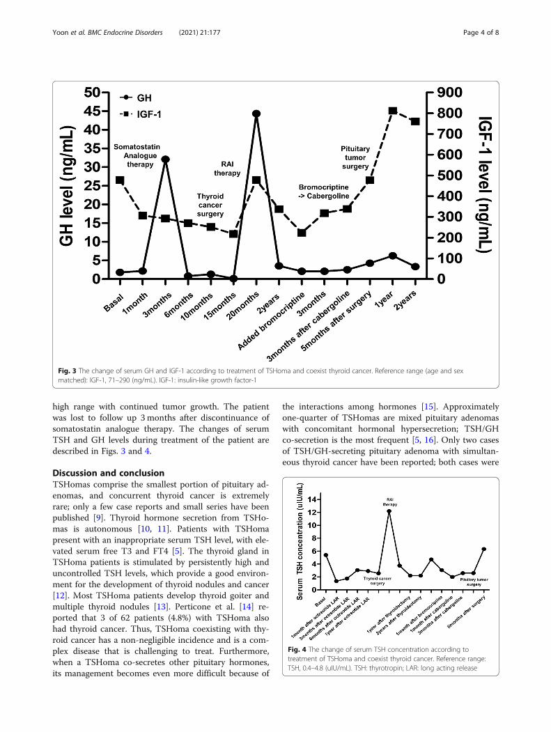
high range with continued tumor growth. The patientwas lost to follow up 3months after discontinuance ofsomatostatin analogue therapy. The changes of serumTSH and GH levels during treatment of the patient aredescribed in Figs. 3 and 4.
Discussion and conclusionTSHomas comprise the smallest portion of pituitary ad-enomas, and concurrent thyroid cancer is extremelyrare; only a few case reports and small series have beenpublished [9]. Thyroid hormone secretion from TSHo-mas is autonomous [10, 11]. Patients with TSHomapresent with an inappropriate serum TSH level, with ele-vated serum free T3 and FT4 [5]. The thyroid gland inTSHoma patients is stimulated by persistently high anduncontrolled TSH levels, which provide a good environ-ment for the development of thyroid nodules and cancer[12]. Most TSHoma patients develop thyroid goiter andmultiple thyroid nodules [13]. Perticone et al. [14] re-ported that 3 of 62 patients (4.8%) with TSHoma alsohad thyroid cancer. Thus, TSHoma coexisting with thy-roid cancer has a non-negligible incidence and is a com-plex disease that is challenging to treat. Furthermore,when a TSHoma co-secretes other pituitary hormones,its management becomes even more difficult because of
the interactions among hormones [15]. Approximatelyone-quarter of TSHomas are mixed pituitary adenomaswith concomitant hormonal hypersecretion; TSH/GHco-secretion is the most frequent [5, 16]. Only two casesof TSH/GH-secreting pituitary adenoma with simultan-eous thyroid cancer have been reported; both cases were
Fig. 3 The change of serum GH and IGF-1 according to treatment of TSHoma and coexist thyroid cancer. Reference range (age and sexmatched): IGF-1, 71–290 (ng/mL). IGF-1: insulin-like growth factor-1
Fig. 4 The change of serum TSH concentration according totreatment of TSHoma and coexist thyroid cancer. Reference range:TSH, 0.4–4.8 (uIU/mL). TSH: thyrotropin; LAR: long acting release
Yoon et al. BMC Endocrine Disorders (2021) 21:177 Page 4 of 8
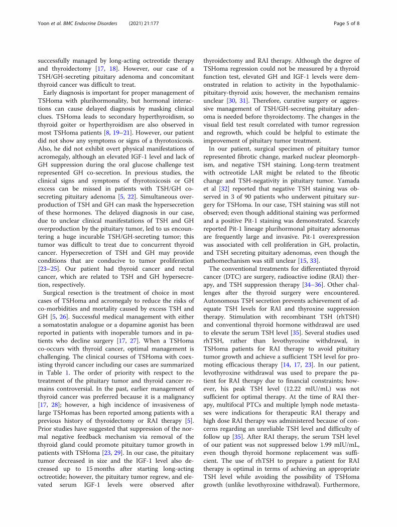
successfully managed by long-acting octreotide therapyand thyroidectomy [17, 18]. However, our case of aTSH/GH-secreting pituitary adenoma and concomitantthyroid cancer was difficult to treat.Early diagnosis is important for proper management of
TSHoma with plurihormonality, but hormonal interac-tions can cause delayed diagnosis by masking clinicalclues. TSHoma leads to secondary hyperthyroidism, sothyroid goiter or hyperthyroidism are also observed inmost TSHoma patients [8, 19–21]. However, our patientdid not show any symptoms or signs of a thyrotoxicosis.Also, he did not exhibit overt physical manifestations ofacromegaly, although an elevated IGF-1 level and lack ofGH suppression during the oral glucose challenge testrepresented GH co-secretion. In previous studies, theclinical signs and symptoms of thyrotoxicosis or GHexcess can be missed in patients with TSH/GH co-secreting pituitary adenoma [5, 22]. Simultaneous over-production of TSH and GH can mask the hypersecretionof these hormones. The delayed diagnosis in our case,due to unclear clinical manifestations of TSH and GHoverproduction by the pituitary tumor, led to us encoun-tering a huge incurable TSH/GH-secreting tumor; thistumor was difficult to treat due to concurrent thyroidcancer. Hypersecretion of TSH and GH may provideconditions that are conducive to tumor proliferation[23–25]. Our patient had thyroid cancer and rectalcancer, which are related to TSH and GH hypersecre-tion, respectively.Surgical resection is the treatment of choice in most
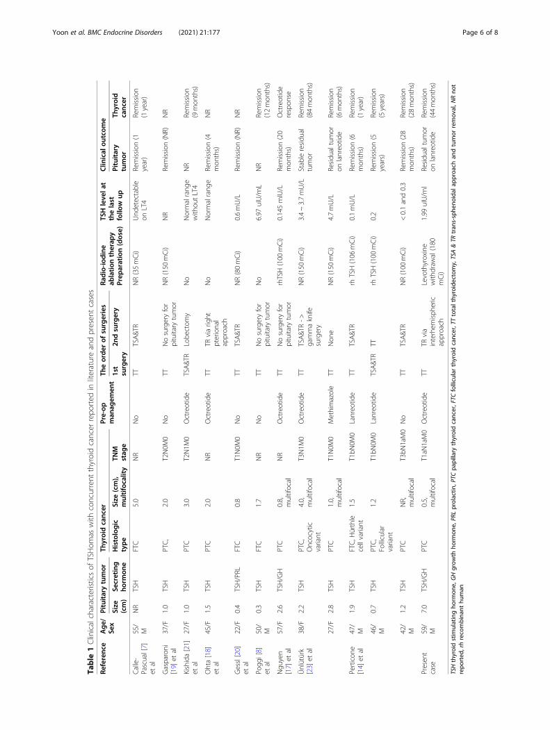
cases of TSHoma and acromegaly to reduce the risks ofco-morbidities and mortality caused by excess TSH andGH [5, 26]. Successful medical management with eithera somatostatin analogue or a dopamine agonist has beenreported in patients with inoperable tumors and in pa-tients who decline surgery [17, 27]. When a TSHomaco-occurs with thyroid cancer, optimal management ischallenging. The clinical courses of TSHoma with coex-isting thyroid cancer including our cases are summarizedin Table 1. The order of priority with respect to thetreatment of the pituitary tumor and thyroid cancer re-mains controversial. In the past, earlier management ofthyroid cancer was preferred because it is a malignancy[17, 28]; however, a high incidence of invasiveness oflarge TSHomas has been reported among patients with aprevious history of thyroidectomy or RAI therapy [5].Prior studies have suggested that suppression of the nor-mal negative feedback mechanism via removal of thethyroid gland could promote pituitary tumor growth inpatients with TSHoma [23, 29]. In our case, the pituitarytumor decreased in size and the IGF-1 level also de-creased up to 15months after starting long-actingoctreotide; however, the pituitary tumor regrew, and ele-vated serum IGF-1 levels were observed after
thyroidectomy and RAI therapy. Although the degree ofTSHoma regression could not be measured by a thyroidfunction test, elevated GH and IGF-1 levels were dem-onstrated in relation to activity in the hypothalamic-pituitary-thyroid axis; however, the mechanism remainsunclear [30, 31]. Therefore, curative surgery or aggres-sive management of TSH/GH-secreting pituitary aden-oma is needed before thyroidectomy. The changes in thevisual field test result correlated with tumor regressionand regrowth, which could be helpful to estimate theimprovement of pituitary tumor treatment.In our patient, surgical specimen of pituitary tumor
represented fibrotic change, marked nuclear pleomorph-ism, and negative TSH staining. Long-term treatmentwith octreotide LAR might be related to the fibroticchange and TSH-negativity in pituitary tumor. Yamadaet al [32] reported that negative TSH staining was ob-served in 3 of 90 patients who underwent pituitary sur-gery for TSHoma. In our case, TSH staining was still notobserved; even though additional staining was performedand a positive Pit-1 staining was demonstrated. Scarcelyreported Pit-1 lineage plurihormonal pituitary adenomasare frequently large and invasive. Pit-1 overexpressionwas associated with cell proliferation in GH, prolactin,and TSH secreting pituitary adenomas, even though thepathomechanism was still unclear [15, 33].The conventional treatments for differentiated thyroid
cancer (DTC) are surgery, radioactive iodine (RAI) ther-apy, and TSH suppression therapy [34–36]. Other chal-lenges after the thyroid surgery were encountered.Autonomous TSH secretion prevents achievement of ad-equate TSH levels for RAI and thyroxine suppressiontherapy. Stimulation with recombinant TSH (rhTSH)and conventional thyroid hormone withdrawal are usedto elevate the serum TSH level [35]. Several studies usedrhTSH, rather than levothyroxine withdrawal, inTSHoma patients for RAI therapy to avoid pituitarytumor growth and achieve a sufficient TSH level for pro-moting efficacious therapy [14, 17, 23]. In our patient,levothyroxine withdrawal was used to prepare the pa-tient for RAI therapy due to financial constraints; how-ever, his peak TSH level (12.22 mIU/mL) was notsufficient for optimal therapy. At the time of RAI ther-apy, multifocal PTCs and multiple lymph node metasta-ses were indications for therapeutic RAI therapy andhigh dose RAI therapy was administered because of con-cerns regarding an unreliable TSH level and difficulty offollow up [35]. After RAI therapy, the serum TSH levelof our patient was not suppressed below 1.99 mIU/mL,even though thyroid hormone replacement was suffi-cient. The use of rhTSH to prepare a patient for RAItherapy is optimal in terms of achieving an appropriateTSH level while avoiding the possibility of TSHomagrowth (unlike levothyroxine withdrawal). Furthermore,
Yoon et al. BMC Endocrine Disorders (2021) 21:177 Page 5 of 8
Table
1Clinicalcharacteristicsof
TSHom
aswith
concurrent
thyroidcancer
repo
rted
inliteratureandpresen
tcases
Referenc
eAge/
Sex
Pituitarytumor
Thyroidcanc
erPre-op
man
agem
ent
Theorde
rof
surgeries
Radio-io
dine
ablation
therap
yPrep
aration(dose)
TSHleve
lat
thelast
follo
wup
Clin
ical
outcom
e
Size
(cm)
Secreting
horm
one
Histologic
type
Size
(cm),
multifocality
TNM
stag
e1st
surgery
2ndsurgery
Pituitary
tumor
Thyroid
canc
er
Calle-
Pascual[7]
etal
55/
MNR
TSH
FTC
5.0
NR
No
TTTSA&T
RNR(35mCi)
Und
etectable
onLT4
Remission
(1year)
Remission
(1year)
Gasparoni
[19]
etal
37/F
1.0
TSH
PTC,
2.0
T2N0M
0No
TTNosurgeryfor
pituitary
tumor
NR(150
mCi)
NR
Remission
(NR)
NR
Kishida[21]
etal
27/F
1.0
TSH
PTC
3.0
T2N1M
0Octreotide
TSA&T
RLobe
ctom
yNo
Normalrang
ewith
outLT4
NR
Remission
(9mon
ths)
Ohta[18]
etal
45/F
1.5
TSH
PTC
2.0
NR
Octreotide
TTTR
viarig
htpterional
approach
No
Normalrang
eRemission
(4mon
ths)
NR
Gessl[20]
etal
22/F
0.4
TSH/PRL
FTC
0.8
T1N0M
0No
TTTSA&T
RNR(80mCi)
0.6mU/L
Remission
(NR)
NR
Pogg
i[8]
etal
50/
M0.3
TSH
FTC
1.7
NR
No
TTNosurgeryfor
pituitary
tumor
No
6.97
uIU/m
LNR
Remission
(12mon
ths)
Ngu
yen
[17]
etal
57/F
2.6
TSH/GH
PTC
0.8,
multifocal
NR
Octreotide
TTNosurgeryfor
pituitary
tumor
rhTSH(100
mCi)
0.145mIU/L
Remission
(20
mon
ths)
Octreotide
respon
se
Ünlütürk
[23]
etal
38/F
2.2
TSH
PTC,
Oncocytic
variant
4.0,
multifocal
T3N1M
0Octreotide
TTTSA&T
R->
gammaknife
surgery
NR(150
mCi)
3.4~3.7mU/L
Stableresidu
altumor
Remission
(84mon
ths)
27/F
2.8
TSH
PTC
1.0,
multifocal
T1N0M
0Methimazole
TTNon
eNR(150
mCi)
4.7mU/L
Residu
altumor
onlanreo
tide
Remission
(6mon
ths)
Perticon
e[14]
etal
47/
M1.9
TSH
FTC,H
ürthle
cellvariant
1.5
T1bN
0M0
Lanreo
tide
TTTSA&T
Rrh
TSH(106
mCi)
0.1mU/L
Remission
(6mon
ths)
Remission
(1year)
46/
M0.7
TSH
PTC,
Follicular
variant
1.2
T1bN
0M0
Lanreo
tide
TSA&T
RTT
rhTSH(100
mCi)
0.2
Remission
(5years)
Remission
(5years)
42/
M1.2
TSH
PTC
NR,
multifocal
T3bN
1aM0
No
TTTSA&T
RNR(100
mCi)
<0.1and0.3
Remission
(28
mon
ths)
Remission
(28mon
ths)
Presen
tcase
59/
M7.0
TSH/GH
PTC
0.5,
multifocal
T1aN
1aM0
Octreotide
TTTR
via
interhem
isph
eric
approach
Levothyroxine
with
draw
al(180
mCi)
1.99
uIU/m
lResidu
altumor
onlanreo
tide
Remission
(44mon
ths)
TSHthyroidstim
ulatingho
rmon
e,GHgrow
thho
rmon
e,PR
Lprolactin
,PTC
papillary
thyroidcancer,FTC
follicularthyroidcancer,TTtotalthy
roidectomy,TSA&TR
tran
s-sphe
noidal
approa
chan
dtumor
remov
al,N
Rno
trepo
rted
,rhrecombina
nthu
man
Yoon et al. BMC Endocrine Disorders (2021) 21:177 Page 6 of 8
the serum TSH level does not reflect the thyroid statusor the severity of TSHoma in cases where the TSHomais not treated curatively. Further studies are necessary toidentify a better method to evaluate thyroid functionalstatus and the extent of TSH secretions by TSHomas.In conclusion, our case involved a large Pit-1 lineage
TSH/GH co-secreting pituitary macroadenoma withconcomitant thyroid cancer was challenging to diagnoseand treat. Controlling the pituitary tumor before thy-roidectomy is important to avoid rapid rebound of pitu-itary tumor growth due to the attenuation of thyroidgland-mediated negative feedback. An appropriate thy-roid cancer management strategy, such as using rhTSHto prepare the patient for RAI therapy followed by me-ticulous surveillance after thyroid surgery, is necessaryfor the successful treatment of TSHoma and concomi-tant thyroid cancer.
AbbreviationsFDG: Fluorodeoxyglucose; GH: Growth hormone; IGF-1: Insulin-like growthfactor-1; MRI: Magnetic resonance imaging; PET-CT: Positron emissiontomography-computed tomography; PTMC: Papillary thyroidmicrocarcinoma; RAI: Radioactive iodine; T3: Triiodothyronine; T4: Thyroxine;TSH: Thyroid stimulating hormone; US: Ultrasonography
Supplementary InformationThe online version contains supplementary material available at https://doi.org/10.1186/s12902-021-00839-x.
Additional file 1: Supplementary table 1. Baseline pituitary hormonetest
Additional file 2: Supplementary figure 1. The change of the visualfield examination. A: At the time of the first diagnosis of TSH/GH co-secreting tumor. B: Pre-operative work up before thyroid cancer surgery
AcknowledgmentsNot applicable.
Authors’ contributionsJHY drafted the manuscript. ARH, JYP and WSC participated in the analysisand the interpretation of the data. SSK performed additionalimmunohistochemical staining and pathological reviewing. HKK and HCKcritically revised the manuscript. All the authors read and approved the finalarticle.
FundingNot applicable.
Availability of data and materialsAll the data generated and/or analyzed during this study are included in thisarticle.
Declarations
Ethics approval and consent to participateThis study was approved by the Institutional Review Board of ChonnamNational University Hwasun Hospital and a written informed consent wasobtained from the patient.
Consent for publicationWritten informed consent was obtained from the patient for publication ofthis case report and any accompanying images.
Competing interestsAll authors declare no conflict of interest.
Author details1Department of Internal Medicine, Chonnam National University MedicalSchool, 160, Baekseo-ro, Dong-gu, 61469 Gwangju, South Korea.2Department of Pathology, Chonnam National University Medical School,160, Baekseo-ro, Dong-gu, 61469 Gwangju, South Korea.
Received: 4 November 2020 Accepted: 16 August 2021
References1. Beck-Peccoz P, Persani L, Lania A. Thyrotropin-secreting pituitary adenomas.
In: De Groot LJ, Beck-Peccoz P, Chrousos G, Dungan K, Grossman A,Hershman JM, Koch C, McLachlan R, New M, Rebar R, et al., editors.Endotext. South Dartmouth: MDText.com, Inc.; 2000.
2. Beck-Peccoz P, Brucker-Davis F, Persani L, Smallridge RC, Weintraub BD.Thyrotropin-secreting pituitary tumors. Endocr Rev. 1996;17(6):610–38.
3. Nishioka H, Inoshita N. New WHO classification of pituitary adenomas (4thedition): assessment of pituitary transcription factors and the prognostichistological factors. Brain Tumor Pathol. 2018;35(2):57–61.
4. Jenkins PJ. Cancers associated with acromegaly. Neuroendocrinology. 2006;83(3–4):218–23.
5. Beck-Peccoz P, Persani L, Mannavola D, Campi I. Pituitary tumours: TSH-secreting adenomas. Best Pract Res Clin Endocrinol Metab. 2009;23(5):597–606.
6. Lombardi G, Di Somma C, Grasso LF, Savanelli MC, Colao A, Pivonello R. Thecardiovascular system in growth hormone excess and growth hormonedeficiency. J Endocrinol Investig. 2012;35(11):1021–9.
7. Calle-Pascual AL, Yuste E, Martin P, Aramendi T, Garcia-Maurino ML, ArgenteJ, et al. Association of a thyrotropin-secreting pituitary adenoma and athyroid follicular carcinoma. J Endocrinol Investig. 1991;14(6):499–502.
8. Poggi M, Monti S, Pascucci C, Toscano V. A rare case of follicular thyroidcarcinoma in a patient with thyrotropin-secreting pituitary adenoma. Am JMed Sci. 2009;337(6):462–5.
9. Kesmodel SB, Terhune KP, Canter RJ, Mandel SJ, LiVolsi VA, Baloch ZW, et al.The diagnostic dilemma of follicular variant of papillary thyroid carcinoma.Surgery. 2003;134(6):1005–12.
10. Foppiani L, Del Monte P, Ruelle A, Bandelloni R, Quilici P, Bernasconi D. TSH-secreting adenomas: rare pituitary tumors with multifaceted clinical andbiological features. J Endocrinol Investig. 2007;30(7):603–9.
11. Ness-Abramof R, Ishay A, Harel G, Sylvetzky N, Baron E, Greenman Y, et al.TSH-secreting pituitary adenomas: follow-up of 11 cases and review of theliterature. Pituitary. 2007;10(3):307–10.
12. Fiore E, Vitti P. Serum TSH and risk of papillary thyroid cancer in nodularthyroid disease. J Clin Endocrinol Metab. 2012;97(4):1134–45.
13. Beck-Peccoz P, Giavoli C, Lania A. A 2019 update on TSH-secreting pituitaryadenomas. J Endocrinol Investig. 2019;42(12):1401–6.
14. Perticone F, Pigliaru F, Mariotti S, Deiana L, Furlani L, Mortini P, et al. Is theincidence of differentiated thyroid cancer increased in patients withthyrotropin-secreting adenomas? Report of three cases from a largeconsecutive series. Thyroid. 2015;25(4):417–24.
15. Ng HY, Namboodiri D, Learoyd D, Davidson A, Champion B, Preda V. Clinicalchallenges of a co-secreting TSH/GH pituitary adenoma. Endocrinoldiabetes Metab Case Rep. 2019;17:EDM190068.
16. Mantovani G, Asteria C, Pellegrini C, Bosari S, Alberti L, Bondioni S, et al.HESX1 expression in human normal pituitaries and pituitary adenomas. MolCell Endocrinol. 2006;247(1–2):135–9.
17. Nguyen HD, Galitz MS, Mai VQ, Clyde PW, Glister BC, Shakir MK.Management of coexisting thyrotropin/growth-hormone-secreting pituitaryadenoma and papillary thyroid carcinoma: a therapeutic challenge. Thyroid.2010;20(1):99–103.
18. Ohta S, Nishizawa S, Oki Y, Namba H. Coexistence of thyrotropin-producingpituitary adenoma with papillary adenocarcinoma of the thyroid--a casereport and surgical strategy. Pituitary. 2001;4(4):271–4.
19. Gasparoni P, Rubello D, Persani L, Beck-Peccoz P. Unusual associationbetween a thyrotropin-secreting pituitary adenoma and a papillary thyroidcarcinoma. Thyroid. 1998;8(2):181–3.
20. Gessl A, Vierhapper H, Feichtinger H. Non-suppressible TSH in a patientthyroidectomized due to follicular thyroid carcinoma. Exp Clin EndocrinolDiabetes. 2006;114(7):389–92.
Yoon et al. BMC Endocrine Disorders (2021) 21:177 Page 7 of 8

21. Kishida M, Otsuka F, Kataoka H, Yokota K, Oishi T, Yamauchi T, et al.Hyperthyroidism in a patient with TSH-producing pituitary adenomacoexisting with thyroid papillary adenocarcinoma. Endocr J. 2000;47(6):731–8.
22. Johnston PC, Hamrahian AH, Prayson RA, Kennedy L, Weil RJ. Thyrotoxicosiswith absence of clinical features of acromegaly in a TSH- and GH-secreting,invasive pituitary macroadenoma. Endocrinol Diabetes Metab Case Rep.2015;2015:140070.
23. Unluturk U, Sriphrapradang C, Erdogan MF, Emral R, Guldiken S, Refetoff S,et al. Management of differentiated thyroid cancer in the presence ofresistance to thyroid hormone and TSH-secreting adenomas: a report of fourcases and review of the literature. J Clin Endocrinol Metab. 2013;98(6):2210–7.
24. Kim HK, Lee JS, Park MH, Cho JS, Yoon JH, Kim SJ, et al. Tumorigenesis ofpapillary thyroid cancer is not BRAF-dependent in patients with acromegaly.PLoS One. 2014;9(10):e110241.
25. Petroff D, Tonjes A, Grussendorf M, Droste M, Dimopoulou C, Stalla G, et al. Theincidence of Cancer among acromegaly patients: results from the Germanacromegaly registry. J Clin Endocrinol Metab. 2015;100(10):3894–902.
26. Dekkers OM, Biermasz NR, Pereira AM, Romijn JA, Vandenbroucke JP. Mortalityin acromegaly: a Metaanalysis. J Clin Endocrinol Metab. 2008;93(1):61–7.
27. Atkinson JL, Abboud CF, Lane JI. Dramatic volume reduction of a large GH/TSH secreting pituitary tumor with short term Octreotide therapy. Pituitary.2005;8(2):89–91.
28. Safer JD, Colan SD, Fraser LM, Wondisford FE. A pituitary tumor in a patientwith thyroid hormone resistance: a diagnostic dilemma. Thyroid. 2001;11(3):281–91.
29. Beck-Peccoz P, Persani L. Medical management of thyrotropin-secretingpituitary adenomas. Pituitary. 2002;5(2):83–8.
30. Jones PM, Burrin JM, Ghatei MA, O'Halloran DJ, Legon S, Bloom SR. Theinfluence of thyroid hormone status on the hypothalamo-hypophysealgrowth hormone axis. Endocrinology. 1990;126(3):1374–9.
31. Laron Z. Interactions between the thyroid hormones and the hormones ofthe growth hormone axis. Pediatr Endocrinol. 2003;1(Suppl 2):244–249-discussion 250.
32. Yamada S, Fukuhara N, Horiguchi K, Yamaguchi-Okada M, Nishioka H,Takeshita A, et al. Clinicopathological characteristics and therapeuticoutcomes in thyrotropin-secreting pituitary adenomas: a single-center studyof 90 cases. J Neurosurg. 2014;121(6):1462–73.
33. Lee JC, Pekmezci M, Lavezo JL, Vogel H, Katznelson L, Fraenkel M, et al.Utility of Pit-1 Immunostaining in distinguishing pituitary adenomas ofprimitive differentiation from null cell adenomas. Endocr Pathol. 2017;28(4):287–92.
34. Schlumberger MJ. Papillary and follicular thyroid carcinoma. N Engl J Med.1998;338(5):297–306.
35. Mallick UK. The revised American Thyroid Association managementguidelines 2009 for patients with differentiated thyroid cancer: an evidence-based risk-adapted approach. Clin Oncol. 2010;22(6):472–4.
36. Haugen BR, Alexander EK, Bible KC, Doherty GM, Mandel SJ, Nikiforov YE,et al. 2015 American Thyroid Association management guidelines for adultpatients with thyroid nodules and differentiated thyroid Cancer: theAmerican Thyroid Association guidelines task force on thyroid nodules anddifferentiated thyroid Cancer. Thyroid. 2016;26(1):1–133.
Publisher’s NoteSpringer Nature remains neutral with regard to jurisdictional claims inpublished maps and institutional affiliations.
Yoon et al. BMC Endocrine Disorders (2021) 21:177 Page 8 of 8