Embed Size (px)
Citation preview

Anterior pituitary insufficiency
Endocrine block 2013

Anterior pituitary disorders
① Non-functional pituitary tumor: mass-effect
② Prolactin secreting cell disorder:
prolactinoma
③ Growth hormone secreting cell disorder:
acromegaly
④ ACTH secreting cell disorders: Cushing’s
⑤ TSH secreting cell tumor: TSH-oma
⑥ Gonadotropin secreting cell disorder

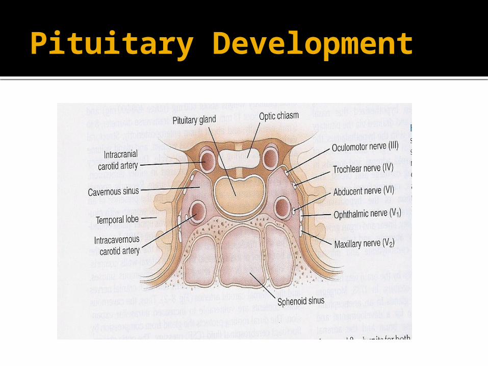
Pituitary Development
Anterior pituitary is recognizable by 4- 5th wk of gestation
Full maturation by 20th wk From Rathke’s pouch, Ectodermal evagination of
oropharynx Migrate to join neurohypophysis Portion of Rathke’s pouch →→ Intermediate lobe Remnant of Rathke’s pouch cell in oral cavity →→
pharyngeal pituitary Lies at the base of the skull as sella turcica Roof is formed by diaphragma sellae Floor by the roof of sphenoid sinus

Pituitary Development
Posterior pituitary from neural cells as an outpouching from the floor of 3rd ventricle
Pituitary stalk in midline joins the pituitary gland with hypothalamus that is below 3rd ventricle
Development of pituitary cells is controlled by a set of transcription growth factors like pit-1, Prop-1, Pitx2

Pituitary Development
Pituitary stalk and its blood vessels pass through the diaphragm
Lateral wall by cavernous sinus containing III, IV, VI, V1, V2 cranial nerves and internal carotid artery with sympathetic fibers. Both adjacent to temporal lobes
Pituitary gland measures 15 X 10 X 6 mm, weighs 500 mg but about 1 g in women
Optic chiasm lies 10 mm above the gland and anterior to the stalk
Blood supply : superior, middle, inferior hypophysial arteries ( internal carotid artery) running in median eminence from hypothalamus
Venous drainage: to superior and inferior petrosal sinsuses to jugular vein
Pituitary Development

Normal Pituitary Anatomy
Modified from Lechan RM. Neuroendocrinology of Pituitary Hormone Regulation. Endocrinology and Metabolism Clinics 16:475-501, 1987

Pituitary Development

Endocrine system

Anterior Pituitary Function
Corticotroph Gonadotroph Thyrotroph Lactotroph Somatotroph
Hormone POMC, ACTH FSH, LH TSH Prolactin GH
Stimulators CRH, AVP, gp-130
cytokines
GnRH, Estrogen
TRH Estrogen, TRH
GHRH, GHS
Inhibitors Glucocorticoids Sex steroids, inhibin
T3, T4, Dopamine,
Somatostatin, GH
Dopamine Somatostatin, IGF-1,
Activins
Target Gland
Adrenals Ovary, Testes Thyroid Breast and other
tissues
Liver, bone and other
tissues
Trophic Effects
Steroid production
Sex Steroid, Follicular growth,
Germ Cell maturation
T4 synthesis and
secretion
Milk Productio
n
IGF-1 production,
Growth induction,
Insulin antagonis
m
Adapted from: William’s Textbook of Endocrinology, 10th ed., Figure 8-4, pg 180.


Etiology of Pituitary Masses

Etiology of Pituitary-Hypothalamic Lesions Non-Functioning Pituitary Adenomas
Endocrine active pituitary adenomas Prolactinoma Somatotropinoma Corticotropinoma Thyrotropinoma Other mixed endocrine active adenomas
Malignant pituitary tumors: Functional and non-functional pituitary carcinoma
Metastases in the pituitary (breast, lung, stomach, kidney)
Pituitary cysts: Rathke's cleft cyst, Mucocoeles, Others
Empty sella syndrome
Pituitary abscess
Lymphocytic hypophysitis
Carotid aneursym
\

Clinical presentations of sellar mass


Evaluation of Pituitary mass
Pituitary adenoma: 10 % of all pituitary lesions
Genetic-related
MEN-1, Gs-alpha mutation, PTTG gene, FGF
receptor-4
Pituitary incidentaloma:
1.5 -31% in autopsy ( prevalence)
10 % by MRI most of them < 1 cm

Evaluation of Pituitary lesions
Functional adenomas ( hormonal-secreting)
Non-Functional adenomas

Evaluation of Pituitary lesion

Evaluation of Pituitary lesion
Non-Functional pituitary lesion:
Absence of signs and symptoms of hormonal hypersecretion
25 % of pituitary tumor Needs evaluation either micro or macroadenoma Average age 50 – 55 yrs old, more in male

Non- functional pituitary adenoma
Presentation of NFPA:
As incidentaloma by imaging
Symptoms of mass effects ( mechanical pressure)
Hypopituitarism ( mechanism)
Gonadal hypersecretion

Non- functional pituitary adenoma

Non- functional pituitary adenoma
Treatment: Surgery if indicated
- recurrence rate 17 % if gross removal, 40 % with residual tumor- predictors of recurrence: young male,
cavernous sinus invasion, extent of suprasellar extention of residual tumor, duration of follow up, marker; Ki-67
Observation with annual follow up for 5 years and then as needed, visual field exam Q 6-12 month if close to optic chiasm. Slow growing tumour
Adjunctive therapy:- Radiation therapy- Dopamine agonist- Somatostatin analogue

Functional pituitary mass

Prolactinoma


Prolactinoma
Prolactin:

Growth hormone
Pituitary tumor as mass effect →→ Growth hormone deficiency
Hyperfunctioning mass →→ Acromegaly

Growth hormone deficiency
Diagnosis in children and adult

Diagnosis of GH-deficiency and management
GH, IGF-I level Dynamic testing: clonidine
stimulation test, glucagon stimulation, exercise testing, arginine-GHRH, insulin tolerance testing
X-ray of hands: delayed bone age In Adult: Insulin tolerance testing,
MRI pituitary to rule out pituitary adenoma
Management: GH replacement

Acromegaly

Growth hormone disorder


Growth hormone disorder

Acromegaly
Clinical picture and presentation GH level ( not-reliable, pulsatile) IGF-I 75 g OGTT tolerance test for GH suppression Fasting and random blood sugar, HbA1c Lipid profile Cardiac disease is a major cause of morbidity and
mortality 50 % died before age of 50 HTN in 40% LVH in 50% Diastolic dysfunction as an early sign of
cardiomyopathy

Growth hormone disorder-Acromegaly
Medical treatment:
Somatostatin analogue
Surgical resection of the tumor

HPA-axis
2nd adrenal insufficiency glucgocorticoid replacement Circadian rhythm of cortisol secretion Early morning cortisol between 8-9 am

ACTH-disorders

ACTH-disorders
Hypoadrenalism
Nausea Vomiting Abdominal pain Diarrhoea Muscle ache Dizziness and weakness Tiredness Weight loss Hypotension
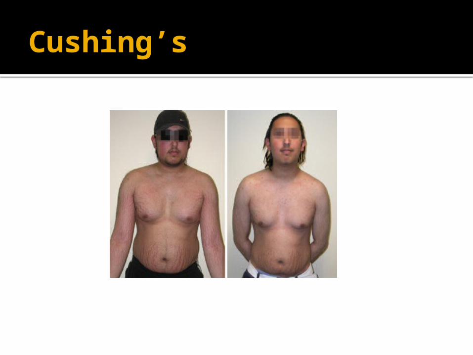
Cushing’s

HPA-axis ( excessive cortisol)

HPA-axis ( excessive cortisol) 80 % HTN LVH Diastolic dysfunction, intraventricular septal hypertrophy ECG needed: high QRS voltage, inverted T-wave Echocardiogram preop OSA: 33% mild, 18% severe. Needs respiratory
assessment and careful use of sedative during surgery Glucose intolerance in 60%, control of hyperglycemia Osteoporosis with vertebral fracture→→ positioning of
patient in OR ( 50 %), 20 % with fracture thin skin→→ difficult IV cannulation, poor wound healing

Cushing’s-Management
Surgical resection of pituitary Medical Treatment
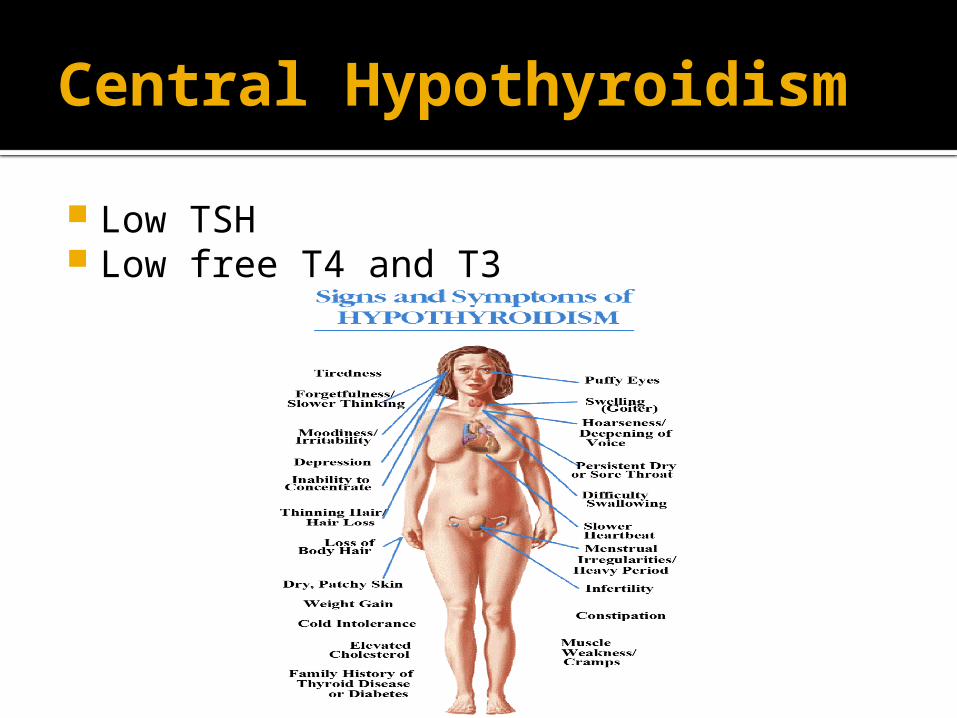
Central Hypothyroidism
Low TSH Low free T4 and T3

Central Hypothyroidism
Thyroxine replacement Surgical removal of pituitary adenoma

TSH-Producing adenoma
Very rare < 2.8 %
Signs of hyperthyroidism
High TSH, FT4, FT3
Treatment preop with anti-thyroid meds pre-op
Surgical resection of adenoma
Medical therapy: Somatostatin Analogue


assessment of pituitary function
Baseline: TSH, FT4, FT3, LH, FSH, Prolactin, GH, IGF-I,Testosterone, Estradiol
MRI brain Neuropthalmic evaluation of visual field Cardiac and respiratory assessment Anesthesiologist for airway and perioperative
monitoring Neurosurgeon ENT for Endonasal evaluation for surgical approach Preop hormonal replacement: all pituitary adenoma
should be covered with stress dose of HC