Embed Size (px)
Citation preview

A Human T Cell Lymphoma Secreting an Immunoglobulin ESpecific Helper FactorMichael C. Young, Harb Harfi, Rajih Sabbah, Donald Y. M. Leung, and Raif S. GehaDivision of Allergy, The Children's Hospital and Department of Pediatrics, Harvard Medical School, Boston, Massachusetts 02115; andDepartments of Pediatrics, Oncology, and Pathology, The King Faisal Specialist Hospital and Research Center, Riyadh, Saudi Arabia
AbstractAn 8-yr-old nonallergic girl with non-Hodgkin's lymphomahad markedly elevated serum IgE at presentation (>10,000IU/ml), negative skin tests to a battery of 24 commonallergens,and no evidence of parasitic infestation. Serum levels of IgG,IgA, and IgM were normal. Remission after cytotoxic chemo-therapy was accompanied by a marked reduction in serum IgElevels (to <200 IU/ml) with no change in the level of serumIgG, IgM, or IgA. Recurrence of the lymphoma 7 mo afterremission was accompanied by an isotype specific rise in serumIgE (to 3,850 IU/ml). Isoelectric focusing revealed that theIgE was polyclonal.
Phenotypic analysis of the lymphoma obtained duringrelapse revealed all (>98%) cells to be T3+, T4+, and T8+.Incubation of lymphoma cells with human myeloma IgE followedby immunosorbent purified fluorescein tagged goat anti-humanIgE (anti-IgE PS-adsorbed over IgE ADZ) stained 25% of thecells. In contrast, <1%of the cells were stained after incubationwith human IgG followed by fluorescein conjugated goat anti-human IgE. Supernatants from lymphoma cells (5 X 10⁶/ml,48 h) enhanced IgE production in B cells derived from fourpatients with allergic rhinitis (mean±SD picograms per milliliterof net IgE 930±320 in unstimulated cultures versus 2,450±650in cultures stimulated with lymphoma supernatants; P < 0.01)but did not induce IgE synthesis in B cells from two normalsubjects that synthesized no IgE spontaneously. Lymphomasupernatants failed to enhance IgG synthesis by B cells ofboth allergic and nonallergic subjects.
These results indicate that a T cell lymphoma comprisedof cells bearing Fc receptors for IgE with a phenotype char-acteristic of immature T cells (i.e., T3+, T4+, T8+) exhibitedIgE specific helper function. This lymphoma may representthe monoclonal expansion of a subpopulation of IgE specifichelper T cells.
Introduction
Substantial evidence in experimental animals indicates thatIgE synthesis is under the control of regulatory T cells (1, 2).
Portions of this work were presented at the 76th annual meeting ofthe American Society for Clinical Investigation, Washington, DC, May4-7, 1984.
Dr. Young received National Research Service Award 1 F32A1067 17.01. Dr. Leung received New Investigator Award5R23HL30082-02. Dr. Geha received Allergic Diseases AcademicAward K07 A10440-01. Address correspondence to Dr. Geha, TheChildren's Hospital, Division of Allergy, 300 Longwood Ave., Boston,MA02115.
Receivedfor publication 30 November 1984 and in revisedform 19February 1985.
IgE specific helper T cells have been described in the rat byIshizaka and co-workers (3, 4). They appear to bear Fcreceptors for IgE (FcR)' and to secrete glycoproteins that bindto IgE, i.e., IgE binding factors. Functional studies indicatethat highly glycosylated IgE binding factors potentiate IgEsynthesis in an isotype specific manner. We have recentlyderived FcR positive T cell lines from the blood of patientswith the hyper-IgE syndrome and we have shown that theselines secrete into their supernatants a factor(s) that providesIgE specific help (5). This factor binds to IgE, is glycoproteinin nature, and acts on IgE bearing B cells.
In this study we describe a patient with a non-Hodgkin'slymphoma and extremely elevated serum IgE. The serum IgEin this patient returned to the normal range with the regressionof the tumor only to rise again when the tumor recurred. Cellsurface analysis of the tumor cells revealed them to be T3+,T4+, and T8+ and to bear FcR. The tumor cells releasedinto their culture supernatants an IgE specific enhancing factorthat had affinity for IgE. The patient's tumor may represent amalignant proliferation of a subpopulation of IgE specifichelper T cells.
Case report. An 8-yr-old female was admitted to the KingFaisal Hospital, Riyadh, Saudi Arabia, with a 3-mo history ofchest pain, cough, and shortness of breath. Physical examinationrevealed a thin child, 28 kg in weight, 129 cm in height, witha bulging left hemithorax, absent breath sounds over the leftchest field, and decreased breath sounds over the right chestfield. Cervical, axillary, and left inguinal lymph nodes weremoderately enlarged. The liver and spleen edges were each 5cm below the costal margin. Chest x-ray revealed a completelyopaque left hemithorax and a mediastinal shift to the right.Computerized axial tomography scan revealed a solid tumorfilling the left hemithorax. Pleural fluid from the left chestrevealed no malignant cells but abundant eosinophils and fewlymphocytes. Histologic examination of the chest mass biopsyrevealed cells with irregular nuclei, most of which were con-voluted; scanty cytoplasm, and undifferentiated and inconspic-uous nucleoli. There were occasional plasma cells and a smallnumber of eosinophils. Fig. 1 shows an electron micrographof the tumor, which illustrates the irregular convoluted nucleiobserved in the majority of the tumor cells and a high nuclearto cytoplasmic ratio. There was a lack of intercellular junctions,of neurosecretory granules, and of cytoplasmic filaments.
On admission, the leukocyte count was 11,400 cells/mm3with 67% eosinophils, 31% neutrophils, 7% lymphocytes, and5%monocytes. Bone marrow aspiration revealed a hypercellularmarrow with increased eosinophil precursors. No malignantcells were observed in peripheral blood or bone marrowsmears. Because of the blood and bone marrow eosinophilia,a serum IgE level was taken and was >10,000 IU/ml. This
1. Abbreviations used in this paper: E, erythrocytes; Fc.R, Fc receptorsfor IgE; FcR+, FcR positive; FCS, fetal calf serum; HS, horse serum;
PBMC, peripheral blood mononuclear cells.
Human T Cell Lymphoma Secreting Immunoglobulin E Helper Factor 1977
J. Clin. Invest.© The American Society for Clinical Investigation, Inc.002 1-9738/85/06/1977/06 $ 1.00Volume 75, June 1985, 1977-1982
PRED '60(img/daJ go L
VC,AIA0 a 0 0 0 oVP-01 1 I 1 I I I
io -~~~~~~~~~~~~~~~~~10
8- -sT v v v s v v t 8
21.4- 1 -.- 4
No --orlleI ~ ~ u Jl u SI UC 0o
W ,: @',4S14~~~~~~~~~~~~~IAE[n '''e''9~~~~~~~~~g
Figure 1. Electron micrograph of the tumor. Note the convolutednuclei, the lack of nucleoli, and the large nuclear to cytoplasmicratio.
was confirmed by a repeat determination. There was nopersonal or family history of allergic disease. A battery of 24hypersensitivity skin tests for inhalant allergens was negativeas were repeated stool examinations (X 5) for ova and parasites.Analysis of the serum IgE by isoelectric focusing was performedby Dr. Z. Audeh (Center for Blood Research, Boston, MA)and revealed a polyclonal pattern. IgG was 1,388 mg/100 ml(normal, 568-1,483 mg/100 ml), IgA was 123 (normal, 57-414 mg/100 ml), and IgM was 84 mg/100 ml (normal, 120-274 mg/100 ml). An SMA20 analysis was normal except foran elevated lactate dehydrogenase (1,128 U vs. a normal of100-225 U/ml) and a low albumin of 2.5 g/100 m.
Fig. 2 summarizes the changes in serum IgE levels andtotal eosinophil counts during the course of the therapy. 8 wkafter the initiation of treatment (Feb. 1983), there was noclinical or radiologic evidence of tumor. Serum IgE at thattime was 767 IU/ml. Serum IgM, IgG, and IgA remainedessentially unchanged (<10% change). 5 mo later (May 1983),there was still no evidence of disease and the patient's serumIgE at that time was 200 IU/ml. Serum IgM, IgG, and IgAwere unchanged. 7 wk later a mild enlargement of the left sideof the mediastinum was observed on chest radiography. Sub-sequently, over a 3-mo period, a left medistinal mass becameclearly visible and grew larger despite the continued adminis-tration of cytotoxic drugs. This was accompanied by a rise inserum IgE (up to 3,900 IU/ml). At that time a course of ara-C (300 mgX 3 d) and of VP16 (250 mg for 2 d) was admin-istered. This was quickly followed by a drop in serum IgE,
Figure 2. Schematic representation of the clinical course of thepatient. (-*-), IgE (IU/ml) X IO-'; (---o---), eosinophils, mm3X lo0-. The tumor mass was estimated from the chest radiograph.Courses of Vincristine (V), Cyclophosphamide (C), and Adriamycin(A), and of ara-C and V-P 16 are indicated by arrows. PREDNIS,prednisone.
from 3,900 IU/ml to 746 IU/ml in a period of 8 d. In contrast,serum IgG, IgM, and IgA remained in the normal range.
Examination of Fig. 2 shows the peripheral blood eosino-philia present initially rapidly resolved with the administrationof prednisone. However, the blood eosinophil count rose
rapidly with the recurrence of the tumor mass. Although atthat time the neutrophil count rose by 2.5-fold, the eosinophilcount concomitantly rose from 100/mm3 to 6,800/mm'.
Methods
Isolation of tumor cells. Tumor tissue was teased with fine scissors inmedium RPMI 1640 containing 10% fetal calf serum (FCS). Debriswas allowed to settle and the suspension was filtered through a shortcolumn of glass wool. These cells were incubated for two cycles, 1 heach, at 370C in Petri dishes. Nonadherent cells were collected, washedthree times, and suspended at 5 X 10⁶ cells/ml in RPMI-10% FCS.
Tumor cell lines. The human tumor T cell line CEM-6 was a giftof Dr. A. Fauci (National Institutes of Health, Bethesda, MD). Thetumor cell line RPMI 8866 was a gift of Dr. K. Ishizaka (JohnsHopkins, Baltimore, MD). This human lymphoblastoid B cell lineexpresses receptors for human IgE.
Isolation of peripheral blood mononuclear cells (PBMC). PBMCwere isolated from heparinized peripheral blood by Ficoll-Hypaque(Pharmacia Fine Chemicals, Piscataway, NJ) density gradient centrif-ugation. The isolated PBMCwere washed three times in Hank'sbalanced salt solution and suspended in RPMI 1640 medium containing10% FCS.
Isolation of B cells. Suspensions of PBMC were depleted ofadherent cells by allowing them to incubate for I h at 370C in plasticPetri dishes (5 X 10⁶ cells/ml). Cell suspensions enriched in B cellswere prepared by rosetting nonadherent mononuclear cells with sheeperythrocytes (E) pretreated with neuraminidase and recovering thenonrosette-forming cells by centrifugation over Ficoll-Hypaque (6).After repeating the rosetting procedure a second time, the nonrosette-forming cells contained 70-85% surface Ig' cells, and <2% E+ cells.
Analysis of surface markers. T cell surface markers of the tumorcells were analyzed by indirect immunofluorescence using the mono-clonal antibodies OKT3, OKT4, and OKT8 (Ortho Diagnostic Systems,Inc., Raritan, NJ), control mouse ascites, and fluorescein conjugatedF(ab')2 goat anti-mouse Ig (Tago Inc., Burlingame, CA) as describedpreviously (7). The percent of cells bearing surface Igs was determined
1978 M. C. Young, H. Harfi, R. Sabbah, D. Y. M. Leung, and R. S. Geha
>1
Now Doc ion Feb Mar Apr May Jun July Aug Sept Oct Nov4090 11 -IQR'A

by direct immunofluorescence using fluorescein conjugated polyvalentgoat anti-human Igs (Cappel Laboratories, Cochranville, PA). Back-ground staining was always <2% and was subtracted from the experi-mental values.
Fluorescence was read under a fluorescent microscope (Carl Zeiss,Inc., Thornwood, NY) with epi-illumination by two individuals whohad no knowledge of the contents of the samples. The average of thetwo readings was taken. The two readings were always in closeagreement with each other.
Analysis of Fc receptors for IgE. Tumor cells were incubated at 5X 10⁶/ml in RPMI 1640 10% FCS and 0.1% sodium azide with asolution of 10 ug/ml of human myeloma IgE or with a solution of 10,ug/ml of human IgG. This IgG was a Cohn's Fraction II (MassachusettsBiologicals, Jamaica Plain, MA) purified further over DEAE. Afterincubation for 45 min on ice, the cells were washed three times in theabove medium. The cells were resuspended to the original volume andincubated with a 1:10 dilution of a 10-25 1Ag/ml of fluoresceinconjugated goat anti-human IgE. This antiserum was obtained byimmunizing a goat with human IgE, isolating the IgG fraction of theantiserum, and absorbing it with human IgM, IgG, IgA, myelomas,and K and L Bence Jones proteins followed by immunosorbentpurification over a column of IgE myeloma. The goat anti-human IgEwas then fluorescein conjugated according to the procedure describedin (8). After 45 min at 40C the cells were washed three times andfluorescence was determined as described above. Background fluores-cence of cells preincubated with IgG was <2% and was subtractedfrom the experimental value.
Assay for the secretion of helper factors by tumor cells. Cells wereincubated at 5 X 10⁶ cells/ml in RPMI 1640- 10% FCS for 48 h at370C. Culture supernatants were collected and then passed through aCF50-A Centriflo membrane (Amicon Corp., Danvers, MA) with amolecular weight cutoff of 50,000 D. The filtrate was sterilized bypassage through a 0.45-,gm millipore filter and was frozen at -20°Cuntil tested.
Induction of IgE synthesis in B cells. Tumor cell supernatants wereassessed for their capacity to enhance IgE synthesis by cultures of Bcells obtained from four subjects with allergic rhinitis whose serumIgE levels ranged from 200 to 400 IU/ml, respectively, and on B cellsfrom two normal subjects (serum IgE of <10 IU/ml).
The E rosette-negative cells were washed and resuspended inRPMI 1640 with 10% FCS at a concentration of 1.0 X 10⁶ cells/ml.The cells were placed in 0.5-1 ml volumes (12 X 75-mm plastic tubes)in the presence or absence of tumor cell supernatants used at a finalconcentration of 10%. After 7 d the supernatants were collected, andtheir IgE and IgG contents were determined. All cultures were madein duplicate. In vivo preformed cytophilic IgE was determined by acidelution of B cells on day 0 as described by Turner et al. (9). After 7 dof culture, supernatants were collected and their IgE and IgG contentswere determined. Net IgE synthesis was calculated by subtracting thevalues for IgE obtained in day 0 acid eluted cell pellets, i.e., preformedIgG from IgE values of day 7 supernatants.
Radioimmunoassay (RIA) for IgE. Duplicate samples from eachculture were analyzed for their IgE content. The RIA for IgE wasperformed in flexible flat-bottom microtiter plates (Cooke LaboratoryProducts, Alexandria, VA). The wells were filled with 0.1 ml of a 10,ug/ml solution of a 1:1 mixture of two monoclonal antihuman IgEFc-specific antibodies. These two antibodies (8, 9) were a kind gift ofDr. Siraganian (National Institutes of Health, Bethesda, MD). Afterincubation for 16 h, the coating solution was removed, and the wellswere washed and blocked with 10% horse serum (HS) in phosphate-buffered saline (PBS) for 2 h. After washing three times with PBScontaining 1% HS, 0.1 ml of culture supernatant or of IgE standardwas added to each of the triplicate wells and incubated for 16 h in ahumidified chamber at room temperature. The wells were then washedtwo times with PBS 1% HS containing 0.5% Tween 20 and twice withPBS 1% HS; then 0.1 ml of Phadebas radioallergo-sorbent test 1251_anti-human IgE (Pharmacia Fine Chemicals; sp act 12 ACi/zg) wasadded to each well. 6 h later the radiolabeled anti-IgE was removedand then wells were washed three times with PBS 1% HS and eight
times under running distilled water. The wells were cut out andcounted in a Gammaspectrometer (Tracor Analytic, Elk GroveVillage, IL). Standard curves were constructed using dilutions of theIgE standards obtained from Pharmacia Fine Chemicals. The con-centration of IgE in the supernatants was read from the standardcurve. The lower limit of sensitivity of this assay varied from 150 to300 pg/ml.
The specificity and sensitivity of our IgE RIA was confirmed in arecently completed multi-institutional study coordinated by the MayoClinic (Yunginger, J. W., R. M. Helm, and G. J. Gleich; manuscriptin preparation). Coded samples with varying quantities of polyclonalIgE in the presence of other Ig isotypes sent to our laboratory wereevaluated using our IgE RIA. In no test sample were falsely elevatedIgE values measured; this ruled out the possibility of cross-reactivitywith other isotypes. Furthermore, values of IgE > 300 pg IgE/ml weremeasured accurately within 10% of expected values.
Determination of IgG. IgG in culture supernatants was measuredby a solid-phase competitive RIA as previously described (10).
IgE binding capacity of T lymphoma supernatants. To demonstratethe IgE binding capacity of the tumor cell supernatants, the supernatantswere circulated over a column of human IgE (PS) conjugated toSepharose 4B overnight at 40C. Sepharose beads conjugated to humanIgG DEAEpurified Cohn fraction II (Massachusetts Biologicals) wereused as controls. After collection of the effluent, the beads were washedwith PBS, and eluted with 0.2 Mglycine-HCI at pH 3.0, 40C. Theeluate was collected into tubes containing an appropriate amount of0.1 M Tris buffer, pH 8.6, so that the final pH of the eluate wasbetween 7.0 and 8.0. The final volume of the eluate was made up withPBS to the original volume. The eluates were then immediatelydialyzed (Spectropor dialysis membrane with a cutoff of 3,500 D)against RPMI 1640, passed through a CF5OA Centriflo membrane,and subsequently sterilized by filtration through a 0.45-,Om milliporefilter and frozen at -20°C until tested.
ResultsSurface phenotype of tumor cells. The surface phenotype ofthe tumor cell is shown in Table I. Almost all tumor cellsexpressed the T3, T4, and T8 antigens. There were no detectableB cells as evidenced by lack of surface Ig positive cells. 25%of the tumor cells expressed Fc receptors for IgE as determinedby indirect immunofluorescence. Using the same method, 85%of the RPMI 8866 cell line was found to be FcR positive(FcR+), whereas no FcR+ cells could be detected in theCEM-6 cell line. The phenotypic analysis of peripheral bloodlymphocytes of the patient was determined simultaneouslyand was found to be normal.
Enhancement of IgE synthesis by tumor cell supernatants.The effect of the tumor cell supernatants on IgE synthesis wasexamined by adding the supernatants at a final solution of 1:10 to cultures containing B cells obtained from allergic donorsor from normal subjects.
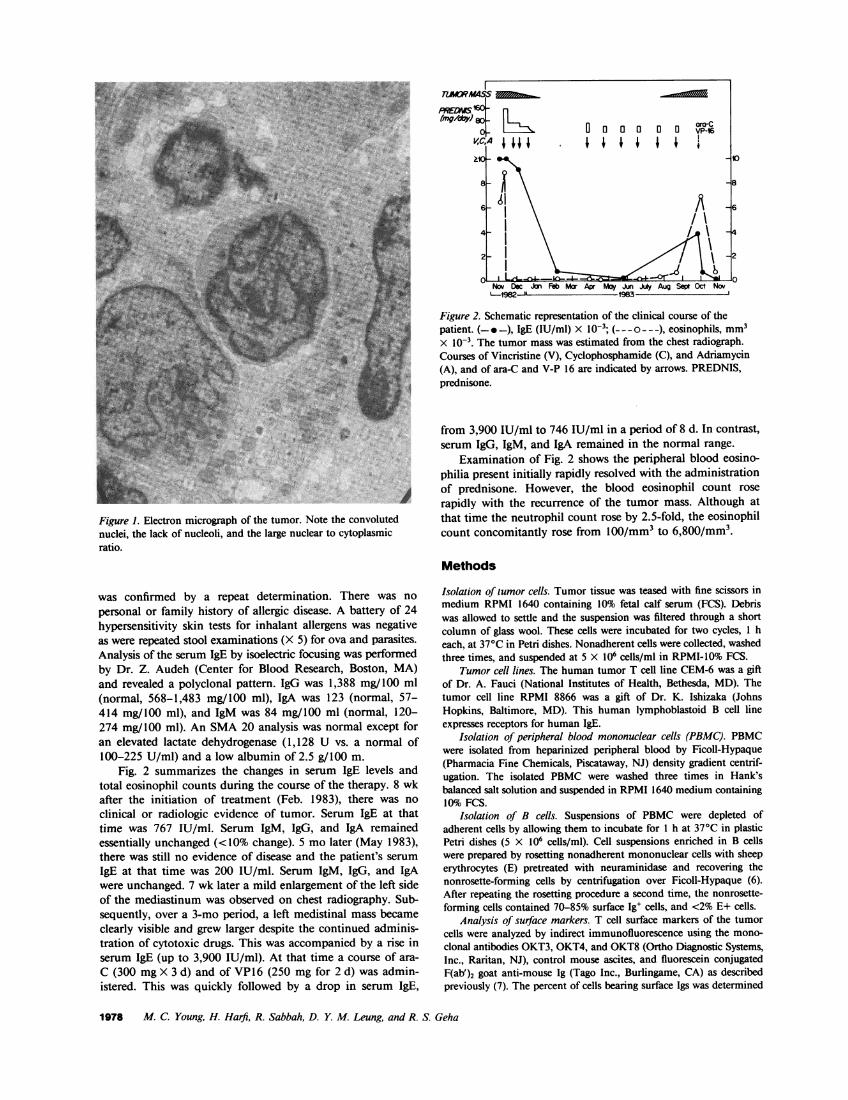
Fig. 3 shows that the tumor B cell supernatants selectivelyenhanced IgE synthesis in the B cells of four allergic subjectsstudied. The preformed IgE in acid treated cell pellets wasequivalent in B cells to which medium or T lymphomasupernatants were added. The mean total IgE present in 7-dcultures of B cells that received T lymphoma supernatants(2,450±650 pg/ml) was significantly higher (P < 0.01) thanthe mean total IgE present in B cells cultured with mediumalone (930±320 pg/ml). In contrast, T lymphoma supernatantsdid not enhance IgG synthesis in the B cells from the allergicsubjects (Fig. 3).
The effect of T lymphoma supernatants was studied on Bcells from normal subjects. These cells contained no detectablepreformed IgG (<200 pg/ml) and failed to synthesize anydetectable IgE regardless of the presence or absence of added
Human T Cell Lymphoma Secreting Immunoglobulin E Helper Factor 1979

Table I. Phenotypic Analysis of the Patient's Lymphoma
Percent positive cells
Patient's Patient'sMarker tumor RPMI 8866 CEM-6 PBMC
T3 98 <1 <1 75T4 98 . 1 98 48T8 98 <1 60 23slg <1 96 <1 5Fc IgE 25 85 <1 <1
The surface phenotype of the patient's suspended lymphoma cells,RPMI 8866 cells, CEM-6 cells, and PBMCfrom the patient was de-termined by immunofluorescence as described in Methods. All deter-minations were done on coded samples, the identities of which werewithheld from the investigators reading the immunofluorescence.
T cell supernatants. Furthermore, their spontaneous IgG syn-thesis was not enhanced by T lymphoma supernatants.
Table II summarizes the data obtained in all experiments.Data are expressed as net IgE and IgG synthesis by subtractingthe preformed values of IgE and IgG from the values in 7-dcultures.
It was important to ascertain that the observed increasedIgE values in supernatants of cultures stimulated with Tlymphoma supernatants represented de novo IgE synthesis. Bcells from four separate allergic subjects were cultured in thepresence or absence of the protein synthesis inhibitor cyclo-heximide (100 gg/ml) and the culture supernatants wereassayed for IgE and IgG after a 7-d incubation period. Theresults in Table III clearly show that cycloheximide inhibitedthe induction of IgE synthesis by T lymphoma supernatants.
Affinity of IgE enhancing factor for IgE. Supernatants fromthe tumor cells were examined for their capacity to enhanceIgE synthesis in allergic B cells before and after passage ofthese supernatants over columns of Sepharose cross-linkedwith human IgE or IgG. Fig. 4 depicts the results of two suchexperiments. Passage of T lymphoma supernatants over Se-
3
2
o
IgE IgG
Medium T Lymph Sup Medium T Lymph Sup
1.0
0
Figure 3. Effect of T lymphoma supernatants (T Lymph Sup) on IgEand IgG synthesis in cultures of B cells from allergic subjects (n = 4).Preformed IgE and IgG were measured by acid treatment of cellpellets (1i) on day 0. IgE and IgG were measured in supernatants of7-d cultures of B cells (o). T lymphoma supernatant was added at afinal dilution of 1: 1O. P < 0.01.
pharose IgE columns depleted their capacity to enhance IgEsynthesis. In contrast, passage over Sepharose IgG columnsdid not affect the capacity of the T lymphoma supernatants toenhance IgE synthesis. Materials that bound to IgE and IgGcolumns were eluted with glycine HC1 buffer pH 3. This acideluate was neutralized, dialyzed against RPMI 1640, andpassed through a CF5OA filter to remove any Ig eluted off theSepharose and then was tested for IgE enhancing activity inthe same B cell cultures. IgE enhancing activity was recoveredin acid eluates of IgE Sepharose columns but not of IgGSepharose columns (Fig. 4).
Discussion
This study documents the capacity of a human T cell lymphomato secrete IgE isotype specific enhancing factor(s). These fac-tor(s), like those secreted by T cells from parasite infested rats(3, 4) and by T cells of patients with the hyper-IgE syndrome(5), had affinity for IgE.
The occurrence of extremely elevated serum IgE levels ina patient with non-Hodgkin's lymphoma who was nonatopicand had no family history of allergic disease brought up theintriguing possibility that these two findings were interrelated.This was strengthened by the observation that during tumorremission induced by cytotoxic drugs, serum IgE levels droppeddramatically, whereas relapse of the lymphoma was associatedwith a marked rise in serum IgE levels (Fig. 2) but with nochanges in serum levels of other isotypes. The increased serumIgE was not due to a monoclonal proliferation of IgE B Cellsbecause isoelectric focusing of serum IgE revealed a polyclonalpattern.
The morphology of the patient's tumor cells, in particularthe presence of convoluted nuclei (Fig. 1), was suggestive of Tcells. The T cell nature of these cells was confirmed byphenotypic analysis of tumor cells obtained during the relapse.This showed 98% of cells to be T3+, T4+, and T8+ (TableI). This phenotype is thought to be charascteristic of immaturethymocytes (stage III thymocytes), which have not yet differ-entiated along either the T4 or the T8 pathway, despiteacquiring the T3 antigen, which is specific for T cells (1 1). Inthis regard, circulating T cells coexpressing the T3, T4, T8,and T10 surface antigens have been reported to occur in somepatients with severe combined immunodeficiency syndrome(12). In these patients a block in T cell maturation anddifferentiation could have resulted in the appearance of im-mature T cells in the periphery.
Since T cells are known to play an important role in thepotentiation of IgE synthesis, we further investigated thefunctional activity of soluble factors secreted by this patient'slymphoma cells. These studies revealed that her T cell lym-phoma produced factors that enhanced IgE synthesis by Bcells from patients with allergic rhinitis but not by B cells fromnormal donors (Table II, Fig. 3). This enhancement wasisotype specific because no changes were observed in IgGsynthesis by the same B cell cultures after addition of lymphomasupernatants. Characterization of the IgE potentiating factorrevealed that it could bind to immobilized IgE but not toimmobilized IgG and was, thus, an IgE binding factor (Fig.4). The observation that this factor exerted biologic activityon B cells from allergic donors, which spontaneously secreteIgE but not on normal B cells that do not synthesize IgEspontaneously, suggests that this factor exerts its effect on IgEB cells that are spontaneously activated in vivo and that
1980 M. C. Young, H. Harfi, R. Sabbah, D. Y. M. Leung, and R. S. Geha
0.5
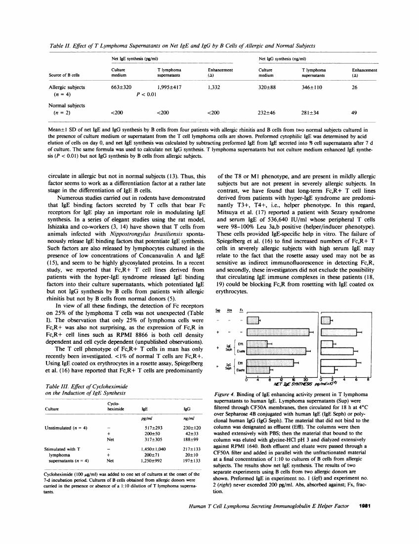
Table II. Effect of T Lymphoma Supernatants on Net IgE and IgG by B Cells of Allergic and Normal Subjects
Net IgE synthesis (pg/ml) Net IgG synthesis (ng/ml)
Culture T lymphoma Enhancement Culture T lymphoma EnhancementSource of B cells medium supernatants (a) medium supernatants (A)
Allergic subjects 663±320 1,995±417 1,332 320±88 346±110 26(n = 4) P < 0.01
Normal subjects(n = 2) <200 <200 <200 232±46 281±34 49
Mean± 1 SD of net IgE and IgG synthesis by B cells from four patients with allergic rhinitis and B cells from two normal subjects cultured inthe presence of culture medium or supernatant from the T cell lymphoma cells are shown. Preformed cytophilic IgE was determined by acidelution of cells on day 0, and net IgE synthesis was calculated by subtracting preformed IgE from IgE secreted into B. cell supernatants after 7 dof culture. The same formula was used to calculate net IgG synthesis. T lymphoma supernatants but not culture medium enhanced IgE synthe-sis (P < 0.01) but not IgG synthesis by B cells from allergic subjects.
circulate in allergic but not in normal subjects (13). Thus, thisfactor seems to work as a differentiation factor at a rather latestage in the differentiation of IgE B cells.
Numerous studies carried out in rodents have demonstratedthat IgE binding factors secreted by T cells that bear Fcreceptors for IgE play an important role in modulating IgEsynthesis. In a series of elegant studies using the rat model,Ishizaka and co-workers (3, 14) have shown that T cells fromanimals infected with Nippostrongylus brasiliensis sponta-neously release IgE binding factors that potentiate IgE synthesis.Such factors are also released by lymphocytes cultured in thepresence of low concentrations of Concanavalin A and IgE(15), and seem to be highly glycosylated proteins. In a recentstudy, we reported that FcfR+ T cell lines derived frompatients with the hyper-IgE syndrome released IgE bindingfactors into their culture supernatants, which potentiated IgEbut not IgG synthesis by B cells from patients with allergicrhinitis but not by B cells from normal donors (5).
In view of all these findings, the detection of Fc receptorson 25% of the lymphoma T cells was not unexpected (TableI). The observation that only 25% of lymphoma cells wereFcR+ was also not surprising, as the expression of FcR inFcfR+ cell lines such as RPMI 8866 is both cell densitydependent and cell cycle dependent (unpublished observations).
The T cell phenotype of FcR+ T cells in man has onlyrecently been investigated. <1% of normal T cells are FcR+.Using IgE coated ox erythrocytes in a rosette assay, Spiegelberget al. (16) have reported that FcR+ T cells are predominantly
Table III. Effect of Cycloheximideon the Induction of IgE Synthesis
Cyclo-Culture heximide IgE IgG
pg/mi ng/ml
lJnstimulated (n = 4) - 517±293 230±120+ 200±50 42±33Net 317±305 188±99
Stimulated with T - 1,450±1,040 217±133lymphoma + 200±71 20±10supernatants (n = 4) Net 1,250±992 197±133
Cycloheximide (100 ug/ml) was added to one set of cultures at the onset of the7-d incubation period. Cultures of B cells obtained from allergic donors werecarried in the presence or absence of a 1:10 dilution of T lymphoma superna-tants.
of the T8 or Ml phenotype, and are present in mildly allergicsubjects but are not present in severely allergic subjects. Incontrast, we have found that long-term FcR+ T cell linesderived from patients with hyper-IgE syndrome are predomi-nantly T3+, T4+, i.e., helper phenotype. In this regard,Mitsuya et al. (17) reported a patient with Sezary syndromeand serum IgE of 536,640 IU/ml whose peripheral T cellswere 98-100% Leu 3a,b positive (helper/inducer phenotype).These cells provided IgE-specific help in vitro. The failure ofSpiegelberg et al. (16) to find increased numbers of FcfR+ Tcells in severely allergic subjects with high serum IgE mayrelate to the fact that the rosette assay used may not be assensitive as indirect immunofluorescence in detecting FcR,and secondly, these investigators did not exclude the possibilitythat circulating IgE immune complexes in these patients (18,19) could be blocking FcR from rosetting with IgE coated oxerythrocytes.
Su Abs
+
El+ gG [ Eli
+ SehE
Fx
_.:....... ... ... .: -:-:.;::::::::::::::::::.::::::::::::: ......:: ::- _ :: :D -:'T.
...--::: : - -:::--: -:...- :::: :..
:
0 4L 8 2 Is 0 0 2 4 6Arr ig sywhe pglvxo--)
Figure 4. Binding of IgE enhancing activity present in T lymphomasupernatants to human IgE. Lymphoma supernatants (Sup) werefiltered through CF5OAmembranes, then circulated for 18 h at 4°Cover Sepharose 4B conjugated with human IgE (IgE Seph) or poly-clonal human IgG (IgG Seph). The material that did not bind to thecolumn was designated as effluent (Effil). The columns were thenwashed extensively with PBS; then the material that bound to thecolumn was eluted with glycine-HCI pH 3 and dialyzed extensivelyagainst RPMI 1640. Both effluent and eluate were passed through aCF5OA filter and added in parallel with the unfractionated materialat a final concentration of 1:10 to cultures of B cells from allergicsubjects. The results show net IgE synthesis. The results of twoseparate experiments using B cells from two allergic donors are
shown. Preformed IgE in experiment no. I (left) and experiment no.2 (right) never exceeded 200 pg/ml. Abs, absorbed against; Fx, frac-tion.
Human T Cell Lymphoma Secreting Immunoglobulin E Helper Factor 1981

If the T3+, T4+, T8+, FcR+ lymphoma currently studiedrepresents the monoclonal expansion of a normally occurringIgE specific human helper T cell subpopulation, it wouldsuggest that the T4 or T8 surface markers may not benecessarily indicative of IgE helper vs. suppressor function.Recent studies on human T cell clones have suggested thatthe T4 and T8 surface antigen play an important role in therecognition of HLA-DR and HLA-A,B histocompatibility an-tigens rather than imparting helper or suppressor/cytotoxic Tcell function ( 19-22). Further studies are in progress to delineatethe heterogeneity of human T cell subpopulations that play arole in regulating IgE synthesis in human.
Acknowledgments
The authors wish to thank Miss Melissa Smith for excellent secretarialassistance.
The research for this paper was supported by U. S. Public HealthServices grants AI-21163 and AI20373; and by grants from theNational Foundation and the March of Dimes.
References
1. Ishizaka, K., and T. Ishizaka, 1978. Mechanisms of reaginichypersensitivity and IgE antibody response. Immunol. Rev. 41:109-148.
2. Katz, D. H. 1980. Recent studies on the regulation of IgEantibody synthesis in experimental animals and man. Immunology.41:1-24.
3. Yodoi, J., and K. Ishizaka. 1980. Lymphocytes bearing Fcreceptors for IgE. IV. Formation of IgE-binding factor by rat Tlymphocytes. J. Immunol. 124:1322-1329.
4. Hirashima, M., J. Yodoi, and K. Ishizaka. 1980. Regulatory roleof IgE-binding factors from rat T lymphocytes. III. IgE-specific sup-pressive factor with IgE-binding activity. J. Immunol. 125:1442-1448.
5. Young, M. C., D. Y. M. Leung, and R. S. Geha. 1984.Production of IgE potentiating factor in man by T cell lines bearingFc receptors for IgE. Eur. J. Immunol. 14:871-878.
6. Geha, R. S., F. S. Rosen, and E. Merler. 1973. Identificationand characterization of subpopulations and lymphocytes in humanperipheral blood after fractionation on discontinuous gradients ofalbumin. J. Clin. Invest. 52:1726-1734.
7. Leung, D. Y. M., A. R. Rhodes, and R. S. Geha. 1981.Enumeration of T cell subsets in atopic dermatitis with monoclonalantibodies. J. Allergy Clin. Immunol. 67:450-455.
8. Wood, B. T., S. H. Thompson, and G. Gold. 1965. Fluorescentantibody staining. III. Preparation of fluorescein-isothiocyanate labeledantibodies. J. Immunol. 95:225-229.
9. Turner, K. J., P. G. Holt, B. J. Holt, and K. J. Cameron. 1983.
In vitro synthesis of IgE by human peripheral blood leucocytes. III.Release of preformed antibody. Clin. Exp. Immunol. 51:387-394.
10. Geha, R. S., E. L. Reinherz, D. Leung, K. T. McKee, S. F.Schlossman, and F. S. Rosen. 1981. Deficiency of suppressor T cellsin the hyperimmunoglobulinemia E syndrome. J. Clin. Invest. 68:783-791.
11. Bhan, A. K., E. L. Reinherz, S. Poppema, R. T. McCluskey,and S. F. Schlossman. 1980. Location of T cell and major histocom-patability complex antigens in the human thymus. J. Exp. Med. 152:771-782.
12. Reinherz, E. L., M. D. Cooper, S. F. Schlossman, and F. S.Rosen. 1981. Abnormalities of T cell maturation and regulation inhuman beings with immunodeficiency disorders. J. Clin. Invest. 68:699-705.
13. Saryan, J. A., D. Y. M. Leung, and R. S. Geha. 1983. Inductionof human IgE synthesis by a factor derived from T cells of patientswith hyper IgE states. J. Immunol. 130:242-247.
14. Ishizaka, K. 1983. Regulation of IgE response by IgE bindingfactors. Monogr. Allergy. 18:52-80.
15. Yodoi, J., M. Hirashima, and K. Ishizaka. 1981. Lymphocytesbearing Fc receptors for IgE. V. Effect of tunicamycin on the formationof IgE-potentiating factor and IgE-suppressive factor by Con A-activatedlymphocytes. J. Immunol. 126:877-882.
16. Thompson, L. F., M. H. Mellon, R. S. Zeiger, and H. L.Spiegelberg. Characterization with monoclonal antibodies of T lym-phocytes bearing Fc receptors for IgE (T, cells) and IgG (T, cells) inatopic patients. J. Immunol. 131:2772-2776.
17. Mitsuya, H., M. Sata, T. Hirano, K. Fujimoto, F. Kawano,and S. Kishimoto. 1983. Evidence for a malignant proliferation of IgE-class specific helper T cells in a patient with Sezary syndrome exhibitingmassive hyperimmunoglobulinemia E. Clin. Immunol. Immunopathol.26:171-183.
18. Inganas, M., S. G. 0. Johansson, and H. Bennich. 1981. Anti-IgE antibodies in human serum: occurrence and specificity. Int. Arch.Allergy Appl. Immunol. 65:51.
19. Quinti, I., C. Brozek, R. S. Geha, and D. Y. M. Leung. 1984.Circulating IgG antibodies to IgE in atopic syndromes. Clin. Res. 32:146.
20. Krensky, A. M., C. S. Reiss, J. W. Mier, J. L. Strominger, andS. J. Burakoff. 1982. Long-term human cytolytic T-cell lines allospecificfor HLA-DR6 antigen are OKT4+. Proc. Natl. Acad. Sci. USA. 79:2365-2369.
21. Ball, E. J., and P. Stastny. 1982. Cell-mediated cytotoxicityagainst HLA-DR region products expressed in monocytes and Blymphocytes. IV. Characterization of effector cells using monoclonalantibodies against human T cell subsets. Immunogenetics. 16:159.
22. Meuer, S. C., R. E. Hussey, J. C. Hodgdon, T. Hercend, S. F.Schlossman, and E. L. Reinherz. 1982. Surface structures involved intarget recognition of human cytotoxic T lymphocytes. Science (Wash.DC). 218:471.
1982 M. C. Young, H. Harfi, R. Sabbah, D. Y. M. Leung, and R. S. Geha