Embed Size (px)
Citation preview
MAGNETIC RESONANCE IMAGING GUIDED MUSCULOSKELETAL INTERVENTIONS AT 0.23TOptical instrument guidance, bone biopsy, periradicular nerve root therapy, discography, osteoid osteoma laser ablation; a feasibility study
ROBER TOBLANCO SEQUEIROS
Department of Diagnostic Radiology,University of Oulu
OULU 2002


ROBERTO BLANCO SEQUEIROS
MAGNETIC RESONANCE IMAGING GUIDED MUSCULOSKELETAL INTERVENTIONS AT 0.23TOptical instrument guidance, bone biopsy, periradicular nerve root therapy, discography, osteoid osteoma laser ablation; a feasibility study
Academic Dissertation to be presented with the assent ofthe Faculty of Medicine, University of Oulu, for publicdiscussion in the Auditorium 7 of the University Hospitalof Oulu, on January 24th, 2003, at 12 noon.
OULUN YLIOPISTO, OULU 2002
Copyright © 2002University of Oulu, 2002
Supervised byDocent Osmo Tervonen
Reviewed byDocent Seppo KoskinenDocent Pekka Niemi
ISBN 951-42-6903-9 (URL: http://herkules.oulu.fi/isbn9514269039/)
ALSO AVAILABLE IN PRINTED FORMATActa Univ. Oul. D 708, 2002ISBN 951-42-6902-0ISSN 0355-3221 (URL: http://herkules.oulu.fi/issn03553221/)
OULU UNIVERSITY PRESSOULU 2002
Blanco Sequeiros, Roberto, Magnetic resonance imaging guided musculoskeletalinterventions at 0.23T. Optical instrument guidance, bone biopsy, periradicular nerveroot therapy, discography, osteoid osteoma laser ablation; a feasibility studyDepartment of Diagnostic Radiology, University of Oulu, P.O.Box 5000, FIN-90014 University ofOulu, Finland Oulu, Finland2002
Abstract
The purpose of this study was firstly to evaluate the optical instrument tracking system integrated tothe MRI scanner as a guidance facility in performing bone biopsy and secondly to develop andevaluate clinical musculoskeletal applications of interventional MRI at 0,23T. The clinical results andfeasibility of MR-guided bone biopsy (n=14), periradicular nerve root therapy (n=61), discography(n=12) and percutaneous laser therapy of osteiod osteomas (n=5) were studied.
Bone biopsies were performed with the optical instrument tracker and bone biopsy set modifiedfor the tracker system. The biopsy system and optical tracker mounting proved to be safe and reliabletool for bone biopsies. 14 consecutive bone biopsies and 13 fine needle aspirations were performedunder MR-guidance. The clinical accuracy of MR-guided bone biopsy was 95%.
The periradicular therapy was applied to the anatomical region of lumbosacral area of 61consecutive patients with sciatic pain. Procedural success rate was 98,5%. Of patients, 51,5% hadgood or excellent effect with regard to radicular pain from procedure. The therapy effect achievedwith MR-guided procedure was comparable to that achieved with conventional techniques.
MR-guided discography technique and imaging protocol was developed as part of diagnostic painprovocation for patients suspected for intervertebral pain source at lumbosacral area. 34 MR-guideddiscographies were performed on 12 patients. In all patients positive or negative pain provocationresponse was obtained.
Laser induced thermal therapy for osteiod osteoma was studied in MRI. The initial guidance ofthe instrument and monitoring of the thermal procedure were done under MRI control. All the 5patients were successfully treated.
The MR-guidance in musculoskeletal applications seems safe and accurate.
Keywords: bone biopsy, discography, interventional, laser therapy, magnetic resonance im-aging, musculoskeletal, periradicular therapy


Para mi familia
“proceed, proceed: we will begin these rites, As we trust they’ll end in true delights”William Shakespeare

Acknowledgements
This study was done at the Department of Radiology in the Oulu University during theyears 1999-2002.
This work would not have been possible without my supervisor, docent OsmoTervonen M.D., who has made it possible for me to conduct very fruitful research. I owehim my sincerest thanks as a supervisor and collaborator. As the professor of theDepartment of Radiology, Ilkka Suramo M.D., has been in key role in realizing this workfrom a mere vision to concrete, and I want to thank him especially for the vital notesduring the last laps of the work. To Risto Ojala M.D. Ph.D., who has been my colleagueboth in science and other aspects of life, I want to present my deepest thanks, particularlyfor sharing the same vision for the art of science, which made this work so enjoyable.
I would like to express my gratitude to my co-workers Rauli Klemola M.D. and LasseJyrkinen Ph.D., for providing me enthusiastic conversations during the research. I alsowish to thank Teuvo Vaara Ph.D. and Erkki Vahala Ph.D., for numerous last minute savesconsidering the manuscripts. I owe my sincerest thanks to my co-workers in other clinicsand organizations, docent Ylermi Soini M.D., Elisa Lappi-Blanco M.D., Alberto BlancoSequeiros Ph.D. and Pekka Hyvönen M.D., for invaluable assistance throughout thestudy.
I want thank docent Seppo Koskinen M.D. and docent Pekka Niemi M.D., for theexcellent and careful review of the thesis. For the statistical analysis I want to thankMarianne Haapea M.Sc. The revision of the English language is credited to Ms. AnnaVuolteenaho.
I want express my warmest thanks to Andreas Blanco Sequeiros M.D. and KristianBlanco Sequeiros for helping out the older brother from technological obstacles.
I express my great gratitude to all my mentors in radiology for pointing out the essencein radiology. Especially I want to thank Jaakko Karumo M.D., Ph.D and docent SeppoLähde M.D. for leading the way.
I want to express my warmest thanks to the staff of the Departments of InterventionalMRI and Northern Central Radiology for constructive attitude and help. Especially Iwould like to thank my colleagues Jukka Perälä M.D., Ph.D, Juho Kariniemi M.D., Matti
Isokangas M.D., docent Sami Leinonen M.D and docent Topi Siniluoto M.D. I also wantto thank radiographers Kyösti Palomaa, Salme Meriläinen, Anu Kauppila and RaijaYlävaara for invaluable assistance during the study.
There are no words to describe my thanks to my parents, Guillermo and Marjatta. Youhave given me the heart and mind to start with.
Finally I want to express my thanks to the lights of my life, to my beloved wife Elisa,whose love never ceases to fill me with amaze and to my children, Sofia, Werner andAmanda, whose tap of little feet and general buzzing around is a fountain of happinessand never-ending stimulation for me.

Abbreviations
2D two-dimensional3D three-dimensionalC-BASS completely balanced steady stateCT computed tomographyESR electron spin resonanceETL echo train lengthEXPRESS Philips Medical Systems acronym for SSFSEFA flip angleFE field (gradient) echoFNA fine needle aspirationFOV field of viewFSE fast spin echoFUS focused ultrasound surgeryHU Hounsfield unitIMRI interventional magnetic resonance imagingIR infra redLITT laser-induced thermotherapyLoLo local lookMR magnetic resonanceMRI magnetic resonance imagingRF radio frequencySENSE sensitivity encodingSMASH simultaneous acquisition of spatial harmonicsSNR signal-to-noise-ratioSSFSE single-shot fast-spin echoSTIR short tau inversion recoveryT teslaT1 longitudinal relaxation timeT2 transverse relaxation timeTA time of acquisitionTE time of echo

TR time of repetitionTrue-FISP fast imaging with steady-state precessionUS ultrasonography, ultrasoundXVGA extended video graphics array
List of original publications
This thesis was based on the following articles, which are referred to in the text by theirRoman numerals.
I Ojala R, Sequeiros RB, Klemola R, Vahala E, Jyrkinen L & Tervonen O (2002)MR-guided bone biopsy: preliminary report of a new guiding method. J MagnReson Imaging 15: 82-86
II Blanco Sequeiros R, Klemola R, Ojala R, Jyrkinen L, Lappi-Blanco E, Soini Y &Tervonen O (2002) MRI-guided trephine biopsy and fine-needle aspiration in thediagnosis of bone lesions in low-field (0.23T) MRI system using optical instrumenttracking. Eur Radiol 12: 830-5
III Sequeiros R, Ojala RO, Klemola R, Vaara TJ, Jyrkinen L & Tervonen OA (2002)MRI-guided periradicular nerve root infiltration therapy in low-field (0.23T) MRI-system using optical instrument tracking. Eur Radiol 12: 1331-7
IV Blanco Sequeiros R, Klemola R, Ojala R, Jyrkinen L, Vaara T & Tervonen O (2002)Percutaneous MR-guided discography in a low-field system using optical instrumenttracking: a feasibility study J Magn Reson Imaging, in press
V Blanco Sequeiros R, Hyvönen P, Blanco Sequeiros A, Jyrkinen L, Ojala R, KlemolaR & Tervonen O (2002) MR imaging-guided laser ablation of osteoid osteomas withuse of optical instrument guidance at 0.23 Tesla. Submitted for publication.

Contents
Abstract Acknowledgements Abbreviations List of original publications 1 Introduction . . . . . . . . . . . . . . . . . . . . . . . . . . . . . . . . . . . . . . . . . . . . . . . . . . . . . . . . 152 Review of the literature . . . . . . . . . . . . . . . . . . . . . . . . . . . . . . . . . . . . . . . . . . . . . . . 16
2.1 Image guided interventions . . . . . . . . . . . . . . . . . . . . . . . . . . . . . . . . . . . . . . . . 162.1.1 Bone biopsy . . . . . . . . . . . . . . . . . . . . . . . . . . . . . . . . . . . . . . . . . . . . . . . 172.1.2 Periradicular therapy . . . . . . . . . . . . . . . . . . . . . . . . . . . . . . . . . . . . . . . . 172.1.3 Discography . . . . . . . . . . . . . . . . . . . . . . . . . . . . . . . . . . . . . . . . . . . . . . . 182.1.4 Laser induced bone thermotherapy of osteoid osteoma . . . . . . . . . . . . . . 18
2.2 Imaging modalities for interventional procedures . . . . . . . . . . . . . . . . . . . . . . 192.2.1 X-ray fluoroscopy . . . . . . . . . . . . . . . . . . . . . . . . . . . . . . . . . . . . . . . . . . 192.2.2 Ultrasound . . . . . . . . . . . . . . . . . . . . . . . . . . . . . . . . . . . . . . . . . . . . . . . . 202.2.3 Computed tomography . . . . . . . . . . . . . . . . . . . . . . . . . . . . . . . . . . . . . . . 202.2.4 Magnetic resonance imaging . . . . . . . . . . . . . . . . . . . . . . . . . . . . . . . . . . 212.2.5 Instrument tracking in interventional procedures in MRI . . . . . . . . . . . . 232.2.6 Implications for this study . . . . . . . . . . . . . . . . . . . . . . . . . . . . . . . . . . . . 24
3 Purpose of the study . . . . . . . . . . . . . . . . . . . . . . . . . . . . . . . . . . . . . . . . . . . . . . . . . . 254 Materials and methods . . . . . . . . . . . . . . . . . . . . . . . . . . . . . . . . . . . . . . . . . . . . . . . . 26
4.1 Patients and procedures . . . . . . . . . . . . . . . . . . . . . . . . . . . . . . . . . . . . . . . . . . . 264.1.1 MRI-system for interventional procedures . . . . . . . . . . . . . . . . . . . . . . . 264.1.2 Procedural set-up . . . . . . . . . . . . . . . . . . . . . . . . . . . . . . . . . . . . . . . . . . . 29
4.2 Guidance method for MR-guided bone biopsy . . . . . . . . . . . . . . . . . . . . . . . . . 304.3 MR-guided bone biopsy . . . . . . . . . . . . . . . . . . . . . . . . . . . . . . . . . . . . . . . . . . 314.4 MR-guided periradicular therapy . . . . . . . . . . . . . . . . . . . . . . . . . . . . . . . . . . . 344.5 MR-guided discography . . . . . . . . . . . . . . . . . . . . . . . . . . . . . . . . . . . . . . . . . . 384.6 MR-guided laser induced thermotherapy of osteiod osteoma . . . . . . . . . . . . . . 41
5 Results . . . . . . . . . . . . . . . . . . . . . . . . . . . . . . . . . . . . . . . . . . . . . . . . . . . . . . . . . . . . 455.1 Guidance method for MR-guided bone biopsy . . . . . . . . . . . . . . . . . . . . . . . . . 455.2 MR-guided bone biopsy . . . . . . . . . . . . . . . . . . . . . . . . . . . . . . . . . . . . . . . . . . 465.3 MR-guided periradicular therapy . . . . . . . . . . . . . . . . . . . . . . . . . . . . . . . . . . . 475.4 MR-guided discography . . . . . . . . . . . . . . . . . . . . . . . . . . . . . . . . . . . . . . . . . . 49

5.5 MR-guided laser induced thermotherapy of osteiod osteoma . . . . . . . . . . . . . . 506 Discussion . . . . . . . . . . . . . . . . . . . . . . . . . . . . . . . . . . . . . . . . . . . . . . . . . . . . . . . . . 51
6.1 Low field imaging & integrated optical instrument tracking . . . . . . . . . . . . . . 516.2 Guidance method for MR-guided bone biopsy . . . . . . . . . . . . . . . . . . . . . . . . . 536.3 MR-guided bone biopsy . . . . . . . . . . . . . . . . . . . . . . . . . . . . . . . . . . . . . . . . . . 536.4 MR-guided periradicular therapy . . . . . . . . . . . . . . . . . . . . . . . . . . . . . . . . . . . 566.5 MR-guided discography . . . . . . . . . . . . . . . . . . . . . . . . . . . . . . . . . . . . . . . . . . 576.6 MR-guided laser thermotherapy of osteiod osteoma . . . . . . . . . . . . . . . . . . . . 59
7 Conclusions . . . . . . . . . . . . . . . . . . . . . . . . . . . . . . . . . . . . . . . . . . . . . . . . . . . . . . . . 638 References . . . . . . . . . . . . . . . . . . . . . . . . . . . . . . . . . . . . . . . . . . . . . . . . . . . . . . . . . 64
1 Introduction
Interventional radiology was launched shortly after the discovery of X-rays. The first stepwas visualisation of blood vessels with angiography, followed by fluoroscopically guidedbiopsies and balloon angioplasties. Computed tomography (CT) and ultrasound-guidedinterventions emerged in the 1970s, and ultimately magnetic resonance imaging (MRI)guided interventions were introduced in the 1980s. The rapid development ofinterventional techniques has been led by the integration of imaging with computers, newtherapy devices and operating room like conditions. This has facilitated faster and moreaccurate imaging and more demanding procedures have thus been applied to therepertoire of the interventional radiologist. In combining features of various otherimaging modalities and adding some more to them, interventional MRI (IMRI) haspotential to take further the possibilities of interventional radiology, minimally invasivetherapies and surgery.
Acting as a guidance modality for interventional procedures has by no means been thebirthright of MRI. The hardware has been large and clumsy for interventional use. Accessto patient has been far from ergonomic and in some cases impossible. Image updatefrequency has been unacceptable for most interventional procedures. Strong magneticfields and artifacts have effectively obstructed the use of interventional instruments inMRI suites. This is all very much true even today, at least when considering the oldergeneration MRI devices. The advantages of MRI as an imaging modality have been soobvious that considerable development has taken place in the 20-year history of MRI.
The image quality has become better, ever faster software, new innovative sequences,better MRI hardware and increased computing power have accelerated imaging speed andimage quality to a totally new level. Perhaps the most important feature in the recentdevelopment has been the introduction of open configuration MRI devices in the early1990s; this enabled direct patient access and utilization of the MRI as an interventionaldevice.
This study aimed to develop image-guided diagnostic and therapeutic musculoskeletalinterventions for low field (0,23T) MRI surroundings and investigate the feasibility andclinical results of the specific musculoskeletal interventional procedures.

2 Review of the literature
2.1 Image guided interventions
Image-guided interventions to obtain diagnostic information are an integral part ofevidence-based medicine. This information can be collected e.g. as bacterial, cytologicaland histological specimens upon pathological processes. Interventions can also be usedfor therapeutic purposes, much like surgery. The key concept in modern image guidedprocedures is minimal invasiveness, which leads to better patient compliance and oftenbetter treatment results.
The first interventional study reflecting the time to become was performed in 1896,when an angiography of a cadaver hand was obtained (Haschek & Lindenthal 1896).Angiographies on live patients were introduced during the 1920s. First image guidedbiopsies were performed on the 1930s as aspiration biopsies of skeletal neoplasms. Dotterand Judkins developed percutaneous transluminar angioplasty in 1964 (Dotter & Judkins1964).
The development of ultrasound and CT boosted the use of interventional image guidedprocedures to a new level, -well planned, safe and effective diagnostic and therapeuticprocedures were now possible. It is possible to do biopsies, aspirations, drainages,palliative tumour therapies and procedures under imaging guidance (Martino et al. 1984,Sones 1984, Livraghi 2001) Magnetic resonance imaging (MRI) was established as apromising diagnostic tool in the beginning of 1980s, it was soon recognized that MRI wasa superior diagnostic imaging modality in diagnosing many pathological conditions andcould be used for interventional procedures as a guidance method (Lufkin et al. 1987,Lufkin et al. 1988, Jolesz & Blumenfeld 1994, Lewin et al. 1998, Kettenbach et al. 2000).

17
2.1.1 Bone biopsy
Correct assessment of information obtainable from lesions of infectious, malignant orbenign origin is essential for the selection of effective treatment in bone diseases.Although surgical biopsy is often considered the method of choice to obtain diagnosis inbone lesions, a substantial proportion (18.2%) of biopsy samples obtained at surgicalbiopsies may be unsatisfactorily diagnosed (Mankin et al. 1982). The percutaneousbiopsy technique reduces the cost of the procedure, the anesthetic requirements, and therisk of complications when compared to the surgical biopsy technique (Fraser-Hill et al.1992). Percutaneous biopsy is performed by inserting a trephine needle through the skinto the desired location under imaging guidance. The sample is then collected byforwarding the drill to the lesion through the trephine. The drill is then removed and acylindrical biopsy sample is collected. Almost any location in the human body can bereached with percutaneous biopsy techniques, but the most common site for biopsies arethe long bone of extremities, reflecting the distribution of bone pathology in general.
Cross-sectional imaging modalities, such as CT and MRI, are more time-consumingbut provide better visualization of the bone lesion than fluoroscopy (Neuerburg et al.1998a). A percutaneous approach using x-ray fluoroscopy or CT- guidance is feasible inbone biopsies, with a reported initial success rate of 97 to 100 % (Settle et al. 1990,Tikkakoski et al. 1992, Jelinek et al. 1996, Leffler & Chew 1999). The complication rateis low, with no reported complications (Jelinek et al. 1996, Leffler & Chew 1999).
2.1.2 Periradicular therapy
In selective nerve root therapy a mixture of therapeutic agents, usually a corticosteroid-anesthetics combination, is injected to the periradicular nerve root channel. This is calledperiradicular nerve root infiltration.
Selective periradicular nerve root infiltration with local corticosteroids and anestheticshas been used for preoperative evaluation of lumbosacral pain and sciatica patients inorder to determine the not always clear correlation between the clinical symptoms andimaging findings (Krempen et al. 1975, Wilppula & Jussila 1977, Boden et al. 1990,Jensen et al. 1994). Periradicular nerve root infiltration has also a significant therapeuticeffect in discogenic radicular lumbosacral and sciatic pain (Weiner & Fraser 1997, Lutz etal. 1998, Ojala et al. 2000, Karppinen et al. 2001). Weiner and Fraser reported that 78.5% of patients with lumbar radicular pain had considerable and sustained relief from theirsymptoms (Weiner & Fraser 1997). In a study where CT was used as a guidance modality55% of patients experienced excellent therapy effect free of symptoms, or hadexperienced some improvement when evaluated 4 months after treatment (Uhlenbrock &Arlinghaus 1997). Karppinen et al. reported that combined injection of anesthetic andsteroid to the periradicular space resulted in at least short-term pain relief from sciaticaand facilitated conservative treatment in contained herniation situation (Karppinen et al.2001).
18
2.1.3 Discography
Discography is a minimally invasive procedure where diagnostic contrast agent isinjected to an intervertebral disc to assess disc pathology and origin of the pain(Tehranzadeh 1998). It is also used as a provocative test to evaluate possible concordantpain to patients’ spinal area symptoms (Weishaupt et al. 2001). An effective use ofdiscography as a diagnostic tool is achieved when it is used for pre-surgical evaluation onpatients considered for vertebral fusion operation (Vamvanij et al. 1998, Derby et al.1999, Carragee 2000) .
Discography is performed under fluoroscopic control from an oblique posteriorapproach in the lumbar spine. The lumbar disc is punctured with a needle and diagnosticdye is injected into the disc. Sometimes a CT-study is added to obtain more informationabout disc characteristics after dye injection (Sachs et al. 1987).
Magnetic resonance imaging (MRI) is generally considered to be the most informativeform of medical imaging in describing intervertebral disc degeneration and relatedpathology. Despite the advantages of MRI the more invasive discography is stillsometimes needed to verify the location of the pain. The disadvantage of discography isthe high initial radiation dose due to procedural times that are often long in fluoroscopy,especially if complemented by CT- study. MRI discography has also been reported(Huang et al. 2002), in this study the results indicated high concurrence between CT- andMRI-discography.
2.1.4 Laser induced bone thermotherapy of osteoid osteoma
Osteoid osteoma is a benign bone neoplasm. Its incidence has a male prevalence. The agerange of patients varies from 2 to 50 years, with more than 50 % of tumors occurringbefore 20 years of age (Kransdorf et al. 1991) The typical symptoms consist of localfluctuating pain at the site of tumor, which is worse at night and dramatically improvedby aspirin treatment. The radiological diagnosis is very accurate when a combination ofX-ray-, CT- and/or MRI modalities is used (Greenspan 1993, Assoun et al. 1994,Shankman et al. 1997, Radcliffe et al. 1998, Spouge & Thain 2000, Yildiz et al. 2001).Together with clinical findings a high confidence imaging-based diagnosis is possible(Roger et al. 1996, Cerase & Priolo 1998, Sans et al. 1999). Traditional treatment issurgical operation and curettage of nidus with possible autologous bone patch(Campanacci et al. 1999). Percutanous interstitial laser treatment and radiofrequency hasbeen successfully applied to osteoid osteoma under CT guidance (de Berg et al. 1995,Gangi et al. 1997a, Rosenthal et al. 1998, Barei et al. 2000, Cove et al. 2000, Witt et al.2000, Lindner et al. 2001, Woertler et al. 2001).
MRI is frequently used for thermal therapy monitoring and guidance. Interstitial tumortherapy can be achieved via radiation and chemical or thermal coagulation of tissue.Brachytherapy is widely in use. Alcohol and other cytotoxic substances are frequentlyused (Livraghi 2001) for chemical cell destruction. Thermal coagulation in tissue can beachieved by heating the tissue with laser (Vogl et al. 1999a, Fiedler et al. 2001),radiofrequency energy (Solbiati et al. 2001), microwaves (Midorikawa et al. 2000),
19
Cryotherapy (Neeleman et al. 2001) and focused ultrasound (FUS) (Hynynen et al. 2001,Jolesz et al. 2002). Depending on the indication and therapy modality used the resultsvary from good to excellent when compared to surgery (Jenkins et al. 1997, Seifert et al.2000, Dick et al. 2002)
Laser energy has been successfully used under MRI control for thermal tumourablation. Vogl et al. demonstrated a series where hepatic tumours were treated with lasertumor ablation (Vogl et al. 1998).
RF-energy can also be used for tumor therapy (Livraghi et al. 2000), although specialequipment is needed with MRI (Zhang et al. 1998, Huppert et al. 2000).
Microwaves affect the tissue much in the same way as RF-energy by coagulatingtissue. The affected area is smaller than in laser or RF-therapy.
Cryotherapy destroys the tissue by a freezing effect and it can be done under MR-guidance (Mala et al. 2001). The effect of the therapy is distributed via shattering the cellstructure. The affected tissue area in cryotherapy is of the same size as in laser and RF-energy.
FUS is a unique method amongst all these methods since it is truly non-invasive sinceit needs no skin penetration is needed. The feasibility of FUS in breast tumor therapy hasbeen investigated (Hynynen et al. 2001).
The uniting factor in percutaneous tumor therapies is the low morbidity and lowmortality associated with these procedures (Vogl et al. 1999b, Dick et al. 2002). Thermaltherapy is not confined to soft tissue tumours, also bone tumours can be treated(Gronemeyer et al. 2002). Primary success rate of over 90% has been reached withoutinitial complications and with minimal invasiveness (Gronemeyer et al. 2002).
2.2 Imaging modalities for interventional procedures
2.2.1 X-ray fluoroscopy
As a guidance modality, X-ray fluoroscopy has been the mainstay for interventionalprocedures for more than 60 years. It is relatively cheap, easily adapted and based uponwell-tested and reliable technology.
Fluoroscopy provides real-time control over the procedure. Both uniplanar andbiplanar fluoroscopy can be used, and fluoroscopy can be combined with endoscopy, CT,US and angiography (Arthur et al. 2002, Tanaka et al. 2002). Fluoroscopy providesexcellent visualization of bone structures, but soft tissue resolution is poor. When usingfluoroscopy for interventional procedures the operator must rely upon secondaryanatomical landmarks when targeting soft tissue lesions unless contrast is used. Contrastmaterial is needed to visualize tubular structures with fluoroscopy (Pfirrmann et al. 2001).A further disadvantage of fluoroscopy is the definitive radiation exposure to the operatorand the patient. The radiation dose varies according to fluoroscopy time, which again isdependent upon the procedure initialized (Vehmas 1997, McParland 1998). For instancein many cases in periradicular therapy multiple sessions are needed to obtain a
20
satisfactory result (Narozny et al. 2001). Also complex anatomical structures are alsodifficult to figure in fluoroscopy, leading to technical difficulties in performingprocedures such as sacral periradicular therapy (Viton et al. 1998).
2.2.2 Ultrasound
The possibilities of this sectional imaging modality for interventional use were openedwhen the technique for ultrasound guided percutaneous puncture was described by Holmin 1972 (Holm et al. 1972). With the application of the Seldinger technique to theultrasound guided punctures the indications for US-guided procedures were broadenedeven further (Gronvall et al. 1977). Ultrasound combines a set of advantages. Ultrasoundprovides excellent soft tissue resolution and speed with real-time imaging added withmultiplanar imaging, lack of ionizing radiation and relatively low cost. It is a very gooddiagnostic and interventional tool in experienced hands (Schafroth et al. 1987, Tang et al.2002). The major disadvantage is the sensitivity to changes in echo properties of tissueand the occasional difficulty in visualizing the instrument in the tissue. Ultrasound (US)is fast and cheap and provides real-time guidance, but it has limited soft tissue contrastand visualization of structures beneath gas or bone is poor. For instance, unless extensivestructural destruction is present, US can be used to merely outline the surface of the bone.However, it has also been reported that successful biopsy of bone is also possible withultrasound with an initial success rate of 98.4% (Saifuddin et al. 2000).
2.2.3 Computed tomography
Since its development in early the 1970s computed tomography (CT) has been thecornerstone diagnostic tool of radiological departments. This was also reflected in theinterventional applications of CT. As an end-of-the-line imaging tool CT-guidance wasused in cases where other imaging modalities had failed or were not applicable. CT hasproved an excellent tool in performing interventional procedures. CT has good spatialresolution, especially in bone tissue. CT allows quantification of densities of tissue, thusenabling the determination of tissue properties and further increasing the informative feedfor the interventional radiologist during procedure. These properties are reflected in thegood safety and efficacy of CT-guided procedures (Gangi et al. 1997a). The capability ofinterventional CT has been recently enhanced due to the development of helical andmulti-detector CT. CT-fluoroscopy is possible, this enables real-time guidance ininterventions and makes straight-forward and more complex procedures possible (Whiteet al. 1997, Laufer et al. 2001). In selected indications CT has been the gold standard asan interventional radiological guidance modality (Derby et al. 1992, Seibel et al. 1997).The drawback of CT- guidance is the inherent radiation to which the operator and thepatient are exposed (Silverman et al. 1999, Paulson et al. 2001, Teeuwisse et al. 2001)Another weakness is the ability to do real-time imaging in one plane only. Image
21
reconstructions are possible, but these are usually time consuming and do not serve as atool during procedure, although they are of value in procedural planning though (Wolf etal. 2001).
2.2.4 Magnetic resonance imaging
First interventional procedures under MRI were done in the 1980s (Mueller et al. 1986).They were started as aspiration biopsies and biopsies (Duckwiler et al. 1989). In 1988vanSonnenberg et al. (vanSonnenberg et al. 1988) described a system for MR-guidedbiopsy and drainage. Experimental MR-guided therapies were reported in 1992 by Clineet al. and Matsumoto et al. (Cline et al. 1992, Matsumoto et al. 1992) The applicationsand indications of MR-guided interventions have increased steadily.
MRI interventions are done in both high field (Salomonowitz 2001) and low fielddevices (Popowski et al. 2000). The scanners are usually of closed or open type,depending of their structural and functional properties. Typically high field devices areclosed bore magnets since the stronger magnetic field requires more robust shielding andgradient structure to maintain field homogeneity. The advantage of higher magnetic fieldis reflected in better spatial and temporal resolution. The high field scanners field strengthis typically 1.5 T and above. Low field scanners are less resolute upon structuralconfiguration of the magnet, thus it has been possible to construct open configurationMRI scanners on which one side is usually open for patient access. Scanners of this typeare obviously more suited to a bed-side type interventional procedures than closedsystems (Gronemeyer et al. 1991, Schenck et al. 1995). The open magnet’s field strengthvaries from 0.2 to 1.0 T. There is trade-off in image quality towards less resolution due toopen structure of these systems. The image quality of low field scanners is however quitesufficient for interventional use (Ojala et al. 2000, Ojala et al. 2001)
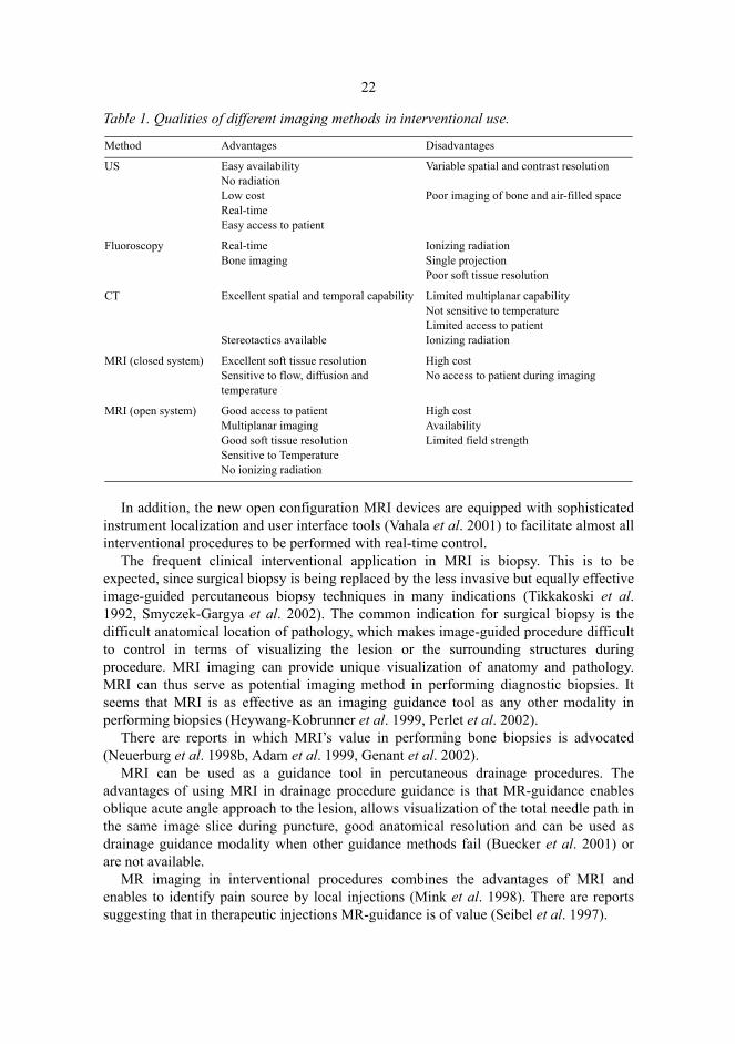
There are overall numerous facts that support the use of MRI in interventions. The lackof ionizing radiation is considered an important advantage; this alone may lead to theincreased use of MRI in interventions in future. But there are further advantages to theMRI and these are not easily, if at all, matched by any other imaging modality; firstly,MRI provides relatively good spatial and temporal resolution (Jager & Reiser 2001).Secondly, high intrinsic contrast in tissue without or with the use of contrast medium(Tung & Davis 1993). Thirdly, multiplanar imaging capability with optional two andthree-dimensional view (Murphy & Totty 1986). Furthermore, MRI has the ability tomeasure and quantify flow, diffusion and perfusion (Morvan et al. 1993, Rordorf et al.1998, Pedersen et al. 2002). An important feature is also the temperature sensitivity ofMRI, which allows the assessment of temperature changes (Quesson et al. 2000).Qualities of different modalities used for guidance in interventions are demonstrated inTable 1.
22
Table 1. Qualities of different imaging methods in interventional use.
In addition, the new open configuration MRI devices are equipped with sophisticatedinstrument localization and user interface tools (Vahala et al. 2001) to facilitate almost allinterventional procedures to be performed with real-time control.
The frequent clinical interventional application in MRI is biopsy. This is to beexpected, since surgical biopsy is being replaced by the less invasive but equally effectiveimage-guided percutaneous biopsy techniques in many indications (Tikkakoski et al.1992, Smyczek-Gargya et al. 2002). The common indication for surgical biopsy is thedifficult anatomical location of pathology, which makes image-guided procedure difficultto control in terms of visualizing the lesion or the surrounding structures duringprocedure. MRI imaging can provide unique visualization of anatomy and pathology.MRI can thus serve as potential imaging method in performing diagnostic biopsies. Itseems that MRI is as effective as an imaging guidance tool as any other modality inperforming biopsies (Heywang-Kobrunner et al. 1999, Perlet et al. 2002).
There are reports in which MRI’s value in performing bone biopsies is advocated(Neuerburg et al. 1998b, Adam et al. 1999, Genant et al. 2002).
MRI can be used as a guidance tool in percutaneous drainage procedures. Theadvantages of using MRI in drainage procedure guidance is that MR-guidance enablesoblique acute angle approach to the lesion, allows visualization of the total needle path inthe same image slice during puncture, good anatomical resolution and can be used asdrainage guidance modality when other guidance methods fail (Buecker et al. 2001) orare not available.
MR imaging in interventional procedures combines the advantages of MRI andenables to identify pain source by local injections (Mink et al. 1998). There are reportssuggesting that in therapeutic injections MR-guidance is of value (Seibel et al. 1997).
Method Advantages Disadvantages
US Easy availabilityNo radiationLow costReal-timeEasy access to patient
Variable spatial and contrast resolution Poor imaging of bone and air-filled space
Fluoroscopy Real-timeBone imaging
Ionizing radiationSingle projectionPoor soft tissue resolution
CT Excellent spatial and temporal capability Stereotactics available
Limited multiplanar capabilityNot sensitive to temperatureLimited access to patientIonizing radiation
MRI (closed system) Excellent soft tissue resolution Sensitive to flow, diffusion and temperature
High costNo access to patient during imaging
MRI (open system) Good access to patientMultiplanar imagingGood soft tissue resolutionSensitive to TemperatureNo ionizing radiation
High costAvailabilityLimited field strength
23
Image guided tumor therapies are a wide entity with lots of applications. There arenumerous possibilities for interventionalists to use both in imaging modalities and therapymodes. As an imaging modality MRI has the widest range of qualities, which makes MRIthe most interesting modality to be used for therapy planning and guidance. MR-guidancehas been successfully applied in interstitial laser treatment guidance and monitoring ofsoft tissue tumor ablation (Adam et al. 1999, de Jode et al. 1999, Vogl et al. 1999b, Law& Regan 2000, Fiedler et al. 2001).
Optimization of thermal therapies demands methods measuring temperature in vivoduring therapy. In this respect MRI provides interesting and unique potential to measurethermal deposition in tissue during thermal treatment. This can be achieved by usingdifferent MRI techniques suitable for the purpose: T1 relaxation time of water protons,molecular diffusion constant of water, water proton resonance frequency, Protonspectroscopic imaging, temperature sensitive contrast agents, and theoretically even spindensity or magnetization transfer can namely be used for MRI thermometry (Quesson etal. 2002).
2.2.5 Instrument tracking in interventional procedures in MRI
Instrument tracking in MRI is based upon creative use of scanner hardware, software,sequences and tracking options (Smits et al. 1999, Konings et al. 2001). MRIinterventions can be performed in a straightforward diagnostic MRI unit with standardsoftware, but it is much more feasible and also safer to perform them using user interfacedesigned for MRI interventions (Hinks et al. 1998, Yrjana et al. 2002) (Jyrkinen et al.2000). This type a software allows planning, imaging and performing the interventionalprocedure in a predetermined way using default settings for imaging and imagewindowing. This allows categorizing the interventions and providing custom-madeimaging features for each of them. The software usually comes with hardware thatenables monitoring the procedure from the imaging room and user interface hardwarethat can be used in MRI environment.
Imaging sequences in interventional MRI (IMRI) are somewhat different from theones used in diagnostic MRI. This is due to the fact that fast imaging speed is related togood spatial and temporal resolution. It is difficult to achieve all these simultaneously;there is trade-off between image speed, signal to noise ratio, and resolution (Busch et al.1998). Therefore almost all the sequences used in IMRI are custom-made and originatefrom fast imaging sequences. These include various gradient echo techniques generallywith short TR; also different strategies for k-space sampling have been developed in orderto speed up the imaging. These include LoLo (Buecker et al. 1998), keyhole (Duerk et al.1996), segmented k-space (Heid et al. 1995) and wavelet encoded data acquisitiontechniques (Wendt et al. 1998). New techniques like SMASH and SENSE are likely to setthe standard for image quality in coming years (Sodickson et al. 1999, Weiger et al.2002).
Instrument tracking gives the possibility to obtain images in the instrument planesimultaneously during the procedure. This leads to a multiplanar interactive scanningenvironment, where the ability to interactively localize, plan and monitor the procedure is
24
an essential feature. This kind of setting requires active instrument tracking. Activeinstrument tracking can be achieved in at least two ways; the instrument can be trackedwith infrared camera when an appropriate number of mirrors with known locale isprovided (Vahala et al. 2001). Other method to achieve active tracking is to use a built-inreceiver or active coils in the instrument to achieve exact positional information of thedevice (Ladd et al. 1998, Joensuu et al. 2000). Locally induced field inhomogenities canalso be used to pinpoint the instrument position (Glowinski et al. 1998). In this method acurrent is applied through a wire built in the wall of the instrument causing fieldinhomogeneity and thus signal void. There are also other potential methods for activeinstrument tracking, such as using electron spin resonance (ESR) (Ehnholm et al. 1999),or inducing a signal from the instrument tip from an external source (Konings et al.2001).
Ideally IMRI procedures would be performed in real time imaging setting and thussimple passive instrument tracking would be sufficient; this is also an option whenimmediate image update is not necessary due to the nature of the procedure. Passiveinstrument tracking is based on the inherent susceptibility artifact caused by theinstrument in the image data also this can be enhanced with modified catheter structure(Bakker et al. 1996). At the moment passive tracking methods are in frequent clinical use,namely instrument artifacts follow-up (Adam et al. 1999, Salomonowitz et al. 2000). Ofactive tracking methods, optical tracking is clinically in use (Jolesz 1998, Ojala et al.2000).
2.2.6 Implications for this study
Intreventional procedures are in effect minimally invasive procedures of surgical nature.These procedures have been performed a under cluster of radiological imagingmodalities, of which most recent is MRI. There are various MRI systems in use in whichMRI’s feasibility as a potential platform for interventional procedures has beendemonstrated. Low field MRI scanner and open configuration high-field scanners seem tobe most suitable for interventional procedures, but there is lack of clinical studiesdemonstrating the clinical feasibility of MR-guidance in interventional procedures. Theinformation available upon procedural strategies and indications for MRI interventions islimited. The procedural instrument guidance methods are still being developed. Therehave been no procedural reports of MR-guided bone biopsy with associated fine needlebiopsy available. Neither are any studies available with statistical results upon MR-guided nerve root therapies. Discography procedure has not been performed in low-fieldMRI surroundings. Thermal therapies are being increasingly used for tumor palliationand treatment; this is also being done under MR-guidance.

3 Purpose of the study
The purpose of this study was:1. To study the feasibility of optical guidance method in conjunction with MR-guidance
in bone biopsies at 0.23T.2. To investigate clinical feasibility of MR-guidance in performing bone biopsies at
0.23T.3. To evaluate MR-guided nerve root therapies at 0.23T.4. To study feasibility of MR-guidance in performing discography.5. To explore the feasibility of MR-guided thermal laser therapy in treating osteoid
osteoma at 0.23T.

4 Materials and methods
4.1 Patients and procedures
The total number of patients included to these studies was 97. Bone biopsies wereperformed in five patients with the optical instrument tracker and bone biopsy setmodified for the tracker system. The sequel study consisted of clinical experiment of 14consecutive bone biopsies (n=14), and 13 fine needle aspirations performed under MR-guidance.
Periradicular nerve root infiltration therapy was performed on 61 consecutive patients(n=61), with sciatic pain. The periradicular therapy was applied to the anatomical regionof the lumbosacral area.
MR-guided discography was performed as part of diagnostic pain provocation forpatients suspected of intervertebral pain source at lumbosacral area. 34 MR-guideddiscographies were performed on 12 patients (n=12). Laser induced thermal therapy forosteiod osteoma therapy was applied under MR-guidance. 5 patients were treated (n=5).Informed consent was obtained from all patients. All the studies were accepted by theethical committee of Oulu University Hospital.
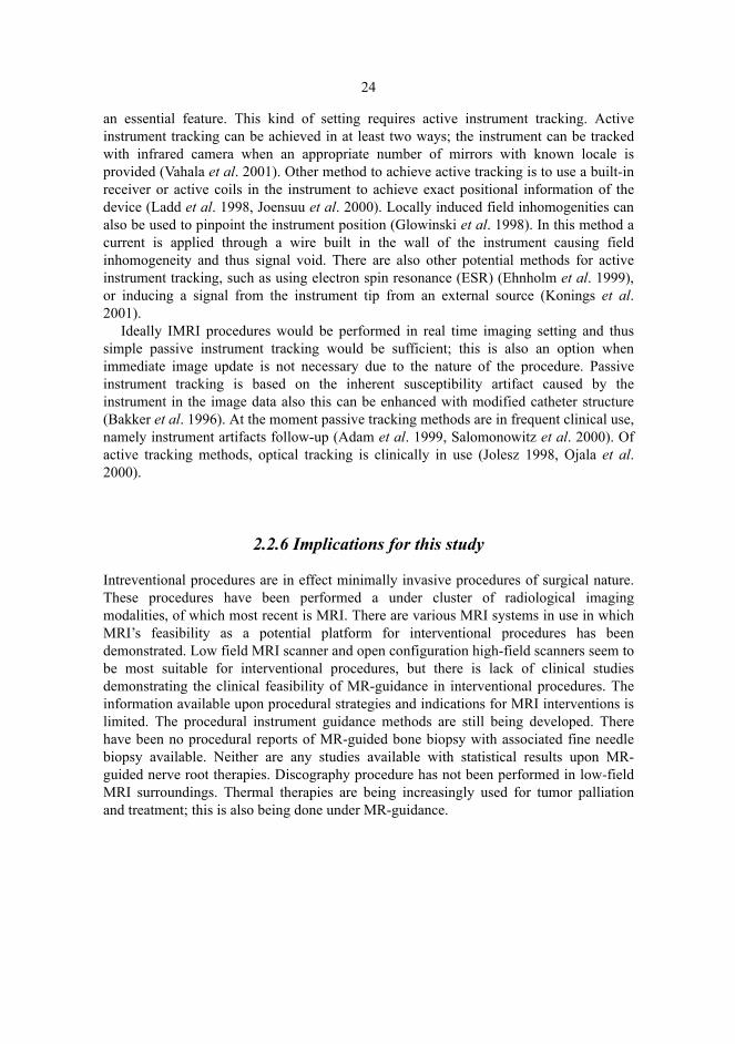
4.1.1 MRI-system for interventional procedures
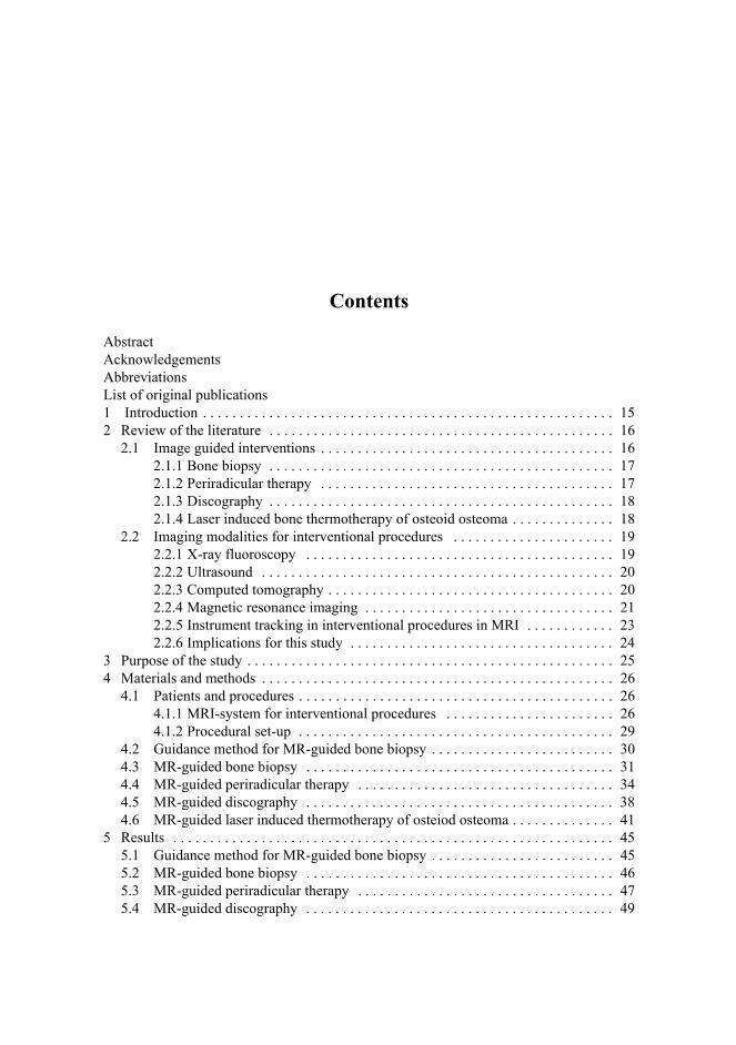
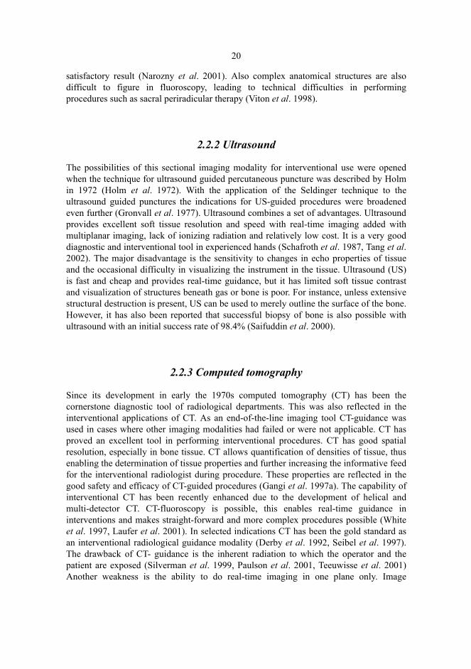
All the procedures were performed in the interventional MRI system which consisted of alow field MRI scanner, in-room monitor, in-room workstation with imaging capability,infra-red camera, fixed infra-red mirrors and a foot pedal for the operator whichcontrolled sequence start. MRI scanner used was a resistive 0.23 T open configuration(C-shaped) MRI with interventional optical tracking equipment and software (OutlookProview, Philips Medical Systems, MR Technologies Finland) (Figure 1), which wasinstalled into a full-scale operating room. The open configuration construction with onepillar supporting the scanner allows wide passage to the patient through a 44 cm diameterhorizontal gap.
27
Fig. 1. An open interventional MRI system.
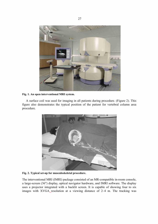
A surface coil was used for imaging in all patients during procedure. (Figure 2). Thisfigure also demonstrates the typical position of the patient for vertebral column areaprocedure.
Fig. 2. Typical set-up for musculoskeletal procedure.
The interventional MRI (IMRI) package consisted of an MR-compatible in-room console,a large-screen (36") display, optical navigator hardware, and IMRI software. The displayuses a projector integrated with a backlit screen. It is capable of showing four to siximages with XVGA resolution at a viewing distance of 2–4 m. The tracking was

28
performed with an infrared navigator camera. The camera detects two infrared-reflectivetrackers simultaneously, one of which is attached to the instrument and the other to themagnet pole-piece, which thereby provides a fixed reference frame.
The navigator camera is placed on an adjustable and movable stand. The camerautilizes infrared (IR) passive tracking, whereby the position of the instrument beingobserved is calculated from the IR pulses reflected from the spheres attached rigidly tothe bone biopsy set. The camera is capable of differentiating between instrument typesbased on the geometrically unique configurations of the spheres on the instruments. Thefixed reference frame attached to the upper pole-piece of the magnet allows repositioningof the camera during the operation, in case the line-of-sight to the needle holder becomesblocked.
The software supports instrument-guided imaging, so that the image set centre followsthe instrument tip and the image plane follows the instrument orientation. The navigationsoftware communicates with the scanner in real time, allowing real-time changes in theimage plane. The default image planes were typically set in three orthogonal planes inrelation to the instrument axis. Imaging can also be assessed in fixed plane mode wherethe relation of imaging plane to the operator is fixed. Alternatively imaging in relation tothe magnetic field co-ordinates can be assessed. Alternate types of sequences with avariable number of slices (1-9 slices typically) can be assessed from preset values. Theinstrument dimensions were calibrated with tracking software before the puncture. Theinstrument and its trajectory shows as a real-time graphic overlay in the image or in theimage set during procedure. The guidance software allows determination of the target as agraphic overlay item (target-point) and also provides spatial feedback from instrumentsposition as a graphic overlay in relation to the determined target-point. The instrumentwas connected to the optical tracking system with a wireless instrument holder (Figure 3).
Fig. 3. Instrument holder with optical tracking system.
29
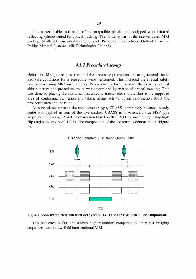
It is a sterilizable tool made of biocompatible plastic and equipped with infraredreflecting spheres suited for optical tracking. The holder is part of the interventional MRIpackage (iPath 200) provided by the magnet (Proview) manufacturer (Outlook Proview,Philips Medical Systems, MR Technologies Finland).
4.1.2 Procedural set-up
Before the MR-guided procedure, all the necessary precautions ensuring normal sterileand safe conditions for a procedure were performed. This included the special safetyissues concerning MRI surroundings. When starting the procedure the possible site ofskin puncture and procedural route was determined by means of optical tracking. Thiswas done by placing the instrument mounted to tracker close to the skin at the supposedarea of containing the lesion and taking image sets to obtain information about theprocedure area and the route.
As a novel sequence to the used scanner type, CBASS (completely balanced steadystate) was applied in four of the five studies. CBASS is in essence a true-FISP typesequence combining T2 and T1 expression based on the T2/T1 balance in high using highflip angles (Duerk et al. 1998). The composition of the sequence is demonstrated (Figure4).
Fig. 4. CBASS (completely balanced steady state), i.e. True-FISP sequence. The composition.
This sequence is fast and allows high resolution compared to other fast imagingsequences used in low-field interventional MRI.
30
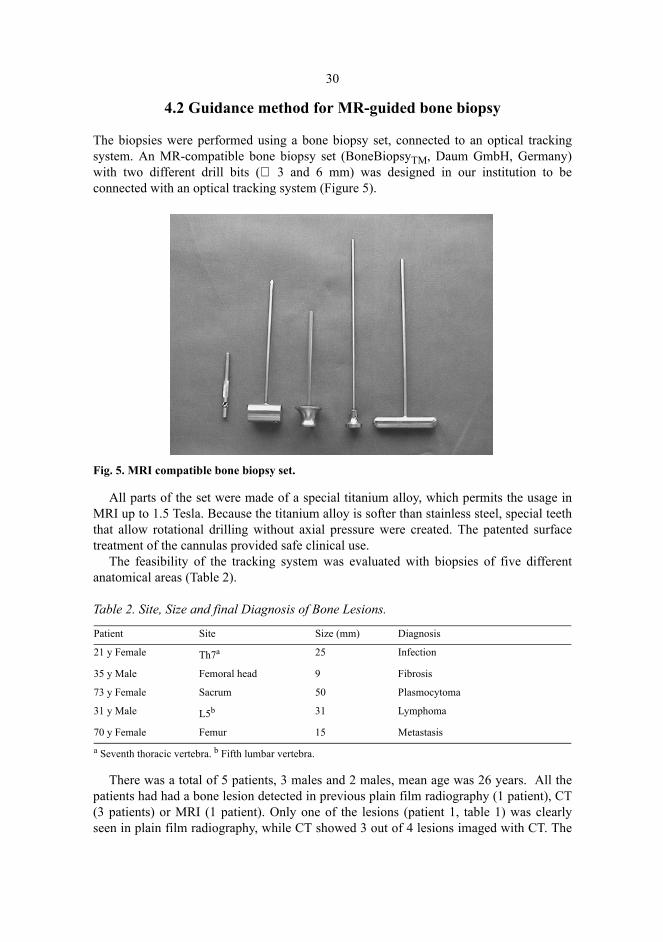
4.2 Guidance method for MR-guided bone biopsy
The biopsies were performed using a bone biopsy set, connected to an optical trackingsystem. An MR-compatible bone biopsy set (BoneBiopsyTM, Daum GmbH, Germany)with two different drill bits (∅ 3 and 6 mm) was designed in our institution to beconnected with an optical tracking system (Figure 5).
Fig. 5. MRI compatible bone biopsy set.
All parts of the set were made of a special titanium alloy, which permits the usage inMRI up to 1.5 Tesla. Because the titanium alloy is softer than stainless steel, special teeththat allow rotational drilling without axial pressure were created. The patented surfacetreatment of the cannulas provided safe clinical use.
The feasibility of the tracking system was evaluated with biopsies of five differentanatomical areas (Table 2).
Table 2. Site, Size and final Diagnosis of Bone Lesions.
There was a total of 5 patients, 3 males and 2 males, mean age was 26 years. All thepatients had had a bone lesion detected in previous plain film radiography (1 patient), CT(3 patients) or MRI (1 patient). Only one of the lesions (patient 1, table 1) was clearlyseen in plain film radiography, while CT showed 3 out of 4 lesions imaged with CT. The
Patient Site Size (mm) Diagnosis
21 y Female Th7a 25 Infection
35 y Male Femoral head 9 Fibrosis
73 y Female Sacrum 50 Plasmocytoma
31 y Male L5b 31 Lymphoma
70 y Female Femur 15 Metastasisa Seventh thoracic vertebra. b Fifth lumbar vertebra.
31
sizes of the lesions varied from 9 to 50 mm (mean 26 mm). Two of the lesions were in thespine, one in the sacral bone, one in the femoral head and one in the femoral diaphysis. Aprevious malignancy was known in one patient. Two biopsies were performed undergeneral anesthesia and three under spinal anesthesia.
Preoperatively, the lesion and the puncture route were visualized with T1-weightedfast spin echo (FSE; TR/TE 400/16 ms, echo train length (ETL) 4, flip angle (FA) 90,field of view (FOV) 380 mm x 380 mm, acquisition matrix 324 x 324, 7-mm slices, timeof acquisition (TA) 22 s, number of slices 5) in a longitudinal, either sagittal or coronal,plane, depending on the anatomical area. The following needle guidance sequences wereused: 1. T1-weighted gradient echo (TR/TE 95/7, ETL 1, FA 60, FOV 380 x 380, matrix300 x 300, 7-mm slices, TA 18 s/5 slices), 2. CBASS 2D (completely balanced steadystate sequence) (TR/TE 9.1/4.5, ETL 1, FA 60, FOV 380 x 380, matrix 216 x 216, 10-mmslice, TA 2 s / one slice) and 3. CBASS 3D (TR/TE 8.4/4.2, ETL 1, FA 45, FOV 380 x380, matrix 256 x 256, 5-mm slices, TA 24 s/8 slices). The biopsies were targeted to theenhancing part of the tumor, if one had been present in the previous MRI. When thelesion’s border was reached, the puncture needle was removed and a trephine bore wasintroduced through a trocar. The bore was then drilled into the lesion, and a sample wasremoved and fixed in 10% formalin. The patients remained in hospital for 24 hours forroutine follow-up.
4.3 MR-guided bone biopsy
All consecutive patients referred for bone biopsy at our institution during September 11999- September 1 2000 were included in the study. The series consisted of 14 patients,who underwent altogether 20 percutaneous bone biopsies. 19 MR-guided trephine biopsysamples from 13 patients were collected. On 4 patients double trephine biopsy wasperformed during one session, on one patient triple trephine biopsy was done during onesession.
In combination with MR-guided trephine biopsy thirteen FNAs targeted to the lesionarea were performed on 13 patients. FNA was performed through the trochar from thechannel drilled to the bone with trephine as a complementary procedure. Additionalbacterial samples were aspirated in 4 cases to establish possible microbiologicalinvolvement in the lesion. A MRI compatible 20 gauge needle (MReye, Cook,Bloomington, IN, USA) was used in FNA and in aspiration for bacterial stains. Eightpatients were biopsied under general anesthesia, and 6 biopsies were made under spinalanesthesia.
The mean age of the patients was 50 years (6 males, 8 females). All the patients hadhad a bone lesion detected in a previous radiographic, CT or MRI examination. Theindications for bone biopsy were: bone lesion of unknown origin n= 5, suspectedosteomyelitis n=2, suspected primary malignancy n=2, suspected metastatic lesion n=5.The bone lesion was suspected of being either neoplastic or infectious in nature. In caseswith suspected osteomyelitis there was no preprocedural evidence of extraosseusinfectious agent. The lesion size varied from 0.7 cm to 12 cm the mean being 3.4 cm.
32
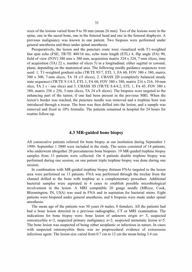
The locations of the lesions were: pelvis n=4, femur n=4, lumbar spine n=2, thoracicspine n=3, tibia n=1. The lesions were graded by preoperative imaging being edematousn=7, destructive n=5 or sclerotic n=2 of nature. Lesion sizes and localizations aredemonstrated in Table 3.
Table 3. Bone biopsy lesion sizes, localization and diagnosis.
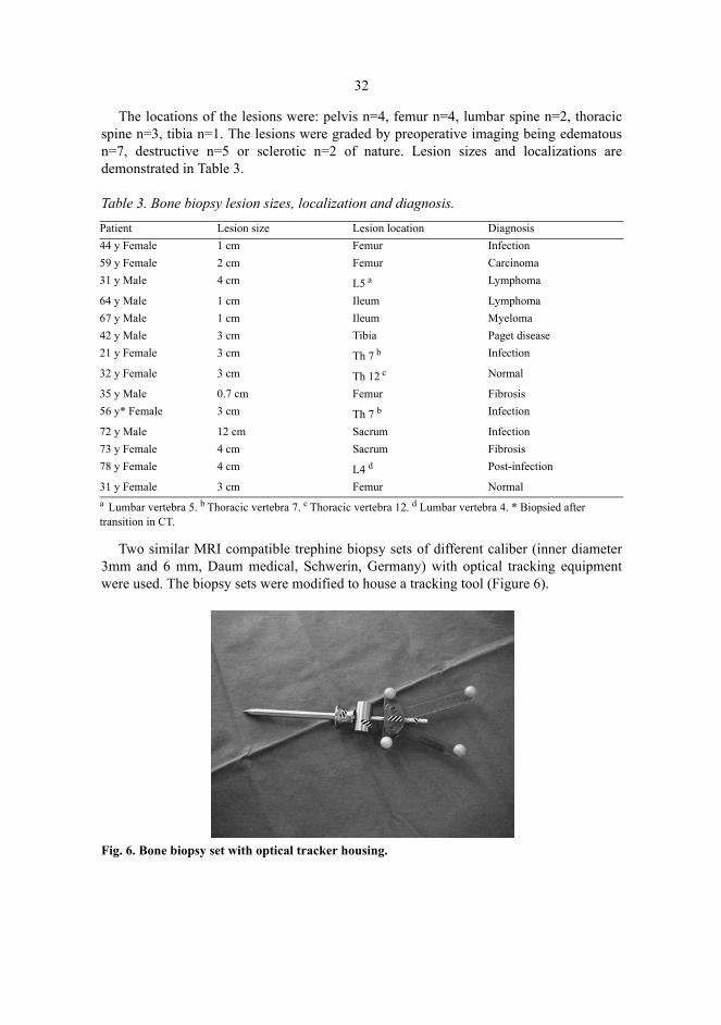
Two similar MRI compatible trephine biopsy sets of different caliber (inner diameter3mm and 6 mm, Daum medical, Schwerin, Germany) with optical tracking equipmentwere used. The biopsy sets were modified to house a tracking tool (Figure 6).
Fig. 6. Bone biopsy set with optical tracker housing.
Patient Lesion size Lesion location Diagnosis44 y Female 1 cm Femur Infection59 y Female 2 cm Femur Carcinoma31 y Male 4 cm L5 a Lymphoma
64 y Male 1 cm Ileum Lymphoma67 y Male 1 cm Ileum Myeloma42 y Male 3 cm Tibia Paget disease21 y Female 3 cm Th 7 b Infection
32 y Female 3 cm Th 12 c Normal
35 y Male 0.7 cm Femur Fibrosis56 y* Female 3 cm Th 7 b Infection
72 y Male 12 cm Sacrum Infection73 y Female 4 cm Sacrum Fibrosis78 y Female 4 cm L4 d Post-infection
31 y Female 3 cm Femur Normala Lumbar vertebra 5. b Thoracic vertebra 7. c Thoracic vertebra 12. d Lumbar vertebra 4. * Biopsied after transition in CT.
33
A prerequisite for the procedure was a free route to the lesion. The route wasdetermined with T1-weighted fast spin echo imaging (5 slices, FSE, TE 400 ms, TR 16ms, slice thickness/ interval 7,0 mm/ 8.0 mm, FOV 380x380, matrix 324x324, acquisitiontime 23 seconds), CBASS imaging (8 slices, CBASS, TR 8.4 ms, TE 4.2 ms, slicethickness/ interval 5.0 mm/ 5.0 mm, FOV 380x380, matrix 256x256, acquisition time 24seconds), and T2-weighted fast spin echo images (9 slices, FSE, TE 3500 ms, TR 150ms, slice thickness/interval 7,0 mm/8,0 mm, FOV 380x380, matrix 192x192, acquisitiontime 35 seconds), were also obtained whenever no previous MRI imaging had been made.A representative, and most probably, viable part of the lesion was selected as a target. Theparts of the lesion that represented extraosseous tissue extension or were not covered bycortical or osteosclerotic bone were preferred for biopsy.
The set-up for biopsy is demonstrated in Figure 2. To reduce post-procedural pain in patients who underwent the procedure under general
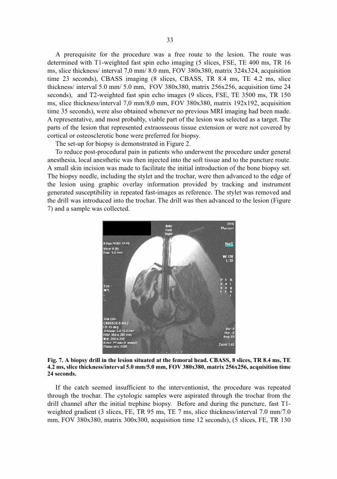
anesthesia, local anesthetic was then injected into the soft tissue and to the puncture route.A small skin incision was made to facilitate the initial introduction of the bone biopsy set.The biopsy needle, including the stylet and the trochar, were then advanced to the edge ofthe lesion using graphic overlay information provided by tracking and instrumentgenerated susceptibility in repeated fast-images as reference. The stylet was removed andthe drill was introduced into the trochar. The drill was then advanced to the lesion (Figure7) and a sample was collected.
Fig. 7. A biopsy drill in the lesion situated at the femoral head. CBASS, 8 slices, TR 8.4 ms, TE4.2 ms, slice thickness/interval 5.0 mm/5.0 mm, FOV 380x380, matrix 256x256, acquisition time24 seconds.
If the catch seemed insufficient to the interventionist, the procedure was repeatedthrough the trochar. The cytologic samples were aspirated through the trochar from thedrill channel after the initial trephine biopsy. Before and during the puncture, fast T1-weighted gradient (3 slices, FE, TR 95 ms, TE 7 ms, slice thickness/interval 7.0 mm/7.0mm, FOV 380x380, matrix 300x300, acquisition time 12 seconds), (5 slices, FE, TR 130
34
ms, TE 11 ms, slice thickness/interval 10,0 mm/10.0 mm, FOV 380x380, matrix256x256, acquisition time 18 seconds), or CBASS imaging ( 8 slices, CBASS, TR 8.4 ms,TE 4.2 ms, slice thickness/interval 5.0 mm/5.0 mm, FOV 380x380, matrix 256x256,acquisition time 24 seconds), (1 slice, CBASS, TR 9.1 ms, TE 4.5 ms, slice thickness/interval 10.0 mm/ 10.0 mm, FOV 380x380, matrix 256x256 acquisition time 1.5seconds) was performed in order to visualize the biopsy set.
If the immediate preoperative MRI visualization of the lesion was not adequate orpreoperative imaging suggested that using contrast agent might be beneficial invisualizing the most active part of lesion, intravenous contrast medium (15 cc ofgadolinium) was introduced directly prior to the puncture. Contrast was used in 5 lesionsgraded edematous, one graded destructive and in one graded sclerotic. T1-weighted fastspin echo imaging (5 slices, FSE, TE 400 ms, TR 16 ms, slice thickness/interval 7,0 mm/8.0 mm, FOV 380x380, matrix 324x324, acquisition time 23 seconds) of the lesion wasthen performed. The patients remained in hospital for 24 hours for routine follow-up.
The trephine biopsy samples were fixed in 10% formalin. The FNA material wasprocessed by fixing the sample in 50% alcohol for preparation by the filtration technique.The FNA findings were rated as cytologic samples: C0 -insufficient; C1 -normal; C2 -benign atypia; C3 -possibly malignant; C4 was highly suspicious of malignancy and C5was malignant. Grades C3, C4 and C5 were determined to concur with malignanthistology. A pathologist determined the amount of obtained trephine biopsy material andFNA material as either sufficient or insufficient for diagnostic use. Histopathological andcytological diagnosis based on trephine biopsy material and FNA material wasdetermined by pathologists. Double reading of the samples was used. After initialhistopathological and cytological diagnosis, the samples were re-evaluated by apathologist experienced in bone pathology. Possible change in diagnosis after this re-evaluation was recorded. The diagnostic accuracy for histology, FNA and bacterial stainswas reported as true-positive or true-negative and false-positive or false-negative. Casesdetermined as being false negative were divided into two groups after 6 months’ clinicalfollow-up: wrong diagnosis or sampling error. Insufficient sample in FNA and necrosis asa diagnosis in trephine biopsy were determined as a non-diagnostic being false negative,and a new biopsy (MR-guided or surgical) was scheduled. The evaluation of diagnosticaccuracy was achieved by comparing the histopathological diagnosis, cytologicaldiagnosis and microbiological diagnosis with current or final diagnosis made during 6months’ clinical follow-up.
4.4 MR-guided periradicular therapy
Patients referred for percutaneous periradicular nerve root infiltration therapy at ourinstitution during April 1 1999-March 1 2001 were included in the study. The seriesconsisted of 61 patients, who underwent altogether 67 percutaneous periradicular nerveroot infiltrations. The periradicular nerve root infiltration was always targeted to thesymptomatic side of the patient. 58 infiltrations were targeted to first sacral root, 7infiltrations to 5th lumbosacral root and 2 to 4th lumbosacral root. On 5 patients doubleinfiltration was performed from 4 to 10 months after to the first procedure due to renewed
35
radicular pain. One patient had periradicular nerve root infiltration also to the contra-lateral nerve root. The mean age of the patients was 46 years (range 22-71 years); 41patients had an unoperated disc disease and 20 patients had history of previous backsurgery. All patients had lumbosacral radicular pain or sciatica diagnosis set by surgeonor rehabilitation specialist before referral to the procedure. Patients with indication foracute surgery, such as cauda equina, were excluded from the study. The final outcome ofprocedural pain relief obtained was evaluated after 6 months’ clinical follow-up andquestionnaire. The effect to the radicular pain was graded: 1. Good to excellent = no pain or not disturbing pain allowing normal physical activity
at 3 months from the procedure, 2. Temporary = temporary relief of pain (1-4 weeks), 3. No relief of pain 4. Worsening of pain.
All the patients had a preprocedural MRI or CT study made.The possible correlation between the effect of nerve root infiltration therapy and the
etiology of the pain was studied. To facilitate this the findings of vertebral disc anatomyand pathology causing possible narrowing in subsequent path of the symptomatic nerveroot in individual patients were classified into 3 categories at the lumbosacral levels fromL1 to S1. This classification was done by inspecting the pre-procedural imaging data(MRI or CT) obtained from the lumbosacral area.
The categories were; 1. Normal or minor protrusion of intervertebral disc without narrowing of the nerve root
space.2. Intervertebral disc herniation narrowing the nerve root space.3. Narrowing of the nerve root channel due post-operational scarring and/or osseal
prominence.An MRI compatible 20 gauge needle (MReye, Cook, Bloomington, IN, USA or
Manan, MD Tech, Fl, USA) was used for nerve root infiltration puncture and injection oftherapeutic substance.
A prerequisite for the procedure was a free route to the periradicular nerve root area.The route was determined with T1-weighted fast spin echo imaging (5 slices, FSE, TE400 ms, TR 16 ms, slice thickness/ interval 7,0 mm/ 8.0 mm, FOV 380x380, matrix324x324, acquisition time 23 seconds), CBASS (completely balanced steady state)imaging (8 slices, CBASS, TR 8.4 ms, TE 4.2 ms, slice thickness/interval 5.0 mm/5.0mm, FOV 380x380, matrix 256x256, acquisition time 24 seconds), and T2-weighted fastspin echo images (9 slices, FSE, TE 3500 ms, TR 150 ms, slice thickness/interval 7.0mm/8.0 mm, FOV 380x380, matrix 192x192, acquisition time 35 seconds), were alsoobtained whenever no previous MRI imaging had been made. The used sequence,CBASS (completely balanced steady state) is in essence true-FISP type sequencecombining T2 and T1 expression based on the T2/T1 balance in using high flip angles.This sequence is fast and allows high resolution compared to other fast imagingsequences used in low-field interventional MRI (Duerk et al. 1998).
The nerve root determined preoperatively as being the most probable pain source byclinical inspection and imaging studies was selected as target.
The set-up for therapeutic MR-guided injection and the surface coil used aredemonstrated in Figure 2.
36
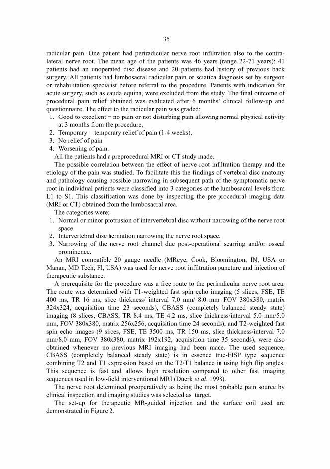
To reduce procedural subcutaneous pain, the local anesthetic lidocaine (10%),maximum 5ml, was then injected into the subcutaneous soft tissue. No other medication,sedation or monitoring was used. The needle was then advanced through thesubcutaneous fat and dorsal sacrolumbar ligaments to the connective tissue surroundingthe nerve root, i.e. periradicular space, using graphic overlay information provided bytracking and instrument generated susceptibility in repeated fast-images as reference,(Figure 8A). The mandrel was removed and 2 ml of saline was introduced into theperiradicular space (Figure 8B). This technique was used in the last 62 infiltrations. Thepossible pain provocation generated and the nature of it were recorded simultaneously.
Fig. 8. A Needle at periradicular space of the S1 nerve. 8 slices, CBASS, TR 8.4 ms, TE 4.2 ms,slice thickness/interval 5.0 mm/5.0 mm, FOV 380x380, matrix 256x256, acquisition time 24seconds.

37
Fig. 8. B Saline is visualized at periradicular space. 5 slices, EXPRESS, TR 9000 ms, TE 274 ms,slice thickness/interval 7mm/7mm, FOV 380x380, matrix 256x256, acquisition time 9 seconds.
Before and during the puncture, fast T1-weighted gradient echo (3 slices, FE, TR 95ms, TE 7 ms, slice thickness/interval 7.0 mm/7.0 mm, FOV 380x380, matrix 300x300,acquisition time 12 seconds), (5 slices, FE, TR 130 ms, TE 11 ms, slice thickness/interval10,0 mm/10.0 mm, FOV 380x380, matrix 256x256, acquisition time 18 seconds), orCBASS imaging (8 slices, CBASS, TR 8.4 ms, TE 4.2 ms, slice thickness/interval 5.0mm/5.0 mm, FOV 380x380, matrix 256x256, acquisition time 24 seconds), (1 slice,CBASS, TR 9.1 ms, TE 4.5 ms, slice thickness/interval 10.0 mm/10.0 mm, FOV380x380, matrix 256x256 acquisition time 1.5 seconds) was performed in order tovisualize the needle and anatomic structures to enable correct needle positioning to thetarget.
Immediately before and after the introduction of saline to the periradicular nerve rootspace, a single shot fast spin echo (SSFSE) imaging (5 slices, EXPRESS, TR 9000 ms,TE 274 ms, slice thickness/ interval 7mm/7mm, FOV 380x380, matrix 256x256,acquisition time 9 seconds) was performed to detect fluid signal around or parallel to thenerve root. The final proof of correct placement of the needle was obtained with thismethod. Possible pain provocation by the injection of saline was also used as an adjunctconfirmation of the right placement of the needle.
After this the therapeutic agent was introduced through the needle, either 2 ml ofmethylprednisolone-bupivacaine solution (Solomet, methylprednisolone 40 mg/ml-bupivacaine 5 mg/ml; Orion, Finland) (63 infiltrations) or 2ml bupivacaine (5%), (4infiltrations) was injected into the periradicular space and the fluid signal around orparallel to the nerve root was again detected with the SSFSE sequence. In the first 8procedures fluoroscopy was used with MRI as an adjunct modality either for determiningthe puncture site to the skin or to confirm the final placement of the needle in theconnective tissue surrounding the nerve sheath with a contrast agent.
38
4.5 MR-guided discography
Between April 1 2000 and March 1 2001 patients referred for percutaneous discographyat our institution were included in the study. The series consisted of 12 patients, 6 malesand 6 females, mean age being 41 years. Altogether 35 disc punctures were initializedand 34 percutaneous discograms were obtained. All patients had clinical suspicion oflumbar discogenic pain and/or a suggestive finding of lumbar disc or lumbar spinedegeneration in imaging studies (MRI, CT, x-ray). Pre-procedural degenerative spinefindings and clinical diagnosis of possible lumbar instability verified in lumbar regionextension-flexion x-ray images was reported. MR-guided discography was performed inorder to determine possible pain provocation during contrast injection as a preliminarytest to evaluate the possibility of surgical spinal fusion as a treatment option forcontinuous pain. The discography was always performed from the less symptomatic side.Patients with indication for acute surgery, such as cauda equina, were excluded from thestudy. Also excluded were patients with suggestive imaging findings of acute discherniation, neoplasm, spinal stenosis and infection. All the patients received pre-procedural intra-venous antibiotics, 1.5 g cefuroxime.
The final incidence of possible procedural complications was evaluated after 6months’ clinical follow-up. The primary result of possible surgical operation wasevaluated 6 months after surgery. 9 patients underwent MRI study of the lower back areaimmediately before the MR-guided discography. The MRI study with same sequentialconfiguration was repeated immediately after the MR-guided discography for these 9patients. The diagnostic MRI study used consisted of non-contrast sagittal FSE T1 (13slices, FSE, TE 380 ms, TR 18 ms, slice thickness/interval 5.0 mm/5.5 mm, FOV320x320, matrix 256x256, acquisition time 5 min 46 seconds), FSE T2 (13 slices, FSE,TE 4500 ms, TR 120 ms, slice thickness/interval 5.0 mm/5.5 mm, FOV 320x320, matrix256x256, acquisition time 7 min 12 seconds). In addition, axial 3D T1 gradient echoimaging (24 slices, FE3D, TE 33 ms, TR 9 ms, slice thickness/interval 3.0 mm/3.0 mm,FOV 350x350, matrix 256x256, acquisition time 7 min 36 seconds) was performedpreoperatively and after injection of contrast media to obtain MRI discograms.
A prerequisite for the procedure was a free route to the lumbar disc area. The routewas determined with T1-weighted fast spin echo imaging (5 slices, FSE, TE 400 ms, TR16 ms, slice thickness/interval 7.0 mm/8.0 mm, FOV 380x380, matrix 324x324,acquisition time 23 seconds) and CBASS imaging (8 slices, CBASS, TR 8.4 ms, TE 4.2ms, slice thickness/ interval 5.0 mm/5.0 mm, FOV 380x380, matrix 256x256, acquisitiontime 24 seconds).
The typical patient set-up for MR-guided discography is demonstrated in Figure 2.To reduce procedural subcutaneous pain the local anesthetic lidocaine (5ml) was then
injected into the subcutaneous soft tissue. No other medication, sedation or monitoringwas used.
Before and during the puncture, a T1-weighted gradient echo (3 slices, FE, TR 95 ms,TE 7 ms, slice thickness/interval 7.0 mm/7.0 mm, FOV 380x380, matrix 300x300,acquisition time 12 seconds), (5 slices, FE, TR 95 ms, TE 7 ms, slice thickness/interval7,0 mm/7.0 mm, FOV 380x380, matrix 256x256, acquisition time 18 seconds), orCBASS imaging (8 slices, CBASS, TR 8.4 ms, TE 4.2 ms, slice thickness/interval 5.0mm/5.0 mm, FOV 380x380, matrix 256x256, acquisition time 24 seconds), (1 slice,
39
CBASS, TR 9.1 ms, TE 4.5 ms, slice thickness/interval 10.0 mm/ 10.0 mm, FOV380x380, matrix 256x256 acquisition time 1.5 seconds) was performed in order tovisualize the needle.
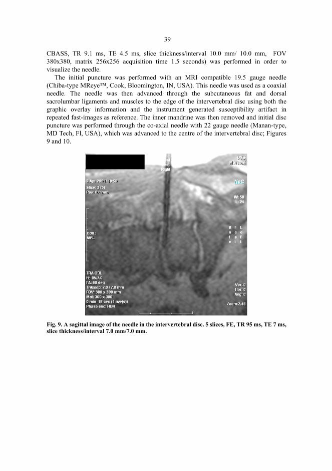
The initial puncture was performed with an MRI compatible 19.5 gauge needle(Chiba-type MReye™, Cook, Bloomington, IN, USA). This needle was used as a coaxialneedle. The needle was then advanced through the subcutaneous fat and dorsalsacrolumbar ligaments and muscles to the edge of the intervertebral disc using both thegraphic overlay information and the instrument generated susceptibility artifact inrepeated fast-images as reference. The inner mandrine was then removed and initial discpuncture was performed through the co-axial needle with 22 gauge needle (Manan-type,MD Tech, Fl, USA), which was advanced to the centre of the intervertebral disc; Figures9 and 10.
Fig. 9. A sagittal image of the needle in the intervertebral disc. 5 slices, FE, TR 95 ms, TE 7 ms,slice thickness/interval 7.0 mm/7.0 mm.

40
Fig. 10. An axial image of the needle in the paravertebral disc. 5 slices, FE, TR 95 ms, TE 7 ms,slice thickness/interval 7.0 mm/7.0 mm.
After disc puncture 1-2ml of gadolinium-saline mixture (1:8) was injected into thedisc. The possible pain provocation generated and the nature of it were recordedsimultaneously. Immediately after injection sagittal FE T1 weighted images wereobtained to verify the formation of MRI discogram (Figure 11).
Fig. 11. Sagittal FE T1 weighted discogram image. 5 slices, FE, TR 95 ms, TE 7 ms, slicethickness/interval 7.0 mm/7.0 mm.
41
Evaluation of the pain patient experienced was done during injection of contrast byevaluating pain concordance with patients’ existing pain symptoms using the DallasDiscogram Description scale (Sachs et al. 1987) for injection-generated pain: nosensation, pressure, dissimilar pain, similar pain, exact pain reproduction. For this studypain reproduction was classified as concordant when the patient felt similar or exactreproduction of pain. Non-concordant pain reproduction was reported when the patientfelt the sensation of dissimilar pain, pressure or no pain during injection of contrast.
4.6 MR-guided laser induced thermotherapy of osteiod osteoma
Between September 1 2001 and May 1 2002 5 consecutive patients (mean age 26 years, 3females, 2 males) with clinical and imaging findings suggesting osteoid osteoma weretreated with interstitial laser treatment at our department.
The patients’ lesion localization sites were; femur (n=4), talus (n=1), mean nidus sizewas 1.3 cm. Lesion sizes and localizations are demonstrated in Table 4.
Table 4. Osteoid osteoma patients’ age, lesion size, localization, diagnosis and outcome.
Strict sterility was maintained throughout the procedure. All the patients receivedprophylactic antibiotics before procedure, cefuroxime 1,5 g iv.
Depending from the patients age, lesion localization and patient co-operation eithergeneral anesthesia or spinal anesthesia was used. If general anesthesia was used localanesthetics were applied (lidocain 5%) subcutaneously to the treatment area to ease post-procedural local pain.
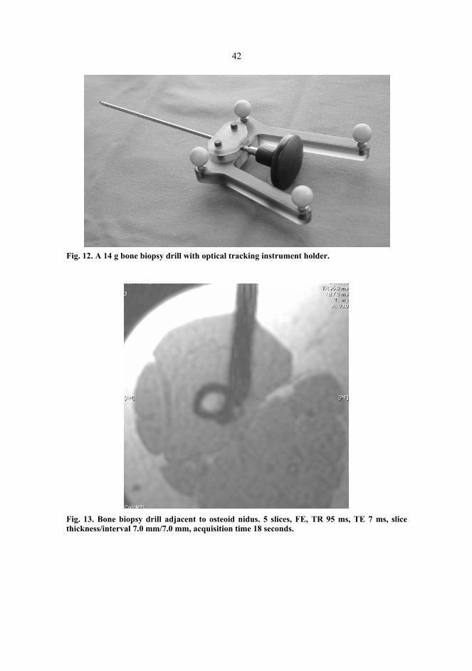
Initial puncture to the lesion was done under MR-guidance using the described opticalinstrument guidance. The needle was then advanced through the subcutaneous fat to thetarget vicinity using graphic overlay information provided by tracking and instrumentgenerated susceptibility in repeated fast-images as reference. Before and during thepuncture CBASS imaging (8 slices, CBASS, TR 8.4 ms, TE 4.2 ms, slice thickness/interval 5.0 mm/5.0 mm, FOV 380x380, matrix 256x256, acquisition time 24 seconds), (1slice, CBASS, TR 9.1 ms, TE 4.5 ms, slice thickness/interval 10.0 mm/10.0 mm, FOV380x380, matrix 256x256 acquisition time 1.5 seconds) and FE imaging (5 slices, FE, TR95 ms, TE 7 ms, slice thickness/interval 7.0 mm/7.0 mm, acquisition time 18 seconds)was performed in order to visualize the instrument and anatomic structures to enablecorrect instrument positioning to the target.
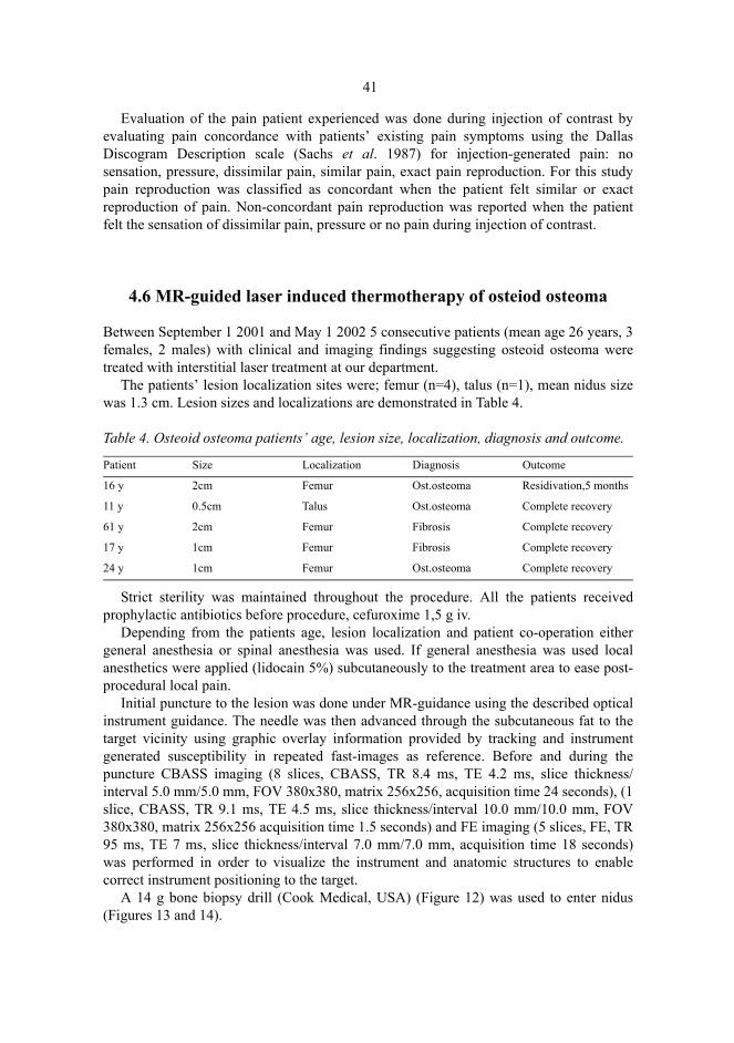
A 14 g bone biopsy drill (Cook Medical, USA) (Figure 12) was used to enter nidus(Figures 13 and 14).
Patient Size Localization Diagnosis Outcome
16 y 2cm Femur Ost.osteoma Residivation,5 months
11 y 0.5cm Talus Ost.osteoma Complete recovery
61 y 2cm Femur Fibrosis Complete recovery
17 y 1cm Femur Fibrosis Complete recovery
24 y 1cm Femur Ost.osteoma Complete recovery
42
Fig. 12. A 14 g bone biopsy drill with optical tracking instrument holder.
Fig. 13. Bone biopsy drill adjacent to osteoid nidus. 5 slices, FE, TR 95 ms, TE 7 ms, slicethickness/interval 7.0 mm/7.0 mm, acquisition time 18 seconds.
43
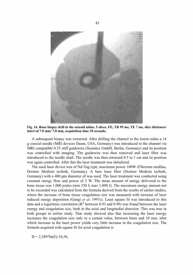
Fig. 14. Bone biopsy drill in the osteoid nidus. 5 slices, FE, TR 95 ms, TE 7 ms, slice thickness/interval 7.0 mm/ 7.0 mm, acquisition time 18 seconds.
A subsequent biopsy was extracted. After drilling the channel to the lesion nidus a 14g coaxial needle (MRI devices Daum, USA, Germany) was introduced to the channel viaMRI compatible 0.35 stiff guidewire (Somatex GmbH, Berlin, Germany) and its positionwas controlled with imaging. The guidewire was then removed and laser fiber wasintroduced to the needle shaft. The needle was then retracted 0.5 to 1 cm and its positionwas again controlled. After this the laser treatment was initialized.
The used laser device was of Nd-Yag type, maximum power 100W (Fibertom medilas,Dornier Medizin technik, Germany). A bare laser fiber (Dornier Medizin technik,Germany) with a 400 µm diameter of was used. The laser treatment was conducted usingconstant energy flow and power of 2 W. The mean amount of energy delivered to thebone tissue was 1,000 joules (min 350 J, max 1,800 J). The maximum energy amount notto be exceeded was calculated from the formula derived from the results of earlier studies,where the increase of bone tissue coagulation size was measured with increase of laserinduced energy deposition (Gangi et al. 1997c). Least square fit was introduced to thisdata and a logaritmic correlation (R2 between 0.93 and 0.99) was found between the laserenergy and coagulation size, both in the axial and longitudial direction. This was true inboth groups in earlier study. That study showed also that increasing the laser energyincreases the coagulation size only to a certain value, between 6mm and 10 mm, afterwhich increase in the laser power yields very little increase in the coagulation size. Theformula acquired with square fit for axial coagulation is
D = 2,389*ln(E)-10,58,

44
where D is the coagulation diameter in millimeters and E the laser energy. The treatmentwas monitored by using CBASS MRI imaging, (CBASS 3D, TE 7.7 ms, TR 3.8 ms, 8slices, slice thickness/interval 6.0 mm/6.0 mm, FOV 380x380, matrix 160x160, FA 63deg., acquisition time 24 seconds), by following the change in signal intensity expressedas signal void in the image caused by the temperature change in the tissue heated.Imaging sequence was repeated at 1minute 30 seconds intervals. When possible signalvoid was detected and maximum amount of energy delivered the treatment wasconsidered complete. After the treatment the patients were monitored for 6 hours in therecovery room. The patients were dispatched from the hospital after the first clinicalcontrol performed within 24 hours of the treatment. A further clinical control was carriedout within 3 weeks after the procedure. A questionnaire for each patient on the status ofpain symptoms was filled in by the patients 3 and 6 months after the treatment. Thequestionnaire was structured as 1. total relief of pain 2. some relief pain 3. no relief ofpain. Only total relief of pain was considered as successful outcome. In case of persistingpain patients were rescheduled for control MRI study and re-treatment with laserablation.

5 Results
5.1 Guidance method for MR-guided bone biopsy
The bone biopsy system was successfully applied to all patients, and it provided a safeand accurate guidance method for all phases of the procedure. Optical trackingimplemented as part of the biopsy set allowed the puncture route to be chosen fast andreliably before needle insertion, and it was also useful during the biopsy procedurewhenever the angle of approach had to be changed. Optical tracking also allowed almostreal-time imaging in the plane of the instrument, which made the procedure easy toaccomplish in different anatomical areas and at different angles of approach, allowing theprocedural time from the insertion of the needle through skin to needle retraction to beless than 40 minutes.
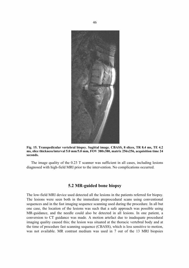
The biopsies performed using this system yielded sufficient samples in all cases (Table2). One of the vertebral body biopsies (Th 7) was performed via the transpedicular routewithout difficulties (Figure 15).
46
Fig. 15. Transpedicular vertebral biopsy. Sagittal image. CBASS, 8 slices, TR 8.4 ms, TE 4.2ms, slice thickness/interval 5.0 mm/5.0 mm, FOV 380x380, matrix 256x256, acquisition time 24seconds.
The image quality of the 0.23 T scanner was sufficient in all cases, including lesionsdiagnosed with high-field MRI prior to the intervention. No complications occurred.
5.2 MR-guided bone biopsy
The low-field MRI device used detected all the lesions in the patients referred for biopsy.The lesions were seen both in the immediate preprocedural scans using conventionalsequences and in the fast imaging sequence scanning used during the procedure. In all butone case, the location of the lesions was such that a safe approach was possible usingMR-guidance, and the needle could also be detected in all lesions. In one patient, aconversion to CT guidance was made. A motion artefact due to inadequate proceduralimaging quality caused this; the lesion was situated at the thoracic vertebral body and atthe time of procedure fast scanning sequence (CBASS), which is less sensitive to motion,was not available. MR contrast medium was used in 7 out of the 13 MRI biopsies
47
performed in order to improve visualization of the active component of the lesion.However, no improvement in visualization of the lesion was found in cases when contrastwas used.
Out of the 13 patients biopsied in MRI, the trephine biopsy yielded a diagnosis in allbut one case. All the double and triple samples concurred. Double reading of samplematerial did not result in changes in initial diagnosis. One of the trephine biopsy findingswas considered false negative during 6 months’ clinical follow up. This was categorizedas a sampling error and the final diagnosis was achieved with surgical biopsy. Diagnosticaccuracy in the trephine biopsy series was thus 95%. The trephine biopsy yielded amalignant diagnosis in 4 patients, a post-infectious or granulomatous reaction in 4patients, a benign fibrotic lesion in 2 patients and Paget´s disease in one patient, whiletwo patients had a normal finding after a 6-month clinical follow-up. MRI guidedtrephine biopsy excluded malignancy in 3 patients with a known previous malignantdisease and detected malignancy in 3 patients without a previous diagnosis of malignantdisease.
The FNA sample material was adequate for diagnostic evaluation in all cases. TheFNA cytological diagnosis concurred with the trephine biopsy diagnosis in 54% of cases.There were thus 46% of false negatives in FNA diagnoses, which were all determined tobe due to sampling error. However, the cytological diagnosis concurred with histologicaldiagnosis in all cases with malignant histology. The bacterial stains for microbiologicaldiagnostics were true negative in all of the 4 cases where they were collected with a 6-month clinical follow-up.
The mean puncture time from the introduction of the needle through skin to needleretraction was 1 hour 20 minutes. The mean operative time from entering theinterventional MRI operation theater to leaving it was 2 hours 55 minutes, maximum 4hours 30 minutes and minimum 1 hour 45 minutes, this included the anesthesiaprocedures needed. The mean number of sequences used during the procedure was 26,maximum 58 and minimum 8 sequences. There were no procedural complications.
5.3 MR-guided periradicular therapy
All the nerve roots selected for targeted infiltration were detected in the low-field MRIdevice used. MR-guidance allowed correct needle positioning in all but one case (98.5%)when MR-guidance was used. The failure was caused by degeneration-induced changesin anatomy obstructing path to the nerve root canal. There was no need to reposition theneedle after initial MRI imaging (8 slices, CBASS, TR 8.4 ms, TE 4.2 ms, slicethickness/interval 5.0 mm/5.0 mm, FOV 380x380, matrix 256x256, acquisition time 24seconds) had confirmed the localization of the needle in the direct vicinity of the nerveroot sheath.
The mean puncture time from the introduction of the needle through skin to needleretraction was 12 minutes, minimum 2 minutes and maximum 60 minutes. The meanprocedural time from starting the interventional MRI procedure to completing it was 33minutes, minimum 9 minutes and maximum 84 minutes, this included the anesthesiaprocedures needed. These times do not include the time required for preparing the patient
48
and operation room. The mean total number of sequences used in all scans during theprocedure was 13, minimum 7 sequences and maximum 26. The mean number ofsequences used for puncturing was 10, minimum 2 and maximum 20. There was one casewhere post-procedural pain was categorized as a post-procedural complication. Threepatients underwent back surgery during follow-up period due to persisting clinicalsymptoms and worsened disc pathology finding in imaging studies. All these patients hadrelief to the pain from surgery.
The therapy effect outcome of all performed punctures is shown in Table 5.
Table 5. The effect of nerve root infiltration to the radicular pain in all performedpunctures.
Effect of nerve root infiltration on the radicular pain according to the operationalsituation before the procedure is shown in Table 6.
Table 6. Effect of nerve root infiltration on radicular pain according to the operationalsituation before the procedure.
The therapy effect on patients with different types of nerve root channel narrowing isshown in Table 7.
The effect of nerve root infiltration on radicular pain N %
No pain or not disturbing pain 35 52.2
Temporary relief of pain 15 22.4
No relief of pain 14 20.9
Worsening of pain 3 4.5
Total 67 100.0
For these data statistical analysis was performed with a chi-square test where p< 0.001, df =3, and χ² = 31.806.
The effect of nerve root infiltrationon radicular pain
Patients’ operational situation before nerve root infiltration
Operated Not operated
N % n %
No pain or not disturbing pain 15 78.9 18 41.9
Temporary relief of pain 2 10.5 11 25.6
No relief of pain 2 10.5 11 25.6
Worsening of pain - 0.0 3 7.0
Total 19 100.0 43 100.0

49
Table 7. The therapy effect on patients with different types of nerve root channelnarrowing.
Of all infiltrations, 63 were done with methylprednisolone-bupivacaine solution, theoutcome being good to excellent in (n=33) 52.4%, temporary relief in (n=15) 23.8%,(n=12) 19% had no effect, and (3) 4.8% had worsening of pain. With the four infiltrationsdone with bupivacaine alone, (n=2) 50% patients had good or excellent outcome and (2)50% patients had no effect.
Saline solution served as the contrast agent for heavily T2-weighted SSFSE imagingsequences in the last 62 infiltrations. In all of these the contrast to noise ratio wassufficient to confirm the right placement of the needle and allowed visualization of thepath of the nerve root treated.
5.4 MR-guided discography
A total of 35 discs were punctured and 34 procedural MRI-discograms were obtained on12 patients. This yielded the procedural success rate of 97%. The anatomic distribution ofpunctured disks was following: 3 (9%) discs at the L2-to L3 level, 12 (35%) discs at theL3-to L4 level, 12 (35%) discs at the L4-to- L5 level and 7 (21%) discs at the L5- to S1level. In all injections a positive or negative pain response from targeted discs wasobtained, generating 100% success in assessing pain response.
The average time for performing MR-guided discography was 1 hour 25 minutes (min.45 minutes, max. 2 hours 15 minutes) for 3 discs and the average number of imagingsequences used for puncturing one disc was 12. Average time to perform MR-guideddiscography on one disc was 19 minutes (min. 5 minutes, max. 50 minutes). There wasone complication reported (disc collapse) during follow-up. In one disc the puncturefailed and discogram was not acquired. Post-procedural axial MRI-discogram wasacquired from 9 patients (Figure 16), making up a total of 25 discs.
The effect of nerve root infiltration to theradicular pain
Type of finding in preprocedural imaging
A disc herniation finding
Narrowing of the nerve root channel findings due to post-operational scarring and/or osseal prominence
Normal or symmetrically protruded disc without narrowing of the nerve root channel findings
n % 5 % n %
No pain or not disturbing pain 23 62.2 2 18.2 3 25.0
Temporary relief of pain 8 21.6 6 54.5 1 8.3
No relief of pain 4 10.8 3 27.3 7 58.3
Worsening of pain 2 5.7 - 0.0 1 8.3
Total 37 100.0 11 100.0 12 100.0
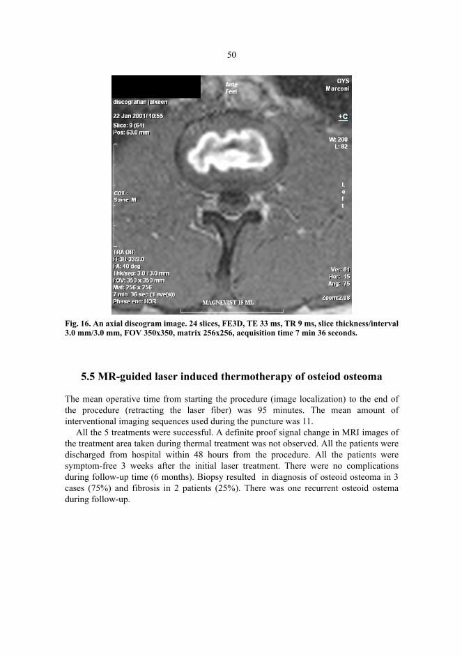
50
Fig. 16. An axial discogram image. 24 slices, FE3D, TE 33 ms, TR 9 ms, slice thickness/interval3.0 mm/3.0 mm, FOV 350x350, matrix 256x256, acquisition time 7 min 36 seconds.
5.5 MR-guided laser induced thermotherapy of osteiod osteoma
The mean operative time from starting the procedure (image localization) to the end ofthe procedure (retracting the laser fiber) was 95 minutes. The mean amount ofinterventional imaging sequences used during the puncture was 11.
All the 5 treatments were successful. A definite proof signal change in MRI images ofthe treatment area taken during thermal treatment was not observed. All the patients weredischarged from hospital within 48 hours from the procedure. All the patients weresymptom-free 3 weeks after the initial laser treatment. There were no complicationsduring follow-up time (6 months). Biopsy resulted in diagnosis of osteoid osteoma in 3cases (75%) and fibrosis in 2 patients (25%). There was one recurrent osteoid ostemaduring follow-up.

6 Discussion
6.1 Low field imaging & integrated optical instrument tracking
Open configuration MRI scanners possess several attributes that make them favorable asan interventional system compared to other imaging modalities or closed high-field MRIsystems. High field scanners put far greater demands on patient safety due to high B0.Patient access is also a problem.
An open configuration MRI device allows good access to the patient. Furthermore,there is seldom any need to move the patient during the operation. A low field scannercan typically be easily and quickly shut down facilitating fast conversion in procedures ifneeded. Supplementary and accessory technical hardware is easier to protect and use inlow- or mid-field surroundings. These facts are bound to affect patient safety.
When compared to conventional imaging modalities MR-guidance allows the freedomto choose any imaging plane. This facilitates the technical task of performinginterventional procedures at oblique angles. The optical tracking facilitates real-timecontrol of instrument position in relation to the image data and is easy to use. Combinedwith the operator controlled sequence start with foot pedal and fast imaging the systemincreases operator control, thus increasing the speed and accuracy of the procedure. Theadvantage of this was also shown in our studies, where high technical accuracy wasachieved almost without complications. The disadvantages of optical tracking are thetrackers’ need of line-of-sight and relative insensitivity to needle bending.
The most critical phase of performing minimally invasive procedures is the accurateand safe positioning of the needle. Dissimilar needle artifact sizes in different sequencesunder MR-guidance must be taken in account for (Butts et al. 1999). In general, needleartifact in MRI image is much more prominent in gradient echo images than in T1weighted FSE and T2 weighted FSE images. The needle thickness also affects artifactsize; the removal of mandrel diminishes the artifact size in MRI image. The needleposition in relation to the main magnetic field (B0) also has considerable effect. A moreperpendicular needle position with respect to B0 orientation produces more pronouncedneedle artifact. The needle path should be planned accordingly.
52
Needle bending due to needle material (titanium-alloys) must be taken into account, asthe correct position should be unambiguously ascertained. In practice this meansconfirmatory images where the needle artifact or injected contrast agent are clearlyvisible. In MR-guided nerve root infiltration needle bending can be easily controlled bythicker slice set if necessary.
With MR-guidance there is no irradiation burden on the patient or the physician, whichis advantageous in performing procedures on adolescents and potentially pregnantfemales but particularly important for physicians when considering the interventionalradiologist’s radiation burden general. It is also generally acknowledged thatinterventional procedures should be performed without ionizing radiation wheneverfeasible. The European Union also has established guidelines advocating the use of MRIinstead of modalities with ionizing radiation involved when the expected level ofinformation or therapeutic result is equal (European Commission 2001, and EuropeanUnion 1997).
Still, MRI interventions are not as easily conducted as fluoroscopic interventions. Interms of imaging speed, MRI is comparable to conventional CT but not to CTfluoroscopy. The slower imaging speed in MRI is not a problem if real-time opticaltracking is available. CT has superior axial resolution, and the ability to detectpneumothorax favors the use of CT in the thoracic region. However, in all of our series itwas found that image quality of operative images obtained with a 0.23 T scanner wassufficient to guarantee a safe procedure.
The superiority of MRI as a guidance modality is evident due to its good sensitivity,good visualization of the lesion and accuracy. The potential and actual excellent clinicalresults have established MRI as a guidance modality comparable to the others inperforming interventions.
MR-guidance is used successfully in many indications where the use of MRI has thepotential of yielding supplementary information on the target and its surroundings. MRIhas already emerged as a reliable tool for numerous interventional procedures (Adam etal. 1999, de Jode et al. 1999, Law & Regan 2000, Fiedler et al. 2001, Koenig et al. 2001,Vogl et al. 2001).
MR-guidance in interventions has several advantages when compared to traditionalmodalities. MRI is able to detect lesions not seen at all in other modalities. MRI also hassuperior soft tissue resolution, with or without contrast, even in interventional sequences.Also bone interventions can be reliably conducted (Koenig et al. 2001).
Despite all the inherent advantages of MRI in interventional use there are also somechallenges that must be met. All MR-guided interventions require special safety demandsto be fulfilled in order to ensure a safe procedure. All the equipment within the imagingarea must be MRI-safe, naturally including all the instrumentation. Instrument safety isone of the key issues in IMRI. At the moment, MRI-compatible instrumentation isrelatively highly priced and not so easy to come by. Although most of the majorinstrument manufacturers do have MRI compatible equipment to offer, none seems tohave a truly extensive coverage of the instruments needed at the field of IMRI. The initialhigh cost of the IMRI system is relative a burden, low-field MRI systems are comparableto CT devices in this respect. In can be stated, however, that these facts do not challengethe actual feasibility of MRI interventions. Further assessment of clinical data is howeveressential to establish cost effectiveness of IMRI procedures.
53
In this study the difference of modality specific procedural time between differentmodalities used for interventional procedures was not evaluated. It is to be noted that suchstudies are warranted.
6.2 Guidance method for MR-guided bone biopsy
Real-time imaging with MRI is often limited by the achievable signal-to-noise ratio andtissue contrast. Therefore, a modality-independent method for localizing the instrument inreal time is beneficial on less frequently updated images: an optical tracking systemaugments this localization by providing instantaneous feedback (Schenck et al. 1995,Silverman et al. 1995), while intraoperative MRI is used for generating updatedroadmaps along the needle path. Compared with passive tracking, where the needleartifact alone is used for deducing the instrument’s position and orientation (Lewin et al.1996), optical tracking offers distinct advantages. The initial needle orientation at thepuncture site can be determined with the aid of graphic tools and/or needle-guided scoutimages, whereas passive tracking requires more elaborate slice positioning and MR-visible markers. In this series, determination of the puncture site was possible with opticaltracking overlays in one or two MRI sets, and no skin markers were needed. Difficultiesin pinpointing the exact orientation of the needle from its artifact have adverse effects onthe overall accuracy of needle alignment, and the acquisition of affirmative imagesaccrues a time penalty. Other active guidance systems, such as ultrasound probes andmechanical arms (Barnett et al. 1993, Rohling et al. 1995) could also be used instead ofoptical systems, but they lack the flexibility of wireless instrument tracking.
Optical tracking has been successfully used in CT (Jacob et al. 2000) and MR-guidedneedle biopsies and injections (Silverman et al. 1995) as well as in surgical operations(Caversaccio et al. 1999, Vorbeck et al. 2000). Direct attachment of the needle to theoptical tracking system was beneficial in deep neural drug injections (Ojala et al. 2000).Bending of the needle makes confirmatory imaging more important in fine-needlebiopsies and injections than in procedures done with a stiff instrument, such as bonebiopsy which was applied in our study. In bone biopsy, optical tracking potentiallyreduces the need for imaging during the procedure and thus diminishes the proceduretime. A wireless solution for optical tracking was even more important in procedureswhere the instruments are heavy and clumsy, as it helped to avoid the extra disturbancecaused by handling the instrument by wires.
The MR-guidance system described here is a comparable method to conventional CT,and although CT-fluoroscopy can provide true real-time imaging its use is limitedbecause of ionizing radiation.
6.3 MR-guided bone biopsy
This study suggests that percutaneous MR-guided bone biopsy is safe and accurate.
54
Percutaneous or surgical biopsy is the method of choice for diagnosing bony lesions.The most common presentation of a bone lesion is that of a lytic or blastic lesion, and in apatient with a neoplastic history the diagnosis is straightforward. However, if there is noneoplastic history or such history dates back several years, a biopsy is certainly needed.Even if the lesion is presumably metastatic, it is sometimes useful to obtain a biopsy,particularly if there is no previous history of metastatic disease. Correct assessment ofinformation obtainable from lesions of infectious, malignant or benign origin is essentialfor the selection of effective treatment.
Due to its good sensitivity, MRI is able to detect lesions not seen at all in othermodalities. Particularly, lesions with edema, often seen only in MRI as a brightinvolvement of bone marrow in heavily T2 weighted sequences and STIR sequences canbe reliably visualized and readily biopsied (Hoane et al. 1991, Kaplan et al. 1998, Adamet al. 1999). MRI also provides superior tissue contrast without contrast medium. Thereare also results suggesting that the use of contrast medium may contribute to the accuracyof bone lesion detection in MRI. This is the case especially in recurrent malignancies andin certain types of cartilaginous lesion (Geirnaerdt et al. 1998). Contrast media has beenused effectively in MRI guided biopsies (Parkkola et al. 2001). However, in our study noadvantage in lesion visualization was found when contrast was used. It is to be noted thatour data did not include any primary malignancy of bone, soft tissue or cartilaginoustumors and this may reflect the poor result of contrast contribution in our study. In takingbiopsy from primary bone malignancies it is advisable to use contrast medium, as it islikely to increase the accuracy of biopsy by pointing out the enhancing dimensions oftumor (Parkkola et al. 2001). It is also important to know the exact anatomic proportionsof the tumor. In primary bone tumors, choosing the proper biopsy route is of utmostimportance since removal of the biopsy channel is mandatory during surgery.
CT has superior axial resolution, which favors the use of CT as a guidance modality inbone biopsies. However, in our series all the bone lesions detected preoperatively withCT, high-field MRI or scintigraphy were also seen in operative images obtained with a0.23 T scanner. Image quality was sufficient in the thoracic region to guarantee a safeprocedure. However, we had one conversion to a CT-guided procedure in a patient withthoracic vertebral body lesion. This was before the initialization of fast imagingsequences took place and at that time image quality was considered not sufficient to allowprocedure in the thoracic area. After fast imaging sequences were introduced into routineuse, no conversions were needed. The spatial resolution capability of MRI remains acontroversial issue, at least in the low-field scanners. Unless fast sequences withrelatively good resolution, such as CBASS/true-FISP, are used, the procedures targeted tothe areas containing fine bony structures should be done under CT-guidance. In thevertebral region the pedicles and cervical area must be considered as such structures.
General anesthesia was used in 6 patients and spinal anesthetics in 8 patients.Although bone biopsies have traditionally been made under local anesthesia, there is ageneral consensus in the Oulu University Hospital after 15 years of experience inpercutaneous bone biopsies that patients do feel more uncomfortable and more pain whenusing local anesthetics. It is felt that in a patient group where sacro-lumbar and lowerextremity region is biopsied, best patient compliance is achieved with spinal anesthetics.General anesthesia is used when performing bone biopsy in children and young patientsor patients with lesion in thoracic or head and neck area. Thoracic area biopsy is
55
controversial since optimal visualization in low field is acquired trough minimizingmotion-generated artifacts in image. Hence in the thoracic area the breathing motioncauses artifact. To minimize this motion during biopsy procedure in thoracic area breath-hold technique can be used. This is however only possible in local anesthesia since spinalanesthesia without ventilation cannot be safely applied when operating at the thoracicarea. The next choice is to use generated apnea under general anesthesia during imagingto achieve best image quality. To minimize patient motion and pain and to maximizeimage quality general anesthesia was used in thoracic area biopsies.
Complete statistical analysis was not made due to small number of patients.Despite the small number of patients, however, the trephine biopsy results of a success
rate of 92% are comparable to the results obtained in CT studies published on this topic.Tikkakoski et al. reported diagnostic success rate of 84% in their study which wasperformed in CT (Tikkakoski et al. 1992). Leffler et al reported an 82% positivepredictive accuracy of CT-guided percutaneous bone biopsy (Leffler & Chew 1999). Nget al. reported an initial success rate of 95% in their study (Ng et al. 1998).
In a previous MRI bone biopsy study, sub-optimal sample quality hindered diagnosticefficacy (Neuerburg et al. 1998a). This was due to the packing of the sample, whichresulted in destruction of the fine structure of the bone. There was no such experience inthe series discussed here, and the drill type used seems to provide good-quality samplesregardless of the density of the target tissue. There were no procedural complications inthis series of MR-guided percutaneous bone biopsies.
FNA has been shown to be informative when taken in conjunction with bone biopsy(Tikkakoski et al. 1992). The results of our study are however controversial, with onlynominal information advantage gained from performing FNA. The obvious advantage ofFNA diagnostics is the relative quickness when compared to bone histology, where thepreparation of the sample can take weeks, whereas cytological diagnosis can be obtainedwithin hours. It must be stated, however, that despite the less than optimal overallperformance, the cytological diagnosis concurred with histological diagnosis in all caseswith malignant histology. This result is probably due to general morphological changesassociated with the malignant disease in bone tissue: in malignant disease there is aprofound damage in bone structure caused by malignant infiltration, and thus it is easierto obtain cell material in FNA biopsy.
It is concluded that although the number of cases is limited in this study it seems thatMR-guided percutaneous bone biopsy is safe, accurate and feasible to perform. It seemsalso that no significant complications appear to be associated with the procedure. Fineneedle biopsy is advantageous when malignant disease is suspected. MRI can be used asthe sole modality or with optical tracking guidance in bone biopsies or as a backupmodality in performing procedures not possible otherwise. When available, MR-guidancecan be favored as a method of choice in performing percutaneous bone biopsies.
56
6.4 MR-guided periradicular therapy
In this study it was shown that MR-guided nerve root infiltration is safe and accurate.Fluoroscopy or CT-guided procedure is usually the method of choice for nerve rootinfiltration therapy in the lumbosacral area. Although fluoroscopy is cheap and easy toapply, it does have disadvantages of use in guiding interventions. Firstly, the proceduralsteering must be done according to bony anatomical landmarks and any variation orchange in the soft tissue morphology due to anatomical variation or pathology is notdetected. The use of contrast media does little to overcome this dilemma, since whencontrast is injected only the edges of adjacent structures to the needle are readilyvisualized. Secondly, there is radiation burden to the operator and to the patient.Although CT-guidance is far better in identifying anatomical structures at the procedurearea, it too has restrictions. Despite excellent temporal and spatial resolution CT is not sogood in differentiating various soft tissue structures from each other without specialwindowing or HU analysis. CT also shares the problem of radiation with fluoroscopy.
As an adjunct to optical tracking and direct visualization of the needle saline solutionwas used as contrast agent to visualize the nerve root sheath injected. This proved to be avery effective means of confirming the right placement of the needle. Gadoliniumcompounds with T1-weighted sequences have been used for this purpose (Seibel et al.1997). However, to this date these compounds have not been accepted for intrathecal useand using saline as contrast agent makes it possible to avoid the possible side-effects ofintrathecal or epidural gadolinium infiltration.
There was one patient in whom the worsening of the radicular pain was categorized asa minor complication due to the puncture. This was probably caused by nerve rootirritation from the puncture or either of the therapeutic substances used.
Low back disorders are extremely prevalent in all societies, and the rate of causeddisability, as well as the costs, have increased - independently of disease prevalence -over recent decades (Frymoyer & Cats-Baril 1991). It is obvious that we cannot solve thisdilemma with increasing surgical interventions. Minimally invasive interventionalprocedures are an option to relieve pain and minimize the risk of disability.
To the authors knowledge this is the first study where the usefulness of MR-guidancein lumbar nerve root infiltration is evaluated. In the referred studies (Seibel et al. 1997,Uhlenbrock & Arlinghaus 1997, Weiner & Fraser 1997, Viton et al. 1998) the outcome ofthe first sacral root and lumbar root infiltrations have been evaluated in fluoroscopy-, CT-and MR- guidance. Since 1995, over 1,400 nerve root infiltrations have been performedwith fluoroscopy guidance at Oulu University Hospital, and in our experience this methodallows the needle to be placed only into the dorsal root canal or dorsal root sheath of thelumbosacral nerve roots and the depth is difficult to estimate. MRI overcomes thisproblem.
In our study the length of the procedural time decreased gradually from 60 minutesreaching a minimum of 2 minutes. This can be seen as a learning curve development inimplementing the procedure.
Previous reports concerning the effect of nerve root infiltration therapy on radicularpain have been promising. In a study of CT-guided lumbar nerve root infiltrations(Uhlenbrock & Arlinghaus 1997), 55% of patients were free of symptoms or had hadsome improvement when evaluated approximately 4 months after the treatment; 30%

57
reported temporary improvement, and in 15% there was no change compared with thepre-treatment symptoms. In the recent study (Seibel et al. 1997) only 15% of the failedback surgery patients got relief to their symptoms after a single CT- or MR-guided nerveroot infiltration, and after four treatments 72% of the patients were symptom-free.
In the present study the patients with irritation after failed back surgery had anexcellent outcome from nerve root infiltration therapy, with almost 80% of initial painfree result. It has been reported that 75.4% of patients with radicular leg pain had asuccessful long-term outcome after lumbar transforaminal epidural steroid injection (Lutzet al. 1998). 22 of the 28 patients with lumbar disc herniation had considerable andsustained relief from their symptoms in another study (Weiner & Fraser 1997). Theoutcome was not evaluated separately for lumbar and sacral nerve roots in these studies.In our series the outcome of nerve root infiltration therapy was in accordance with theseother studies using fluoroscopy or CT- guidance (Uhlenbrock & Arlinghaus 1997, Weiner& Fraser 1997, Viton et al. 1998). A recent published controlled trial is in accordancewith these studies as well (Karppinen et al. 2001).
In our study the patients with normal to minor disc pathology and the patients withnerve root channel narrowing due to bony or scar involvement did not have such a goodoutcome from nerve root infiltration therapy. This is probably due to a more passivenature of underlying disease. In these patient groups the feasibility of the procedureremains controversial. It must be stated, however, that percutaneous nerve root infiltrationtherapy is an almost complication free, patient friendly procedure when compared tosurgery, it can very easily be repeated and can be mastered by any radiologist.
On the basis of the present study, MR-guided needle introduction is an accurateprocedure and may be used to substitute conventional techniques for nerve rootinfiltration. This would be especially useful in cases where there are substantialanatomical or structural changes due variation or pathology. It is concluded that MR-guided nerve root infiltration therapy is safe, accurate and feasible to perform. Nerve rootinfiltration therapy is effective treatment form to control radicular pain and can be used tosubstitute more invasive procedures in a selected patient group.
6.5 MR-guided discography
Percutaneous discography is a controversial procedure.Literature lists it used as a clinical tool in at least 8 indications (Tehranzadeh 1998,
Anderson & Flanagan 2000, Carragee 2000); Table 8.
58
Table 8. Discography indications.
Some of these indications have much argued grounds (Carragee 2000). However, theunifying element in all indications of discography is the low back or rather spinal areapain, which is usually conjoined to a situation where other tests or imaging studiesfindings are not sufficient to allow diagnosis and treatment. It must be noted that the useof discography to assess diagnostics on litigation basis has poor results (Carragee 2000).The most effective use of discography as a diagnostic tool is achieved when it is used forpre-surgical evaluation on patients forwarded for fusion operation (Vamvanij et al. 1998,Derby et al. 1999, Carragee 2000). All the patients included in this study were forwardedto the discography by orthopedic surgeon in order to determine disc pathology, whichwould affect possible prognosis and the extent of fusion operation.
In our study we wanted to find out whether MRI guided discography is feasible or not.In this study the technique for diagnostic MRI-guided discography in a low fieldsurroundings is presented. To the author’s knowledge this has not been reported before.MRI-discography has been reported in a high field system (Huang et al. 2002). In thestudy of Huang et al. it was concluded that MRI-discography could substituteconventional discography in selected indications. These indications would be allergy toionated contrast or patients whose radiation exposure must be limited.
The contrast used in our study was gadolinium, which was diluted to saline in a ratioof 1 to 8. Gadolininum contrast is considered very safe and has long been used inintravascular applications with a low complication rate. There are no reports of toxicproperties of gadolinium contrast towards any human tissue. Gadolinium contrast has notyet been approved for intrathecal use. In discography procedure there is a potential threatof intrathecal leagage of gadolinium. Gadolinium has been reported to have been used inanimals and humans intrathecally without complications (Jinkins et al. 1999, Zeng et al.1999). Having this information we proceeded with our study plan.
We had one complication reported in our study. The patient experienced continuouspain in the lumbar area after discography. The complication resulted in collapse of thedisc space. Infectious agents were not found in laboratory cultivation and the exactetiology of disc collapse remained unknown. Discitis and disc collapse are well-knowncomplications of discography procedure (Guyer et al. 1997, Junila et al. 1997) and in thisrespect, although unfortunate, it is concluded that the one complication does not challenge
List of most frequent indications
1. Negative MRI, CT or myelogram with equivocal findings for disc disease.
2. Positive multiple finding for disc disease in MRI, CT or myelogram. Discogram discerns symptomatic disc.
3. Presence of equivocal MRI, CT, or myelogram
4. Recurrent back pain in post surgical spine, to differentiate recurrent disc versus scar tissue.
5. To assess diagnostics between recurrent disc and pseudoarthrosis in post-surgical fused spine.
6. Preliminary test to spinal fusion to evaluate discs above or below the would-be fusion level.
7. Preliminary test for chemonucleolysis
8. Preliminary test prior to cortisone or anesthetic injection to disc.

59
the feasibility of MR-guidance in performing discography. In this respect the role ofprophylactic antibiotics in performing percutaneous discography cannot beunderestimated (Osti et al. 1990).
Diagnostic MRI has surpassed conventional x-ray fluoroscopy and CT- discography asan imaging tool to assess disk morphology. MRI is efficient in pinpointing the painsources of degenerative spine (Weishaupt et al. 2001). However, MRI also detectspathology in patients without symptoms. It is recognized that discography remains a goodtool in providing the surgeon with information of the pain source by pointing out the disclevel to be stabilized.
In our study we had a high procedural success rate showing MR-guidance to befeasible in discography. We had one procedural puncture failure and this puncture wastargeted to collapsed intervertebral disk space at L5-to-S1 level. Our results show thatpain response was adequately assessed in MR-guided puncture and injection, similarly tothe principles in conventional discography. Pain provocation from the contrast injectioncould be graded as concordant or non-concordant. We did not grade pain intensity. Theevaluation of concordant and non-concordant pain as well as pain intensity evaluation indiscography is highly debated (Carragee 2000). However, there are also reports in favorof it (Walsh et al. 1990). It is felt that assessment of pain concordance is a key proceduralelement of discography and there is no replacement for it in evaluating discogenic pain. Itmust be noted that patients’ emotional and chronic pain behavior has a strong affect onpain response in discography and such history in discography subject should be observed.
In our study the obtained MRI resolution of the injected contrast media appearing asan MRI discogram in intervertebral disc was less than that in conventional fluoroscopy.This could be due to diluted contrast and technical image formation factors in low fieldMRI or to the inherent physiological behavior of gadolininum contrast in vertebral diskwhere metabolism is distributed trough diffusion (Kerttula et al. 2000, Kurunlahti et al.2001). Further evaluation of these factors was however out of scope of this study.
In this study the technique of MR-guided discography in low field scanner waspresented and its feasibility discussed in interventional MRI and low field imagingsurroundings. It is concluded that although the study has its restrictions in the form of asmall amount of patients included in the study, it is shown that MR-guided discography isfeasible and comparable to conventional discography. The use of MRI as a guidancemethod for discography also enables simultaneous conventional MRI data acquisition,thus broadening the possible diagnostic spectrum of the procedure.
It is apparent that further research with larger patient groups is warranted to determinethe role of MR-guidance in discography.
6.6 MR-guided laser thermotherapy of osteiod osteoma
Osteioid osteoma is a benign lesion, that causes remarkably difficult symptoms andaffects mainly young individuals at the peak of their physical activity. This sum-up leadsto decreased level of physical activity, which can be very difficult both emotionally andphysically for these patients and their families. Effective treatment of this benign lesion ischallenging, not only because of the reasons mentioned above, but due to the fact that
60
osteiod ostema frequently affects proximal or distal ends of long bones situating itself inthe vicinity of chondral structures (Greenspan 1993). These lesions put great demands onoperative surgery, which is the treatment of choice today in osteoid osteoma. Withoutimage guidance the surgeon has to rely upon large amounts of regional bone tissue to beremoved to be sure of complete removal of the nidus. If the nidus is located in thevicinity of joint structures this leads to long lasting (3 to 4 weeks) post-operativerestrictions of stress and motor activity of the affected extremity, even if bone patch isused, placing further demands on the patient’s endurance (Campanacci et al. 1999). Theuse of open surgical approach may also increase the risk of infectious operationalcomplication.
Although percutaneous laser thermotherapy is considered a mini-invasive procedure,prophylactic antibiotics were used in our study. This was done with knowledge of theadvantages of antibiotic prophylaxis in open orthopedic surgery (Norden 1991, Oishi etal. 1993, Mazza 2000). Infection complication associated with the percutaneous removalof osteoid osteoma has also been reported (Sans et al. 1999). The risk of an infectionassociated with the procedure was to be minimized.
Percutaneous interstitial laser treatment has been reported as an effective treatment forosteiod osteomas (Gangi et al. 1997a). With laser treatment tumor nidus is destroyedthrough heating. A defined volume of predictable size and shape can be obtained (Gangiet al. 1997c). This leaves the affected bone tissue volume significantly smaller than insurgical treatment without compromising the treatment effect. The smaller destructionarea leads to little or no restrictions in extremity stress (max. one week) and thus shortensoverall recovery time. Percutaneous approach is minimally invasive, and also diminishingdamage to the skin and subcutaneous tissue, further favoring rapid recovery. With thermaldestruction there is no need to support the destructed area since the damage to the corticalstructures is usually minimal and even if coagulated, the destructed bone tissue has afilling effect on the area treated. This means that the diminishing effect of the treatment tothe structural support delivered by the treated bone tissue is almost insignificant. Ourresults are in conclusion with earlier reports on interstitial laser treatment efficiency intreating osteid osteomas thus supporting the advantages of the percutaneous treatmentapproach over the surgical one.
Different therapeutic methods of osteoid osteoma and their results are demonstrated inTable 9.
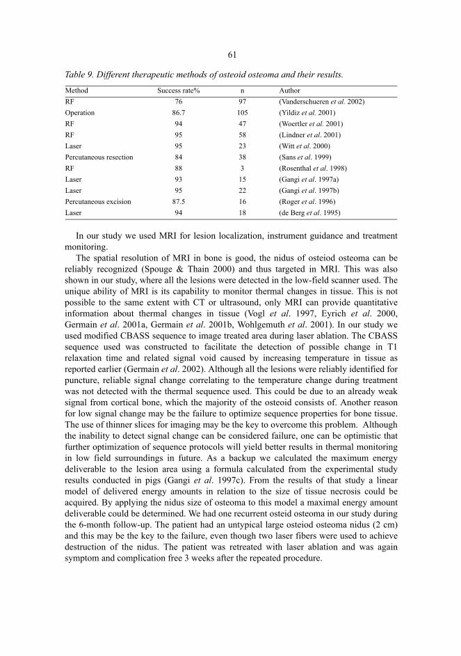
61
Table 9. Different therapeutic methods of osteoid osteoma and their results.
In our study we used MRI for lesion localization, instrument guidance and treatmentmonitoring.
The spatial resolution of MRI in bone is good, the nidus of osteiod osteoma can bereliably recognized (Spouge & Thain 2000) and thus targeted in MRI. This was alsoshown in our study, where all the lesions were detected in the low-field scanner used. Theunique ability of MRI is its capability to monitor thermal changes in tissue. This is notpossible to the same extent with CT or ultrasound, only MRI can provide quantitativeinformation about thermal changes in tissue (Vogl et al. 1997, Eyrich et al. 2000,Germain et al. 2001a, Germain et al. 2001b, Wohlgemuth et al. 2001). In our study weused modified CBASS sequence to image treated area during laser ablation. The CBASSsequence used was constructed to facilitate the detection of possible change in T1relaxation time and related signal void caused by increasing temperature in tissue asreported earlier (Germain et al. 2002). Although all the lesions were reliably identified forpuncture, reliable signal change correlating to the temperature change during treatmentwas not detected with the thermal sequence used. This could be due to an already weaksignal from cortical bone, which the majority of the osteoid consists of. Another reasonfor low signal change may be the failure to optimize sequence properties for bone tissue.The use of thinner slices for imaging may be the key to overcome this problem. Althoughthe inability to detect signal change can be considered failure, one can be optimistic thatfurther optimization of sequence protocols will yield better results in thermal monitoringin low field surroundings in future. As a backup we calculated the maximum energydeliverable to the lesion area using a formula calculated from the experimental studyresults conducted in pigs (Gangi et al. 1997c). From the results of that study a linearmodel of delivered energy amounts in relation to the size of tissue necrosis could beacquired. By applying the nidus size of osteoma to this model a maximal energy amountdeliverable could be determined. We had one recurrent osteid osteoma in our study duringthe 6-month follow-up. The patient had an untypical large osteiod osteoma nidus (2 cm)and this may be the key to the failure, even though two laser fibers were used to achievedestruction of the nidus. The patient was retreated with laser ablation and was againsymptom and complication free 3 weeks after the repeated procedure.
Method Success rate% n AuthorRF 76 97 (Vanderschueren et al. 2002)Operation 86.7 105 (Yildiz et al. 2001)RF 94 47 (Woertler et al. 2001)RF 95 58 (Lindner et al. 2001)Laser 95 23 (Witt et al. 2000)Percutaneous resection 84 38 (Sans et al. 1999)RF 88 3 (Rosenthal et al. 1998)Laser 93 15 (Gangi et al. 1997a)Laser 95 22 (Gangi et al. 1997b)Percutaneous excision 87.5 16 (Roger et al. 1996)Laser 94 18 (de Berg et al. 1995)
62
Although there was a recurrent osteoid osteoma in our patient data, this studydemonstrates the preliminary feasibility of MRI in guiding percutaneous interstitial laserablation treatment of osteoid osteomas. MRI can be used as a solo guidance modality forthis procedure and is in this respect comparable to CT.
7 Conclusions
1. The optical guidance method in conjunction with MR-guidance in bone biopsiesprovides precise and safe instrument control during MR-guided procedure at 0.23 T.
2. MR-guidance in performing bone biopsies is clinically viable in various anatomicalareas at 0.23 T.
3. MR-guided nerve root therapies are safe and the same therapeutic effect as withconventional guidance methods can be achieved under MR- guidance at 0.23T.
4. MR-guided discography can be adequately performed at 0.23T.5. Thermal laser therapy in treating osteoid osteoma is feasible under MR-guidance at
0.23 T.

8 References
Adam G, Bucker A, Nolte-Ernsting C, Tacke J & Gunther RW (1999) Interventional MR imaging:percutaneous abdominal and skeletal biopsies and drainages of the abdomen. Eur Radiol 9(8):1471-8.
Anderson SR & Flanagan B (2000) Discography. Curr Rev Pain 4(5): 345-52.Arthur WR, Dhawan J, Norell MS, Hunter AJ & Clark AL (2002) Does cardiologist- or radiographer-
operated fluoroscopy and image acquisition influence optimization of patient radiation exposureduring routine coronary angiography? Br J Radiol 75(897): 748-53.
Assoun J, Richardi G, Railhac JJ, Baunin C, Fajadet P, Giron J, Maquin P, Haddad J & BonnevialleP (1994) Osteoid osteoma: MR imaging versus CT. Radiology 191(1): 217-23.
Bakker CJ, Hoogeveen RM, Weber J, van Vaals JJ, Viergever MA & Mali WP (1996) Visualizationof dedicated catheters using fast scanning techniques with potential for MR-guided vascularinterventions. Magn Reson Med 36(6): 816-20.
Barei DP, Moreau G, Scarborough MT & Neel MD (2000) Percutaneous radiofrequency ablation ofosteoid osteoma. Clin Orthop(373): 115-24.
Barnett GH, Kormos DW, Steiner CP & Weisenberger J (1993) Use of a frameless, armlessstereotactic wand for brain tumor localization with two-dimensional and three-dimensionalneuroimaging. Neurosurgery 33(4): 674-8.
Boden SD, Davis DO, Dina TS, Patronas NJ & Wiesel SW (1990) Abnormal magnetic-resonancescans of the lumbar spine in asymptomatic subjects. A prospective investigation. J Bone JointSurg Am 72(3): 403-8.
Buecker A, Adam G, Neuerburg JM, Glowinski A, van Vaals JJ & Guenther RW (1998) MR-guidedbiopsy using a T2-weighted single-shot zoom imaging sequence (Local Look technique). J MagnReson Imaging 8(4): 955-9.
Buecker A, Neuerburg JM, Adam GB, Nolte-Ernsting CC, Hunter DW, Glowinski A, van Vaals JJ& Guenther RW (2001) MR-guided percutaneous drainage of abdominal fluid collections incombination with X-ray fluoroscopy: initial clinical experience. Eur Radiol 11(4): 670-4.
Busch M, Bornstedt A, Wendt M, Duerk JL, Lewin JS & Gronemeyer D (1998) Fast "real time"imaging with different k-space update strategies for interventional procedures. J Magn ResonImaging 8(4): 944-54.
Butts K, Pauly JM, Daniel BL, Kee S & Norbash AM (1999) Management of biopsy needle artifacts:techniques for RF-refocused MRI. J Magn Reson Imaging 9(4): 586-95.
Campanacci M, Ruggieri P, Gasbarrini A, Ferraro A & Campanacci L (1999) Osteoid osteoma.Direct visual identification and intralesional excision of the nidus with minimal removal of bone.J Bone Joint Surg Br 81(5): 814-20.
65
Carragee EJ (2000) Is lumbar discography a determinate of discogenic low back pain: provocativediscography reconsidered. Curr Rev Pain 4(4): 301-8.
Caversaccio M, Bachler R, Ladrach K, Schroth G, Nolte LP & Hausler R (1999) The "Bernese"frameless optical computer aided surgery system. Comput Aided Surg 4(6): 328-34.
Cerase A & Priolo F (1998) Skeletal benign bone-forming lesions. Eur J Radiol 27(Suppl 1): S91-7.Cline HE, Schenck JF, Hynynen K, Watkins RD, Souza SP & Jolesz FA (1992) MR-guided focused
ultrasound surgery. J Comput Assist Tomogr 16(6): 956-65.Cove JA, Taminiau AH, Obermann WR & Vanderschueren GM (2000) Osteoid osteoma of the spine
treated with percutaneous computed tomography-guided thermocoagulation. Spine 25(10): 1283-6.
de Berg JC, Pattynama PM, Obermann WR, Bode PJ, Vielvoye GJ & Taminiau AH (1995)Percutaneous computed-tomography-guided thermocoagulation for osteoid osteomas. Lancet346(8971): 350-1.
de Jode MG, Vale JA & Gedroyc WM (1999) MR-guided laser thermoablation of inoperable renaltumors in an open-configuration interventional MR scanner: preliminary clinical experience inthree cases. J Magn Reson Imaging 10(4): 545-9.
Derby R, Howard MW, Grant JM, Lettice JJ, Van Peteghem PK & Ryan DP (1999) The ability ofpressure-controlled discography to predict surgical and nonsurgical outcomes. Spine 24(4): 364-71; discussion 371-2.
Derby R, Kine G, Saal JA, Reynolds J, Goldthwaite N, White AH, Hsu K & Zucherman J (1992)Response to steroid and duration of radicular pain as predictors of surgical outcome. Spine 17(6Suppl): S176-83.
Dick EA, Taylor-Robinson SD, Thomas HC & Gedroyc WM (2002) Ablative therapy for livertumours. Gut 50(5): 733-9.
Dotter CT & Judkins MP (1964) Transluminar treatment of arteriosclerotic obstuctions: descriptionsof a new technique and a preliminary report of its application. Circulation 30: 654-77.
Duckwiler G, Lufkin RB & Hanafee WN (1989) MR-directed needle biopsies. Radiol Clin North Am27(2): 255-63.
Duerk JL, Lewin JS, Wendt M & Petersilge C (1998) Remember true FISP? A high SNR, near 1-second imaging method for T2-like contrast in interventional MRI at .2 T. J Magn Reson Imaging8(1): 203-8.
Duerk JL, Lewin JS & Wu DH (1996) Application of keyhole imaging to interventional MRI: asimulation study to predict sequence requirements. J Magn Reson Imaging 6(6): 918-24.
Ehnholm GJ, Vahala ET, Kinnunen J, Nieminen JE, Standertskjold-Nordenstam C & Uusitalo MA(1999) Electron spin resonance (ESR) probe for interventional MRI instrument localization. JMagn Reson Imaging 10(2): 216-9.
Eyrich GK, Bruder E, Hilfiker P, Dubno B, Quick HH, Patak MA, Gratz KW & Sailer HF (2000)Temperature mapping of magnetic resonance-guided laser interstitial thermal therapy (LITT) inlymphangiomas of the head and neck. Lasers Surg Med 26(5): 467-76.
Fiedler VU, Schwarzmaier HJ, Eickmeyer F, Muller FP, Schoepp C & Verreet PR (2001) Laser-induced interstitial thermotherapy of liver metastases in an interventional 0.5 Tesla MRI system:technique and first clinical experiences. J Magn Reson Imaging 13(5): 729-37.
Fraser-Hill MA, Renfrew DL & Hilsenrath PE (1992) Percutaneous needle biopsy of musculoskeletallesions. 2. Cost-effectiveness. AJR Am J Roentgenol 158(4): 813-8.
Frymoyer JW & Cats-Baril WL (1991) An overview of the incidences and costs of low back pain.Orthop Clin North Am 22(2): 263-71.
Gangi A, Dietemann JL, Gasser B, Mortazavi R, Brunner P, Mourou MY, Dosch JC, Durckel J,Marescaux J & Roy C (1997a) Interstitial laser photocoagulation of osteoid osteomas with use ofCT guidance. Radiology 203(3): 843-8.
66
Gangi A, Dietemann JL, Gasser B, Mortazavi R, Dosch JC, Dupuis M, Durckel J & Roy C (1997b)Percutaneous Laser Photocoagulation of Osteoid Osteomas. Semin Musculoskelet Radiol 1(2):273-280.
Gangi A, Gasser B, De Unamuno S, Fogarrassy E, Fuchs C, Siffert P, Dietemann JL & Roy C (1997c)New Trends in Interstitial Laser Photocoagulation of Bones. Semin Musculoskelet Radiol 1(2):331-338.
Geirnaerdt MJ, Bloem JL, van der Woude HJ, Taminiau AH, Nooy MA & Hogendoorn PC (1998)Chondroblastic osteosarcoma: characterisation by gadolinium-enhanced MR imaging correlatedwith histopathology. Skeletal Radiol 27(3): 145-53.
Genant JW, Vandevenne JE, Bergman AG, Beaulieu CF, Kee ST, Norbash AM & Lang P (2002)Interventional musculoskeletal procedures performed by using MR imaging guidance with avertically open MR unit: assessment of techniques and applicability. Radiology 223(1): 127-36.
Germain D, Chevallier P, Laurent A & Saint-Jalmes H (2001a) MR monitoring of tumour thermaltherapy. Magma 13(1): 47-59.
Germain D, Chevallier P, Laurent A, Savart M, Wassef M & Saint-Jalmes H (2001b) MR monitoringof laser-induced lesions of the liver in vivo in a low-field open magnet: temperature mapping andlesion size prediction. J Magn Reson Imaging 13(1): 42-9.
Germain D, Vahala E, Ehnholm GJ, Vaara T, Ylihautala M, Savart M, Laurent A, Tanttu J & Saint-Jalmes H (2002) MR temperature measurement in liver tissue at 0.23 T with a steady-state freeprecession sequence. Magn Reson Med 47(5): 940-7.
Glowinski A, Kursch J, Adam G, Bucker A, Noll TG & Gunther RW (1998) Device visualization forinterventional MRI using local magnetic fields: basic theory and its application to cathetervisualization. IEEE Trans Med Imaging 17(5): 786-93.
Greenspan A (1993) Benign bone-forming lesions: osteoma, osteoid osteoma, and osteoblastoma.Clinical, imaging, pathologic, and differential considerations. Skeletal Radiol 22(7): 485-500.
Gronemeyer DH, Schirp S & Gevargez A (2002) Image-guided radiofrequency ablation of spinaltumors: preliminary experience with an expandable array electrode. Cancer J 8(1): 33-9.
Gronemeyer DH, Seibel RM & Kaufman L (1991) Low-field design eases MRI-guided biopsies.Diagn Imaging (San Franc) 13(3): 139-43.
Gronvall J, Gronvall S & Hegedus V (1977) Ultrasound-guided drainage of fluid-containing massesusing angiographic catheterization techniques. AJR Am J Roentgenol 129(6): 997-1002.
Guyer RD, Ohnmeiss DD, Mason SL & Shelokov AP (1997) Complications of cervical discography:findings in a large series. J Spinal Disord 10(2): 95-101.
Haschek E & Lindenthal O (1896) A contribution to the practical use of the photography accordingto Röntgen. In: Stanley Baum, editor, Abrams' Angiography. Boston: Little, Brown andCompany; 1997, p 3.
Heid O, Deimling M & Huk WJ (1995) Ultra-rapid gradient echo imaging. Magn Reson Med 33(1):143-9.
Heywang-Kobrunner SH, Heinig A, Schaumloffel U, Viehweg P, Buchmann J, Lampe D &Spielmann R (1999) MR-guided percutaneous excisional and incisional biopsy of breast lesions.Eur Radiol 9(8): 1656-65.
Hinks RS, Bronskill MJ, Kucharczyk W, Bernstein M, Collick BD & Henkelman RM (1998) MRsystems for image-guided therapy. J Magn Reson Imaging 8(1): 19-25.
Hoane BR, Shields AF, Porter BA & Shulman HM (1991) Detection of lymphomatous bone marrowinvolvement with magnetic resonance imaging. Blood 78(3): 728-38.
Holm HH, Kristensen JK, Rasmussen SN, Northeved A & Barlebo H (1972) Ultrasound as a guidein percutaneous puncture technique. Ultrasonics 10(2): 83-6.
Huang TS, Zucherman JF, Hsu KY, Shapiro M, Lentz D & Gartland J (2002) Gadopentetatedimeglumine as an intradiscal contrast agent. Spine 27(8): 839-43.
67
Huppert PE, Trubenbach J, Schick F, Pereira P, Konig C & Claussen CD (2000) [MRI-guidedpercutaneous radiofrequency ablation of hepatic neoplasms--first technical and clinicalexperiences]. Rofo Fortschr Geb Rontgenstr Neuen Bildgeb Verfahr 172(8): 692-700.
Hynynen K, Pomeroy O, Smith DN, Huber PE, McDannold NJ, Kettenbach J, Baum J, Singer S &Jolesz FA (2001) MR imaging-guided focused ultrasound surgery of fibroadenomas in the breast:a feasibility study. Radiology 219(1): 176-85.
Jacob AL, Messmer P, Kaim A, Suhm N, Regazzoni P & Baumann B (2000) A whole-bodyregistration-free navigation system for image-guided surgery and interventional radiology. InvestRadiol 35(5): 279-88.
Jager L & Reiser M (2001) CT and MR imaging of the normal and pathologic conditions of the facialnerve. Eur J Radiol 40(2): 133-46.
Jelinek JS, Kransdorf MJ, Gray R, Aboulafia AJ & Malawer MM (1996) Percutaneous transpedicularbiopsy of vertebral body lesions. Spine 21(17): 2035-40.
Jenkins LT, Millikan KW, Bines SD, Staren ED & Doolas A (1997) Hepatic resection for metastaticcolorectal cancer. Am Surg 63(7): 605-10.
Jensen MC, Brant-Zawadzki MN, Obuchowski N, Modic MT, Malkasian D & Ross JS (1994)Magnetic resonance imaging of the lumbar spine in people without back pain. N Engl J Med331(2): 69-73.
Jinkins JR, Williams RF & Xiong L (1999) Evaluation of gadopentetate dimeglumine magneticresonance cisternography in an animal model: preliminary report. Invest Radiol 34(2): 156-9.
Joensuu RP, Sepponen RE, Lamminen AE & Standertskjold-Nordenstam CG (2000) A shieldedOverhauser marker for MR tracking of interventional devices. Magn Reson Med 43(1): 139-45.
Jolesz FA (1998) Interventional and intraoperative MRI: a general overview of the field. J MagnReson Imaging 8(1): 3-7.
Jolesz FA & Blumenfeld SM (1994) Interventional use of magnetic resonance imaging. Magn ResonQ 10(2): 85-96.
Jolesz FA, Hynynen K, Pomeroy O, Smith DN, Huber PE, McDannold NJ, Kettenbach J, Baum J &Singer S (2002) Magnetic resonance image-guided focused ultrasound surgery
MR imaging-guided focused ultrasound surgery of fibroadenomas in the breast: a feasibility study.Cancer J 8(Suppl 1): S100-12.
Junila J, Niinimaki T & Tervonen O (1997) Epidural abscess after lumbar discography. A case report.Spine 22(18): 2191-3.
Jyrkinen L, Ojala R, Haataja L, Blanco Sequeiros R, Klemola R, Silven O & Tervonen O (2000)Managing the Complexity of the User Interface of a MRI Guided Surgery System. In: Proceedingsof the 6th ERCIM Workshop "user interfaces for All", Florence: 213-9.
Kaplan GR, Saifuddin A, Pringle JA, Noordeen MH & Mehta MH (1998) Langerhans' cellhistiocytosis of the spine: use of MRI in guiding biopsy. Skeletal Radiol 27(12): 673-6.
Karppinen J, Malmivaara A, Kurunlahti M, Kyllonen E, Pienimaki T, Nieminen P, Ohinmaa A,Tervonen O & Vanharanta H (2001) Periradicular infiltration for sciatica: a randomized controlledtrial. Spine 26(9): 1059-67.
Kerttula LI, Jauhiainen JP, Tervonen O, Suramo IJ, Koivula A & Oikarinen JT (2000) Apparentdiffusion coefficient in thoracolumbar intervertebral discs of healthy young volunteers. J MagnReson Imaging 12(2): 255-60.
Kettenbach J, Kacher DF, Koskinen SK, Silverman SG, Nabavi A, Gering D, Tempany CM,Schwartz RB, Kikinis R, Black PM & Jolesz FA (2000) Interventional and intraoperativemagnetic resonance imaging. Annu Rev Biomed Eng 2: 661-90.
Koenig CW, Duda SH, Truebenbach J, Schott UG, Maurer F, Claussen CD & Pereira PL (2001) MR-guided biopsy of musculoskeletal lesions in a low-field system. J Magn Reson Imaging 13(5):761-8.

68
Konings MK, Bartels LW, van Swol CF & Bakker CJ (2001) Development of an MR-safe trackingcatheter with a laser-driven tip coil. J Magn Reson Imaging 13(1): 131-5.
Kransdorf MJ, Stull MA, Gilkey FW & Moser RP, Jr. (1991) Osteoid osteoma. Radiographics 11(4):671-96.
Krempen JF, Smith BS & DeFreest LJ (1975) Selective nerve root infiltration for the evaluation ofsciatica. Orthop Clin North Am 6(1): 311-5.
Kurunlahti M, Kerttula L, Jauhiainen J, Karppinen J & Tervonen O (2001) Correlation of diffusionin lumbar intervertebral disks with occlusion of lumbar arteries: a study in adult volunteers.Radiology 221(3): 779-86.
Ladd ME, Zimmermann GG, Quick HH, Debatin JF, Boesiger P, von Schulthess GK & McKinnonGC (1998) Active MR visualization of a vascular guidewire in vivo. J Magn Reson Imaging 8(1):220-5.
Laufer U, Kirchner J, Kickuth R, Adams S, Jendreck M & Liermann D (2001) A comparative studyof CT fluoroscopy combined with fluoroscopy versus fluoroscopy alone for percutaneoustranshepatic biliary drainage. Cardiovasc Intervent Radiol 24(4): 240-4.
Law P & Regan L (2000) Interstitial thermo-ablation under MRI guidance for the treatment offibroids. Curr Opin Obstet Gynecol 12(4): 277-82.
Leffler SG & Chew FS (1999) CT-guided percutaneous biopsy of sclerotic bone lesions: diagnosticyield and accuracy. AJR Am J Roentgenol 172(5): 1389-92.
Lewin JS, Duerk JL, Jain VR, Petersilge CA, Chao CP & Haaga JR (1996) Needle localization inMR-guided biopsy and aspiration: effects of field strength, sequence design, and magnetic fieldorientation. AJR Am J Roentgenol 166(6): 1337-45.
Lewin JS, Petersilge CA, Hatem SF, Duerk JL, Lenz G, Clampitt ME, Williams ML, Kaczynski KR,Lanzieri CF, Wise AL & Haaga JR (1998) Interactive MR imaging-guided biopsy and aspirationwith a modified clinical C-arm system. AJR Am J Roentgenol 170(6): 1593-601.
Lindner NJ, Ozaki T, Roedl R, Gosheger G, Winkelmann W & Wortler K (2001) Percutaneousradiofrequency ablation in osteoid osteoma. J Bone Joint Surg Br 83(3): 391-6.
Livraghi T (2001) Percutaneous ethanol injection in the treatment of hepatocellular carcinoma incirrhosis. Hepatogastroenterology 48(37): 20-4.
Livraghi T, Goldberg SN, Lazzaroni S, Meloni F, Ierace T, Solbiati L & Gazelle GS (2000)Hepatocellular carcinoma: radio-frequency ablation of medium and large lesions. Radiology214(3): 761-8.
Lufkin R, Teresi L, Chiu L & Hanafee W (1988) A technique for MR-guided needle placement. AJRAm J Roentgenol 151(1): 193-6.
Lufkin R, Teresi L & Hanafee W (1987) New needle for MR-guided aspiration cytology of the headand neck. AJR Am J Roentgenol 149(2): 380-2.
Lutz GE, Vad VB & Wisneski RJ (1998) Fluoroscopic transforaminal lumbar epidural steroids: anoutcome study. Arch Phys Med Rehabil 79(11): 1362-6.
Mala T, Edwin B, Samset E, Gladhaug I, Hol PK, Fosse E, Mathisen O, Bergan A & Soreide O (2001)Magnetic-resonance-guided percutaneous cryoablation of hepatic tumours. Eur J Surg 167(8):610-7.
Mankin HJ, Lange TA & Spanier SS (1982) The hazards of biopsy in patients with malignant primarybone and soft-tissue tumors. J Bone Joint Surg [Am] 64(8): 1121-7.
Martino CR, Haaga JR, Bryan PJ, LiPuma JP, El Yousef SJ & Alfidi RJ (1984) CT-guided liverbiopsies: eight years' experience. Work in progress. Radiology 152(3): 755-7.
Matsumoto R, Selig AM, Colucci VM & Jolesz FA (1992) Interstitial Nd:YAG laser ablation innormal rabbit liver: trial to maximize the size of laser-induced lesions. Lasers Surg Med 12(6):650-8.
Mazza A (2000) Ceftriaxone as short-term antibiotic prophylaxis in orthopedic surgery: a cost-benefitanalysis involving 808 patients. J Chemother 12 Suppl 3: 29-33.
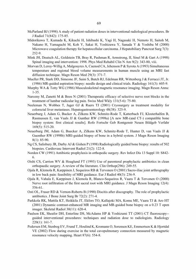
69
McParland BJ (1998) A study of patient radiation doses in interventional radiological procedures. BrJ Radiol 71(842): 175-85.
Midorikawa T, Kumada K, Kikuchi H, Ishibashi K, Yagi H, Nagasaki H, Nemoto H, Saitoh M,Nakano H, Yamaguchi M, Koh Y, Sakai H, Yoshizawa Y, Sanada Y & Yoshiba M (2000)Microwave coagulation therapy for hepatocellular carcinoma. J Hepatobiliary Pancreat Surg 7(3):252-9.
Mink JH, Deutsch AL, Goldstein TB, Bray R, Pashman R, Armstrong, II, Sinel M & Gart A (1998)Spinal imaging and intervention: 1998. Phys Med Rehabil Clin N Am 9(2): 343-80, viii.
Morvan D, Leroy-Willig A, Malgouyres A, Cuenod CA, Jehenson P & Syrota A (1993) Simultaneoustemperature and regional blood volume measurements in human muscle using an MRI fastdiffusion technique. Magn Reson Med 29(3): 371-7.
Mueller PR, Stark DD, Simeone JF, Saini S, Butch RJ, Edelman RR, Wittenberg J & Ferrucci JT, Jr.(1986) MR-guided aspiration biopsy: needle design and clinical trials. Radiology 161(3): 605-9.
Murphy WA & Totty WG (1986) Musculoskeletal magnetic resonance imaging. Magn Reson Annu:1-35.
Narozny M, Zanetti M & Boos N (2001) Therapeutic efficacy of selective nerve root blocks in thetreatment of lumbar radicular leg pain. Swiss Med Wkly 131(5-6): 75-80.
Neeleman N, Wobbes T, Jager GJ & Ruers TJ (2001) Cryosurgery as treatment modality forcolorectal liver metastases. Hepatogastroenterology 48(38): 325-9.
Neuerburg J, Adam G, Bucker A, Zilkens KW, Schmitz-Rode T, Katterbach FJ, Klosterhalfen B,Rasmussen E, van Vaals JJ & Gunther RW (1998a) [A new MR-(and CT-) compatible bonebiopsy system: first clinical results]. Rofo Fortschr Geb Rontgenstr Neuen Bildgeb Verfahr169(5): 515-20.
Neuerburg JM, Adam G, Buecker A, Zilkens KW, Schmitz-Rode T, Hunter D, van Vaals JJ &Guenther RW (1998b) MRI-guided biopsy of bone in a hybrid system. J Magn Reson Imaging8(1): 85-90.
Ng CS, Salisbury JR, Darby AJ & Gishen P (1998) Radiologically guided bone biopsy: results of 502biopsies. Cardiovasc Intervent Radiol 21(2): 122-8.
Norden CW (1991) Antibiotic prophylaxis in orthopedic surgery. Rev Infect Dis 13 Suppl 10: S842-6.
Oishi CS, Carrion WV & Hoaglund FT (1993) Use of parenteral prophylactic antibiotics in cleanorthopaedic surgery. A review of the literature. Clin Orthop(296): 249-55.
Ojala R, Klemola R, Karppinen J, Sequeiros RB & Tervonen O (2001) Sacro-iliac joint arthrographyin low back pain: feasibility of MRI guidance. Eur J Radiol 40(3): 236-9.
Ojala R, Vahala E, Karppinen J, Klemola R, Blanco-Sequeiros R, Vaara T & Tervonen O (2000)Nerve root infiltration of the first sacral root with MRI guidance. J Magn Reson Imaging 12(4):556-61.
Osti OL, Fraser RD & Vernon-Roberts B (1990) Discitis after discography. The role of prophylacticantibiotics. J Bone Joint Surg Br 72(2): 271-4.
Parkkola RK, Mattila KT, Heikkila JT, Ekfors TO, Kallajoki MA, Komu ME, Vaara TJ & Aro HT(2001) Dynamic contrast-enhanced MR imaging and MR-guided bone biopsy on a 0.23 T openimager. Skeletal Radiol 30(11): 620-4.
Paulson EK, Sheafor DH, Enterline DS, McAdams HP & Yoshizumi TT (2001) CT fluoroscopy--guided interventional procedures: techniques and radiation dose to radiologists. Radiology220(1): 161-7.
Pedersen EM, Stenbog EV, Frund T, Houlind K, Kromann O, Sorensen KE, Emmertsen K & HjortdalVE (2002) Flow during exercise in the total cavopulmonary connection measured by magneticresonance velocity mapping. Heart 87(6): 554-8.
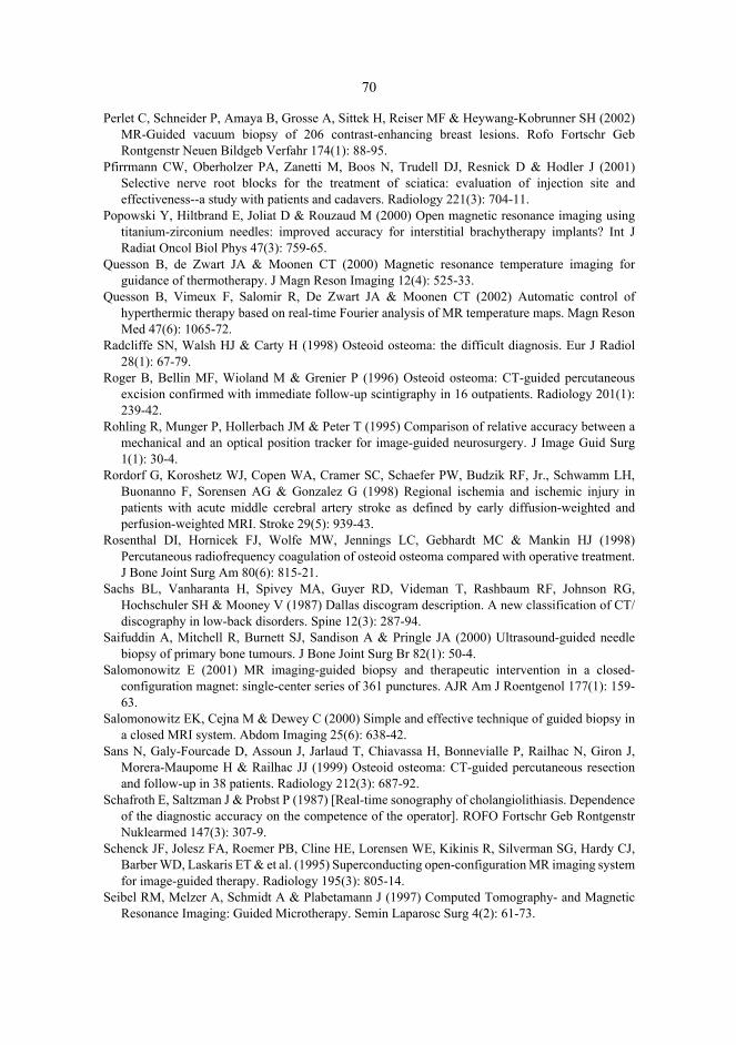
70
Perlet C, Schneider P, Amaya B, Grosse A, Sittek H, Reiser MF & Heywang-Kobrunner SH (2002)MR-Guided vacuum biopsy of 206 contrast-enhancing breast lesions. Rofo Fortschr GebRontgenstr Neuen Bildgeb Verfahr 174(1): 88-95.
Pfirrmann CW, Oberholzer PA, Zanetti M, Boos N, Trudell DJ, Resnick D & Hodler J (2001)Selective nerve root blocks for the treatment of sciatica: evaluation of injection site andeffectiveness--a study with patients and cadavers. Radiology 221(3): 704-11.
Popowski Y, Hiltbrand E, Joliat D & Rouzaud M (2000) Open magnetic resonance imaging usingtitanium-zirconium needles: improved accuracy for interstitial brachytherapy implants? Int JRadiat Oncol Biol Phys 47(3): 759-65.
Quesson B, de Zwart JA & Moonen CT (2000) Magnetic resonance temperature imaging forguidance of thermotherapy. J Magn Reson Imaging 12(4): 525-33.
Quesson B, Vimeux F, Salomir R, De Zwart JA & Moonen CT (2002) Automatic control ofhyperthermic therapy based on real-time Fourier analysis of MR temperature maps. Magn ResonMed 47(6): 1065-72.
Radcliffe SN, Walsh HJ & Carty H (1998) Osteoid osteoma: the difficult diagnosis. Eur J Radiol28(1): 67-79.
Roger B, Bellin MF, Wioland M & Grenier P (1996) Osteoid osteoma: CT-guided percutaneousexcision confirmed with immediate follow-up scintigraphy in 16 outpatients. Radiology 201(1):239-42.
Rohling R, Munger P, Hollerbach JM & Peter T (1995) Comparison of relative accuracy between amechanical and an optical position tracker for image-guided neurosurgery. J Image Guid Surg1(1): 30-4.
Rordorf G, Koroshetz WJ, Copen WA, Cramer SC, Schaefer PW, Budzik RF, Jr., Schwamm LH,Buonanno F, Sorensen AG & Gonzalez G (1998) Regional ischemia and ischemic injury inpatients with acute middle cerebral artery stroke as defined by early diffusion-weighted andperfusion-weighted MRI. Stroke 29(5): 939-43.
Rosenthal DI, Hornicek FJ, Wolfe MW, Jennings LC, Gebhardt MC & Mankin HJ (1998)Percutaneous radiofrequency coagulation of osteoid osteoma compared with operative treatment.J Bone Joint Surg Am 80(6): 815-21.
Sachs BL, Vanharanta H, Spivey MA, Guyer RD, Videman T, Rashbaum RF, Johnson RG,Hochschuler SH & Mooney V (1987) Dallas discogram description. A new classification of CT/discography in low-back disorders. Spine 12(3): 287-94.
Saifuddin A, Mitchell R, Burnett SJ, Sandison A & Pringle JA (2000) Ultrasound-guided needlebiopsy of primary bone tumours. J Bone Joint Surg Br 82(1): 50-4.
Salomonowitz E (2001) MR imaging-guided biopsy and therapeutic intervention in a closed-configuration magnet: single-center series of 361 punctures. AJR Am J Roentgenol 177(1): 159-63.
Salomonowitz EK, Cejna M & Dewey C (2000) Simple and effective technique of guided biopsy ina closed MRI system. Abdom Imaging 25(6): 638-42.
Sans N, Galy-Fourcade D, Assoun J, Jarlaud T, Chiavassa H, Bonnevialle P, Railhac N, Giron J,Morera-Maupome H & Railhac JJ (1999) Osteoid osteoma: CT-guided percutaneous resectionand follow-up in 38 patients. Radiology 212(3): 687-92.
Schafroth E, Saltzman J & Probst P (1987) [Real-time sonography of cholangiolithiasis. Dependenceof the diagnostic accuracy on the competence of the operator]. ROFO Fortschr Geb RontgenstrNuklearmed 147(3): 307-9.
Schenck JF, Jolesz FA, Roemer PB, Cline HE, Lorensen WE, Kikinis R, Silverman SG, Hardy CJ,Barber WD, Laskaris ET & et al. (1995) Superconducting open-configuration MR imaging systemfor image-guided therapy. Radiology 195(3): 805-14.
Seibel RM, Melzer A, Schmidt A & Plabetamann J (1997) Computed Tomography- and MagneticResonance Imaging: Guided Microtherapy. Semin Laparosc Surg 4(2): 61-73.
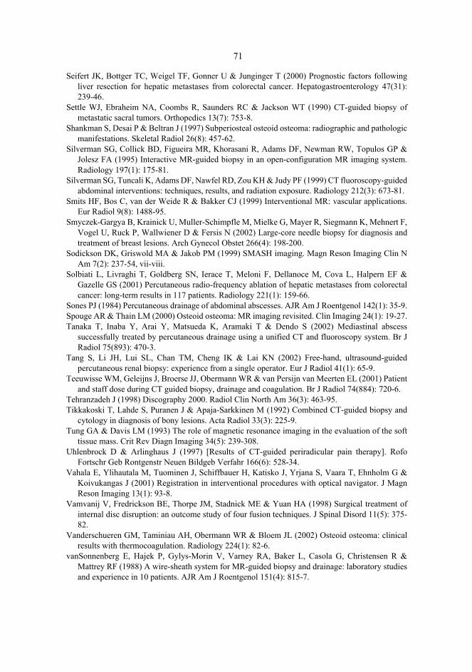
71
Seifert JK, Bottger TC, Weigel TF, Gonner U & Junginger T (2000) Prognostic factors followingliver resection for hepatic metastases from colorectal cancer. Hepatogastroenterology 47(31):239-46.
Settle WJ, Ebraheim NA, Coombs R, Saunders RC & Jackson WT (1990) CT-guided biopsy ofmetastatic sacral tumors. Orthopedics 13(7): 753-8.
Shankman S, Desai P & Beltran J (1997) Subperiosteal osteoid osteoma: radiographic and pathologicmanifestations. Skeletal Radiol 26(8): 457-62.
Silverman SG, Collick BD, Figueira MR, Khorasani R, Adams DF, Newman RW, Topulos GP &Jolesz FA (1995) Interactive MR-guided biopsy in an open-configuration MR imaging system.Radiology 197(1): 175-81.
Silverman SG, Tuncali K, Adams DF, Nawfel RD, Zou KH & Judy PF (1999) CT fluoroscopy-guidedabdominal interventions: techniques, results, and radiation exposure. Radiology 212(3): 673-81.
Smits HF, Bos C, van der Weide R & Bakker CJ (1999) Interventional MR: vascular applications.Eur Radiol 9(8): 1488-95.
Smyczek-Gargya B, Krainick U, Muller-Schimpfle M, Mielke G, Mayer R, Siegmann K, Mehnert F,Vogel U, Ruck P, Wallwiener D & Fersis N (2002) Large-core needle biopsy for diagnosis andtreatment of breast lesions. Arch Gynecol Obstet 266(4): 198-200.
Sodickson DK, Griswold MA & Jakob PM (1999) SMASH imaging. Magn Reson Imaging Clin NAm 7(2): 237-54, vii-viii.
Solbiati L, Livraghi T, Goldberg SN, Ierace T, Meloni F, Dellanoce M, Cova L, Halpern EF &Gazelle GS (2001) Percutaneous radio-frequency ablation of hepatic metastases from colorectalcancer: long-term results in 117 patients. Radiology 221(1): 159-66.
Sones PJ (1984) Percutaneous drainage of abdominal abscesses. AJR Am J Roentgenol 142(1): 35-9.Spouge AR & Thain LM (2000) Osteoid osteoma: MR imaging revisited. Clin Imaging 24(1): 19-27.Tanaka T, Inaba Y, Arai Y, Matsueda K, Aramaki T & Dendo S (2002) Mediastinal abscess
successfully treated by percutaneous drainage using a unified CT and fluoroscopy system. Br JRadiol 75(893): 470-3.
Tang S, Li JH, Lui SL, Chan TM, Cheng IK & Lai KN (2002) Free-hand, ultrasound-guidedpercutaneous renal biopsy: experience from a single operator. Eur J Radiol 41(1): 65-9.
Teeuwisse WM, Geleijns J, Broerse JJ, Obermann WR & van Persijn van Meerten EL (2001) Patientand staff dose during CT guided biopsy, drainage and coagulation. Br J Radiol 74(884): 720-6.
Tehranzadeh J (1998) Discography 2000. Radiol Clin North Am 36(3): 463-95.Tikkakoski T, Lahde S, Puranen J & Apaja-Sarkkinen M (1992) Combined CT-guided biopsy and
cytology in diagnosis of bony lesions. Acta Radiol 33(3): 225-9.Tung GA & Davis LM (1993) The role of magnetic resonance imaging in the evaluation of the soft
tissue mass. Crit Rev Diagn Imaging 34(5): 239-308.Uhlenbrock D & Arlinghaus J (1997) [Results of CT-guided periradicular pain therapy]. Rofo
Fortschr Geb Rontgenstr Neuen Bildgeb Verfahr 166(6): 528-34.Vahala E, Ylihautala M, Tuominen J, Schiffbauer H, Katisko J, Yrjana S, Vaara T, Ehnholm G &
Koivukangas J (2001) Registration in interventional procedures with optical navigator. J MagnReson Imaging 13(1): 93-8.
Vamvanij V, Fredrickson BE, Thorpe JM, Stadnick ME & Yuan HA (1998) Surgical treatment ofinternal disc disruption: an outcome study of four fusion techniques. J Spinal Disord 11(5): 375-82.
Vanderschueren GM, Taminiau AH, Obermann WR & Bloem JL (2002) Osteoid osteoma: clinicalresults with thermocoagulation. Radiology 224(1): 82-6.
vanSonnenberg E, Hajek P, Gylys-Morin V, Varney RA, Baker L, Casola G, Christensen R &Mattrey RF (1988) A wire-sheath system for MR-guided biopsy and drainage: laboratory studiesand experience in 10 patients. AJR Am J Roentgenol 151(4): 815-7.
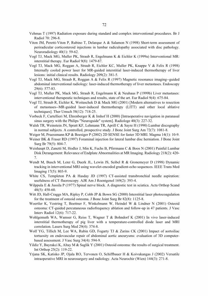
72
Vehmas T (1997) Radiation exposure during standard and complex interventional procedures. Br JRadiol 70: 296-8.
Viton JM, Peretti-Viton P, Rubino T, Delarque A & Salamon N (1998) Short-term assessment ofperiradicular corticosteroid injections in lumbar radiculopathy associated with disc pathology.Neuroradiology 40(1): 59-62.
Vogl TJ, Mack MG, Muller PK, Straub R, Engelmann K & Eichler K (1999a) Interventional MR:interstitial therapy. Eur Radiol 9(8): 1479-87.
Vogl TJ, Mack MG, Roggan A, Straub R, Eichler KC, Muller PK, Knappe V & Felix R (1998)Internally cooled power laser for MR-guided interstitial laser-induced thermotherapy of liverlesions: initial clinical results. Radiology 209(2): 381-5.
Vogl TJ, Mack MG, Straub R, Roggan A & Felix R (1997) Magnetic resonance imaging--guidedabdominal interventional radiology: laser-induced thermotherapy of liver metastases. Endoscopy29(6): 577-83.
Vogl TJ, Muller PK, Mack MG, Straub R, Engelmann K & Neuhaus P (1999b) Liver metastases:interventional therapeutic techniques and results, state of the art. Eur Radiol 9(4): 675-84.
Vogl TJ, Straub R, Eichler K, Woitaschek D & Mack MG (2001) [Modern alternatives to resectionof metastases--MR-guided laser-induced thermotherapy (LITT) and other local ablativetechniques]. Ther Umsch 58(12): 718-25.
Vorbeck F, Cartellieri M, Ehrenberger K & Imhof H (2000) [Intraoperative navigation in paranasalsinus surgery with the Philips "Neuroguide" system]. Radiologe 40(3): 227-32.
Walsh TR, Weinstein JN, Spratt KF, Lehmann TR, Aprill C & Sayre H (1990) Lumbar discographyin normal subjects. A controlled, prospective study. J Bone Joint Surg Am 72(7): 1081-8.
Weiger M, Pruessmann KP & Boesiger P (2002) 2D SENSE for faster 3D MRI. Magma 14(1): 10-9.Weiner BK & Fraser RD (1997) Foraminal injection for lateral lumbar disc herniation. J Bone Joint
Surg Br 79(5): 804-7.Weishaupt D, Zanetti M, Hodler J, Min K, Fuchs B, Pfirrmann C & Boos N (2001) Painful Lumbar
Disk Derangement: Relevance of Endplate Abnormalities at MR Imaging. Radiology 218(2): 420-7.
Wendt M, Busch M, Lenz G, Duerk JL, Lewin JS, Seibel R & Gronemeyer D (1998) Dynamictracking in interventional MRI using wavelet-encoded gradient-echo sequences. IEEE Trans MedImaging 17(5): 803-9.
White CS, Templeton PA & Hasday JD (1997) CT-assisted transbronchial needle aspiration:usefulness of CT fluoroscopy. AJR Am J Roentgenol 169(2): 393-4.
Wilppula E & Jussila P (1977) Spinal nerve block. A diagnostic test in sciatica. Acta Orthop Scand48(5): 458-60.
Witt JD, Hall-Craggs MA, Ripley P, Cobb JP & Bown SG (2000) Interstitial laser photocoagulationfor the treatment of osteoid osteoma. J Bone Joint Surg Br 82(8): 1125-8.
Woertler K, Vestring T, Boettner F, Winkelmann W, Heindel W & Lindner N (2001) Osteoidosteoma: CT-guided percutaneous radiofrequency ablation and follow-up in 47 patients. J VascInterv Radiol 12(6): 717-22.
Wohlgemuth WA, Wamser G, Reiss T, Wagner T & Bohndorf K (2001) In vivo laser-inducedinterstitial thermotherapy of pig liver with a temperature-controlled diode laser and MRIcorrelation. Lasers Surg Med 29(4): 374-8.
Wolf YG, Tillich M, Lee WA, Rubin GD, Fogarty TJ & Zarins CK (2001) Impact of aortoiliactortuosity on endovascular repair of abdominal aortic aneurysms: evaluation of 3D computer-based assessment. J Vasc Surg 34(4): 594-9.
Yildiz Y, Bayrakci K, Altay M & Saglik Y (2001) Osteoid osteoma: the results of surgical treatment.Int Orthop 25(2): 119-22.
Yrjana SK, Katisko JP, Ojala RO, Tervonen O, Schiffbauer H & Koivukangas J (2002) Versatileintraoperative MRI in neurosurgery and radiology. Acta Neurochir (Wien) 144(3): 271-8.
73
Zeng Q, Xiong L, Jinkins JR, Fan Z & Liu Z (1999) Intrathecal gadolinium-enhanced MRmyelography and cisternography: a pilot study in human patients. AJR Am J Roentgenol 173(4):1109-15.
Zhang Q, Chung YC, Lewin JS & Duerk JL (1998) A method for simultaneous RF ablation and MRI.J Magn Reson Imaging 8(1): 110-4.