Embed Size (px)
Citation preview
Magnetic Nanoparticle Sensors
CitationKoh, Isaac, and Lee Josephson. 2009. Magnetic nanoparticle sensors. Sensors 9(10): 8130-8145.
Published Versiondoi:10.3390/s91008130
Permanent linkhttp://nrs.harvard.edu/urn-3:HUL.InstRepos:9527627
Terms of UseThis article was downloaded from Harvard University’s DASH repository, and is made available under the terms and conditions applicable to Other Posted Material, as set forth at http://nrs.harvard.edu/urn-3:HUL.InstRepos:dash.current.terms-of-use#LAA
Share Your StoryThe Harvard community has made this article openly available.Please share how this access benefits you. Submit a story .
Accessibility

Sensors 2009, 9, 8130-8145; doi:10.3390/s91008130
sensors ISSN 1424-8220
www.mdpi.com/journal/sensors
Review
Magnetic Nanoparticle Sensors
Isaac Koh 1 and Lee Josephson 2,*
1 T2 Biosystems, 286 Cardinal Medieros Ave, Cambridge, MA 02141, USA;
E-Mail: [email protected] 2 Center for Translational Nuclear Medicine, Department of Nuclear Medicine and Molecular
Imaging and Center for Molecular Imaging Research, Massachusetts General Hospital/Harvard
Medical School, 149 13th Street, Charlestown, MA 02129, USA
* Author to whom correspondence should be addressed; E-Mail: [email protected];
Tel.: +1 617-726-6478; Fax: +1 617-723-7212.
Received: 3 August 2009; in revised form: 29 September 2009 / Accepted: 30 September 2009 /
Published: 16 October 2009
Abstract: Many types of biosensors employ magnetic nanoparticles (diameter = 5–300 nm)
or magnetic particles (diameter = 300–5,000 nm) which have been surface functionalized
to recognize specific molecular targets. Here we cover three types of biosensors that
employ different biosensing principles, magnetic materials, and instrumentation. The first
type consists of magnetic relaxation switch assay-sensors, which are based on the effects
magnetic particles exert on water proton relaxation rates. The second type consists of
magnetic particle relaxation sensors, which determine the relaxation of the magnetic
moment within the magnetic particle. The third type is magnetoresistive sensors, which
detect the presence of magnetic particles on the surface of electronic devices that are
sensitive to changes in magnetic fields on their surface. Recent improvements in the design
of magnetic nanoparticles (and magnetic particles), together with improvements in
instrumentation, suggest that magnetic material-based biosensors may become widely used
in the future.
Keywords: magnetic particles; magnetic nanoparticles; target molecules; biosensors;
magnetization
OPEN ACCESS
Sensors 2009, 9
8131
1. Introduction
Nanoscale magnetic materials are an important source of labels for biosensing due to their strong
magnetic properties which are not found in biological systems. Modulation of the composition, size
and magnetic properties of these materials permits their use in a variety of instruments and formats for
biosensing [1,2]. New types of instrumentation are promising for the use of nanoscale magnetic
materials in point of care sensors in variety of applications. Here, we cover three biosensors that
employ magnetic nanoparticle labels with different sensing principles and instrumentation:
(i) magnetic relaxation switches, (ii) magnetic particle relaxation sensors, and (iii) magnetoresistive
sensors.
2. Magnetic Relaxation Switches (MRSws)
Superparamagnetic nanoparticles made of iron oxide and a polymeric coating are clinically proven
magnetic resonance (MR) contrast agents and widely used in pre-clinical, targeted molecular imaging
applications [2,3]. When used as targeted contrast agents, surface-modified nanoparticles (NPs) bind
specific molecules producing local inhomogenieties in the applied magnetic field in tissues where
molecular targets are present. These inhomogeneities result in decreases in the T2 relaxation time (or
increases in 1/T2, the T2 relaxation rate), and these, in turn, lead to changes in the contrast of
MR images.
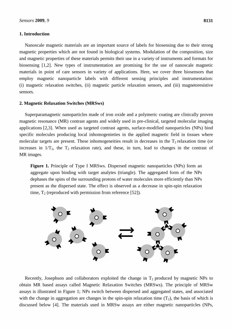
Figure 1. Principle of Type I MRSws. Dispersed magnetic nanoparticles (NPs) form an
aggregate upon binding with target analytes (triangle). The aggregated form of the NPs
dephases the spins of the surrounding protons of water molecules more efficiently than NPs
present as the dispersed state. The effect is observed as a decrease in spin-spin relaxation
time, T2 (reproduced with permission from reference [52]).
Recently, Josephson and collaborators exploited the change in T2 produced by magnetic NPs to
obtain MR based assays called Magnetic Relaxation Switches (MRSws). The principle of MRSw
assays is illustrated in Figure 1; NPs switch between dispersed and aggregated states, and associated
with the change in aggregation are changes in the spin-spin relaxation time (T2), the basis of which is
discussed below [4]. The materials used in MRSw assays are either magnetic nanoparticles (NPs,

Sensors 2009, 9
8132
diameter 5–300 nm) or micometer-sized magnetic particles (MPs, diameter 300–5,000 nm). As shown
in Figure 1, MRSws are homogeneous particle aggregation/disaggregation-based assays similar to
aggregation assays using Latex particles, red blood cell hemagglutination, and antibody reactions with
proteins (nephelometry). Unlike optically-based assays, MRSws employ radiofrequency radiation
which penetrates biological samples regardless of their optical properties [5]. Since the dispersed and
aggregated states of NPs (or MPs) can be reversed by such factors such as temperature, pH, and a high
concentration of competing analytes, and hence are referred to as “relaxation switches”. The
aggregated and dispersed states of magnetic NPs or MPs have different transverse spin-spin relaxation
times (values of T2). NP aggregation and the size range of the resulting aggregates depends on the type
of analyte and analyte concentration [6].
2.1. Mechanism of MRSws
MRSw assays exploit the fact that for both nanoparticles (NPs) and larger magnetic particles (MPs)
transverse relaxation times (T2) differ between dispersed and aggregated states. However, for Type I,
NP-based systems, T2 decreases with the aggregation, while with type II, MP-based systems T2
increases with aggregation. The basis of this is as follows [6–8].
The general theory of how magnetic spheres alter T2 is termed outer sphere relaxation theory. This
theory uses two parameters of Dw and tD. Dw is the difference in angular frequencies between the local
field experienced by a proton at the equatorial line of the sphere's surface and in the bulk
(Dw = mOMg/3, where mO is the vacuum magnetic permeability, M is the particle magnetization, and g
is the proton gyromagnetic ratio). Then tD is the translational diffusion time of water around the sphere
(tD = Ra2/D, where Ra is the sphere radius and D is the water diffusion coefficient). The outer sphere
diffusion theory is applied when the motional average condition is fulfilled as DwtD < 1 [7,8]. In this
condition, the relaxation rate R2 (= 1/T2) increases as the sphere's size is increased. As the definitions
of Dw and tD imply, the motional average condition is not fulfilled with increased size of the particles
such as MPs (DwtD > 1) and the relaxation rate of 1/T2 decreases with the formation of MP aggregates.
See the detailed discussion of this phenomenon in a review [8].
Thus, when present in solution magnetic NPs (or MPs) induce local magnetic field inhomogeneities,
which cause a dephasing (loss of phase coherence) of the proton spin precession, and these
inhomgeoneities lead to a reduction of the T2 relaxation time. When NPs aggregate (Type I MRSw), a
smaller number of larger magnetic field inhomogeneities result. These larger inhomogeneities are more
effective dephasers of proton relaxation and T2 drops. Here DwtD < 1. When MPs aggregate (Type II
MRSw), a smaller number of larger magnetic field inhomogeneities again results. However, there now
so few aggregates, and spaces between them so great, that many water proteins fail to diffuse in and out
of these homogeneities during the time course of the measurement. This is termed the "diffusion
limited case" for the enhancement of proton relaxation by magnetic microspheres. Here DwtD > 1.
Relaxivity is an important measure of the potency of magnetic materials and an important factor to
selecting evaluating materials for use in MRSw assays. Materials with higher relaxivities are more
detectable by the relaxometry and can detect lower concentrations of analyte [8].
Sensors 2009, 9
8133
R2 = (1/T2(+) – 1/T2(−))/C (1)
where R2 is relaxivity of the particle (in moles of metal) expressed as (mM sec)−1, C is the
concentration of the paramagnetic center in mM, and 1/T2(+) and 1/T2(−) are the transverse relaxation
rates (sec−1) in the presence and absence of the nanoparticle, respectively. C is typically expressed as
the concentration of paramagnetic metal, but it can also be expressed as the concentration of NPs or
MPs in solution. Here the R2 per metal is multiplied times the number of paramagnetic metal atoms per
particle. Magnetic particles with larger numbers of metals per particle are more potent in MRSw
assays, see below.
2.2. Magnetic Particles
Magnetic particles can be categorized by their size, with nanoparticles (NPs) being between 10 and
300 nm in diameter, while larger magnetic particles (MPs) are between 300 and 5,000 nm in diameter.
Since the first publication demonstrating the MRSw assay principle in 2001 [4], NPs with surfaces of
cross-linked iron oxide (CLIO) have been used for sensing for analytes ranging from small molecules
to mammalian cells [5,9–12]. CLIO is an excellent NP both for in vivo MR imaging [13] and for
MRSw assay applications, because of its stability in a variety of fluids, including aqueous buffers and
blood, and because of its functional handle of amino groups. CLIO is prepared by two-step treatment of
the monocrystalline iron oxide nanoparticle known as MION. The MION NP features a dextran coating
which is first cross-linked with epichlorohydrin and then reacted with ammonia to obtain amino groups
on the crosslinked dextran surface. MION and CLIO NPs have an iron oxide cores of about 5 nm in
diameter and dextran shell (or crosslinked dextran shell) about 10 nm in thickness, so that both NPs
have overall diameters between 25 nm and 30 nm.
Recently, magnetic NPs and MPs with improved magnetic properties, and higher detectability per
particle, have been described for use with in vivo MR imaging and in vitro biosensor
applications [1,14,15]. One strategy is to increase the R2 relaxivity of NPs by increasing M or d, since
R2 is proportional to M2d2. Here M is the saturation magnetization per mole of metal or per gram of
metal atoms within the particle and d is the particle diameter. [16–18]. Core/shell NPs have been
designed with Fe metal cores (not iron oxide cores) and these have an increased Ms and a thin iron
oxide shell to block oxidation metal oxidation. They show an enhanced sensitivity compared to CLIO
for the detection of bacterial cells [17]. Another strategy employs Mn-doped metal oxide NPs; these
also have high Ms and high R2s, and have been synthesized with sizes of 10, 12 and 16 nm. These NPs
have been used in the sensitive detection of unprocessed cancer cells, with as few as two cells
per 1 µL being detected with miniaturized relaxometer [16]. Another approach to improving the
sensitivity of MRSw assays is the use of MPs rather than NPs. These MPs have far more metal atoms
per particle than NPs and a far larger per magnetic moments per particle, even though their values or M
per metal are typical of older NPs [6,19]. In an MRSw assay of immunoreactive antibodies to
influenza, MPs of 1 µm in diameter were employed that had a similar R2 relaxivity to CLIO NPs on a
per iron atom basis. However, the larger MPs had 350,000 fold more irons per particle than CLIO NPs.
In the MRSw assay for anti-Tag peptide antibody, MPs had 186,000 fold enhanced sensitivity (relative
to CLIO). The improvement in sensitivity was achieved by a combination of factors including the use
Sensors 2009, 9
8134
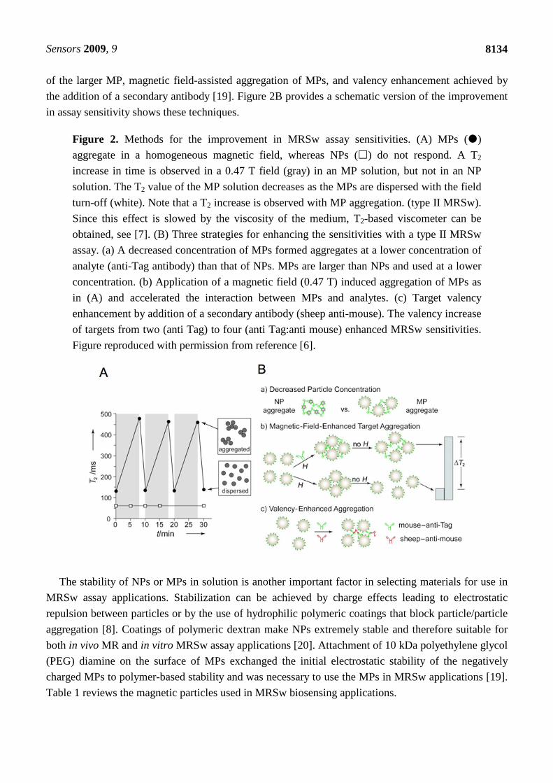
of the larger MP, magnetic field-assisted aggregation of MPs, and valency enhancement achieved by
the addition of a secondary antibody [19]. Figure 2B provides a schematic version of the improvement
in assay sensitivity shows these techniques.
Figure 2. Methods for the improvement in MRSw assay sensitivities. (A) MPs (�)
aggregate in a homogeneous magnetic field, whereas NPs (�) do not respond. A T2
increase in time is observed in a 0.47 T field (gray) in an MP solution, but not in an NP
solution. The T2 value of the MP solution decreases as the MPs are dispersed with the field
turn-off (white). Note that a T2 increase is observed with MP aggregation. (type II MRSw).
Since this effect is slowed by the viscosity of the medium, T2-based viscometer can be
obtained, see [7]. (B) Three strategies for enhancing the sensitivities with a type II MRSw
assay. (a) A decreased concentration of MPs formed aggregates at a lower concentration of
analyte (anti-Tag antibody) than that of NPs. MPs are larger than NPs and used at a lower
concentration. (b) Application of a magnetic field (0.47 T) induced aggregation of MPs as
in (A) and accelerated the interaction between MPs and analytes. (c) Target valency
enhancement by addition of a secondary antibody (sheep anti-mouse). The valency increase
of targets from two (anti Tag) to four (anti Tag:anti mouse) enhanced MRSw sensitivities.
Figure reproduced with permission from reference [6].
The stability of NPs or MPs in solution is another important factor in selecting materials for use in
MRSw assay applications. Stabilization can be achieved by charge effects leading to electrostatic
repulsion between particles or by the use of hydrophilic polymeric coatings that block particle/particle
aggregation [8]. Coatings of polymeric dextran make NPs extremely stable and therefore suitable for
both in vivo MR and in vitro MRSw assay applications [20]. Attachment of 10 kDa polyethylene glycol
(PEG) diamine on the surface of MPs exchanged the initial electrostatic stability of the negatively
charged MPs to polymer-based stability and was necessary to use the MPs in MRSw applications [19].
Table 1 reviews the magnetic particles used in MRSw biosensing applications.

Sensors 2009, 9
8135
Table 1. Characteristics of magnetic particles used for biosensing applications.
Particle Size Composition Characteristics Reference CLIO ~30 nm 5 nm core, 10 nm dextran
coating MRSw,
R2 = 50 (s⋅mM Fe)-1 [5]
Core/shell 16 nm Fe core, iron oxide shell, 2.5 nm shell thickness
MRSw, R2 = 260 (s⋅mM Fe)-1
[17]
Mn-MNPa 16 nm Mn-doped iron oxide MRSw, R2 = 420 (s⋅mM
metal)-1
[16]
MP 1000 nm Commercial (Dynabeads) MRSw, R2 = 43 (s⋅mM Fe)-1
[19]
Iron oxide 56 nm Commercial (Quantum Magnetics, Miltenyi
Biotech)
SQUID [35,36]
Iron oxide 19.5 nm AC susceptometer [42] Cubic FeCo 12.8 nm 1.5 nm oxidized shell GMR [49]
SAFb 100 nm Multilayers of ferromagnetic, interlayer of nonmagnetic material
GMR, disk shape
[47]
Magnetic bead 130, 250 nm Commercial (Micromod Partikeltechnologie)
SQUID [43]
(a) MNP: magnetic nanoparticle, (b) SAF: synthetic antiferromagnetic.
2.3. Instrumentation
Point of care (POC) sensors would benefit home users, clinicians and physicians, and aid in the
preparations for bio-warfare and pandemics. The miniaturization of MR relaxometers holds great
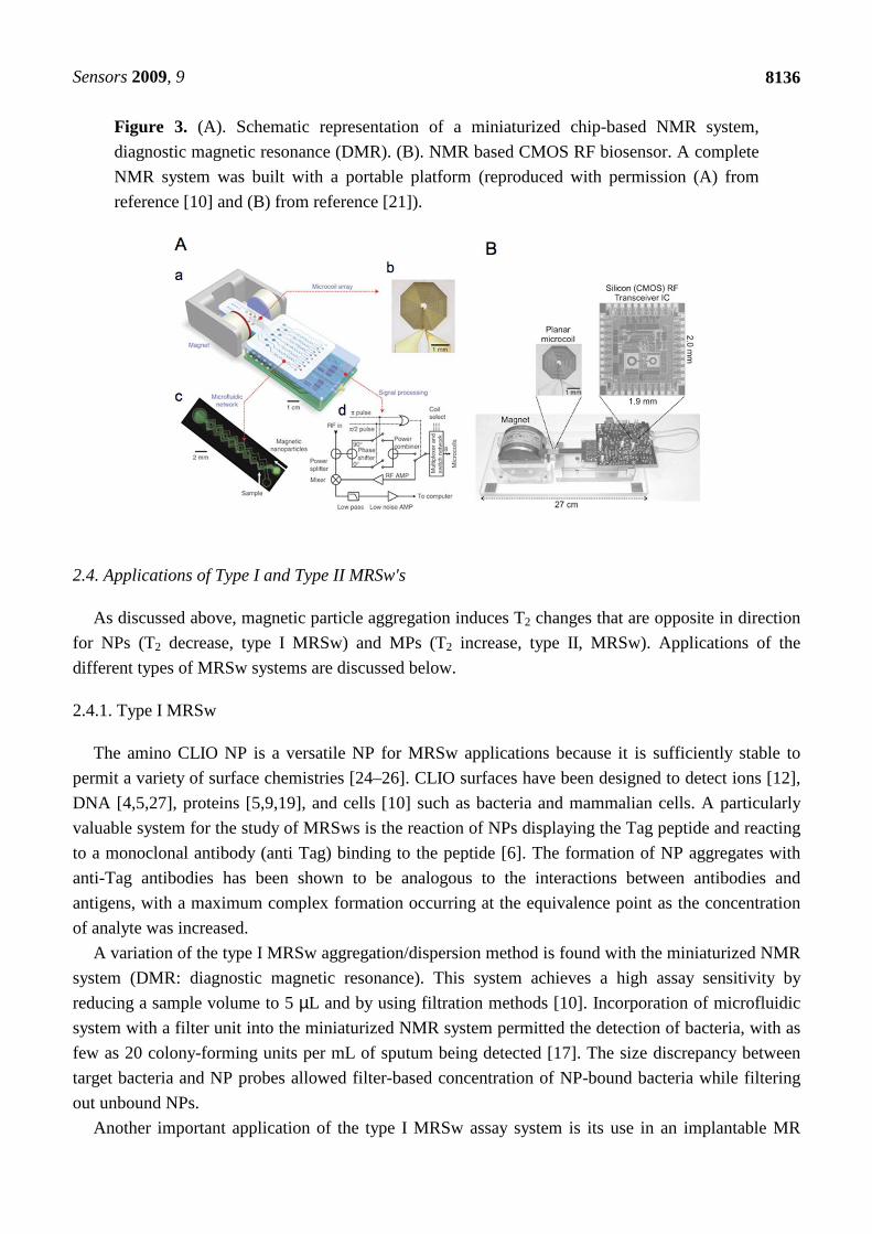
promise for use as instrumentation with POC [10,16,17,21].
The MR relaxometers used for MRSw assays have three basic components, a magnet, a coil, and a
transceiver. Currently MRSw assays depend on the commercial bench top relaxometers such as the
0.47 T Minispec, 20 MHz instrument made by Bruker, Billerica, MA [5, 19]. High throughput MRSw
assays have been demonstrated in 384-well plates through the use of a 1.5 T MR scanner [5,22,23].
However, the relaxometer and MR scanner above are impractical as POC sensors due to their high
cost, which results principally from the large magnets employed and lack of miniaturized electronic
components [21].
The magnets used in relaxometers can be relatively weak (0.1 to 0.5 T) and can provide less
homogeneous magnetic fields than those used in MR imagers. One of the first miniaturized MR
relaxometry systems consisted of a small palm-sized permanent magnet and on-board NMR electronics
and planar microcoils with integrated microfluidic channels [10] (see Figure 3A). A multiplexed
detection of biomarkers was achieved using an 8 microcoil array and demonstrated the potential
application of the microNMR system for high throughput MRSw assays. Optimization of circuit
designing in development of RF transceiver integrated circuits led to a small but complete NMR
system [21] (Figure 3B).
Sensors 2009, 9
8136
Figure 3. (A). Schematic representation of a miniaturized chip-based NMR system,
diagnostic magnetic resonance (DMR). (B). NMR based CMOS RF biosensor. A complete
NMR system was built with a portable platform (reproduced with permission (A) from
reference [10] and (B) from reference [21]).
2.4. Applications of Type I and Type II MRSw's
As discussed above, magnetic particle aggregation induces T2 changes that are opposite in direction
for NPs (T2 decrease, type I MRSw) and MPs (T2 increase, type II, MRSw). Applications of the
different types of MRSw systems are discussed below.
2.4.1. Type I MRSw
The amino CLIO NP is a versatile NP for MRSw applications because it is sufficiently stable to
permit a variety of surface chemistries [24–26]. CLIO surfaces have been designed to detect ions [12],
DNA [4,5,27], proteins [5,9,19], and cells [10] such as bacteria and mammalian cells. A particularly
valuable system for the study of MRSws is the reaction of NPs displaying the Tag peptide and reacting
to a monoclonal antibody (anti Tag) binding to the peptide [6]. The formation of NP aggregates with
anti-Tag antibodies has been shown to be analogous to the interactions between antibodies and
antigens, with a maximum complex formation occurring at the equivalence point as the concentration
of analyte was increased.
A variation of the type I MRSw aggregation/dispersion method is found with the miniaturized NMR
system (DMR: diagnostic magnetic resonance). This system achieves a high assay sensitivity by
reducing a sample volume to 5 µL and by using filtration methods [10]. Incorporation of microfluidic
system with a filter unit into the miniaturized NMR system permitted the detection of bacteria, with as
few as 20 colony-forming units per mL of sputum being detected [17]. The size discrepancy between
target bacteria and NP probes allowed filter-based concentration of NP-bound bacteria while filtering
out unbound NPs.
Another important application of the type I MRSw assay system is its use in an implantable MR

Sensors 2009, 9
8137
based, water relaxation sensor. A semi-permeable membrane was employed with a size cutoff that
permitted small analytes, like glucose, to diffuse in and out while the larger CLIO NPs were retained
within the sensor [25]. Continuous monitoring of the T2 values of the solution inside the membrane
showed a competitive assay type-response of glucose-functionalized CLIO to glucose [23,25]. The
proof of concept sensing obtained with glucose was translated to an implantable water relaxation
sensor detecting hCG as a cancer biomarker [28,29]. The implantable device had a reservoir that was
covered with a semi-permeable polycarbonate membrane and contained CLIO functionalized with
antibodies to the hCG cancer biomarker. In vivo MR imaging was used to monitor the T2 values from
inside the sensor device. When implanted in a tumor bearing mouse model, the MR signal from the
sensor showed significant decreases in 1-4 days due to diffusion of the cancer biomarker hCG into the
reservoir and the resulting aggregation of the CLIO NP.
2.4.2. Type II MRSw
Type II MRSws, where biomolecules are attached to MPs and aggregated by reaction with
molecular targets, exhibit an increased T2 when aggregated by reaction with a target analyte. With their
greater numbers of iron atoms per particle, MPs can be used at concentrations far below than that of
NPs in MRSw assays. With the lower concentration of MPs, lower concentrations of analyte are
needed to induce aggregation and this results in greatly improved sensitivity [6,19].
When placed in a homogeneous magnetic field, MPs with charge-based or polymer layer-based
stability, will aggregate, while NPs will not respond in this fashion [6,7,19,30–33]. The magnetic field-
induced MP aggregation is lost when the magnetic field was removed and Brownian effects break
down aggregates. The rate of self-assembly formation of MPs in a magnetic field is a function of
viscosity and can be used to make a T2 based viscometer. See Figure 2A and [7]. Recently, magnetic
field-induced MP aggregation has also been used to accelerate analyte-mediated formation of MP
aggregates [19,31,34]. The applied magnetic field enhanced the kinetics of molecular interactions
between multivalent analytes, (e.g., a monoclonal Tag antibody), and multivalent MPs displaying the
Tag peptide. This technique is referred to as magnetic field enhanced target aggregation and shown in
Figure 2B, frame (b).
3. Magnetic Particle Relaxation-Based Sensors
The relaxation of the magnetic moments within magnetic particles have been used as a basis for
magnetic particle-based assays.
3.1. Theory
Magnetic particles in a liquid, with magnetic moments aligned by an applied magnetic field, employ
two relaxation mechanisms when magnetic field is turned off: (i) Brownian relaxation and
(ii) Néel relaxation. Brownian relaxation is governed by the physical rotation of the entire particle and
characterized by the Brownian relaxation time, τB. Here:
τB = 3VHη/kT (2)
where VH is the hydrodynamic volume, η is the viscosity of the medium, k is the Boltzmann’s
Sensors 2009, 9
8138
constant, and T is the absolute temperature. The monodomain magnetic particle has an anisotropy
energy, Ea, which is proportional to the crystal volume.
Ea = KaV (3)
where Ka is the anisotropy constant and V is the volume of the crystal. When the applied field is
removed, the magnetization vector within the particle returns to the lowest energy state along the easy
axis with a characteristic Néel relaxation time, τN:
τN = τ0 exp(Ea/kT) (4)
where τ0 is the preexponential factor that decreases as the anisotropy energy increases. Note that τN is
an exponential function of the anisotropy energy that is proportional to the crystal volume.
The effective relaxation rate is expressed as the sum of the Brownian relaxation rate and the Néel
relaxation rate:
1/τ = 1/τB + 1/τN (5)
As Equation (5) shows, faster relaxation time between the two governs the effective relaxation
process. Target induced aggregation can decrease the rates of the Brownian or Neel relaxations and this
assays for molecular targets are generated. See [8].
3.2. Assays
3.2.1. Néel Relaxation Sensors
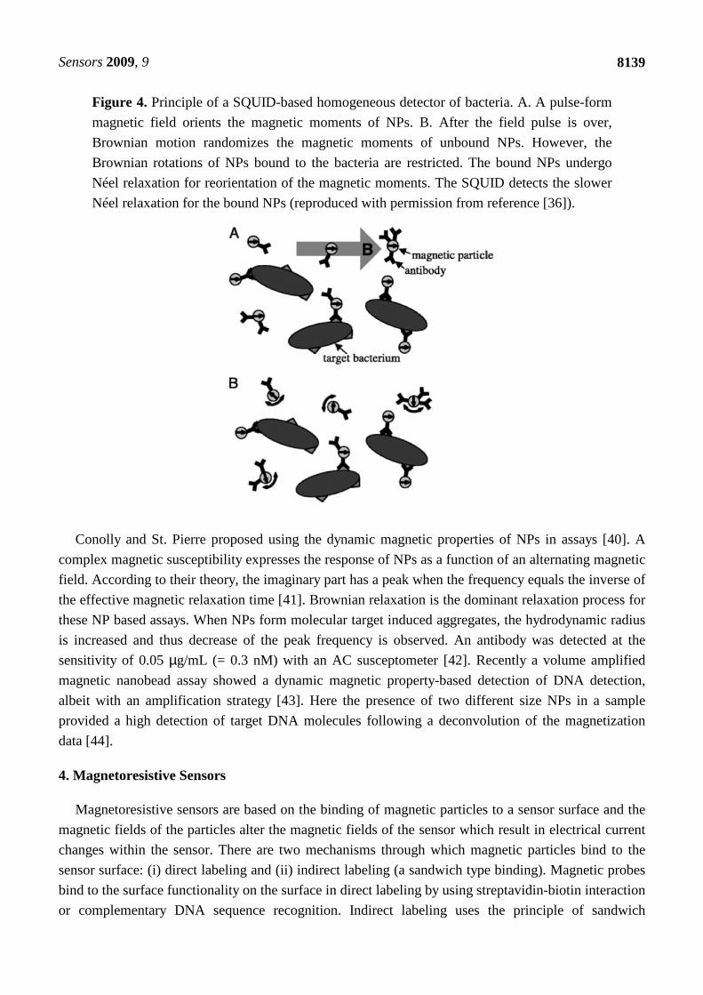
Superconducting Quantum Interference Devices (SQUIDs) have been used for measurements of the
relaxation of particle magnetic moments. The Brownian relaxation is much faster than the Néel
relaxation. For a 20 nm single domain magnetite particle in solution, the calculated relaxation times
were τB ~1 µs and τN ~1 s [35]. The difference in the relaxation time scales was a basis for a
homogeneous immunoassay [35] and a bacterial detection [36]. See Figure 4. The Brownian relaxation
time scale of a single unbound magnetic particle was so short, it was out of the detectable range
between 1 ms and 1 s of the SQUID. The free Brownian rotation of particles was then restricted when
the magnetic particles bound a bacterium. The Néel relaxation was within the detection window of a
SQUID, which was used to determine the relaxation time of surface bound particles. The
SQUID-based detection of the Néel relaxation time showed a limit of detection of 5 × 10⁴ NPs for a
substrate based assay and 1.1 × 10⁵ bacteria in a 20 µL sample volume. Development of a gradiometer
instead of a magnetometer suggested a two-order improvement in sensitivity was possible [37].
3.2.2. Brownian Relaxation Sensors
Measurements of static and dynamic magnetic susceptibility using alternate currents (ac) have
permitted use of the Brownian relaxation of NPs for biosensing. As Equation (2) suggests, the NP
aggregates that form in recognition of target analytes have a larger hydrodynamic size and thus show
slower Brownian relaxation responses than a single NP. The resulting decrease in relaxation was
sensed in buffer [38], and in serum [39] by using a SQUID or an ac magnetosusceptometer [39].
Sensors 2009, 9
8139
Figure 4. Principle of a SQUID-based homogeneous detector of bacteria. A. A pulse-form
magnetic field orients the magnetic moments of NPs. B. After the field pulse is over,
Brownian motion randomizes the magnetic moments of unbound NPs. However, the
Brownian rotations of NPs bound to the bacteria are restricted. The bound NPs undergo
Néel relaxation for reorientation of the magnetic moments. The SQUID detects the slower
Néel relaxation for the bound NPs (reproduced with permission from reference [36]).
Conolly and St. Pierre proposed using the dynamic magnetic properties of NPs in assays [40]. A
complex magnetic susceptibility expresses the response of NPs as a function of an alternating magnetic
field. According to their theory, the imaginary part has a peak when the frequency equals the inverse of
the effective magnetic relaxation time [41]. Brownian relaxation is the dominant relaxation process for
these NP based assays. When NPs form molecular target induced aggregates, the hydrodynamic radius
is increased and thus decrease of the peak frequency is observed. An antibody was detected at the
sensitivity of 0.05 µg/mL (= 0.3 nM) with an AC susceptometer [42]. Recently a volume amplified
magnetic nanobead assay showed a dynamic magnetic property-based detection of DNA detection,
albeit with an amplification strategy [43]. Here the presence of two different size NPs in a sample
provided a high detection of target DNA molecules following a deconvolution of the magnetization
data [44].
4. Magnetoresistive Sensors
Magnetoresistive sensors are based on the binding of magnetic particles to a sensor surface and the
magnetic fields of the particles alter the magnetic fields of the sensor which result in electrical current
changes within the sensor. There are two mechanisms through which magnetic particles bind to the
sensor surface: (i) direct labeling and (ii) indirect labeling (a sandwich type binding). Magnetic probes
bind to the surface functionality on the surface in direct labeling by using streptavidin-biotin interaction
or complementary DNA sequence recognition. Indirect labeling uses the principle of sandwich
Sensors 2009, 9
8140
immunoassay in ELISA. For example, antibodies that bind to the target protein are immobilized on the
surface. After treatment of the surface with a sample solution containing the target proteins, second
antibodies that are biotinylated are added to the system. Finally Streptavidin coated magnetic particles
are applied for tagging the biotinylated antibodies.
Giant magnetoresistance (GMR) spin valve (SV) or magnetic tunnel junction (MTJ) sensors have
been successfully used to sense MPs. Sensors are composed of multiple layers of ferromagnetic
materials. A biologically active molecule can be deposited on an Au layer or SiO2 layer to obtain a
surface for the attachment of biomolecules. For a review of the structure of magnetoresistive sensors
see [45].
Superparamagnetic particles with different sizes have been used in magnetoresistive biosensing.
Earlier applications used relatively large magnetic particles, with diameters between 0.1 and 3 µm [46].
Micrometer sized particles have the advantages of facile observation under light microscope and a
higher particle-based magnetic moment that permits detection very small numbers of particles.
However, recently magnetic NPs have replaced the larger particles because the NPs are stable in
suspension and are less prone to particle clustering in an applied magnetic field [45,47–49].
Streptavidin coated MPs were applied to spin valve sensors in the protein marker detection at
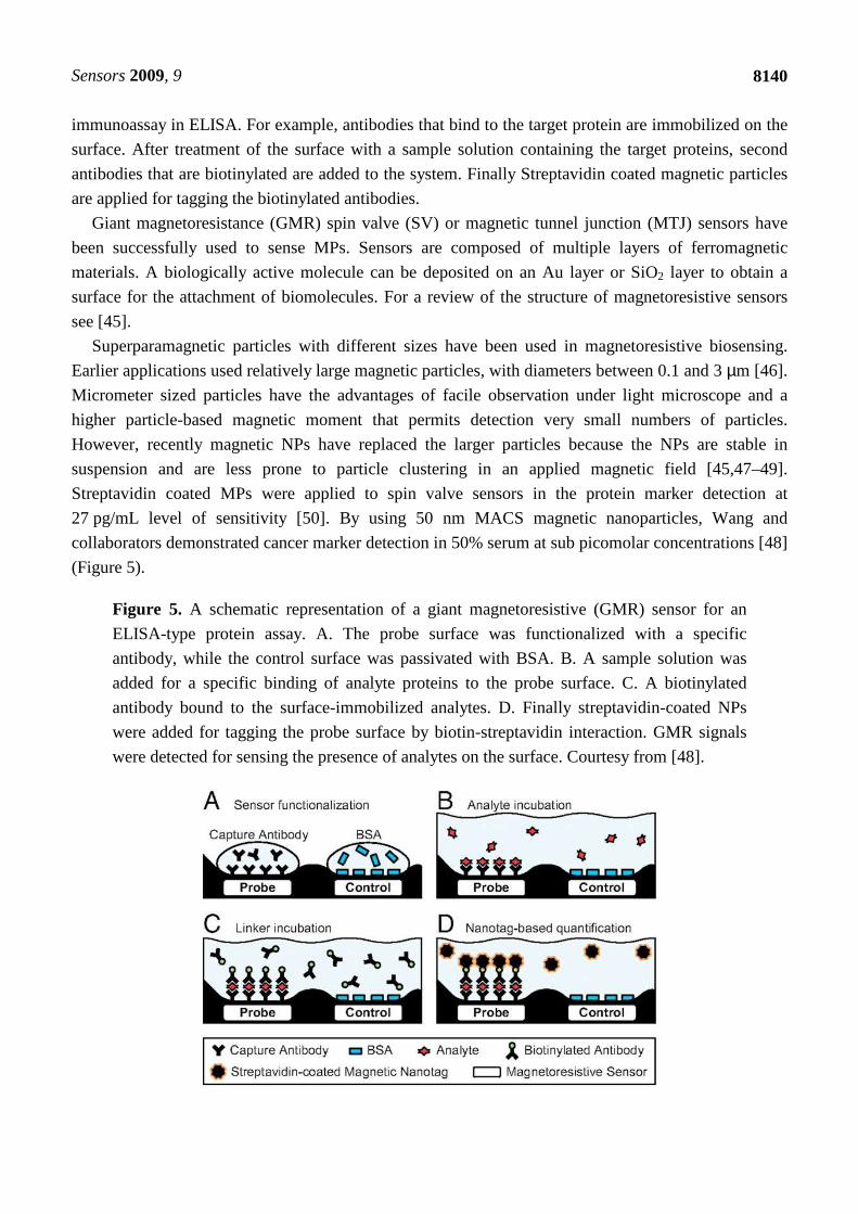
27 pg/mL level of sensitivity [50]. By using 50 nm MACS magnetic nanoparticles, Wang and
collaborators demonstrated cancer marker detection in 50% serum at sub picomolar concentrations [48]
(Figure 5).
Figure 5. A schematic representation of a giant magnetoresistive (GMR) sensor for an
ELISA-type protein assay. A. The probe surface was functionalized with a specific
antibody, while the control surface was passivated with BSA. B. A sample solution was
added for a specific binding of analyte proteins to the probe surface. C. A biotinylated
antibody bound to the surface-immobilized analytes. D. Finally streptavidin-coated NPs
were added for tagging the probe surface by biotin-streptavidin interaction. GMR signals
were detected for sensing the presence of analytes on the surface. Courtesy from [48].
Sensors 2009, 9
8141
Improvement of spin valve sensors was achieved by reducing the passivation layer to 30 nm and led
to an enhanced sensitivity. A signal amplification strategy that had multiple layers of streptavidin
coated NPs and biotinylated antibodies in the sandwich type immunoassay also showed enhanced
signals. Multiplex sensing of different protein markers in serum was demonstrated on a single chip by
carefully selecting antibodies and by employing the signal enhancing strategy with multiple layers of
NPs. Wang and his group in Standford University used nanoimprint lithography to synthesize
antiferromagnetic nanoparticles of 100 nm size with high magnetic moment and zero remanence [51].
The antiferromagnetic nanoparticles that have a disk shape were composed of multiple layers of
ferromagnetic material separated by a nonmagnetic interlayer. NPs with high magnetic moments were
functionalized with streptavidin and permitted the detection of DNA at concentrations as low as
10 pM [47].
Another effort to synthesize magnetic nanoparticles with high magnetic moment utilized cubic-
shaped FeCo nanoparticles of 12.8 nm in a GMR based sensor [49]. The cubic nanoparticles were
surface functionalized with silane chemistry for attachment of Streptavidin or antibody. Direct labeling
of biotinylated surface with Streptavidin coated nanoparticles allowed detection of 600 nanoparticle
binding. Indirect labeling in ELISA type assay produced signals as low as 2 × 10⁶ molecules of a
biomarker protein.
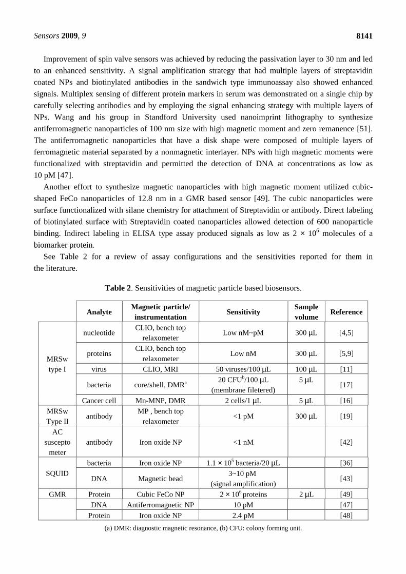
See Table 2 for a review of assay configurations and the sensitivities reported for them in
the literature.
Table 2. Sensitivities of magnetic particle based biosensors.
Analyte Magnetic particle/ instrumentation
Sensitivity Sample volume
Reference
MRSw type I
nucleotide CLIO, bench top
relaxometer Low nM~pM 300 µL [4,5]
proteins CLIO, bench top
relaxometer Low nM 300 µL [5,9]
virus CLIO, MRI 50 viruses/100 µL 100 µL [11]
bacteria core/shell, DMRa 20 CFUb/100 µL
(membrane filetered) 5 µL
[17]
Cancer cell Mn-MNP, DMR 2 cells/1 µL 5 µL [16]
MRSw Type II
antibody MP , bench top
relaxometer <1 pM 300 µL [19]
AC suscepto
meter antibody Iron oxide NP <1 nM [42]
SQUID bacteria Iron oxide NP 1.1 × 10⁵ bacteria/20 µL [36]
DNA Magnetic bead 3~10 pM
(signal amplification) [43]
GMR Protein Cubic FeCo NP 2 × 10⁶ proteins 2 µL [49]
DNA Antiferromagnetic NP 10 pM [47]
Protein Iron oxide NP 2.4 pM [48]
(a) DMR: diagnostic magnetic resonance, (b) CFU: colony forming unit.
Sensors 2009, 9
8142
5. Conclusions
Magnetic NPs and MPs have been used in different types of biosensors based on different physical
principles. Some achieve high sensitivity and, with rapid advances in instrumentation, maybe useful as
point-of-care sensors. The continued rapid development of sensors using magnetic materials
seems assured.
Acknowledgment
This work was supported in part by NIH grants U54 CA119349 and P50 CA08635.
References
1. Lee, J.H.; Huh, Y.M.; Jun, Y.W.; Seo, J.W.; Jang, J.T.; Song, H.T.; Kim, S.; Cho, E.J.; Yoon,
H.G.; Suh, J.S.; Cheon, J. Artificially engineered magnetic nanoparticles for ultra-sensitive
molecular imaging. Nat. Med. 2007, 13, 95–99.
2. Weissleder, R.; Pittet, M.J. Imaging in the era of molecular oncology. Nature 2008, 452, 580–589.
3. Weissleder, R. Molecular imaging in cancer. Science 2006, 312, 1168–1171.
4. Josephson, L.; Perez, J.M.; Weissleder, R. Magnetic nanosensors for the detection of
oligonucleotide sequences. Angew. Chem. Int. Ed. 2001, 40, 3204–3208.
5. Perez, J.M.; Josephson, L.; O'Loughlin, T.; Hogemann, D.; Weissleder, R. Magnetic relaxation
switches capable of sensing molecular interactions. Nat. Biotechnol. 2002, 20, 816–820.
6. Koh, I.; Hong, R.; Weissleder, R.; Josephson, L. Nanoparticle-target interactions parallel
antibody-protein interactions. Anal. Chem. 2009, 81, 3618–3622.
7. Hong, R.; Cima, M.J.; Weissleder, R.; Josephson, L. Magnetic microparticle aggregation for
viscosity determination by MR. Magn. Reson. Med. 2008, 59, 515–520.
8. Laurent, S.; Forge, D.; Port, M.; Roch, A.; Robic, C.; Vander Elst, L.; Muller, R.N. Magnetic iron
oxide nanoparticles: synthesis, stabilization, vectorization, physicochemical characterizations, and
biological applications. Chem. Rev. 2008, 108, 2064–2110.
9. Kim, G.Y.; Josephson, L.; Langer, R.; Cima, M.J. Magnetic relaxation switch detection of human
chorionic gonadotrophin. Bioconjug. Chem. 2007, 18, 2024–2028.
10. Lee, H.; Sun, E.; Ham, D.; Weissleder, R. Chip-NMR biosensor for detection and molecular
analysis of cells. Nat. Med. 2008, 14, 869–874.
11. Perez, J.M.; Simeone, F.J.; Saeki, Y.; Josephson, L.; Weissleder, R. Viral-induced self-assembly
of magnetic nanoparticles allows the detection of viral particles in biological media. J. Am. Chem.
Soc. 2003, 125, 10192–10193.
12. Taktak, S.; Weissleder, R.; Josephson, L. Electrode chemistry yields a nanoparticle-based nmr
sensor for Calcium. Langmuir 2008, 24, 7596–7598.
13. Harisinghani, M.G.; Barentsz, J.; Hahn, P.F.; Deserno, W.M.; Tabatabaei, S.; van de Kaa, C.H.;
de la Rosette, J.; Weissleder, R. Noninvasive detection of clinically occult lymph-node metastases
in prostate cancer. N. Engl. J. Med. 2003, 348, 2491–2499.

Sensors 2009, 9
8143
14. Jun, Y.W.; Huh, Y.M.; Choi, J.S.; Lee, J. H.; Song, H.T.; Kim, S.; Yoon, S.; Kim, K.S.; Shin,
J.S.; Suh, J.S.; Cheon, J. Nanoscale size effect of magnetic nanocrystals and their utilization for
cancer diagnosis via magnetic resonance imaging. J. Am. Chem. Soc. 2005, 127, 5732–5733.
15. Park, J.; An, K.J.; Hwang, Y.S.; Park, J.G.; Noh, H.J.; Kim, J.Y.; Park, J.H.; Hwang, N.M.;
Hyeon, T. Ultra-large-scale syntheses of monodisperse nanocrystals. Nat. Mater. 2004, 3,
891–895.
16. Lee, H.; Yoon, T.J.; Figueiredo, J.L.; Swirski, F.K.; Weissleder, R. Rapid detection and profiling
of cancer cells in fine-needle aspirates. Proc. Natl. Acad. Sci. USA 2009, doi: 10.1073/pnas. 0902365106.
17. Lee, H.; Yoon, T.J.; Weissleder, R. Ultrasensitive detection of bacteria using core-shell
nanoparticles and an NMR-filter system. Angew.Chem. Int. Ed. 2009, 48, 5657–5660.
18. Park, J.H.; von Maltzahn, G.; Zhang, L.L.; Schwartz, M.P.; Ruoslahti, E.; Bhatia, S.N.; Sailor,
M.J. Magnetic iron oxide nanoworms for tumor targeting and imaging. Adv. Mater. 2008, 20,
1630–1635.
19. Koh, I.; Hong, R.; Weissleder, R.; Josephson, L. Sensitive NMR sensors detect antibodies to
influenza. Angew. Chem. Int. Ed. 2008, 47, 4119–4121.
20. Perez, J.M.; Josephson, L.; Weissleder, R. Use of magnetic nanoparticles as nanosensors to probe
for molecular interactions. Chembiochem 2004, 5, 261–264.
21. Sun, N.; Liu, Y.; Lee, H.; Weissleder, R.; Ham, D. CMOS RF biosensor utilizing nuclear
magnetic resonance. IEEE J. Solid-State Circuits 2009, 44, 1629–1643.
22. Grimm, J.; Perez, J.M.; Josephson, L.; Weissleder, R. Novel nanosensors for rapid analysis of
telomerase activity. Cancer Res. 2004, 64, 639–643.
23. Tsourkas, A.; Hofstetter, O.; Hofstetter, H.; Weissleder, R.; Josephson, L. Magnetic relaxation
switch immunosensors detect enantiomeric impurities. Angew. Chem. Int. Ed. 2004, 43,
2395–2399.
24. Sun, E.Y.; Josephson, L.; Weissleder, R. “Clickable” nanoparticles for targeted imaging. Mol.
Imaging 2006, 5, 122–128.
25. Sun, E.Y.; Weissleder, R.; Josephson, L. Continuous analyte sensing with magnetic nanoswitches.
Small 2006, 2, 1144–1147.
26. Weissleder, R.; Kelly, K.; Sun, E.Y.; Shtatland, T.; Josephson, L. Cell-specific targeting of
nanoparticles by multivalent attachment of small molecules. Nat. Biotechnol. 2005, 23,
1418–1423.
27. Perez, J.M.; O'Loughin, T.; Simeone, F.J.; Weissleder, R.; Josephson, L. DNA-based magnetic
nanoparticle assembly acts as a magnetic relaxation nanoswitch allowing screening of DNA-
cleaving agents. J. Am. Chem. Soc. 2002, 124, 2856–2857.
28. Daniel, K.D.; Kim, G.Y.; Vassiliou, C.C.; Galindo, M.; Guimaraes, A.R.; Weissleder, R.; Charest,
A.; Langer, R.; Cima, M.J. Implantable diagnostic device for cancer monitoring. Biosens.
Bioelectron. 2009, 24, 3252–3257.
29. Daniel, K.D.; Kim, G.Y.; Vassiliou, C.C.; Jalali-Yazdi, F.; Langer, R.; Cima, M.J., Multi-
reservoir device for detecting a soluble cancer biomarker. Lab. Chip. 2007, 7, 1288–1293.
Sensors 2009, 9
8144
30. Doyle, P.S.; Bibette, J.; Bancaud, A.; Viovy, J.L. Self-assembled magnetic matrices for DNA
separation chips. Science 2002, 295, 2237.
31. Baudry, J.; Rouzeau, C.; Goubault, C.; Robic, C.; Cohen-Tannoudji, L.; Koenig, A.; Bertrand, E.;
Bibette, J. Acceleration of the recognition rate between grafted ligands and receptors with
magnetic forces. Proc. Natl. Acad. Sci. USA 2006, 103, 16076–16078.
32. Singh, H.; Laibinis, P.E.; Hatton, T.A. Rigid, superparamagnetic chains of permanently linked
beads coated with magnetic nanoparticles. Synthesis and rotational dynamics under applied
magnetic fields. Langmuir 2005, 21, 11500–11509.
33. Zerrouki, D.; Baudry, J.; Pine, D.; Chaikin, P.; Bibette, J. Chiral colloidal clusters. Nature 2008,
455, 380–382.
34. Cohen-Tannoudji, L.; Bertrand, E.; Baudry, J.; Robic, C.; Goubault, C.; Pellissier, M.; Johner, A.;
Thalmann, F.; Lee, N.K.; Marques, C.M.; Bibette, J. Measuring the kinetics of biomolecular
recognition with magnetic colloids. Phys. Rev. Lett. 2008, 100, 108301:1–108301:4.
35. Chemla, Y.R.; Grossman, H.L.; Poon, Y.; McDermott, R.; Stevens, R.; Alper, M.D.; Clarke, J.
Ultrasensitive magnetic biosensor for homogeneous immunoassay. Proc. Natl. Acad. Sci. USA
2000, 97, 14268–14272.
36. Grossman, H.L.; Myers, W.R.; Vreeland, V.J.; Bruehl, R.; Alper, M.D.; Bertozzi, C.R.; Clarke, J.
Detection of bacteria in suspension by using a superconducting quantum interference device. Proc.
Natl. Acad. Sci. USA 2004, 101, 129–134.
37. Lee, S.; Myers, W.R.; Grossman, H.L.; Cho, H.M.; Chemla, Y.R.; Clarke, J. Magnetic
gradiometer based on a high-transition temperature superconducting quantum interference device
for improved sensitivity of a biosensor. Appl. Phys. Lett. 2002, 81, 3094–3096.
38. Hong, C.Y.; Wu, C.C.; Chiu, Y.C.; Yang, S.Y.; Horng, H.E.; Yang, H.C. Magnetic susceptibility
reduction method for magnetically labeled immunoassay. Appl. Phys. Lett. 2006, 88,
212512:1–212512:3.
39. Hong, C.Y.; Chen, W.S.; Jian, Z.F.; Yang, S.Y.; Horng, H.E.; Yang, L.C.; Yang, H.C. Wash-free
immunomagnetic detection for serum through magnetic susceptibility reduction. Appl. Phys. Lett.
2007, 90, 074105:1–074105:3.
40. Connolly, J.; St Pierre, T.G. Proposed biosensors based on time-dependent properties of magnetic
fluids. J. Magn. Magn. Mater. 2001, 225, 156–160.
41. Chung, S.H.; Hoffmann, A.; Bader, S.D.; Liu, C.; Kay, B.; Makowski, L.; Chen, L. Biological
sensors based on Brownian relaxation of magnetic nanoparticles. Appl. Phys. Lett. 2004, 85,
2971–2973.
42. Fornara, A.; Johansson, P.; Petersson, K.; Gustafsson, S.; Qin, J.; Olsson, E.; Ilver, D.; Krozer, A.;
Muhammed, M.; Johansson, C. Tailored magnetic nanoparticles for direct and sensitive detection
of biomolecules in biological samples. Nano Lett. 2008, 8, 3423–3428.
43. Stromberg, M.; Goransson, J.; Gunnarsson, K.; Nilsson, M.; Svedlindh, P.; Stromme, M. Sensitive
molecular diagnostics using volume-amplified magnetic nanobeads. Nano Lett. 2008, 8, 816–821.
44. Stromberg, M.; Zardan Gomez de la Torre, T.; Goransson, J.; Gunnarsson, K.; Nilsson, M.;
Svedlindh, P.; Stromme, M. Multiplex detection of DNA sequences using the volume-amplified
magnetic nanobead detection assay. Anal. Chem. 2009, 81, 3398–3406.
Sensors 2009, 9
8145
45. Wang, S.X.; Li, G. Advances in giant magnetoresistance biosensors with magnetic nanoparticle
tags: Review and outlook. IEEE Trans. Magn. 2008, 44, 1687–1702.
46. Graham, D.L.; Ferreira, H.A.; Freitas, P.P. Magnetoresistive-based biosensors and biochips.
Trends Biotechnol. 2004, 22, 455–462.
47. Fu, A.; Hu, W.; Xu, L.; Wilson, R.J.; Yu, H.; Osterfeld, S.J.; Gambhir, S.S.; Wang, S.X. Protein-
functionalized synthetic antiferromagnetic nanoparticles for biomolecule detection and magnetic
manipulation. Angew. Chem. Int. Ed. 2009, 48, 1620–1624.
48. Osterfeld, S.J.; Yu, H.; Gaster, R.S.; Caramuta, S.; Xu, L.; Han, S.J.; Hall, D.A.; Wilson, R.J.;
Sun, S.; White, R.L.; Davis, R.W.; Pourmand, N.; Wang, S.X. Multiplex protein assays based on
real-time magnetic nanotag sensing. Proc. Natl. Acad. Sci. USA 2008, 105, 20637–20640.
49. Srinivasan, B.; Li, Y.; Jing, Y.; Xu, Y.; Yao, X.; Xing, C.; Wang, J.P. A detection system based
on giant magnetoresistive sensors and high-moment magnetic nanoparticles demonstrates
zeptomole sensitivity: potential for personalized medicine. Angew. Chem. Int. Ed. 2009, 48, 2764–
2767.
50. De Palma, R.; Reekmans, G.; Liu, C.; Wirix-Speetjens, R.; Laureyn, W.; Nilsson, O.; Lagae, L.
Magnetic bead sensing platform for the detection of proteins. Anal. Chem. 2007, 79, 8669–8677.
51. Hu, W.; Wilson, C.R.J.; Koh, A.; Fu, A.H.; Faranesh, A.Z.; Earhart, C.M.; Osterfeld, S.J.; Han,
S.J.; Xu, L.; Guccione, S.; Sinclair, R.; Wang, S.X. High-moment antiferromagnetic nanoparticles
with tunable magnetic properties. Adv. Mater. 2008, 20, 1479–1483.
52. Lowery, T.J.; Palazzolo, R.; Wong, S.M.; Prado, P.J.; Taktak, S. Single-coil, multisample, proton
relaxation method for magnetic relaxation switch assays. Anal. Chem. 2008, 80, 1118–1123.
© 2009 by the authors; licensee Molecular Diversity Preservation International, Basel, Switzerland.
This article is an open-access article distributed under the terms and conditions of the Creative
Commons Attribution license (http://creativecommons.org/licenses/by/3.0/).