Embed Size (px)
Citation preview
© 2014. Published by The Company of Biologists Ltd | Disease Models & Mechanisms (2014) 7, 785-797 doi:10.1242/dmm.015594
785
ABSTRACTStudying macrophage biology in the context of a whole livingorganism provides unique possibilities to understand the contributionof this extremely dynamic cell subset in the reaction to infections, andhas revealed the relevance of cellular and molecular processes thatare fundamental to the cell-mediated innate immune response. Inparticular, various recently established zebrafish infectious diseasemodels are contributing substantially to our understanding of themechanisms by which different pathogens interact with macrophagesand evade host innate immunity. Transgenic zebrafish lines withfluorescently labeled macrophages and other leukocyte populationsenable non-invasive imaging at the optically transparent early lifestages. Furthermore, there is a continuously expanding availability ofvital reporters for subcellular compartments and for probing activationof immune defense mechanisms. These are powerful tools tovisualize the activity of phagocytic cells in real time and shed light onthe intriguing paradoxical roles of these cells in both limiting infectionand supporting the dissemination of intracellular pathogens. ThisReview will discuss how several bacterial and fungal infection modelsin zebrafish embryos have led to new insights into the dynamicmolecular and cellular mechanisms at play when pathogensencounter host macrophages. We also describe how these insightsare inspiring novel therapeutic strategies for infectious diseasetreatment.
KEY WORDS: Leukocyte biology, Innate immunity, Infectiousdisease, Host-directed therapy, Mycobacterium, Salmonella,Burkholderia, Staphylococcus, Shigella, Candida
IntroductionThe immune system has evolved through the constant interplaybetween microbes and their multicellular hosts. Selective forcesacting on both sides have driven the evolution of a wide variety ofvirulence mechanisms in pathogens and alternative controlmechanisms in their hosts. In vivo modeling of infectious disease isessential for understanding this complexity and translating it intonovel therapeutic interventions. The immune system, innate andadaptive, is well-conserved among vertebrates. The zebrafish (Daniorerio) offers an optically and genetically accessible vertebrate modelto study host-pathogen interactions (Renshaw and Trede, 2012; vander Vaart et al., 2012; Ramakrishnan, 2013). At the embryonic andearly larval stages, zebrafish provide the opportunity of studying therelevance of innate immunity in a context where no adaptive
REVIEW
Institute of Biology, Leiden University, Einsteinweg 55, 2333 CC, Leiden, The Netherlands.
*Author for correspondence ([email protected])
This is an Open Access article distributed under the terms of the Creative CommonsAttribution License (http://creativecommons.org/licenses/by/3.0), which permits unrestricteduse, distribution and reproduction in any medium provided that the original work is properlyattributed.
response has yet been developed, given that early lymphocytes maketheir first appearance in 4-day-old larvae and a full adaptiveimmunity requires several weeks to be mounted (Lam et al., 2004;Page et al., 2013).
Macrophages and neutrophils are the main phagocytic cell typesof the innate immune system. Zebrafish models provide unique toolsfor studying the function of phagocytic cells, and these studies caneffectively complement studies in other infectious disease models.Other recent reviews highlighted the use of zebrafish forunderstanding neutrophil biology (Henry et al., 2013; Shelef et al.,2013). Here, we will discuss six zebrafish models for importanthuman pathogens (Mycobacterium, Salmonella, Burkholderia,Staphylococcus, Shigella and Candida), emphasizing the novelinsights that these models have recently provided into macrophagebiology and highlighting how this could lead to the finding of newhost-derived therapeutic strategies.
Zebrafish macrophage biology and tools for investigatingmacrophage functionOntogeny and properties of early macrophages in zebrafishThe first macrophage precursors appear in the zebrafish embryo asearly as 20 hours post-fertilization (hpf) from the anterior lateralplate mesoderm (Herbomel et al., 1999). Following migration to theyolk sac, they differentiate and either invade the head mesenchyme,where they will later differentiate into microglial cells (the residentmacrophages of the brain), or enter the blood circulation (Herbomelet al., 1999; Herbomel et al., 2001). These cells, named primitivemacrophages, retain proliferative capability and have been reportedto exist in mammals too (Takahashi et al., 1996; Herbomel et al.,1999). They can remove apoptotic cells, are able to sense andrespond to invading microbes, and can eradicate non-pathogenicinfections. Primitive macrophages readily phagocytose microbesfrom the blood circulation or when present in tissues. In contrast,neutrophils (which develop slightly later) are less efficient inphagocytosing microbes in the blood, but are potent scavengers ofsurface-associated bacteria (Colucci-Guyon et al., 2011).
Primitive macrophages are gradually replaced by differentlineages of macrophages deriving from definitive hematopoiesis, theprocess that will produce specialized pluripotent cells with theability to differentiate into all types of mature blood cells. The firstwave of definitive hematopoiesis starts at 24 hpf in the posteriorblood island or caudal hematopoietic tissue (CHT) with thedifferentiation of erythromyeloid progenitors (Bertrand et al., 2007).By 48 hpf, these pluripotent progenitors are replaced with anothersubset of hematopoietic stem and progenitor cells (HSPCs), nowable to also differentiate into the lymphoid lineage. These cellsoriginate from the AGM (aorta, gonads and mesonephros), derivedfrom the lateral posterior mesoderm. After leaving the AGM, theymigrate to and nest in the CHT, and will provide the second waveof definitive hematopoiesis (Murayama et al., 2006; Bertrand et al.,2007). Development of HSPCs and their emergence from aortic
Macrophage-pathogen interactions in infectious diseases: new therapeutic insights from the zebrafish host modelVincenzo Torraca, Samrah Masud, Herman P. Spaink and Annemarie H. Meijer*
Dis
ease
Mod
els
& M
echa
nism
s

786
endothelium is remarkably conserved between zebrafish andmammals (Bertrand et al., 2010; Kissa and Herbomel, 2010; Boissetet al., 2010).
Following the second wave of hematopoiesis, macrophageprecursors are released into the circulation and will extravasate toseed tissues throughout the whole body, where they differentiateinto tissue macrophages. Starting from 4 days post-fertilization(dpf), the kidney marrow, which is the main hematopoietic tissueof the adult fish, develops and will progressively replace theembryonic hematopoietic system. Another component of themononuclear phagocyte system is represented by the dendritic cell(DC) population, which is also present in zebrafish larvae and canbe detected from 8-12 dpf (Wittamer et al., 2011; Svahn et al.,2013).
The infection studies discussed below, using zebrafish embryoand larval models, do not distinguish macrophages from circulatingmonocytes. Furthermore, possible functional differences betweenmacrophages from primitive or definitive hematopoietic origins aregenerally not addressed. For more detailed and comparativedescriptions of the processes of hematopoiesis in zebrafish andmammals we refer to other reviews (Stachura and Traver, 2011;Jagannathan-Bogdan and Zon, 2013).
Macrophage defense mechanisms and subversion by intracellularpathogensMacrophages sense the presence of infection through microbial-specific molecules and host-derived inflammatory mediators. Theirchemoattraction to the site of infection depends largely on thefunction of G-protein-coupled receptors (Xu et al., 1996; Cotton andClaing, 2009). Scavenger and complement receptors play a majorrole in phagocytosis (Elomaa et al., 1995), and Toll-like receptors(TLRs), in cooperation with other pattern-recognition receptors(PRRs), initiate the innate immune response (O’Neill et al., 2013).TLRs, found on the cell surface and membranes of vesicularcompartments, recognize pathogen- and damage-associatedmolecular patterns (PAMPs and DAMPs, respectively). Anothermain class of PRRs, the NOD-like receptors (NLRs), performs thesame function in the cytosol (Bertin et al., 1999; Inohara et al.,1999). Some NLRs participate in the assembly of theinflammasome, a multiprotein complex able to activate the caspase-1 cascade, which triggers processing of pro-inflammatory cytokines,such as IL1B (interleukin 1 beta), and full activation of the innateimmune response (Martinon et al., 2002).
When engulfed by macrophages, microorganisms are exposed toa number of defense mechanisms within the resulting phagosomeand through its subsequent fusion with lysosomes. These include theproduction of reactive oxygen and nitrogen species (ROS and RNS,respectively) (Minakami and Sumimotoa, 2006; El-Gayar et al.,2003), exposure to antimicrobials, the activity of proteases, andacidification (Schmidtchen et al., 2002; Park et al., 1996; Vandal etal., 2008). Escape from the phagosome triggers septin caging andantibacterial autophagy as additional defense mechanisms (Mostowyet al., 2010; Deretic et al., 2013).
Intracellular pathogens have evolved many strategies tocounteract these defenses. These counter-strategies are mediated byvirulence factors, which are often secreted directly into the host cellvia specialized secretion systems such as the T3SS (type IIIsecretion system) of Gram-negative pathogens and the T7SS (typeVII secretion system) of pathogenic mycobacteria (Abdallah et al.,2007; Baxt et al., 2013). Pathogens can also induce significantreprogramming of their host cells through manipulation of signalingpathways and chromatin remodeling; however, these mechanisms
are still poorly understood (Masaki et al., 2013; Wang et al., 2005).Intracellular pathogens often block phagosome maturation andfusion with lysosomes or manipulate the vesicular system such thatthe phagosome is modified to resemble the endoplasmic reticulumor a Golgi-like compartment (Duclos and Desjardins, 2000).Furthermore, several pathogens inject virulence factors that promoteactin polymerization to actively stimulate their uptake by both non-phagocytic and phagocytic cells (Ogawa et al., 2008). Pathogens thatare able to escape from the phagosome have mechanisms to evadeautophagy and can spread from the initially infected cell to othercells by acquiring actin-based motility (Ogawa et al., 2005; Ogawaet al., 2008). Other virulence mechanisms can induce inflammationand different cell-death programs to facilitate the dissemination ofinfection (Hilbi et al., 1998). These different virulence strategies areschematically depicted in Fig. 1.
Macrophage markers and transgenic linesThe development of transgenic zebrafish lines with fluorescentlylabeled leukocytes (supplementary material Table S1) has been keyto the successful application of zebrafish for immunological studies.However, until recently, the lack of a specific reporter for themacrophage lineage limited the study of this myeloid subset. Thishas now been remedied with the development of the csf1ra andmpeg1 reporter lines (Gray et al., 2011; Ellett et al., 2011). Thesegenes are robust markers for macrophages at embryonic and larvalstages, because they are co-expressed with the pan-leukocyticmarker lcp1 but not with the neutrophil markers mpx and lyz (Meijeret al., 2008; Zakrzewska et al., 2010). Despite the fact that csf1ra ismacrophage-specific within the immune cell types, it is alsoexpressed in neural crest cells and derivatives, such as thexanthophores. Nevertheless, the highly motile macrophages can bedistinguished easily from the immobile xanthophores in time-courseexperiments (Gray et al., 2011). Reporter lines using the mpeg1promoter label macrophages but not xanthophores (Fig. 2A;supplementary material Movie 1) and, combined with a neutrophilmarker, can show the different kinetics of macrophage andneutrophil responses to infection and wounding, as well as thedynamic interactions between the two cell types (Ellett et al., 2011).The mpeg1 reporter also labels microglia and it has been suggestedto label other antigen-presenting cells, such as the Langerhansdendritic cells, but these could not be detected before 8-9 dpf (Svahnet al., 2013).
Expression of the Gal4 transcription factor under the control ofmacrophage or neutrophil promoters in combination with a UAS-nitroreductase-mCherry line allows for the specific ablation of oneof the two phagocyte populations. This approach can be used toinvestigate their individual contributions to the immune responseand infectious disease pathogenesis (Gray et al., 2011; Prajsnar etal., 2012). Alternatively, spi1/pu.1 antisense morpholino knockdowncan be used to block the development of either macrophagesexclusively or of both macrophages and neutrophils, depending onthe concentration used (Su et al., 2007). Similarly, irf8 tools havealso been used to deplete specific myeloid cell populations and toskew the development of their progenitors towards macrophages orneutrophils. Morpholino knockdown of irf8 can completely depletemacrophage differentiation while stimulating an increased output ofneutrophils, and irf8 overexpression can direct myeloid developmenttowards macrophage differentiation (Li et al., 2011).
Many other transgenic lines that label either the entire myeloidpopulation, the early myeloid subset, microglia or all antigen-presenting cells are also very useful for the study of macrophagebiology (supplementary material Table S1).
REVIEW Disease Models & Mechanisms (2014) doi:10.1242/dmm.015594
Dis
ease
Mod
els
& M
echa
nism
s
In vivo visualization of macrophage functionVisualization of live macrophage behavior in zebrafish embryos canbe achieved with great structural detail using digitally enhanceddifferential interference contrast (DIC) microscopy (Herbomel et al.,1999; Davis et al., 2002; Herbomel and Levraud, 2005; Davis andRamakrishnan, 2009). More recently, there has been tremendousprogress in the use of transgenic marker lines (supplementary materialTable S1) and labeled pathogens that facilitate live imaging in spatialand temporal dimensions (Fig. 2; supplementary material Movies 2,3). Photoconvertable fluorescent proteins such as Kaede and Dendra2have been exploited to show that cells from the CHT can be recruited
distally to infection foci and wounds (Yoo et al., 2011), and thatmycobacterium-infected macrophages egress from primarygranulomas to initiate secondary infection foci (Davis andRamakrishnan, 2009). For imaging of phagocyte migration, pathogensor specific chemoattractants can be injected subcutaneously or intobody cavities such as the otic vesicle and hindbrain ventricle, whichcan be reached without generating extensive tissue damage, therebypreventing wound-induced leukocyte mobilization (Colucci-Guyon etal., 2011; Benard et al., 2012; Sarris et al., 2012; Yang et al., 2012;Deng et al., 2013; Cambier et al., 2014). To visualize phagocytosisand the intracellular fate of bacteria, the pHrodo dye can be
787
REVIEW Disease Models & Mechanisms (2014) doi:10.1242/dmm.015594
Redirection ofvesiculartrafficking
Control of PRR-induced
innate immunity
Plasma membrane PRRs
Vesicular PRRs
Cytosolic PRRs
Bactericidial mechanisms
Modulation ofInflammasome
Casp1IL1β
Inflammasome
PAMPs
ER
Fusion with vesicular compartments
Non-lytic expulsion
Replication in specialized
vacuoles
Pyroptosis
Lysososomalfusion and acidification Injectosome
Mitochondrial stress and modulation
of apoptosis Cytochrome cROSCalcium
Cytosolicreplication
Necro(pto)sis
Manipulation of cytoskeleton
Actin-basedprotrusion
ROS/RNSAntimicrobialsProteases
Mitochondrion
Escape to cytosol
Galectin decorationUbiquitylation
Proteasome
Actinfilaments
Septin caging
Proteasomaldegradation
Autophagy
NF-κB and other
transcriptionfactors
Control of gene expression
mRNAsDNA
Chromatin remodelingand reprogramming
Nucleus
CytokinesChemokinesPro/anti-inflammatory mediators
Virulence factors
Virulence factors
Membrane-polarity andadhesion molecules
Phagocytosis
Adhesion molecules
Ionic pumps
1
23
6
8
12
9
10
13
4
5
11
1415
16
20
17
18
19
7
22
23
24
21Phagosome
Ribosome
Pathogen
Macrophage
Activatory mechanism
Inhibitory mechanism
Key
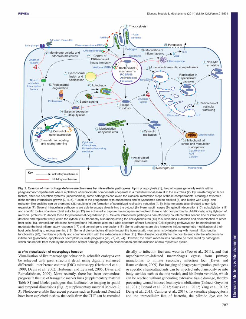
Fig. 1. Evasion of macrophage defense mechanisms by intracellular pathogens. Upon phagocytosis (1), the pathogens generally reside withinphagosomal compartments where a plethora of microbicidal components cooperate in a multidirectional assault to the microbes (2). By transferring virulencefactors, often via secretion systems (injectosomes), some pathogens can avoid the classical maturation steps of these compartments, creating a favorableniche for their intracellular growth (3, 4, 5). Fusion of the phagosome with endosomes and/or lysosomes can be blocked (6) and fusion with Golgi- andreticulum-like vesicles can be promoted (3), resulting in the formation of specialized replicative vacuoles (4, 5), in some cases also directed to non-lyticexpulsion (7). Several intracellular pathogens are able to escape directly into the cytosol (8). Here, septin cages (9), galectin decoration (10), ubiquitylation (11)and specific routes of antimicrobial autophagy (12) are activated to capture the escapers and redirect them to lytic compartments. Additionally, ubiquitylation ofmicrobial proteins (11) labels these for proteasomal degradation (13). Several intracellular pathogens can efficiently counteract this second line of intracellulardefense and replicate freely within the cytosol (14), frequently also manipulating the cell cytoskeleton (15) to sustain their extrusion and dissemination to otherhost cells (16). Intracellular infections have profound influences also on a wide spectrum of host functions. Cell signaling pathways can be manipulated tomodulate the host inflammatory response (17) and control gene expression (18). Some pathogens are also known to induce epigenetic modification of theirhost cells, leading to reprogramming (19). Some virulence factors directly impact the homeostatic mechanisms by interfering with normal mitochondrialfunctionality (20), membrane polarity and communication with the extracellular milieu (21). The ultimate possibility for the host to eradicate the infection is toinitiate cell (pyroptotic, apoptotic or necroptotic) suicide programs (20, 22, 23, 24). However, the death mechanisms can also be modulated by pathogens,which can benefit from them by the induction of host damage, pathogen dissemination and the initiation of new replicative cycles.
Dis
ease
Mod
els
& M
echa
nism
s

788
conjugated to bioparticles or to live or heat-killed bacteria (Fig. 2D),providing constitutive fluorescence in one channel and additionalfluorescence in another channel following exposure to an acidicenvironment (Hall et al., 2009). Furthermore, the nature of thecompartments where the pathogens reside can be investigated withcombinations of different vital stains, most of which are permeableinto zebrafish embryos when added to the water. Several pH-sensitivedyes (LysoSensor and LysoTracker) do not distinguish betweenlysosome-dependent or -independent phagosome acidificationmechanisms, but they can be used simultaneously with methods fordetection of the activity of lysosomal proteases (MagicRed-Cathepsinand DQ-BSA) (Peri and Nüsslein-Volhard, 2008). Different methodsallow in situ detection of ROS and RNS responses during infection inzebrafish embryos (Hall et al., 2012; Hall et al., 2013; Bilan et al.,2013; Elks et al., 2013). Also, tools for visualizing ATP, calciumeffluxes and apoptosis have been efficiently used in zebrafish (Periand Nüsslein-Volhard, 2008; Cheung et al., 2011; Sieger et al., 2012).Furthermore, an increasing number of transgenic marker lines forvesicular compartments are becoming available that will help inelucidating the subcellular locations where pathogens reside in vivo(supplementary material Table S2).
New insights into macrophage-pathogen interactionsMycobacterium marinumM. marinum is a natural pathogen of zebrafish that causesgranulomatous necrotic lesion formation in host tissues. These
lesions are histologically very similar to those generated byMycobacterium tuberculosis, the etiological agent of humantuberculosis (Prouty et al., 2003; Swaim et al., 2006). In adultzebrafish, M. marinum can cause a latent infection and the bacteriacan be reactivated from dormancy by immunosuppressive treatment,as is the case for M. tuberculosis, which is estimated to haveinfected one third of the world population (Parikka et al., 2012;Gengenbacher and Kaufmann, 2012). Tuberculosis therapy islimited by a number of problems, including the poor response ofdormant mycobacteria to antibiotics, the increasing prevalence ofmultidrug-resistant strains and the lack of an effective vaccineagainst latent or reactivated tuberculosis (Ottenhoff and Kaufmann,2012). The lack of a mouse model for tuberculosis that fullyrecapitulates the disease and the risk of working with the humanpathogen owing to its airborne transmission emphasize the need foralternative models. The unique accessibility of the early stages ofgranuloma formation in zebrafish larvae has made the zebrafish-M.marinum host-pathogen pair one of the most productive models usedto unravel the core pathogenic processes of mycobacterial infections(Davis et al., 2002; Ramakrishnan, 2013; Cronan and Tobin, 2014)(Fig. 3A). Notably, the use of this model has already providedseveral direct translational applications for human disease treatments(Table 1).
Infection of zebrafish embryos with M. marinum hasdemonstrated that macrophages are sufficient to initiate granulomaformation in the absence of adaptive immunity (Davis et al., 2002).
REVIEW Disease Models & Mechanisms (2014) doi:10.1242/dmm.015594
MacrophagesMm
47�
54� 57� 60�
50� 52�B
Macrophages
A C 8h 39�8h 37� 8h 47�
10h 17�9h 06� 9h 31� MacrophagesMm
C
Macrophages Mm Overlay
D
Acidification
Fig. 2. In vivo imaging of macrophage responses to infection. (A) A 3-dpf Tg(mpeg1:Gal4-VP16/UAS-E1b:Kaede) zebrafish embryo showing thedistribution pattern of macrophages (green). Random patrolling of macrophages is shown in supplementary material Movie 1. (B) Phagocytosis of M. marinum(Mm; green) injected into the subcutaneous area overlying a somite in a 2-dpf Tg(mpeg1:mCherry-F) embryo. The arrow points at a macrophage (red) in theprocess of phagocytosis between 47 and 60 minutes post-infection. The images are particulars and stills from supplementary material Movie 2 (10 to60 minutes post-infection). (C) Macrophage-mediated dissemination of M. marinum infection. The white track represents the path of an infected macrophagemigrating away from the infection focus. The images are stills and particulars from supplementary material Movie 3, which was taken from the same embryo asin B at a more advanced stage of infection (~8 to ~10 hours post-infection). (D) Partial acidification of phagocytosed M. marinum. Bacteria double-labeled withconstitutive mCrimson and pH-sensitive green pHrodo are contained within subcellular compartments of macrophages, which are intensely labeled by themembrane-bound mCherry of the Tg(mpeg1:mCherry-F) line. White arrows point at bacteria in acidified compartments, where the pHrodo dye is activated.Yellow arrows point at bacteria in non-acidified compartments. Note that most of the intracellular mycobacteria are not acidified, consistent with the ability ofthis pathogen to counteract phagosome maturation. Macrophages were imaged in the yolk sac circulation valley 5 hours after injection of bacteria into thecaudal vein at 2 dpf. Images in A-C were acquired with the Zeiss Observer 6.5.32 laser-scanning confocal, with 10× (A) or 20× (B,C) objectives. Images in Dwere acquired with Leica TCS SPE confocal with a 20× objective. Figures and movies were processed with ImageJ. The zebrafish transgenic linesTg(mpeg1:Gal4-VP16/UAS-E1b:Kaede) and Tg(mpeg1:mCherry-F) were previously described in other reports (Ellett et al., 2011; Bernut et al., 2014). Scalebars: (A) 200 μm; (B,C) 25 μm; (D) 10 μm.
Dis
ease
Mod
els
& M
echa
nism
s

789
REVIEW Disease Models & Mechanisms (2014) doi:10.1242/dmm.015594
Phagocytosis Intracellular resistance
Escapeto cytosol
Granuloma formation
Newly recruited macrophages
Dissemination
Seeding of secondary granulomas
Cytokine storm
Pyroptotic cell death
Replication within SCVs
Uncontrolledimmune response
and lethality
Redirection of vescicular trafficking
Phagocytosis
Non-lytic expulsionReplication within intracellular vacuoles
PhagocytosisCell aggregates
A Mycobacterium marinum
B Salmonella typhimurium
C Burkholderia cenocepacia
Cell death
Resistance to intracellular killing
Immunological bottleneck
Expansion in aprivileged niche
Clonal focal abscesses
Phagocytosis oflarge inoculum
D Staphylococcus aureus
Extracellular replication
Partial infection control in neutrophils
Intraphagosomalreplication
Uncontrolledbacteremiaand lethality
PhagocytosisResistance to
intraphagocyte killingPartial control by
septin caging and autophagy
Non-immunecell invasion
Escape and replication
Actin-based motility
Cell-to-cell dissemination Inefficient cytosolic control
E Shigella flexneri
Cell deathUncontrolledbacteremiaand lethality
Scavengingneutrophils
PhagocytosisIntracellular replication
without hyphal transitionF Candida albicans Extracellular
release
Systemic candidiasis
Extracellular yeast-to-hyphal switch
Macro-phage Neutrophil Pyroptosis
Non-immune cell
Necro(pto)sis
Key Actin tails
Septin cages
AutophagyMicrobe-containing vacuoles and compartments
Hyphal transition
Fig. 3. See next page for legend. Dis
ease
Mod
els
& M
echa
nism
s
790
Subsequently, this model changed the widespread view ofgranulomas, historically regarded solely as host-protectivestructures, by showing that early granulomas promote mycobacterialdissemination (Fig. 3A) and that their formation is driven byvirulence determinants of the RD1 locus, encoding ESX1, asecretion system conserved in all pathogenic mycobacteria(Volkman et al., 2004; Abdallah et al., 2007; Davis andRamakrishnan, 2009; Ramakrishnan, 2013). Furthermore, the ESX-1-secreted protein ESAT-6 (Early secreted antigenic target 6) wasfound to induce matrix metalloproteinase Mmp9 production byepithelial cells surrounding the infection focus, which in turnfacilitates macrophage infiltration. As a result, the application ofMmp9 antagonists has been suggested as a host-directed anti-tuberculosis therapy (Volkman et al., 2010) (Table 1). The notionthat granulomas are dynamic structures, even during latent infection,is supported by a study of M. tuberculosis infection in the macaquemodel (Lin et al., 2009).
Mycobacteria are well known for developing drug tolerance. Thezebrafish embryo model has demonstrated that their intramacrophagelocalization correlates with development of resistance and thatgranulomas promote dissemination of this resistant population.Upregulation of bacterial efflux pumps, which are required forintracellular growth, can mediate drug tolerance both in M. marinum-infected zebrafish and in M. tuberculosis-infected humanmacrophages. Efflux-pump inhibitors, already available on the market,can reduce this tolerance, and their addition to standard anti-tuberculosis therapy might therefore shorten treatment duration(Adams et al., 2011; Adams et al., 2014) (Table 1).
Another important insight into tuberculosis pathogenesis concernsthe relevance of the inflammatory status. A zebrafish mutagenesisscreen revealed Lta4h (Leukotriene A4 hydrolase) as a host factorthat strongly correlates with M. marinum susceptibility (Tobin et al.,2010). This enzyme is required for producing LTB4 (LeukotrieneB4), a powerful proinflammatory chemoattractant. Lta4h deficiencycorrelates with a less inflamed status, due to redirection of itssubstrates to anti-inflammatory lipoxins, resulting in reduced levelsof proinflammatory cytokines such as tumor necrosis factor (TNF).The crucial role of TNF in controlling mycobacterial infection isexemplified by the increased risk of tuberculosis in patients withchronic inflammatory disorders treated with TNF antagonists(Wallis, 2008). However, excessive production of TNF is alsoassociated with higher susceptibility to tuberculosis. This has beenshown in zebrafish and other animal models as well as intuberculosis meningitis patients, where a polymorphism at theLTA4H locus that causes increased TNF production has been linkedwith more progressive disease symptoms (Tsenova et al., 1999;Tobin et al., 2010). Hyper-inflamed and hypo-inflamed statuses haveboth been associated with necrotic death of infected macrophagesand consequent extracellular release of the pathogen (Clay et al.,2008; Roca and Ramakrishnan, 2013). In conditions of lowinflammation, macrophages passively undergo necrotic deathbecause they are unable to control intracellular bacterial growth,whereas, in conditions of high inflammation, macrophages activelyinitiate two ROS-mediated necroptotic pathways, dependent on theactivation of the mitochondrial permeability transition pore complex(mPTPC) and of the lysosomal acid sphingomyelinase (aSMase).Drugs targeting these pathways prevent activation of the necroptoticprogram in the zebrafish model (Roca and Ramakrishnan, 2013).The crucial role of the inflammatory status emphasizes theimportance of designing personalized patient therapies: patients withthe proinflammatory LTA4H genotype might benefit from classicalanti-inflammatory drugs [such as corticosteroids or non-steroidalanti-inflammatory drugs (NSAIDs)]; however, these drugs shouldbe avoided in patients with the opposite genotype (Tobin et al.,2010; Tobin et al., 2012). Drugs that directly block the ROS-linkednecroptotic pathways will benefit the proinflammatory genotypeswithout generating detrimental effects on the other genotypes,because the necroptotic pathways are exclusively triggered in hyper-inflamed conditions (Roca and Ramakrishnan, 2013) (Table 1).
The inflammatory response is initiated by TLR recognition ofPAMPs. Myd88, a central adaptor in TLR signaling, was recentlyshown to be required for control of systemic M. marinum infectionin zebrafish embryos (van der Vaart et al., 2013). In contrast, theinitial recruitment of macrophages to a localized M. marinuminfection in the hindbrain was found to be largely Myd88independent (Cambier et al., 2014). This effect was linked to thepresence of PDIM (phthiocerol dimycoceroserate) lipid on thesurface of pathogenic mycobacteria, which masks the underlyingPAMPs. Non-pathogenic mycobacteria, which lack PDIM, induce arobust immune response and are efficiently contained. Mutation ofthe PDIM transporter (ΔmmpL7) and of a factor involved in PDIMsynthesis (Δmas) can restore Myd88-dependent macrophagerecruitment and allow an efficient intracellular RNS responseagainst invading bacteria. Interestingly, in the absence of a TLRresponse, macrophages can still be recruited. This Myd88-independent recruitment was found to be mediated by cell-surfacephenolic glycolipids (PGLs), which induce macrophage recruitmentthrough a pathway that is analogous to the mammalian CCL2-CCR2(CC-motif chemokine ligand–receptor 2) axis. The macrophagesrecruited in this situation are suggested to be more permissive to
REVIEW Disease Models & Mechanisms (2014) doi:10.1242/dmm.015594
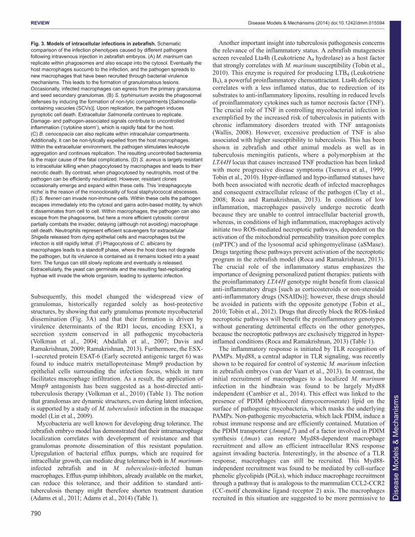
Fig. 3. Models of intracellular infections in zebrafish. Schematiccomparison of the infection phenotypes caused by different pathogensfollowing intravenous injection in zebrafish embryos. (A) M. marinum canreplicate within phagosomes and also escape into the cytosol. Eventually thehost macrophages succumb to the infection, and the pathogen spreads tonew macrophages that have been recruited through bacterial virulencemechanisms. This leads to the formation of granulomatous lesions.Occasionally, infected macrophages can egress from the primary granulomaand seed secondary granulomas. (B) S. typhimurium avoids the phagosomaldefenses by inducing the formation of non-lytic compartments [Salmonella-containing vacuoles (SCVs)]. Upon replication, the pathogen inducespyroptotic cell death. Extracellular Salmonella continues to replicate.Damage- and pathogen-associated signals contribute to uncontrolledinflammation (‘cytokine storm’), which is rapidly fatal for the host. (C) B. cenocepacia can also replicate within intracellular compartments.Additionally, it can be non-lytically expelled from the host macrophages.Within the extracellular environment, the pathogen stimulates leukocyteaggregation and continues replication. The resulting uncontrolled bacteremiais the major cause of the fatal complications. (D) S. aureus is largely resistantto intracellular killing when phagocytosed by macrophages and leads to theirnecrotic death. By contrast, when phagocytosed by neutrophils, most of thepathogen can be efficiently neutralized. However, resistant clonesoccasionally emerge and expand within these cells. This ‘intraphagocyteniche’ is the reason of the monoclonality of focal staphylococcal abscesses.(E) S. flexneri can invade non-immune cells. Within these cells the pathogenescapes immediately into the cytosol and gains actin-based motility, by whichit disseminates from cell to cell. Within macrophages, the pathogen can alsoescape from the phagosome, but here a more efficient cytosolic controlpartially combats the invader, delaying (although not avoiding) macrophagecell death. Neutrophils represent efficient scavengers for extracellularShigella released from dying epithelial cells and macrophages but theinfection is still rapidly lethal. (F) Phagocytosis of C. albicans bymacrophages leads to a standoff phase, where the host does not degradethe pathogen, but its virulence is contained as it remains locked into a yeastform. The fungus can still slowly replicate and eventually is released.Extracellularly, the yeast can germinate and the resulting fast-replicatinghyphae will invade the whole organism, leading to systemic infection.
Dis
ease
Mod
els
& M
echa
nism
s

intracellular bacterial growth, because they do not drive the strongintracellular RNS response. These observations might also explainwhy M. tuberculosis establishes infection in the lower rather than inthe higher respiratory tracts, because the latter is exposed to residentand environmental microbes that can make macrophages morecompetent for intracellular killing via a continuous transduction ofTLR-dependent immune signaling (Cambier et al., 2014).
Although macrophages are the main cell type infected by M.marinum following intravenous injection, neutrophils are alsoimportant for early infection control in zebrafish. In the earlygranuloma, the protective role of neutrophils was found to dependon ROS production (Yang et al., 2012). Prior to granulomaformation, neutrophils, both infected and non-infected, also produceRNS (Elks et al., 2013). This RNS production is dependent oninducible nitric oxide synthase (iNOS) and can be modulated bygenetic or pharmacological modulation of Hif-α (Hypoxia induciblefactor alpha) transcription factors. Increasing Hif-1α signaling ordecreasing Hif-2α signaling primes neutrophils with higher levels ofRNS prior to infection, thereby limiting susceptibility tomycobacterial infection. Increasing host RNS output by therapeutictargeting of the Hif-α pathway might shift the balance in favor of the
host and can thereby be explored as a strategy to complementantibiotic interventions (Table 1).
In addition to the classical microbicidal mechanisms ofmacrophages, antibacterial autophagy has emerged as an importantsupplementary control mechanism in mycobacterial infections(Deretic et al., 2013). Using the zebrafish model, colocalization ofmycobacteria with the autophagic marker Lc3 has beendemonstrated in vivo (van der Vaart et al., 2012). Moreover, actin-tail formation and recruitment of septin cages have been visualized,the latter also being associated with Lc3 and autophagy (Mostowyet al., 2013). The induction of dram1 (DNA damage-regulatedautophagy modulator 1) during infection suggests an immunologicalfunction of this autophagy modulator, and the zebrafish model couldbe further exploited to investigate therapeutic targeting of selectiveautophagy pathways (Mostowy et al., 2013; Meijer et al., 2014).
Very recently, a zebrafish larval model has also been establishedto study a rapidly growing mycobacterium, Mycobacteriumabscessus, an emerging pathogen that causes severe pulmonaryinfections in individuals with cystic fibrosis (Bernut et al., 2014).The study showed that the virulent rough morphotype of M.abscessus is transported to the central nervous system by
791
REVIEW Disease Models & Mechanisms (2014) doi:10.1242/dmm.015594
Table 1. Therapeutic strategies inspired by the zebrafish model to counteract intracellular infections
Target Susceptible pathogens Desired drug effect
Drugs tested in zebrafish Reference
Mmp9 (matrix metalloproteinase 9)
Mycobacteria Reduction of ESAT6–Mmp9-dependent macrophage infiltration to mycobacterial infection
Not tested Volkman et al., 2010
Efflux pumps (multiple bacterial genes)
Wide spectrum of bacteria
Prevention of drug-tolerance development mediated by a macrophage-induced efflux mechanism
Verapamil1,4, Reserpine1
Adams et al., 2011; Adams et al., 2014
GR (glucocorticoid receptor) Mycobacteria Suppression of detrimental inflammation in mycobacterial infection in patients with proinflammatory genotypes
Glucocorticoids1 Tobin et al., 2012
COXs (cyclooxygenases) Mycobacteria Suppression of detrimental inflammation in mycobacterial infection in patients with proinflammatory genotypes
Acetylsalicylic acid (aspirin)1
Tobin et al., 2012
BLT1 (LTB4 receptor) Mycobacteria Suppression of detrimental inflammation in mycobacterial infection in patients with proinflammatory genotypes
U-753023 Tobin et al., 2012
CYPD (cyclophilin D) Mycobacteria Prevention of mitochondrial permeability transition, limiting ROS-dependent necroptosis
Alisporivir2 Roca and Ramakrishnan, 2013
ASMase (acid sphingomyelinase) Mycobacteria Prevention of ceramide production, limiting ROS-dependent necroptosis
Desipramine1 Roca and Ramakrishnan, 2013
ROS (reactive oxygen species) Wide spectrum of intracellular pathogens
Scavenging of ROS N-acetylcysteine1, Amifostine1, Tempol1
Roca and Ramakrishnan, 2013
Hif-1 /Hif-2 pathway (hypoxia-induced factor 1 and 2 )
Wide spectrum of pathogens
Stimulation of protective Hif- and iNOS-mediated RNS production in neutrophils
Not tested Elks et al., 2013
iNOS (inducible nitric oxide synthase)
Wide spectrum of pathogens
Stimulation of protective RNS production and beneficial emergency hematopoiesis
Not tested Hall et al., 2012; Elks et al., 2013
Irg1 pathway (immunoresponsive gene 1)
Wide spectrum of pathogens
Modulation of fatty-acid catabolism and of mitochondrial ROS production
Not tested Hall et al., 2013
SQSTM1/p62 (sequestosome 1)
Wide spectrum of intracellular pathogens
Enhancement of infection control by selective autophagy mechanisms
Not tested Mostowy et al., 2013
The zebrafish host model has contributed extensively to the investigation of novel therapeutic strategies, oriented on modulating host-derived responses. Metalloproteinase inhibitors can reduce the tissue inflammatory response guiding phagocytes towards mycobacterial infections, thus attenuating the expansion of primary granulomas and the seeding of secondary infectious foci. Efflux pumps, although not representing a host target, impact directly on the capability of the pathogens to adapt to the intracellular growth and their blockade can reduce drug tolerance. Several classes of established anti-inflammatory drugs are beneficial for subsets of tuberculosis and leprosy patients, dependent on their genotypically determined inflammatory status (suppression of excessive inflammatory response). Levels of ROS and RNS work as a double-edged sword and their tight control can stimulate a positive outcome of the infectious process. Drugs that scavenge, suppress or boost ROS or RNS production are thereby valuable therapeutic tweezers to fine-tune their balance. Finally, the possibility of stimulating pathogen-selective autophagy is a promising therapeutic approach. Specific drugs tested in the zebrafish model in support of these approaches are listed: 1accepted drug; 2drug accepted for clinical trial; 3not accepted for clinical trial; 4drug validated in human THP1 macrophages infected with M. tuberculosis.
Dis
ease
Mod
els
& M
echa
nism
s

792
macrophages, where bacteria released from dying cells formmassive amounts of serpentine cords that grow too large to bephagocytosed, leading to acute and lethal infection. Furthermore, M.tuberculosis can also be disseminated by zebrafish larvalmacrophages and is sensitive to antibiotic treatment in this model(Carvalho et al., 2011). It will be of interest to investigate noveltherapeutic strategies emerging from the study of M. marinum(Table 1) also in the zebrafish models for these human pathogens.
Salmonella enterica serovar Typhimurium (S. typhimurium)Like many other Gram-negative enterobacteria, Salmonella caninfect a diverse range of hosts and cause zoonotic diseases (Fàbregaand Vila, 2013). The S. enterica serovar Typhimurium, often referredto as S. typhimurium, represents a common agent of enteric fever,gastroenteritis and bacteremia, often linked to food poisoning.Although the bacterium is not a natural fish pathogen, zebrafishembryos are strongly susceptible to S. typhimurium in experimentalsettings. Pathogenesis in fish involves some of the acute symptomsseen in humans, including bacteremia and a strong proinflammatoryhost response (‘cytokine storm’), which are associated with earlylethality of the zebrafish embryos (van der Sar et al., 2003;Stockhammer et al., 2009). The life cycle of S. typhimurium hasbeen well characterized in infected cell cultures and in mammaliansystems: the pathogen is able to alternate phases of intracellularreplication within phagocytes and extracellular growth within thedamaged tissue (Dunlap et al., 1992; Salcedo et al., 2001). Similarobservations have been made in the zebrafish embryo model. S.typhimurium, like M. marinum, can survive intracellularly inmacrophages of zebrafish embryos (Fig. 3B), but infected cells donot disseminate into tissues. Instead, following intravenous injection,the infection remains restricted to the vasculature, with bacteriamultiplying both in macrophages and extracellularly at theepithelium of blood vessels (van der Sar et al., 2003). Althoughinfection with wild-type bacteria causes early lethality, bacteria withmutations in lipopolysaccharide (LPS) are attenuated inmacrophages and are more sensitive to extracellular lysis, likely dueto complement factors (van der Sar et al., 2003).
In sharp contrast with M. marinum infection, S. typhimuriuminfection leads to a cytokine storm within hours after intravenousinjection (Stockhammer et al., 2009; Ordas et al., 2011; van derVaart et al., 2012). At the transcriptional level, this response issimilar to that observed in infection with the natural fish pathogenEdwardsiella tarda (van der Vaart et al., 2013). Thisproinflammatory transcriptional response provides a useful readoutfor characterizing the consequences of mutation or knockdown ofhost genes involved in the immune response. Deficiencies in theTLR signaling components Myd88 and Traf6 strongly reduceexpression of transcriptional regulators, signaling components andeffectors of the immune response (Stockhammer et al., 2010; vander Vaart et al., 2013). Conversely, these gene groups are hyper-induced following knockdown of the protein tyrosine phosphataseShp1 (also known as Ptpn6) (Kanwal et al., 2013). Theseobservations are consistent with the function of these genes inmammalian animal models and human patients, where MYD88 andTRAF6 mutations are associated with immunodeficiencies andSHP1 mutations cause inflammatory phenotypes and autoimmunedefects. Control of S. typhimurium and other infections in zebrafishis impaired both under conditions of a reduced or hyper-inducedimmune response, indicating the importance of highly balancedregulatory mechanisms (van der Vaart et al., 2013; Kanwal et al.,2013). Micro-RNAs (miRNAs), including members of the miR-146family, have been implicated in fine-tuning of the mammalian innate
immune response and, in zebrafish embryos, miR-146 is induced byS. typhimurium in a Myd88-Traf6-dependent manner. Although nomajor effects on known targets of the Myd88-Traf6 pathway wereobserved, apolipoprotein-mediated lipid transport emerged as anewly identified infection-inducible pathway under control of thismiRNA family (Ordas et al., 2013).
The signals involved in recruitment of phagocytes to localinfection remain to be elucidated. Chemokines, such as Cxcl8 (Il8)and the orphan ligand Cxcl-c1c, are highly induced rapidly upon S.typhimurium infection (Stockhammer et al., 2009). Using otherbacterial infection models, the function of the CXCL8-CXCR2signaling axis in neutrophil recruitment has been shown to beconserved in zebrafish (Sarris et al., 2012; Deng et al., 2013). LocalS. typhimurium infection has also been shown to induce therecruitment of macrophages via the chemokine receptor Cxcr3.2,one of the zebrafish orthologs of human CXCR3 (Zakrzewska et al.,2010). The ligand association of Cxcr3.2 remains to be established.
In addition to phagocyte recruitment, localized infection alsotriggers emergency-driven hematopoiesis (Hall et al., 2012). Earlyneutropenia is a frequent outcome of S. typhimurium hindbraininfection in zebrafish embryos, and this is compensated for byincreased granulopoiesis. The activity of iNOS (and thus theproduction of the pleiotropic mediator nitric oxide), was found to benecessary to stimulate the expansion and proliferation of HSPCs inresponse to infection-dependent neutropenia. The induction of iNOSis dependent on expression of the transcription factor C/ebpβ(CCAAT enhancer-binding protein β) in HSPCs. This is suggestedto be an effect of elevated circulating levels of Gcsf (Granulocytecolony stimulating factor), produced by activated macrophages atthe infection site (Hall et al., 2012).
The S. typhimurium infection model has recently led to newinsight into the connection between infection control and host cellmetabolic modulation (Hall et al., 2013). Profound adaptations inglucose and lipid metabolism occur within infected immune cells.For example, in response to stimulation by Salmonella pathogenicfactors, macrophages increase their uptake of lipids to fuel ROSproduction (Hall et al., 2013). In line with this, S. typhimuriuminfection induces the expression of the mitochondria-associatedenzyme Irg1 (Immunoresponsive gene 1) within infected zebrafishmacrophages. This protein directs the catabolism of fatty acids tosustain mitochondrial oxidative phosphorylation and in turn leads tothe production of mitochondrial ROS, contributing to intracellulardegradation of phagocytosed bacteria. Irg1 holds promise as a newtherapeutic target at the interface of inflammation and metabolism(Hall et al., 2013) (Table 1).
Burkholderia cenocepaciaThe B. cepacia complex (Bcc) is represented by several closelyrelated Gram-negative species that are able to survive freely in theenvironment or replicate within different hosts, including amoebae,invertebrates, vertebrates and plants (Mahenthiralingam et al., 2008).In humans, opportunistic infection by Bcc frequently occurs in cysticfibrosis or immunocompromised individuals and represents arecurrent cause of fatal complications (Saiman and Siegel, 2004).The capability to survive and infect a wide range of hosts suggeststhat Bcc species are highly adaptable to different niches and producemultiple virulence factors; however, the complex mechanisms ofhost-pathogen interactions underlying infections with B.cenocepacia and other Bcc strains remain largely unknown. Inparticular, it has been difficult to establish conclusively whether B.cenocepacia can survive intracellularly. Visualizing infection inzebrafish embryos helped to answer this key question, by
REVIEW Disease Models & Mechanisms (2014) doi:10.1242/dmm.015594
Dis
ease
Mod
els
& M
echa
nism
s

demonstrating the ability of this pathogen to survive withinmacrophages (Fig. 3C). Following the creation of anintramacrophage replication niche, the bacterial infectiondisseminates by non-lytic expulsion from infected cells, inducesimmune cell aggregations and ultimately causes fatal systemicbacteremia (Vergunst et al., 2010).
The zebrafish model system has also provided a valuablecontribution to the investigation of the in vivo relevance of severalB. cenocepacia virulence factors. A quorum-sensing-deficient cepRstrain was shown to be strongly attenuated, indicated by a reducedability to replicate intracellularly and to disseminate efficiently frominfected macrophages. Furthermore, differences in virulence wereobserved between strains from a panel of clinical isolates (Vergunstet al., 2010). Loss of the third replicon, pC3 (a non-essentialmegaplasmid associated with several virulence determinants), resultsin highly attenuated infection in multiple hosts, including zebrafishembryos (Agnoli et al., 2012; Agnoli et al., 2014). Mutants in pC3are not able to grow significantly in vivo, but are not eradicated,suggesting that the pC3-linked virulence factors are dispensable forintramacrophage survival (Agnoli et al., 2012). A function inadaptation to a wide range of environments, rather than a direct rolein modulating intracellular growth, might explain the prevalence ofthe pC3 replicon among Bcc isolates (Agnoli et al., 2014).
Together, these studies demonstrate the usefulness of the zebrafishmodel for analysis of B. cenocepacia mechanisms of intracellularsurvival and virulence.
Staphylococcus aureusS. aureus is the causative agent of a wide range of infectiouspathologies such as sty, pneumonia, endocarditis, osteomyelitis andsepticemia (Lowy, 1998), which remain important causes ofmorbidity and of complications in hospitalized patients. Althoughnot a natural pathogen of zebrafish, both embryos (Prajsnar et al.,2008) and adults (Lin et al., 2007) exhibit clear acute symptomswhen infected with this Gram-positive pathogen, providing a usefulmodel for bacteremia (Fig. 3D).
S. aureus has long been considered an extracellular pathogen, butthere is accumulating evidence that it can also survive and replicatein phagocytes (Rigby and DeLeo, 2012). The zebrafish embryomodel has contributed significantly to our understanding of thenature and relevance of the intracellular phase in the life cycle ofthis pathogen (Prajsnar et al., 2008; Prajsnar et al., 2012). Liveimaging showed that, upon intravenous infection, S. aureus iscompletely phagocytosed by macrophages and neutrophils (Prajsnaret al., 2008; Li and Hu, 2012). Although some embryos clear theinfection in a phagocyte-dependent manner, other embryos developoverwhelming infection, indicating that the bacteria can subvert thephagocyte-killing mechanisms (Prajsnar et al., 2012).
When larvae are co-infected with two isogenic, but differentlylabeled, S. aureus clones, the infection evolves by forming focallesions that are predominantly monoclonal and, during the course ofoverwhelming infection, the ratio between the original strains isoften skewed towards one predominating strain (Prajsnar et al.,2012). This phenomenon is fully dependent on phagocyte activity.These data suggest that most of the phagocytes are able to clear theinfection but a population of phagocytes provides an intracellularprotective niche in which some bacteria gain the ability to replicateand resist, to ultimately be released and disseminate. Consistent withthis, co-infection in a murine sepsis model resulted in kidneyabscesses that contained predominantly one strain of S. aureus andthus were likely founded by single bacteria (Holtfreter et al., 2013).The relevance of this work for clinical treatments is underscored by
a recent study showing that the use of sub-curative antibiotic dosescan support the preferential expansion of antibiotic-resistant bacteriaduring a mixed infection (McVicker et al., 2014). Selective ablationof macrophages or neutrophils in the zebrafish model has revealedthat neutrophils are most likely to form the privileged nicheresponsible for disseminated infection of S. aureus (Prajsnar et al.,2012). Interesting remaining questions include elucidation of themechanisms by which some bacteria from the initial inoculum areable to avoid being killed by neutrophils, and determination ofwhether S. aureus can also resist macrophages in vivo, as suggestedby human cell culture studies (Kubica et al., 2008).
Shigella flexneriS. flexneri, a human-adapted Escherichia coli species, is a causativeagent of diarrhea and dysentery in humans, generally deriving fromorofecal contaminations. Like other enterobacteria, it mostly affectsthe digestive tract; however, in advanced infectious stages, it canlead to bacteremia and systemic sepsis. In the early phase ofinfection, the pathogen can interact with membranes of host cells,inject virulence determinants, and induce ruffling and internalization(Ogawa et al., 2008). In this actively induced ingestion mechanismresides its capability to establish intracellular infection in non-phagocytic cells, such as epithelial cells associated with the digestivetract. Once it is internalized, it can escape from phagosomes andsurvive freely in the cytosol. Subsequently, the pathogen can spreadthrough intestinal epithelial cells by actin-based motility (Fig. 3E).Microfold cells allow Shigella to transverse the intestinal epithelium,where they encounter macrophages. Death of infected macrophagesand subsequent destabilization of the epithelium due toinflammation allows more Shigella to infiltrate the tissue and invadeepithelial cells through the basal membrane. Survival and replicationin macrophages, eventually followed by macrophage pyroptosis, isfundamental to allowing dissemination and extensive colonizationof the intestinal epithelium (Ashida et al., 2011).
Recently, a zebrafish model for S. flexneri was established, andthis has been used to show that intravenously administrated bacteriacan survive and replicate both in macrophages and in non-immunecells (Mostowy et al., 2013). The pathogenicity of Shigella is highlydependent on the presence of T3SS virulence factors, and avirulentstrains can be successfully combated by the zebrafish innate immunesystem and are not able to induce phagocytosis in non-phagocyticcells. Live imaging shows that replication of S. flexneri inmacrophages ultimately induces cell death, whereas bacteria aremore efficiently contained and degraded within neutrophils.Neutrophils also act as scavengers, eliminating infected dead cells.Macrophages are not able to retain the infection within vacuoles andbacteria spread into the cytosol, where they can colocalize with actinand septin cages (Fig. 3E).
In mammalian cultured cells, cytosolic S. flexneri can betargeted for autophagy via both ubiquitin-dependent and -independent pathways, and, as a counteractive mechanism, thebacteria can secrete virulence factors to escape autophagy (Ogawaet al., 2005; Mostowy et al., 2011). Colocalization with theautophagy marker Lc3 followed by electron microscopic analysisin the zebrafish model confirmed that autophagy targeting isassociated with entrapment of S. flexneri in septin cage-likestructures (Mostowy et al., 2010; Mostowy et al., 2013). Reductionof autophagy, via knockdown of the autophagy-related receptorp62, increases the infection burden of zebrafish larvae and thiseffect is specific only for the T3SS-positive strain that is able toescape into the cytosol (Mostowy et al., 2013). These data supportthe hypothesis that antibacterial protection provided by efficient
793
REVIEW Disease Models & Mechanisms (2014) doi:10.1242/dmm.015594
Dis
ease
Mod
els
& M
echa
nism
s

794
autophagic machinery is essential to properly counteract S. flexneriinfection. The ability to monitor S. flexneri infection in atransparent zebrafish host provides new possibilities to assess therelevance of autophagy in vivo in immune and non-immune cells,and to develop new strategies for anti-bacterial therapies targetingthis process (Table 1).
Candida albicansC. albicans is an opportunistic dimorphic fungus that grows in yeastand hyphal forms (Gow et al., 2011). Most of the human populationcarries C. albicans as a harmless constituent of the epidermal,mucosal and intestinal flora. However, uncontrolled systemiccandidiasis and fungal growth on the mucosal surfaces can causesevere and life-threatening infectious complications, particularly inimmunocompromised individuals. In zebrafish embryos, as inhumans, C. albicans can be phagocytosed by both neutrophils andmacrophages (Chao et al., 2010; Brothers et al., 2011). Liveobservations reveal that, in a non-compromised zebrafish host, thisintracellular localization leads to a transitory standoff phase in whichthe yeast form survives and replicates, but does not germinate orlyse the host cell (Fig. 3F). Subsequently, the fungi switch to themore virulent hyphal form and proliferate exuberantly in individualsthat fail to contain the infection, whereas they revert to the yeastform in most surviving embryos (Brothers et al., 2011). Intracellularyeast forms are unable to undergo the yeast-to-hyphal transition,even under conditions of impaired oxidative-stress response, incontrast with previous in vitro data in which filamentous growth wasobserved within cultured macrophages (Brothers et al., 2013).Therefore, macrophages apparently have an enhanced ability tocontrol infection in the in vivo environment.
Although germination was shown to be independent of thephagocyte-specific NADPH oxidase (PHOX), this enzyme was foundto be essential to produce an efficient oxidative-stress response againstC. albicans and to control filamentous growth (Brothers et al., 2011;Brothers et al., 2013). Previously, the limitation of fungal growth wasascribed mainly to direct fungal destruction by ROS; however,imaging in zebrafish revealed a non-canonical role for PHOX and forthe epithelial dual NADPH oxidase (DUOX) in recruitment ofphagocytes to C. albicans infection sites. Therefore, impairedphagocyte recruitment to invading Candida under conditions ofNADPH oxidase deficiency seems to be the cause of the overallreduction in containment of the infection and, consequently, ofmassive extracellular hyphal growth. Although localized infectionwith wild-type C. albicans is unable to induce chemoattraction underconditions of NADPH oxidase deficiency, infection with a yeast-locked mutant strain (edt1Δ/Δ) can be efficiently counteracted byphagocyte recruitment and internalization, even in pan-NADPH-oxidase-depleted conditions. This suggests that the hyphal transition(or another edt1-associated program) is also able to attenuate ROS-independent phagocyte recruitment, thus explaining the relevance ofhost ROS-driven chemoattraction mechanisms to counteract C.albicans infection (Brothers et al., 2013).
Concluding remarksThe study of intracellular pathogens in zebrafish macrophages hasled to new mechanistic insights that are inspiring novel host-directedtherapeutic strategies (Table 1). The real-time imaging possibilitiesin zebrafish will also be very useful for elucidating the mechanismsunderlying macrophage migration processes, as has already beendemonstrated by the study of neutrophils in the larval system (Sarriset al., 2012; Henry et al., 2013; Shelef et al., 2013). A question thatis very relevant both for infectious diseases and for cancer biology
concerns the presence of different pro- and anti-inflammatorymacrophage subtypes in zebrafish. Classically activated (M1) andalternatively activated (M2) macrophages, resembling thephenotypes of mammalian macrophages, have been identified indifferent fish species (Forlenza et al., 2011). That differentmacrophage subtypes might already be present in early zebrafishlarvae has been suggested, but this remains to be further investigated(Feng et al., 2010). The early larval stages, which are optimallysuited for imaging and for genetic and pharmacologicalinterventions, can give much information on the intracellularsurvival mechanisms of pathogens, as demonstrated by the studiesdiscussed herein. The early larval stages are also very useful forstudying the response of microglia to brain injuries or infection,contributing to a deeper understanding of the role of thesespecialized macrophages in neurodegenerative diseases (Sieger etal., 2012; Sieger and Peri, 2013). Studying the antigen-presentationfunction of macrophages and DCs at later developmental stages isbecoming increasingly feasible owing to advances in technologiesfor generating stable mutant lines (Clark et al., 2011; Blackburn etal., 2013; Kettleborough et al., 2013). Dynamic interactions betweenmacrophages and neutrophils that are emerging from recent studiesin zebrafish are of considerable interest for further study (Ellett etal., 2011; Yang et al., 2012; Elks et al., 2013). The use of thezebrafish model has already provided insights into the in vivorelevance of intracellular defense mechanisms such as ROS andRNS production and autophagy. We expect that further use of thispowerful model will continue to make important contributionstowards the understanding of innate immunity and of the virulencestrategies that pathogens use to subvert innate host defenses.
This article is part of a Special Issue, Spotlight on Zebrafish: Translational Impact.See all the articles in the issue at http://dmm.biologists.org/content/7/7.toc.
AcknowledgementsThe authors thank Georges Lutfalla (University of Montpellier II) and Graham J.Lieschke (Monash University) for the kind gift of the Tg(mpeg1:mCherry-F) andTg(mpeg1:Gal4-VP16/UAS-E1b:Kaede) lines, respectively. These lines were usedto make Fig. 2 and the movies.
Competing interestsThe authors declare no competing financial interests.
FundingV.T. is a Marie Curie fellow in the Initial Training Network FishForPharma (PITN-GA-2011-289209) funded by the 7th Framework Programme of the EuropeanCommission. S.M. is supported by a fellowship from the Higher EducationCommission of Pakistan. Work that led to this Review was additionally supportedby the European 7th framework project ZF-Health (FP7-Health-2009-242048), bythe Smart Mix Program (NWOA_6QY9BM) of The Netherlands Ministry ofEconomic Affairs and The Netherlands Ministry of Education, Culture and Science,and by the Leiden University Fund (LUF).
Supplementary materialSupplementary material available online athttp://dmm.biologists.org/lookup/suppl/doi:10.1242/dmm.015594/-/DC1
ReferencesAbdallah, A. M., Gey van Pittius, N. C., Champion, P. A., Cox, J., Luirink, J.,
Vandenbroucke-Grauls, C. M., Appelmelk, B. J. and Bitter, W. (2007). Type VIIsecretion – mycobacteria show the way. Nat. Rev. Microbiol. 5, 883-891.
Adams, K. N., Takaki, K., Connolly, L. E., Wiedenhoft, H., Winglee, K., Humbert,O., Edelstein, P. H., Cosma, C. L. and Ramakrishnan, L. (2011). Drug tolerance inreplicating mycobacteria mediated by a macrophage-induced efflux mechanism. Cell145, 39-53.
Adams, K. N., Szumowski, J. D. and Ramakrishnan, L. (2014). Verapamil, and itsmetabolite norverapamil, inhibit macrophage-induced, bacterial efflux pump-mediated tolerance to multiple anti-tubercular drugs. J. Infect. Dis. [Epub ahead ofprint] doi:10.1093/infdis/jiu095.
Agnoli, K., Schwager, S., Uehlinger, S., Vergunst, A., Viteri, D. F., Nguyen, D. T.,Sokol, P. A., Carlier, A. and Eberl, L. (2012). Exposing the third chromosome of
REVIEW Disease Models & Mechanisms (2014) doi:10.1242/dmm.015594
Dis
ease
Mod
els
& M
echa
nism
s

Burkholderia cepacia complex strains as a virulence plasmid. Mol. Microbiol. 83,362-378.
Agnoli, K., Frauenknecht, C., Freitag, R., Schwager, S., Jenul, C., Vergunst, A.,Carlier, A. and Eberl, L. (2014). The third replicon of members of the Burkholderiacepacia Complex, plasmid pC3, plays a role in stress tolerance. Appl. Environ.Microbiol. 80, 1340-1348.
Ashida, H., Ogawa, M., Kim, M., Suzuki, S., Sanada, T., Punginelli, C., Mimuro, H.and Sasakawa, C. (2011). Shigella deploy multiple countermeasures against hostinnate immune responses. Curr. Opin. Microbiol. 14, 16-23.
Baxt, L. A., Garza-Mayers, A. C. and Goldberg, M. B. (2013). Bacterial subversion ofhost innate immune pathways. Science 340, 697-701.
Benard, E. L., van der Sar, A. M., Ellett, F., Lieschke, G. J., Spaink, H. P. andMeijer, A. H. (2012). Infection of zebrafish embryos with intracellular bacterialpathogens. J. Vis. Exp. 61, 3781.
Bernut, A., Herrmann, J. L., Kissa, K., Dubremetz, J. F., Gaillard, J. L., Lutfalla, G.and Kremer, L. (2014). Mycobacterium abscessus cording prevents phagocytosisand promotes abscess formation. Proc. Natl. Acad. Sci. USA 111, E943-E952.
Bertin, J., Nir, W. J., Fischer, C. M., Tayber, O. V., Errada, P. R., Grant, J. R., Keilty,J. J., Gosselin, M. L., Robison, K. E., Wong, G. H. et al. (1999). Human CARD4protein is a novel CED-4/Apaf-1 cell death family member that activates NF-kappaB.J. Biol. Chem. 274, 12955-12958.
Bertrand, J. Y., Kim, A. D., Violette, E. P., Stachura, D. L., Cisson, J. L. and Traver,D. (2007). Definitive hematopoiesis initiates through a committed erythromyeloidprogenitor in the zebrafish embryo. Development 134, 4147-4156.
Bertrand, J. Y., Chi, N. C., Santoso, B., Teng, S., Stainier, D. Y. and Traver, D.(2010). Haematopoietic stem cells derive directly from aortic endothelium duringdevelopment. Nature 464, 108-111.
Bilan, D. S., Pase, L., Joosen, L., Gorokhovatsky, A. Y., Ermakova, Y. G., Gadella,T. W., Grabher, C., Schultz, C., Lukyanov, S. and Belousov, V. V. (2013). HyPer-3:a genetically encoded H(2)O(2) probe with improved performance for ratiometric andfluorescence lifetime imaging. ACS Chem. Biol. 8, 535-542.
Blackburn, P. R., Campbell, J. M., Clark, K. J. and Ekker, S. C. (2013). The CRISPRsystem – keeping zebrafish gene targeting fresh. Zebrafish 10, 116-118.
Boisset, J. C., van Cappellen, W., Andrieu-Soler, C., Galjart, N., Dzierzak, E. andRobin, C. (2010). In vivo imaging of haematopoietic cells emerging from the mouseaortic endothelium. Nature 464, 116-120.
Brothers, K. M., Newman, Z. R. and Wheeler, R. T. (2011). Live imaging ofdisseminated candidiasis in zebrafish reveals role of phagocyte oxidase in limitingfilamentous growth. Eukaryot. Cell 10, 932-944.
Brothers, K. M., Gratacap, R. L., Barker, S. E., Newman, Z. R., Norum, A. andWheeler, R. T. (2013). NADPH oxidase-driven phagocyte recruitment controls Candidaalbicans filamentous growth and prevents mortality. PLoS Pathog. 9, e1003634.
Cambier, C. J., Takaki, K. K., Larson, R. P., Hernandez, R. E., Tobin, D. M., Urdahl,K. B., Cosma, C. L. and Ramakrishnan, L. (2014). Mycobacteria manipulatemacrophage recruitment through coordinated use of membrane lipids. Nature 505,218-222.
Carvalho, R., de Sonneville, J., Stockhammer, O. W., Savage, N. D., Veneman, W.J., Ottenhoff, T. H., Dirks, R. P., Meijer, A. H. and Spaink, H. P. (2011). A high-throughput screen for tuberculosis progression. PLoS ONE 6, e16779.
Chao, C. C., Hsu, P. C., Jen, C. F., Chen, I. H., Wang, C. H., Chan, H. C., Tsai, P. W.,Tung, K. C., Wang, C. H., Lan, C. Y. et al. (2010). Zebrafish as a model host forCandida albicans infection. Infect. Immun. 78, 2512-2521.
Cheung, C. Y., Webb, S. E., Love, D. R. and Miller, A. L. (2011). Visualization,characterization and modulation of calcium signaling during the development of slowmuscle cells in intact zebrafish embryos. Int. J. Dev. Biol. 55, 153-174.
Clark, K. J., Voytas, D. F. and Ekker, S. C. (2011). A TALE of two nucleases: genetargeting for the masses? Zebrafish 8, 147-149.
Clay, H., Volkman, H. E. and Ramakrishnan, L. (2008). Tumor necrosis factorsignaling mediates resistance to mycobacteria by inhibiting bacterial growth andmacrophage death. Immunity 29, 283-294.
Colucci-Guyon, E., Tinevez, J. Y., Renshaw, S. A. and Herbomel, P. (2011).Strategies of professional phagocytes in vivo: unlike macrophages, neutrophilsengulf only surface-associated microbes. J. Cell Sci. 124, 3053-3059.
Cotton, M. and Claing, A. (2009). G protein-coupled receptors stimulation and thecontrol of cell migration. Cell. Signal. 21, 1045-1053.
Cronan, M. R. and Tobin, D. M. (2014). Fit for consumption: zebrafish as a model fortuberculosis. Dis. Model. Mech. 7, 777-784
Davis, J. M. and Ramakrishnan, L. (2009). The role of the granuloma in expansionand dissemination of early tuberculous infection. Cell 136, 37-49.
Davis, J. M., Clay, H., Lewis, J. L., Ghori, N., Herbomel, P. and Ramakrishnan, L.(2002). Real-time visualization of mycobacterium-macrophage interactions leading toinitiation of granuloma formation in zebrafish embryos. Immunity 17, 693-702.
Deng, Q., Sarris, M., Bennin, D. A., Green, J. M., Herbomel, P. and Huttenlocher,A. (2013). Localized bacterial infection induces systemic activation of neutrophilsthrough Cxcr2 signaling in zebrafish. J. Leukoc. Biol. 93, 761-769.
Deretic, V., Saitoh, T. and Akira, S. (2013). Autophagy in infection, inflammation andimmunity. Nat. Rev. Immunol. 13, 722-737.
Duclos, S. and Desjardins, M. (2000). Subversion of a young phagosome: thesurvival strategies of intracellular pathogens. Cell. Microbiol. 2, 365-377.
Dunlap, N. E., Benjamin, W. H., Jr, Berry, A. K., Eldridge, J. H. and Briles, D. E.(1992). A ‘safe-site’ for Salmonella typhimurium is within splenic polymorphonuclearcells. Microb. Pathog. 13, 181-190.
El-Gayar, S., Thüring-Nahler, H., Pfeilschifter, J., Röllinghoff, M. and Bogdan, C.(2003). Translational control of inducible nitric oxide synthase by IL-13 and arginineavailability in inflammatory macrophages. J. Immunol. 171, 4561-4568.
Elks, P. M., Brizee, S., van der Vaart, M., Walmsley, S. R., van Eeden, F. J.,Renshaw, S. A. and Meijer, A. H. (2013). Hypoxia inducible factor signalingmodulates susceptibility to mycobacterial infection via a nitric oxide dependentmechanism. PLoS Pathog. 9, e1003789.
Ellett, F., Pase, L., Hayman, J. W., Andrianopoulos, A. and Lieschke, G. J. (2011).mpeg1 promoter transgenes direct macrophage-lineage expression in zebrafish.Blood 117, e49-e56.
Elomaa, O., Kangas, M., Sahlberg, C., Tuukkanen, J., Sormunen, R., Liakka, A.,Thesleff, I., Kraal, G. and Tryggvason, K. (1995). Cloning of a novel bacteria-binding receptor structurally related to scavenger receptors and expressed in asubset of macrophages. Cell 80, 603-609.
Fàbrega, A. and Vila, J. (2013). Salmonella enterica serovar Typhimurium skills tosucceed in the host: virulence and regulation. Clin. Microbiol. Rev. 26, 308-341.
Feng, Y., Santoriello, C., Mione, M., Hurlstone, A. and Martin, P. (2010). Liveimaging of innate immune cell sensing of transformed cells in zebrafish larvae:parallels between tumor initiation and wound inflammation. PLoS Biol. 8, e1000562.
Forlenza, M., Fink, I. R., Raes, G. and Wiegertjes, G. F. (2011). Heterogeneity ofmacrophage activation in fish. Dev. Comp. Immunol. 35, 1246-1255.
Gengenbacher, M. and Kaufmann, S. H. (2012). Mycobacterium tuberculosis:success through dormancy. FEMS Microbiol. Rev. 36, 514-532.
Gow, N. A., van de Veerdonk, F. L., Brown, A. J. and Netea, M. G. (2011).Candida albicans morphogenesis and host defence: discriminating invasion fromcolonization. Nat. Rev. Microbiol. 10, 112-122.
Gray, C., Loynes, C. A., Whyte, M. K., Crossman, D. C., Renshaw, S. A. andChico, T. J. (2011). Simultaneous intravital imaging of macrophage and neutrophilbehaviour during inflammation using a novel transgenic zebrafish. Thromb.Haemost. 105, 811-819.
Hall, C., Flores, M. V., Chien, A., Davidson, A., Crosier, K. and Crosier, P. (2009).Transgenic zebrafish reporter lines reveal conserved Toll-like receptor signalingpotential in embryonic myeloid leukocytes and adult immune cell lineages. J.Leukoc. Biol. 85, 751-765.
Hall, C. J., Flores, M. V., Oehlers, S. H., Sanderson, L. E., Lam, E. Y., Crosier, K.E. and Crosier, P. S. (2012). Infection-responsive expansion of the hematopoieticstem and progenitor cell compartment in zebrafish is dependent upon inducible nitricoxide. Cell Stem Cell 10, 198-209.
Hall, C. J., Boyle, R. H., Astin, J. W., Flores, M. V., Oehlers, S. H., Sanderson, L.E., Ellett, F., Lieschke, G. J., Crosier, K. E. and Crosier, P. S. (2013).Immunoresponsive gene 1 augments bactericidal activity of macrophage-lineagecells by regulating β-oxidation-dependent mitochondrial ROS production. Cell Metab.18, 265-278.
Henry, K. M., Loynes, C. A., Whyte, M. K. and Renshaw, S. A. (2013). Zebrafishas a model for the study of neutrophil biology. J. Leukoc. Biol. 94, 633-642.
Herbomel, P. and Levraud, J. P. (2005). Imaging early macrophage differentiation,migration, and behaviors in live zebrafish embryos. Methods Mol. Med. 105, 199-214.
Herbomel, P., Thisse, B. and Thisse, C. (1999). Ontogeny and behaviour of earlymacrophages in the zebrafish embryo. Development 126, 3735-3745.
Herbomel, P., Thisse, B. and Thisse, C. (2001). Zebrafish early macrophagescolonize cephalic mesenchyme and developing brain, retina, and epidermis througha M-CSF receptor-dependent invasive process. Dev. Biol. 238, 274-288.
Hilbi, H., Moss, J. E., Hersh, D., Chen, Y., Arondel, J., Banerjee, S., Flavell, R.A., Yuan, J., Sansonetti, P. J. and Zychlinsky, A. (1998). Shigella-inducedapoptosis is dependent on caspase-1 which binds to IpaB. J. Biol. Chem. 273,32895-32900.
Holtfreter, S., Radcliff, F. J., Grumann, D., Read, H., Johnson, S., Monecke, S.,Ritchie, S., Clow, F., Goerke, C., Bröker, B. M. et al. (2013). Characterization ofa mouse-adapted Staphylococcus aureus strain. PLoS ONE 8, e71142.
Inohara, N., Koseki, T., del Peso, L., Hu, Y., Yee, C., Chen, S., Carrio, R.,Merino, J., Liu, D., Ni, J. et al. (1999). Nod1, an Apaf-1-like activator of caspase-9and nuclear factor-kappaB. J. Biol. Chem. 274, 14560-14567.
Jagannathan-Bogdan, M. and Zon, L. I. (2013). Hematopoiesis. Development 140,2463-2467.
Kanwal, Z., Zakrzewska, A., den Hertog, J., Spaink, H. P., Schaaf, M. J. andMeijer, A. H. (2013). Deficiency in hematopoietic phosphatase ptpn6/Shp1hyperactivates the innate immune system and impairs control of bacterial infectionsin zebrafish embryos. J. Immunol. 190, 1631-1645.
Kettleborough, R. N., Busch-Nentwich, E. M., Harvey, S. A., Dooley, C. M., deBruijn, E., van Eeden, F., Sealy, I., White, R. J., Herd, C., Nijman, I. J. et al.(2013). A systematic genome-wide analysis of zebrafish protein-coding genefunction. Nature 496, 494-497.
Kissa, K. and Herbomel, P. (2010). Blood stem cells emerge from aortic endotheliumby a novel type of cell transition. Nature 464, 112-115.
Kubica, M., Guzik, K., Koziel, J., Zarebski, M., Richter, W., Gajkowska, B.,Golda, A., Maciag-Gudowska, A., Brix, K., Shaw, L. et al. (2008). A potentialnew pathway for Staphylococcus aureus dissemination: the silent survival of S.aureus phagocytosed by human monocyte-derived macrophages. PLoS ONE 3,e1409.
Lam, S. H., Chua, H. L., Gong, Z., Lam, T. J. and Sin, Y. M. (2004). Developmentand maturation of the immune system in zebrafish, Danio rerio: a gene expressionprofiling, in situ hybridization and immunological study. Dev. Comp. Immunol. 28, 9-28.
Li, Y. J. and Hu, B. (2012). Establishment of multi-site infection model in zebrafishlarvae for studying Staphylococcus aureus infectious disease. J. Genet. Genomics39, 521-534.
795
REVIEW Disease Models & Mechanisms (2014) doi:10.1242/dmm.015594
Dis
ease
Mod
els
& M
echa
nism
s

796
Li, L., Jin, H., Xu, J., Shi, Y. and Wen, Z. (2011). Irf8 regulates macrophage versusneutrophil fate during zebrafish primitive myelopoiesis. Blood 117, 1359-1369.
Lin, B., Chen, S., Cao, Z., Lin, Y., Mo, D., Zhang, H., Gu, J., Dong, M., Liu, Z. andXu, A. (2007). Acute phase response in zebrafish upon Aeromonas salmonicida andStaphylococcus aureus infection: striking similarities and obvious differences withmammals. Mol. Immunol. 44, 295-301.
Lin, P. L., Rodgers, M., Smith, L., Bigbee, M., Myers, A., Bigbee, C., Chiosea, I.,Capuano, S. V., Fuhrman, C., Klein, E. et al. (2009). Quantitative comparison ofactive and latent tuberculosis in the cynomolgus macaque model. Infect. Immun. 77,4631-4642.
Lowy, F. D. (1998). Staphylococcus aureus infections. N. Engl. J. Med. 339, 520-532. Mahenthiralingam, E., Baldwin, A. and Dowson, C. G. (2008). Burkholderia
cepacia complex bacteria: opportunistic pathogens with important natural biology. J.Appl. Microbiol. 104, 1539-1551.
Martinon, F., Burns, K. and Tschopp, J. (2002). The inflammasome: a molecularplatform triggering activation of inflammatory caspases and processing of proIL-beta.Mol. Cell 10, 417-426.
Masaki, T., Qu, J., Cholewa-Waclaw, J., Burr, K., Raaum, R. and Rambukkana,A. (2013). Reprogramming adult Schwann cells to stem cell-like cells by leprosybacilli promotes dissemination of infection. Cell 152, 51-67.
McVicker, G., Prajsnar, T. K., Williams, A., Wagner, N. L., Boots, M., Renshaw,S. A. and Foster, S. J. (2014). Clonal expansion during Staphylococcus aureusinfection dynamics reveals the effect of antibiotic intervention. PLoS Pathog. 10,e1003959.
Meijer, A. H., van der Sar, A. M., Cunha, C., Lamers, G. E., Laplante, M. A.,Kikuta, H., Bitter, W., Becker, T. S. and Spaink, H. P. (2008). Identification andreal-time imaging of a myc-expressing neutrophil population involved in inflammationand mycobacterial granuloma formation in zebrafish. Dev. Comp. Immunol. 32, 36-49.
Meijer, A. H., van der Vaart, M. and Spaink, H. P. (2014). Real-time imaging andgenetic dissection of host-microbe interactions in zebrafish. Cell. Microbiol. 16, 39-49. PubMed
Minakami, R. and Sumimotoa, H. (2006). Phagocytosis-coupled activation of thesuperoxide-producing phagocyte oxidase, a member of the NADPH oxidase (nox)family. Int. J. Hematol. 84, 193-198.
Mostowy, S., Bonazzi, M., Hamon, M. A., Tham, T. N., Mallet, A., Lelek, M.,Gouin, E., Demangel, C., Brosch, R., Zimmer, C. et al. (2010). Entrapment ofintracytosolic bacteria by septin cage-like structures. Cell Host Microbe 8, 433-444.
Mostowy, S., Sancho-Shimizu, V., Hamon, M. A., Simeone, R., Brosch, R.,Johansen, T. and Cossart, P. (2011). p62 and NDP52 proteins target intracytosolicShigella and Listeria to different autophagy pathways. J. Biol. Chem. 286, 26987-26995.
Mostowy, S., Boucontet, L., Mazon Moya, M. J., Sirianni, A., Boudinot, P.,Hollinshead, M., Cossart, P., Herbomel, P., Levraud, J. P. and Colucci-Guyon, E. (2013). The zebrafish as a new model for the in vivo study of Shigellaflexneri interaction with phagocytes and bacterial autophagy. PLoS Pathog. 9,e1003588.
Murayama, E., Kissa, K., Zapata, A., Mordelet, E., Briolat, V., Lin, H. F., Handin, R.I. and Herbomel, P. (2006). Tracing hematopoietic precursor migration to successivehematopoietic organs during zebrafish development. Immunity 25, 963-975.
O’Neill, L. A., Golenbock, D. and Bowie, A. G. (2013). The history of Toll-likereceptors – redefining innate immunity. Nat. Rev. Immunol. 13, 453-460.
Ogawa, M., Yoshimori, T., Suzuki, T., Sagara, H., Mizushima, N. and Sasakawa,C. (2005). Escape of intracellular Shigella from autophagy. Science 307, 727-731.
Ogawa, M., Handa, Y., Ashida, H., Suzuki, M. and Sasakawa, C. (2008). Theversatility of Shigella effectors. Nat. Rev. Microbiol. 6, 11-16.
Ordas, A., Hegedus, Z., Henkel, C. V., Stockhammer, O. W., Butler, D., Jansen,H. J., Racz, P., Mink, M., Spaink, H. P. and Meijer, A. H. (2011). Deep sequencingof the innate immune transcriptomic response of zebrafish embryos to Salmonellainfection. Fish Shellfish Immunol. 31, 716-724.
Ordas, A., Kanwal, Z., Lindenberg, V., Rougeot, J., Mink, M., Spaink, H. P. andMeijer, A. H. (2013). MicroRNA-146 function in the innate immune transcriptomeresponse of zebrafish embryos to Salmonella typhimurium infection. BMC Genomics14, 696.
Ottenhoff, T. H. and Kaufmann, S. H. (2012). Vaccines against tuberculosis: whereare we and where do we need to go? PLoS Pathog. 8, e1002607.
Page, D. M., Wittamer, V., Bertrand, J. Y., Lewis, K. L., Pratt, D. N., Delgado, N.,Schale, S. E., McGue, C., Jacobsen, B. H., Doty, A. et al. (2013). Anevolutionarily conserved program of B-cell development and activation in zebrafish.Blood 122, e1-e11.
Parikka, M., Hammarén, M. M., Harjula, S. K., Halfpenny, N. J., Oksanen, K. E.,Lahtinen, M. J., Pajula, E. T., Iivanainen, A., Pesu, M. and Rämet, M. (2012).Mycobacterium marinum causes a latent infection that can be reactivated by gammairradiation in adult zebrafish. PLoS Pathog. 8, e1002944.
Park, Y. K., Bearson, B., Bang, S. H., Bang, I. S. and Foster, J. W. (1996). InternalpH crisis, lysine decarboxylase and the acid tolerance response of Salmonellatyphimurium. Mol. Microbiol. 20, 605-611.
Peri, F. and Nüsslein-Volhard, C. (2008). Live imaging of neuronal degradation bymicroglia reveals a role for v0-ATPase a1 in phagosomal fusion in vivo. Cell 133,916-927.
Prajsnar, T. K., Cunliffe, V. T., Foster, S. J. and Renshaw, S. A. (2008). A novelvertebrate model of Staphylococcus aureus infection reveals phagocyte-dependentresistance of zebrafish to non-host specialized pathogens. Cell. Microbiol. 10, 2312-2325.
Prajsnar, T. K., Hamilton, R., Garcia-Lara, J., McVicker, G., Williams, A., Boots,M., Foster, S. J. and Renshaw, S. A. (2012). A privileged intraphagocyte niche is
responsible for disseminated infection of Staphylococcus aureus in a zebrafishmodel. Cell. Microbiol. 14, 1600-1619.
Prouty, M. G., Correa, N. E., Barker, L. P., Jagadeeswaran, P. and Klose, K. E.(2003). Zebrafish-Mycobacterium marinum model for mycobacterial pathogenesis.FEMS Microbiol. Lett. 225, 177-182.
Ramakrishnan, L. (2013). Looking within the zebrafish to understand the tuberculousgranuloma. Adv. Exp. Med. Biol. 783, 251-266.
Renshaw, S. A. and Trede, N. S. (2012). A model 450 million years in the making:zebrafish and vertebrate immunity. Dis. Model. Mech. 5, 38-47.
Rigby, K. M. and DeLeo, F. R. (2012). Neutrophils in innate host defense againstStaphylococcus aureus infections. Semin. Immunopathol. 34, 237-259.
Roca, F. J. and Ramakrishnan, L. (2013). TNF dually mediates resistance andsusceptibility to mycobacteria via mitochondrial reactive oxygen species. Cell 153,521-534.
Saiman, L. and Siegel, J. (2004). Infection control in cystic fibrosis. Clin. Microbiol.Rev. 17, 57-71.
Salcedo, S. P., Noursadeghi, M., Cohen, J. and Holden, D. W. (2001). Intracellularreplication of Salmonella typhimurium strains in specific subsets of splenicmacrophages in vivo. Cell. Microbiol. 3, 587-597.
Sarris, M., Masson, J. B., Maurin, D., Van der Aa, L. M., Boudinot, P., Lortat-Jacob, H. and Herbomel, P. (2012). Inflammatory chemokines direct and restrictleukocyte migration within live tissues as glycan-bound gradients. Curr. Biol. 22,2375-2382.
Schmidtchen, A., Frick, I. M., Andersson, E., Tapper, H. and Björck, L. (2002).Proteinases of common pathogenic bacteria degrade and inactivate the antibacterialpeptide LL-37. Mol. Microbiol. 46, 157-168.
Shelef, M. A., Tauzin, S. and Huttenlocher, A. (2013). Neutrophil migration: movingfrom zebrafish models to human autoimmunity. Immunol. Rev. 256, 269-281.
Sieger, D. and Peri, F. (2013). Animal models for studying microglia: the first, thepopular, and the new. Glia 61, 3-9.
Sieger, D., Moritz, C., Ziegenhals, T., Prykhozhij, S. and Peri, F. (2012). Long-range Ca2+ waves transmit brain-damage signals to microglia. Dev. Cell 22, 1138-1148.
Stachura, D. L. and Traver, D. (2011). Cellular dissection of zebrafish hematopoiesis.Methods Cell Biol. 101, 75-110.
Stockhammer, O. W., Zakrzewska, A., Hegedûs, Z., Spaink, H. P. and Meijer, A.H. (2009). Transcriptome profiling and functional analyses of the zebrafish embryonicinnate immune response to Salmonella infection. J. Immunol. 182, 5641-5653.
Stockhammer, O. W., Rauwerda, H., Wittink, F. R., Breit, T. M., Meijer, A. H. andSpaink, H. P. (2010). Transcriptome analysis of Traf6 function in the innate immuneresponse of zebrafish embryos. Mol. Immunol. 48, 179-190.
Su, F., Juarez, M. A., Cooke, C. L., Lapointe, L., Shavit, J. A., Yamaoka, J. S.and Lyons, S. E. (2007). Differential regulation of primitive myelopoiesis in thezebrafish by Spi-1/Pu.1 and C/ebp1. Zebrafish 4, 187-199.
Svahn, A. J., Graeber, M. B., Ellett, F., Lieschke, G. J., Rinkwitz, S., Bennett, M.R. and Becker, T. S. (2013). Development of ramified microglia from earlymacrophages in the zebrafish optic tectum. Dev. Neurobiol. 73, 60-71.
Swaim, L. E., Connolly, L. E., Volkman, H. E., Humbert, O., Born, D. E. andRamakrishnan, L. (2006). Mycobacterium marinum infection of adult zebrafishcauses caseating granulomatous tuberculosis and is moderated by adaptiveimmunity. Infect. Immun. 74, 6108-6117.
Takahashi, K., Naito, M. and Takeya, M. (1996). Development and heterogeneity ofmacrophages and their related cells through their differentiation pathways. Pathol.Int. 46, 473-485.
Tobin, D. M., Vary, J. C., Jr, Ray, J. P., Walsh, G. S., Dunstan, S. J., Bang, N. D.,Hagge, D. A., Khadge, S., King, M. C., Hawn, T. R. et al. (2010). The lta4h locusmodulates susceptibility to mycobacterial infection in zebrafish and humans. Cell140, 717-730.
Tobin, D. M., Roca, F. J., Oh, S. F., McFarland, R., Vickery, T. W., Ray, J. P., Ko,D. C., Zou, Y., Bang, N. D., Chau, T. T. et al. (2012). Host genotype-specifictherapies can optimize the inflammatory response to mycobacterial infections. Cell148, 434-446.
Tsenova, L., Bergtold, A., Freedman, V. H., Young, R. A. and Kaplan, G. (1999).Tumor necrosis factor alpha is a determinant of pathogenesis and diseaseprogression in mycobacterial infection in the central nervous system. Proc. Natl.Acad. Sci. USA 96, 5657-5662.
van der Sar, A. M., Musters, R. J., van Eeden, F. J., Appelmelk, B. J.,Vandenbroucke-Grauls, C. M. and Bitter, W. (2003). Zebrafish embryos as amodel host for the real time analysis of Salmonella typhimurium infections. Cell.Microbiol. 5, 601-611.
van der Vaart, M., Spaink, H. P. and Meijer, A. H. (2012). Pathogen recognition andactivation of the innate immune response in zebrafish. Adv. Hematol. 2012, 159807.
van der Vaart, M., van Soest, J. J., Spaink, H. P. and Meijer, A. H. (2013).Functional analysis of a zebrafish myd88 mutant identifies key transcriptionalcomponents of the innate immune system. Dis. Model. Mech. 6, 841-854.
Vandal, O. H., Pierini, L. M., Schnappinger, D., Nathan, C. F. and Ehrt, S. (2008).A membrane protein preserves intrabacterial pH in intraphagosomal Mycobacteriumtuberculosis. Nat. Med. 14, 849-854.
Vergunst, A. C., Meijer, A. H., Renshaw, S. A. and O’Callaghan, D. (2010).Burkholderia cenocepacia creates an intramacrophage replication niche in zebrafishembryos, followed by bacterial dissemination and establishment of systemicinfection. Infect. Immun. 78, 1495-1508.
Volkman, H. E., Clay, H., Beery, D., Chang, J. C., Sherman, D. R. andRamakrishnan, L. (2004). Tuberculous granuloma formation is enhanced by amycobacterium virulence determinant. PLoS Biol. 2, e367.
REVIEW Disease Models & Mechanisms (2014) doi:10.1242/dmm.015594
Dis
ease
Mod
els
& M
echa
nism
s

Volkman, H. E., Pozos, T. C., Zheng, J., Davis, J. M., Rawls, J. F. andRamakrishnan, L. (2010). Tuberculous granuloma induction via interaction of abacterial secreted protein with host epithelium. Science 327, 466-469.
Wallis, R. S. (2008). Tumour necrosis factor antagonists: structure, function, andtuberculosis risks. Lancet Infect. Dis. 8, 601-611.
Wang, Y., Curry, H. M., Zwilling, B. S. and Lafuse, W. P. (2005). Mycobacteriainhibition of IFN-gamma induced HLA-DR gene expression by up-regulating histonedeacetylation at the promoter region in human THP-1 monocytic cells. J. Immunol.174, 5687-5694.
Wittamer, V., Bertrand, J. Y., Gutschow, P. W. and Traver, D. (2011).Characterization of the mononuclear phagocyte system in zebrafish. Blood 117,7126-7135.
Xu, L. L., Warren, M. K., Rose, W. L., Gong, W. and Wang, J. M. (1996). Humanrecombinant monocyte chemotactic protein and other C-C chemokines bind andinduce directional migration of dendritic cells in vitro. J. Leukoc. Biol. 60, 365-371.
Yang, C. T., Cambier, C. J., Davis, J. M., Hall, C. J., Crosier, P. S. andRamakrishnan, L. (2012). Neutrophils exert protection in the early tuberculousgranuloma by oxidative killing of mycobacteria phagocytosed from infectedmacrophages. Cell Host Microbe 12, 301-312.
Yoo, S. K., Starnes, T. W., Deng, Q. and Huttenlocher, A. (2011). Lyn is a redoxsensor that mediates leukocyte wound attraction in vivo. Nature 480, 109-112.
Zakrzewska, A., Cui, C., Stockhammer, O. W., Benard, E. L., Spaink, H. P. andMeijer, A. H. (2010). Macrophage-specific gene functions in Spi1-directed innateimmunity. Blood 116, e1-e11.
797
REVIEW Disease Models & Mechanisms (2014) doi:10.1242/dmm.015594
Dis
ease
Mod
els
& M
echa
nism
s