Embed Size (px)
Citation preview

M1 AND M4 MUSCARINIC ACETYLCHOLINE RECEPTOR REGULATION OF
NEUROTRANSMISSION AND CELL EXCITABILITY IN RODENT
HIPPOCAMPUS AND PREFRONTAL CORTEX
By
Jana Kristin Shirey-Rice
Dissertation
Submitted to the Faculty of the
Graduate School of Vanderbilt University
in partial fulfillment of the requirements
for the degree of
DOCTOR OF PHILOSOPHY
in
Pharmacology
May, 2010
Nashville, Tennessee
Approved:
Professor P. Jeffrey Conn
Professor Ariel Y. Deutch
Professor Danny G. Winder
Professor C. David Weaver

ABSTRACT
Muscarinic acetylcholine receptors (mAChRs), specifically M1 and M4 subtypes,
provide viable targets for the treatment of multiple central nervous system disorders.
However, highly selective activators of either M1 or M4 have not been available, making
it difficult to determine the in vivo effects of selective activation of these receptors. We
have used cheminformatics and medicinal chemistry to develop new, highly selective M1
and M4 positive allosteric modulators (PAMs). VU10010 potentiated the functional M4
response to acetylcholine while having no activity at other mAChR subtypes. Whole-cell
patch clamp recordings revealed that VU10010 increased carbachol-induced depression
of transmission at excitatory but not inhibitory synapses at the Schaffer collateral-CA1
(SC-CA1) synapse in the hippocampus. Chemical optimization of VU10010 afforded
two centrally penetrant analogs, VU0152099 and VU0152100, which are also potent,
selective M4 PAMs. Interestingly, these compounds reversed amphetamine-induced
hyperlocomotion in rats, a model that is predictive of clinical antipsychotic efficacy in
humans.
A growing body of literature also supports M1 receptors as a viable target for
treatment of disorders involving impaired cognitive function. Data in this thesis reports
the molecular characterization of a novel compound, BQCA, which is a potent, highly
selective PAM of the rat M1 receptor. BQCA induced a robust inward current and
increased spontaneous EPSCs in mPFC layer V pyramidal cells, effects which were
absent in acute slices from M1 receptor knockout mice. Furthermore, multiple single-unit
recordings were obtained from the mPFC of rats which showed that BQCA increased
ii

firing of pyramidal cells in vivo. BQCA also restored discrimination reversal learning in
a transgenic mouse model of AD and regulated non-amyloidogenic APP processing in
vitro.
Together, these studies provide compelling evidence while M4 inhibits excitatory
transmission at the SC-CA1 synapse, M1 receptor activation induces a dramatic excitation
of PFC neurons. Newly developed highly selective ligands that activate or potentiate M1
and M4 provide exciting tools that will be useful in further delineating the individual roles
of these receptors in the efficacy of drugs like acetyl cholinesterase inhibitors and
xanomeline.
iii

ACKNOWLEDGEMENTS
First and foremost, I want to extend my gratitude to my advisor, Dr. P. Jeffrey
Conn for allowing me to join his laboratory and perform my graduate work there. Dr.
Conn has provided unwavering support and guidance throughout the time I’ve spent as a
graduate student in his lab. He has served as a role model for me not only as a scientist
but as a person; he has such a passion for science coupled with an exceptionally kind
heart and a genuine desire to contribute to our understanding of human disease with the
ultimate goal being the development of better treatments for devastating CNS disorders.
I have been continually inspired not just by his relentless pursuit of scientific progress but
by the dignity and diplomacy with which he does so. His ability to bring together a
bright, like-minded group of researchers has also greatly contributed to my happiness and
success in lab over the years. I thank all the members of the Conn lab, past and present,
who have infinitely blessed and enriched my life both in and outside the lab. None of the
research in this thesis would have been possible without my funding sources (NIH T32
Training Grant and NIMH Ruth L. Kirschstein Individual Predoctoral National Research
Service Award). I would like to acknowledge my thesis committee members; Drs. Ariel
Deutch, C. David Weaver, and Danny Winder have taught me so much. They were
always available to answer questions and not only offer direction for my thesis project
but also to give great advice on how to successfully maneuver my way through graduate
school life. They gave me confidence through the many times when I doubted my
abilities or my work and helped me believe in myself because they did. I’m especially
iv

grateful for invaluable advice from Dr. Winder on how to find and obtain a post-doctoral
position during the final stages of my thesis research.
My family and friends have also provided endless support. I’m so blessed to have
parents who always believe I can do anything I set out to do; thank you, mom and dad,
for constantly reminding me how proud you are of me and for your encouragement
throughout every stage of my life. I am so grateful for the friendships I’ve developed
with my lab mates over the years. The numerous members of the Conn lab have
committed tremendous time and effort to my training and have been great mentors and
friends to me. My heartfelt gratitude goes to Drs. Ashley Brady, Alice Rodriguez, Doug
Sheffler, Zixiu Xiang, Paulianda Jones, Jennifer Ayala, and Meredith Noetzel not only
for their help with experiments and publications but also for their amazing friendship and
boundless support. I would also like to thank Dr. Elaine Sanders-Bush for giving me a
job as a research assistant before I entered graduate school; it was in her lab that I first
discovered my interests in pharmacology and neuroscience. She was such a shining
example of a strong, bright, determined female scientist who really inspired me.
I would finally like to acknowledge my husband, Jarrett, who has been a
consistently positive force in my life; he kept me grounded as I faced the difficult and
stressful times that are part of getting a graduate degree. He so often saw me at my worst
and showed me nothing but open arms. I couldn’t imagine a more perfect person with
whom to share this exciting time in my life. I contribute much of my success to his
selfless support, and I am deeply grateful to have found my perfect match.
v
TABLE OF CONTENTS
Page
ABSTRACT ...........................................................................................................................ii ACKNOWLEDGEMENTS ...................................................................................................iv LIST OF TABLES .................................................................................................................x LIST OF FIGURES ...............................................................................................................xi LIST OF ABBREVIATIONS ................................................................................................xiv Chapter I. INTRODUCTION .............................................................................................................1 Cholinergic biosynthesis and neuroanatomy ...............................................................1 Acetylcholine receptors ......................................................................................4 Muscarinic acetylcholine receptor subtypes, signaling, and function ................4 Muscarinic regulation of hippocampal physiology ......................................................10 Electrophysiological effects of mAChR activation in the hippocampus ............12 Muscarinic modulation of GABAergic transmission in the hippocampus .........15 NMDA receptor modulation by mAChRs ..........................................................17 Presynaptic M2 and M4 may regulate excitatory and inhibitory synaptic transmission ........................................................................................................18 Role of muscarinic cholinergic signaling in hippocampal LTP ..........................19 Muscarinic modulation and induction of LTD in the hippocampus ...................24
mAChR modulation of hippocampal physiology: conclusions ..........................26 Prefrontal cortical physiology and function .................................................................27 Role of mAChRs in the mPFC .............................................................................28 Postsynaptic effects of mAChRs in cortical neurons ...........................................29 Cholinergic modulation of cortical neurotransmission ........................................32 mAChRs modulation of long-term changes in cortical synaptic strength ...........35 mAChR modulation of cortical physiology: conclusions ....................................42 Pharmacology of muscarinic receptors ........................................................................43 Models of receptor activation ..............................................................................43 Allosteric ligands of mAChRs .............................................................................45 Development of early allosteric ligands of mAChRs ..........................................48 New ligands are highly selective for the M4 mAChR subtype ............................50 Advances in the development of novel selective activators of M1 ......................52 mAChR pharmacology and novel allosteric ligands: conclusions ......................61
vi

Muscarinic receptors as therapeutic targets .................................................................62 Xanomeline ..........................................................................................................65 mAChR subtypes involved in the therapeutic efficacy of xanomeline ................66 The dopamine hypothesis of schizophrenia .........................................................67 Therapeutic promise of mAChR ligands: conclusions ........................................69 II: MATERIALS AND METHODS .....................................................................................70 Compounds and materials ............................................................................................70 Chemistry methods ......................................................................................................70 Chemical database mining for LY2033298 analogs ...........................................70 General syntheses of compounds VU10001-VU10010 ......................................71 General medicinal chemistry methods, M4 PAM libraries .................................74 General medicinal chemistry methods, M1 PAM libraries .................................77 JetMilling ............................................................................................................82 Stable recombinant cell line establishment and cell culture ........................................82 Calcium mobilization assays ........................................................................................83 GIRK-mediated thallium flux assay ............................................................................85 Ancillary pharmacology assays ...................................................................................86 Equilibrium radioligand binding assays .......................................................................86 [35S]GTPγS binding assays ..........................................................................................88 Electrophysiology ........................................................................................................88 Hippocampal whole cell patch clamp recordings ................................................89 mPFC layer V whole cell patch clamp recordings ...............................................91 In vivo mPFC unit activity recordings .................................................................93 Pharmacokinetic profiling ............................................................................................94 M4 PAMS VU0152099 and VU0152100 ............................................................94 M1 PAM BQCA ...................................................................................................96 Mouse and rat behavior ................................................................................................98 Amphetamine-induced hyperlocomotion .............................................................98 Reversal learning .................................................................................................99 Amyloid precursor protein processing .........................................................................105 IIIa: M4 POSITIVE ALLOSTERIC MODULATORS REDUCE EXCITATORY POSTSYNAPTIC CURRENTS AT THE SC-CA1 SYNAPSE ...................................107 Introduction ..................................................................................................................107 Results ..........................................................................................................................112 Database mining of the ChemBridge chemical library yields a focused library of 232 compounds ....................................................................................112 Primary screening hits are robust, subtype selective allosteric potentiators of rat M4 ...........................................................................................116 Chemical optimization of primary M4 allosteric potentiator hits .........................117 Characterization of novel compounds in the VU10000 series ..............................119 VU10010 binds allosterically and increases M4 receptor affinity for ACh ..........125 VU10010 enhances M4 coupling to downstream effector proteins ......................128
vii

VU10010 enhances muscarinic depression of excitatory post-synaptic currents in hippocampal CA1 pyramidal cells .....................................................131 Muscarinic depression of inhibitory post synaptic currents is not affected by M4 potentiation.....................................................................................................134 VU10010 potentiates CCh-induced reduction of EPSCs in wild-type but not M4 knockout mice ..........................................................................................136 Discussion………………………………………………………. ...............................137 IIIb: CENTRALLY ACTIVE ALLOSTERIC POTENTIATORS OF THE M4
MUSCARINIC ACETYLCHOLINE RECEPTOR REVERSE AMPHETAMINE- INDUCED HYPERLOCOMOTOR ACTIVITY IN RATS….. ..................................141 Introduction…………………………………………………… ..................................141 Results ..........................................................................................................................144 Chemical lead optimization .................................................................................144 Screening paradigm for analog libraries ..............................................................147 VU0152099 and VU0152100 are potent positive allosteric modulators of M4 in two independent in vitro assays .................................................................150 VU0152099 and VU0152100 are selective for the M4 mAChR subtype ............154 VU0152099 and VU0152100 bind to an allosteric site on the M4 receptor and increase ACh affinity ...........................................................................................159 VU0152099 and VU0152100 exhibit improved physiochemical and pharmacokinetic properties compared to VU10010 ............................................161 VU0152099 and VU0152100 exhibit in vivo activity in rat ................................163
Discussion ..................................................................................................................165
IV: A SELECTIVE ALLOSTERIC POTENTIATOR OF THE M1 mAChR INCREASES ACTIVITY OF MEDIAL PREFRONTAL CORITCAL NEURONS AND RESTORES IMPAIRMENTS IN REVERSAL LEARNING ...............................169 Introduction ..................................................................................................................169 Results ..........................................................................................................................171 A panel of 21 compounds related to BQCA has a range of activities as allosteric potentiators at the rat M1 mAChR ........................................................171 BQCA is a potent and selective positive allosteric modulator of the rat M1
receptor in vitro ....................................................................................................175 BQCA is functionally selective for the M1 mAChR subtype...............................177 BQCA does not compete for orthosteric antagonist binding but increases ACh affinity at the rM1 receptor ..........................................................................181 Activation of the M1 receptor induces an inward current in rat mPFC layer V pyramidal cells and this effect is potentiated by BQCA ......................................183 BQCA does not potentiate CCh-mediated inward currents in M1 knockout mice ......................................................................................................................186 CCh increases mPFC spontaneous EPSC amplitude and frequency ...................188 The effect of CCh on sEPSC amplitude and frequency is inhibited by M1 antagonist VU0255035 ........................................................................................190
viii

BQCA increases sEPSCs and potentiates the effect of a sub-threshold concentration of CCh on sEPSC frequency .........................................................192 CCh and BQCA have no effect on miniature EPSCs ..........................................194 BQCA has no effect on sEPSCs in slices from M1 receptor knockout mice .......196 BQCA has excellent brain penetration and increases the firing rate of mPFC neurons in vivo in rats ..........................................................................................200 Acute administration of BQCA restores impairment in reversal learning in Tg2576 mice ........................................................................................................204 BQCA regulates non-amyloidogenic APP processing ........................................207 Discussion ....................................................................................................................210 V: SUMMARY AND FUTURE DIRECTIONS ..................................................................213 REFERENCES ......................................................................................................................231
ix

LIST OF TABLES
Table Page 1 Media and Odor Pairings for reversal learning .......................................................104 2 Example of Experimental Design for reversal learning ..........................................104 3 Identification numbers of compounds and hits from the ChemBridge library .......114 4 Radioligand binding parameters for mAChR-expressing cell lines .......................126 5 Structures, activities and ACh CRC fold-shifts of M4 PAM library
five analogs .............................................................................................................149 6 Selectivity of VU0152099 and VU0152100 determination in radioligand binding assays in the LeadProfilingScreen® by MDS Pharma ..............................156 7 Pharmacokinetic analysis of VU0152099 and VU0152100 ...................................162 8 Pharmacokinetic analysis of BQCA .......................................................................201 9 BQCA reverses impairments in discrimination learning in Tg2576 mice ..............205
x
LIST OF FIGURES
Figure Page 1 Schematic model of cholinergic biosynthesis and neurotransmission in the central nervous system .....................................................................................…3 2 Database mining yields a series of compounds from the ChemBridge library with allosteric potentiator activity at the rat M4 receptor ...............................111 3 Lead compounds robustly potentiate M4-mediated calcium mobilization and are selective for M4 relative to other mAChR subtypes ............................................115 4 Synthesis of compounds in the VU10000 series........................................................118 5 Chemical optimization generates compounds that potentiate M4-mediated calcium
mobilization with greater efficacy than lead compound 791236 ...............................121
6 VU10010 is selective for the rM4 receptor relative to the other mAChRs ................123 7 VU10010 showed no agonist, antagonist, or potentiator activity at two GPCRs
unrelated to rM4 mAChR ...........................................................................................124 8 Compound VU10010 binds to an allosteric site on M4 mAChR and causes an increase in affinity for ACh and M4-mediated [35S]GTPγS binding .........................127 9 VU10010 does not compete for binding at rM1, hM2, hM3, or hM5 .........................130 10 VU10010 potentiates CCh-induced reduction of EPSCs but not IPSCs at rat
hippocampal Shaffer collateral-CA1 synapses ..........................................................133
11 VU10010 potentiates CCh-induced reduction of EPSCs in wild-type but not in M4 knockout mice ......................................................................................................135 12 Chemical structures of xanomeline and VU10010 ....................................................143 13 Chemical optimization of VU10010 using a diversity-oriented approach to
achieve soluble, centrally penetrant M4 positive allosteric modulators .....................146 14 Screening paradigm for analog libraries 7-15 allowing for the rapid triage of
inactive analogs ..........................................................................................................148 15 VU0152099 and VU0152100 are potent positive allosteric modulators of rM4 in vitro .............................................................................................................151
xi

16 VU0152099 and VU0152100 potentiate GIRK-mediated thallium flux in response to ACh in HEK cells expressing human M4 ...............................................153 17 VU0152099 and VU0152100 are functionally selective for the M4 mAChR
subtype .......................................................................................................................155
18 Millipore GPCR Profiler™ Functional Screen for VU0152099 selectivity ..............158 19 VU0152099 and VU0152100 bind allosterically and increase ACh affinity at rM4 .............................................................................................................................160 20 Pharmacokinetic profiling of VU0152099 and VU0152100 in rats ..........................162 21 VU0152099 and VU0152100 inhibit amphetamine-induced hyperlocomotor
activity in rats without causing sedation ....................................................................164 22 Twenty-one putative M1 receptor PAMs were synthesized and evaluated at the
rM1 mAChR for their ability to potentiate an EC20 concentration of ACh ................173 23 Four of the compounds initially identified as robust M1 potentiators of the calcium response were further characterized in vitro at the rM1 mAChR .................174 24 BQCA (VU0238386) is a potent positive allosteric modulator of the rM1
receptor in vitro ..........................................................................................................176 25 The presence of BQCA has no effect on the ACh concentration response curve at any other mAChR subtype ...........................................................................179 26 Millipore GPCR Profiler BQCA selectivity data .......................................................180 27 BQCA does not compete for orthosteric antagonist binding and induces a robust leftward shift in ACh affinity at the rM1 receptor...........................................182 28 CCh-induced inward current in mPFC layer V pyramidal cells is reduced by M1 receptor antagonist VU0255035 and potentiated by BQCA ................................185 29 BQCA does not potentiate CCh-mediated inward currents in M1 knockout mice ....187 30 Muscarinic receptor activation increases mPFC spontaneous EPSC amplitude and frequency .............................................................................................................189 31 The effect of CCh on sEPSC amplitude and frequency is inhibited by VU0255035 ................................................................................................................191 32 BQCA increases sEPSCs and potentiates a sub-threshold concentration
of CCh to increase sEPSC frequency .........................................................................193
xii
33 CCh and BQCA have no effect on miniature EPSC amplitude and frequency in rat mPFC layer V pyramidal cells ..........................................................................195 34 BQCA has no effect and does not potentiate the CCh effect on sEPSCs in M1
receptor knockout mice ..............................................................................................198 35 Pharmacokinetic profiling of BQCA in rats ..............................................................201 36 BQCA increases the firing rate of mPFC neurons in vivo in rats ..............................203 37 Effects of acute administration of BQCA on discrimination learning in Tg2576
mice ............................................................................................................................206 38 BQCA regulates non-amyloidogenic APP processing ..............................................209
xiii

LIST OF ABBREVIATIONS
77-LH-28-1 Centrally penetrant M1 agonist
Aβ Amyloid precursor protein cleavage product produced by β-secretase and subsequent γ-secretase cleavage
AC42 M1 allosteric agonist
ACh Acetylcholine
AChE Acetyl cholinesterase
ACSF Artificial cerebrospinal fluid
AD Alzheimer’s disease
AF267 Muscarinic receptor agonist reported to be M1-selective
ANOVA Analysis of variance
AP-5 (2R)-amino-5-phosphonovaleric acid, competitive NMDAR antagonist
APP Amyloid precursor protein
ATP Adenosine triphosphate
Brucine First reported M1 positive allosteric modulator
BQCA Benzylquinolone carboxylic acid, M1 PAM
CAR Conditioned avoidance responding
CCh Carbachol, non-selective muscarinic agonist
CD Compound discrimination
CDR Compound discrimination reversal
c-fos Transcription factor belonging to the immediate early gene family
CID Collision-induced dissociation
xiv
CNS Central nervous system
CNQX 6-cyano-7-nitroquinoxaline-2,3-dione, AMPA/kainate receptor antagonist
CRC Concentration response curve
D2 D2 dopamine receptor
DA Dopamine
DAR Dopamine receptor
DCC Dicyclohexylcarbodiimide
DIEA Diisopropylethyl amine
DMEM Dulbecco's Modified Eagle Medium
DMSO Dimethyl sulfoxide
EDTA Ethylenediaminetetraacetic acid
EGTA Ethylene glycol tetraacetic acid
ELISA Enzyme-linked immunosorbent assay
EPSC Excitatory postsynaptic current
EPSP Excitatory postsynaptic potential
ERK Extracellular signal-regulated kinase
GABA γ-aminobutyric acid
GDP Guanosine 5'-diphosphate
GFP Green fluorescent protein
GIRK G protein-coupled inwardly rectifying potassium
Glu Glutamic acid
GPCR G protein-coupled receptor
xv
Gqi5 Chimeric Gqα protein containing the five c-terminal amino acid residues corresponding to those of Giα
GTPγS Guanosine 5´-[γ-thio]triphosphate
HOBt Hydroxybenzotriazole
i.p. Intraperitoneally
IPSC Inhibitory postsynaptic current
IPSP Inhibitory postsynaptic potential
[3H]-NMS l-[N-methyl-3H]scopolamine
LC-MS-MS Liquid chromatography followed by tandem mass spectrometry
LTD Long-term depression
LTP Long-term potentiation
LY2033298 M4 positive allosteric modulator
M1 – M5 Muscarinic acetylcholine receptor subtypes 1 - 5
mAChR Muscarinic acetylcholine receptor
MAPK Mitogen-activated protein kinase
mGluR Metabotropic glutamate receptor
mPFC Medial prefrontal cortex
NAM Negative allosteric modulator
NIH National Institutes of Health
NMDA N-Methyl-D-aspartate
Oxotremorine Non-selective muscarinic receptor agonist
PAM Positive allosteric modulator
P-gp P-glycoprotein
PLCβ Phospholipase C β
xvi

xvii
PPI Prepulse inhibition (of the acoustic startle reflex)
PPR Paired pulse ratio
rpm Revolutions per minute
SAR Structure-Activity-Relationship
s.c. Subcutaneously
SC-CA1 Shaffer collateral-CA1
SD Simple discrimination
SDR Simple discrimination reversal
SDS-PAGE Sodium dodecyl sulfate polyacrylamide gel electrophoresis
S.E.M. Standard error of the mean
TBPB M1 allosteric agonist Tg2576 mice Mice over-expressing a 695 amino acid splice form (Swedish
mutations K670N and M671L) of the human amyloid precursor protein (APP695)
VU0029767 M1 positive allosteric modulator
VU0090157 M1 positive allosteric modulator
VU0152099 Centrally penetrant M4 positive allosteric modulator
VU0152100 Centrally penetrant M4 positive allosteric modulator
VU0177548 M1 allosteric agonist
VU0184670 M1 allosteric agonist
VU0207811 M1 allosteric agonist
VU0255035 Centrally penetrant orthosteric M1-selective antagonist
VU0357017 M1 allosteric agonist
VU10010 M4 positive allosteric modulator

1
CHAPTER I
INTRODUCTION
Cholinergic biosynthesis and neuroanatomy
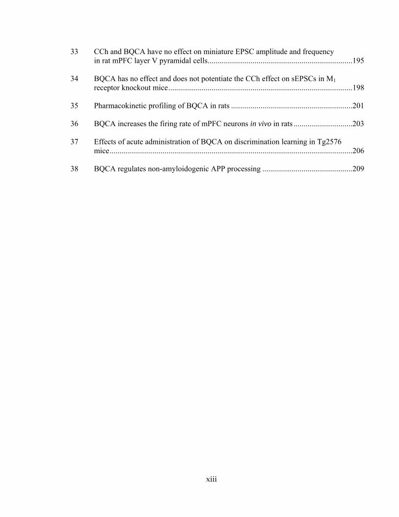
A detailed understanding of cholinergic synthesis and neurotransmission is now
available as a result of diverse research techniques, including biochemical analysis,
molecular genetics, and microscopy (Fig. 1) (Krnjevic, 1969; Csillik, 1975). In the
presynaptic terminal, acetylcholine (ACh) is synthesized from its precursors acetyl
coenzyme A (acetyl CoA) and choline by the enzyme choline acetyltransferase (ChAT).
ACh is packaged into synaptic vesicles by the vesicular acetylcholine transporter
(VAChT) and subsequently released into the synaptic cleft. Synaptic vesicles containing
the neurotransmitter can bind and activate pre- and post-synaptic acetylcholine receptors
only for a brief time until it is degraded by acetylcholinesterase (AChE). Following
degradation, the precursor choline is taken up into the presynaptic axon terminal by a
high affinity choline transporter (CHT). This reuptake process is the rate limiting step in
acetylcholine biosynthesis.
Cholinergic nuclei in the basal forebrain, a group of structures in the medial and
ventral telencephalon, contain large numbers of neurons that project to the hippocampus,
amygdala, and cerebral cortex and provide the majority of cholinergic innervation to
these areas (Mufson et al., 2000). The basal forebrain is divided into several distinct
regions that send bundles of axons to their respective targets. Projection cholinergic
neurons in the medial septum and the diagonal band of Broca project to the hippocampus,

2
while neurons in the nucleus basalis of Meynert project broadly to the neocortex as well
as to the amygdala. These cholinergic projections provide one of the most crucial
neuromodulatory inputs to the forebrain in humans and other mammals. Cholinergic
interneurons in the striatum also release ACh within local circuits where muscarinic
receptors modulate the activity of medium spiny neurons. Cholinergic neurons are
distinguished among other features by the presence of ChAT, the enzyme that is
responsible for ACh biosynthesis these neurons.

3
ACh AChE Choline+
Acetate
mAChRnAChR
Acetyl CoA+
Choline
ChATACh
CHT
VAChT
Figure 1. Schematic model of cholinergic biosynthesis and neurotransmission in the central nervous system. Acetylcholine (ACh) is synthesized in presynaptic nerve terminals by the enzyme choline acetyltransferase (ChAT), packaged into synaptic vesicles by the vesicular acetylcholine transporter (VAChT), and released into the synaptic cleft. Intact ACh can bind and activate nicotinic and muscarinic acetylcholine receptors on both pre- and post-synaptic membranes until it is degraded by the enzyme acetylcholinesterase (AChE). The biosynthetic precursor choline is then taken up into the presynaptic terminal by the high affinity choline transporter (CHT) with choline reuptake being the rate-limiting step in ACh synthesis.

4
Acetylcholine Receptors
ACh, the first neurotransmitter to be identified, activates two families of receptors
that mediate its action in target tissues: nicotinic receptors, which function as ligand-
gated cation channels that participate in rapid postsynaptic neurotransmission, and
muscarinic receptors (mAChR), which are members of family A G-protein coupled
receptors (GPCRs) and play a role in modulating the activity of many circuits within the
CNS. These two classes of receptor families were originally named for their specific
activation by nicotine and muscarine, respectively, but have been extensively
characterized since that time on a molecular basis. This thesis will focus on muscarinic
acetylcholine receptor subtypes. The diversity and complexity of muscarinic cholinergic
signaling is facilitated in part by five distinct receptor subtypes, M1-M5, the genes for
which were cloned in the mid to late 1980s (Bonner et al., 1987; Peralta et al., 1987;
Bonner et al., 1988). These intronless genes encode muscarinic receptor proteins that
have the typical structural features of the seven transmembrane helix GPCR superfamily,
the largest family of cell-surface receptors and key regulators of a wide variety of
physiological processes (Lefkowitz, 2007). In general, when the receptor is activated
GDP is converted to GTP on the G protein resulting in its dissociation from the receptor.
Subsequently, the G protein dissociated into α and βγ subunits, which both activate
downstream signaling cascades.
Muscarinic acetylcholine receptor subtypes, signaling, and function
The five receptor subtypes are highly homologous in the ACh binding domain;
their signaling properties are quite different, however. M1, M3, and M5 preferentially
5
couple to Gαq proteins to activate phospholipase C (PLC) and cause a subsequent release
of calcium from intracellular stores as well as an increase in phosphatidylinositol
turnover and activation of protein kinase C (PKC) (Wess, 1996). The major mechanism
of calcium release from endoplasmic reticulum stores in CA1 pyramidal neurons is via
production of inositol 1,4,5-triphosphate (IP3) induced by mAChR activation. In addition
M1, M3 and M5 receptors have also been shown to activate phospholipase A2 (PLA2),
phospholipase D (PLD) and tyrosine kinases (Wess, 1996). M2 and M4 receptors couple
predominantly to Gαi/o proteins to inhibit adenylate cyclase and cause a decrease in cyclic
AMP levels. Activation of Giβγ subunits by M2 and M4 subtypes also modulate a variety
of ionic channels including voltage-gated calcium channels as well as inwardly rectifying
potassium channels. All subtypes of muscarinic receptors have been shown to activate
extracellular signal related kinase (ERK), a signaling protein involved in cell growth,
differentiation and survival (Hulme (a) et al., 2003).
In situ hybridization experiments following the cloning of mAChR subtype genes
revealed that individual subtypes were expressed in partially overlapping tissues, with
some regions, including the hippocampus, expressing all five mAChR subtypes (Buckley
et al., 1988; Weiner et al., 1990). A series of studies using subtype-selective antibodies
has illustrated the distinct neuroanatomical localization of the mAChR subtypes in brain
and have provided important clues as to their function in neural circuits (Levey et al.,
1991; Mrzljak et al., 1993; Levey et al., 1994; Hersch and Levey, 1995; Levey et al.,
1995b; Rouse and Levey, 1996; Rouse et al., 1998; Rouse et al., 2000b).
The M1 receptor is expressed at very high levels in multiple brain regions
including cortex, hippocampus, and striatum (Wess, 2003). In the cortex, M1 is localized

6
to pyramidal cells and is prominent in the neuropil of layers II/III and VI (Levey et al.,
1991). M1 is expressed broadly throughout the hippocampus, including in pyramidal
neuron cell bodies and dendritic processes in the stratum radiatum and stratum oriens,
and in the molecular layer and granule cells of the dentate gyrus (Levey et al., 1995b). In
the striatum, M1 is found in the majority of dopamine-2 receptor (D2)-expressing medium
spiny neurons as well as in the neuropil. At the electron microscopy level, M1 can be
visualized at the postsynaptic density of asymmetrical synapses, suggesting a role in
modulating excitatory neurotransmission (Hersch et al., 1994). Studies using knockout
mice have suggested a role for M1 in learning and memory, and it has been demonstrated
that M1 potentiates NMDA receptor currents (Marino et al., 1998) and is the sole mAChR
responsible for muscarinic activation of extracellular signal-regulated kinase (ERK 1/2)
in the hippocampus, a protein involved in synaptic plasticity (Berkeley et al., 2001;
Hamilton and Nathanson, 2001). M1 knockout mice are less susceptible to pilocarpine-
induced seizures and have a phenotype that is similar to that seen in animal models of
psychosis, including hyperactivity, increased dopamine release, and a heightened
response to amphetamine (Gerber et al., 2001; Miyakawa et al., 2001). Interestingly, M1
knockout mice perform as well as their wild-type littermates in the Morris water maze, a
paradigm commonly used to assess hippocampal-dependent spatial memory; however,
performance was impaired under certain experimental conditions in the eight-arm radial
maze and in fear conditioning studies (Miyakawa et al., 2001). These animals only
exhibited a mild reduction in hippocampal long-term potentiation (LTP), an
electrophysiological phenomenon associated with learning and memory (Anagnostaras et
al., 2003). The profound hyperactivity phenotype of these animals (Gerber et al., 2001;

7
Miyakawa et al., 2001) makes it unclear whether some of the behavioral impairments are
actually due to cognitive impairments. This caveat aside, M1-/- mice display significant
impairments in non-matching-to-sample working memory and consolidation
(Anagnostaras et al., 2003), implicating these receptors in cortical memory function and
tasks requiring prefrontal cortical signaling (Hamilton and Nathanson, 2001). Based on
these studies using genetically altered mice as well as a growing body of clinical and pre-
clinical data, the M1 receptor subtype is viewed as one of the most exciting therapeutic
targets for the treatment of neurological disorders such as Alzheimer’s disease (AD) and
schizophrenia (Fisher, 2008b; Conn et al., 2009b).
M2 is the most widespread mAChR subtype in brain; it is expressed
predominantly on presynaptic terminals and has been shown to function as an
autoreceptor controlling ACh release in the hippocampus and cerebral cortex but not in
striatum (Zhang et al., 2002a). M2 has a distinct laminar distribution in the cortical
neuropil of layer IV and the junction between layers V/VI, and is also present on
interneuron cell bodies (Levey et al., 1991). Close inspection revealed that M2 is
expressed both pre-and post-synaptically (Mrzljak et al., 1993). In the hippocampus, M2
localizes to discrete bands of cell bodies and processes along the oriens/alveus border,
and is also found in processes along the pyramidal cell layer, most prominently in the
CA3 region (Levey et al., 1995b). There is a high expression level of M2 in the basal
forebrain, in both cholinergic and noncholinergic cells, as well as in the neuropil (Levey
et al., 1995a). M2 also mediates parasympathetic decreases in the force of contraction
and in the rate of cardiac contraction by inhibiting voltage-gated calcium channels and
activating inwardly rectifying potassium channels (Caulfield and Birdsall, 1998). Studies

8
in M2 knockout mice have demonstrated a physiological role for this protein in
locomotion, regulation of body temperature, and response to pain (Gomeza et al., 2001).
These mice have deficits in behavioral flexibility and working memory as well as in
passive avoidance (Seeger et al., 2004; Tzavara et al., 2004).
The M3 receptor is expressed at relatively low levels in brain, accounting for only
5-10% of total mAChRs in various brain regions (Levey et al., 1994). By
immunohistochemistry, M3 can be seen to localize to multiple brain regions, including
cortex, hippocampus, olfactory bulb, amygdala, striatum, thalamus, and pons.
Subcellularly, M3 appears in cell bodies and proximal dendrites, suggesting a
postsynaptic localization, and also as a diffuse, punctate reaction product in the neuropil
that may reflect presynaptic terminals or dendritic processes. In the CNS, M3 helps
regulate the release of several neurotransmitters, including dopamine in the striatum,
GABA and glycine in the dorsal horns of the spinal cord, and endocannabinoids (Zhang
et al., 2002a; Ohno-Shosaku et al., 2003; Zhang et al., 2006; Zhang et al., 2007). Studies
in M3 knockout mice have also implicated this subtype in multiple peripheral and
autonomic functions, including arterial vasodilation, insulin release, glandular secretion
(including salivation), weight gain, and smooth muscle contraction in the stomach,
trachea, and urinary bladder (Matsui et al., 2000; Yamada et al., 2001b; Duttaroy et al.,
2004; Khurana et al., 2004). It is believed that activation of peripheral M3 receptors leads
to severe side effects such as excess salivation and GI distress induced by cholinergic
agonists and AChE inhibitors used to treat AD.
The M4 receptor is expressed at somewhat lower levels than other mAChR
subtypes in cortical laminae, and is localized to discrete layers in the hippocampus,
9
including the stratum radiatum and stratum oriens in CA1 and the inner molecular layer
of the dentate gyrus. In the striatum where M4 is most highly expressed, dense patches of
receptor expression are observed that correspond to postsynaptic sites on medium spiny
neurons. Analogous to the autoinhibitory role that M2 plays in the hippocampus and
cortex, M4 is the major autoreceptor in the striatum responsible for feedback regulation of
ACh release from the presynaptic terminal (Zhang et al., 2002b). M4 has been shown to
participate in regulating dopaminergic signaling and release, and M4 knockout mice show
increased basal and dopamine-regulated locomotor responses (Gomeza et al., 1999;
Zhang et al., 2002a). These animals are hypersensitive to agents that disrupt prepulse
inhibition of the acoustic startle response, a measure of sensorimotor gating which is also
disrupted in schizophrenic patients (Felder et al., 2001). In vivo microdialysis studies
revealed that M4 knockout mice also have elevated basal dopamine levels in the nucleus
accumbens and that these mice show heightened dopamine efflux in response to
psychostimulants like D-amphetamine and phencylidine (Tzavara et al., 2004).
Levels of the M5 receptor approach the lower limits of specific detection in brain
as determined by quantitative immunoprecipitation and immunohistochemistry, although
M5 mRNA is detectable in multiple brain tissues. M5 knockout mice have revealed roles
for M5 in dilation of cerebral blood vessels and in reward and reinforcement behaviors,
specifically in response to drugs of abuse such as morphine and cocaine (Yamada et al.,
2001a; Basile et al., 2002; Thomsen et al., 2005). Further support for the role of M5 in
drug addiction came with the finding that these animals exhibit less severe withdrawal
symptoms after chronic morphine exposure as well as decreased cocaine conditioned

10
place preference and reduced acute cocaine self-administration (Fink-Jensen et al., 2003;
Thomsen et al., 2005).
Muscarinic regulation of hippocampal physiology and function
The hippocampus is a key cortical structure that plays an important role in a
number of normal physiological processes, including processing of complex spatial and
temporal patterns and formation of short- and long-term memory. In addition, the
hippocampus is a primary site of pathology in certain neurological disorders, such as AD
and temporal lobe epilepsy. Because of this, much effort has been directed at developing
a detailed understanding of the synaptic organization of the hippocampus as well as the
cellular mechanisms involved in regulation of synaptic transmission in this structure (see
(Brown and Zador, 1990) for review). Glutamate is the primary neurotransmitter at each
of the three major excitatory synapses in the hippocampal formation. In addition,
neuromodulators from extrinsic afferents (ie. acetylcholine, serotonin, norepinephrine)
regulate transmission through the hippocampus by activating GTP-binding protein-linked
receptors. Regulation of hippocampal function by these neuromodulators dramatically
influences net transmission through the hippocampus and participates in a variety of
different physiological and pathological conditions.
The hippocampus is commonly viewed as a relatively simple circuit consisting of
three major excitatory synapses. The primary input to the hippocampus is from the
entorhinal cortex which sends excitatory afferents to the dentate gyrus (DG) via the
perforant path; the entorhinal cortex collects polymodal information from other cortical
areas and relays this information to dentate granule cells. Mossy fibers from the dentate

11
granule cells project to area CA3 of the hippocampus which in turn sends afferents via
the Schaffer collateral to hippocampal area CA1. Afferents from CA1 pyramidal cells
then provide the major output of the hippocampus. Each subregion of the hippocampus
also has intrinsic circuits and connections within (associational) and between
(commissural) hippocampi. Glutamate is the excitatory neurotransmitter at each of the
three major excitatory synapses in the hippocampal formation. In addition,
neuromodulators from extrinsic afferents (ie. acetylcholine, serotonin, norepinephrine)
regulate transmission through the hippocampus by activating GTP-binding protein-linked
receptors. Regulation of hippocampal function by these neuromodulators dramatically
influences net transmission through the hippocampus and participates in a variety of
different physiological and pathological conditions.
One of the major neuromodulatory inputs to the hippocampus is a large bundle of
cholinergic projections from the medial septum and the diagonal band of Broca (Brown
and Zador, 1990), and these cholinergic projections make synaptic contact with
widespread but highly specific targets in the hippocampus. A large number of animal
and human studies suggest that cholinergic projections to the hippocampus play a critical
role in memory and attention mechanisms. For instance, blockade of muscarinic
receptors or lesions of the septo-hippocampal projections produce memory and
attentional deficits (Drachman and Leavitt, 1974; Bartus et al., 1982; Dekker et al., 1991;
Fibiger et al., 1991; Nilsson et al., 1992; Callahan et al., 1993). Also, the theta rhythm,
an electroencephalographic measure of the arousal response in the hippocampus which
may be involved in attention and filtering of sensory information, is regulated by
cholinergic septal input and can be induced by muscarinic agonists and abolished by

12
muscarinic antagonists (Colom et al., 1991; Vinogradova et al., 1993). Furthermore,
abundant evidence suggests that the clinical syndrome associated with AD results, at least
in part, from failed neurotransmission at cholinergic synapses in the hippocampus and
neocortex. The basal forebrain neurons that provide the majority of cholinergic
innervation of the neocortex and hippocampus degenerate with AD (Whitehouse et al.,
1982; Arendt et al., 1983) and this is accompanied by a depletion of presynaptic
cholinergic markers in these brain regions (Bowen et al., 1976; Davies and Maloney,
1976; Perry et al., 1978; Whitehouse et al., 1982). Furthermore, lesions of basal
forebrain neurons or pharmacological blockade of muscarinic receptors, experimentally
in animals (Dunnett, 1985; Dekker et al., 1991; Fibiger, 1991; Voytko et al., 1994) or
naturally in humans (Drachman and Leavitt, 1974; Drachman, 1977; Bartus et al., 1982;
Damasio et al., 1985), impairs learning, memory, and attention. Evidence suggests that
cholinergic transmission in the hippocampus is mediated primarily by mAChRs and
thatthese receptors are likely to mediate the cholinergic involvement in learning and
memory (Coyle et al., 1983; Brown and Zador, 1990; Fibiger, 1991).
Electrophysiological effects of mAChR activation in the hippocampus
Multiple mAChR subtypes are expressed in the hippocampus (Hulme et al., 1990;
Caulfield, 1993; Levey et al., 1995b) where they are involved in regulating various
aspects of hippocampal physiology. Early studies showed that mAChR activation
induces a number of direct excitatory effects on hippocampal pyramidal cells and reduces
both excitatory (Hounsgaard, 1978; Segal, 1982; Dutar and Nicoll, 1988; Sheridan and
Sutor, 1990; Williams and Johnston, 1990; Burgard et al., 1993) and inhibitory (Krnjevic

13
et al., 1981; Bilkey and Goddard, 1985) synaptic transmission in the hippocampus.
Another well characterized electrophysiological effect of muscarinic receptor activation
in hippocampal pyramidal cells using the intracellular recording technique is a slow post-
synaptic potential (Cole and Nicoll, 1984a); repetitive electrical stimulation of
cholinergic fibers terminating in stratum oriens evokes a series of membrane potential
changes. First is a series of fast excitatory postsynaptic potentials (EPSPs) followed by
an inhibitory postsynaptic potential (IPSP); these are followed by a slow EPSP that lasts
on the order of 20-30 seconds. The slow EPSP could be induced by ionophoretic
application of ACh and was blocked by atropine and was dependent on action potential
firing and calcium (Cole and Nicoll, 1984a).
Muscarinic receptors modulate a large number of ionic conductances in pyramidal
neurons through both direct and indirect biochemical interactions; the conductances
known to be modulated by mAChRs in hippocampal pyramidal cells include several
potassium currents (IM, the muscarine sensitive K+ current; IAHP, the Ca2+-activated K+
current underlying spike frequency adaptation; Ileak, the background leak current)
(Halliwell, 1990). Exogenously applied muscarinic agonists induce a pronounced
membrane potential depolarization and increased membrane resistance (Cole and Nicoll,
1984b), and direct electrical stimulation of cholinergic afferents in the hippocampus
causes a similar mAChR-dependent membrane potential depolarization (Segal, 1982;
Cole and Nicoll, 1984b; Madison et al., 1987; Pitler and Alger, 1990; Morton and Davies,
1997). This response often results in a sustained action potential discharge, in part
arising from a pronounced reduction in spike frequency adaptation (Cole and Nicoll,
1984b). Activation of mAChRs increases cell firing and depresses the IAHP that is due to

14
a calcium-activated potassium conductance, as mentioned above. It is thought that this
conductance, at least in part, is responsible for a dampening of action potential discharge
during depolarizing current injections. These excitatory effects of ACh are mediated by
mAChRs because they are completely blocked by atropine, a non-selective muscarinic
antagonist, but not by nicotinic antagonists. mAChR activation also potentiates two
mixed cation currents (Ih, the hyperpolarization-activated cation current; Icat, the Ca2+-
dependent non-specific cation current) (Halliwell, 1990; Colino and Halliwell, 1993) and
modulates the activity of both voltage-dependent Ca2+ currents and several ligand-gated
receptors including N-methyl-D-aspartate (NMDA) receptors (Markram and Segal,
1990b, a, 1992; Harvey et al., 1993; Marino et al., 1998; Sur et al., 2003). In addition,
mAChR agonists increase pyramidal cell excitability indirectly by reducing GABA-
mediated synaptic inhibition (Krnjevic et al., 1981; Bilkey and Goddard, 1985).
Presynaptically, activation of mAChRs inhibits excitatory afferents, reducing the release
of glutamate through inhibition of voltage-gated calcium channels (Qian and Saggau,
1997; Fernandez de Sevilla et al., 2002; Fernandez de Sevilla and Buno, 2003). More
recently, muscarinic agonists have also been shown to inhibit L-type calcium currents in
superior cervical ganglion (SCG) neurons (Liu et al., 2006) and enhance R-type, but not
T-type, Ca2+ currents in hippocampal CA1 pyramidal neurons, an effect that required
PKC activation (Tai et al., 2006).
Subsequent studies examining specific mAChR subtypes involved in some of
these electrophysiological effects have shown that depolarization of hippocampal
pyramidal neurons is likely mediated, at least in part, by the M1 receptor subtype as
inward currents recorded in voltage clamp mode from CA3 pyramidal cells were

15
markedly reduced in M1 knockout mice as compared to wild-types (Fisahn et al., 2002).
The muscarinic potentiation of ICAT contributes to the depolarization of pyramidal
neurons. Muscarine caused an increase in magnitude of Ih in wild-type but not M1
knockout mice, indicated that the M1 subtype is responsible for this effect in CA3 cells;
conversely, there was no effect of muscarinic agonists on IM in the M1 knockout
supporting data by Rouse et al. (Rouse et al., 2000a) indicating a lack of M1 modulation
of this current in CA1 pyramidal cells.
Muscarinic modulation of GABAergic transmission in the hippocampus
Extrahippocampal γ-aminobutyric acid (GABA)ergic afferents originating from
the medial septum and diagonal band of Broca innervate the hippocampus and target
solely hippocampal interneurons and cholinergic afferents which in turn target both
pyramidal cells and interneurons (Frotscher and Leranth, 1985). Acetylcholine is a
powerful presynaptic modulator of synaptic transmission at both excitatory glutamatergic
and inhibitory GABAergic synapses with modulation being both cell type and pathway
specific. Furthermore, GABAergic interneurons can inhibit cholinergic release
presynaptically through GABAB receptors (Morton et al., 2001). The role of mAChRs in
modulating activity of GABAergic inhibitory neurons is complex; studies have shown
that mAChR activation of individual interneurons in the hippocampus yields differential
effects on resting membrane potential (McQuiston and Madison, 1999a, b). Among
several subpopulations of GABAergic interneurons, muscarinic receptor activation
produces a pure hyperpolarizing response, a biphasic response in which an initial
hyerpolarization is followed by a secondary depolarizing phase, a slow membrane
16
potential oscillatory response, or no response. Immunocytochemical findings suggested
cell type-specific localization of mAChRs in different subtypes of interneurons (Levey et
al., 1995b) which likely explains the variable muscarinic effects observed in these cells.
This diversity of interneuron responses also likely reflects a highly heterogeneous
population of GABAergic interneurons with respect to their connectivity and
neurochemistry. A more recent study investigated mAChR function in a morphologically
identifiable class of stratum oriens interneurons, the stratum oriens-lacunosum moleulare
(O-LM) interneurons; they exhibit a muscarinic-induced afterdepolarization that is
associated with the inhibition of several potassium conductances and the activation of
ICAT (Lawrence et al., 2006). In these interneurons, muscarine abolished the
afterhyperpolarization (AHP) current and induced a switch in firing frequency from
accommodating to accelerating during the depolarizing current injection. The AHP was
replaced by a prominent afterdepolarization in the presence of muscarine. In another set
of cells that exhibited strong spike frequency accommodation and broad rebound spikes,
mAChR activation was accompanied by a reduction in input resistance and shunting of
firing. Finally, muscarinic receptor activation increases the frequency and amplitude of
spontaneous IPSCs but depresses monosynaptically evoked IPSCs and the frequency of
miniature IPSCs (Behrends and ten Bruggencate, 1993).

17
NMDAR modulation by mAChRs
As previously stated, one of the most prominent effects of mAChR activation in
the hippocampus and other forebrain regions is potentiation of currents through the
NMDA subtype of ionotropic glutamate receptor (Markram and Segal, 1990b; Harvey et
al., 1993; Calabresi et al., 1998; Marino et al., 1998; Lu et al., 1999; Marino and Conn,
2002). The M1 receptor likely enhances currents through NMDARs through a PKC-
dependent activation of the non-receptor tyrosine kinase (Src) signaling cascade
(Calabresi et al., 1998; Lu et al., 1999). The NMDA receptor plays a critical role in
regulating hippocampal and cortical function and is thought to be important for the
cognition-enhancing and attention-promoting effects of mAChR activation. In addition,
the NMDA receptor may play an important role in regulation of circuits that are disrupted
in schizophrenia and other psychotic disorders (Coyle et al., 2002; Marino and Conn,
2002; Tsai and Coyle, 2002). Competitive and non-competitive antagonists of the
NMDA receptor can induce a psychotic state that closely resembles that seen in
schizophrenic patients. Furthermore, co-agonists at the NMDA receptor, such as glycine
and D-cycloserine produce improvements in the symptoms of schizophrenic patients.
Thus, a large number of clinical and animal studies have led to the hypothesis that
potentiation of NMDA receptor currents in these regions could have an antipsychotic
action. Based on this, it is possible that mAChR-induced potentiation of NMDA receptor
function plays important roles in the therapeutic efficacy of mAChR activation in
psychotic disorders.

18
Presynaptic M2 and M4 receptors may regulate excitatory and inhibitory synaptic transmission Many studies have indicated that mAChR-induced reduction of transmission at
excitatory synapses in the hippocampus is mediated presynaptically. Based on early
immunocytochemical studies, the most likely mAChRs involved in the presynaptic
actions of mAChR agonists are M2 and M4 since both of these receptors are
predominantly localized presynaptically in each major subsector of the hippocampus.
Consistent with this, a large body of research suggests that presynaptic receptors involved
in regulating neurotransmitter release are often coupled to inhibition of adenylyl cyclase,
and M2 and M4 both couple to this effector system. While both M2 and M4 are localized
presynaptically, M2 immunoreactivity is not present in granule cells and pyramidal cells,
but is highly localized in inhibitory interneurons (Levey et al., 1995b). Since dentate
granule cells and CA3 pyramidal cells provide the majority of excitatory input to areas
CA3 and CA1 respectively, this makes M4 a more likely candidate for the mAChR
involved in regulating glutamate release. In contrast, if M2 is localized on presynaptic
terminals of inhibitory interneurons, M2 would be in an ideal position for regulating
GABA release and thereby reducing GABA-mediated synaptic inhibition.
One mechanism of presynaptic inhibition of neurotransmitter release involves the
modulation of presynaptic calcium channels that are involved in vesicle fusion and
release. Early indirect (Valentino and Dingledine, 1981) and more recent studies have
shown that muscarinic inhibition of synaptic transmission in the hippocampus is
presynaptic and relies at least in part on muscarinic blockade of N-type and P/Q-type
voltage dependent calcium channels at the SC-CA1 synapse (Qian and Saggau, 1997) and
in the associational-commissural fiber system of CA3 (Vogt and Regehr, 2001).
19
Activation of mAChRs with carbachol (CCh), a muscarinic agonist, increases the paired-
pulse facilitation index of evoked EPSCs at CA1 synapses and also decreased the
coefficient of variation ratio, indicating that CCh inhibits synaptic transmission via
activation of presynaptic mAChRs which causes a reduction in the reliability of
glutamate release (Fernandez de Sevilla et al., 2002). Electrical stimulation of
cholinergic terminals in stratum oriens/alveus also causes a decrease in evoked CA1
EPSCs by presynaptically inhibiting glutamate release via activation of mAChRs as
indicated by parallel changes in PPF index and EPSC variance (Fernandez de Sevilla and
Buno, 2003). Although ACh has been shown to suppress excitatory transmission at
mossy fiber synapses in CA3 through modulation of GABAB receptor activity (K. E.
Vogt and W. G. Regehr, 2001), presynaptic inhibition at CA1 seems to be independent of
GABAergic transmission (Kremin et al., 2006). Although experiments to confirm a
presynaptic mechanism of action were not carried out, we have found that selective
potentiation of M4 receptors with the PAM VU10010 enhances muscarinic depression of
evoked excitatory but not inhibitory postsynaptic currents in CA1 neurons (Chapter
IIIa).
Role of muscarinic cholinergic signaling in hippocampal LTP
Of particular relevance to issues of synaptic plasticity, mAChR activation
enhances agonist-evoked currents through the NMDA subtype of glutamate receptor
(Markram and Segal, 1990b; Harvey et al., 1993; Marino et al., 1998). The NMDA
receptor is known to play a critical role in several forms of hippocampal long-lasting
synaptic plasticity which are thought to underlie learning and memory. Consistent with

20
this, mAChR activation can also induce or enhance long term potentiation (LTP) of
excitatory synaptic responses in the hippocampus (Blitzer et al., 1990; Burgard and
Sarvey, 1990; Markram and Segal, 1990b; Abe et al., 1994; Auerbach and Segal, 1996;
Shinoe et al., 2005). Because long-term alterations in efficacy of glutamate transmission
contribute to memory mechanisms (Bliss and Collingridge, 1993; Malenka and Bear,
2004), induction of long-term synaptic changes by cholinergic receptors may underlies
the cholinergic dependence of normal memory processing. Furthermore, because of the
postulated role of NMDA receptors and LTP in learning and memory, it has been
suggested that the mAChR subtype that mediates these responses may be an excellent
target for therapeutic agents useful in the treatment of AD.
Early studies using sharp microelectrodes revealed that ionophoretic application
of ACh caused an initial reduction in EPSP amplitude followed by a gradual and long-
lasting facilitation of EPSP amplitude in CA1 pyramidal cells that was not associated
with a change in input resistance (Markram and Segal, 1990b). Atropine blocked both
the suppressing and facilitating effects of ACh. These studies also indicated that ACh
enhanced the slow NMDA-mediated component of the EPSP and that responses to
NMDA application in current clamp mode were also potently facilitated by both ACh and
oxotremorine-M, a muscarinic agonist. Muscarinic facilitation of both EPSP amplitude
and NMDA responses were independent of changes in voltage or K+ conductances as the
facilitation was not affected by clamping cells at resting membrane potentials or by the
inclusion of cesium in recording electrodes.
Burgard and Sarvey (1990) showed a concentration-dependent ability of
muscarine in the dentate gyrus to facilitate LTP induction by subthreshold titanic

21
stimulation (Burgard and Sarvey, 1990); Blitzer et al. (1990) demonstrated a significant
depression of the CA1 field EPSP with high doses of CCh which, when controlled for,
enhanced tetanus-induced LTP (Blitzer et al., 1990). Subsequent reports indicated that
bath application of 750 nM carbachol also induced a long-lasting (>45 min post drug
washout) facilitation of intracellularly recorded EPSPs; muscarinic potentiation of field
EPSPs was also seen and was dependent on CCh concentration and time of drug
application (Auerbach and Segal, 1994). Only with 20-minute CCh application was
sustained LTP produced. This phenomenon was termed LTPm and was completely
blocked by atropine; when antagonist was added to the bath after the establishment of
LTP, no effect was seen indicating that induction but not maintenance of LTPm was
mediated by mAChRs. These studies also indicated that LTPm requires involvement of
intracellular calcium stores and protein kinases but is activity independent. Interestingly,
CCh still induced LTPm in the presence of the 10 µM NMDAR antagonist (2R)-amino-5-
phosphonopentanoate (APV), a concentration that did block tetanus-induced LTP,
suggesting that the two mechanisms were somewhat divergent. It was also demonstrated
that 0.1 µM CCh (a concentration that did not induce LTP alone) facilitated LTP caused
by a subthreshold titanic stimulus and that LTPm and tetanus-induced LTP were mutually
occlusive phenomena. Therefore, the mechanisms of LTPm and tetanus-induced LTP
seemed to converge at a point downstream of NMDA receptor activation (Auerbach and
Segal, 1994). LTPm was later shown to not be accompanied by a change in the size of the
afferent fiber volley or by a change in paired-pulse potentiation, consistent with a
postsynaptic locus of CCh action (Auerbach and Segal, 1996). Intracellular recordings
from CA1 pyramidal cells in voltage clamp mode revealed that 0.5 µM CCh transiently
22
potentiated NMDA responses but that responses to AMPA increased gradually and
remained potentiated after drug washout.
The M1 receptor is a likely candidate for the mAChR subtype involved in the
facilitation of LTP induction. For instance, evidence suggests that mAChR-induced
facilitation of LTP is mediated by a postsynaptic mechanism, and depends on the release
of calcium from intracellular stores (Auerbach and Segal, 1994). The M1 receptor is the
most abundant postsynaptically localized mAChR subtype in CA1 pyramidal cells.
Furthermore, M1 is coupled to phosphoinositide hydrolysis and activation of this receptor
leads to release of intracellular calcium. Thus, it seemed plausible that the mAChR-
induced facilitation of LTP induction is mediated by the M1 receptor. Recent studies
have indicated that M1 is indeed the subtype most likely mediating cholinergic
modulation of synaptic plasticity in the mouse hippocampus. Experiments focusing on
enhancement of electrically induced LTP by low concentrations of CCh or by repetitive
stimulation in the stratum oriens, which presumably trigger release of endogenous ACh
from cholinergic terminals, revealed that the enhancing effect was abolished in M1
knockout but not in M3 knockout mice (Shinoe et al., 2005). While concentrations of 500
nM to 5 µM CCh induced a transient depression of field excitatory postsynaptic
potentials (fEPSPs) at the SC-CA1 synapse, 50 nM had no effect alone on fEPSPs. This
is in contrast to what had been reported in rat where low doses of CCh induced LTP.
This could be a result of differences in mAChR expression and receptor density between
species; as shown in Chapters IIIa and IV, we have also seen a difference in potency of
CCh on various electrophysiological responses between rat and mouse. Regardless,
Shinoe et al. reported that 50 nM CCh significantly increased LTP elicited by high
23
frequency stimulation (HFS, 100 Hz for 1 sec) of SC afferent fibers. Stimulation of
stratum oriens thirty seconds prior to the HFS to cause endogenous ACh release also
potentiated the degree of LTP. While HFS-LTP remained intact in both M1 and M3
knockout animals, CCh-induced enhancement of LTP was absent in only the M1
knockout supporting a role for M1 in mediating this effect. The same was shown to be
true after stratum oriens stimulation (Shinoe et al., 2005).
One recent set of studies by Fernández de Sevilla et al. (2008) revealed that a
brief puff of ACh applied at the apical dendritic shaft of a CA1 pyramidal cell induces a
postsynaptic calcium elevation and LTP of excitatory postsynaptic currents (EPSCs)
evoked by stimulation of SC-CA1 afferents. Because changes in postsynaptic responses
can be mediated by a change in α-amino-3-hydroxyl-5-methyl-4-isoxazole-propionate
receptor (AMPAR) density at the postsynaptic membrane, the content of recombinant
tagged GluR1 and GluR2, subtunits of AMPARs, was analyzed before and after ACh
treatment using two-photon microscopy. Indeed, ACh induced an increase in GluR1 and
GluR2 containing AMPARs at the spine surface without a change in spine volume.
Results also suggested that LTP induced by ACh was mediated through a postsynaptic
mechanism as shown by a lack of effect of ACh on the paired-pulse ratio or the fiber
volley of fEPSPs evoked by Schafer Collateral (SC) stimulation, and LTP in this case
was NMDA independent. Because levels of ACh released from the medial septum rise
during tasks that require attention, it could be postulated that this form of plasticity could
be induced in vivo. Tetanic stimulation of the medial septum using chronically implanted
stimulating electrodes did in fact induce a long-lasting synaptic enhancement of fEPSPs
at CA1 synapses in vivo (Fernandez de Sevilla et al., 2008). A key difference between

24
the ACh-induced LTP reported by Fernández de Sevilla et al. and the synaptic
enhancement induced by CCh in earlier studies (Auerbach and Segal, 1994, 1996) is a
dependence on NMDAR activity.
Muscarinic modulation and induction of LTD in the hippocampus
Interestingly, M1 activation also seems to induce a novel form of long-term
depression (LTD) often termed mLTD (Scheiderer et al., 2006). At hippocampal CA3-
CA1 synapses, bath application of a high concentration of CCh (50 µM) elicits a robust
transient depression of the dendritic fEPSP which is followed by LTD after agonist
washout. This mLTD was prevented by atropine, by the M1 toxin MTx-7 (Potter, 2001),
and by pirenzepine but not by the nAChR antagonist methyllycaconitine. The expression
of mLTD was independent of GABAA receptor activity and appeared to be via a
postsynaptic mechanism. It was also postulated that the acute presynaptic depression and
mLTD involve separate mechanisms and that the presynaptic depression is not required
for mLTD induction. Lastly, similar to LTPm described by Auerbach and Segal (1994),
mLTD was blocked in the presence of the NMDAR antagonist D,L-APV (Auerbach and
Segal, 1994). In contrast to LTPm however, mLTD is activity dependent; mLTD requires
presynaptic activity because cessation of stimulation of presynaptic afferents during CCh
application prevented induction of plasticity when CA3 cell bodies were removed from
the slice. Thus mLTD is both activity- and NMDAR-dependent. Interestingly, this
Hebbian form of LTD is lost after medial septal lesioning but is rescued by sympathetic
sprouting of noradrenergic fibers from the superior cervical ganglia into hippocampus.
This sprouting of sympathetic fibers appears to stimulate cholinergic reinnervation as

25
indicated by new VAChT-positive fibers in the hippocampus, and the appearance of these
new fibers correlates with the rescue of mLTD.
In a subsequent publication, McCutchen et al. (2006) found that mLTD did not
affect subsequent electrical induction of LTP; mLTD was also able to depotentiate LTP.
That is, application of CCh could cause a population of CA1 pyramidal cells exhibiting
LTP after high frequency stimulation to undergo an acute depression and then return to
the baseline fEPSP slope level (McCutchen et al., 2006).
Electrical induction of LTD using low frequency stimulation (LFS-LTD) is both
activity- and NMDA receptor dependent; since both LFS-LTD and mLTD share those
characteristics, occlusion studies were performed to confirm a shared mechanism
between the two types of LTD. Not surprisingly, prior saturation of LFS-LTD occluded
induction of mLTD by CCh application. Unexpectedly, however, when the converse
experiment was performed, saturating levels of mLTD did not occlude further depression
induced by subsequent LFS. In other words, even after multiple CCh applications to
insure saturated mLTD, application of LFS induced roughly 25% more depression of the
fEPSP. Further studies revealed that CCh treatment prior to LFS caused LFS-LTD to
become independent of NMDARs at hippocampal CA1 synapses but not in layer IV-layer
II/III synapses in visual cortex where LFS-LTD is also normally NMDA-dependent. The
fact that the switch in LFS-LTD does not occur in visual cortex suggests that this may be
a cholinergic mechanisms specific to hippocampal synapses and that there is perhaps
more flexibility in the induction mechanisms available to mediate hippocampal LTD
(McCoy and McMahon, 2007). Finally, induction of mLTD seems to require ERK
activation; application of U0126, a MEK inhibitor, before and during CCh treatment
26
completely blocks mLTD. Induction of this form of synaptic plasticity also required
activity of Src kinase but not PLC (Scheiderer et al., 2008).
Muscarinic modulation of hippocampal physiology: conclusions
It is likely that these cellular actions of mAChR activation are directly related to
the behavioral effects of activation of hippocampal mAChRs. For instance, the combined
reduction of excitatory synaptic transmission with an increase in excitability of pyramidal
cells could increase the signal to noise ratio of signaling through the hippocampus. Such
modulation of signal to noise ratio occurs with some other neurotransmitters and has been
proposed to play an important role in regulation of attentiveness to sensory stimuli
(Madison and Nicoll, 1986). In addition, selective potentiation of NMDA receptor
responses could critically modulate synaptic plasticity that is involved in learning and
memory. Consistent with this, mAChR activation modulates LTP and LTD of excitatory
synaptic responses in the hippocampus. A complete understanding of the roles of
mAChRs in both normal and pathological hippocampal function will require a detailed
understanding of the cellular mechanisms involved in these responses as well as the
specific mAChR subtypes that mediate each of these responses. While preliminary
studies using mice lacking each of the individual mAChR subtypes have provided
important clues about the function of these subtypes in mediating many of the
electrophysiological effects described above, we now have selective activators of M1 and
M4 that will be described further in subsequent chapters. It will be important to confirm
initial findings in knockout mice as well as to further delineate the roles of these two
receptor subtypes in modulating hippocampal physiology. Studies using the M4 positive
27
allosteric modulator VU10010 (see Chapter IIIa) revealed that this mAChR subtype is
involved in modulation of excitatory but not inhibitory transmission at the SC-CA1
synapse in rats and mice.
Prefrontal cortical physiology and function
An important projection from the hippocampus is to the prefrontal cortex (PFC), a
cortical region that integrates information from multiple other brain regions; PFC
function is the target of many neuromodulators including acetylcholine. The PFC
receives glutamatergic inputs from the thalamus and primary sensory cortices, from the
brainstem, and from the hippocampus (Groenewegen and Uylings, 2000; Dalley et al.,
2004; Vertes, 2006), and this merging of information in the PFC is implicated in higher
executive functioning. Cholinergic input to the PFC regulates attention (Muir et al.,
1992; Hasselmo, 1995), memory (DeSousa et al., 1994), and anxiety induction processes
(Berntson et al., 1998). Dysfunction of mAChRs and muscarinic-mediated signaling
pathways has been linked to Fragile X mental retardation (Volk et al., 2007), AD (Fisher,
2008b), and aging (Joseph et al., 1993). Lesions of the medial PFC (mPFC) result in
cognitive deficits that resemble symptoms of schizophrenia and Alzheimer’s disease
(Kolb, 1984, 1990; Heckers et al., 1998).
Among a number of glutamatergic afferents projecting to the PFC, the afferents
originating in the hippocampus are especially crucial in supporting memory and cognitive
function. Previous work has shown that the CA1 and subiculum of ventral hippocampus
send ipsilateral, unidirectional projections that terminate on neurons in the mPFC (Jay
and Witter, 1991; Jay et al., 1992). This hipocampal-mPFC pathway has been described

28
as projecting anteriorly through the fornix, and it ultimately ascends dorsally to terminate
on neurons within the nucleus accumbens and a region of the mPFC that includes the
infralimbic and prelimbic cortices. The functional integrity of the hippocampal-mPFC
network and the flow of information between these two brain regions are critical to the
proper functioning of the mPFC in memory formation (O'Donnell and Grace, 1995;
O'Donnell et al., 2002; Goto and O'Donnell, 2003).
Role of mAChRs in the mPFC
Cholinergic afferents to the prelimbic and infralimbic cortices, the major
components of the medial prefrontal cortex (mPFC), originate primarily in the basal
forebrain nucleus basalis magnocellularis, the diagonal band of Broca, and the
mesopontine laterodorsal nucleus (Lehmann et al., 1980; Satoh and Fibiger, 1986;
Gaykema et al., 1990). Cholinergic input to the cortex plays a vital role in supporting
processes requiring arousal, attention, memory, and learning (Phillis, 2005); in addition
to being released from inputs from the basal forebrain nuclei, ACh is also released from
local circuit neurons in the mPFC, and ACh release increases during performance of
attentional tasks (Passetti et al., 2000). In contrast, lesions of cholinergic nuclei that
project to the mPFC impair cue detection and attentional performance whereas
augmentation of cholinergic transmission enhances behavioral arousal and locomotor
activity (Day et al., 1991; McGaughy et al., 2002; Parikh et al., 2007). Of the five
mAChR subtypes, M1 and M2 are the predominant subtypes expressed in the mPFC with
M1 mainly localizing postsynaptically at asymmetrical (glutamatergic) synapses and M2
localizing both pre- and postsynaptically (Volpicelli and Levey, 2004). Activation of
29
mAChRs results in an array of effects on the excitability and firing properties of cortical
neurons, and these effects involve different cellular mechanisms. In addition, mAChRs
play a role in modulating long-term strengthening and weakening of synaptic
transmission in this brain region which appears to be input-specific. Modulation of
electrophysiological behavior and plasticity of cortical circuits, particularly in the PFC,
by ACh is thought to play a critical role in attention and memory (Gill et al., 2000;
Passetti et al., 2000; Anagnostaras et al., 2003; Sarter et al., 2005).
Postsynaptic effects of mAChRs in cortical neurons
Similar to the effects seen in hippocampal pyramidal cells, ACh causes a marked
depolarization and increase in membrane excitability and repetitive spiking in cortical
pyramidal cells (Benardo and Prince, 1982; Andrade, 1991; Krnjevic, 2004; Carr and
Surmeier, 2007). Many ionic currents have been implicated in mediating this
depolarization; one of these is a voltage-dependent nonselective cation current (Haj-
Dahmane and Andrade, 1996; Klink and Alonso, 1997; Fisahn et al., 2002). Studies in
hippocampal and sensorimotor cortices have indicated this increase in excitability can be
attributed to the inhibition of three main potassium currents: the calcium-activated
potassium current underlying the afterhyperpolarization (IAHP), a voltage-dependent leak
potassium current (Ileak), and the voltage-dependent M-current (IM). The involvement of
a potassium current-mediated mechanism was supported by the increase in input
resistance and a dependence on membrane potential; barium and tetraethylammonium
(TEA), potassium channel blockers, also reduced CCh-induced inward currents (Haj-
Dahmane and Andrade, 1996). However, the CCh-induced current did not reverse at the

30
reveral potential of potassium (EK) but persisted as an inward current at membrane
potentials 10-20 mV negative to the predicted EK for potassium. Intracellular cesium,
which blocks a wide range of potassium channels, also did not inhibit the CCh-induced
inward current. These results indicated that another ion conductance was likely involved
in this muscarinic effect. Subsequent studies implicated the involvement of a non-
selective cation current in muscarinic depolarization of mPFC layer V neurons (Haj-
Dahmane and Andrade, 1996). Similar findings in entorhinal cortex did not support a
role for potassium conductances in this effect but rather a calcium-dependent cationic
conductance largely permeable to sodium (Klink and Alonso, 1997). Conflicting data
support the involvement of an inwardly rectifying potassium current through a channel
containing Kir2 subunits; these channels are open at the resting membrane potential and
help set the resting membrane potential in PFC pyramidal neurons and also play a role in
integration of excitatory synaptic inputs (Day et al., 2005). Carr and Surmeier (2007)
found the CCh-induced inward current to be sensitive to barium and cesium that block
Kir2 channels, and showed that mAChR activation reduces Kir2 channel currents by
depleting membrane phosphatidylinositol 4,5-bisphosphate (PIP2) as this modulation was
blocked by PLC but not PKC inhibitors (Carr and Surmeier, 2007).
The calcium-dependent non-selective cation current mentioned above has also
been shown to contribute to the fast afterdepolarization (fADP) that follows an action
potential triggered by depolarizing current injection (Haj-Dahmane and Andrade, 1997).
This fADP is prominently displayed by pyramidal neurons of layer V in rat PFC.
Interestingly, these cells also exhibit a muscarinic-induced slow afterdepolarization
(sADP) (Haj-Dahmane and Andrade, 1998). Buffering of intracellular calcium prevented

31
the mAChR-induced sADP, and ion substitution experiments implicated the flow of
sodium ions into the cell but not the flux of potassium or chloride. The muscarinic sADP
therefore is likely mediated by a calcium- and voltage-sensitive nonselective cation
current and could represent a mechanism by which mAChRs modulate neuronal
excitability in the PFC. Due to the calcium-dependence of the sADP, the involvement of
calcium channel modulation by mAChRs has been investigated. A recent study has
implicated TRPC (transient receptor potential) ion channels in the muscarinic sADP (Yan
et al., 2009); these channels allow calcium flux into cells in response to a variety of
stimuli usually linked to phosphatidylinositol signal transduction pathways. Data
indicates that the current underlying the sADP induced by mAChR activation is carried
through TRPC channels containing TRPC5 or TRPC6 subunits and occurs through a
mechanism involving Gαq and PLCβ1 signaling.
Pyramidal cells normally respond to a sustained current injection with a high
initial firing rate followed by a slowing of this rate. The voltage-sensitive potassium
current (IM) and voltage-dependent calcium influx which activates a calcium-sensitive
potassium current (IAHP) are both thought to underlie this accommodation or adaptation in
cell firing rate by pyramidal cells in response to sustained depolarization. Activation of
mAChRs decreases both of these potassium currents which, in turn, allows neurons to
fire in a more sustained manner in response to sustained afferent input. Cholinergic
agonists also cause a strong increase in the firing rate of cortical neurons, and this
increase in firing rate was demonstrated to result from a slow depolarization of cortical
pyramidal cells due at least in part to blockade of potassium currents (Krnjevic et al.,
1971; Hasselmo and McGaughy, 2004). The muscarinic inhibition of the calcium-

32
dependent potassium currents underlying afterhyperpolarization also supports a role of
mAChRs in modulating voltage-dependent calcium channels. Indeed, mAChR
modulation of L-, N-, P/Q- and R-type calcium channels pre- and postsynaptically in both
hippocampal and cortical neurons occurs through G-protein dependent mechanisms
(Perez-Burgos et al., 2008).
In accordance with the excitatory effects of selective M1 receptor activation in rat
and mouse layer V mPFC pyramidal cells described in this thesis, a recent study using M1
knockout mice shows that activation of this receptor subtype mediates cholinergic
modulation of excitability in these neurons (Gulledge et al., 2009). Interestingly, short
focal application of ACh near the soma of the recorded neuron produced a
hyperpolarization and pronounced inhibition of spiking followed by spike acceleration
when ACh was delivered during periods of action potential generation by depolarizing
current injection in layer V but not as robustly in layer II/III neurons. This effect was
abolished in slices from M1 knockout but not M3 or M5 knockout mice. Tonic
cholinergic excitation using bath-applied CCh depolarized layer V neurons and also
suppressed the afterhyperpolarization in addition to generating a slow afterdepolarization,
effects that were also absent in M1 knockout slices (Gulledge et al., 2009).
Cholinergic modulation of cortical neurotransmission
Despite a consistent reporting of increases in cortical neuronal excitability in
response to cholinergic agonists, many discrepancies exist in studies examining the
effects of cholinomimetic drugs on glutamatergic transmission; data suggests that the
effect of ACh on synaptic transmission is region- and input specific (Hasselmo and

33
Bower, 1992; Gil et al., 1997; Gulledge et al., 2007; Eggermann and Feldmeyer, 2009;
Gulledge et al., 2009). In addition, varying concentrations of ACh induce either
depression or facilitation of glutamatergic transmission; this principle holds true when
examining the effects of CCh on long-term changes in synaptic transmission as discussed
below. By employing stimulation of either layer Ia or Ib in rat piriform cortex slices,
Hasselmo and Bower found that CCh, muscarine, and ACh strongly reduced responses
stimulated from intrinsic fibers in layer Ib but not from afferent fibers from layer Ia; this
suggested that mAChR activation decreases excitatory transmission within intracortical
circuits but not from afferent inputs (Hasselmo and Bower, 1992). In slices containing
somatosensory cortex and ventrobasal thalamus, muscarine depressed EPSPs at both
thalamocoritcal (TC) and intracortical (IC) synapses onto layer III pyramidal cells
whereas nicotine selectively enhanced responses from TC synapses and had no effect on
IC synapses. Muscarine increased the paired-pulse ratio at both TC and IC synapses,
suggesting a presynaptic mechanism of action (Gil et al., 1997). In addition, GABA
released by an intracortical stimulus selectively suppresses transmitter release from IC
synapses but not TC synapses by activating presynaptic GABAB receptors. Taken
together, these results indicate that cholinergic receptors selectively modulation of
intrinsic versus afferent synapses.
Acetylcholine also modulates activity and GABA release from cortical
GABAergic interneurons differentially depending on cell subtype (Kawaguchi, 1997).
Despite the relatively low abundance of inhibitory neocortical interneurons, these cells
have profuse axonal arborizations such that a single GABAergic interneuron can control
hundreds of excitatory pyramidal cells (Kruglikov and Rudy, 2008). Recordings from
34
these interneurons revealed that ACh induces inhibitory postsynaptic potentials in
neocortical cells indirectly through cortical GABAergic cells; CCh also induces an
increase in spontaneous IPSC in frontal cortical pyramidal cells, an effect that was
dependent on action potential firing as it was suppressed by the fast sodium channel
blocker tetrodotoxin (TTX). While fast-spiking and late-spiking GABAergic cells were
not depolarized by CCh, this muscarinic agonist did appear to depolarize somatostatin-
immunoreactive and vasoactive intestinal peptide (VIP)-positive GABAergic cells
(Kawaguchi, 1997) indicating that muscarinic modulation of inhibitory interneurons is
cell-type specific. Paradoxically, muscarine and oxotremorine decrease the amplitude of
electrically evoked inhibitory postsynaptic currents in layer II/II neurons in rat auditory
cortex (Salgado et al., 2007) and in the mouse layer V somatosensory cortex (Kruglikov
and Rudy, 2008) via a presynaptic mechanism. Furthermore, mAChR activation
potentiates GABAA currents in acutely dissociated PFC neurons, an effect that required
pretreatment with insulin. This effect was blocked by inhibitors of PKC, broad-spectrum
protein tyrosine kinase, and Src inhibitors (Ma et al., 2003). This array of muscarinic
effects on GABAergic release and transmission likely results from the cortical expression
of multiple mAChR subtypes both pre- and postsynaptically where they have both
excitatory and inhibitory effects on different populations of inhibitory neurons.
Of particular relevance to muscarinic modulation of prefrontal cortical signaling
in AD, a relationship between beta amyloid (Aβ), ACh, and GABAergic signaling has
been established. Exposure of cortical slices to fibril Aβ peptides activated PKC and
CamKII in addition to inducing action potential firing and enhancing spontaneous EPSCs
in mPFC layer V neurons; these effects were blocked by oxotremorine. In addition,

35
mAChR activation caused a PKC-dependent increase in spontaneous IPSC amplitude and
frequency (Gu et al., 2003). Interestingly, this muscarinic modulation of sIPSCs was
impaired in an APP mutant mouse model of AD (Zhong et al., 2003). These authors also
showed that persistent spiking of layer V neurons induced by CCh was independent of
PKC activation and was intact in APP transgenic mice, suggesting that muscarinic
modulation of inhibitory transmission is selectively impaired in these animals.
mAChRs modulation of long-term changes in cortical synaptic strength
Cholinergic receptors have been shown to play a role in cortical synaptic
plasticity in a number of cortical regions. Similar to the cholinergic activity-dependent
long-term depression of EPSPs observed in hippocampus (Scheiderer et al., 2006),
carbachol induces LTD of synaptic responses when paired-pulse stimulation is delivered
to layer IV and field EPSPs were recorded from layer III of rat visual cortex (Kirkwood
et al., 1999). A key difference is that in hippocampus, agonist treatment alone without
any low-frequency stimulation induced LTD; a paired pulse stimulation protocol was
required to reveal muscarinic LTD in visual cortex. Therefore CCh, in conjunction with
paired-pulse stimulation, induces an activity-dependent and homosynaptic form of LTD.
CCh also facilitated LTD induction using a low-frequency stimulation protocol in visual
cortex. Muscarinic-induced LTD required NMDAR activity as it was blocked in the
presence of AP5.
Subsequent studies have shown that mLTD in visual cortex has a postsynaptic
locus of expression as indicated by the fact that there was no change in the paired pulse
ratio during expression of the long-lasting depression induced by CCh (McCoy and

36
McMahon, 2007). A change in PPR is usually indicative of a presynaptic locus of
expression, whereas no change indicates a postsynaptic locus. In contrast to results
reported by Kirkwood et al. (1999), these authors found no effect of the NMDA
antagonist APV in mLTD induction or maintenance. They did however show that the L-
type Ca2+ blocker nifedipine inhibited mLTD whereas picrotoxin had no effect which
indicates that mLTD induction is not due to an increase in inhibitory transmission but
requires activity of L-type Ca+2 channels. McCoy and McMahon also found that mLTD
in visual cortex was independent of PKC but was partially dependent on PLC activation.
A prominent signaling cascade downstream of M1 receptor activation is the Src tyrosine
kinase which leads to the phosphorylation and activation of MEK which subsequently
phosphorylates and activates ERK1/2 (Chan et al., 2005). The Src inhibitor PP2
completely blocked mLTD as did the ERK1/2 inhibitor U0126, indicating that activation
of the MEK/ERK1/2 cascade is required for this form of plasticity to occur. In the
hippocampus, Src activation has also been implicated in muscarinic modulation of
NMDA currents (Lu et al., 1999); since mLTD in visual cortex may or may not require
NMDAR activity, it is unclear whether there is a link between these two phenomena.
Lastly, the maintenance of mLTD in visual cortex which can last on the order of 2.5
hours can be blocked by protein synthesis inhibitors (McCoy and McMahon, 2007).
Translation of new proteins is not required for the induction of mLTD but for
maintenance of the depression long-term. Inhibiting transcription of mRNAs had no
effect on either the induction, expression, or the long-term maintenance of mLTD.
Reduction of cortical cholinergic innervations produces a shift from LTP to LTD
that can be prevented by exogenous application of ACh (Kuczewski et al., 2005); this
37
suggests a regulatory action of ACh on the direction of synaptic plasticity (LTP vs. LTD).
In a recent study by Origlia et al. (2006), an approach using mAChR knockout mice
indicated that M2/M4 receptors were necessary for TBS-induced LTP whereas M1/M3
receptors were necessary for LFS-LTP in visual cortex. Interestingly, this group showed
that in the absence of any muscarinic agonist, atropine was able to fully block TBS-
induced LTP and LFS-LTD in layer II/III of visual cortex after stimulation of layer IV.
This data indicates that mAChR activity, unlike in hippocampus, is required for induction
of LTP and LTD using standard electrical stimulus protocols. Furthermore, theta-burst
stimulation failed to induce LTP in M2/M4 double knock-outs, and LFS failed to induce
LTD in but instead induced LTP in M1/M3 double knock-outs. In single knockout
studies, M2 and M4 knockouts exhibited normal LTP after TBS whereas the M3 knockout
showed no LTD after LFS and the M1 knockout actually exhibited LTP after a low
frequency stimulation protocol that induces LTD in wild-type animals. Application of
the PLC inhibitor U73122 had a similar effect on the response to LFS as when M1
receptors were absent; in other words, blocking PLC caused LTP to be induced by the
LFS protocol. Similarly, treatment of slices with pertussis toxin blocked the ability of
TBS to induce LTP supporting the idea that activation of M2/M4 receptors and the
downstream activation of Gi/o proteins underlie LTP induction. These studies indicate
that the direction of synaptic plasticity in visual cortex depends on the combined activity
of different mAChR subtypes (Origlia et al., 2006). It could be postulated that if there
was a loss or decrease in particular mAChR subtype expression or function, this would
tip the balance of muscarinic modulation of LTP and LTD and induce a bias in one
direction or another.

38
More directly relevant to the studies described in this thesis is the finding that
mAChR activation also induces very robust LTD in layer V pyramidal cells of the mPFC
(Huang and Hsu, 2009). Whole cell patch clamp studies in voltage clamp mode revealed
that CCh induces a dose-dependent acute depression of EPSCs evoked by stimulation of
layer V apical dendrites, and that the acute depression is followed by a long-lasting
depression of excitatory transmission. Very high concentrations of CCh were used (10,
50, and 100 µM), but the short- and long-term effects of 50 µM CCh were completely
blocked by 1 µM atropine and 100 nM pirenzepine, indicating that the depression is
indeed mediated by mAChRs. CCh also transiently depressed monosynaptic IPSCs in
the presence of 6-cyano-7-nitroquinoxaline-2,3-dione (CNQX) and APV, but IPSCs
returned to baseline levels upon agonist washout indicating that mLTD is selective for
excitatory synaptic transmission. This depression of excitatory synaptic transmission still
occurred in the presence of NMDA antagonist D-APV indicating that mLTD is NMDAR
independent, a finding similar to what was observed in the visual cortex by McCoy and
McMahon (2007). In contrast to mLTD at CA1 and visual cortical synapses, however,
this form of plasticity in layer V mPFC is activity independent; if electrical stimulation
ceased during CCh addition, mLTD was still induced when stimulation resumed.
Activation of ionotropic and metabotropic glutamate receptors was also not required for
induction of mLTD. GDPβS which disrupts G protein-coupled signaling blocked the
long-term but not the acute depression induced by CCh, suggesting that the acute
depression is likely presynaptic whereas the LTD requires postsynaptic G protein
activation. mLTD also required activation of PLC, PKC, and IP3 receptors. Intriguingly,
as opposed to mLTD in visual cortex (McCoy and McMahon, 2007) and hippocampus
39
(Scheiderer et al., 2008), long-term depression induced by CCh in layer V mPFC
pyramidal cells was not blocked by Src kinase and MEK inhibitors PP2 and U0126,
respectively (Huang and Hsu, 2009). Another discrepancy was seen with the use of the
L-type Ca2+ blocker nimodipine which inhibited mLTD in visual cortex but not in mPFC.
mLTD in the mPFC appeared to have a presynaptic locus of expression that did not
require postsynaptic clathrin-mediated endocytosis of AMPARs. Data indicated that
activation of postsynaptic mAChR signaling suppresses glutamatergic synaptic
transmission through a presynaptic effect on neurotransmitter release; it was therefore
hypothesized that a retrograde messenger was involved in the expression of mLTD. The
nitric oxide synthase (NOS) inhibitor L-NAME blocked mLTD induction; inhibitor
studies also showed that mLTD was dependent on activation of presynaptic sGC/PKG
signaling pathway initiated by postsynaptic nitric oxide (NO) production. Finally, to
ascertain whether endogenous ACh release, as opposed to exogenously applied CCh, can
modulate LTD, a paired-pulse LFS (PP-LFS) protocol was used to induce a stable form
of LTD measured using field EPSPs. The magnitude of PP-LFS-LTD was significantly
reduced by atropine and pirenzepine and enhanced by the cholinesterase inhibitor eserine.
These results indicate that endogenous ACh does in fact play and critical role in LTD in
these neurons. Occlusion experiments also showed that PP-LFS-LTD and mLTD share a
common mechanism (Huang and Hsu, 2009).
Also of particular interest in relation to our findings in mPFC layer V neurons is
the finding that mAChRs modulate excitatory afferents from the hippocampus which
project to the mPFC (Wang and Yuan, 2009). Parent et al. (2009) identified a bundle of
afferent fibers that project to the mPFC from the ventral hippocampus in acute slice

40
preparations; in vivo track tracing and optic studies revealed that the afferent bundle did
not include contributions from the mediodorsal nucleus of the thalamus, amygdala, or
lateral hypothalamus/medial forebrain bundle (Parent et al., 2009). Although there have
been numerous studies examining the result of hippocampal monosynaptic projections to
the mPFC in vivo (Laroche et al., 1990; Degenetais et al., 2003; Romcy-Pereira and
Pavlides, 2004), the majority of electrophysiological studies examining
neurotransmission in vitro in acute slices of the mPFC have employed placement of a
stimulating electrode in different layers of the mPFC. This is possible due to the fact that
pyramidal neurons in this brain region have elaborate dendrites that extend into both deep
and superficial layers of the cortex. The mPFC, like other cortical regions, not only
contains local circuits but receives innervations of afferents that are not spatially
resolved. Therefore synaptic responses resulting from general laminar stimulation are a
product of stimulating mixed populations of axonal fibers, and this type of stimulation
often results in polysynaptic responses. Studies by Parent et al. (2009) indicate that
hippocampal afferent fibers can be identified and stimulated in modified coronal mouse
brain slices containing the mPFC, and that stimulation of these fibers produces
monosynaptic responses that undergo bidirectional plasticity in response to patterned
stimulation. This finding provides an exciting opportunity to further explore
neurotransmission and informational flow from the hippocampus to the mPFC and the
ways in which mAChRs may contribute to and modulate this flow.
Employing this modified coronal slice preparation, Wang and Yuan (2009)
recorded monosynaptic EPSPs in current clamp mode from mPFC layer II/III and layer V
pyramidal cells during stimulation of afferents from the ventral hippocampus. They

41
found that bath application of 20 µM CCh for 10 min depressed EPSP slope and that this
depression lasted >45 min after agonist washout; this muscarinic effect was evident in
recordings from both layer II/II and layer V. In other words, muscarinic LTD occurs not
only in hippocampus and visual cortex but also in mPFC neurons, and this mLTD can be
induced by CCh application after stimulation of deep or superficial cortical layers or after
stimulation of newly proposed hippocampal afferents to the mPFC. It was also shown
that mLTD at hippocampal-mPFC glutamatergic synapses was not affected by GABAA
and GABAB antagonists (Wang and Yuan, 2009). It is important to note that when layer
I was stimulated and whole cell EPSPs recorded in layer II/III, CCh induced an acute
depression but not a long-lasting one; Huang and Hsu (as described above, 2009) saw
confounding results when stimulating and recording from layer V or when stimulating
layer II/III and recording in layer V. In both scenarios, mLTD could be induced. One
critical difference is that Wang and Yuan used slices from mice whereas studies from
Huang and Hsu employed rat slices. This may go to show, however, that the stimulation
and recording location within the mPFC have quite an impact on plasticity phenomena
and that a more consistent, direct examination of the hippocampal-mPFC pathway may
yield more reliable data.
Paired pulse facilitation studies indicated that mLTD upon stimulation of
hippocampal afferents was induced via a presynaptic mechanism, and interruption of
electrical stimulation during CCh application prevented mLTD which indicated activity
dependence (Wang and Yuan, 2009). Acute and long-term depression of EPSPs by CCh
were both intact in the presence of NMDAR antagonist APV but not with the addition of
the L-type Ca2+ blocker nifedipine indicating that mLTD at hippocampal-mPFC synapses

42
is NMDA independent but relies on Ca2+ influx through voltage gated L-type calcium
channels. Upon the observations that mAChR antagonists AF-DX 116 and pirenzepine
induced differential modulation of mLTD and that the mechanism of depression seemed
presynaptic, the authors concluded that presynaptic M2 receptors mediated mLTD. These
antagonists are not selective, however, and evidence presented by Huang and Hsu (2009)
supporting a role for the retrograde signaling molecule nitric oxide seems much more
conclusive (Huang and Hsu, 2009). This scenario would explain the involvement of a
post-synaptic Gq-coupled mAChR as well as presynaptic inhibition of glutamate release
seen by both sets of researchers.
mAChR modulation of cortical physiology: conclusions
Acetylcholine release in cortical areas of the brain results in a multi-faceted array
of electrophysiological responses in both pyramidal neurons and GABAergic
interneurons by activating both mAChRs and nAChRs. ACh is a powerful modulator of
numerous ion channels that contribute to the excitability and firing patterns of cortical
neurons both at rest and in response to intracortical and afferent inputs from multiple
brain regions including the thalamus and hippocampus. The diversity of cholinergic
receptor subtypes and layer-specific expression contributes to the complex cell type- and
input-dependent consequences of mAChR activation. In this thesis, we show that the
selective activation of the M1 receptor induces an inward current and an increase in
spontaneous EPSCs in layer V pyramidal neurons in rat and mouse mPFC, a cortical
region implicated in attentional processes and cognition. These effects are decreased in
M1 knockout mice. Potentiation of M1 also increases the firing rate of rat mPFC neurons

43
in vivo. Finally, the selective M1 PAM BQCA reverses impairments in a form of
learning that requires normal functioning of the PFC in a mouse model of AD. These
studies lend further support for a critical role of M1 in supporting normal function of the
PFC.
Pharmacology of muscarinic receptors
Our lack of understanding of the physiological roles of specific mAChRs is largely
due to the historical lack of pharmacological agents that are highly selective for individual
subtypes. Previous efforts have failed to develop highly selective traditional (orthosteric)
agonists of the individual muscarinic receptors. This is likely due to the high conservation
of the orthosteric (primary) ACh binding region across muscarinic receptor subtypes
(Eglen, 2005), making it difficult to develop subtype selective ligands that bind to this site
(Felder et al., 2000). However, in recent years, we and others have been highly successful
in developing allosteric modulators and agonists of GPCRs. These compounds provide
unprecedented selectivity for the intended receptor and can have behavioral effects in vivo
that are very similar to those of direct acting agonists.
Models of receptor activation
The binding of an agonist to the receptor can be theoretically modeled. One type
of model, the two-state model also referred to as the ternary model of binding posits that
the receptor exists in two conformations; an inactive conformation and an active
conformation (Gether, 2000). In this model, the agonists are able to bind to receptors in the
active conformation and thus shift the equilibrium toward that of activated receptors.

44
Antagonists bind to both the inactive and active conformations, resulting in no change in
equilibrium. In contrast, inverse agonists bind to the inactive form of the receptor resulting
in a shift in equilibrium towards inactive receptors (Gether, 2000). This is quantified by
the cooperativity factor α, which is based on the affinity of the ligand for the active state of
the receptor compared to the affinity for the inactive state (Christopoulos and Kenakin,
2002; Hulme (a) et al., 2003). This model is expanded further in the extended ternary
complex model to incorporate binding of G proteins to the receptor (Hulme (a) et al.,
2003). In this case, the receptor again exists in inactive and active conformations.
However, the receptor in the active state (either bound or unbound by ligand) now binds to
the G protein (Hulme (a) et al., 2003). It should be noted that the G protein could also bind
to the inactive form of the receptor (Christopoulos and Kenakin, 2002). In this model it is
also possible that multiple G proteins have the ability to bind to the active receptor
conformation, which would allow for potential specificity in receptor signaling (Kenakin,
2003).
Molecules bind to the receptor in a specific manner to activate (agonists), block
(antagonists) or decrease (inverse agonists) receptor activation. Agonists have both affinity
and intrinsic efficacy for the receptor, whereas antagonists possess only affinity; an inverse
agonist has negative efficacy (Christopoulos and El-Fakahany, 1999). The action of an
agonist is dependent on the system that it is being studied, thus it can only be defined as a
partial or full agonist for the system in which the response is measured. When a system has
spare receptors this means that only a fraction of the receptors need to be activated to get a
maximal response. Additionally, it is important to note that a receptor saturating
concentration of a drug in one system may not be saturating in a different system.

45
There are different classes of allosteric modulators; positive allosteric modulators
or potentiators are thought to result in a conformation change at the orthosteric site that
results in higher affinity for the orthosteric ligand. Allosteric agonists result in increased G
protein coupling irrespective of the action on the orthosteric site; and allosteric antagonists
result in a decrease in receptor affinity and/or make the G protein less likely to couple to
the receptor (Christopoulos and Kenakin, 2002; Wess, 2005).
Furthermore, ligands that bind in an allosteric manner can induce conformational
changes in the receptor which can result in the modulation of receptor activity
(Christopoulos, 2002). In allosteric interactions the binding of one ligand affects the ability
of another ligand to bind. This occurs in a reciprocal manner between the two ligands
(Birdsall et al., 1996). Changes in receptor conformation can result in change in receptor
affinity. While positive cooperativity results in an increase in affinity, negative
cooperativity results in a decrease in affinity and neutral cooperativity results in no change
in affinity (Christopoulos, 2002). The action of a particular allosteric modulator may also
be ligand specific. Thus, it may be possible to get some degree of subtype selectivity based
in cooperativity rather than affinity (Lanzafame et al., 2006).
Allosteric ligands of mAChRs
Despite tremendous efforts by multiple industry and academic scientists, previous
efforts have failed to produce highly selective traditional (orthosteric) agonists of the M1
muscarinic receptor. This is likely due to the fact that the ACh binding site is highly
conserved, making it difficult to develop agonists at M1 that are devoid of activity at the
other mAChR subtypes. In addition to the difficulty in developing M1 agonists that are
46
highly selective, there are a number of problems associated with the use of direct acting
agonists as drugs. These include adverse effects associated with excessive activation of the
receptor, greater receptor desensitization than may occur with more indirect approaches,
and loss of activity dependence of receptor activation. Another approach to achieving
receptor activation that has been highly successful for ligand-gated ion channels is
development of allosteric modulators of receptor function. Allosteric ligands do not bind
to the orthosteric neurotransmitter binding site and do not directly activate the receptor but
bind to another site on the receptor and potentiate the effects of a orthosteric agonist. A
classical example of this approach is the use of benzodiazepines to potentiate GABAA
receptor function (Mohler et al., 2002). Because allosteric potentiators require the
endogenous agonist for activity, they maintain activity-dependence of receptor activation.
This is thought to reduce adverse effects that can be associated with direct-acting agonists
and may also reduce the level of desensitization. In recent years, we (Marino et al., 2003;
O'Brien et al., 2003; Lindsley et al., 2004; O'Brien et al., 2004; Galici et al., 2005; Kinney
et al., 2005; Brady et al., 2008; Shirey et al., 2008; Bridges et al., 2009; Marlo et al., 2009)
and others (Christopoulos, 2002; Gasparini et al., 2002; Rees et al., 2002; Soudijn et al.,
2002; Waelbroeck, 2003) have been highly successful in developing allosteric potentiators
of GPCRs. In many cases, these compounds provide unprecedented selectivity for the
intended receptor and can have behavioral effects in vivo that are very similar to those of
direct acting agonists. In the case of mAChRs, a major goal has been to develop allosteric
ligands that selectively increase the activity of M1 or M4 receptors. Allosteric activators
can also include allosteric agonists, which act at a site removed from the orthosteric site to
directly activate the receptor in the absence of ACh, in contrast to positive allosteric
47
modulators (PAMs), which do not activate the receptor directly but potentiate activation of
the receptor by the orthosteric agonists. Lastly, it is possible for a single molecule to have
both allosteric potentiator and allosteric agonist activity (May et al., 2007; Conn et al.,
2009a).
As stated earlier, previous attempts to develop orthosteric agonists that are highly
selective for individual mAChR subtypes have failed because of the high degree of
conservation of the orthosteric ACh-binding site. Although there have been reports of
subtype-selective M1 agonists in the patent and primary literature, subsequent studies
across multiple systems have revealed that previous orthosteric agonists are not highly
selective when evaluated across multiple systems. A reason for this is that the claims of
selectivity of these agents were based on functional efficacy rather than selective binding to
individual mAChR subtypes. Because of this, a weak partial agonist at multiple mAChR
subtypes might seem to be selective in cell lines in which there is little or no receptor
reserve but might have robust agonist activity at these same mAChR subtypes in native
systems that have high receptor reserve. Because high receptor reserve is common for
mAChRs in native tissues, these agents often activate multiple mAChR subtypes in animal
models and humans (Felder et al., 2000; Wess et al., 2007; Langmead et al., 2008b). A
recent example, AF267, was suggested to have selective agonist activity at M1 but displays
a similar selectivity profile to previous orthosteric agonists and activates multiple mAChR
subtypes (i.e. M1, M3 and M5) (Jones et al., 2008). As discussed further elsewhere in this
introduction, the muscarinic agonist developed by Eli Lilly, xanomeline, suffers from this
same lack of selectivity across multiple mAChR subtypes (Langmead et al., 2008b). These
studies do not rule out the possibility that highly selective agonists of individual mAChR
48
subtypes will eventually be developed but do raise a need for also considering other
strategies for identifying agents that selectively activate individual subtypes of this receptor
family.
Development of early allosteric ligands of mAChRs
Work by several laboratories (Jakubik et al., 1997; Lazareno et al., 1998; Fawzi et
al., 2001; Lazareno, 2004) identified a diverse array of chemotypes that possess PAM
activity at the M1 and M4 mAChR subtypes. The first mAChR PAM reported was brucine
(Jakubik et al., 1997; Lazareno et al., 1998), which acts as an allosteric potentiator at M1.
Brucine is a weak M1 PAM requiring high micromolar concentrations for activity and
induces only a two-fold increase in ACh affinity at the M1 receptor. However, brucine is
relatively selective for M1 and this provided an important proof of concept that it would be
possible to develop selective PAMs for this important mAChR subtype. Equilibrium and
non-equilibrium radioligand-binding studies with brucine, the mAChR antagonist
radioligand [3H]-N-methyl-scopolamine ([3H]NMS) and unlabeled ACh were consistent
with a ternary allosteric model in which both the orthosteric and allosteric ligands bind to
the receptor simultaneously and modify the affinities of each other.
Discovery of brucine was followed by the discovery of thiochrome as an M4 PAM
(Lazareno et al., 2004) and SCH-202676 as a PAM of multiple GPCR subtypes including
M1 (Fawzi et al., 2001; Lanzafame and Christopoulos, 2004). Thiochrome was especially
interesting in that it provided M4 subtype selectivity. This compound increases the affinity
of ACh three- to fivefold at M4 and increased ACh potency in activating M4 in functional
assays but has no effect on ACh affinity at M1, M2 or M5 (Lazareno et al., 2004).
49
Interestingly, thiochrome has no effect on the equilibrium binding of [3H]-NMS to the five
human mAChRs (M1–M5) but inhibits the [3H]NMS dissociation from M1–M4. This
indicates that the selectivity of thiochrome for M4 is due to selective cooperativity rather
than selective interactions with this single mAChR subtype.
Another major breakthrough in mAChR pharmacology came when Spalding et al.
(2002) identified AC42 as the first in a novel class of compounds that bind to a secondary
ectopic site on the M1 receptor (Spalding et al., 2002). AC42 acts at a site that is clearly
distinct from the orthosteric ACh site and involves transmembrane domains one and seven
(but not three, five, and six, which contribute to the orthosteric site). AC42 is an allosteric
agonist rather than an allosteric potentiator in that it activates the receptor directly rather
than potentiating the response to ACh. This compound fully activates the M1 receptor and
is highly selective for M1 relative to other mAChR subtypes. As with allosteric
potentiators, this selectivity is likely to be achieved by targeting a site distinct from the
orthosteric ACh binding site. Mutations that render the receptor insensitive to ACh
agonists do not alter activity of AC42. Also, activity of AC42 can be eliminated by
mutations that do not alter activation of the receptor by ACh (Spalding et al., 2002).
Unfortunately, brucine, AC-42 and other early mAChR allosteric agonists and
PAMs lacked the pharmacological profiles and physicochemical properties required to be
useful tools to probe the effect of allosteric activation of mAChRs in more complex native
systems. For instance, these compounds have off-target activities at other receptors and
have relatively low potencies at M1 or M4. This, combined with poor solubility in
physiological buffer systems, prevented their use for studies in tissue or animal model
systems. However, discovery of these early compounds provided an important advance in

50
establishing the potential of developing more selective activators of M1 and other mAChR
subtypes by targeting allosteric sites.
New ligands are highly selective for the M4 mAChR subtype
Another major breakthrough came with the discovery of a novel highly selective
allosteric potentiator of M4, LY2033298, by Felder and coworkers at Eli Lilly (Chan et al.,
2008). This compound does not activate M4 directly but induces a robust potentiation of
the M4-mediated response to ACh. This compound is highly selective at M4 and has no
discernable activity at the other mAChR subtypes. Site-directed mutagenesis studies
revealed that residue D432 in the third extracellular loop (o3) of the receptor is involved in
the potentiating effects of LY2033298. When co-administered with a low dose (0.1 – 0.3
mg/kg) of oxotremorine, LY2033298 attenuated conditioned avoidance responding (CAR)
and reversed apomorphine-induced disruption of the prepulse inhibition (PPI) of the
acoustic startle response. Potentiation of M4 also augmented oxotremorine-stimulated
dopamine (DA) release in dialysate from prefrontal cortex but not nucleus accumbens
supporting a role for M4 in modulating mesocortical but not mesolimbic DA levels.
Authors reported a diminished potency of this compound at the rat M4 (rM4) receptor
compared to the human receptor (hM4), and this statement was based on the ability of
LY2033298 to enhance binding of [3H]-oxotremorine-M to CHO hM4 cell membranes and
rat striatal membranes in a dose-dependent manner. The lack of effect of the PAM when
administered alone on CAR, PPI, and DA release was therefore postulated to be due to this
species difference (Chan et al., 2008). Functional data using recombinant cell lines
expressing rM4 or hM4 wild-type and mutant receptors indicated that the response to ACh

51
was similar between species, but no data examining potentiation by LY2033298 in this
assay was shown.
Employing a cheminformatics approach based on LY2033298, coupled with
medicinal chemistry, we identified a new series of ligands that interact with an allosteric
site on the M4 receptor (no displacement of [3H]-NMS in binding assays) which not only
potentiate the receptor, but also confer complete subtype selectivity versus M1, M2, M3 and
M5 (Shirey et al., 2008). The lead compound in this series, VU100010, possesses an EC50
for potentiation of ~400 nM and shifts the functional ACh response curve 47-fold to the
left. As described further in this thesis in Chapter IIIa, binding studies indicate that
VU100010 exerts its allosteric potentiation of M4 by increasing the affinity of the receptor
for ACh and downstream coupling to G proteins. Electrophysiological studies indicate that
VU100010 modulates hippocampal synaptic transmission at excitatory but inhibitory CA1
synapses in acute slices (Shirey et al., 2008).
Despite this notable advance in mAChR pharmacology, VU10010 suffered from
poor physiochemical properties (logP~4.5) and in vivo studies proved infeasible because
we were unable to formulate VU10010 into a homogeneous solution in any acceptable
vehicle, regardless of salt form or particle size. Several nonhomogenous suspensions were
prepared and dosed i.p., but VU10010 was not found to be centrally active. In order to
evaluate the role of selective M4 activation in vivo, VU10010 required further chemical
optimization. The subsequent development and characterization of two novel analogs of
VU10010 that are CNS penetrant following systemic administration are described in
Chapter IIIb of this thesis (Brady et al., 2008). VU0152099 and VU0152100 were
identified as M4 PAMS with EC50 values for potentiation of the response to ACh of

52
approximately 400 nM. These novel modulators maintained the high degree of mAChR
subtype selectivity shown by VU10010 and also showed functional selectivity in a screen
of 15 other GPCRs that are highly expressed in the CNS. Similar to the binding profile of
VU10010, VU0152099 and VU0152100 did not compete for orthosteric [3H]NMS binding
but increased M4 receptor affinity for ACh. The most exciting finding in this series of
studies was that VU0152099 and VU0152100 exhibited improved physiochemical and
pharmacokinetic properties as compared to VU10010; in contrast, to the high logP of
VU10010 (4.5), both VU0152099 and VU0152100 possessed logPs of 3.65 and 3.6,
respectively, a full order of magnitude less lipophilic than VU10010. As a consequence,
both VU0152099 and VU0152100 displayed improved physiochemical properties and
afforded homogeneous dosing solutions in multiple vehicles acceptable for in vivo studies.
Furthermore, both compounds exhibited substantial systemic absorption and brain
penetration after systemic dosing with the PK profile of VU0152100 being superior as
shown by a higher AUC (0-∞) value, a measurement of bioavailability. Subsequent studies
in an animal model used to predict antipsychotic efficacy showed that both novel M4
PAMS substantially reduced amphetamine-induced hyperlocomotion (Brady et al., 2008).
It will be of critical importance to further evaluate these compounds and their efficacy in
other animal models of psychosis and cognition to yield a deeper understanding of the role
of this receptor subtype in human diseases like schizophrenia and AD.
Advances in the development of novel selective activators of M1
Over the past few years, several novel selective M1 agonists and allosteric
potentiators have been identified. These compounds are providing important new tools to
53
evaluate the potential utility of selective activators of M1 for treatment of AD and other
CNS disorders. For instance, TBPB (Jones et al., 2008) and 77-LH-28-1 (Langmead et al.,
2008b; Thomas et al., 2008) have been reported as agonists of M1. Since the discovery of
AC42, Acadia has developed a number of related compounds that have unprecedented
selectivity for M1 relative to other mAChR subtypes, are orally bioavailable, and readily
cross the blood brain barrier. These compounds provide an exciting new approach for
developing therapeutic agents to selectively activate M1. In addition, they provide new
tools that can be used to determine the physiological roles of M1 in the central nervous
system and test the hypothesis that activation of M1 has effects that would predict
antipsychotic efficacy in humans. Both of these compounds are systemically active and are
proving to be useful for in vivo studies of M1 activation.
TBPB selectively activates M1 in cell lines and has no agonist activity at any other
mAChR subtypes. Mutations that reduce the activity of orthosteric agonists have no effect
on the response to TBPB, and Schild analysis of the blockade of TBPB effects with the
orthosteric antagonist atropine reveals that TBPB does not interact with the orthosteric site
in a competitive manner. These data are consistent with the predictions of an allosteric
ternary complex model for the actions of two molecules that interact with distinct sites
(May et al., 2007; Conn et al., 2009a) and are consistent with the hypothesis that TBPB
acts as an allosteric M1 agonist. However, recent studies have shown that TBPB acts as an
antagonist at M2-M5 receptors (Lebois et al., 2009) and also has off-target activity at D2
receptors (Jones et al., 2008). Despite the fact that TBPB does not significantly occupy D2
sites at doses that produced efficacy in vivo, these findings do complicate data
interpretation from studies using this compound.

54
Interestingly, TBPB induces M1-dependent potentiation of NMDA currents in
hippocampal pyramidal cells, an action that is thought to be important for the cognition-
enhancing effects of mAChR activation. By contrast, TBPB does not reduce inhibitory or
excitatory synaptic transmission in these neurons, effects thought to be mediated by M2 and
M4 mAChRs, respectively (Jones et al., 2008). Of particular relevance to AD pathology,
early studies in cell lines indicate that TBPB induces an increase in processing of the
amyloid precursor protein (APP) towards the non-amyloidogenic pathway and decreased
Aβ production in vitro (Jones et al., 2008). This is consistent with previous extensive
studies indicating that mAChR activation has favorable effects on amyloid precursor
protein processing in animal models and in humans (Nitsch et al., 2000; Fisher, 2008a;
Caccamo et al., 2009) and provides further support to the hypothesis that selective
activation of M1 might have disease-modifying effects in the treatment of AD in addition to
its potential efficacy in acutely enhancing cognition in patients suffering from this disorder.
TBPB is systemically active and crosses the blood–brain barrier, making this a useful tool
for studies of cognition-enhancing effects of M1-selective agonists. These encouraging
results provide strong support for the utility of M1 allosteric agonists in activating this
crucial mAChR subtype in the CNS without inducing the adverse effects associated with
less selective mAChR agonists.
A second systemically active M1 agonist, 77-LH-28-1 was discovered as a
structural analog of AC-42 (Langmead et al., 2008b; Thomas et al., 2008). 77-LH-28-1 is
somewhat selective for M1 relative to other mAChR subtypes but does have weak agonist
activity at M3 at higher concentrations. Interestingly, in contrast to the effects of atropine
on the response to TBPB, the orthosteric antagonist scopolamine induces parallel rightward

55
shifts in the 77-LH-28-1 concentration–response relationship (Langmead et al., 2008b).
Consistent with this, recent radioligand-binding studies reveal that 77-LH-28-1 binds to the
orthosteric site in a competitive manner (Thomas et al., 2008). However, extensive
mutagenesis studies combined with functional studies and analysis of the effects of 77-LH-
28-1 on binding of an orthosteric antagonist are consistent with an allosteric mode of
agonist action of this compound. These studies raise the possibility that 77-LH-28-1 might
be what has been referred to as a ‘bi-topic’ ligand that binds to a site that overlaps with the
orthosteric site but also includes an allosteric site that modulates orthosteric-site affinity
(Lebon et al., 2009). Electrophysiological studies indicated that 77-LH-28-1 increased
activity of hippocampal CA1 pyramidal cells in vitro and in vivo and also induced
synchronous network activity in the gamma and/or theta frequency bands in the
hippocampus (Langmead et al., 2008b). Interestingly, 77-LH-28-1 selectively activates the
coupling of M1 to Gαq and Gαs signaling without activating the coupling of M1 to Gαi in
Chinese hamster ovary cells (Thomas et al., 2008); this indicates that this compound might
preferentially activate different responses to M1 activation. Such differential effects of
allosteric agonists on various responses to M1 activation could prove to be crucially
important in determining the in vivo and ultimately therapeutic potential of allosteric M1
agonists.
None of the novel M1 allosteric agonists described above are entirely selective for
the M1 subtype and have properties that limit their use in determining the roles of M1 in
native systems; an exciting development in the field of muscarinic pharmacology has been
the recent discovery of M1 allosteric agonists VU0184670 and VU0357017 (Lebois et al.,
2009). These compounds are chemically optimized analogs of M1 allosteric agonists

56
identified in a primary high-throughput screen in the Vanderbilt Screening Center for
GPCRs, Ion Channels and Transporters, initiated and supported by the NIH Molecular
Libraries Roadmap. A diversity-oriented synthesis/iterative screening approach was used
to rapidly explore structure-activity relationships (SAR) of novel compounds with the goal
being to improve potency while maintaining subtype selectivity. Neither initial hits from
the primary screen (VU0207811, VU0177548) nor the two chemically optimized analogs
(VU0184670, VU0357017) displayed any agonist or antagonist activity at M2-M5, and the
optimized leads also possessed an exceptionally clean ancillary pharmacology profile when
assessed in a Panlab lead profiling screen of sixty-eight GPCRs, ion channels, and
transporter targets (Lebois et al., 2009). Thus, these newer generation M1 allosteric
agonists represent a major breakthrough and are much more highly selective and have
improved physiochemical properties compared to previous allosteric or orthosteric M1
agonists. Mutagenesis studies revealed that residues located in the extracellular loop three
(o3) and the first helical turn of transmembrane seven (TM7) of the M1 receptor were
critical for activity of VU0184670. This same region of M4 was also implicated in the
actions of M4 PAM LY2033298 (Chan et al., 2008). Both M1 allosteric agonist lead
compounds were found to possess a high degree of solubility in aqueous solutions and were
centrally penetrant after systemic administration with VU0184670 possessing a superior
pharmacokinetic profile. Similar to what was found with TBPB, VU0184670 also
potentiates NMDAR currents evoked by NMDA application in hippocampal CA1
pyramidal cells. An exciting finding was that acute i.p. administration of VU0357017
reversed cognitive deficits induced by the orthosteric mAChR antagonist scopolamine in a
57
contextual fear conditioning paradigm, a measure of hippocampal dependent learning
(Lebois et al., 2009).
In addition to the discovery of novel M1-selective allosteric agonists, exciting
progress has been made in the discovery of novel M1 PAMs that serve as allosteric
potentiators of this receptor. For instance, multiple M1 allosteric potentiators have recently
been identified in a high-throughput functional-screening campaign (Marlo et al., 2009).
These molecules belong to several structurally distinct chemical scaffolds and include
compounds that are selective for M1 relative to other mAChR subtypes. None of the
compounds identified had agonist activity but each behaved as a pure PAM and induced
parallel leftward shifts in the ACh concentration–response relationships. None of the novel
M1 PAMs competes for binding at the orthosteric ACh-binding site. The two most
selective compounds, VU0090157 and VU0029767, were studied in detail and induced
progressive shifts in ACh affinity at M1 that are consistent with their effects in a functional
assay; this indicates that they mechanistically enhance M1 activity by increasing ACh
affinity. However, these compounds were strikingly different in their ability to potentiate
responses at a mutant M1 receptor that exhibits decreased affinity for ACh and in their
ability to affect responses of the allosteric M1 agonist TBPB. Furthermore, VU0090157
induced similar potentiation in M1 activation of PLC and PLD activity, whereas
VU0029767 potentiated activation of PLC but not PLD signaling. This provides another
example of an ability of novel M1 PAMs to differentially regulate coupling of the receptor
to different signaling pathways. Based on this, it is possible that different M1 PAMs will
have different actions on different responses to M1 activation in native systems.

58
In a recent publication, researchers reported an encouraging advance in the
discovery of benzyl quinolone carboxylic acid (BQCA) as a highly selective and
efficacious allosteric potentiator for M1 (Ma et al., 2009). As with VU0090157 and
VU0029767, BQCA has no direct agonist activity but induces a robust leftward shift of the
concentration–response relationship of ACh at activating M1. BQCA has no activity at M2,
M3, M4, or M5. This compound markedly increased two markers of neuronal activation, c-
fos and arc, in multiple brain regions including cortex in wild-type but not M1 knockout
mice as detected by in situ hybridization. Similar to data from studies in our lab, BQCA
did not compete for orthosteric [3H]NMS binding but increased affinity of M1 for ACh (see
Chapter IV). Residues Y179 and W400 which lie in the second (o2) and third (o3)
extracellular loop of the receptor were shown to be required for the potentiation of M1 by
BQCA as mutations of these residues completely illuminated potentiation. Interestingly,
BQCA is systemically active and fully reversed the cognitive impairment induced by
scopolamine in a contextual fear-conditioning model of episodic-like memory (Ma et al.,
2009). In addition, BQCA induces changes in electroencephalography oscillations in a
manner that is consistent with a potential cognition-enhancing effect. These findings
provide exciting new data in support of the hypothesis that it will be possible to develop
highly selective M1 PAMs that have potential for cognition-enhancing effects in animal
models.
In agreement with findings by Ma et al. (2009), our lab has also characterized the
M1 PAM, BQCA; our results are discussed in detail in Chapter IV of this thesis. We also
found BQCA to be highly selective for M1 relative to other mAChRs as well as to a panel
of other GPCRs (Shirey et al., 2009). Because studies in M1 knockout mice supported a
59
role for this receptor in supporting cognitive behaviors requiring normal functioning of the
prefrontal cortex (PFC) (Anagnostaras et al, 2003), we sought to examine the effect of
selective M1 activation in mPFC pyramidal cells. Nonselective mAChR activation with
CCh induced a robust inward current, and BQCA increased the inward current induced by
a low concentration of CCh. We also found that CCh dose-dependently increased
excitatory drive to these cells as seen by an increase in spontaneous EPSCs, and that
BQCA potentiates this effect as well. Furthermore, systemic administration of BQCA
drastically increases the firing rate of mPFC pyramidal cells in vivo in multichannel single
unit recordings and is able to reverse impairments in PFC-dependent learning in a mouse
model of AD (Shirey et al., 2009). Together, studies by our group (Lebois et al., 2009;
Shirey et al., 2009) and Ma et al. (2009) support a pivotal role for the M1 receptor in
modulating both hippocamapal- and PFC-dependent learning and suggest that this mAChR
subtype represents an exciting potential therapeutic target for the treatment of CNS
disorders characterized by memory and cognitive impairment.
Lastly, it is important to note that, at present, the mechanisms underlying the
selectivities of muscarinic allosteric agonists and PAMs are not fully established. The
initial rationale driving this approach is that allosteric ligands bind to evolutionarily less-
conserved allosteric sites on the M1 or M4 receptor, as opposed to the highly conserved
orthosteric (ACh) binding site and might, therefore, bind selectively to allosteric sites on
individual mAChR subtypes. This hypothesis has been clearly established for
metabotropic glutamate receptors where selective binding of allosteric ligands to individual
subtypes has been established as the basis for the selectivity of some ligands (Conn et al.,
2009a). However, this does not rule out the possibility that these compounds bind to
60
allosteric sites on multiple mAChR subtypes with similar affinities but that selectivity is
conferred by selective cooperativity with orthosteric-site agonists (May et al., 2007; Conn
et al., 2009a), as has been suggested for the functionally selective PAM activity of
thiochrome on M4 (Lazareno et al., 2004). If this is the case, it is possible that selectivity
of these compounds will vary depending on the system in which their effects are measured.
Thus, it will be crucial to develop a clear understanding of the molecular basis for the
observed selectivity in future studies. Optimally, this should be addressed with
radioligands that act at the allosteric but not the orthosteric sites. However, studies
involving detailed analysis of effects of allosteric compounds on binding of orthosteric-site
ligands in addition to mutagenesis studies will also shed light on this important issue.
Finally, it will be critical to understand whether selectivity of PAMs holds constant in the
presence of multiple different classes of orthosteric agonists; examples of probe
dependence of allosteric modulators are now surfacing (Kenakin, 2008). Preliminary
findings by our group and others with the M4 PAM LY2033298 and related analogs
suggest that selectivity of these compounds across mAChR subtypes exists in the presence
of ACh but not another orthosteric agonist, oxotremorine. Considering that co-
administration of low doses of systemically active muscarinic agonists may be required to
see efficacy of PAMs in different behavioral models, it will be critical to explore selectivity
profiles of allosteric modulators not only in careful radioligand binding experiments but
also in multiple functional assays in vitro with different classes of orthosteric agonists.
61
mAChR pharmacology and novel allosteric ligands: conclusions
Exciting new data from animal and clinical studies indicate that ligands at mAChR
subtypes might provide a novel approach to the treatment of multiple CNS disorders. This
includes compelling evidence that selective increases in the activity of M1 and M4 could
provide a novel approach to the treatment of AD and schizophrenia and perhaps have
efficacy in treating psychosis and cognitive impairment in other neurodegenerative
diseases. Major advances have been made in establishing selective allosteric agonists and
PAMs of both M1 and M4 as an alternative approach to orthosteric agonists for the
development of selective receptor activators. These molecules have now been optimized
for use in animal models, and early studies indicate that they have effects in animal models
that predict efficacy in the treatment of these disorders. In addition to potential clinical
utility of selective mAChR activators, abundant clinical and animal studies indicate that
highly selective antagonists of specific mAChR subtypes might have utility in the treatment
of other CNS disorders including dystonia, Parkinson's disease, epilepsy and others
(Bymaster et al., 2002; Bymaster et al., 2003c; Bymaster et al., 2003b; Katzenschlager et
al., 2003; Wess et al., 2007; Fisher, 2008b; Langmead et al., 2008a; Conn et al., 2009b). In
each case, subtype selectivity will be a key to achieving clinical efficacy in the absence of
adverse effects. For other GPCRs, it has been possible to discover allosteric modulators
that serve as allosteric activators or inhibitors (i.e. NAMs) of receptor function (Gasparini
et al., 1999; Mannaioni et al., 2001; Campbell et al., 2004). Thus, it might be possible to
discover selective allosteric antagonists of mAChR subtypes. Indeed, a recent high-
throughput screening campaign that focused on M1 PAMs was also successful in
identifying NAMs of mAChR subtypes that are still under development (Marlo et al.,
62
2009). These exciting advances are providing a fundamental advance in our approaches to
regulating mAChRs as drug targets and may provide a novel approach to treatment of AD
and schizophrenia in addition to other CNS-related disorders.
Muscarinic receptors as therapeutic targets
A prominent characteristic of AD is a degeneration of a group of cholinergic
projection neurons in the basal forebrain that project from the medial septum and nucleus
basalis of Meynert to the hippocampus and other forebrain areas involved in cognition,
learning and memory (Francis et al., 1999). There is a subsequent decrease in the
synaptic levels of ACh that seem to correlate with cognitive impairment, and in AD, there
is can be up to a ninety percent loss of cholinergic neurons in the basal forebrain. A large
number of animal and human studies suggest that these cholinergic projections play a
critical role in memory and attention mechanisms and that the clinical syndrome
associated with AD results, at least in part, from failed neurotransmission at cholinergic
synapses (Bartus et al., 1982; Whitehouse et al., 1982; Arendt et al., 1983; Damasio et al.,
1985; Reinikainen et al., 1990; Fibiger, 1991; Voytko et al., 1994; Auld et al., 2002).
Evidence suggests that cholinergic transmission in the forebrain is mediated primarily by
mAChRs and these receptors are likely to mediate the cholinergic involvement in
learning and memory (Coyle et al., 1983; Brown and Zador, 1990; Fibiger et al., 1991).
Despite a loss in presynaptic cholinergic markers such as the vesicular ACh transporter
and the choline transporter as well as presynaptic M2 receptor expression, it appears that
postsynaptic M1 and M3 receptors remain intact whereas presynaptic M4 levels have been
shown to increase in AD (Flynn et al., 1995b; Flynn et al., 1995a).

63
These studies suggest that agents that enhance cholinergic transmission could be
clinically useful for ameliorating the loss of cognitive function in patients with AD and
other memory disorders. Some of the earliest therapies developed for the treatment of AD
employed the strategy of inhibiting the enzyme responsible for degrading ACh,
acetylcholinesterase (AChE). The cholinergic hypothesis of AD has been partially
substantiated by clinical trials with AChE inhibitors such as tacrine, rivastigmine,
donepezil, and galantamine, establishing dose-related improvements in measures of
cognitive performance and quality of life (Davis et al., 1992; Farlow et al., 1992; Knapp et
al., 1994; Rogers et al., 1998; Raskind et al., 1999; Wilkinson and Murray, 2001; Munoz-
Torrero, 2008). Several factors have limited this therapeutic approach, the first being that
AChE inhibitors increase levels of ACh not only in the CNS but in the periphery. This
leads to a host of adverse side effects mediated primarily by peripheral mAChRs. The
most prominent adverse effects of AChE inhibitors are mediated by activation of peripheral
M2 and M3 mAChRs and include bradycardia, GI distress, excessive salivation, and
sweating (Krejci and Tucek, 2002; Bymaster et al., 2003c; Bymaster et al., 2003a; Unno et
al., 2005).
An alternative therapeutic strategy involves the used and development of
cholinergic agonists that activate mAChRs directly and would replace the deficiency in
endogenous acetylcholine signaling. This approach would circumvent one of the
limitations of AChE inhibitors in that those drugs rely on intact ACh release; as discussed
above, one of the prominent pathologies underlying AD and the cholinergic hypothesis is
the degeneration of cholinergic inputs. Direct acting agonists therefore would not rely on
presynaptic cholinergic projections to be intact and functional.

64
The M1 receptor subtype has been viewed as the most likely candidate for
mediating the beneficial effects of cholinergic therapies on cognition, attention
mechanisms, and sensory processing. Because of this, considerable effort has been focused
on developing selective M1 agonists for treatment of AD (Fisher, 1997; Korczyn, 2000;
Messer et al., 2000; Greenlee et al., 2001; Fisher et al., 2002; Messer, 2002a). The
development and use of compounds such as AF102B and AF267B, talsaclidine and
xanomeline (Pittel et al., 1996; Bodick et al., 1997b; Nitsch et al., 2000; Fisher et al., 2002;
Messer, 2002a; Shekhar et al., 2008) have provided important proof of concept evidence
that this is a viable approach. Treatment with these compounds caused a modest
improvement in cognitive impairments measured by the AD assessment scale; however,
these efforts were limited because cholinergic side effects similar to those seen with AChE
inhibitors were seen. These findings indicate that despite claims of M1 selectivity in vitro,
these compounds are not selective for M1 in vivo. Differences in receptor expression levels
in recombinant systems and the use of varying functional readouts of receptor activation
does lead to discrepancies in selectivity profiles of many compounds when characterized
by different research groups. Our lab and others have found that AF267B (Fisher et al.,
2002; Caccamo et al., 2009), also activates M3 and M5 receptor subtypes (Jones et al.,
2008; Langmead et al., 2008b). Furthermore, we and others have found that xanomeline
not only activates M1 and M4 but M2, M3, and M5 in recombinant cell lines (Langmead et
al., 2008b).

65
Xanomeline
Of the mAChR agonists to date, xanomeline has progressed furthest in clinical
development. In an early large multicenter trial, it had a modest effect on cognitive
function in AD patients that barely reached significance (Bodick et al., 1997b; Bodick et
al., 1997a). Although the cognition-enhancing effects of this compound in Phase III trials
were somewhat disappointing, the most exciting and surprising finding in the xanomeline
trial was that this agonist had robust therapeutic effects on psychotic symptoms and
behavioral disturbances such as vocal outbursts, suspiciousness, delusions, agitation, and
hallucinations that are associated with AD (Bodick et al., 1997a). These data indicate that
activation of mAChRs could be a therapeutic strategy for treatment of psychosis and
behavioral disturbances in patients suffering from a broad range of disorders including
schizophrenia, AD and other neurodegenerative disorders (Felder et al., 2000; Bymaster et
al., 2002; Mirza et al., 2003; Langmead et al., 2008a; Conn et al., 2009b). Indeed, initial
clinical findings from a small-scale double-blind, placebo-controlled study testing the
efficacy of xanomeline in treating the various positive, negative, and cognitive symptoms
in twenty subjects with schizophrenia, significant improvements in positive and negative
symptoms occurred with treatment compared to placebo. Subjects in the xanomeline group
also showed improvements in cognitive measures of verbal learning and short-term
memory function. Together, these studies provide strong clinical validation of mAChR
agonists as novel therapeutic agents used for the treatment of psychosis and behavioral
disturbances in patients suffering from a broad range of disorders including schizophrenia,
AD and other neurodegenerative disorders. Unfortunately, because xanomeline lacks true
M1/M4 specificity and has significant affinity and efficacy at M2, M3, and M5 (Jakubik et

66
al., 2006; Langmead et al., 2008b; Noetzel et al., 2009) and is also poorly brain penetrable
so that high plasma concentrations are required to achieve sufficient brain levels for M1
activation, it suffers from the same peripheral side effects as those of AChE inhibitors.
This dose-limiting toxicity prevented determination of whether higher doses would have
induced a more robust effect on cognition (Bodick et al., 1997b; Bodick et al., 1997a;
Shekhar et al., 2008).
mAChR subtypes involved in the therapeutic efficacy of xanomeline
Although the exact mAChR subtype responsible for the effects of xanomeline in
schizophrenia patients agents is not certain, xanomeline is an M1/M4-preferring agonist and
multiple animal studies indicate that one or both of these mAChR subtypes is likely to be
responsible for the clinical efficacy of this compound (Felder et al., 2001; Gerber et al.,
2001; Miyakawa et al., 2001; Bymaster et al., 2002; Marino and Conn, 2002; Anagnostaras
et al., 2003; Tzavara et al., 2004; Langmead et al., 2008a). Thus, it will be important to
rigorously evaluate the potential roles of both of these receptor subtypes in animal models
that predict efficacy in the different symptom clusters associated with schizophrenia (i.e.
positive, negative and cognitive symptoms). Interestingly, the M1-selective allosteric
agonist TBPB has activity in multiple animal models used to predict the efficacy in
treatment of positive symptoms of schizophrenia. These include the reversal of
amphetamine-induced hyperlocomotor activity and disruption of prepulse inhibition, in
addition to changes in c-fos expression that are almost identical to changes induced by
atypical antipsychotic agents (Jones et al., 2008). Furthermore, the M1-selective allosteric
agonist 77-LH-28-1 increases hippocampal pyramidal cell firing and initiates gamma

67
frequency network oscillations in slices and increased pyramidal cell firing in rat
hippocampus in vivo (Langmead et al., 2008b). Our studies show that the selective M1
PAM BQCA increases spontaneous EPSCs and firing rate of mPFC neurons in slices and
in vivo and acute administration of this compound also reverses impairments in reversal
learning in an animal model of AD (Shirey et al., 2009). BQCA also increases c-fos
expression and ERK phosphorylation in rodent cortex and hippocampus in addition to
dose-dependently reversing scopolamine-induced memory deficits in contextual fear
conditioning, a measure of hippocampal-dependent memory (Ma et al., 2009). Despite
data from M1 knockout mice showing a lack of major impairments in hippocampal-
dependent learning in behavioral paradigms (Anagnostaras et al., 2003) current data
employing selective pharmacological activation of M1 strongly support the hypothesis that
is a promising therapeutic target for psychosis and cognitive impairment.
The dopamine hypothesis of schizophrenia
Another neurotransmitter system involved in the etiology of schizophrenia is the
dopminergic system. Clinically relevant antipsychotic drugs block dopamine D2 receptors,
and drugs such as amphetamine and cocaine that cause increases in DA levels are
psychotomimetics that can mimic positive symptoms of schizophrenia in healthy
individuals (Bymaster and Felder, 2002; Raedler et al., 2007). The dopamine hypothesis of
schizophrenia (Kapur et al., 2005) also suggests that hyperactivity of the mesolimbic
dopaminergic pathway underlies the positive symptoms and that hypoactivity of the
mesocortical dopaminergic pathways causes the negative and cognitive symptoms of the
disease. Typical and atypical antipsychotics do not possess efficacy in treatment of many

68
of the negative and cognitive symptoms, however, implying that dopaminergic dysfunction
cannot fully account for the array of symptoms seen in this patient population (Gold, 2004).
In addition, these therapies also are often poorly tolerated because of numerous side effects
including sedation, weight gain, sexual dysfunction, diabetes, and Parkinson's disease-like
symptoms related to DAR blockade. Furthermore, greater than 25% of schizophrenia
patients do not respond to these dopamine-based therapies (Hirsch and Barnes, 1995).
Thus, although it is evident that dopamine does play a prominent role in the pathogenesis
and treatment of schizophrenia, the dopamine hyperfunction hypothesis of schizophrenia
fails to account for all aspects of this disorder, and it is increasingly evident that other
neurotransmitter systems are probably involved. Interestingly, M1 and M4 knockout mice
both display significantly elevated dopamine neurotransmission in basal ganglia structures,
increased basal locomotor activity, and increased responsiveness to the stimulatory effects
of amphetamine (Gerber et al., 2001; Tzavara et al., 2004). M4 knockout animals also
showed elevated basal levels and stimulated dopamine efflux and metabolites in the
nucleus accumbens compared to wild-types; basal levels of ACh in the midbrain were also
significantly increased (Tzavara et al., 2004). These data indicate that M4 controls ACh
release from cholinergic projections to the midbrain and also controls mAChR-regulated
dopamine release. It has been speculated that selective activation of M4 may alleviate mid-
brain dopamine hyperactivity that is attributed to the psychosis observed in schizophrenia
(Langmead et al., 2008a). Growing data supporting this hypothesis includes the finding
that the selective M4 PAM LY2033298 potentiates oxotremorine-stimulated dopamine
release in the PFC but not in nucleus accumbens; when the compound was administered
subcutaneously, it attenuated conditioned avoidance responding and apomorphine-induced

69
disruption of prepulse inhibition of the acoustic startle reflex when co-administered with a
subthreshold dose of oxotremorine, two preclinical animal models predictive of
antipsychotic efficacy (Chan et al., 2008). Additionally, as described in this thesis, we
found that the related M4 PAMs VU0152100 and VU0152099 reversed amphetamine-
induced hyperlocomotion without affecting baseline levels of motor performance as
assessed by the rotorod test (Brady et al., 2008).
Therapeutic promise of mAChR ligands: conclusions
In summary, abundant and burgeoning preclinical and clinical data bolster the
theory that selective activation of either or both M1 and M4 receptors represents an exciting
and promising approach to the treatment of numerous CNS disorders including AD and
schizophrenia, bringing together the potential for both cognitive improvements via M1 and
dopaminergic imbalances via M4. New tools described in this thesis including BQCA and
VU0152100 provide an exciting opportunity to activate M1 and M4 receptors in vivo with
exquisite selectivity. It remains to be seen if selective activation of either receptor alone
will be enough to replicate and improve upon the antipsychotic efficacy and cognitive-
improving effects of xanomeline without inducing undesirable, dose-limiting cholinergic
side effects.
70
CHAPTER II
METHODS
Compounds and Materials
Commercially available test compounds from the M4 PAM analog structure
similarity database mining search were purchased from ChemBridge Research
Laboratories (San Diego, CA). Probenecid, dimethyl sulfoxide (DMSO), acetylcholine
chloride (ACh), adenosine-5´triphosphate (ATP), guanosine 5´-[γ-thio]triphosphate
(GTPγS), guanosine 5'-diphosphate (GDP) and atropine were purchased from Sigma-
Aldrich (St. Louis, MO), and glutamate was purchased from Tocris Cookson (Ellisville,
MO). All tissue culture reagents as well as fluo-4 AM were purchased from Invitrogen
(Carlsbad, CA). BioCoat poly-D-lysine 384-well culture plates were obtained from
Becton Dickinson Biosciences Discovery Labware (Bedford, MA); Greiner optical
bottom TC-treated 384-well culture plates and Axygen polypropylene 96-well deep well
plates were obtained from VWR Scientific Products (Sewanee, GA). l-[N-methyl-
3H]scopolamine was purchased from GE Healthcare (Little Chalfont, Buckinghamshire,
UK); [35S]GTPγS, Unifilter-96 GF/B plates and MicroScint-20 were obtained from
PerkinElmer Life and Analytical Sciences (Boston, MA).
Chemical Database Mining for M4 PAM LY2033298 Analogs: Chapter IIIa
Substructures searches were performed using ChemFinder (CambridgeSoft Corp.,
Cambridge, MA) to search the commercially available database of ChemBridge Corp.
71
(San Diego, CA). The thienopyridine-2-carboxamide core of LY2033298 (Chan et al.,
2008) was used to search the library yielding approximately 500 substructure hits. The
set of compounds was then limited by properties such as molecular weight, polar surface
area, rotatable bonds and number of hydrogen bond donor/acceptors to provide a library
of 232 compounds.
General procedure for the synthesis of compounds VU10001-VU10010: Chapter IIIa 2-bromo-N-(4-chlorobenzyl)acetamide: To a solution of 4-chlorobenzylamine (12.0 g,
85.1 mmol) in CH2Cl2 (300 mL) at 0 ºC was added Et3N (14.32 g, 141.8 mmol, 1.7
equiv) and bromoacetyl bromide (21.49 g, 106.4 mmol, 1.25 equiv). The reaction was
stirred for 2 h. The reaction was quenched with the addition of water (400 mL) and
mixture was extracted with CH2Cl2 (3 x 300 mL). The combined organic layers were
dried (MgSO4), filtered, and concentrated under reduced pressure to afford the
bromoamide. (22.3g, quant.) as a light brown solid. 1H-NMR (400 MHz, CDCl3) δ 7.35
(d, J = 8.5 Hz, 2H), 7.25 (d, J = 8.5 Hz, 2H), 6.80 (s, 1H), 4.47 (d, J = 6 Hz, 2H), 3.96 (s,
2H). MS (m/z): calcd for (C9H9BrClNO+H)+, 262; found, 262.
3-amino-N-(4-chlorobenzyl)-4,6-dimethylthieno[2,3-b]pyridine-2-carboxamide
(M4 PAM VU10010) synthesis: To a solution of mercaptopyridine (5.0 g, 30.48 mmol)
in DMF (100 mL) was added bromo amide (11.95 g, 45.73 mmol, 1.5 equiv) followed by
KOH (6.82 g, 121.9 mmol, 5 equiv) and water (4 mL). The reaction was stirred at room
temperature for 15 h and then heated to 50 ºC for 24 h. The reaction mixture was diluted
with water (500 mL) and extracted with EtOAc (2 x 400 mL). The combined organic
layers were washed with brine (300 mL), dried (MgSO4), filtered, and concentrated

72
under reduced pressure to afford the free amine title compound (6.75 g, 64%) as a light
brown solid. The free amine was suspended in acetonitrile (30 mL) and water (5 mL)
and TFA was added (2 mL). The mixture was filtered and the filtrate was concentrated to
give the title compound as the mono TFA salt.
VU10001: 1H-NMR (400 MHz, DMSO-d6): δ 7.68 (t, J = 6.0 Hz, 1H), 7.01 (s, 1H), 3.01
(t, J = 6 Hz, 2H), 2.70 (s, 3H), 2.50 (s, 3H), 1.89-1.77 (m, 1H), 0.85 (d, J = 6 Hz, 6H).
13C-NMR (100 MHz, DMSO-d6): δ 165.6, 158.7, 158.6, 147.8, 145.0, 122.2, 98.2, 46.7,
28.5, 24.1, 20.6, 20.1. HRMS (m/z): calcd for (C14H19ClN3OS+H)+, 278.1327; found,
278.1324.
VU10002: 1H-NMR (400 MHz, DMSO-d6): δ 7.66 (t, J = 6.0 Hz, 1H), 7.00 (s, 1H), 3.19-
3.10 (m, 2H), 2.70 (s, 3H), 2.50 (s, 3H), 1.56-1.41 (m, 2H), 0.85 (t, J = 7 Hz, 3H). 13C-
NMR (100 MHz, DMSO-d6): δ 165.2, 158.5, 158.3, 147.4, 144.4, 123.2, 121.7, 97.7,
40.6, 23.7, 22.6, 19.7, 11.4. HRMS (m/z): calcd for (C13H17N3OS+H)+, 264.1171; found,
264.1173.
VU10003: 1H-NMR (400 MHz, DMSO-d6): δ 7.02 (s, 1H), 2.71 (s, 3H), 2.50 (s, 3H),
1.37 (s, 9H). 13C-NMR (100 MHz, DMSO-d6): δ 165.8, 158.5, 158.3, 147.6, 145.1,
123.8, 122.2, 99.1, 51.6, 29.1, 24.0, 20.2. HRMS (m/z): calcd for (C14H19N3OS+H)+,
278.1327; found, 278.1330.
VU10004: 1H-NMR (400 MHz, DMSO-d6): δ 7.72-7.66 (m, 1H), 7.99 (s, 1H), 2.80-2.72
(m, 1H), 2.69 (s, 3H), 2.47 (s, 3H), 0.67-0.52 (m, 4H). 13C-NMR (100 MHz, DMSO-d6):
δ 167.0, 158.9, 158.7, 148.0, 144.9, 123.5, 122.1, 97.7, 24.1, 23.3, 20.1, 6.2. HRMS
(m/z): calcd for (C13H15N3OS+H)+, 262.1014; found, 262.1015.

73
VU10005: 1H-NMR (400 MHz, DMSO-d6): δ 8.28 (t, J = 6.0 Hz, 1H), 7.37-7.30 (m,
2H), 7.17-7.09 (m, 2H), 7.03 (s, 1H), 4.37 (d, J = 6 Hz, 2H), 2.70 (s, 3H), 2.50 (s, 3H).
13C-NMR (100 MHz, DMSO-d6): δ 165.6, 158.7, 158.5, 148.3, 145.1, 136.6, 129.6,
123.6, 122.2, 115.4, 115.2, 97.6, 42.0, 24.1, 20.1. HRMS (m/z): calcd for
(C17H16FN3OS+H)+, 330.1076; found, 330.1078.
VU10006: 1H-NMR (400 MHz, DMSO-d6): δ 8.01 (d, J = 6.0 Hz, 1H), 7.42-7.16 (m,
5H), 7.01 (s, 1H), 5.19-5.10 (m, 1H), 2.70 (s, 3H), 2.50 (s, 3H), 1.46 (d, J = 8 Hz, 3H).
13C-NMR (100 MHz, DMSO-d6): δ 165.0, 158.9, 158.7, 148.3, 145.4, 145.0, 128.5,
126.9, 126.5, 123.6, 122.2, 97.8, 48.6, 24.1, 22.2, 20.2. HRMS (m/z): calcd for
(C18H19N3OS+H)+, 326.1327, found, 326.1330.
VU10007: 1H-NMR (400 MHz, DMSO-d6): δ 8.01 (d, J = 6.0 Hz, 1H), 7.42-7.16 (m,
5H), 7.01 (s, 1H), 5.19-5.10 (m, 1H), 2.70 (s, 3H), 2.50 (s, 3H), 1.46 (d, J = 8 Hz, 3H).
13C-NMR (100 MHz, DMSO-d6): δ 165.0, 158.9, 158.7, 148.3, 145.4, 145.0, 128.5,
126.9, 126.5, 123.6, 122.2, 97.8, 48.6, 24.1, 22.2, 20.2. HRMS (m/z): calcd for
(C18H19N3OS+H)+, 326.1327; found, 326.1329.
VU10008: 1H-NMR (400 MHz, DMSO-d6): δ 8.36 (t, J = 6.0 Hz, 1H), 7.68 (d, J = 8 Hz,
2H), 7.51, (d, J = 8 Hz, 2H), 7.03 (s, 1H), 4.46 (d, J = 6 Hz, 2H), 2.70 (s, 3H), 2.50 (s,
3H). 13C-NMR (100 MHz, DMSO-d6): δ 165.8, 158.9, 158.8, 148.4, 145.3, 145.2, 128.2,
125.6, 125.5, 123.6, 122.3, 97.3, 42.4, 24.1, 20.1. HRMS (m/z): calcd for
(C18H16F3N3OS+H)+, 380.1044; found, 380.1051.
VU10009: 1H-NMR (400 MHz, DMSO-d6): δ 7.73-7.64 (t, J = 6.0 Hz, 1H), 7.01 (s, 1H),
3.28-3.16 (m, 2H), 2.70 (s, 3H), 2.50 (s, 3H), 1.08 (t, J = 8 Hz, 3H). 13C-NMR (100 MHz,

74
DMSO-d6): δ 165.4, 158.6, 158.5, 147.8, 145.0, 123.7, 122.2, 98.2, 34.1, 24.1, 20.1,
15.4. HRMS (m/z): calcd for (C12H15N3OS+H)+, 250.1014; found, 250.1019.
VU10010: 1H-NMR (300 MHz, DMSO-d6): δ 8.30 (t, J = 6.0 Hz, 1H), 7.39 (d, J = 8.5
Hz, 2H), 7.33 (d, J = 8.5 Hz, 2H), 7.05 (s, 1H), 4.38 (d, J = 6 Hz, 2H), 2.72 (s, 3H), 2.50
(s, 3H). 13C-NMR (100 MHz, DMSO-d6): δ 166.1, 159.3, 159.2, 148.8, 145.6, 140.0,
132.0, 130.0, 129.0, 124.1, 122.7, 98.0, 42.6, 24.5, 20.6. MS (m/z): calcd for
(C17H16ClN3OS+H)+, 346; found, 346. Analysis (calcd, found for C19H17ClF3N3O3S): C
(49.62, 49.96), H (3.73, 3.72), N (9.14, 8.94).
General Medicinal Chemistry Methods: Chapter IIIb
All NMR spectra were recorded on a 400-MHz Bruker NMR. 1H chemical shifts
were reported in values in parts per million down-field from tetramethylsilane as the
internal standard in DMSO. Data are reported as follows: chemical shift, multiplicity (s =
singlet, d = doublet, t = triplet, q = quartet, br = broad, and m = multiplet), integration,
and coupling constant (in Hertz). 13C chemical shifts are reported in values in parts per
million with the DMSO carbon peak set to 39.5 ppm. Low resolution mass spectra were
obtained on an Agilent 1200 LC/MS with electrospray ionization (Agilent Technologies,
Santa Clara, CA). High-resolution mass spectra were recorded on a Quadrupole Time of
Flight (Q-ToF)-API-US plus Acquity system (Waters, Milford, MA). Analytical thin-
layer chromatography was performed on 250-µm Silica Gel 60 F254 plates. Analytical
HPLC was performed on an Agilent 1200 analytical LC/MS with UV detection at 214
and 254 nm along with ELSD detection. Preparative purification was performed on a
custom Agilent 1200 preparative LC/MS with collection triggered by mass detection.
75
Solvents for extraction, washing, and chromatography were of HPLC grade. All reagents
were purchased from Aldrich Chemical Co. (Milwaukee, WI), Maybridge Chemicals
(Trevillet, UK), ChemBridge Corporation (San Diego, CA), and SPECS Technologies
Corporation (Sarasota, FL) and were used without purification. All polymer-supported
reagents were purchased from Biotage AB (Uppsala, Sweden).
General Procedure for M4 PAM Library Synthesis of Analogs 7: Each of the 31 glass
vials containing 3 ml of CH2Cl2 was loaded with N,N-diisopropylethylamine (0.3 ml, 1.70
mmol), AM-807/25050004 (50 mg, 0.225 mmol; SPECS Technologies Corporation), 1-
hydroxybenzotriazole hydrate (30.4 mg, 0.225 mmol, 1.0 equivalents), polystyrene-bound
N-cyclohexylcarbodiimide (317 mg, 0.450 mmol, 1.42 mmol/g, 2.0 equivalents), and one
of 31 amines (0.225 mmol, 1.0 equivalents). The reactions were stirred for 48 h at room
temperature. Macroporous triethylammonium methylpolystyrene carbonate (145 mg,
0.225 mmol, 3.11 mmol/g, 2.0 equivalents) was added, and the reactions were stirred for
an additional 3 h at room temperature. Then, the reactions were filtered and concentrated
on a heat-air block to afford 84 to 99% pure products. Those <99% pure were purified by
mass-directed HPLC.
M4 PAM VU0152099 Synthesis: The following components were added to a stirred
solution of 3-amino-4,6-dimethylthioenol[2,3-b]-pyridine-2-carboxylic acid (3.0 g, 13.51
mmol; ChemBridge Corporation) in CH2Cl2 (90 ml) at 25°C under room atmosphere:
N,N-diisopropylethylamine (10 ml, 56.66 mmol); 1-hydroxybenzotriazole hydrate (1.83
g, 13.51 mmol, 1.0 equivalents); 4-methoxybenzylamine (2.04 g, 14.86 mmol, 1.1
equivalents); and N-(3-dimethylaminopropyl)-N'-ethyl-carbodiimide hydrochloride (5.18
g, 27.02 mmol, 2.0 equivalents). After 48 h, macroporous triethylammonium
76
methylpolystyrene carbonate (4.4 g, 13.51 mmol, 3.077 mmol/g, 1.0 equivalents) was
added to the solution, which was then stirred for 3 h at 25°C under room atmosphere. The
solution was vacuum-filtered next, and the filtrate was separated with citric acid (1.0 M in
water) and CH2Cl2. The organics were dried over MgSO4 and concentrated in vacuo to
produce a dark yellow solid. The solid was purified by column chromatography (silica
gel, fixed 1:2 EtOAc/hexanes) to afford 2.5 g (7.33 mmol, 54%) of the title compound as
a bright yellow solid. Analytical LC/MS (J-Sphere80-S4, 3.0 x 50 mm, 4.0-min gradient,
5%[CH3CN]: 95%[0.1% trifluoroacetic acid/H2O] to 100%[CH3CN]): 2.773 min, >99%
(214 nm and ELSD), M + 1 peak m/e 342.12; 1H NMR (400 MHz, DMSO-d6) 7.27 (d, J
= 8.8 Hz, 2H), 6.89 (s, 1H), 6.86 (d, J = 8.8 Hz, 2H), 6.34 (br s, 2H), 5.80 (s, 1H), 4.53
(d, J = 6.0 Hz, 2H), 3.79 (s, 3H), 2.73 (s, 3H), 2.57 (s, 3H); 13C NMR (100 MHz, DMSO-
d6) 165.9, 159.3, 159.2, 147.7, 143.9, 130.7, 129.3, 123.7, 122.4, 114.4, 98.5, 55.5, 43.3,
24.5, 20.4; high-resolution mass spectroscopy (Q-ToF): m/z calc for C18H19N3O2S[M +
H]: 342.1198; found, 342.1276.
M4 PAM VU0152100 Synthesis: The following components were added to a stirred
solution of 3-amino-4,6-dimethylthioenol[2,3-b]-pyridine-2-carboxylic acid (2.50 g,
11.26 mmol; ChemBridge Corporation) in CH2Cl2 (90 ml) at 25°C under room
atmosphere: N,N-diisopropylethylamine (10 ml, 56.66 mmol); 1-hydroxybenzotriazole
hydrate (1.52 g, 11.26 mmol, 1.0 equivalents); piperonylamine (1.87 g, 12.38 mmol, 1.1
equivalents); and N-(3-dimethylaminopropyl)-N'-ethyl-carbodiimide hydrochloride (4.32
g, 22.52 mmol, 2 equivalents). After 48 h, macroporous triethylammonium
methylpolystyrene carbonate (3.66 g, 11.26 mmol, 3.077 mmol/g, 1.0 equivalents) was
added to the solution, which was then stirred for 3 h at 25°C under room atmosphere. The
77
solution was vacuum-filtered next, and the filtrate was separated with citric acid (1.0 M in
water) and CH2Cl2. The organics were dried over MgSO4 and concentrated in vacuo to
produce a dark yellow solid. The solid was purified by column chromatography (silica
gel, fixed 1:2 EtOAc/hexanes) to afford 2.0 g (5.63 mmol, 50%) of the title compound as
a yellow solid. Analytical LC/MS (J-Sphere80-S4, 3.0 x 50 mm, 4.0 min gradient,
5%[CH3CN]: 95%[0.1% trifluoroacetic acid/H2O] to 100%[CH3CN]): 2.740 min, >99%
(214 nm and ELSD), M + 1 peak m/e 356.10; 1H NMR (400 MHz, DMSO-d6) 8.38 (s,
1H), 7.18 (s, 1H), 6.88 (s, 1H), 6.84 (d, J = 8.0 Hz, 1H), 6.78 (d, J = 8.0 Hz, 1H), 5.98 (br
s, 2H), 5.97 (s, 2H), 4.30 (d, J = 5.2 Hz, 2H), 2.77 (s, 3H), 2.57 (s, 3H); 13CNMR (100
MHz, DMSO-d6) 179.9, 164.8, 161.7, 158.0, 153.4, 147.4, 133.8, 122.4, 121.9, 120.5,
108.0, 107.9, 100.8, 92.8, 42.1, 22.4, 20.0; high-resolution mass spectroscopy (Q-ToF):
m/z calc for C18H17N3O3S[M + H]: 356.0991; found, 356.1069.
General Medicinal Chemistry Methods: Chapter IV
All NMR spectra were recorded on a 400 MHz Bruker NMR. 1H chemical shifts
are reported in δ values in ppm downfield from TMS as the internal standard in DMSO.
Data are reported as follows: chemical shift, multiplicity (s = singlet, d = doublet, t =
triplet, q = quartet, br = broad, m = multiplet), integration, coupling constant (Hz). 13C
chemical shifts are reported in δ values in ppm with the DMSO carbon peak set to 39.5
ppm. Low resolution mass spectra were obtained on an Agilent 1200 LCMS with
electrospray ionization. High resolution mass spectra were recorded on a Waters QToF-
API-US Plus Acquity system. Analytical thin layer chromatography was performed on
250 mM silica gel 60 F254 plates. Analytical HPLC was performed on an Agilent 1200

78
analytical LCMS with UV detection at 214 nm and 254 nm along with ELSD detection.
Preparative purification was performed on a custom Agilent 1200 preparative LCMS
with collection triggered by mass detection. Solvents for extraction, washing and
chromatography were HPLC grade. All reagents were purchased from Aldrich Chemical
Co., Ryan Scientific, Maybridge, and BioBlocks, and were used without purification. All
polymer-supported reagents were purchased from Biotage, Inc.
General Procedure for M1 PAM Library I Synthesis: Each of seven glass vials
containing 2 mL of DMF were loaded with ethyl 8-fluoro-4-oxo-1,4-dihydroquinoline-3-
carboxylate (25 mg, 0.106 mmol, Maybridge BTB02003EA), K2CO3 (30 mg, 0.212
mmol, 2.0 equivalents), KI (2 mg, 0.011 mmol, 0.1 equivalents), and one of seven benzyl
bromides (0.319 mmol, 3.0 equivalents). The reactions were stirred for 24 hours at room
temperature before receiving polystyrene-bound thiophenol (0.159 mmol, 1.5
equivalents) each, and then stirred for an additional 3 hours. The reactions were then
judged complete by LCMS, filtered, and separated into CH2Cl2 and H2O. The organics
were washed with brine, dried over MgSO4, filtered, and concentrated in vacuo yielding
seven benzyl-substituted ethyl 8-fluoro-4-oxo-1,4-dihydroquinoline-3-carboxylates
confirmed by analytical LCMS. Next, crude products (0.1 mmol) and LiOH (8 mg, 0.3
mmol, 3.0 equivalents) were dissolved in 3 mL of THF:H2O (9:1) in glass vials. The
reactions were microwave irradiated at 120°C for 10 minutes and then separated into
EtOAc and H2O, which was acidified to pH 4 drop-wise using 1N HCl. Organics were
dried over MgSO4, filtered, and concentrated in vacuo yielding seven benzyl-substituted
8-fluoro-4-oxo-1,4-dihydroquinoline-3-carboxylic acids confirmed by LCMS.

79
Purification using mass-directed HPLC afforded the seven compounds (25-85% total
yield) as TFA salts with >98% purity.
General Procedure for M1 PAM Library II Synthesis: Each of seven glass vials
containing 2 mL of DMF were loaded with ethyl 4-oxo-1,4-dihydroquinoline-3-
carboxylate (25 mg, 0.115 mmol, Ryan Scientific 6J-050), K2CO3 (32 mg, 0.230 mmol,
2.0 equivalents), KI (2 mg, 0.012 mmol, 0.1 equivalents), and one of seven benzyl
bromides (0.345 mmol, 3.0 equivalents). The reactions were stirred for 24 hours at room
temperature before receiving polystyrene-bound thiophenol (0.173 mmol, 1.5
equivalents) each, and then stirred for an additional 3 hours. The reactions were then
judged complete by LCMS, filtered, and separated into CH2Cl2 and H2O. The organics
were washed with brine, dried over MgSO4, filtered, and concentrated in vacuo yielding
seven benzyl-substituted ethyl 4-oxo-1,4-dihydroquinoline-3-carboxylates confirmed by
analytical LCMS. Next, crude products (0.1 mmol) and LiOH (8 mg, 0.3 mmol, 3.0
equivalents) were dissolved in 3 mL of THF:H2O (9:1) in glass vials. The reactions were
microwave irradiated at 120°C for 10 minutes and then separated into EtOAc and H2O,
which was acidified to pH 4 drop-wise using 1N HCl. Organics were dried over MgSO4,
filtered, and concentrated in vacuo yielding seven benzyl-substituted 4-oxo-1,4-
dihydroquinoline-3-carboxylic acids confirmed by LCMS. Purification using mass-
directed HPLC afforded the seven compounds (25-85% total yield) as TFA salts with
>98% purity.
General Procedure for M1 PAM Library III Synthesis: Each of seven glass vials
containing 2 mL of DMF were loaded with ethyl 5,8-difluoro-4-oxo-1,4-
dihydroquinoline-3-carboxylate (25 mg, 0.099 mmol, Ryan Scientific 6J-020), K2CO3
80
(27 mg, 0.198 mmol, 2.0 equivalents), KI (2 mg, 0.099 mmol, 0.1 equivalents), and one
of seven benzyl bromides (0.297 mmol, 3.0 equivalents). The reactions were stirred for
24 hours at room temperature and atmosphere before receiving polystyrene-bound
thiophenol (0.149 mmol, 1.5 equivalents) each, and then stirred for an additional 3 hours.
The reactions were then judged complete by LCMS, filtered, and separated into CH2Cl2
and H2O. The organics were washed with brine, dried over MgSO4, and concentrated in
vacuo yielding seven benzyl-substituted ethyl 5,8-difluoro-4-oxo-1,4-dihydroquinoline-3-
carboxylates confirmed by analytical LCMS. Next, crude products (0.1 mmol) and LiOH
(8 mg, 0.3 mmol, 3.0 equivalents) were dissolved in 3 mL of THF:H2O (9:1) in glass
vials. The reactions were microwave irradiated at 120°C for 10 minutes and then
separated into EtOAc and H2O, which was acidified to pH 4 drop-wise using 1N HCl.
Organics were dried over MgSO4 and concentrated in vacuo yielding seven benzyl-
substituted 5,8-difluoro-4-oxo-1,4-dihydroquinoline-3-carboxylic acids confirmed by
LCMS. Purification using mass-directed HPLC afforded the seven compounds (25-85%
total yield) as TFA salts with >98% purity.
Sodium 1-(4-methoxybenzyl)-4-oxo-1,4-dihydroquinoline-3-carboxylate (BQCA)
synthesis: To stirred solution of 200 mL DMF in a glass flask was added ethyl 4-oxo-
1,4-dihydroquinoline-3-carboxylate (3.40 g, 15.66 mmol, Ryan Scientific 6J-050), K2CO3
(4.33 g, 31.32 mmol, 2.0 equivalents), KI (260 mg, 1.57 mmol, 0.1 equivalents), and 4-
methoxybenzyl bromide (4.70 g, 23.49 mmol, 1.5 equivalents). After 48 hours of stirring
at room temperature and atmosphere, the reaction was monitored by LCMS and judged
complete. The reaction was then partitioned into CH2Cl2 and H2O, and the organics were
washed with brine, dried over MgSO4, and concentrated in vacuo. Purification by diethyl

81
ether washing (6 x 50 mL) afforded the intermediate product ethyl 1-(4-methoxybenzyl)-
4-oxo-1,4-dihydroquinoline-3-carboxylate (4.99 g, 14.83 mmol, 95%) as an off-white
solid at >98% purity by LCMS. To a glass vial containing ethyl 1-(4-methoxybenzyl)-4-
oxo-1,4-dihydroquinoline-3-carboxylate (4.99 g, 14.83 mmol) in 90 mL THF:H2O (5:1)
was added LiOH (1.07 g, 44.49 mmol, 3.0 equivalents). The reaction was microwave
irradiated at 120°C for 10 minutes and then partitioned into CH2Cl2 and H2O. The
solution was re-acidified to pH 5 drop-wise using 2N HCl. The organics were dried over
MgSO4, filtered, concentrated in vacuo, and analyzed by LCMS. The crude product was
purified by diethyl ether washing (6 x 50 mL) and additional H2O wash (1 x 100 mL) to
afford the intermediate product 1-(4-methoxybenzyl)-4-oxo-1,4-dihydroquinoline-3-
carboxylic acid (3.20 g, 10.35 mmol, 70%) as an off-white crystalline solid at >98%
purity by LCMS. To a stirred solution of 1-(4-methoxybenzyl)-4-oxo-1,4-
dihydroquinoline-3-carboxylic acid (1.89 g, 6.11 mmol) in 25 mL DMF in a glass flask at
0°C was added NaH (143 mg, 5.99 mmol, 0.98 equivalents). The reaction was allowed
to warm to room temperature and stirred for 1 hour before concentration in vacuo. The
crude product was washed with diethyl ether (3 x 30 mL) to afford the title compound
(1.80 g, 5.44 mmol, 89%) as a white solid at >98% purity by LCMS. 1H NMR (400
MHz, D2O): δ = 9.07 (s, 1H), 8.25 (d, J = 8.0 Hz, 1H), 7.53 (t, J = 8.4 Hz, 1H), 7.45 (d, J
= 8.4 Hz, 1H), 7.39 (t, J = 8.0 Hz, 1H), 7.11 (d, J = 8.8 Hz, 2H), 6.79 (d, J = 8.8 Hz, 2H),
5.35 (s, 2H), 3.67 (s, 3H). 13C NMR (100 MHz, D2O, externally referenced to DMSO-
d6): δ = 176.3, 172.2, 158.0, 147.4, 138.5, 132.3, 127.5, 127.2, 126.9, 125.5, 124.5, 117.5,
116.9, 113.8, 55.8, 54.7. HRMS calcd for C18H14NO4Na2 [M + 2Na] 354.0718, found
354.0716.
82
JetMilling
M4 PAMS VU0152099 and VU0152100 as well as M1 PAM BQCA were Jet-
milled, to afford uniform nanoparticles, before vehicle formulation and in vivo studies
employing a Model 00 Jet-O-Mizer with a High-Yield® Collection Module from Fluid
Energy Processing & Equipment Company.
Stable Recombinant Cell Line Establishment and Cell Culture
Chinese hamster ovary (CHO) cells stably expressing rat M1 were purchased from
the American Type Culture Collection (ATCC) and cultured following their
recommendations. CHO cells stably expressing human M2 (hM2), human M3 (hM3), and
human M5 (hM5) were generously provided by A. Levey (Emory Medical School,
Atlanta, GA); rat M4 (rM4) cDNA provided by T.I. Bonner (National Institutes of Health,
Bethesda, MD) was used to stably transfect CHO-K1 cells purchased from the ATCC
using Lipofectamine 2000 reagent according to manufacturer protocol. To make stable
hM2 and rM4 cell lines for use in calcium mobilization assays, these cells were also stably
co-transfected with a chimeric G protein, Gqi5 (Coward et al., 1999), obtained from Bruce
Conklin at the University of California using Lipofectamine 2000. Gqi5 is a Gqα protein
containing the five c-terminal amino acid residues corresponding to those of Giα
(EYNLV DCGLF) and was expressed in a pcDNA3.1 vector containing a hygromycin
resistance cassette. Large quantities of Gqi5 DNA were made by transforming DH5α E.
coli cells with the construct, plating bacteria for individual colonies and expanding a
single colony in liquid growth medium (LB containing hygromycin and tetracycline). A
Qiagen MaxiPrep kit was used to isolate DNA from the bacterial culture. rM1, hM3, and

83
hM5 cells were grown in Ham’s F-12 medium containing 10% heat-inactivated fetal
bovine serum (FBS), 2 mM GlutaMax I, 20 mM HEPES, and 50 μg ml–1 G418 sulfate.
hM2-Gqi5 cells were grown in the same medium also containing 500 μg ml–1 Hygromycin
B. Stable rM4 cells lacking the Gqi5 construct were grown in Dulbecco’s modified
Eagle’s medium (DMEM) containing 10% heat-inactivated FBS, 2 mM GlutaMax I, 1
mM sodium pyruvate, 0.1 mM nonessential amino acids, 20 mM HEPES, and 400 μg ml–
1 G418 sulfate; rM4-Gqi5 cells were grown in the same medium also containing 500 μg
ml–1 Hygromycin B. Parental untransfected CHO-K1 cells were cultured in Dulbecco’s
modified Eagle’s medium (DMEM) containing 10% heat-inactivated FBS, 2 mM
GlutaMax I, 1 mM sodium pyruvate, 0.1 mM nonessential amino acids, and 20 mM
HEPES. Human embryonic kidney (HEK) 293A cells stably expressing rat mGluR5a
were grown in DMEM containing 10% heat-inactivated FBS, 2 mM GlutaMax I, 0.1 mM
nonessential amino acids, 20 mM HEPES, and 500 μg ml–1 G418 sulfate.
Calcium Mobilization Assays
For experiments in Chapter III, recombinant muscarinic cell lines were plated at
a seeding density of 3 to 5 x 10⁵ cells per well in Greiner clear-bottomed, TC-treated 384-
well plates (VWR Scientific, Suwanee, GA) in antibiotic-free growth medium and
incubated overnight at 37°C in 5% CO2. Rat mGluR5a HEK cells were plated in an
identical manner in clear-bottomed, poly-D-lysine-coated 96-well plates in glutamate-
glutamine-free medium for growth overnight. For experiments in Chapter IV, all
recombinant Chinese Hamster Ovary (CHO-K1) cell lines stably expressing rM1, hM3, or
hM5 receptors were plated at a seeding density of 50,000 cells/100µl/well in Greiner
84
clear-bottomed, TC-treated 384-well plates. CHO-K1 cells stably co-expressing hM2-
Gqi5 and rM4-Gqi5 were plated at a seeding density of 60,000 cells/100µl per well. Cells
were incubated in antibiotic-free medium overnight at 37°C/5% CO2 and assayed the
following day. The following day, medium was replaced with 2 μM calcium indicator
dye, fluo-4 AM, for 1 h at 37°C. Dye was removed and replaced with 20 µl per well
assay buffer containing 1x Hanks balanced salt solution (Invitrogen, Carlsbad, CA), 20
mM HEPES, and 2.5 mM probenecid, pH 7.4.
For experiments in Chapter IIIa/b, cell plates were then loaded into the
Functional Drug Screening System 2000 (FDSS 6000, Hamamatsu, Japan), and cells
were preincubated with either vehicle or test compound for 5 min before a 1 min
incubation with agonists (i.e., ACh, glutamate, or ATP). All test compounds were
dissolved and diluted in 100% DMSO and then serially diluted into assay buffer for a
2.5x stock in 0.25% DMSO; stock compounds were then added to the assay for a final
DMSO concentration of 0.1%. ACh, ATP, and Glu were prepared as 5x stock solutions
in assay buffer before addition to the assay plate. Calcium mobilization was measured
using the FDSS 6000 and data recorded as fold over basal fluorescence; raw data was
then normalized to the maximal response to agonist in the presence of vehicle for each
receptor type.
For experiments in Chapter IV, all compounds were serially diluted in assay
buffer for a final 2X stock in 0.6% DMSO. This stock was then added to the assay plate
for a final DMSO concentration of 0.3%. ACh (EC20 concentration or full dose-response
curve) was prepared at a 10X stock solution in assay buffer prior to addition to assay
plates. Calcium mobilization was measured at 25°C using a FLEXstation II (Molecular

85
Devices, Sunnyvale, CA). Cells were preincubated with test compound (or vehicle) for
1.5 min prior to the addition of the agonist, ACh. Cells were then stimulated for 50 sec
with a submaximal concentration (EC20) or a full dose-response curve of ACh. The
signal amplitude was first normalized to baseline and then as a percentage of the maximal
response to ACh.
GIRK-Mediated Thallium Flux Assay
Because the M4 receptor couples to Gi/o G proteins, we were able to use a novel
functional assay recently developed in our lab that takes advantage of the ability of
endogenous Gi/oβγ subunits of Gi/o-coupled GPCRs to alter the kinetics of G-protein
regulated inwardly rectifying K+ (GIRK) channels to conduct the ion, thallium
(Niswender et al., 2008). For these studies, HEK293 cells stably co-expressing
heteromeric GIRK1/2 channels and the hM4 muscarinic receptor were pre-incubated with
test compound and then stimulated with agonist (ACh) in the presence of thallium ion.
Cells were incubated with 80 µl/well of 1.7 µM benzothiazole coumarin-acetoxymethyl
ester indicator dye [prepared as a stock in DMSO and mixed in a 1:1 ratio with 10%
Pluronic acid F-127 in assay buffer (1x Hanks' balanced salt solution supplemented with
20 mM HEPES)] for 1 h at room temperature in the dark. The dye was then replaced
with 40 µl of assay buffer. Test compounds were prepared as described above.
Acetylcholine (EC20 concentration or full dose-response curve) was prepared as a 5x
stock solution in thallium buffer (pH 7.3, 12 mM thallium sulfate, 1 mM MgSO4, 1.8 mM
CaSO4, 5 mM glucose, and 10 mM HEPES) to which 125 mM NaHCO3 was added
immediately before use. Thallium flux was measured at 25°C using the FLEXstation II,

86
as described above for calcium mobilization assays. The slope of the fluorescence
increase was obtained over a 10-s window beginning at 5 s after agonist/thallium
addition. The signal amplitude was first normalized to baseline and then as a percentage
of the maximal response to acetylcholine.
Ancillary Pharmacology Assays
Before conducting in vivo experiments, M4 PAMs VU0152099 and VU0152100
were submitted to MDS Pharma Services (King of Prussia, PA) and evaluated in the
LeadProfiling Screen, a radioligand binding assay panel employing 68 GPCRs, ion
channels, transporters, and enzymes, to ensure a clean ancillary pharmacology profile.
VU0152099 was also submitted to GPCR Profiler Service (Millipore Corporation,
Billerica, MA) where it was evaluated for agonist, antagonist, and allosteric potentiator
activity against a panel of 16 GPCRs in a functional screening paradigm. M1 PAM
BQCA was also submitted to Millipore’s GPCR Profiler™.
Equilibrium Radioligand Binding Assays
To prepare membranes from CHO-K1 cells stably expressing rM1, hM2, hM3,
rM4, or hM5, cells were washed once with ice-cold phosphate-buffered saline, pH 7.4.
Cells were then harvested with a cell scraper, resuspended in ice-cold buffer (20 mM
HEPES, 100 mM NaCl, 10 mM MgCl2, pH 7.4), and homogenized using a glass
homogenizer (Dounce). The homogenate was centrifuged at 20,000xg for 20 min at 4°C.
This final step was repeated twice more with homogenization between centrifugations,
and the final pellets were resuspended and homogenized in ice-cold buffer in aliquots at a

87
final protein concentration of 1-3 mg ml–1 and stored at -80°C until use. Protein
concentrations were measured using the Bio-Rad Protein Assay Kit (Bio-Rad, Hercules,
CA) with serum albumin (Pierce Chemical, Rockford, IL) as the standard. For each
binding experiment, membranes were resuspended and homogenized in ice-cold buffer.
Non-specific binding, calculated in the presence of 1 µM atropine, was never greater than
10% of total binding; in both saturation and competition binding experiments, bound
radioligand did not exceed 10%.
For competition binding experiments in Chapter IIIa/b and IV, VU10010,
BQCA and atropine were dissolved in 100% DMSO, diluted in assay buffer to make a 3x
stock, and then added to the assay plate for a final maximum DMSO concentration of
0.3% for VU10010 and 0.01% for atropine. Vehicle for each condition was DMSO-
matched control made in binding buffer. For ACh competition experiments, VU10010
was diluted in binding buffer to make a 5x stock and then added to the assay plate for a
maximum final DMSO concentration of 0.1%. Binding reactions were incubated at room
temperature for 2 h on a Titer Plate Shaker (Lab-Line Instruments, Melrose, IL), and
equilibrium binding was terminated by rapid filtration through Unifilter-96 GF/B filter
plates presoaked for at least 2 h with 0.1% polyethylenimine (PEI, Sigma). Filter plates
were washed three times with ice-cold harvesting buffer (50 mM Tris-HCl, 150 mM
NaCl, pH 7.4) using a 96-well Brandel harvester (Brandel Inc., Gaithersburg, MD).
Plates were then dried overnight and 35 μl MicroScint-20 was added to each well the
following day. Radioactivity was counted using a TopCount NXT microplate
scintillation and luminescence counter (PerkinElmer Life and Analytical Sciences,
Downers Grove, IL). All binding reactions were carried out in 1 ml volumes containing

88
12-25 μg membrane protein, and non-specific binding was determined in the presence of
1 µM atropine. [3H]NMS saturation binding was performed to calculate the Bmax and
NMS Kd for each mAChR-expressing cell line using GraphPad Prizm software.
Competition binding reactions contained membrane protein, appropriate concentrations
of test compounds or vehicle, and 0.1 nM [3H]NMS in 96-well deep well plates. Counts
were normalized to maximal specific binding in the presence of vehicle.
[35S]GTPγS Binding Assays
Membranes were prepared from rM4-CHO cells that were not transfected with the
Gqi5 construct as described above for [3H]NMS binding. After thawing, membranes were
diluted and homogenized in ice-cold assay buffer containing 20 mM HEPES, 150 mM
NaCl, 10 mM MgCl2, and 1 mM EDTA, pH 7.4, and 5 μM GDP. Assay reactions
contained 25 μg membrane protein, 10 μM VU10010 or vehicle, ACh, and 0.1 nM
[35S]GTPγS in a total volume of 100 μl; samples were incubated in a 96-well deep well
plate for 1 h at room temperature. Nonspecific binding in each experiment was
determined in the presence of 10 μM unlabeled GTPγS. Equilibrium binding was
terminated and radioactivity counted as described above. Counts were normalized to
maximal specific binding in the presence of vehicle and 100 μM ACh.
Animals: Slice Electrophysiology in Chapter IIIa/b and IV, behavior in Chapter III
All experiments were conducted in accordance with the National Institutes of
Health regulations of animal care covered in Principles of Laboratory Animal Care
(National Institutes of Health publication 85-23, revised 1985) and were approved by the

89
Institutional Animal Care and Use Committee. Subjects were housed in groups of four to
five per cage in a large colony room under a 12-h light/dark cycle (lights on at 6:00 a.m.)
with food and water provided ad libitum. Animals were housed in an Association for
Assessment and Accreditation of Laboratory Animal Care (AALAC) approved, and all
efforts were made to minimize animal suffering and to reduce the number of animals
used.
Brain Slice Electrophysiology: Chapter IIIa
All whole-cell recordings were carried out using hippocampal slices prepared
from 16- to 23-day old Sprague Dawley rats (Charles River Laboratories, MA, USA) or
15- to 23-day WT and M4 knockout (KO). Animals were anesthetized with isoflurane
and decapitated. The brain was rapidly removed from the skull and submerged in ice-cold
modified artificial cerebrospinal fluid (ACSF), which was oxygenated with 95% O2 and
5% CO2 and was composed of (in mM) 230 sucrose, 2.5 KCl, 0.5 CaCl2, 10 MgSO4, 1.25
NaH2PO4, 26 NaHCO3, and 10 D-glucose. The brain was then blocked in the horizontal
plane, glued to the stage of a vibratome (Vibratome, St. Louis, MO, USA) that was filled
with ice-cold modified ACSF, and cut at a thickness of 300 μm for rat hippocampal slices
and 275-280 μm for mice. Slices were then incubated in oxygenated normal ACSF (in
mM, 126 NaCl, 2.5 KCl, 2 CaCl2, 2 MgSO4, 1.25 NaH2PO4, 26 NaHCO3, and 10 D-
glucose) at 31-32°C for 1 h and maintained at room temperature afterward until
transferred individually to a recording chamber, which was continuously perfused with
oxygenated ACSF at 31-32°C. Whole-cell recordings were made from visually identified
hippocampal CA1 pyramidal neuron soma under an Olympus BX50WI upright

90
microscope (Olympus, Lake Success, NY, USA). A low-power objective (4x) was used
to identify CA1 region of the hippocampus, and a 40x water immersion objective coupled
with Hoffman optics and infrared video was used to visualize individual pyramidal cells.
A MultiClamp amplifier (Molecular devices, Union City, CA) was used for current- and
voltage-clamp recordings. Patch pipettes (2-3 MΩ) were prepared from borosilicate glass
(World Precision Instrument, Sarasota, FL, USA) using a Narashige vertical patch pipette
puller (Narashige, Japan). Excitatory or inhibitory postsynaptic currents (EPSCs or
IPSCs) were evoked in CA1 pyramidal cells by electrical stimulation of Schaffer
collaterals using a concentric bipolar stimulating electrode (FHC, Bowdoinham, ME,
USA) in the presence of the GABAA receptor antagonist bicuculine (20 μM) or
ionotropic glutamate receptor antagonists CNQX (10 μM) and AP-5 (50 μM),
respectively. For recordings of EPSCs, the recording pipette solution contained (in mM)
123 K-gluconate, 7 KCl, 1 MgCl2, 0.025 CaCl2, 10 HEPES, and 0.1 EGTA, 2 Mg-ATP,
and 0.2 Na-GTP. For recordings of IPSCs, the recording pipette solution contained (in
mM) 65 K-gluconate, 65 CsCl, 3.5 NaCl, 1 MgCl2, 0.025 CaCl2, 10 HEPES, 0.1 EGTA,
2 Mg-ATP, and 0.2 Na-GTP. The pH of the pipette solutions were adjusted to 7.3 with 1
M KOH, and osmolarity was adjusted to ~295 mOsm. All drugs were bath applied.
Cells were typically voltage-clamped at -60 mV. IPSCs were inward currents in such
recording conditions because the calculated Cl- equilibrium potential was approximately -
20 mV. The access resistance was continuously monitored, and only recordings that had
access resistance less than 20 MΩ with < 15% change were used for further analysis.
Data analysis was performed using a PC computer equipped with pClamp data
acquisition and analysis system (Molecular Devices, Union City, CA).

91
Statistical analysis: Electrophysiology data was analyzed using the nonparametric
Wilcoxon matched-pairs signed-ranks test or Mann-Whitney U test, and p-values less
than 0.05 (P < 0.05) were considered to be statistically significant. All cell-based,
binding, and electrophysiological data are presented as mean ± S.E.M.
Brain Slice Electrophysiology: Chapter IV
Brain slices were prepared from Sprague-Dawley rats (Charles River,
Wilmington, MA), wild-type C57Bl/6Hsd (Harlan, Indianapolis, IN) or M1 receptor KO
mice (Taconic, Cambridge City, IN with permission from J. Wess); all animals were
postnatal day 16-26. Animals were anesthetized with isoflorane. Brains were rapidly
removed and submerged in ice-cold modified oxygenated artificial cerebrospinal fluid
(ACSF) composed of 126 mM choline chloride, 2.5 mM KCl, 8 mM MgSO4, 1.3 mM
MgCl, 1.2 mM NaH2PO4, 26 mM NaHCO3, 10 mM D-glucose, 5 µM glutathione, and 0.5
mM sodium pyruvate. Coronal brain slices (295-300 µm) containing the mPFC were
made using a Leica VT1000S or 3000 vibratome (St. Louis, MO). Slices were incubated
in oxygenated ACSF at 32°C for 30-60 min and then maintained at 20-22°C (room
temperature) for 1-6 hr until they were transferred to a recording chamber. The recording
chamber was continuously perfused at 30 ± 0.2°C with oxygenated ACSF containing 126
mM NaCl, 2.5 mM KCl, 3.0 mM CaCl2, 2.0 mM MgSO4, 1.25 mM NaH2PO4, 26 mM
NaHCO3, and 10 mM D-glucose. Spontaneous and miniature EPSCs were recorded from
layer V pyramidal cells in whole-cell voltage-clamp mode using either an Axon
Multiclamp 700B amplifier (Molecular Devices, Sunnyvale, CA) or a Warner 501A
amplifier (Warner Instruments, Hamden, CT) and visualized with an Olympus BX50WI

92
upright microscope (Olympus, Lake Success, NY) coupled with a 40x water immersion
objective and Hoffman optics. Borosilicate glass (World Precision Instruments, Sarasota,
FL) patch pipettes were prepared using a Flaming-Brown micropipette puller (Model P-
97; Sutter Instruments, Novato, CA) and filled with 123 mM potassium gluconate, 7 mM
KCl, 1 mM MgCl2, 0.025 mM CaCl2, 10 mM HEPES, 0.1 mM EGTA, 2 mM ATP, and
0.2 mM GTP at a pH of 7.3 and osmolarity of 285-295 mOsM. Filled patch pipettes had
resistances of 2 to 4 MΩ. EPSCs were recorded at a holding potential of -70 mV; GABAA
receptor-mediated inhibitory currents were undetectable under these conditions. The
voltage-clamp signal was low-pass-filtered at 5 kHz, digitized at 10 kHz, and acquired
using a Clampex9.2/DigiData1332 system (Molecular Devices, Sunnyvale, CA). All
drugs were bath-applied. Compounds were made in a 100X or 1000X stock in either
purified water or 100% DMSO and diluted in oxygenated ACSF immediately before use.
After a stable baseline was recorded for 5 – 10 min, the effect of each compound on
baseline sEPSC or mEPSC amplitude and frequency was examined. Miniature EPSCs
and inward currents were recorded in the presence of 1 µM tetrodotoxin, a concentration
which completely blocked action potential firing upon depolarizing current injections in
current clamp mode.
Statistical analysis: EPSCs were analyzed using the Mini Analysis Program
(Synaptosoft, Decatur, GA). The peak amplitude and inter-event interval of sEPSCs and
mEPSCs from 2-min episodes during control and drug application were used to generate
cumulative probability plots. The mean values of EPSC amplitude and inter-event
interval from the 2-min episode were grouped (mean ± S.E.M.) and compared using a
paired t-test. Inward current data analysis was performed using Clampfit software (v9.2,

93
Molecular Devices, Sunnyvale, CA). All electrophysiology data was quantified and
graphed using GraphPad Prism (GraphPad Software Inc, San Diego, CA) and Excel
(Microsoft Corp., Redmond, WA). Cumulative probability plots were made using Origin
(v6, Microcal Origin, Northampton, MA). Statistical analysis was performed using the
student's paired or unpaired t-test, and statistical significance was set at P < 0.05.
Averaged data are presented as mean ± standard error of the mean (S.E.M.).
In vivo mPFC Unit Activity Recordings
Multichannel single unit recordings were obtained from extracellular electrode
arrays (NeuroLinc, Corp., New York, NY) chronically implanted in the medial prefrontal
cortex (mPFC) of 300-400g Sprague-Dawley rats performing an auditory detection task
for food reward. For recording sessions, animals were fitted with a HST/16V-G20
miniature headstage 20x pre-amplifier (Plexon Corp., Dallas, TX) and spike event data
(1.1 ms data window) was captured by a Cheetah 32-channel acquisition system
(Neuralynx, Bozeman, MT) for offline processing. Individual data sessions consisted of
a 30-minute pre-injection baseline followed by three 30-minute post-injection (vehicle or
BQCA, 20mg/kg) epochs. Single neurons were isolated offline using a manual spike
sorter (Mclust; A.D. Redish). A sorted file was only considered to emanate from the
activity of a single neuron if bins within +/-1.1 ms (considered absolute refractory period)
of the autocorrelogram contained counts <1% of the overall mean of the autocorrelogram.
In addition, cells with properties characteristic of fast-spiking interneurons (spike width
<250 ms and firing rate > 6 Hz) were eliminated from analysis. Following offline
clustering, the mean firing rate for each neuron within an epoch was calculated by
94
averaging rates across all 10-s pre-tone intervals within an epoch (approximately 50 tone
presentations / 30 minute epoch). The average firing rate in an epoch was expressed as a
percent of the pre-injection baseline rate and data were compared across treatment
conditions with respect to changes in mean rate across the three 30-minute post-injection
epochs.
Brain and Plasma Exposure Pharmacokinetic Profiling of M4 PAMs
Male Sprague-Dawley rats (Harlan Sprague-Dawley, Inc., Indianapolis, IN)
weighing 225 to 250 grams were fasted overnight before dosing. Compounds were
dissolved at a concentration of 56.6 mg/ml in 10% Tween 80 and double deionized water,
with the pH adjusted to approximately 7.0 using 1 N NaOH, and sonicated until a uniform
homogenous solution was obtained. The dose was administered i.p. at 56.6 mg/kg per
compound. Three animals were used for each time point. The rat blood and brain were
collected at 0.5, 1, 2, and 4 h. Animals were euthanized and decapitated, and the brains
were removed and frozen on dry ice. Trunk blood was collected in EDTA vacutainer
tubes, and plasma was separated by centrifugation and stored at -80°C until analysis. For
the brain sample preparation, frozen whole-rat brains were weighed (1.5–1.8 g) and
placed in 3 ml of ice-cold solution of acetonitrile and methanol (1:1, volume) with a
synthetic internal standard (50 ng/ml) and homogenized using a Sonic Dismembrator
model 100 (Thermo Fisher Scientific, Waltham, MA) at maximal speed for 2 min. A 1-
ml aliquot of each homogenate was placed next into 1.5-ml centrifuge tubes and
centrifuged at 16,000 rpm for 5 min. Finally, 100 µl of supernatant was injected into LC-
MS-MS.
95
Plasma samples (100 µl) were combined with 200 µl of ice-cold solution of the
internal standard (100 ng/ml) in acetonitrile with 0.1% formic acid. After vortexing for 1
min, the mixture was centrifuged at 16,000 rpm for 5 min in a bench-top Spectrafuge
16M Microcentrifuge (Labnet, Woodbridge, NJ). The supernatant (100 µl) was injected
again into LC-MS-MS.
For the LC-MS-MS analysis, the LC separation was carried out on a Luna ODS
column (5 µm, 2.1 mm x 5 cm; Phenomenex, Torrance, CA) at a flow rate of 0.3 ml/min.
The gradient started with 80% solvent A (0.1% formic acid in water) and 20% solvent B
(0.1% formic acid in CH3CN), held for 1 min, increased to 100% B in 4 min, and held for
1 min. Mass spectrometry was carried out using a ThermoFinnigan TSQ Quantum Ultra
(Thermo Fisher Scientific, Waltham, MA) mass spectrometer in positive ion mode. The
software Xcalibur version 2.0 was used to control the instrument and collect data. The
electrospray ionization source was fitted with a stainless steel capillary (100 µm i.d.).
Nitrogen was used as both the sheath gas and the auxiliary gas. The ion transfer tube
temperature was 300°C. The spray voltage, tube lens voltage, and pressure of sheath gas
and auxiliary gas were optimized to achieve maximal response using the test compounds
mixing with the mobile phase A (50%) and B (50%) at a flow rate of 0.3 ml/min.
Collision-induced dissociation was performed on the test compounds and internal
standards under 1.0 mTorr of argon. Selected reaction monitoring was carried out using
the transitions from m/z 356 to 205 at 30 eV for VU0152099, m/z 342 to 205 at 27 eV for
VU0152100, and m/z 310 to 223 at 25 eV for our internal standard. The calibration
curves were constructed by spiking known amounts of test compounds in blank brain
homogenates and plasma. The samples went through the same extraction steps as
96
described above. A linear response was achieved from 50 ng/ml to 100 µg/ml in both
matrices. Compound exposure following administration was determined by calculating
AUC0-∞ using the trapezoidal method.
Pharmacokinetics and Brain /Plasma Exposure Profiling of M1 PAM, BQCA
Male Sprague-Dawley rats (Harlan, Indianapolis, USA) weighing 225-250 g,
were injected i.p. with the micro-suspension (containing 10% tween 80) of BQCA at the
dose of 10 mg/kg. The blood and whole brain tissue samples were collected at 0.5, 1, 2,
4 and 8 hours post-injection. Blood samples were collected through cardiac puncture in
EDTA vacutainer tubes. The plasma was separated by centrifugation and stored at -80°C
until analysis. The animals were decapitated and the whole brain tissue were removed
and immediately frozen on dry ice.
Brain tissue was weighed and homogenized in 5 ml of ice-cold phosphate
buffered saline using a Sonic Dismembrator Model 100 (Fisher Scientific) at maximal
speed for 2 min. Five hundred µL of the homogenate samples were treated with 2.0 mL
of an ice-cold solution of acetonitrile containing 0.1% formic acid and VU178 (internal
standard), 100 ng/mL, and vortexed for 1 min. Plasma samples (100 µl) were combined
with 500 µl of ice-cold solution of the internal standard (100 ng/ml) in acetonitrile with
0.1% formic acid and vortexed. The samples were centrifuged at 14,000 rpm for 5 min
using a Spectrafuge 16M Microcentrifuge (Labnet, Woodbridge, NJ). The supernatants
were evaporated and the residues were reconstituted in 100 µl of 80:20 acetonitrile/water,
filtered through 0.2 µm nylon filter and injected onto LC-MS-MS.

97
LC separation was carried out on Waters Acquity UPLC® BEH C18 (1.7 µm 1.0 x
50 mm) column at a flow rate of 0.6 ml/min flow rate. The gradient started with 80%
solvent A (0.1% formic acid in water) and 20% solvent B (0.1% formic acid in
acetonitrile), and held for 1 min. The mobile phase composition was increased to 100%
B by 2 min. and held for 1 min., before it was returned to the initial conditions. The
samples were analyzed in a run time of 6 min. Mass spectrometry was carried out using a
ThermoFinnigan TSQ Quantum Ultra (Thermo Scientific, Waltham, MA) mass
spectrometer in positive ion mode. Xcalibur (version 2.0) software was used for
instrument control and data collection. The ESI source was fitted with a stainless steel
capillary (100 μm i.d.). Nitrogen was used as both the sheath gas and the auxiliary gas.
The ion- transfer capillary tube temperature was 300ºC. The spray voltage, tube lens
voltage, pressure of sheath gas and auxiliary gas were optimized to achieve maximal
response using the test compounds infused with the mobile phase A (50%) and B (50%)
at a flow rate of 0.6 ml/min. Collision-induced dissociation (CID) was performed on the
test compounds and internal standards under 1.0 mTorr of argon. Selected reaction
monitoring (SRM) was carried out using the transitions from m/z 310 to 121 @ 17 eV for
BQCA and m/z 310 to 223 @ 25 eV for the internal standard. The unknown
concentrations were determined against calibration curves constructed by spiking known
amounts of test compounds into the blank brain homogenate and plasma samples. A
linear response was achieved from 10 ng/ml to 2 µg/ml in plasma and 10 ng/ml to 1
µg/ml in brain homogenates. PK parameters were calculated by non-compartmental
analysis of individual concentration-time data using WinNonLin, version 5.2.1 (Pharsight
Corporation, Mountain View, CA).
98
Amphetamine-Induced Hyperlocomotion: Chapter IIIb
All behavioral studies were conducted using male Sprague-Dawley rats (Harlan
Sprague-Dawley, Inc., Indianapolis, IN) weighing 270 to 300 g. Subjects were housed in
pairs in a large colony room under a 12-h light/12-h dark cycle (lights on at 6:00 AM)
with food and water provided ad libitum. Test sessions were performed between 6:00 AM
and 6:00 PM. Dose groups consisted of 8 to 16 rats per dose group. All doses of M4
PAMs VU0152099 and VU0152100 refer to the salt form and were injected in a 1.0
ml/kg volume. Each compound was dissolved in 10% Tween 80 and double deionized
water with the pH adjusted to approximately 7.0 using 1 N NaOH.
Apparatus: Amphetamine-induced hyperlocomotor activity studies were conducted
using a SmartFrame Open Field System (Kinder Scientific., San Diego, CA) equipped
with 32 horizontal (x- and y-axes) infrared photobeams located 1 cm above the floor of
the chamber. Changes in ambulation or locomotor activity were measured as the number
of total photobeam breaks, expressed in 5-min intervals, and were recorded with a
Pentium I computer equipped with the Motor Monitor System software (Kinder
Scientific).
Procedure: Rats were placed in the open-field chambers for a 30-min habituation
interval (data not shown), followed by a pretreatment with vehicle or a 56.6 mg/kg i.p.
dose of either M4 PAM VU0152099 or M4 PAM VU0152100 for an additional 30 min.
Next, all rats received an injection of 1 mg/kg s.c. amphetamine, and locomotor activity
was measured for an additional 60 min. Data were analyzed by a one-way ANOVA with
comparison with the vehicle + amphetamine control group using Dunnett's test.
99
Calculations were performed using JMP version 5.1.2 (SAS Institute Inc., Cary, NC)
statistical software.
Rotorod Test: The effects of M4 PAM VU0152100 on motor performance were
evaluated using a rotorod (Columbus Instruments, Columbus, OH). All rats were given
an initial training trial of 120 s, followed by two additional training trials of 85 s,
approximately 10 min apart, using a rotorod (7.5 cm in diameter) rotating at a constant
speed of 20 revolutions per min. After initial training trials, a baseline trial of 85 s was
conducted, and any rats that did not reach the 85-s criteria were excluded from the study.
Rats were then pretreated for 30 min i.p. with vehicle or dose of M4 PAM VU0152100,
specifically 30, 56.6, or 100 mg/kg, and then the time each animal remained on the
rotorod was recorded; animals not falling off of the rotorod were given a maximal score
of 85 s. Data were analyzed by a one-way ANOVA, with comparison to the vehicle
control group using Dunnett's test. Calculations were performed using JMP version 5.1.2
(SAS Institute Inc.) statistical software.
Reversal Learning: Chapter IV
Forty Tg2576 mice on the 129S6 background were obtained when they were 10 to
12 weeks of age from Taconic (Hudson, NY, USA). Tg2576 APPsw mice over-
expressed a 695 amino acid splice form (Swedish mutation K670N M671L) of the human
amyloid precursor protein (APP695) that results in an five-fold increase in Aβ 1–40 and a
14-fold increase in Aβ 1–42 with increasing age (Hsiao et al., 1996). In this study, 10
hemizygous males and 12 of their wild type male littermates and 9 hemizygous females
and 9 of their wild type female litter mates were individually housed, maintained on a 12

100
hour light:dark cycle (lights on at 8:00 a.m), with ad libitum food and water. At
approximately 12 months of age evaluation of reversal learning began. The mice were
divided into groups and counter-balanced for genotype and treatment type, either BQCA
or vehicle. Experiments were performed during the light cycle. Before the start of
testing, subjects were placed on a restricted food diet of approximately 1 to 2 grams of
food per day, contingent on their performance on the food motivated tasks. A weight
basis of 85% of their pre-food deprivation weight was used as a guideline to avoid
excessive weight loss. Water was available ad libitum during all phases of testing. All
experimental procedures were approved by the Wake Forest University School of
Medicine Animal Care and Use Committee and were conducted in compliance with
guidelines set forth in the NIH Guide for Care and Use of Laboratory Animals.
Apparatus: The reversal-learning test was adapted from a rat set-shifting paradigm.
Subjects were trained to dig in identical terra cotta pots to retrieve a food reward. A
1/20th piece of a Reese’s Peanut Butter Chip (The Hershey Company) was the food
reward. Pots were 1 ¾” in diameter and 1 ½” deep. A square of vinyl window screen
was glued inside the pot to form a cavity underneath the pot in which to place a food
reward that was unobtainable by the subject to serve as a control for odor. Essential Oils
(New Directions Aromatics, San Ramon, CA) were applied to the rim of the pot to
produce a long-lasting odor and media placed inside the pots to a depth that produced
vigorous digging for the subject to reach the food reward. Each odor had unique pots
assigned to it and the pots were filled with the corresponding media and placed in a
plastic sealable container where they were returned after each use.

101
Testing was carried out in the subject’s home cage placed inside an ordinary 24”
(length) x 16” (width) x 16” (depth) cardboard box to shield the subject from seeing
movement within the room. A plexiglass holder was fabricated to insert and remove two
pots at a time from the testing cage. The two pots were separated by a plexiglass
partition. A 1000 ml plastic beaker was painted black and placed over the subject at the
end of the cage closest to the experimenter to create a holding area. When the holder
with the two pots was placed at the opposite end of the cage, the subject was then
released from the black beaker. Upon completion of the dig, the subject was recovered to
the holding area with the black beaker. Between all discriminations, the inter-
discrimination delay was approximately three minutes.
Habituation and Shaping: After three days of food deprivation, subjects in their home
cages were habituated to the test apparatus (holding beaker and pot holder) and then
shaped to dig a reward after release from the holding beaker. Two pots were filled with
Alpha-Dri (Shepherd Specialty Papers) and a reward was randomly placed at the very
bottom of either pot to encourage the subject to dig vigorously to find it. The subjects
were released from the holding area and allowed to dig in the pots. When the reward was
found, the subject was recovered to the holding area using the black beaker. A pot was
re-baited randomly and the trial re-run until a total of ten digs was recorded. If the
subject did not reach ten digs, this habituation procedure was repeated the following day.
No subject required more than two days of habituation.
Testing Paradigm: The reversal learning digging task was used previously in Tg2576
mice (Zhuo et al., 2007; Zhuo et al., 2008). The reversal learning testing was performed
with olfactory discriminations as this has proven, in our hands, to be the more difficult of
102
discriminations compared to using media as the stimulus. One hour before testing,
BQCA or vehicle was administered s.c. to the subjects at 30 mg/kg. The first four trials
of the discriminations (exploratory trials) allowed the subject to explore both pots to find
the reward. If the reward was found, a correct response was recorded and the subject
recovered to the beaker. If digging first occurred in the non-reward pot, an error was
recorded and the subject was allowed to search the other pot for the reward. If the subject
remained motionless for one minute, a “no dig” was recorded, the trial discontinued and
the next trial started. In the subsequent trials after the initial four, a correct dig was
recorded when the subject retrieved the reward and an incorrect dig “error” was recorded
if the subject dug vigorously in the incorrect pot. Vigorous digging was defined as the
subject having its head and shoulders within the pot and using its paws to vigorously
move the media. The subject was limited to 40 trials to reach criteria. No subjects were
eliminated due to the 40 trial limit. Analysis was based on the total number of trials the
subject took to reach the criteria of six correct trials in a row including the first four
exploratory trials but not counting correct trials within the exploratory trials as part of the
six correct trials.
Media and odor for the compound discriminations were established in pairs to
reduce the degrees of freedom (see Table 1). For example, in a simple discrimination
(SD) using odor as the relevant dimension, aniseed odor would always be on one pot
while benzoin odor would always be on the other pot. Alpha-Dri medium was always
used as the irrelevant dimension in both pots in the simple discriminations. In the
compound discrimination (CD), two separate pairs of pots were used and presented to the
subjects in pairs in a random order. For example, the first pair of pots would have the
103
exemplar, jamaroosa root, paired with soft sorbent in one pot and myrrh paired with soft
snow in the other. The second pair of pots would have the exemplar, jamaroosa root,
paired with soft snow in one pot and myrrh paired with soft sorbents in the other.
Two shaping SD’s were run the first day of testing. Each subject was allowed one
discrimination with medium as the relevant dimension and one discrimination with odor
as the relevant dimension. Once a pair of dimensions had been used during the shaping
SD’s, they were not presented to the subjects again in the testing paradigm (for an
example of experimental design, see Table 2).
On the second day of testing, simple odor discrimination was performed first.
Upon reaching criteria for the SD, a simple discrimination reversal (SDR) was performed
so that the pot with the odor that was not rewarded now became the rewarded pot.
Following that, a compound discrimination was performed. An irrelevant dimension
(different media) was added at this point that had no predictive power on the location of
the reward. Upon reaching criteria on the compound discrimination, a compound
discrimination reversal (CDR) was performed so that the pot with the odor that was not
rewarded now became the pot with the odor with the reward.
Statistical analysis: Data for each phase of the digging test (e.g., simple discrimination,
compound discrimination) were analyzed using a chi-square analysis and subsequent
odds ratio calculation to identify the relative likelihood of choice errors on the
discrimination tests in the presence of BQCA within the two groups. Each task phase
was analyzed independently. Since most subjects had excellent performance with no
errors, only those subjects making 1 or more errors were selected for analysis.

104
Table 1. Media and Odor Pairings for reversal learning with exemplars in bold.
Media Pairs Odor Pairs
M1A = CUT FOAM M1B = CUT MOSS O1A = ANISEED O1B = BENZOIN
02A = VANILLA O2B = ORANGE
M3A= SOFT SORBENT
M3B = SOFT SNOW
O3A = JAMAROOSA
ROOTO3B = MYRRH
Table 2. Example of Experimental Design for reversal learning with exemplars in bold. Discriminations Dimensions Exemplar Combinations
Relevant Irrelevant + - Day 1 Shaping
SD1 Odor O1A O1B SD2 Media M1A M1B
Day 2 Testing
SD3 Odor Medium O2A O2B SDR Odor Medium O2B O2A CD Odor Medium O3A/M3A O3B/M3B
CDR Odor Medium O3B/M3B O3A/M3A

105
Amyloid precursor protein (APP) processing
PC12 N21 cells (a gift from Dr. Richard Burry, Ohio State University, Columbus,
OH) were maintained in DMEM containing 10% horse serum, 5% fetal clone, 100 U/ml
penicillin, and 100 µg/ml streptomycin at 37°C and 5% CO2. A humanized amyloid
precursor protein sequence bearing the Swedish mutation (KM670/671/NL) was cloned
in place of green fluorescent protein (GFP) in the FUGW backbone. High titer virus (~1
x 109 infectious particles/ml) was used to transduce PC12 N21 cells. The amyloid
precursor protein (APP)-infected cells were subsequently infected with a lentivirus in
which GFP was replaced by the human M1 muscarinic receptor sequence. For amyloid
processing experiments, cells were plated at 50,000 in 12-well trays 3-4 days before the
experiment. On the day of the experiment, the culture medium was replaced with 450 μL
of Dulbecco’s Modified Eagle’s Medium (DMEM) containing the indicated
concentration of BQCA or DMSO. Following a 10 min pre-treatment, CCh was added in
50 μL DMEM to the indicated final concentrations, and the medium was conditioned for
4 hr at 37°C. Conditioned medium was collected and centrifuged at 17,000 x g for 5 min
to remove any cellular debris. Cells were placed on ice, rinsed with cold PBS, and
harvested in PBS containing protease inhibitor cocktail (Roche Diagnostics). Primary
antibodies included 6E10 (APP Aβ domain; Signet) and C8 (APP C terminus; a gift from
Dr. Dennis Selkoe, Center for Neurologic Diseases, Harvard Medical School, Boston,
MA). Amyloid-β40 (Aβ40) levels were measured using the hAmyloid β40 ELISA (HS) kit
(The Genetics Company) according to the instructions of the manufacturer. Plates were
read at 450 nm on a Spectra Max Plus plate reader (Molecular Devices). For Western
blots, a total of 50 µg of protein from cell extracts or 15 µl of conditioned medium was

106
prepared in Laemmli's sample buffer, separated by SDS-PAGE, and transferred to
Immobilon-P membranes (Millipore, Bedford, MA). Membranes were blocked at room
temperature for 30 min and incubated with primary antibodies overnight at 4°C. Blots
were rinsed and incubated with fluorophore-conjugated secondary antibodies (Invitrogen
and Rockland) for 1 h at room temperature. Blots were imaged, and band intensities were
quantified using an Odyssey Image Station (LI-COR). Statistical analysis was performed
using Graphpad Prism 4.0 software.
107
CHAPTER IIIA
AN ALLOSTERIC POTENTIATOR OF M4 mAChR MODULATES HIPPOCAMPAL SYNAPTIC TRANSMISSION
Introduction
One of the most important neuromodulatory systems responsible for regulation of
multiple aspects of central nervous system (CNS) function is a widespread network of
neurons that employ ACh as the primary neurotransmitter. Cholinergic projections from
the basal forebrain provide major modulatory inputs to the cortex and hippocampus
(Brown and Zador, 1990) and play a critical role in memory and attention mechanisms
(Drachman and Leavitt, 1974; Bartus et al., 1982; Fibiger, 1991). Furthermore, abundant
evidence suggests that the clinical syndrome associated with Alzheimer’s disease (AD)
results, at least in part, from failed neurotransmission at cholinergic synapses in the
hippocampus and neocortex. There are also a number of other critical cholinergic
systems in the central nervous system, including cholinergic interneurons in the striatum
and other midbrain regions, as well as cholinergic projection neurons in various brain
stem regions (Brown and Zador, 1990; Cooper and Sofroniew, 1996). Together, these
cholinergic pathways have been implicated in a wide variety of CNS functions including
nociception, regulation of sleep/wake cycles, motor control, and arousal. Agents that
regulate cholinergic transmission have been proposed to have potential efficacy in a wide
variety of CNS and neurodegenerative disorders including chronic and neuropathic pain,
sleep disorders, epilepsy, schizophrenia, Alzheimer’s disease, and Parkinson’s disease

108
(Hartvig et al., 1989; Eglen et al., 1999; Bymaster et al., 2003a; Katzenschlager et al.,
2003).
Based on this broad influence of cholinergic systems in the CNS, it is surprising
that there have not been greater advances in development of therapeutic agents that target
cholinergic signaling. Efforts to develop agents that enhance cholinergic transmission for
ameliorating the loss of cognitive function in patients with AD and other memory
disorders have been partially successful, and clinical trials with tacrine and other acetyl
cholinesterase (AChE) inhibitors have established dose-related improvements in
measures of cognitive performance and quality of life (Davis et al., 1992; Rogers et al.,
1998; Raskind et al., 1999; Wilkinson and Murray, 2001). More recently, cholinergic
agents have been shown to reduce behavioral disturbances and psychotic symptoms in
patients suffering from AD as well as a variety of other neurodegenerative disorders
including Lewy body dementia, Parkinson’s disease dementia, vascular dementia, and
schizophrenia (Kaufer et al., 1998; Bullock and Cameron, 2002; Feldman, 2002; Rosler,
2002; Mirza et al., 2003). However, while this clinical validation of the efficacy of
cholinergic agents is exciting, all cholinergic agents developed thus far have dose
limiting adverse effects that prevent widespread use in the clinic. These adverse effects
are primarily due to activation of muscarinic acetylcholine receptors (mAChRs) in the
periphery. To take advantage of the therapeutic potential of manipulating cholinergic
systems, it will be critical to develop new approaches for selectively regulating
cholinergic signaling in central circuits involved in CNS disorders while avoiding the
peripheral adverse effects associated with currently available treatments.

109
Evidence suggests that cholinergic transmission in many of the most critical CNS
circuits is mediated primarily by mAChRs (Brown and Zador, 1990). Of the five
mAChR subtypes that have been identified (termed M1 – M5), M1 and M4 are most
heavily expressed in the CNS and are the most likely candidates for mediating the effects
on cognition, attention mechanisms, and sensory processing (Marino et al., 1998; Rouse
et al., 1999; Rouse et al., 2000b). In contrast, the most prominent adverse effects of
cholinergic agents (bradycardia, GI distress, salivation, and sweating) are mediated by
activation of peripheral M2 and M3 mAChRs (Okamoto et al., 2002; Bymaster et al.,
2003b; Bymaster et al., 2003a). To attempt to avoid these peripheral side effects,
considerable effort has been focused on developing subtype-selective agonists for
treatment of AD and other CNS disorders (Messer, 2002a). Unfortunately, these efforts
have been largely unsuccessful because of an inability to develop compounds that are
highly selective for a single mAChR relative to other mAChR subtypes (Caulfield and
Birdsall, 1998; Felder et al., 2000).
The difficulty in developing mAChR subtype-selective compounds is likely due
to the high level of conservation of the ACh binding site (Wess, 1996). In recent years,
we have been very successful in developing highly selective allosteric potentiators of
metabotropic glutamate receptors (mGluRs) (Marino et al., 2003; O'Brien et al., 2003;
Kinney et al., 2005; Galici et al., 2006), which are members of the family III/C GPCRs.
These compounds do not activate the receptor directly but interact with an allosteric site
to potentiate responses to activation of the receptor by traditional agonists. These
compounds provide unprecedented selectivity for the intended receptor and can have
behavioral effects in vivo that are very similar to those of direct acting agonists.

110
Interestingly, the strychnine analog, brucine, is a weak allosteric potentiator of M1
(Lazareno et al., 1998), and thiochrome has been reported as an M4 allosteric potentiator
(Lazareno et al., 2004). These findings suggest that this approach can also be applied to
mAChRs. More recently, a report showed that a novel compound, LY2033298 (Fig. 2),
is a robust allosteric potentiator highly selective for the human M4 mAChR subtype
(Chan et al., 2008). We used this structure to initiate cheminformatics and medicinal
chemistry studies in which we have now identified a series of highly selective, robust
allosteric potentiators of rat M4 (rM4). Using these compounds along with M4 KO mice,
we now report that M4 is one of the primary mAChR subtypes responsible for regulating
excitatory but not inhibitory synaptic transmission at a key synapse in the hippocampal
formation that is thought to be important for cholinergic regulation of certain aspects of
cognitive function.

111
N S
NH2
MeO
O
HN
Cl
N S
NH2O
HN
N S
NH2O
HNPh
N S
NH2O
HNO
LY2033298
7904200
7911703
7912361
A.
ACh EC
AChEC
7904
200
7911
703
7912
361
0
20
40
60
80
100
120
Nor
mal
ized
flu
ores
cenc
e(%
max
AC
hre
spon
se)
20max
-20
0
20
40
60
80
100
120
0 60 120 180 240 300 360 420
Nor
mal
ized
Fluo
resc
ence
(%M
axA
Ch
Res
pons
e)
Time(s)
vehicle ortest compound
ACh EC + vehicle20
ACh EC + test compound20
ACh EC + vehiclemax
AChB.
C.
Figure 2. Database mining yields a series of compounds from the ChemBridge library with allosteric potentiator activity at the rat M4 receptor. A. Chemical structures of LY2033298 and test compounds 7904200, 7911703, and 7912361. B. Effect of a representative allosteric potentiator hit on the rat M4 calcium mobilization response to an EC20 concentration of ACh (15 nM). The responses to an EC20 concentration of ACh in the presence of vehicle () and test compound 7912361 (10 μM, ---) are shown. Also shown is the response to a maximally effective concentration of ACh (10 μM, ). Traces are from one representative experiment. C. The activity of the most robust allosteric potentiators from the ChemBridge series is shown (black bars); responses were normalized as percentage of the maximum response to 10 μM ACh after preincubation with vehicle (white bar) and compared to the EC20 response in the presence of vehicle (grey bar); bars represent the mean ± S.E.M. from four independent experiments, each performed in singlicate. Database mining performed by A. Rodriguez.

112
Results
Database mining of the ChemBridge chemical library yields a focused library of 232 compounds with possible M4 PAM activity The structure of the novel M4 mAChR allosteric potentiator, LY2033298 (Chan et
al., 2008), is shown in Fig. 2. We performed a search of compounds contained within the
chemical database of ChemBridge Corp. (San Diego, CA) for compounds containing a
core structure similar to this compound. This search yielded a set of 232 compounds
whose chemical ID numbers are listed in Table 3. These compounds were purchased and
tested for activity as allosteric potentiators of M4 in Chinese hamster ovary (CHO) cells
stably expressing rM4 and the chimeric G-protein, Gqi5 (Coward et al., 1999). Activation
of rM4 was assessed using a functional fluorescence-based calcium assay. To identify
compounds that potentiated agonist activation of rM4, we determined the response to an
EC20 concentration of acetylcholine (ACh, 15 nM) in the absence and presence of test
compound. Representative calcium fluorescence traces illustrating the effects of EC20
(15 nM, ) and EC100 (10 μM, ) concentrations of ACh after pre-incubation with
vehicle are shown in Fig. 2. In addition, the effect of a representative test compound that
potentiated the effect of 15 nM ACh is shown (−−). Compounds that increased the
response to 15 nM ACh at least 2 standard deviations above the vehicle control in at least
three of four independent experiments were defined as putative allosteric potentiators of
the receptor. Compounds that met this criterion are highlighted in bold within Table 3.
Secondary assays were performed to confirm initial hits, and compounds 7904200,
7911703, and 7912361 were found to be the most robust potentiators in this collection;
the activity of these compounds is shown in Fig. 3, and the structures of these three lead

113
compounds are shown in Fig. 2. These compounds had no effect on M4 alone but
increased the functional response to a sub-maximal concentration of ACh when
compared to vehicle control.

114
Table 3. Identification numbers of compounds and hits from the ChemBridge library.
Compounds that potentiated the rM4 response to ACh at least 2 standard deviations above the mean EC20 response compared to vehicle in at least three of four independent experiments are shown in bold.
5169108 6387497 7870176 7412870 6659786 7904152 5212048 7490526 7870578 7414550 6660698 7904200 5231589 7494270 7854487 7424915 6662916 7904411 5240462 7498365 7857288 6388075 6663230 7904463 5240467 7499445 7859283 6389172 7870944 7911696 5251219 7506534 7863792 6392658 7871282 7911698 5255524 7512235 7866797 6394090 7872987 7911699 5271808 7514919 7867196 6394713 7874317 7911700 5275133 7530053 7870176 6395313 7874468 7911703 5276901 7535026 7870578 6397197 7875849 7911705 5277323 7535375 6684242 6399377 7878578 7911706 5277481 7538140 6686650 6399865 7879850 7912361 5278619 7542163 6688345 6403645 7880651 7932590 5280655 7543470 6688888 6406928 7880971 7936360 5280786 7549000 6691423 6410109 7881499 7938195 5282186 7562976 6792256 6417144 7903663 7940724 5282257 7566498 6801885 6418448 7903681 7940923 5282377 7613501 6816325 6419303 7903683 7950152 5285006 7659652 6819140 6419575 7903694 7951001 5377101 7673326 6830908 6420481 7903706 7953070 5468907 7674625 6837167 6422275 7903729 7954474 5474303 7674750 6851361 6422766 7903731 7957062 5716785 7745085 6867990 6426854 7903735 7958103 5728212 7746407 6933087 6626765 7903737 7959199 5788658 7753203 6946842 6631832 7903799 7964860 5835275 7832304 6955524 6632612 7903810 7980513 5835470 7845204 6975540 6638434 7903841 7981803 5835633 7845460 6976958 6640339 7903867 7993127 5919559 7847172 6983578 6640650 7903947 7993539 6034849 7847589 7084505 6642571 7903960 7993727 6043941 7850005 7139207 6643255 7903986 7995223 6045083 7852383 7139699 6645298 7904013 7997155 6047710 7852850 7149817 6646017 7904046 9000975 6048642 7854487 7191812 6646204 7904047 9003285 6157275 7857288 7210223 6646749 7904048 9003877 6244219 7859283 7354970 6648211 7904049 9004590 6342131 7863792 7358962 6652559 7904064 5809177 6350328 7866797 7403247 6653720 7904095 6362515 7867196 7410242 6656495 7904104
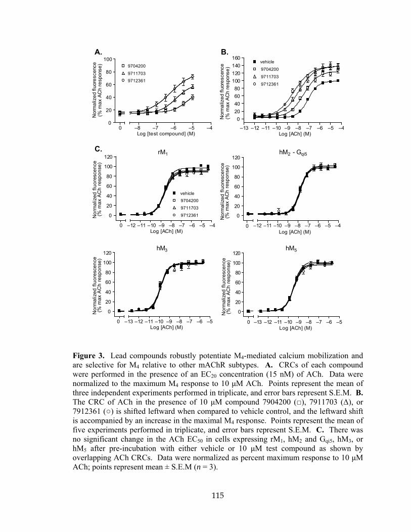
115
B.
–13 –12 –11 –10 –9 –8 –7 –6 –5 –4
020406080
100120140160
vehicle
9704200
9711703
9712361
Log [ACh] (M)
Nor
mal
ized
fluo
resc
ence
(%m
ax A
Ch
resp
onse
)
A.
00
20
40
60
80
100
–8 –7 –6 –5 –4Log [test compound] (M)
Nor
mal
ized
fluo
resc
ence
(%m
axAC
hre
spon
se) 9704200
97117039712361
rM
0
0
20
40
60
80
100
120
vehicle970420097117039712361
–12 –11 –10 –9 –8 –7 –6 –5 –4Log [ACh] (M)
Nor
mal
ized
fluo
resc
ence
(%m
axA
Ch
resp
onse
)
hM - Gqi5
0
0
20
40
60
80
100
120
–12 –11 –10 –9 –8 –7 –6 –5 –4Log [ACh] (M)
Nor
mal
ized
fluo
resc
ence
(%m
axAC
hre
spon
se)
hM
0
0
20
40
60
80
100
120
–13 –12 –11 –10 –9 –8 –7 –6 –5Log [ACh] (M)
Nor
mal
ized
fluo
resc
ence
(%m
axA
Ch
resp
onse
)
hM
0
0
20
40
60
80
100
120
–13 –12 –11 –10 –9 –8 –7 –6 –5Log [ACh] (M)
Nor
mal
ized
fluo
resc
ence
(%m
axA
Ch
resp
onse
)
1
53
2C.
Figure 3. Lead compounds robustly potentiate M4-mediated calcium mobilization and are selective for M4 relative to other mAChR subtypes. A. CRCs of each compound were performed in the presence of an EC20 concentration (15 nM) of ACh. Data were normalized to the maximum M4 response to 10 μM ACh. Points represent the mean of three independent experiments performed in triplicate, and error bars represent S.E.M. B. The CRC of ACh in the presence of 10 μM compound 7904200 (), 7911703 (Δ), or 7912361 () is shifted leftward when compared to vehicle control, and the leftward shift is accompanied by an increase in the maximal M4 response. Points represent the mean of five experiments performed in triplicate, and error bars represent S.E.M. C. There was no significant change in the ACh EC50 in cells expressing rM1, hM2 and Gqi5, hM3, or hM5 after pre-incubation with either vehicle or 10 μM test compound as shown by overlapping ACh CRCs. Data were normalized as percent maximum response to 10 μM ACh; points represent mean ± S.E.M (n = 3).

116
Primary screening hits are robust, subtype selective allosteric potentiators of rat M4
The potency of each compound was determined by pre-incubating cells with
vehicle or increasing concentrations of test compound followed by the addition of an
EC20 concentration (15 nM) of ACh to yield concentration-response curves (CRCs) as
shown in Fig. 3. The responses to these compounds did not reach a clear plateau in the
concentration range used, and solubility limits prevented further increases in
concentrations. However, in the concentration ranges tested, compound 7912361 was the
most potent with an EC50 of 395 ± 28 nM, while compounds 7904200 and 7911703 had
EC50 values of 6 ± 0.28 μM and 1.3 ± 0.28 μM, respectively.
We next determined the effects of maximal concentrations of each compound on
the CRC of ACh. Cells were pre-incubated with a fixed concentration (10 μM) of test
compound and subsequently stimulated with increasing concentrations of ACh. Each
compound induced a leftward shift of the ACh CRC and increased the maximal response
to ACh (Fig 3). The EC50 value for ACh in the presence of vehicle was 87 ± 12 nM,
whereas the EC50 values were 34, 12, and 4 nM in the presence of compounds 7904200,
7911703, and 7912361, respectively. Thus, compound 7912361 proved to be the most
robust potentiator causing a 21-fold leftward shift in the ACh CRC (Fig. 3).
Experiments were then performed to assess the functional selectivity of our three
lead compounds at potentiating M4 responses relative to other mAChRs. Cells
expressing rM1, rM2-Gqi5, rM3, or rM5 were incubated first with 10 μM test compound or
vehicle, then with increasing concentrations of ACh to test whether these potentiators
would have any effect on functional mAChR responses in these cell lines. As shown in
Fig. 3, among the five muscarinic receptor subtypes, these three novel compounds

117
possess perfect functional subtype selectivity for M4 as shown by their lack of effect on
any other mAChR in the absence or presence of ACh. Because the experimental protocol
measures real-time calcium mobilization during a pre-incubation with each test
compound, any agonist or antagonist as well as allosteric modulator activity is detected.
The ACh CRCs for rM1, rM2, rM3, and rM5 in the presence of vehicle or 10 μM test
compound are virtually overlapping with no effect on the functional ACh EC50.
Chemical optimization of primary M4 allosteric potentiator hits
The results from the 232 compound screen were analyzed to understand the
structure activity relationship of the compounds. The top three lead compounds had a
dimethyl substitution on the pyridine ring, and most compounds that had larger
substitutions lost all activity. Substitution of the primary amine was mostly tolerated but
resulted in loss of some activity. We chose to keep the primary amine and substitute the
amide functionality with side chains related to compounds 7904200 and 7912361. A
collection of 10 amines were acylated with bromoacetyl bromide to give the
corresponding bromo amides (Fig. 4). Subsequent ‘one-pot’ alkylation and cyclization of
mercaptopyridine with the bromo amides using KOH provided compounds VU10001-
VU10010 (Fig. 4) as yellow solids after conversion to their TFA salts.

118
N
CN
SH
N S
NH2O
NHRBrNHR
ORNH2
Br
OBr
Bromoacetyl bromide
Mercaptopyridine
VU10001 - VU10010KOH
DMF, water
ClF
CF3
VU10001 R =
VU10002 R =
VU10003 R =
VU10004 R =
VU10005 R =
VU10006 R =
VU10007 R =
VU10008 R =
VU10009 R =
VU10010 R =
Figure 4. Synthesis of compounds in the VU10000 series. A collection of 10 amines were acylated with bromoacetyl bromide to give the corresponding bromo amides. Subsequent alkylation and cyclization of mercaptopyridine with the bromo amides using KOH provided compounds VU10001-VU10010. Syntheses performed by D. Orton and R. Williams.

119
Characterization of novel compounds in the VU10000 series
To test this small library of ten compounds for rM4 allosteric potentiator activity,
functional experiments identical to the ones described above were performed. Fig. 5
shows the response of cells expressing rM4 and Gqi5 to an EC20 concentration of ACh in
the presence of either vehicle (EC20 grey bar) or test compound (black bars) as compared
to the maximum response to 10 μM ACh after pre-incubation with vehicle (white bar).
All compounds except VU10007 showed allosteric potentiator activity; VU10005,
VU10008, and VU10010 caused the largest potentiation of the EC20 response and these
compounds were selected for further testing. CRCs of these potentiators showed that all
three compounds have submicromolar potencies; compounds VU10005, VU10008, and
VU10010 have EC50 values of 300 ± 75 nM, 715 ± 160 nM, and 400 ± 100 nM,
respectively (Fig. 5b). When comparing the rM4 ACh CRC in the presence of either
vehicle or 10 μM test compound, we found VU10010 to be the most robust allosteric
potentiator as is shown by the 47-fold shift in the ACh EC50 (Fig. 5c). The EC50 value of
ACh was 33 ± 9 nM in the absence of VU10010 and 0.7 ± 0.2 nM in the presence of this
compound. All three lead compounds in the VU10000 series produced greater leftward
shifts in the functional rM4 response to ACh than the best lead compound from the
ChemBridge library, compound 7912361, which induced a 21-fold increase in ACh
potency (Fig. 3 and 5). Compounds VU10005 and VU10008 produced 30- and 37-fold
leftward shifts in the curve, respectively. From these data, we concluded that VU10010
had the best combination of potency and efficacy of all compounds tested, and we chose
this allosteric potentiator for use in all subsequent experiments. The next critical step

120
was to confirm that VU10010 maintained the high degree of subtype selectivity that was
shown for compounds in the ChemBridge series.

121
A.
ECmax
EC20
VU1000
1
VU1000
2
VU1000
3
VU1000
4
VU1000
5
VU1000
6
VU1000
7
VU1000
8
VU1000
9
VU1001
00
20
40
60
80
100
120
Nor
mal
ized
fluor
esce
nce
(%m
axAC
hre
spon
se)
B.
Log [test compound] (M)0
0
20
40
60
80
100
VU10005
VU10010VU10008
–9 –8 –7 –6 –5 –4
Nor
mal
ized
fluor
esce
nce
(%m
axAC
hre
spon
se)
C.
0
0
25
50
75
100
125
150
175vehicle7912361VU10005VU10008VU10010
–12 –11 –10 –9 –8 –7 –6 –5 –4Log [ACh] (M)
Nor
mal
ized
fluo
resc
ence
(%m
axAC
hre
spon
se)
Figure 5. Chemical optimization generates compounds that potentiate M4-mediated calcium mobilization with greater efficacy than lead compound 7912361. A. Nine of the ten compounds synthesized and tested showed allosteric potentiator activity (black bars) compared to the ACh EC20 control (grey bar). Data were normalized to the maximal response to 10 μM ACh (white bar). Bars represent the mean ± S.E.M. of five independent experiments, each performed in triplicate. B. The three compounds that showed the highest degree of potentiation in panel (A.) were further characterized to determine their potencies. As shown by their CRCs, all three compounds have potencies in the submicromolar range. C. When compared to compound 7912361, all three lead compounds in this series induced a more robust leftward shift in the rM4 ACh CRC as well as a larger increase in the maximal response. Points represent mean ± S.E.M. (n = 3).
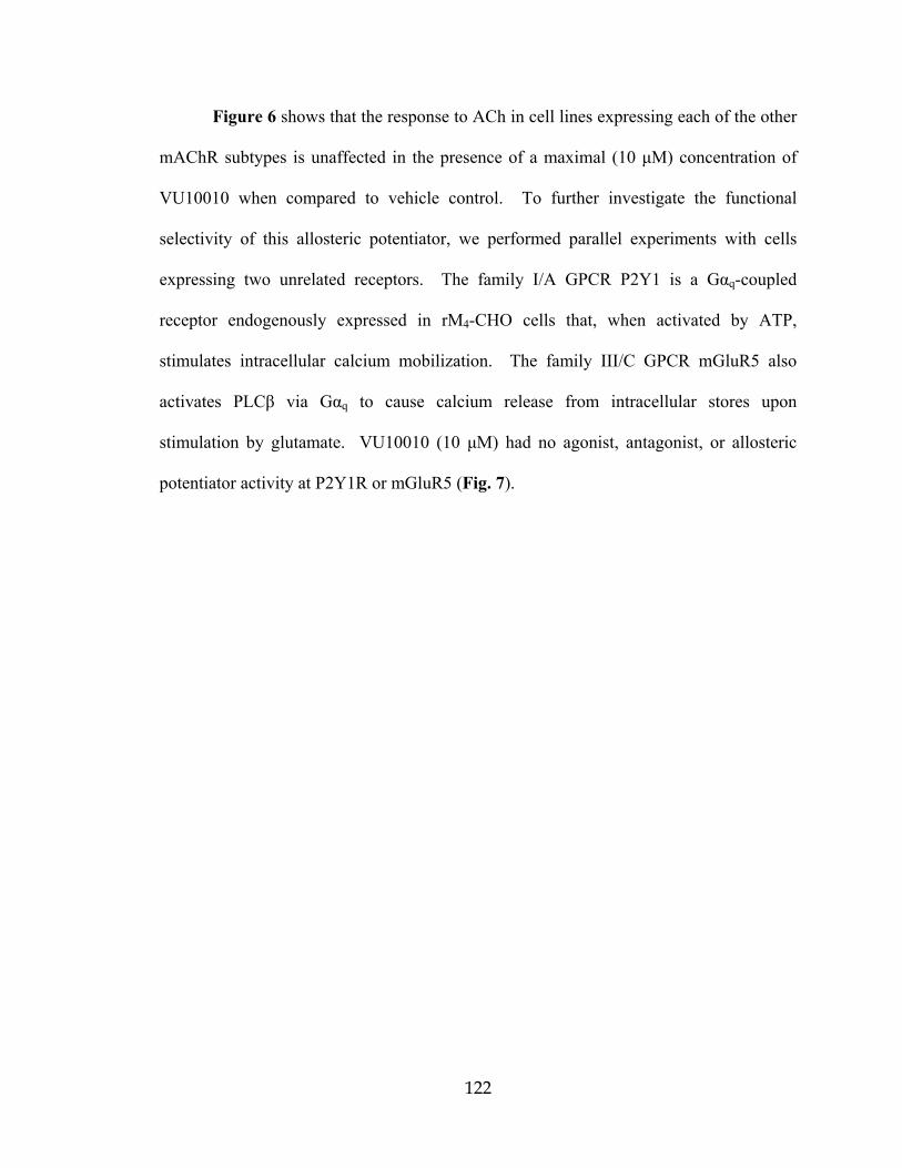
122
Figure 6 shows that the response to ACh in cell lines expressing each of the other
mAChR subtypes is unaffected in the presence of a maximal (10 μM) concentration of
VU10010 when compared to vehicle control. To further investigate the functional
selectivity of this allosteric potentiator, we performed parallel experiments with cells
expressing two unrelated receptors. The family I/A GPCR P2Y1 is a Gαq-coupled
receptor endogenously expressed in rM4-CHO cells that, when activated by ATP,
stimulates intracellular calcium mobilization. The family III/C GPCR mGluR5 also
activates PLCβ via Gαq to cause calcium release from intracellular stores upon
stimulation by glutamate. VU10010 (10 μM) had no agonist, antagonist, or allosteric
potentiator activity at P2Y1R or mGluR5 (Fig. 7).
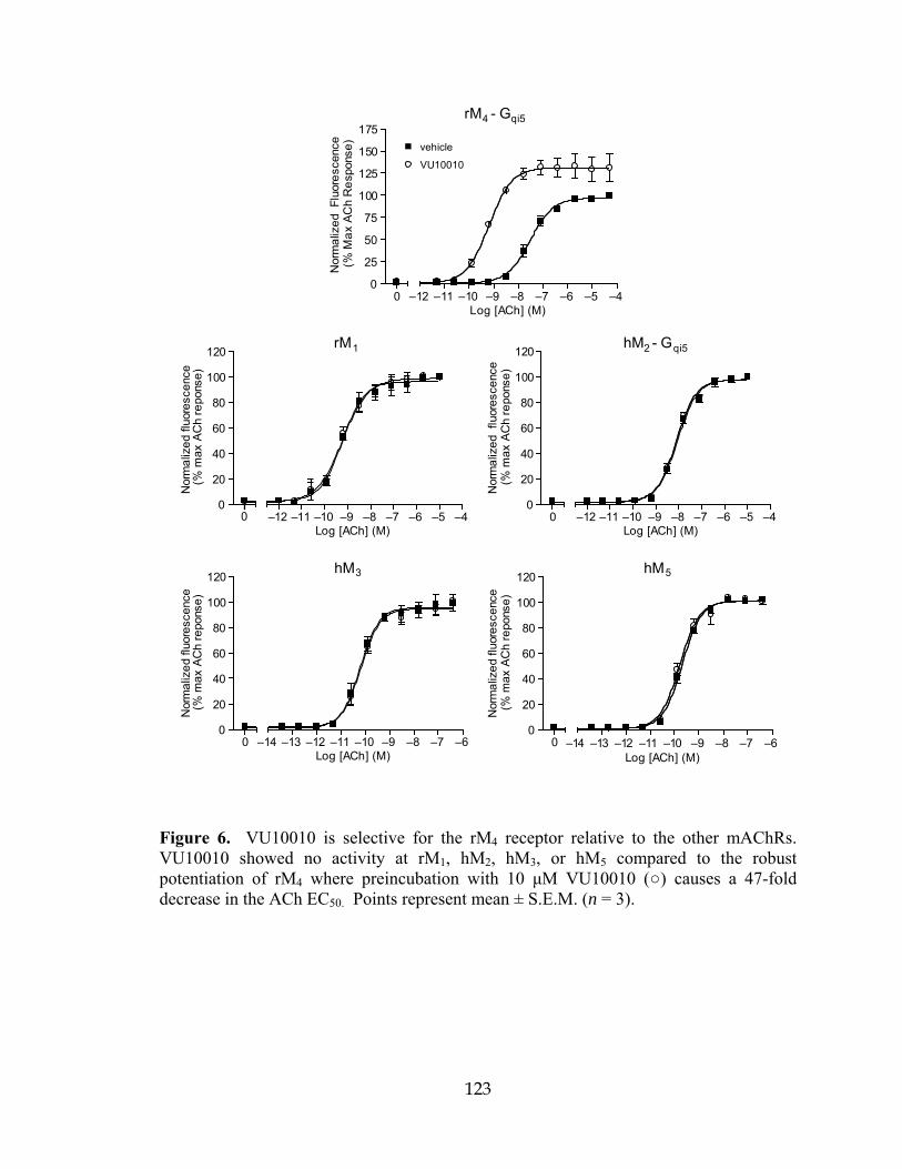
123
rM
0
20
40
60
80
100
120
–12 –11 –10 –9 –8 –7 –6 –5 –4Log [ACh] (M)
Nor
mal
ized
fluo
resc
ence
(%m
axAC
hre
pons
e)
hM - G
00
20
40
60
80
100
120
–12 –11 –10 –9 –8 –7 –6 –5 –4Log [ACh] (M)
Nor
mal
ized
flu
ores
cenc
e(%
max
ACh
repo
nse)
hM
00
20
40
60
80
100
120
–14 –13 –12 –11 –10 –9 –8 –7 –6Log [ACh] (M)
Nor
mal
ized
fluor
esce
nce
(%m
axAC
hre
pons
e)
hM
00
20
40
60
80
100
120
–14 –13 –12 –11 –10 –9 –8 –7 –6Log [ACh] (M)
Nor
mal
ized
fluor
esce
nce
(%m
axAC
hre
pons
e)
rM - Gqi5
00
25
50
75
100
125
150
175vehicle
VU10010
–12 –11 –10 –9 –8 –7 –6 –5 –4Log [ACh] (M)
Nor
mal
ized
Fluo
resc
ence
(%M
axAC
hR
espo
nse)
0
qi52
3
4
5
1
Figure 6. VU10010 is selective for the rM4 receptor relative to the other mAChRs. VU10010 showed no activity at rM1, hM2, hM3, or hM5 compared to the robust potentiation of rM4 where preincubation with 10 μM VU10010 () causes a 47-fold decrease in the ACh EC50. Points represent mean ± S.E.M. (n = 3).

124
A.
B.mGluR5
00
20
40
60
80
100
120
vehicle10 µM VU10010
–9 –8 –7 –6 –5 –4 –3Log [glutamate] (M)
Nor
mal
ized
fluor
esce
nce
(%m
axgl
ure
spon
se)
P2Y1
00
20
40
60
80
100
120
vehicle10 µM VU10010
–9 –8 –7 –6 –5 –4 –3Log [ATP] (M)
Nor
mal
ized
fluor
esce
nce
(%m
axA
TPre
spon
se)
Figure 7. VU10010 showed no agonist, antagonist, or potentiator activity at two GPCRs unrelated to rM4 mAChR. Agonist CRCs from cells expressing the ATP receptor, P2Y1 (A.), or the metabotropic glutamate receptor, mGluR5 (B.), were overlapping in the presence of 10 μM VU10010 () compared to vehicle control. Data points represent mean ± S.E.M. (n = 3).

125
VU10010 binds allosterically and increases M4 receptor affinity for ACh
In order to determine the effects of VU10010 on orthosteric binding and to further
confirm an allosteric mechanism of binding, we performed equilibrium radioligand
binding studies. Membranes prepared from cells expressing rM4 were incubated with a
non-saturating concentration of the orthosteric site antagonist [3H]NMS (0.1 nM) in the
absence or presence of increasing concentrations of either atropine or VU10010.
Consistent with atropine’s high affinity at the orthosteric site, this compound displaced
[3H]NMS binding with an Ki value of 0.52 ± 0.23 nM. In contrast, VU10010 did not
displace [3H]NMS binding at concentrations up to 30 μM, which is 75 times the EC50
value of this compound at potentiating responses to ACh (Fig. 8). While this finding
does not exclude the possibility that VU10010 could alter [3H]NMS binding under some
conditions, it provides strong support for the functional data and further suggests that
VU10010 acts at a site on the M4 receptor that is distinct from the
orthostericantagonist/agonist binding site. To further confirm that VU10010 is selective
for M4 relative to other mAChRs, we performed parallel binding experiments using
membranes from cell lines expressing rM1, rM2, rM3, and rM5. As shown in Fig. 9, this
compound does not displace [3H]NMS binding at any mAChR subtype. The [3H]NMS
Kd, Bmax, and atropine Ki values for each mAChR from saturation and competition
binding experiments are listed in Table 4.
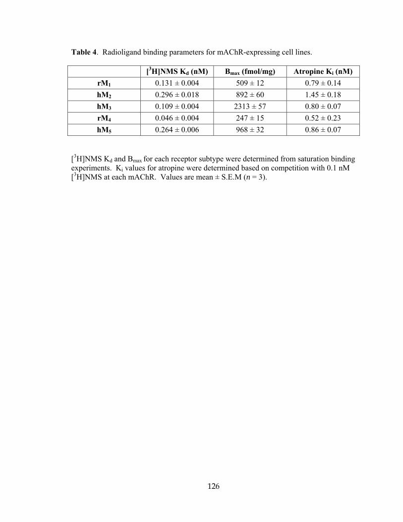
126
Table 4. Radioligand binding parameters for mAChR-expressing cell lines.
[3H]NMS Kd (nM) Bmax (fmol/mg) Atropine Ki (nM) rM1 0.131 ± 0.004 509 ± 12 0.79 ± 0.14 hM2 0.296 ± 0.018 892 ± 60 1.45 ± 0.18 hM3 0.109 ± 0.004 2313 ± 57 0.80 ± 0.07 rM4 0.046 ± 0.004 247 ± 15 0.52 ± 0.23 hM5 0.264 ± 0.006 968 ± 32 0.86 ± 0.07
[3H]NMS Kd and Bmax for each receptor subtype were determined from saturation binding experiments. Ki values for atropine were determined based on competition with 0.1 nM [3H]NMS at each mAChR. Values are mean ± S.E.M (n = 3).

127
A.
B.
–12 –11 –10 –9 –8 –7 –6 –5 –4
0
20
40
60
80
100
120
Atropine
VU10010
Log [cold ligand] (M)
Spec
ific
bind
ing
(%m
ax[3
H]N
MS
boun
d)
-9 –8 –7 –6 –5 –4 –3 –2
0
20
40
60
80
100
120Vehicle
30 nM
100 nM
300 nM
1 µM
3 µM
10 µM
Log [ACh] (M)
Spec
ific
bind
ing
0
020406080
100120140160
Vehicle
10 µM VU10010
–9 –8 –7 –6 –5 –4 –3Log [ACh] (M)
[35S]
GTP
γS B
indi
ng(%
max
ACh
resp
onse
)
C.
(%m
ax[3
H]N
MS
boun
d)
Figure 8. Compound VU10010 binds to an allosteric site on M4 mAChR and causes an increase in affinity for ACh and M4-mediated [35S]GTPγS binding. A. Binding of 0.1 nM [3H]NMS was displaced by atropine (, Ki = 0.52 ± 0.23 nM) but not by VU10010 (). Data are from three independent experiments performed in triplicate. Error bars are S.E.M. B. In a dose-dependent manner, VU10010 shifted the ACh competition curve leftward causing a 14-fold decrease in the Ki at maximal concentration of 10 μM (Δ). Data are mean ± S.E.M. (n = 3). C. VU10010 potentiates the response to ACh in membranes from cells expressing rM4. VU10010 (, 10 μM) induced a 10-fold decrease in the ACh EC50 when compared to vehicle control (). The leftward shift in the CRC was also accompanied by a 43 ± 12% increase in the maximal response. Data represent mean ± S.E.M. (n = 3).

128
One possible mechanism by which a compound could enhance receptor activation
by an agonist is by increasing the receptor’s affinity for that agonist (Christopoulos and
Kenakin, 2002). As shown in Fig. 8, compound VU10010 does in fact induce a
concentration-dependent shift in the potency of ACh at displacing rM4 [3H]NMS binding.
At 10 μM, a concentration that causes maximal potentiation, VU10010 increased the
apparent affinity of ACh 14-fold. Thus, the Ki value for ACh at the orthosteric site
decreased from 540 ± 72 nM in the absence of VU10010 to 39 ± 7 nM in the presence of
10 μM VU10010. These data suggest that this compound binds to an allosteric site on
M4 mAChR and shifts the receptor into a conformation that has higher affinity for ACh.
VU10010 enhances M4 coupling to downstream effector proteins
The finding that VU10010 increases affinity of ACh for the orthosteric site
suggests that this contributes to the ability of this compound to potentiate responses to
ACh. However, the magnitude of this shift (14-fold) is not sufficient to account for the
magnitude of the shift in the ACh potency at increasing calcium mobilization (47-fold).
Also, the shift in agonist potency was also accompanied by an increase in agonist
maximal response which cannot be explained by an increase in affinity alone. Thus, we
examined the effect of VU10010 on the ability of M4 to increase activation of G proteins
by measuring effects of this compound on ACh-induced increases in guanosine 5′-O-(3-
[35S]thiotriphosphate ([35S]GTPγS) binding. To assess this, the effect of increasing
concentrations of ACh on [35S]GTPγS binding to membranes from rM4-CHO cells was
measured in the absence and presence 10 μM compound VU10010. Compound
VU10010 shifted the CRC of ACh at increasing [35S]GTPγS binding to the left and also
129
increased maximal [35S]GTPγS binding by roughly 50% (Fig. 8). Thus, the EC50 value
for ACh was 514 ± 82 nM in the absence of VU10010 and 51 ± 11 nM in the presence of
this compound. These data are consistent with the hypothesis that the allosteric
potentiator not only increases affinity of the receptor for agonist but also increases
coupling efficiency and efficacy.

130
rM
–12 –11 –10 –9 –8 –7 –6 –5 –4
0
20
40
60
80
100
120
AtropineVU10010
Log [cold ligand] (M)
Spec
ific
bind
ing
(%m
ax[3 H
]NM
Sbo
und)
hM
–12 –11 –10 –9 –8 –7 –6 –5 –4
0
20
40
60
80
100
120
Log [cold ligand] (M)
Spec
ific
bind
ing
(%m
ax[3 H
]NM
Sbo
und)
hM
–12 –11 –10 –9 –8 –7 –6 –5 –4
0
20
40
60
80
100
120
Log [cold ligand] (M)
Spec
ific
bind
ing
(%m
ax[3 H
]NM
Sbo
und)
hM
–12 –11 –10 –9 –8 –7 –6 –5 –4
0
20
40
60
80
100
120
140
Log [cold ligand] (M)
Spec
ific
bind
ing
(%m
ax[3 H
]NM
Sbo
und)
1 2
3 5
Figure 9. VU10010 does not compete for binding at rat M1, M2, M3, or M5. Binding of 0.1 nM [3H]NMS was displaced by atropine () but not by VU10010 (). Data are from three independent experiments performed in triplicate, and error bars are S.E.M.

131
VU10010 enhances muscarinic depression of excitatory post synaptic currents in hippocampal CA1 pyramidal cells Discovery of novel highly selective and potent allosteric potentiators of M4
provides an unprecedented opportunity to begin functional studies aimed at determining
the physiological roles of this receptor subtype in native preparations. One of the most
prominent effects of mAChR activation that is seen in a variety of forebrain and cortical
regions is depression of transmission at excitatory glutamatergic synapses; in addition,
muscarinic agonists can reduce transmission at inhibitory GABAergic synapses in these
same circuits (Valentino and Dingledine, 1981; Bilkey and Goddard, 1985; Sheridan and
Sutor, 1990; Pitler and Alger, 1994; Qian and Saggau, 1997; Fernandez de Sevilla and
Buno, 2003). However, the lack of subtype-selective ligands for specific mAChR
subtypes has made it impossible to definitively determine which mAChR subtypes
mediate each of these effects. Thus, we determined the effect of VU10010 on the ability
of the non-selective mAChR agonist, carbachol (CCh), to reduce excitatory and
inhibitory transmission at the Schaffer collateral-CA1 synapse in the rat hippocampus.
Consistent with previous findings, CCh reversibly inhibited evoked EPSCs at Shaffer-
collateral-CA1 synapses in a concentration-dependent manner (Fig. 10). CCh at 100 μM
caused a marked reduction of EPSCs (16.1 ± 4.0 % of control value, n = 5, Fig. 10),
whereas at a sub-maximal concentration of 1 μM, CCh induced a relatively small
reduction of EPSCs (60.5 ± 4.1% of control value, n = 15; Fig. 10). In contrast, VU10010
(5 μM) had no effect on excitatory transmission at this synapse when added alone (EPSC
amplitude 107.9 ± 5.2% of the control, P = 0.16, n = 7; Fig. 10). However, when
VU10010 (5 μM) was co-applied with a sub-maximal concentration of CCh (1 μM), this
compound increased the CCh-induced reduction of EPSCs (36.9 ± 5.7% of control, n = 7,

132
Fig. 10) when compared to the effect of 1 μM CCh alone (60.5 ± 4.1% of control value, n
= 15; P = 0.0038, Fig. 10). Thus, VU10010 potentiates CCh-induced inhibition of
excitatory synaptic transmission at this critical synapse in the hippocampus. As a
negative control, we used VU10007, a closely related analog of VU10010 that is
completely inactive as an allosteric potentiator of M4 in cell-based assays (see Fig. 5).
VU10007 (5 μM) had no effect on the CCh-induced reduction of EPSCs (61.6 ± 5.0% of
control value, n = 7, Fig. 10; compared to 60.5 ± 4.1% of control value by CCh alone, n =
15; P = 1). These results suggest that M4 is one of the mAChRs involved in the
regulation of excitatory synaptic transmission at Schaffer collateral-CA1 synapses.
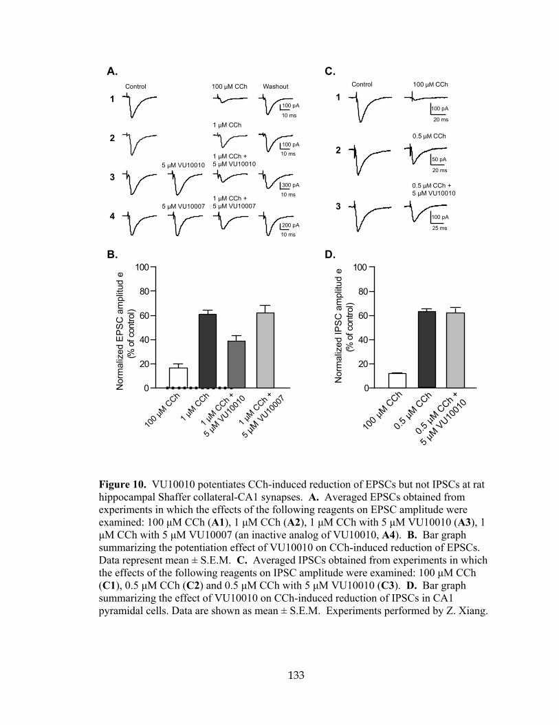
133
100 pA
10 ms
10 ms
10 ms
10 ms
300 pA
100 pA
200 pA
Control
1 μM CCh
Washout
5 μM VU10010
5 μM VU10007
1 μM CCh +5 μM VU10010
1 μM CCh +5 μM VU10007
100 μM CCh
A.
2
4
3
1
B.
0
20
40
60
80
100
Nor
mal
ized
EP
SC
am
plitu
de
(%of
cont
rol)
1 μM C
Ch
1 μM C
Ch +
5 μMVU10
010
1 μM C
Ch +
5 μMVU10
007
100 μM
CCh
50 pA
20 ms
0.5 μM CCh
100 pA
25 ms
100 pA
20 ms
Control 100 μM CCh
0.5 μM CCh +5 μM VU10010
C.
2
3
1
0
20
40
60
80
100
0.5μM
CCh
0.5μM C
Ch +
5 μM VU10
010
100 μM
CCh
Nor
mal
ized
IPS
C a
mpl
itud
e(%
ofco
ntro
l)
D.
Figure 10. VU10010 potentiates CCh-induced reduction of EPSCs but not IPSCs at rat hippocampal Shaffer collateral-CA1 synapses. A. Averaged EPSCs obtained from experiments in which the effects of the following reagents on EPSC amplitude were examined: 100 μM CCh (A1), 1 μM CCh (A2), 1 μM CCh with 5 μM VU10010 (A3), 1 μM CCh with 5 μM VU10007 (an inactive analog of VU10010, A4). B. Bar graph summarizing the potentiation effect of VU10010 on CCh-induced reduction of EPSCs. Data represent mean ± S.E.M. C. Averaged IPSCs obtained from experiments in which the effects of the following reagents on IPSC amplitude were examined: 100 μM CCh (C1), 0.5 μM CCh (C2) and 0.5 μM CCh with 5 μM VU10010 (C3). D. Bar graph summarizing the effect of VU10010 on CCh-induced reduction of IPSCs in CA1 pyramidal cells. Data are shown as mean ± S.E.M. Experiments performed by Z. Xiang.
134
Muscarinic depression of inhibitory post synaptic currents is not affected by M4 potentiation Muscarinic receptors also play a role in regulating inhibitory synaptic
transmission in hippocampal CA1 pyramidal cells (Valentino and Dingledine, 1981;
Pitler and Alger, 1994). Consistent with previous studies, we found that application of
100 μM CCh greatly reduced monosynaptic IPSC amplitude to 10.9 ± 0.9% of control
value (n = 3, Fig. 10), and a sub-maximal concentration of CCh (0.5 μM) caused a
smaller reduction of IPSC amplitude (62.2 ± 3.5% of control, n = 6, Fig. 10). Application
of the M4 potentiator VU10010 (5 μM) alone had no effect on IPSC amplitude (98.5 ±
1.7 of control, n = 4, P = 0.875). Furthermore, VU10010 (5 μM) had no effect on the
depression of IPSCs induced by 0.5 μM CCh (61.7 ± 3.9% with CCh and VU10010, n =
6, versus 62.2 ± 3.5% with CCh alone, n = 6, P = 0.82; Fig. 10). These results indicate
that M4 mAChRs are probably not involved in muscarinic modulation of inhibitory
synaptic transmission in CA1 pyramidal cells. To further test this hypothesis, we
compared the effect of sub-maximal concentration of CCh on IPSCs in hippocampal CA1
pyramidal cells in M4 knockout and WT mice. At 2 μM, CCh caused a reduction of
IPSCs with a comparable magnitude in both WT and M4 KO mice (56.0 ± 4.6% and 60.2
± 2.7% of control values, n = 6 and n = 6, respectively; P = 0.48, Fig. 11). While these
data do not completely rule out a role for M4 in regulating IPSCs in WT animals, the data
are consistent the hypothesis that M4 mAChRs are not involved in modulation of
inhibitory synaptic transmission in CA1 pyramidal cells.

135
10 ms100 pA
10 ms200 pA
10 ms100 pA
10 ms50 pA
10 ms
50 pA
10 ms
100 pA
Control
2 μM CCh
Washout
5 μM VU100102 μM CCh +5 μM VU10010
100 μM CCh
A. Wild-type
2
3
1Control
2 μM CCh
Washout
5 μM VU100102 μM CCh +5 μM VU10010
100 μM CCh
B. M Knockout
Nor
mal
ized
EP
SC
am
plitu
de(%
ofco
ntro
l)
2 μMCCh
2 μMCCh +
5 μMVU10010
100 μM
CCh0
20
40
60
80
100
WT
M KO
C.
2
3
1
4
4
Figure 11. VU10010 potentiates CCh-induced reduction of EPSCs in wild-type (WT) but not in M4 KO mice. A. Averaged EPSCs obtained from experiments in which the effects of the following reagents on EPSC amplitude in WT mice were examined: 100 μM CCh (A1), 2 μM CCh (A2), and 2 μM CCh with 5 μM VU10010 (A3). B. Averaged EPSCs obtained from experiments in which the effects of the same reagents on EPSC amplitude in M4 KO mice were examined: 100 μM CCh (B1), 2 μM CCh (B2), and 2 μM CCh with 5 μM VU10010 (B3). (C) Bar graph summarizing the effect of VU10010 on CCh-induced reduction of EPSCs in WT mice (white bars) and M4 KO mice (black bars). Data represent mean ± S.E.M. Experiments performed by Z. Xiang and J.E. Ayala.

136
VU10010 potentiates CCh-induced reduction of EPSCs in wild-type (WT) but not in M4 knockout (KO) mice To confirm that the potentiation effect of VU10010 on CCh-induced reduction of
EPSCs was due to its action on M4 receptors, we examined the effect of VU10010 in M4
KO mice (Gomeza et al., 1999). As a control, we first assessed the effect of VU10010 in
WT mice of the same mixed genetic background as the M4 KO mice. Similar to the result
obtained from rats, CCh at 100 μM caused a sizable reduction of EPSCs at Schaffer
collateral-CA1 synapses in WT mice (18.0 ± 0.9 % of control value, n = 6, Fig. 11),
whereas at a sub-maximal concentration of 2 μM, CCh induced a smaller inhibition of
EPSCs (53.7 ± 1.4% of control, n = 6; Fig. 11). Compound VU10010 (5 μM) had no
effect on excitatory transmission at this synapse when added alone (EPSC amplitude
102.5 ± 3.8% of the control, P = 0.31, n = 5; Fig. 11). However, when co-applied with 2
μM CCh, VU10010 (5 μM) significantly enhanced the CCh-induced reduction of EPSCs
(39.8 ± 2.7% of the control, n = 5, Fig. 11) when compared to the effect of 2 μM CCh
alone (54.7 ± 1.1% of control, n = 5; P = 0.0079, Fig. 11).
In M4 KO mice, application of 100 μM CCh decreased the EPSC amplitude to
21.1 ± 3.8 % of control value (n = 6, Fig. 11), whereas at the sub-maximal concentration
of 2 μM, CCh induced a relatively small suppression of EPSCs (70.8 ± 3.6% of control
value, n = 9; Fig. 11), which was significantly different than the reduction of EPSCs
caused by 2 μM CCh in WT mice (53.7 ± 1.4% of control, n = 6; P = 0.0076, Fig. 11).
Furthermore, when compound VU10010 was co-applied with 2 μM CCh, it failed to
potentiate the CCh-induced reduction of EPSCs in M4 KO mice (77.5 ± 4.6% of control
value, n = 7, Fig. 11) when compared to the effect of 2 μM CCh alone (70.8 ± 3.6% of
control value, n = 9; P = 0.14, Fig. 11). These data support the conclusion that the
137
potentiation of CCh-induced reduction of EPSCs by VU10010 is solely due to its action
on M4 receptors and that M4 participates in the muscarinic modulation of these synaptic
responses.
Discussion
Over the past two decades, major attention has been focused on developing highly
selective agonists of specific mAChR subtypes to use as research tools in order to achieve
a better understanding of the roles of each subtype in specific organ systems and also to
develop therapeutic agents that lack the adverse effects of AChE inhibitors and
nonselective muscarinic agents. However, the muscarinic receptor agonists developed to
date lack true specificity for individual muscarinic receptor subtypes. This thesis chapter
reports the discovery of a novel series of compounds that act as robust positive allosteric
modulators (allosteric potentiators) of the M4 mAChR subtype. These compounds are
highly selective for M4 and have no detectable activity at any other mAChR subtype. As
with previously discovered allosteric potentiators of metabotropic glutamate receptors
(mGluRs), the novel positive allosteric modulators of M4 reported here have no
detectable affinity for the orthosteric site on M4. The action of these compounds at a site
that is distinct from the highly conserved orthosteric ACh binding site is likely to be
critical for achieving this high degree of subtype selectivity.
Interestingly, the M4 allosteric potentiator reported here increases affinity of ACh
for the orthosteric site. This is similar to previous reports of allosteric modulators of
GPCRs acting by altering affinity of ligands at the orthosteric site; the magnitude of
binding cooperativity or the shift in orthosteric agonist affinity is referred to as the alpha
138
factor (Christopoulos and Kenakin, 2002). However, while VU10010 apparently acts in
part by increasing ACh affinity, the magnitude of this effect was only a 14-fold increase
in ACh affinity whereas VU10010 induced an almost 50-fold shift in the ACh
concentration response curve for activating calcium mobilization. Thus, effects on
agonist binding cannot fully account for the allosteric potentiator activity measured in the
functional assay; potentiation by this compound must also increase efficacy and coupling
to downstream effectors. The present data suggest that VU10010 acts by both increasing
ACh affinity and also through a positively cooperative interaction with ACh for
activation of G proteins and downstream signaling pathways. Studies of the effects of
VU10010 on [35S]GTPγS binding revealed a 10-fold shift in ACh potency; this value is
close to the calculated shift in ACh affinity at the orthosteric site, suggesting that the
increase in ACh affinity could account for the shift in ACh potency at increasing G
protein activation. However, VU10010 also increased the maximal ACh-induced
[35S]GTPγS binding response, suggesting that this potentiator increases the efficiency of
M4 coupling to G proteins. These two factors together likely contribute to the robust shift
in ACh potency for inducing calcium mobilization relative to more modest shift observed
in ACh affinity.
Discovery of a highly selective allosteric modulator of M4 provides an
unprecedented opportunity to selectively increase activity of this receptor and develop a
more detailed understanding of the functional roles of M4 in brain circuits that are heavily
modulated by cholinergic innervation. One of the most important roles of cholinergic
systems in the CNS is modulation of transmission through the hippocampal formation, a
limbic cortical structure that plays a critical role in learning and memory and is thought to
139
be important for cholinergic regulation of cognitive function. Activation of mAChRs in
the hippocampus induces a marked reduction of transmission at both excitatory
glutamatergic and inhibitory GABAergic synapses by presynaptic reduction of
neurotransmitter release (Valentino and Dingledine, 1981; Bilkey and Goddard, 1985;
Fernandez de Sevilla and Buno, 2003). Multiple mAChR subtypes are expressed in the
hippocampus, and evidence suggests that both M2 and M4 are localized in presynaptic
terminals where they could regulate excitatory and inhibitory synaptic transmission
(Levey et al., 1995; Rouse et al., 1999; Rouse et al., 2000a). In addition, evidence
suggests that activation of postsynaptic mAChRs on hippocampal pyramidal cells where
M1 and M4 are expressed could reduce synaptic transmission in the hippocampus by
inducing release of endocannabinoids that activate presynaptic CB1 cannabinoid
receptors (Ohno-Shosaku et al., 2003). Thus, the specific mAChR subtypes involved in
regulating transmission at these important synapses has not been clear. The present
finding that VU10010 potentiates effects of mAChR agonists on excitatory but not
inhibitory synaptic transmission provides strong evidence that M4 is one of the mAChR
subtypes responsible for regulating transmission at glutamatergic synapses in the
hippocampus but is not likely to be involved in regulation of transmission at inhibitory
synapses in this brain region. This selective regulation of excitatory synaptic
transmission is a first critical step in developing a detailed understanding of the roles of
M4 in modulating hippocampal function. In future studies, it will be important to take
advantage of this novel class of allosteric potentiators to further investigate the roles of
M4 in hippocampus as well as other cortical and basal ganglia structures where this
receptor has been postulated to play a critical neuromodulatory role. Also, it will be

140
important to further optimize these compounds for use as tools that will provide a path
for developing an understanding of the impact of selectively increasing M4 activation in
vivo.

141
CHAPTER IIIB
CENTRALLY ACTIVE ALLOSTERIC POTENTIATORS OF THE M4 MUSCARINIC ACETYLCHOLINE RECEPTOR REVERSE AMPHETAMINE-
INDUCED HYPERLOCOMOTOR ACTIVITY IN RATS
Introduction
To date, five muscarinic acetylcholine receptor (mAChR) subtypes have been
identified (M1-M5) and play important roles in mediating the actions of ACh in the
peripheral and central nervous systems (Wess, 1996). Of these, M1 and M4 are the most
heavily expressed in the CNS and represent attractive therapeutic targets for cognition,
Alzheimer’s disease (AD) and schizophrenia (Bymaster et al., 2002; Messer, 2002;
Raedler et al., 2007). In contrast, the adverse effects of cholinergic agents are thought to
be primarily due to activation of peripheral M2 and M3 mAChRs (Bymaster et al., 2003b;
Bymaster et al., 2003a). Due to the high sequence homology and conservation of the
orthosteric ACh binding site among the mAChR subtypes, development of chemical
agents that are selective for a single subtype has been largely unsuccessful, and in the
absence of highly selective activators of M4, it has been impossible to test the role of
selective M4 activation. Clinical trials with xanomeline (Fig. 12), an M1/M4 preferring
orthosteric agonist, demonstrated efficacy as both a cognition enhancing agent, and as an
antipsychotic agent (Bodick et al., 1997b; Shekhar et al., 2001; Shekhar et al., 2008). In
follow-up studies in rats, xanomeline displayed an antipsychotic-like profile comparable
to clozapine (Stanhope et al., 2001). However, a long standing question concerned
whether or not the antipsychotic efficacy or antipsychotic-like activity in animal models

142
is mediated by activation of M1, M4, or a combination of both receptors. Data from
mAChR knock-out mice led to the suggestion that a selective M1 agonist would be
beneficial for cognition, whereas an M4 agonist would provide antipsychotic activity for
the treatment of schizophrenia (Felder et al., 2001; Bymaster et al., 2003b; Bymaster et
al., 2003a). This proposal is further supported by recent studies demonstrating that M4
receptors modulate the dynamics of cholinergic and dopaminergic neurotransmission and
that loss of M4 function results in a state of dopamine hyperfunction (Tzavara et al.,
2004). These data, coupled with findings that schizophrenic patients have altered
hippocampal M4, but not M1, receptor expression (Scarr et al., 2007) suggest that
selective activators of M4 may provide a novel treatment strategy for schizophrenia
patients. However, multiple studies suggest that M1 may also play an important role in
the antipsychotic effects of mAChR agonists and the relative contributions of M1 and M4
to the antipsychotic efficacy of xanomeline or antipsychotic-like effects of this compound
in animal models are not known. Unfortunately, the lack of highly selective, systemically
active activators of M1 and M4 has made it difficult to fully evaluate the effects of
activation of these mAChR subtypes in animal models.
Recently, we reported discovery of a number of positive allosteric modulators for
class C GPCRs that bind to allosteric sites, provide high levels of subtype selectivity and
display behavioral effects in vivo comparable to direct acting agonists (O'Brien et al.,
2003; Lindsley et al., 2004; O'Brien et al., 2004; Kinney et al., 2005; Galici et al., 2006;
Hemstapat et al., 2006; Marino and Conn, 2006; Zhao et al., 2007). In addition, we
identified a highly selective positive allosteric modulator of M4 termed VU10010 (Fig.
12) (Shirey et al., 2008). This compound induces a 47-fold potentiation of the M4 ACh

143
concentration response curve (CRC), possesses an EC50 in the 400 nM range and causes
no activation of the other mAChR subtypes. Additional in vitro pharmacological
characterization studies suggested that VU10010 binds to an allosteric site on the M4
receptor to increase affinity for ACh and coupling to G proteins (Shirey et al., 2008).
Subsequent studies with VU10010 revealed that selective potentiation of M4 increased
carbachol (CCh)-induced depression at excitatory, but not inhibitory, synapses and that
the effect on excitatory currents was not mimicked by an inactive analog of VU10010 or
in M4 knock-out mice (Shirey et al., 2008).
Figure 12. Chemical structures of Xanomeline [3-[3-hexyloxy-1,2,5-thiadiazo-4-yl]-1,2,5,6-tetrahydro-1-methylpyridine] and VU10010 [3-amino-N-(4-chlorobenzyl)-4,6-dimethylthieno[2,3-b]pyridine-2-carboxamide].
Despite this notable advance, VU10010 suffered from poor physiochemical
properties (logP~4.5) and in vivo studies proved infeasible because we were unable to
formulate VU10010 into a homogeneous solution in any acceptable vehicle, regardless of
salt form or particle size. Several suspensions were prepared and dosed i.p., but
VU10010 was not found to be centrally active. In order to evaluate the role of selective
144
M4 activation in vivo, VU10010 would require further chemical optimization. Here we
report the development and characterization of two novel analogs of VU10010 that are
CNS penetrant following systemic administration.
Results
Chemical Lead Optimization
For the chemical optimization of VU10010, undertook a diversity-oriented
synthesis (DOS) approach to explode Structure-Activity-Relationships (SAR) with a
variety of hypothesis-driven, structural changes to the lead compound. The rationale for
this approach for the optimization of VU10010 is that SAR for allosteric ligands is often
‘flat’ or ‘shallow’, with subtle structural modifications leading to a complete loss of
activity, and often only one portion of an allosteric ligand is amenable to change.
Therefore, a multi-dimensional, DOS library approach provides the best opportunity to
quickly identify productive SAR as opposed to a lead optimization strategy based on
classical, single compound synthesis (Lindsley et al., 2004; Zhao et al., 2007). One
explanation for the lack of central activity observed with VU10010 could be the result of
the poor physiochemical properties alone, or in combination with P-glycoprotein (P-gp)
efflux. P-gp is an efflux transporter with broad substrate specificity present on the
luminal membrane of epithelial cells comprising the blood-brain barrier, which is known
to impair the brain penetrability of a number of drugs. The β-aminoamide motif 3 present
in VU10010 represents a potential P-gp liability, which could be removed by cyclization
to analogs such as 2 (Fig. 13A). Alternatively, P-gp susceptibility could also be

145
diminished by electronically attenuating the basicity of the amine moieties by the
incorporation of distal fluorine atoms. Utilizing solution phase parallel synthesis (Fig.
13B), we synthesized small, 12- to 24-member focused libraries around each of the 10
scaffolds 4, 5-13 (Fig. 13C) which were then purified by mass-directed preparative
HPLC to analytical purity (>98%). This collection of VU10010 analogs incorporated
CF3 moieties (scaffolds 7 and 8) to electronically attenuate potential P-gp susceptibility,
deletion of the β-amino moiety (scaffold 12), or replacement of the β-amino moiety with
an isosteric methyl group (scaffold 11). Other scaffolds explored the deletion of
substituents on the pyridine nucleus (9), incorporation of an additional nitrogen atom to
afford a pyrimidine nucleus (10) or removal of the pyridine nitrogen atom in VU10010
(13). Finally, library 5 focused on maintaining the core structure of VU10010, but
explored alternative amides, selected to improve physiochemical properties and lower the
logP value.

146
A.
B.
C.
X
Y
S
R4O
OHRn
DCC, HOBtR1R2NH, 6
CH2Cl2, DIEA
X
Y
SX
R4O
NR1R2
Rn
5 7-15
1 2
3 5 - 13
75 6
108 9
1311 12
4
Figure 13. Chemical optimization of VU10010 using a diversity-oriented approach to achieve soluble, centrally penetrant M4 positive allosteric modulators. A. β-aminoamide as a potential P-gp liability in series 1 and cyclization strategy to diminish this liability in series 2. B. Solution phase parallel synthesis of libraries of VU10010 analogs. Commercial heterocyclic carboxylic acids 3 (X, Y = C or N) were coupled to 12 different amines (HNR1R2, 4) to afford focused VU10010 analog libraries 5-13 in yields ranging from 15% to 99%. C. Generic structures of analogs of VU10010 evaluated in the chemical lead optimization program in an effort to develop soluble, centrally penetrant M4 positive allosteric modulators. Syntheses performed by T.M. Bridges and J.P. Kennedy.

147
Screening Paradigm for Analog Libraries
As observed with positive allosteric modulators of Class C GPCRs, SAR around
VU10010 was relatively flat, possibly due to a shallow binding pocket (Lindsley et al.,
2004; Zhao et al., 2007). An EC20 triage screen, employing a functional, fluorescence-
based Ca2+ assay in CHO-K1 cells stably co-expressing the rat M4 mACh receptor and
the chimeric G protein, Gqi5, (Fig. 14) quickly eliminated all VU10010 analogs except
those in library 5. Within library 5, all aliphatic and non-benzyl amides were inactive,
and only benzyl and heteroaryl methyl congeners of VU10010, 5a-5p, retained M4 PAM
activity (Table 5). Analogs were synthesized as described in the methods section
(Chapter II). To identify compounds that potentiated agonist activation of M4, we
determined the response to an EC20 concentration of ACh in the absence and presence of
test compound. The potency of each compound was determined by pre-incubating cells
with vehicle or increasing concentrations of test compound followed by the addition of an
EC20 concentration of ACh to yield concentration-response curves (CRCs). Subtle
substitution changes on the arene ring lost activity 5-10-fold in terms of M4 EC50 and/or
fold-shift of the ACh CRC (Table 5). For instance, 5d, in which the 4-Cl moiety of
VU100010 is moved to the 3-position results in a loss in potency of over 9-fold (EC50 =
3.7 µM). Similarly, the unsubstituted phenyl congener 5a retains M4 PAM activity (EC50
= 630 nM), but the fold-shift diminishes to 8.6-fold, versus the 47-fold shift observed for
VU10010 (Shirey et al., 2008). In general, functionalized benzyl amides and pyridyl
methyl congeners (5f and 5g) were well tolerated, providing selective M4 PAMs with
EC50 values ranging from 380 nM to 3.7 µM and with fold-shifts of the ACh dose-
response curve from 8.6- to 70-fold (Table 5).

148
EC20
VU
1001
0 1 2 3 4 5 6 7 8 9 10 11 12 13 14 15 16 17 18 19 20 21 22 23 24 25 26 27 28 29 30 31 32 33 34 35 36 37 38 39 40 41 42 43 44 45 46 47 48 49 50 51 52 53 54 55 56 57 58 59 60 61
0
20
40
60
80
100
120
Library 5 Test Compounds (10 µM)
Nor
mal
ized
Flu
ores
cenc
e(%
max
ACh
resp
onse
)*
*
** *
* ** *
* **
* ** *
VU0152099
VU0152100
Figure 14. Screening paradigm for analog libraries 5-13 allowing for the rapid triage of inactive analogs. A representative library of 61 analogs from library 5 (Fig. 13C) were tested at a single concentration (10 µM) for their ability to potentiate an EC20 concentration of ACh in CHO-K1 cells stably co-expressing the rM4 mAChR and the chimeric G protein, Gqi5. Calcium mobilization was measured using a Flexstation II, as described in Methods (Chapter II). Of those tested, 16 compounds (denoted by an asterisk) were selected for further evaluation. The response to an EC20 concentration of ACh alone is shown in the bar on the far left and this level of activity is indicated by the solid line spanning the panel. Thus, test compounds increasing the % max ACh response above this level were considered potentiators of the rM4 mAChR. VU10010 was included as a positive control. Bars represent the mean ± S.E.M. of 3 or more determinations, each performed in duplicate. Experiments performed by A.E. Brady.
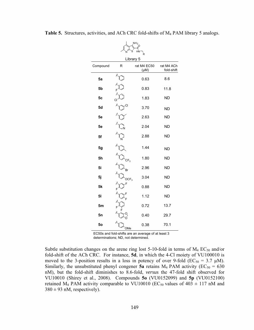
149
Table 5. Structures, activities, and ACh CRC fold-shifts of M4 PAM library 5 analogs.
N S
NH2O
HNR
Library 5
5a 0.63
5b 0.83
5c 1.83
5d 3.70
5e 2.63
5e 2.04
5f 2.88
5g 1.44
5h 1.80
5i 2.96
5j 3.04
5k 0.88
5l 1.12
5m 0.72
5n 0.40
5o 0.38
F
FF
OMe
OO
CF3
Br
Cl
N
N
F
Cl
OCF3
F
FF
8.6
11.8
ND
ND
ND
ND
ND
ND
ND
ND
70.1
29.7
13.7
ND
ND
ND
Compound R rat M4 EC50 rat M4 ACh (µM) fold-shift
EC50s and fold-shifts are an average of at least 3determinations; ND, not determined.
Subtle substitution changes on the arene ring lost 5-10-fold in terms of M4 EC50 and/or fold-shift of the ACh CRC. For instance, 5d, in which the 4-Cl moiety of VU100010 is moved to the 3-position results in a loss in potency of over 9-fold (EC50 = 3.7 µM). Similarly, the unsubstituted phenyl congener 5a retains M4 PAM activity (EC50 = 630 nM), but the fold-shift diminishes to 8.6-fold, versus the 47-fold shift observed for VU10010 (Shirey et al., 2008). Compounds 5o (VU0152099) and 5p (VU0152100) retained M4 PAM activity comparable to VU10010 (EC50 values of 403 ± 117 nM and 380 ± 93 nM, respectively).
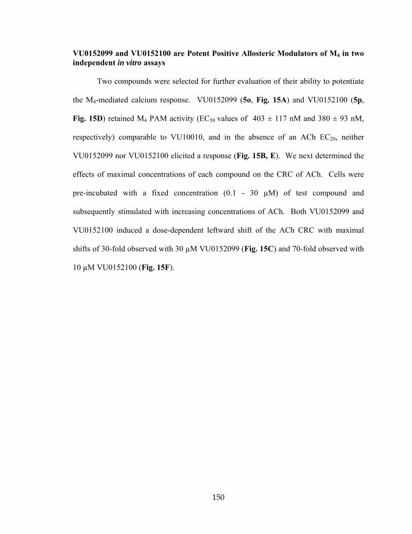
150
VU0152099 and VU0152100 are Potent Positive Allosteric Modulators of M4 in two independent in vitro assays Two compounds were selected for further evaluation of their ability to potentiate
the M4-mediated calcium response. VU0152099 (5o, Fig. 15A) and VU0152100 (5p,
Fig. 15D) retained M4 PAM activity (EC50 values of 403 ± 117 nM and 380 ± 93 nM,
respectively) comparable to VU10010, and in the absence of an ACh EC20, neither
VU0152099 nor VU0152100 elicited a response (Fig. 15B, E). We next determined the
effects of maximal concentrations of each compound on the CRC of ACh. Cells were
pre-incubated with a fixed concentration (0.1 - 30 µM) of test compound and
subsequently stimulated with increasing concentrations of ACh. Both VU0152099 and
VU0152100 induced a dose-dependent leftward shift of the ACh CRC with maximal
shifts of 30-fold observed with 30 µM VU0152099 (Fig. 15C) and 70-fold observed with
10 µM VU0152100 (Fig. 15F).
151
-10 -9 -8 -7 -6 -5 -4
0
20
40
60
80
100
120VU0152099 + ACh EC20
VU0152099 alone
Log [VU0152099] (M)
%M
axA
Ch
Res
pons
e
B.
-13 -12 -11 -10 -9 -8 -7 -6 -5 -4
0
20
40
60
80
100
120
140Vehicle
30 µM10 µM3 µM
Log [ACh] (M)
%M
axA
Ch
Res
pons
e
C.
A. VU0152099
N S
NH2CH3
H3C
O
HN
O
O
-10 -9 -8 -7 -6 -5 -4
0
20
40
60
80
100
120VU0152100 + ACh EC 20
VU0152100 alone
Log [VU0152100] (M)
%M
axA
Ch
Res
pons
e
E.
F.
D. VU0152100
N S
NH2CH3
H3C
O
HN
OMe
-13 -12 -11 -10 -9 -8 -7 -6 -5 -4
0
20
40
60
80
100
120
140 Vehicle
10 µM3 µM
0.3 µM0.1 µM
1 µM
Log [ACh] (M)
%M
axA
Ch
Res
pons
e
Figure 15. VU0152099 and VU0152100 are potent positive allosteric modulators of rM4 in vitro. Chemical structures of VU0152099 (A.) and VU0152100 (D.). B.,E. Potency of VU0152099 (403 ± 117 nM, B.) and VU0152100 (380 ± 93 nM, E.) was evaluated at the rM4 receptor by measuring calcium mobilization in Chinese hamster ovary (CHO) cells stably expressing rM4 and the chimeric G-protein, Gqi5. A range of concentrations of test compound was added to cells, followed 1.5 minutes later by addition of an EC20 concentration of ACh. In the absence of an EC20 concentration of ACh, neither test compound elicited a response. Data were normalized as a percent of the maximal response to 10 µM ACh and represent the mean ± S.E.M. of three independent experiments. C.,F. VU0152099 (C.) and VU0152100 (F.) potentiate the response of rM4 to ACh, as manifest by a dose-dependent leftward shift in the ACh CRC. At the highest concentrations tested, VU0152099 (30 µM) induced a 30-fold shift and VU0152100 (10 µM) induced a 70-fold shift in the ACh CRC. Data were normalized as a percent of the maximal response to 10 µM ACh and represent the mean ± S.E.M. of 3-5 independent experiments. Experiments performed by A.E. Brady.

152
Using calcium mobilization to assess the functional activity of VU0152099 and
VU0152100 at the M4 receptor requires co-expression of the chimeric G protein, Gqi5, in
order to link the Gi/o-coupled M4 receptor to the PLCβ/Ca2+ pathway. As an alternative
approach to measure M4 PAM activity, we chose to take advantage of a novel functional
assay recently developed in our lab that takes advantage of the ability of endogenous Gβγ
subunits of Gi/o-coupled GPCRs to alter the kinetics of G-protein regulated inwardly
rectifying K+ (GIRK) channels to conduct the ion, thallium (Niswender et al., 2008). For
these studies, HEK293 cells stably co-expressing heteromeric GIRK1/2 channels and the
human M4 muscarinic receptor were pre-incubated with test compound and then
stimulated with agonist (ACh) in the presence of thallium ion. Both VU0152099 and
VU0152100 dose-dependently potentiated the response to an EC20 concentration of ACh
with EC50 values of 1.2 ± 0.3 µM and 1.9 ± 0.2 µM, respectively, and increased the
maximal response to ACh to approximately 130 % (Fig 16A). As observed in the
calcium mobilization assays described above, both VU0152099 and VU0152100 (10 µM)
also enhanced the potency of ACh to induce GIRK-mediated thallium flux, as manifest
by a robust (~30-fold) leftward shift in the ACh CRC from 77 ± 1.2 nM to 2.09 ± 0.3 nM
and 2.35 ± 0.5 nM, respectively (Fig 16B). Taken together, these in vitro studies suggest
that VU0152099 and VU0152100 are potent positive allosteric modulators that enhance
the response of the M4 receptor to the endogenous agonist, ACh.

153
-10 -9 -8 -7 -6 -5 -40
20
40
60
80
100
120
140
160VU0152099VU0152100
Log [Compound] (M)
Nor
mal
ized
Slop
e(%
max
AC
hre
spon
se)
-12 -11 -10 -9 -8 -7 -6 -5 -4
020406080
100120140160
VehicleVU0152099VU0152100
Log [ACh] (M)
Nor
mal
ized
Slop
e(%
max
AC
hre
spon
se)
A. B.
Figure 16. VU0152099 and VU0152100 potentiate GIRK-mediated thallium flux in response to ACh in HEK cells expressing the human M4 receptor. A. Both VU0152099 () and VU0152100 () potentiate hM4 -induced GIRK-mediated thallium flux in response to an EC20 concentration of ACh with potencies of 1.2 ± 0.3 μM and 1.9 ± 0.2 μM, respectively. B. In the presence of 10 μM VU0152099 () and VU0152100 (), the ACh CRC for induction of GIRK-mediated thallium flux was leftward shifted (≈30-fold) from 77 ± 1.2 nM ( Veh) to 2.09 ± 0.3 nM (, VU0152099) and 2.35 ± 0.5 nM (, VU0152099). Data were normalized as a percent of the maximal response to 10 μM ACh and represent the mean ± S.E.M. of 3-4 independent experiments performed in quadruplicate. Experiments performed by A.E. Brady.
154
VU0152099 and VU0152100 are selective for M4 relative to other mAChR subtypes
We next evaluated VU0152099 and VU0152100 in calcium mobilization assays
for effects at all mAChR subtypes to determine whether these compounds are selective
for M4. Both VU0152099 and VU0152100 were selective for M4 relative to M1, M2, M3
and M5. Thus, neither VU0152099 (Fig. 17A) nor VU152100 (Fig. 17B) had any effect
on the ACh dose-response curves at these other mAChR subtypes at concentrations up to
30 µM. To further assess selectivity of these compounds for M4 relative to other
potential targets, the activity of VU0152099 and VU0152100 also were evaluated in
radioligand binding assays against a large panel of 68 discrete GPCRs, ion channels,
transporters and enzymes (Table 6). These compounds were largely inactive at each of
the targets in this panel screen. At concentrations of 10 µM, both compounds were
completely inactive at most targets and induced less than 50% displacement of binding
for all targets tested, with the single exception of GABAA receptors (assessed by
flunitrazepam binding), where VU0152099 displayed 51% displacement. This suggests
that VU0152099 may interact with the flunitrazepam site with an IC50 value of
approximately 10 µM, which still provides high selectivity for M4 relative to GABAA
receptors.

155
-14 -13 -12 -11 -10 -9 -8 -7 -6 -5
0
20
40
60
80
100
120
VehVU0152099
Log [ACh] (M)
%M
axAC
hRe
spon
se
rM1
-13 -12 -11 -10 -9 -8 -7 -6
0
20
40
60
80
100
120
VehVU0152099
Log [ACh] (M)
%M
axAC
hRe
spon
se
hM3
-13 -12 -11 -10 -9 -8 -7 -6 -5 -4
0
20
40
60
80
100
120
VehVU0152099
Log [ACh] (M)
%M
axAC
hRe
spon
se
hM2-Gqi5
hM5
-13 -12 -11 -10 -9 -8 -7 -6
0
20
40
60
80
100
120
VehVU0152099
Log [ACh] (M)
%M
axAC
hRe
spon
se
-13 -12 -11 -10 -9 -8 -7 -6 -5 -4
0
20
40
60
80
100
120
VehVU0152100
Log [ACh] (M)
%M
axAC
hRe
spon
se
-14 -13 -12 -11 -10 -9 -8 -7 -6 -5
0
20
40
60
80
100
120VehVU0152100
Log [ACh] (M)
%M
axAC
hRe
spon
se
-13 -12 -11 -10 -9 -8 -7 -6
0
20
40
60
80
100
120VehVU0152100
Log [ACh] (M)
%M
axAC
hRe
spon
se
-13 -12 -11 -10 -9 -8 -7 -6
0
20
40
60
80
100
120
VehVU0152100
Log [ACh] (M)
%M
axAC
hRe
spon
se
rM1 hM2-Gqi5
hM3 hM5
B.
A.
Figure 17. VU0152099 (A.) and VU0152100 (B.) are functionally selective for the M4 mAChR subtype relative to other mAChRs. No shift in the ACh CRC was observed in the presence of 30 μM test compound at CHO-K1 cells stably expressing rM1, hM2-Gqi5, hM3, or hM5. Calcium mobilization was measured in response to increasing concentrations of ACh following preincubation with either vehicle or test compound (30 μM), as described in Methods. Assays of rM1, hM3, and hM5, took advantage of endogenous coupling to Gq proteins, and for hM2, cells co-expressing the chimeric G protein, Gqi5, to allow coupling of this receptor to calcium mobilization. Points represent the mean ± S.E.M. of three independent experiments. Experiments performed by A.E. Brady.

156
Table 6. Selectivity of VU0152099 and VU0152100 determination in radioligand binding assays in the LeadProfilingScreen® by MDS Pharma.
Target Species VU0152099 VU0152100 Target Species VU0152099 VU0152100Adenosine A1 human 28 -1 Histamine H3 human 10 9Adenosine A2 human 46 43 Imidazoline I2, Central rat 7 8Adenosine A3 human 28 40 Interleukin IL-1 mouse -19 -4Adrenergic α1A rat 10 9 Leukotriene, Cysteinyl CysLT1 human -4 -6Adrenergic α1B rat -7 -7 Melatonin MT1 human 15 20Adrenergic α1D human 1 1 Muscarinic M1 human -3 0Adrenergic α2A human 7 17 Muscarinic M2 human -1 -4Adrenergic β1 human 17 17 Muscarinic M3 human -1 -2Adrenergic β2 human 30 35 Neuropeptide Y Y1 human 2 9Androgen (testosterone)AR rat 6 6 Neuropeptide Y Y2 human -2 2Bradykinin B1 human 13 1 Nicotinic Acetylcholine human 6 -6Bradykinin B2 human -5 -9 Nicotinic Acetylcholine α1, Bungarotoxin human -10 -4Calcium channel L-type, benzothiazepine rat 13 7 Opiate δ (OP1, DOP) human 2 -13Calcium channel L-type, dihydropyridine rat 21 17 Opiate κ (OP2, KOP) human 5 2Calcium channel N-type rat -8 -4 Opiate μ (OP3, MOP) human 2 -2Dopamine D1 human 6 7 Phorbol Ester mouse 2 4Dopamine D2S human 2 0 Platelet Activating Factor (PAF) human 13 22Dopamine D3 human -1 5 Potassium Channel [KATP] hamster 3 6Dopamine D4.2 human -13 -11 Potassium Channel hERG human 5 5Endothelin ETA human -8 -8 Prostanoid EP4 human 15 7Endothelin ETB human 2 -4 Purinergic P2X rabbit 6 -4Epidermal Growth Factor (EGF) human 9 -5 Purinergic P2Y rat 15 14Estrogen ERα human -1 -1 Rolipram rat 36 29G protein-coupled receptor GPR103 human -3 -4 Serotonin (5-Hydroxytryptamine) 5-HT1A human 2 2GABAA Flunitrazepam, central rat 51 43 Serotonin (5-Hydroxytryptamine) 5-HT3 human -2 14GABAA Muscimol, central rat 6 9 Sigma σ1 human 2 6GABAB1A human -7 12 Sigma σ2 rat -8 -3Glucocorticoid human -3 8 Sodium Channel, Site 2 rat 11 20Glutamate, Kainate rat 3 -15 Tachykinin NK1 human -15 -18Glutamate, NMDA, Agonism rat 17 22 Thyroid Hormone rat -1 0Glutamate, NMDA, Glycine rat 3 2 Transporter, Dopamine (DAT) human 27 46Glutamate, NMDA, Phencyclidine rat 2 3 Transporter, GABA rat 26 13Histamine H1 human 14 7 Transporter, Norepinephrine (NET) human 34 22Histamine H2 human 7 10 Transporter, Serotonin (5-Hydroxytryptamine) (SERT) human 0 4
% Inhibition% Inhibition
VU0152099 and VU0152100 were tested at 10 µM for orthosteric radioligand displacement against a large panel of 68 GPCRs, ion channels, transporters and enzymes to ensure a clean ancillary pharmacology profile.
157
Since VU0152099 and VU0152100 are allosteric modulators of M4, it is possible
that they have activity at similar allosteric sites on other GPCRs. If so, this would not be
apparent in the radioligand binding assays discussed above. The finding that these
compounds are completely inactive at other mAChR subtypes makes this less likely since
M4 is more closely related to the other mAChR subtypes than to other GPCRs. However,
to further evaluate the selectivity of VU0152099 for M4 relative to other family A
GPCRs, we contracted with Millipore Corp. (St. Charles, MO) to determine the effects of
these compounds on functional responses of a panel of 15 other GPCRs to activation by
their respective agonists. For these studies, we chose family A GPCR subtypes that are
among the closest relatives of mAChRs. We first determined the effects of VU0152099
alone on each receptor and found that these compounds had no agonist activity at any
receptor studied. We then determined the effects of VU0152099 on full concentration
response curves of agonists of each of these receptors. This allows unambiguous
evaluation of whether the compounds possess antagonist activity (either allosteric or
orthosteric) or allosteric potentiator activity at these other GPCRs. Consistent with our
internal studies, VU0152099 induced a robust potentiation of ACh-induced activation of
M4, but had no potentiator activity at M1 (Figure 18). In addition, VU0152099 had no
allosteric potentiator activity at any of the other GPCR subtypes tested (Figure 18). The
only significant activity detected for VU0152099 in this functional panel screen was
weak antagonist activity at the serotonin 5HT2B receptor (Figure 18). Together, these
data suggest that VU0152099 and VU0152100 possess clean ancillary pharmacology
profiles, which would allow us to pursue the behavioral effects of selective M4 activation
in vivo.

158
Histamine H1
-11 -10 -9 -8 -7 -6 -5 -4
0
20
40
60
80
100
120
VU0152099Vehicle
Log [Histamine] (M)
% M
ax R
espo
nse
5HT1A
-11 -10 -9 -8 -7 -6 -5 -4
0
20
40
60
80
100
120
VU0152099Vehicle
Log [8-Hydroxy-DPAT] (M)
% M
ax R
espo
nse
5HT2B
-12 -11 -10 -9 -8 -7 -6 -5
0
20
40
60
80
100
120
VU0152099Vehicle
Log [α-Methyl-5HT] (M)
% M
ax R
espo
nse
Histamine H3
-11 -10 -9 -8 -7 -6 -5 -4
0
20
40
60
80
100
120
VU0152099Vehicle
Log [R(-)a-Methylhistamine] (M)
% M
ax R
espo
nse
5HT2A
-11 -10 -9 -8 -7 -6 -5 -4
0
20
40
60
80
100
120
VU0152099Vehicle
Log [α-Methyl-5HT] (M)
% M
ax R
espo
nse
5HT2C
-11 -10 -9 -8 -7 -6 -5 -4
0
20
40
60
80
100
120
VU0152099Vehicle
Log [α-Methyl-5HT] (M)%
Max
Res
pons
e
Histamine H2
-11 -10 -9 -8 -7 -6 -5 -4
0
20
40
60
80
100
120
VU0152099Vehicle
Log [Amthamine] (M)
% M
ax R
espo
nse
Dopamine D5
-11 -10 -9 -8 -7 -6 -5 -4
0
20
40
60
80
100
120
140
VU0152099Vehicle
Log [A68930] (M)
% M
ax R
espo
nse
Dopamine D2L
Dopamine D5 Histamine H1 Histamine H2 Histamine H3
Serotonin 5HT1A Serotonin 5HT2A Serotonin 5HT2B Serotonin 5HT2C
Dopamine D1
-11 -10 -9 -8 -7 -6 -5 -4
0
20
40
60
80
100
120
VU0152099Vehicle
Log [A68930] (M)
% M
ax R
espo
nse
Dopamine D1α2A-AR
-12 -11 -10 -9 -8 -7 -6 -5
0
20
40
60
80
100
120
VU0152099Vehicle
Log [UK14304] (M)
% M
ax R
espo
nse
α2BAR
-11 -10 -9 -8 -7 -6 -5 -4
0
20
40
60
80
100
120
VU0152099Vehicle
Log [Oxymetazoline] (M)
% M
ax R
espo
nse
α2A-Adrenergic α2B-Adrenergic
α1AAR
-11 -10 -9 -8 -7 -6 -5 -4
0
20
40
60
80
100
120
VU0152099Vehicle
Log [A61603] (M)
% M
ax R
espo
nse
Adenosine A2B
-11 -10 -9 -8 -7 -6 -5 -4
0
20
40
60
80
100
120
VU0152099Vehicle
Log [NECA] (M)
% M
ax R
espo
nse
Adenosine A2B α1A-Adrenergic Muscarinic M1
-11 -10 -9 -8 -7 -6 -5 -4
0
20
40
60
80
100
120
VU0152099Vehicle
Log [Carbachol] (M)
% M
ax R
espo
nse
Muscarinic M1
-11 -10 -9 -8 -7 -6 -5 -4
0
20
40
60
80
100
120
VU0152099Vehicle
Log [Carbachol] (M)
% M
ax R
espo
nse
Muscarinic M4A. B. C. D.
E. F. G. H.
I. J. K. L.
M. N. O. P.
-11 -10 -9 -8 -7 -6 -5 -4
0
20
40
60
80
100
120
140
VU0152099Vehicle
Log [Dopamine] (M)
% M
ax R
espo
nse
Fig 18. Millipore GPCR Profiler™ Functional Screen. Because of the potential liability for off-target activity at other class A GPCRs, VU0152099 was evaluated in Millipore’s GPCR Profiler™ screen for activity at 16 GPCR targets using a Fluorimetric Imaging Plate Reader (FLIPRTetra, Molecular Device Corp). This cell-based assay relies on endogenous expression of the promiscuous G protein, Gα15/16, to couple recombinant receptors to calcium mobilization, and uses a two addition protocol to assess agonist, antagonist, and allosteric modulator activity. VU0152099 exhibited no agonist activity at any of the targets tested and was found to possess antagonist activity only at the 5HT2B receptor (as indicated by an ≈16 fold rightward shift in the agonist CRC in the presence of VU0152099). Potentiator activity was assessed by performing agonist CRCs in the absence (vehicle, ) or presence (VU0152099, ) of 10 μM VU0152099. A. At the human M4 mAChR, 10 μM VU0152099 elicits a robust ≈80-fold leftward shift in the carbachol CRC (EC50 values: CCh alone ≈800nM, CCh + 10 μM VU0152099 ≈ 10 nM). B-P. Similar CRC curves were generated for additional GPCRs: Muscarinic M1 (B.), Adenosine A2B (C.), α1A-, 2A-, 2B-Adrenergic (D-F.), Dopamine D1 ,D2L, D5 (G-I.), Histamine H1, H2, H3 (J-L), and Serotonin 5HT1A, 2A, 2B, 2C (M-P.), with no substantial shift observed at any other target tested. All data represent the mean of duplicate determinations. Experiments performed at Millipore.

159
VU0152099 and VU0152100 bind allosterically and increase ACh affinity
To further confirm an allosteric mechanism of action by the novel M4 PAMs, we
evaluated the effect of VU0152099 and VU0152100 on equilibrium radioligand binding
studies using membranes prepared from cells expressing the rM4 receptor. We first
assessed the ability of increasing concentrations of the two M4 PAMs to displace the
orthosteric radioligand, [3H]NMS (0.1 nM). Unlike the orthosteric antagonist, atropine,
which potently inhibited [3H]NMS binding (Ki = 0.54 ± 0.1 nM), neither M4 PAM
displaced [3H]NMS at concentrations up to 30 µM (Fig. 19A), strongly suggesting that
VU0152099 and VU0152100 act at a site on the M4 receptor that is distinct from the
orthosteric binding site.
In addition, we evaluated the effect of VU0152099 and VU0152100 on the
affinity of ACh for the M4 receptor by assessing the ability of an increasing concentration
of ACh to displace [3H]NMS (0.1 nM) binding in the absence or presence of the M4
potentiators. VU0152099 and VU0152100 were found to induce a 20-25 fold leftward
shift in the potency of ACh to displace [3H]-NMS binding to M4 as manifest by a
reduction in the ACh Ki from 252 ± 17.9 nM (veh) to 10.4 ± 0.91 nM (VU0152099) and
12.2 ± 0.49 nM (VU0152100) (Fig. 19B). These data present a possible mechanism
whereby these compounds could enhance receptor activation by increasing the affinity of
M4 for acetylcholine, and are in agreement with data previously determined for
VU10010, where a 14-fold decrease in the ACh Ki was reported (Shirey et al., 2008).

160
-12 -11 -10 -9 -8 -7 -6 -5 -4
0
20
40
60
80
100
120
140
VU0152099
VU0152100
Atropine
Log [Compound] (M)
%M
ax[3 H
]NM
S B
indi
ng
0
0
20
40
60
80
100
120 Vehicle
VU0152100
VU0152099
VU10010
-9 -8 -7 -6 -5 -4 -3Log [ACh] (M)
A. B.
%M
ax[3 H
]NM
S B
indi
ng
Figure 19. VU0152099 and VU0152100 bind allosterically and increase ACh affinity at rM4. A. In competition binding studies, neither VU0152099 () nor VU0152100 () displaced the orthosteric radioligand, [3H]NMS (0.1 nM), at concentrations up to 30 μM. However, the orthosteric antagonist, atropine (), potently inhibited [3H]NMS binding with a Ki of 0.54 ± 0.1 nM. B. In the presence of vehicle alone, an increasing concentration of ACh displaces [3H]NMS (0.1 nM) binding with a Ki of 252 ± 17.9 nM (). In the presence of a fixed concentration (10 μM) of VU0152099 or VU0152100, the potency of ACh to displace [3H]NMS binding is shifted leftward, yielding Ki values of 10.4 ± 0.91 nM (, VU0152099) and 12.2 ± 0.49 nM (,VU0152100), which represent a 25-fold and 21-fold shift in ACh potency, respectively. Data represent the mean ± S.E.M. of 3 independent experiments, performed in duplicate. Experiments performed by A.E. Brady.

161
VU0152099 and VU0152100 Exhibit Improved Physiochemical and Pharmacokinetic Properties Prior to conducting in vivo studies with VU0152099 and VU0152100,
pharmacokinetic studies were undertaken to assess brain/plasma ratios following
systemic dosing of these compounds. In contrast, to the high logP of VU10010 (4.5),
both VU0152099 and VU0152100 possessed logPs of 3.65 and 3.6, respectively, a full
order of magnitude less lipophilic than VU10010 (Shirey et al., 2008). As a
consequence, both VU0152099 and VU0152100 displayed improved physiochemical
properties and afforded homogeneous dosing solutions in multiple vehicles acceptable for
in vivo studies. Further, we conducted in vivo exposure (brain and plasma) studies in rats
at the dose of 56.6 mg/kg, i.p. Both compounds exhibited substantial systemic absorption
and brain penetration (Fig. 20). After 56.6 mg/kg ip administration, peak brain
concentrations for both the compounds were in the range of 3-5 µg/ml. VU0152100
(Fig. 20B) was far superior to VU0152099 (Fig. 20A) in terms of brain penetration, as
evident from AUC (0-∞) values (Table 7). The AUC brain/AUC plasma ratio, which is
an estimate of CNS bioavailability, for VU0152099 was calculated to be 0.39 ± 0.01,
while the ratio for VU0152100 was determined to be 0.86 ± 0.08 (Table 7). The half-life
of the compounds in the brain was 1.25 ± 0.02 h (VU0152099) and 1.12 ± 0.01 h
(VU0152100) (Table 7). Therefore, our earlier concern of P-glycoprotein (P-gp)
transport susceptibility within this series was likely unwarranted, and the lack of central
activity for VU10010 was most likely due solely to physiochemical properties.
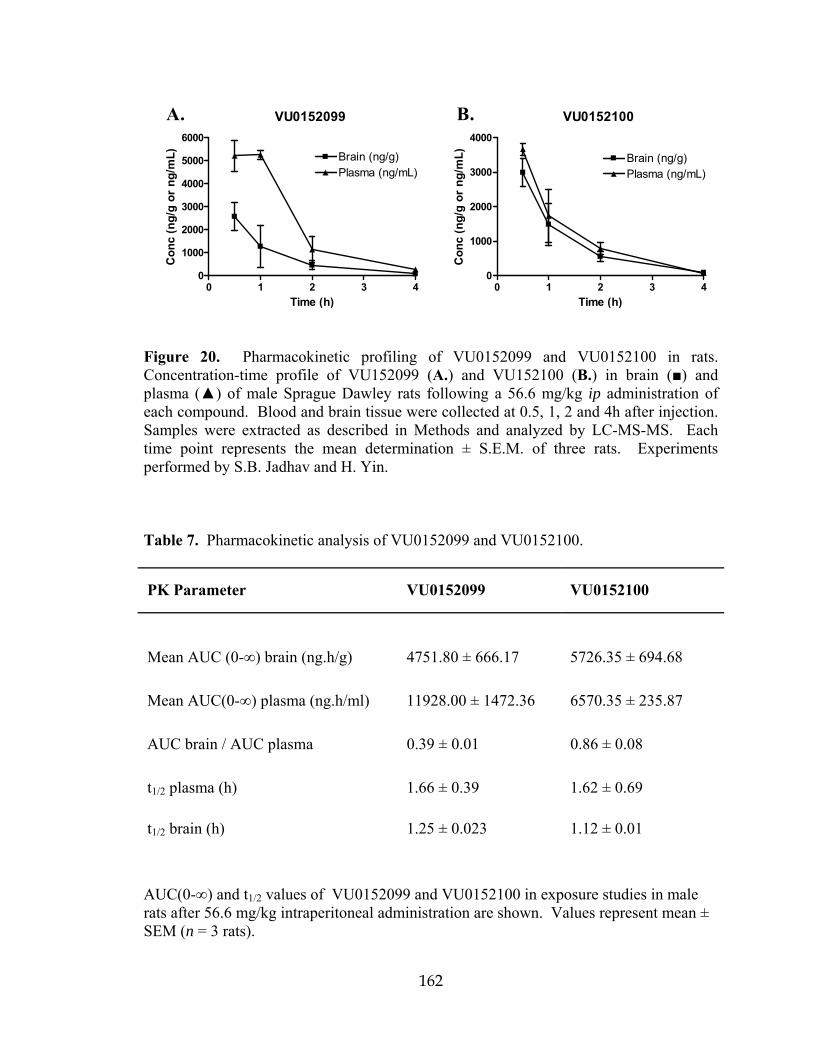
162
VU0152099
0 1 2 3 40
1000
2000
3000
4000
5000
6000
Brain (ng/g)Plasma (ng/mL)
Time (h)
Con
c (n
g/g
or n
g/m
L)
A. B. VU0152100
0 1 2 3 40
1000
2000
3000
4000
Brain (ng/g)Plasma (ng/mL)
Time (h)
Con
c (n
g/g
or n
g/m
L)
Figure 20. Pharmacokinetic profiling of VU0152099 and VU0152100 in rats. Concentration-time profile of VU152099 (A.) and VU152100 (B.) in brain () and plasma () of male Sprague Dawley rats following a 56.6 mg/kg ip administration of each compound. Blood and brain tissue were collected at 0.5, 1, 2 and 4h after injection. Samples were extracted as described in Methods and analyzed by LC-MS-MS. Each time point represents the mean determination ± S.E.M. of three rats. Experiments performed by S.B. Jadhav and H. Yin. Table 7. Pharmacokinetic analysis of VU0152099 and VU0152100.
PK Parameter VU0152099 VU0152100
Mean AUC (0-∞) brain (ng.h/g) 4751.80 ± 666.17 5726.35 ± 694.68
Mean AUC(0-∞) plasma (ng.h/ml) 11928.00 ± 1472.36 6570.35 ± 235.87
AUC brain / AUC plasma 0.39 ± 0.01 0.86 ± 0.08
t1/2 plasma (h) 1.66 ± 0.39 1.62 ± 0.69
t1/2 brain (h) 1.25 ± 0.023 1.12 ± 0.01
AUC(0-∞) and t1/2 values of VU0152099 and VU0152100 in exposure studies in male rats after 56.6 mg/kg intraperitoneal administration are shown. Values represent mean ± SEM (n = 3 rats).

163
VU0152099 and VU0152100 exhibit in vivo activity in rat
Previous studies using the M1/M4-preferring muscarinic agonist xanomeline
produced robust effects in several preclinical models predictive of antipsychotic-like
activity, including reversal of amphetamine-induced hyperlocomotion in rats (Stanhope et
al., 2001). Based on our initial pharmacokinetic studies suggesting that systemic
administration of VU0152099 and VU0152100 provides robust brain levels of these
compounds, the effects of VU0152099 and VU0152100 were evaluated in reversing
amphetamine-induced hyperlocomotion using a dose of 56.6 mg/kg i.p. for each
compound with a 30 min pretreatment interval. As shown in Fig. 21A, both VU0152099
and VU0152100 produced robust decreases in amphetamine-induced hyperlocomotion
over the time course tested. In addition, to provide further confirmation that the M4
PAMs have no effect on baseline levels of motor performance which could complicate
the interpretation of the amphetamine-induced hyperlocomotion data, we evaluated the
effects of one of the M4 PAMs, specifically VU0152100, after administration alone on
performance in the rotorod test (Fig. 21B). As shown, VU0152100 had no effect on
performance in the rotorod test, even when tested at a dose of 100mg/kg, which was
higher than that required to observe reversal of amphetamine-induced hyperlocomotion.
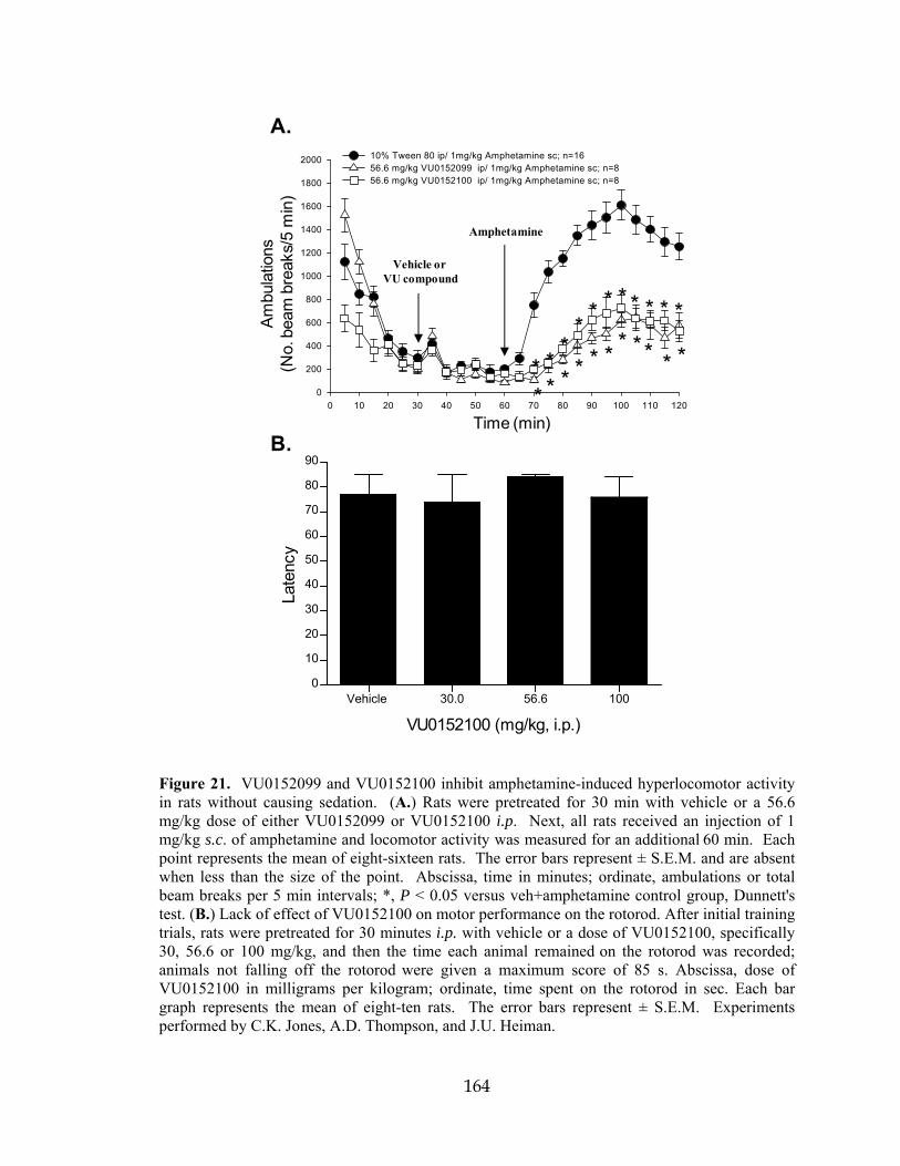
164
Time (min)
0 10 20 30 40 50 60 70 80 90 100 110 120
Ambu
latio
ns
(no.
Bea
m B
reak
s/5
min
)
0
200
400
600
800
1000
1200
1400
1600
1800
2000 10% Tween 80 ip/ 1mg/kg Amphetamine sc; n=1656.6 mg/kg VU0152099 ip/ 1mg/kg Amphetamine sc; n=856.6 mg/kg VU0152100 ip/ 1mg/kg Amphetamine sc; n=8
* * ** * * * * * * *
* * * * * * * ** *
*
Vehicle or VU compound
Amphetamine
A.
B.
Vehicle 30.0 56.6 1000
10
20
30
40
50
60
70
80
90
VU0152100 (mg/kg, i.p.)
Late
ncy
Am
bula
tions
(No.
bea
m b
reak
s/5
min
)
Time (min)
Figure 21. VU0152099 and VU0152100 inhibit amphetamine-induced hyperlocomotor activity in rats without causing sedation. (A.) Rats were pretreated for 30 min with vehicle or a 56.6 mg/kg dose of either VU0152099 or VU0152100 i.p. Next, all rats received an injection of 1 mg/kg s.c. of amphetamine and locomotor activity was measured for an additional 60 min. Each point represents the mean of eight-sixteen rats. The error bars represent ± S.E.M. and are absent when less than the size of the point. Abscissa, time in minutes; ordinate, ambulations or total beam breaks per 5 min intervals; *, P < 0.05 versus veh+amphetamine control group, Dunnett's test. (B.) Lack of effect of VU0152100 on motor performance on the rotorod. After initial training trials, rats were pretreated for 30 minutes i.p. with vehicle or a dose of VU0152100, specifically 30, 56.6 or 100 mg/kg, and then the time each animal remained on the rotorod was recorded; animals not falling off the rotorod were given a maximum score of 85 s. Abscissa, dose of VU0152100 in milligrams per kilogram; ordinate, time spent on the rotorod in sec. Each bar graph represents the mean of eight-ten rats. The error bars represent ± S.E.M. Experiments performed by C.K. Jones, A.D. Thompson, and J.U. Heiman.

165
Discussion
In recent years, the mAChRs have emerged as potential novel targets for the
treatment of schizophrenia. This is based on clinical studies demonstrating efficacy of
mAChR agonists in treatment of positive symptoms in schizophrenia patients, as well as
multiple animal studies suggesting that mAChR agonists could be useful in treatment of
cognitive dysfunction in schizophrenia patients (Bymaster et al., 2002). Furthermore, a
growing body of evidence from clinical and animal studies involving pharmacological
manipulations, post mortem tissue analysis, and brain imaging is consistent with this
hypothesis (Raedler et al., 2007). While recent advances suggesting potential utility of
mAChR activators in treatment of schizophrenia have been exciting, there have been few
selective pharmacological tools available to fully explore this emerging muscarinic
hypothesis of schizophrenia. Unfortunately, previous attempts to develop traditional
orthosteric agonists that are highly selective for individual mAChR subtypes have been
unsuccessful.
The current discovery and optimization of VU0152099 and VU0152100 as highly
selective positive allosteric modulators of M4 provides a major advance in establishing a
new approach for developing highly selective activators of these receptors. The data
presented provide further support for the ability to achieve high subtype selectivity by
targeting allosteric sites and provide exciting new data demonstrating that highly
selective M4 PAMs have robust activity in at least one animal model that is similar to
effects previously described for the non-selective orthosteric mAChR agonist,
xanomeline (Stanhope et al., 2001). The finding that VU0152099 and VU0152100
mimic effects of xanomeline in an animal model that has been used to predict

166
antipsychotic activity of new compounds is especially exciting in light of clinical studies
demonstrating the clinical efficacy of xanomeline in schizophrenia patients. This raises
the exciting possibility that selective activation of M4 may provide a novel approach for
the treatment of some symptoms associated with schizophrenia. Furthermore, the
discovery of systemically active M4 PAMs suggests that this will be a viable approach for
developing selective activators of M1 and other mAChR subtypes.
While the in vitro data for VU0152099 and VU0152100 indicate high
pharmacologic selectivity for M4 relative to any other mAChR subtypes or closely related
GPCRs, the possibility exists that the observed behavioral effects may be due to an off-
target activity not yet identified. In future studies, it will be critical to further validate that
the effects of VU0152099 and VU0152100 observed in vivo are mediated by activation
of M4 using other tools, including structurally distinct M4 PAMs or selective M4
antagonists, as they become available, and/or M4 knockout mice. Unfortunately, studies
in M4 knockout mice will be complex since these mice display fundamentally different
responses to psychomotor stimulants and compounds will need to be optimized for
appropriate pharmacokinetic properties in mice. Also, several behavioral parameters are
substantially altered in the M4 knockout mice, including increases in baseline locomotor
activity, altered responses to amphetamine, and altered dopamine release in the
mesolimbic dopamine circuitry (Gomeza et al., 1999; Gomeza et al., 2001; Tzavara et al.,
2004). These factors will complicate interpretation of studies with M4 knockout mice.
However, while it is not possible to definitively establish an exclusive role for M4 in
mediating the behavioral effects observed here, previous studies have established similar
effects of structurally distinct mAChR agonists (Stanhope et al., 2001). This, coupled

167
with the high selectivities of VU0152099 and VU0152100 for M4 relative to any other
mAChR subtype, suggests that M4 is a likely candidate for mediating these effects. Also,
extensive profiling of VU0152099 and VU0152100 showed no functional effects on
responses to other closely related family A GPCRs and no significant off-target activity
at any of 68 other GPCRs, ion channels, or enzymes. Thus, while it is impossible to
entirely rule out unknown off-target activity, these compounds appear to be much more
highly selective for the targeted receptor than is typical for most orthosteric GPCR
ligands. As new tools become available for targeting M4 and other mAChR subtypes,
they will provide the means to develop a more complete understanding of the roles of
each of the individual mAChR subtypes in regulating CNS function.
In addition to suggesting the potential roles of M4 in vivo, these data have
important implications related to the molecular pharmacology of allosteric modulators of
GPCRs. One of the most promising properties of allosteric modulators of GPCRs has
been that it is often possible to achieve high selectivity for a targeted GPCR subtype
relative to closely related family members (Marino et al., 2003; O'Brien et al., 2003;
Kinney et al., 2005; Galici et al., 2006). However, discovery of these compounds also
raises the question of whether allosteric modulators may have broad activity across other
GPCRs by interacting at potentially promiscuous allosteric sites. The present finding that
VU0152099 and VU0152100 had no major activities across multiple targets in a large
panel radioligand binding screen was encouraging in suggesting the high selectivity of
these compounds, but does not address this critical question. However, the finding that
these compounds had no allosteric modulator activity across a panel of 15 other family A
GPCR subtypes is exciting and suggests that they are not likely to have activity at a site

168
that is shared across multiple GPCRs. While it is impossible to rule out activity at other
unidentified targets that were not tested, these data suggest that it may be possible to
achieve higher subtype selectivity across a range of receptors than has been possible with
many orthosteric ligands.

169
CHAPTER IV
A SELECTIVE ALLOSTERIC POTENTIATOR OF THE M1 MUSCARINIC ACETYLCHOLINE RECEPTOR INCREASES ACTIVITY OF MEDIAL
PREFRONTAL CORTICAL NEURONS AND RESTORES IMPAIRMENTS IN REVERSAL LEARNING
Introduction
The muscarinic acetylcholine (ACh) receptors (mAChRs) play important roles in
regulating higher cognitive function. Non-selective mAChR antagonists induce profound
attention and memory deficits (Aigner et al., 1991; Fibiger, 1991; Miller and Desimone,
1993) and degeneration of forebrain cholinergic neurons is one of the earliest
pathological changes observed in AD (Bartus et al., 1982; Bartus, 2000). Furthermore,
acetylcholinesterase inhibitors (AChEIs) have established efficacy in the treatment of AD
symptoms (Birks, 2006; Munoz-Torrero, 2008).
Of the five mAChR subtypes, the M1 receptor is viewed as the most important
subtype for memory and attention mechanisms (Levey et al., 1991; Felder et al., 2000).
Based on this, selective activators of M1 have been proposed as having potential utility in
treatment of AD (Bodick et al., 1997b; Bodick et al., 1997a; Gu et al., 2003; Caccamo et
al., 2006; Jones et al., 2008; Caccamo et al., 2009). However, recent studies revealed that
genetic deletion of M1 does not alter mAChR excitatory effects on hippocampal
pyramidal cells (Rouse et al., 2000a), impair hippocampal-dependent learning, or alter
cognition-impairing effects of mAChR antagonists (Miyakawa et al., 2001; Anagnostaras
et al., 2003). Interestingly, while hippocampal-dependent learning was intact, M1
receptor knockout mice had specific deficits in forms of learning and memory that

170
require activation of the prefrontal cortex (PFC) (Anagnostaras et al., 2003). Thus, M1
may play a role in regulating PFC function, and M1 receptor-selective activators could
improve deficits in PFC-dependent learning in patients suffering from AD.
Unfortunately, lack of highly selective activators and antagonists of the M1
receptor has prevented detailed studies of the functional consequences of selective M1
receptor activation. The difficulty in developing highly selective M1 agonists is due to
the high sequence homology among the orthosteric binding sites of mAChR subtypes.
However, an alternative strategy for achieving high subtype selectivity is targeting
allosteric binding sites that are distinct from the ACh binding site (Conn et al., 2009a;
Conn et al., 2009b). We recently reported discovery of multiple positive allosteric
modulators (PAMs) of M1 (Marlo et al., 2009). Furthermore, Ma and colleagues (Ma et
al., 2009) report evidence that BQCA is a potent and highly selective PAM at the human
M1 receptor. Based on these preliminary findings, we synthesized a series of molecules
related to BQCA and report that BQCA and related compounds are highly selective rat
M1 PAMs. These compounds do not interact with the ACh site, but dramatically increase
the affinity of the M1 receptor for ACh and potentiate the response to orthosteric agonist.
In addition, activation of the M1 receptor induces an inward current and increases
excitatory synaptic currents in mPFC layer V pyramidal cells. Consistent with this,
BQCA increases firing of mPFC neurons in vivo. Finally, BQCA reverses deficits in a
PFC-dependent form of learning and memory in a transgenic mouse model of AD and
promotes non-amyloidogenic APP processing in vitro. Together, these data suggest that
the M1 receptor plays an important role in regulating excitatory drive to the PFC and that

171
selective potentiation of activity at this receptor can reverse deficits in PFC-dependent
cognitive function.
Results
A panel of 21 compounds related to BQCA has a range of activities as allosteric potentiators at the rat M1 mAChR Ma et al., (2008) recently reported BQCA as a selective positive allosteric
modulator (PAM) of the human M1 muscarinic receptor (hM1). However, GPCR PAMs
can display species specificity, and the effects of BQCA were not extensively evaluated
on the rat M1 receptor (rM1). Thus, in order to determine whether BQCA and related
compounds have properties needed for use in rodent studies, we synthesized BQCA and a
panel of 20 structurally related analogs to identify compounds that can act as selective
PAMs for the rM1 receptor. Effects of BQCA and related compounds were evaluated by
measuring effects on calcium mobilization elicited by a submaximal concentration (EC20)
of ACh (Fig. 22). Libraries I, II, and III each consisted of seven compounds possessing
the same N-benzyl substitutions based on either an 8-fluorinated quinolone carboxylic
acid (Ia-Ig), a quinolone carboxylic acid (IIa-IIg, including BQCA), or a 5,8-
difluorinated quinolone carboxylic acid (IIIa-IIIg) template, respectively (Fig. 22A). The
activity of test compounds was initially assessed by incubating CHO-K1 cells stably
expressing the rM1 receptor with fixed concentrations of each compound at 10, 1, or 0.3
µM (Fig. 22B-D) for 1.5 min prior to the addition of an EC20 concentration of ACh.
From the panel, four compounds that exhibited robust potentiator activity at 0.3 µM were
selected for further evaluation based on their structural diversity. As can be seen in the

172
representative trace, 1 µM BQCA has no effect when added alone, but greatly enhances
the response to an EC20 concentration of ACh when compared to vehicle. A maximal
response to ACh is also shown for comparison (Fig. 22E). To determine the potency of
each of these compounds, full concentration response curves (CRCs) were generated by
pre-incubating rM1 CHO-K1 cells with increasing concentrations of test compound,
followed 1.5 min later by the addition of an EC20 concentration of ACh (Fig. 23A). All
four compounds had similar potencies at rM1 with EC50 values in the 200-400 nM range.
As a second measure of their ability to potentiate the rM1 receptor-mediated calcium
response to ACh, rM1 receptor expressing CHO-K1 cells were pre-incubated with a fixed
concentration (3 µM) of the test compound (or vehicle) and then stimulated with
increasing concentrations of ACh to generate a series of ACh CRCs. Each of the four
test compounds elicited a robust potentiation of the ACh response, as manifest by a
leftward shift in the ACh CRC (9.5- to 18.6-fold shift, Fig. 23B).

173
EC20 Ia Ib Ic Id Ie If Ig IIa IIb IIc IId IIe
BQCA IIg IIIa IIIb IIIc IIId IIIe IIIf IIIg0
20
40
60
80
100 Compounds at 10 µM
%M
axA
Ch
Res
pons
e
B.EC20 Ia Ib Ic Id Ie If Ig IIa IIb IIc IId IIe
BQCA IIg IIIa IIIb IIIc IIId IIIe IIIf IIIg0
20
40
60
80
100Compounds at 1 µM
%M
axA
Ch
Res
pons
e
C.
EC20 Ia Ib Ic Id Ie If Ig IIa IIb IIc IId IIe
BQCA IIg IIIa IIIb IIIc IIId IIIe IIIf IIIg0
20
40
60
80
100 Compounds at 0.3 µM
%M
axA
Ch
Res
pons
e
* **
*
D.
A.
0 25 50 75 100 125 150
0
10000
20000
30000
40000
EC20 + BQCA
EC20 + vehicle
ECmax
Vehicleor BQCA ACh
Time (sec)
Bas
elin
eSu
btra
cted
RFU
E.
Figure 22. Twenty-one putative M1 receptor PAMs were synthesized and evaluated at the rM1 mAChR for their ability to potentiate an EC20 concentration of ACh. A. Synthesis: a. K2CO3, KI, R-Br, DMF, 24 hr. at r.t., b. LiOH, 9:1 THF:H2O 10 min 120°C μwave. Calcium mobilization was measured using a Flexstation II, as described in Methods. Test compounds were evaluated at fixed concentrations of 10 μM (B.), 1 μM (C.), or 0.3 μM (D.) in the presence of an EC20 concentration of ACh. Four compounds, denoted by an asterisk (*) in panel D, were selected for further evaluation based on their structural diversity and ability to potentiate an EC20 concentration of ACh at 0.3 μM. Data were normalized as a percent of the maximal response to 10 μM ACh and represent the mean ± S.E.M. of 3 independent experiments. D. A representative calcium trace from one experiment shows the effect of 1 µM BQCA on the rM1 receptor response to an EC20 concentration of ACh; the response to an EC20
and ECmax concentration of ACh in the presence of vehicle are also shown for comparison. Experiments performed by A.E. Brady.

174
A. B.
-10 -9 -8 -7 -6 -50
20
40
60
80
100VU0238393VU0238389VU0238386VU0238387
Log [Compound] (M)
% M
ax A
Ch
Res
pons
e
-13 -12 -11 -10 -9 -8 -7 -60
20
40
60
80
100
120Vehicle
VU0238393
VU0238389
VU0238386
VU0238387
Log [ACh] (M)
% M
ax A
Ch
Res
pons
e
Figure 23. Four of the compounds initially identified as robust M1 potentiators of the calcium response were further characterized in vitro at the rM1 mAChR. A. Potency of each test compound was determined based on its ability to potentiate an EC20 concentration of ACh, yielding EC50 values of 404 ± 59 nM, VU0238393 (), 187 ± 39 nM, VU0238389 (), 316 ± 102 nM, VU0238386 (BQCA,) and 284 ± 32nM VU0238387 (♦). B. Test compounds were then evaluated at a fixed concentration of 3 µM for their ability to shift the potency of an ACh concentration response curve. ACh alone stimulated calcium mobilization with an EC50 value of 2.09 ± 0.43 nM (). In the presence of each of the four test compounds, a robust leftward shift in the ACh CRC was induced resulting in the following EC50 values and fold shift in the ACh curve (in parentheses): 0.280 ± 0.164 nM (9.5X), VU0238393 (), 0.039 ± 0.017 nM (23.2X), VU0238389 (), 0.142 ± 0.007 nM (16.4X), VU0238386 (BQCA,) and 0.107 ± 0.003 nM (18.6X), VU0238387 (♦). Data were normalized as a percent of the maximal response to 10 µM ACh and represent the mean ± S.E.M. of 3 independent experiments. Experiments performed by Ashley Brady.

175
BQCA is a potent and selective positive allosteric modulator of the rat M1 receptor in vitro Of the molecules tested in this panel screen, BQCA was among the most potent
and efficacious at potentiating rM1 receptor-mediated responses. This is consistent with
its activity at the human receptor (Ma et al., 2009). Based on this and its favorable
physiochemical properties, we chose to pursue studies focusing exclusively on BQCA.
First, we evaluated the potency of BQCA as a positive allosteric modulator of the rM1
receptor by measuring calcium mobilization in CHO-K1 cells stably expressing this
receptor. Cells were incubated with increasing concentrations of BQCA for 1.5 min prior
to the addition of an EC20 concentration of ACh, yielding a concentration response curve
for BQCA with an EC50 value of 267 ± 31 nM (Fig. 24A.). We next determined the
effect of increasing fixed concentrations of BQCA on the ACh CRC. Rat M1 CHO-K1
cells were pre-incubated with a fixed concentration (0.3, 1, and 3 µM) of BQCA and
subsequently stimulated with increasing concentrations of ACh. BQCA induced a dose-
dependent leftward shift in the ACh CRC with a maximal shift of 21-fold observed with
3 µM BQCA (Fig. 24B).

176
-10 -9 -8 -7 -6 -50
20
40
60
80
100
Log [BQCA] (M)
% M
ax A
Ch
Res
pons
e
-13 -12 -11 -10 -9 -8 -7 -6
0
20
40
60
80
100
120Veh
3 μM1 μM0.3 μM
Log [ACh] (M)
% M
ax A
Ch
Res
pons
e
A. B.
Figure 24. BQCA is a potent positive allosteric modulator of the rM1 receptor in vitro. A. The potency of BQCA was evaluated at the rM1 receptor by measuring calcium mobilization in Chinese Hamster Ovary (CHO-K1) cells stably expressing the rM1 receptor. Increasing concentrations of test compound was added to cells, followed 1.5 minutes later by addition of an EC20 concentration of ACh. BQCA robustly potentiated the response to ACh with an EC50 value of 267 ± 31 nM. B. The ability of BQCA to potentiate the response of the rM1 receptor to ACh is also manifest by a dose-dependent leftward shift in the ACh CRC. ACh alone stimulated calcium mobilization with an EC50 value of 2.42 ± 0.337 nM (). In the presence of increasing fixed concentrations of BQCA (0.3 - 3 μM), a robust leftward shift in the ACh CRC was induced, resulting in the following EC50 values (fold shift in the ACh curve is shown in parentheses); 0.3 μM (♦) = 0.762 ± 0.56 nM (3.3-fold), 1.0 μM () = 0.221 ± 0.079 nM (12-fold), 3 μM () = 0.123 ± 0.026 nM (21-fold). Data were normalized as a percent of the maximal response to 10 μM ACh and represent the mean ± S.E.M. of 3-4 independent experiments. Experiments performed by A.E. Brady.

177
BQCA is functionally selective for the M1 mAChR subtype
One of the primary difficulties in developing novel selective ligands for
muscarinic receptors has been the failure to identify compounds that can distinguish
between the highly conserved orthosteric binding site shared by the five members of this
GPCR subfamily. Development of ligands that bind to allosteric sites, both potentiators
and direct acting agonists, has proven to be a practical way to circumvent this issue
(Conn et al., 2009a; Conn et al., 2009b). Thus, it was important to determine whether
BQCA is selective for the M1 mAChR relative to other mAChR subtypes. We evaluated
the effect of BQCA on the ACh CRC in calcium mobilization assays at each of the other
mAChR subtypes. As shown in Fig. 24B, pre-incubation of rM1 receptor-expressing
CHO-K1 cells with 3 µM BQCA results in a robust leftward shift in the CRC for ACh.
However, at this same concentration, BQCA had no effect on the ACh concentration
response curves generated in CHO-K1 cells stably expressing the hM2, hM3, rM4, or hM5
receptors (Fig. 25A-D). To further assess selectivity of BQCA for the M1 receptor
relative to other class A GPCR targets that may also harbor similar allosteric sites, we
took advantage of the GPCR Profiler™ service offered by Millipore Corp. (St. Charles,
MO) to determine the effect of this compound on the functional response of 15 other
closely related GPCRs (Fig. 26). A two-addition protocol afforded the ability to detect
potential agonist, potentiator, and antagonist activity of BQCA at these other GPCR
subtypes. Consistent with our internal studies, BQCA induced robust potentiation at the
hM1 receptor, but had no activity in this assay at the hM4 receptor. Moreover, BQCA had
no effect at any of the other GPCRs tested (Fig. 26A-P). This included a lack of PAM
activity or antagonist activity (either allosteric or orthosteric) at any of these other

178
GPCRs, which would have resulted in a rightward shift in the concentration response
curve. Together, these data suggest that BQCA is highly selective for the M1 mAChR
subtype and has no detectable activity at closely related family A GPCRs that were
tested.

179
-13 -12 -11 -10 -9 -8 -7 -6 -5 -4
0
20
40
60
80
100
120
140
Vehicle3 µM BQCA
Log [ACh] (M)
%M
axA
Ch
Res
pons
e
-13 -12 -11 -10 -9 -8 -7 -6
020406080
100120140
Log [ACh] (M)
%M
axA
Ch
Res
pons
e
-13 -12 -11 -10 -9 -8 -7 -6 -5 -4
020406080
100120140
Log [ACh] (M)
%M
axA
Ch
Res
pons
e
-13 -12 -11 -10 -9 -8 -7 -6
020406080
100120140
Log [ACh] (M)
%M
axA
Ch
Res
pons
e
hM2-Gqi5
rM4 -Gqi5 hM5
hM3
A.
C. D.
B.
Figure 25. The presence of BQCA has no effect on the ACh concentration-response curve at any other mAChR subtype. No shift in the ACh CRC was observed in the presence of 3 μM BQCA in CHO-K1 cells stably expressing M2-Gqi5 (A.), M3 (B.), M4-Gqi5 (C.), or M5 receptors (D.). Calcium mobilization was measured in response to increasing concentrations of ACh following preincubation of cells with either vehicle () or 3 μM BQCA (), as described in Methods. Data represent the mean ± S.E.M. of three independent experiments. Experiments performed by A.E. Brady.

180
Dopamine D2L
Dopamine D5 Histamine H1 Histamine H2 Histamine H3
Serotonin 5HT1A Serotonin 5HT2A Serotonin 5HT2B Serotonin 5HT2C
Dopamine D1α2A‐Adrenergic α2B‐Adrenergic
Adenosine A2B α1A‐Adrenergic Muscarinic M1 Muscarinic M4A. B. C. D.
E. F. G. H.
I. J. K. L.
M. N. O. P.
-11 -10 -9 -8 -7 -6 -5 -4
0
20
40
60
80
100
120
BQCAVehicle
Log [Carbachol] (M)
% M
ax R
espo
nse
-11 -10 -9 -8 -7 -6 -5 -4-10
10
30
50
70
90
110
130
BQCAVehicle
Log [Carbachol] (M)
% M
ax R
espo
nse
-11 -10 -9 -8 -7 -6 -5 -4
0
20
40
60
80
100
120
BQCAVehicle
Log [NECA] (M)
% M
ax R
espo
nse
-12 -11 -10 -9 -8 -7 -6 -5
0
20
40
60
80
100
120BQCAVehicle
Log [A61603] (M)
% M
ax R
espo
nse
-11 -10 -9 -8 -7 -6 -5 -4
0
20
40
60
80
100
120
BQCAVehicle
Log [Oxymetazoline] (M)
% M
ax R
espo
nse
-12 -11 -10 -9 -8 -7 -6
0
20
40
60
80
100
120BQCAVehicle
Log [UK14304] (M)
% M
ax R
espo
nse
-11 -10 -9 -8 -7 -6 -5 -4
0
20
40
60
80
100
120
140
BQCAVehicle
Log [Dopamine] (M)
% M
ax R
espo
nse
-11 -10 -9 -8 -7 -6 -5
0
20
40
60
80
100
120
BQCAVehicle
Log [A68930] (M)
% M
ax R
espo
nse
-11 -10 -9 -8 -7 -6 -5 -4
0
20
40
60
80
100
120
BQCAVehicle
Log [A68930] (M)
% M
ax R
espo
nse
-11 -10 -9 -8 -7 -6 -5 -4
0
20
40
60
80
100
120
BQCAVehicle
Log [Amthamine] (M)
% M
ax R
espo
nse
-11 -10 -9 -8 -7 -6 -5 -4
0
20
40
60
80
100
120BQCAVehicle
Log [Histamine] (M)
% M
ax R
espo
nse
-12 -11 -10 -9 -8 -7 -6 -5
0
20
40
60
80
100
120
140
BQCAVehicle
Log [R (-) α-Methylhistamine] (M)
% M
ax R
espo
nse
-11 -10 -9 -8 -7 -6 -5 -4
0
20
40
60
80
100
120
BQCAVehicle
Log [α-Methyl-5HT] (M)
% M
ax R
espo
nse
-12 -11 -10 -9 -8 -7 -6 -5 -4
0
20
40
60
80
100
120
BQCAVehicle
Log [8-Hydroxy-DPAT] (M)
% M
ax R
espo
nse
-13 -12 -11 -10 -9 -8 -7 -6
0
20
40
60
80
100
120BQCAVehicle
Log [α-Methyl-5HT] (M)
% M
ax R
espo
nse
-11 -10 -9 -8 -7 -6 -5 -4
0
20
40
60
80
100
120BQCAVehicle
Log [α-Methyl-5HT] (M)
% M
ax R
espo
nse
Figure 26. Millipore GPCR Profiler BQCA selectivity data. To confirm that BQCA retains selectivity at M1 when assayed at non-mAChR GPCRs, it was evaluated in Millipore’s GPCR Profiler™ screen for activity at 15 additional Class A GPCR targets. A Fluorimetric Imaging Plate Reader (FLIPRTetra), (Molecular Device Corp), was used to measure calcium mobilization in response to agonist stimulation in a proprietary cell line that relies on endogenous expression of the promiscuous G protein, Gα15/16, to couple recombinant receptors to the PLCβ/Ca2+ pathway. This assay takes advantage of a two addition protocol such that it was possible to detect any agonist, antagonist, or allosteric potentiator activity. BQCA exhibited no agonist or antagonist activity at any of the targets tested. Allosteric potentiator activity was assessed by performing agonist concentration response curves in the absence (vehicle, ) or presence of 10 µM test compound (BQCA, ). A. At the human M1 mAChR, 10 µM BQCA elicits a robust leftward shift in the carbachol concentration-response curve. B-P. Similar CRC curves were generated for additional GPCRs: Muscarinic M4 (B.), Adenosine A2B (C.), α1A-, 2A-, 2B-Adrenergic (D-F.), Dopamine D1 ,D2L, D5 (G-I), Histamine H1, H2, H3 (J-L), and Serotonin 5HT1A, 2A, 2B, 2C (M-P), with no detectable leftward shift in the agonist concentration-response-curve observed at any other target tested. All data represent the mean of duplicate determinations.

181
BQCA does not compete for orthosteric antagonist binding and it induces a robust leftward shift in ACh affinity at the rM1 receptor We previously reported that novel selective PAMs of the rM4 receptor,
exemplified by VU10010 and VU152100, have no detectable affinity at the orthosteric
ACh binding site of the rM4 receptor but allosterically increase affinity of ACh for the
rM4 receptor (Brady et al., 2008; Shirey et al., 2008). To determine whether BQCA
shares this property with the rM4 PAMs, we assessed the ability of this compound to
compete for binding with the orthosteric radioligand, [3H]-NMS (0.1 nM) to the
orthosteric site using membranes prepared from cells expressing the rM1 receptor. BQCA
had little effect on [3H]-NMS binding, with no displacement of radioligand observed at
concentrations up to 10 µM (Fig. 27A). In contrast, the orthosteric antagonist, atropine,
potently inhibited [3H]-NMS binding with a Ki value of 1.35 ± 0.022 nM (Fig. 27A).
The effect of BQCA on the affinity of ACh for the rM1 receptor was also evaluated by
assessing the ability of increasing concentrations of ACh to displace [3H]-NMS (0.1 nM)
binding in the absence or presence of fixed concentrations of the M1 receptor potentiator
(0.3, 1.0, and 3.0 µM). BQCA induced a robust concentration-dependent leftward shift in
the concentration response curve of ACh-induced displacement of [3H]-NMS binding to
the rM1 receptor, with a 30-fold shift observed at the highest concentration tested (3.0
µM). This shift reveals that BQCA induces a reduction in the ACh Ki from 1700 ± 96.4
nM (vehicle) to 348 ± 43.4 nM (0.3 µM), 163 ± 22.9 nM (1.0 µM), and 56.1 ± 4.99 nM
(3.0 µM), respectively (Fig. 27B). Taken together, these data strongly suggest that BQCA
acts at a site on the rM1 receptor that is distinct from the orthosteric binding site and that
it may enhance M1 receptor activation by increasing the affinity for ACh.

182
-12 -11 -10 -9 -8 -7 -6 -5 -4
0
20
40
60
80
100
120
BQCAAtropine
Log [Cold ligand] (M)
%M
ax[3 H
]NM
SB
indi
ng
-10 -9 -8 -7 -6 -5 -4 -30
20
40
60
80
100
120 Vehicle
0.3 µM1 µM3 µM
Log [ACh] (M)
%M
ax[3 H
]NM
SB
indi
ng
A. B.
Fig 27. BQCA does not compete for orthosteric antagonist binding and it induces a robust leftward shift in ACh affinity at the rM1 receptor. A. At concentrations up to 10 μM, BQCA () did not displace the orthosteric radioligand, [3H]-NMS (0.1 nM) in competition binding studies. However, the orthosteric antagonist, atropine (), potently inhibited [3H]-NMS binding with a Ki of 1.35 ± 0.022 nM. B. In the presence of vehicle alone, an increasing concentration of ACh displaces [3H]-NMS (0.1 nM) binding with a Ki of 1700 ± 96.4 nM (). In the presence of increasing fixed concentrations (0.3 - 3.0 μM) of BQCA, the potency of ACh to displace [3H]-NMS binding is shifted leftward, yielding Ki values of 348 ± 43.4 nM (0.3 μM, ), 163 ± 22.9 nM (1.0 μM, ), and 56.1 ± 4.99 nM (3.0 μM, ), which represent 5-fold, 10.6-fold and 30.6-fold shifts in ACh potency, respectively. Data represent the mean ± S.E.M. of 3 independent experiments performed in duplicate. Experiments performed by A.E. Brady.
183
Activation of the M1 receptor induces an inward current in rat mPFC layer V pyramidal cells and this effect is potentiated by BQCA Prefrontal cortical function is required for higher executive function, memory
storage and retrieval, and cognition (Miller and Cohen, 2001). Recent studies suggest
that M1 receptor signaling may play an important role in activation of the prefrontal
cortex by lower brain regions (Anagnostaras et al., 2003). Based on this, it was
postulated that activation of the M1 receptor could increase excitability of mPFC
pyramidal cells or increase excitatory synaptic drive to these neurons. In order to
examine the effects of M1 receptor activation on mPFC pyramidal cells, layer V
pyramidal neurons were visually identified and membrane currents measured using patch
clamp recordings in acute coronal slices. Cell type was confirmed by examining firing
properties upon depolarizing current injection. Typical resting membrane potentials of
these pyramidal neurons were -55 to -65 mV under the conditions used. Holding current
was measured in cells voltage clamped at -70 mV during baseline recording, drug
application, and wash. Bath application of CCh induced a robust, concentration-
dependent inward current as shown in Fig. 28 (10 µM CCh, 16.55 ± 1.93 pA, n = 4; 100
µM CCh, 53.14 ± 5.92 pA, n = 4). Although this CCh–induced inward current is in
agreement with previously reported studies, it is not known whether this response is
mediated by the M1 receptor or another mAChR subtype (Krnjevic, 2004; Carr and
Surmeier, 2007). However, previous studies suggest that the M1 receptor may not be
responsible for induction of inward currents in hippocampal CA1 pyramidal cells (Rouse
et al., 2000a). Before evaluating the effect of BQCA on this current, we determined the
effect of VU0255035, the first highly selective M1 receptor antagonist that was recently
reported (Sheffler et al., 2009), on the CCh-induced inward current. The M1 receptor

184
antagonist, VU0255035 (10 µM), had no effect on holding current alone but significantly
blocked the current induced by 100 µM CCh (P = 0.0202, unpaired t-test). These results
suggest that the CCh-induced inward current in rat mPFC layer V pyramidal cells is
largely mediated by activation of the M1 receptor. If this is the case, we would predict
that the M1 receptor PAM BQCA should potentiate the CCh-induced inward current.
Interestingly, BQCA induced a small change in holding current when applied alone
(21.54 ± 2.42 pA, n = 5, Fig. 28). In addition, BQCA significantly increased the inward
current induced by 10 µM CCh (55.07 ± 6.28 pA upon co-application, n = 5, compared to
10 µM CCh alone, P = 0.0210). These data are consistent with the hypothesis that
activation of the M1 receptor induces an inward current in mPFC layer V pyramidal cells
and that M1 receptor PAMs can induce a marked potentiation of this response.

185
B.A.
10 µM
CCh
100 µ
MCCh
10 µM
BQCA
10 µM
BQCA
+ 10 µ
M CCh
**
10 µM
VU0255035
+ 100 μMCCh
0
20
40
60
80
∆H
oldi
ngC
urre
nt(p
A)10 µM BQCA
10 µM CCh
50 pA
4 min
100 µM CCh
100 pA2 min
10 µM CCh
100 pA1 min
10 µM VU0255035
100 µM CCh
100 pA2 min
Figure 28. CCh-induced inward current in mPFC layer V pyramidal cells is reduced by M1 receptor antagonist VU0255035 and potentiated by BQCA. A. Sample traces from single experiments showing the change in holding currents upon drug treatments. B. Activation of mAChRs by CCh causes a dose-dependent inward current as measured in voltage clamp mode as a change in holding current (16.55 ± 1.93 pA in the presence of 10 µM CCh, n = 4; 53.14 ± 5.92 pA with 100 µM CCh, n = 4). M1 receptor PAM BQCA also caused an inward current when applied alone, and significantly increased the effect of 10 µM CCh (21.54 ± 2.42 pA change with 10 µM BQCA, n = 5; 55.07 ± 6.28 pA when BQCA was co-applied with 10 µM CCh, n = 5, as compared to 10 µM CCh alone, P = 0.0210). The effect of 100 µM CCh was also significantly inhibited by 10 µM M1 receptor antagonist VU0255035 (11.78 ± 1.90 pA in the presence of antagonist, n = 3, compared to 100 µM CCh alone, P = 0.0202). Sample traces represent experiments from single cells, and bars show duration of drug exposure. All changes in holding current were compared to baseline control and are represented as mean ± S.E.M. Asterisks indicate significant differences from control (*, P < 0.05; unpaired t-test).
186
BQCA does not potentiate CCh-mediated inward currents in M1 knockout mice
Application of CCh caused a dose-dependent inward current in mPFC layer V
pyramidcal cells in slices from wild-type (3 µM CCh, 24.0 ± 3.9 pA; 30 µM CCh, 34.4 ±
5.8 pA; Fig. 29A) and M1 knockout mice (3 µM CCh, 2.1 ± 1.2 pA; 30 µM CCh, 6.1 ±
2.0 pA; 100 µM CCh, 42.0 ± 10.5 pA Fig. 29B) but had much lower potency in slices
from knockout animals. While BQCA alone (10 µM) induced a slight inward current in
wildtype cells (17.9 ± 6.0 pA), it induced a much smaller change in holding current in M1
knockout cells (9.1 ± 1.9 pA); co-application of BQCA with 3 µM CCh increased the
inward current induced by this low concentration of CCh (35.2 ± 9.9 pA). In contrast,
BQCA did not potentiate the response to 30 µM CCh in M1 knockout slices (3.5 ± 1.2
pA). Because of the limited availability of knockout animals during this time,
experiments testing the ability of atropine or the M1 antagonist, VU0255035, to block the
inward current caused by 100 µM CCh were not performed. While this critical control is
necessary for conclusive interpretation of these data, trends in the dose response to CCh
between wildtype and M1 knockout animals imply that the M1 subtype mediates, at least
in part, the CCh-induced inward current. While the slight response to BQCA (Fig. 29B)
in knockout animals is somewhat worrisome, holding current stability throughout the
duration of these experiments is highly variable and futher controls to test the
significance of this response will be required.
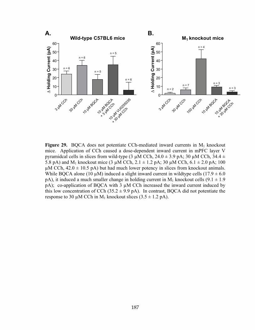
187
Wild-type C57BL6 mice
0
10
20
30
40
50
60
n = 6
n = 6
n = 5
n = 5
n = 6
3 µM C
Ch
30 µM
CCh
10 µM
BQCA
10 µM
BQCA
+ 3 µM
CCh
10 µM
VU0255
035
+ 30 µ
M CCh
M knockout mice
0
10
20
30
40
50
60
n = 2n = 7
n = 4
n = 3
n = 3
3 µM C
Ch
30 µM
CCh
100 µ
M CCh
10 µM
BQCA
10 µM
BQCA
+ 30 µ
M CCh
∆H
oldi
ngC
urre
nt (p
A)
∆H
oldi
ngC
urre
nt (p
A)
1A. B.
Figure 29. BQCA does not potentiate CCh-mediated inward currents in M1 knockout mice. Application of CCh caused a dose-dependent inward current in mPFC layer V pyramidcal cells in slices from wild-type (3 µM CCh, 24.0 ± 3.9 pA; 30 µM CCh, 34.4 ± 5.8 pA) and M1 knockout mice (3 µM CCh, 2.1 ± 1.2 pA; 30 µM CCh, 6.1 ± 2.0 pA; 100 µM CCh, 42.0 ± 10.5 pA) but had much lower potency in slices from knockout animals. While BQCA alone (10 µM) induced a slight inward current in wildtype cells (17.9 ± 6.0 pA), it induced a much smaller change in holding current in M1 knockout cells (9.1 ± 1.9 pA); co-application of BQCA with 3 µM CCh increased the inward current induced by this low concentration of CCh (35.2 ± 9.9 pA). In contrast, BQCA did not potentiate the response to 30 µM CCh in M1 knockout slices (3.5 ± 1.2 pA).

188
mAChR activation increases mPFC spontaneous EPSC amplitude and frequency
It is also possible that activation of the M1 receptor could increase activity of
excitatory synaptic inputs to the mPFC and that this could contribute to the postulated
role of this receptor in increasing PFC activity from in vivo studies in M1 receptor
knockout mice (Anagnostaras et al., 2003). Thus, we determined the effect of mAChR
activation on spontaneous excitatory postsynaptic currents (sEPSCs) in mPFC pyramidal
cells. Application of CCh caused a dramatic, concentration-dependent increase in the
frequency of spontaneous EPSCs; the effect of a maximal concentration of 100 µM CCh
on one representative cell is shown in Fig. 30A. Cumulative probability plots of
amplitude and inter-event interval from the same cell demonstrate significant shifts in the
presence of 100 µM CCh that are reversible upon wash (Fig. 30B). The concentration-
response relationships for CCh effects on sEPSC amplitude and frequency are shown in
Fig. 30C. A concentration of 10 µM CCh was without effect (97.5 ± 4.4% control for
amplitude, 90.1 ± 12.4% control for frequency, n = 6); however, 30 and 100 µM CCh
increased both amplitude and frequency (30 µM amplitude, 108.3 ± 3.9%, frequency,
455.0 ± 101.9% control, n = 7; 100 µM amplitude, 154.3 ± 46.2%, 887.6 ± 268.5%
control for frequency, n = 5). The effects of 30 µM CCh on both amplitude and
frequency were completely blocked by the non-selective muscarinic antagonist, atropine
(5 µM, 102.9 ± 7.8% and 104.4 ± 19.6% control, respectively, n = 3) indicating that the
effect of CCh was due to activation of mAChRs.

189
100 µM CCh
Control
0 10 20 30 40 50 60 70 80 0
0.2
0.4
0.6
0.8
1.0
1.2
Control100 µM CChWash
Cum
ulat
ivePr
obab
ility
Amplitude (pA)
0 1000 2000 3000 40000
0.2
0.4
0.6
0.8
1.0
1.2
Control100 µM CChWash
Cum
ulat
ivePr
obab
ility
Inter-Event Interval (ms)
A. B.
C.
Wash
30 pA
1 s
*n.s. n.s.
n.s.
n.s.
0
50
100
150
200
250
Nor
mal
ized
Ampl
itude
(%C
ontro
l)
Contro
l
10 µM
CCh
30 µM
CCh
100 μ
M CCh
5 µM
Atropin
e
+30
µM C
Ch
*
*
n.s. n.s.
*
0
100
200
400
800
1200
1600
Nor
mal
ized
Freq
uenc
y(%
Con
trol)
Contro
l
10 µM
CCh
30 µM
CCh
100 μ
M CCh
5 µM
Atropin
e
+30
µM C
Ch
Figure 30. Muscarinic receptor activation increases mPFC spontaneous EPSC amplitude and frequency. A. Representative traces from one cell showing the effect of a maximal concentration of 100 µM CCh. B. Change in cumulative probability plots of sEPSC amplitude (top panel) and inter-event interval (bottom panel) upon addition and wash of 100 µM CCh from one representative cell. C. Averaged amplitude and frequency show that CCh treatment induces a dose-dependent increase in both sEPSC amplitude and frequency which is reversible upon washout and is inhibited by 5 µM atropine (amplitudes, 10 µM CCh, 97.5 ± 4.4%, n = 6, P = 0.2727; 30 µM CCh, 108.3 ± 3.9%, n = 7, P = 0.0498; 100 µM CCh, 154.3 ± 46.2%, n = 5, P = 0.2393; 5 µM atropine/30 µM CCh, 102.9 ± 7.8% of control, n = 3, P = 0.5365. 30 µM CCh vs. 5 µM atropine/30 µM CCh, P = 0.4478; frequencies, 10 µM CCh, 90.1 ± 12.4%, P = 0.9364; 30 µM CCh, 455.0 ± 101.9%, P = 0.0139; 100 µM CCh, 887.6 ± 268.5%, P = 0.0314; 5 µM atropine/30 µM CCh, 104.4 ± 19.6% of control, P = 0.6260. 30 µM CCh vs. 5 µM atropine/30 µM CCh, P = 0.0458). All changes in amplitude and frequency were compared to baseline control and are represented as mean ± S.E.M. Asterisks indicate significant differences from control or between conditions (*,P < 0.05; paired or unpaired t-test).

190
The effect of CCh on sEPSC amplitude and frequency is inhibited by VU0255035
To further evaluate the role of the M1 receptor in the effect of CCh on sEPSCs,
slices were treated with the selective M1 receptor antagonist VU0255035 (5 µM) for 2
min. prior to addition of 30 µM CCh (Fig. 31A, B). VU0255035 alone decreased sEPSC
amplitude (92.9 ± 3.4% control, n = 11), and amplitude was further decreased by co-
application with 30 µM CCh (87.1 ± 3.5% control, Fig. 31C). Antagonist alone had no
effect of sEPSC frequency (114.1 ± 25.8% control) but caused a significant decrease in
frequency in the presence of CCh (62.5 ± 10.1% control, Fig. 31C). These data suggest
that the CCh-induced increase in sEPSC amplitude and frequency is mediated by
activation of the M1 receptor. The reversal of the CCh effect on sEPSC frequency in the
presence of the M1 receptor antagonist suggests that blocking the M1 receptor unmasks an
inhibitory action of CCh that may be mediated by another mAChR subtype, possibly M2
or M4 receptors.
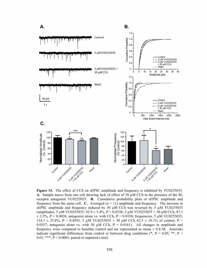
191
Control
5 μM VU0255035 +30 μM CCh
5 μM VU0255035
Wash
A. B.
0 5 10 15 20 25 30 35 40 0
0.2
0.4
0.6
0.8
1.0
1.2
Control5 µM VU02550355 µM VU0255035+ 30 µM CChWash
Cum
ulat
ivePr
obab
ility
Amplitude (pA)
0 500 1000 1500 2000 2500 0
0.2
0.4
0.6
0.8
1.0
1.2
Control5 µM VU02550355 µM VU0255035+ 30 µM CChWash
Cum
ulat
ivePr
obab
ility
Inter-Event Interval (ms)
C.
Contro
l
5 μM
VU0255
035
5 µM
VU0255
035
+ 30
µM C
Ch Was
h
***
0
20
40
60
80
100
120
Nor
mal
ized
Ampl
itude
(%C
ontro
l)
***
*
n.s.
Contro
l
5 μM
VU0255
035
5 µM
VU0255
035
+ 30
µM C
Ch Was
h
*
0
20
40
60
80
100
120
140
Nor
mal
ized
Freq
uenc
y(%
Con
trol)
30 pA
1 s
Figure 31. The effect of CCh on sEPSC amplitude and frequency is inhibited by VU0255035. A. Sample traces from one cell showing lack of effect of 30 µM CCh in the presence of the M1 receptor antagonist VU0255035. B. Cumulative probability plots of sEPSC amplitude and frequency from the same cell. C. Averaged (n = 11) amplitude and frequency. The increase in sEPSC amplitude and frequency induced by 30 µM CCh was reversed by 5 µM VU0255035 (amplitudes, 5 µM VU0255035, 92.9 ± 3.4%, P = 0.0338; 5 µM VU0255035 + 30 µM CCh, 87.1 ± 3.5%, P = 0.0026; antagonist alone vs. with CCh, P = 0.0350; frequencies, 5 µM VU0255035, 114.1 ± 25.8%, P = 0.4543; 5 µM VU0255035 + 30 µM CCh 62.5 ± 10.1% of control, P = 0.0357; antagonist alone vs. with 30 µM CCh, P = 0.0161). All changes in amplitude and frequency were compared to baseline control and are represented as mean ± S.E.M. Asterisks indicate significant differences from control or between drug conditions (*, P < 0.05; **, P < 0.01; ***, P < 0.0001; paired or unpaired t-test).

192
BQCA increases sEPSCs and potentiates the effect of a sub-threshold concentration of CCh on sEPSC frequency Our results thus far suggest that the M1 receptor is responsible for the CCh-
induced increase in sEPSC frequency; therefore, this response should be potentiated by
BQCA. To test this hypothesis, slices were treated with BQCA alone for 2 min. (10 µM)
prior to addition of 10 µM CCh. Sample traces and cumulative probability plots are
shown in Fig. 32A and 32B. Treatment with BQCA alone did not significantly affect
sEPSC amplitude, but increased the frequency of events (108.3 ± 6.6% control, 277.0 ±
97.7% control, respectively, n = 11, Fig. 32C). Co-application of BQCA and 10 µM
CCh induced a further increase in sEPSC frequency (994.5 ± 301.5% control), which
differed significantly from the effect of 10 µM CCh (P = 0.0045, unpaired t-test) (see
Fig. 30C) or BQCA alone (P = 0.0116, paired t-test).

193
A. B.
C.
0 20 40 60 80 100 120 0
0.2
0.4
0.6
0.8
1.0
1.2
Control10 µM BQCA10 µM BQCA +10 µM CChWash
Cum
ulat
ivePr
obab
ility
Amplitude (pA)
0 500 1000 1500 2000 0
0.2
0.4
0.6
0.8
1.0
1.2
Cum
ulat
ivePr
obab
ility
Inter-Event Interval (ms)
*
***
Contro
l
10 µ
MBQCA
10 μ
MBQCA
Was
h
+ 10 µ
M CCh
0
100
200
400
800
1200
1600
Nor
mal
ized
Freq
uenc
y(%
Con
trol
)
Contro
l
10 µM
BQCA
10 μM
BQCAW
ash
+ 10 µ
M CCh
n.s.
n.s.
n.s.
0
50
100
150
200
Nor
mal
ized
Am
plitu
de(%
Con
trol
)
Control
10 μM BQ CA
10 μM BQ CA +10 μM CCh
Wash
30 pA
1 s
Control10 µM BQCA10 µM BQCA +10 µM CChWash
Figure 32. BQCA increases sEPSCs and potentiates a sub-threshold concentration of CCh to increase sEPSC frequency. A. Representative traces from one cell showing the effect of 10 µM BQCA alone and in the presence of 10 µM CCh. This concentration of CCh had no significant effect on sEPSC amplitude or frequency when applied alone. B. Change in cumulative probability plots from one representative cell of sEPSC amplitude (top panel) and inter-event interval (bottom panel) upon addition of 10 µM BQCA with and without 10 µM CCh. C. Averaged (n = 11) amplitude and frequency. Neither drug condition elicited a significant effect on sEPSC amplitude (10 µM BQCA, 108.3 ± 6.6%, P = 0.5013; 10 µM BQCA/10 µM CCh, 136.0 ± 15.3% control, P = 0.0524) but significantly increased frequency (10 µM BQCA, 277.0 ± 97.7%, P = 0.0229; 10 µM BQCA/10 µM CCh, 994.5 ± 301.5% control, P = 0.0045; the effect of combined drug treatment vs. 10 µM BQCA alone was significantly different, P = 0.0116). The effect of 10 µM CCh on frequency in the absence and presence of BQCA was also significantly different (P = 0.0177). All changes in amplitude and frequency were compared to baseline control and are represented as mean ± S.E.M. Asterisks indicate significant differences from control or between drug conditions (*, P < 0.05; **, P < 0.01; paired or unpaired t-test).

194
CCh and BQCA have no effect on miniature EPSC amplitude and frequency in rat mPFC layer V pyramidal cells To determine whether the actions of CCh and BQCA require action potential-
dependent EPSCs, we determined the effects of these compounds on miniature EPSCs
(mEPSCs). mEPSCs were recorded in voltage clamp mode at a holding potential of -70
mV and in the presence of 1 µM tetrodotoxin (TTX) to block voltage-gated sodium
channels. At this concentration, TTX completely eliminates action potential firing and
action potential-mediated synaptic activity (Morisset and Urban, 2001). Under these
conditions, neither CCh nor BQCA elicited any effect on mEPSC amplitude or
frequency. Sample traces from one cell in a slice to which 100 µM CCh was applied in
the presence of TTX show a clear lack of effect (Fig. 33A). Cumulative probability plots
of amplitude and frequency during control, CCh treatment, and wash from the same cell
overlap (Fig. 33B). Pooled amplitude and frequency for all drug treatments are
quantified in Fig. 33C (10 µM CCh, n = 5; 100 µM CCh, n = 4; 10 µM BQCA with and
without 10 µM CCh, n = 4). The only significant effect was that of 10 µM CCh, which
slightly decreased mEPSC amplitude (88.6 ± 3.8% control). The effects of M1 receptor
activation on spontaneous EPSCs thus require action potential firing.
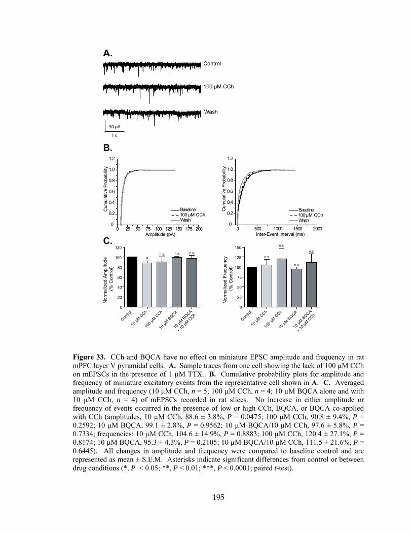
195
A.
B.
C.
Contro
l
10 µM
CCh
100 µ
M CCh
10 µM
BQCA
10 µM
BQCA
+ 10 µ
M CCh
0
20
40
60
80
100
120
Nor
mal
ized
Am
plitu
de(%
Con
trol
)
*n.s. n.s. n.s.
0
25
50
75
100
125
150
Nor
mal
ized
Freq
uenc
y(%
Con
trol
)
n.s.
n.s.
n.s.
0 25 50 75 100 125 150 175 200 0
0.2
0.4
0.6
0.8
1.0
1.2
Baseline 100 µM CChWash
Cum
ulat
ive
Prob
abilit
y
Amplitude (pA)0 500 1000 1500 2000
0
0.2
0.4
0.6
0.8
1.0
1.2
Baseline100 µM CChWash
Cum
ulat
ive
Prob
abilit
y
Inter-Event Interval (ms)
Control
100 µM CCh
Wash
50 pA
1 s
n.s.
Contro
l
10 µM
CCh
100 µ
M CCh
10 µM
BQCA
10 µM
BQCA
+ 10 µ
M CCh
Figure 33. CCh and BQCA have no effect on miniature EPSC amplitude and frequency in rat mPFC layer V pyramidal cells. A. Sample traces from one cell showing the lack of 100 µM CCh on mEPSCs in the presence of 1 µM TTX. B. Cumulative probability plots for amplitude and frequency of miniature excitatory events from the representative cell shown in A. C. Averaged amplitude and frequency (10 µM CCh, n = 5; 100 µM CCh, n = 4; 10 µM BQCA alone and with 10 µM CCh, n = 4) of mEPSCs recorded in rat slices. No increase in either amplitude or frequency of events occurred in the presence of low or high CCh, BQCA, or BQCA co-applied with CCh (amplitudes, 10 µM CCh, 88.6 ± 3.8%, P = 0.0475; 100 µM CCh, 90.8 ± 9.4%, P = 0.2592; 10 µM BQCA, 99.1 ± 2.8%, P = 0.9562; 10 µM BQCA/10 µM CCh, 97.6 ± 5.8%, P = 0.7334; frequencies: 10 µM CCh, 104.6 ± 14.9%, P = 0.8883; 100 µM CCh, 120.4 ± 27.1%, P = 0.8174; 10 µM BQCA, 95.3 ± 4.3%, P = 0.2105; 10 µM BQCA/10 µM CCh, 111.5 ± 21.6%, P = 0.6445). All changes in amplitude and frequency were compared to baseline control and are represented as mean ± S.E.M. Asterisks indicate significant differences from control or between drug conditions (*, P < 0.05; **, P < 0.01; ***, P < 0.0001; paired t-test).

196
BQCA has no effect on sEPSCs in slices from M1 receptor KO mice
In order to confirm that the actions of BQCA were mediated by M1 receptor
activation, recordings of sEPSCs in mPFC layer V neurons were made using slices from
mice lacking the M1 receptor and compared to wild-type (WT) controls. Consistent with
our studies in rat slices, CCh caused a concentration-dependent increase in sEPSC
amplitude and frequency in WT mice (Fig. 34A left panel, 34D black bars). While 3
µM CCh had no effect on amplitude or frequency, 30 µM CCh significantly increased
both parameters (amplitudes, 3 µM CCh, 102.6 ± 11.7% of control, n = 3; 30 µM CCh,
143.1 ± 22.0%, n = 5; frequencies: 3 µM CCh, 83.2 ± 47.1%, 30 µM CCh, 398.3 ±
56.2%). In contrast to effects in rat slices, BQCA had no effect alone in WT slices (10
µM BQCA, 97.3 ± 11.3% control amplitude, 99.8 ± 11.3% control frequency, n = 5), but
induced robust increases in both amplitude and frequency when co-applied with 3 µM
CCh (137.2 ± 16.7% control amplitude, 500.5 ± 212.3% control frequency). In slices
from M1 receptor KO mice, the response to CCh was markedly reduced. In M1 KO mice,
CCh decreased sEPSC amplitude at both concentrations tested and induced a more
modest increase in sEPSC frequency that did not achieve statistical significance
(amplitudes, 3 µM CCh, 79.4 ± 14.9%, n = 4; 30 µM CCh, 80.7 ± 5.2%, n = 4;
frequencies, 3 µM CCh, 186.3 ± 187.4%, 30 µM CCh, 271.7 ± 310.4%). Importantly, the
response to BQCA was completely absent in slices from M1 receptor KO mice. Thus,
BQCA had no effect when applied alone or when co-applied with 3 µM CCh
(amplitudes, 10 µM BQCA, 96.9 ± 10.6%; BQCA/CCh, 84.5 ± 25.3%, n = 5;
frequencies, 10 µM BQCA, 101.3 ± 26.1%; BQCA/CCh, 86.6 ± 17.3%). Responses to
co-application of BQCA and CCh differed significantly between WT and M1 receptor

197
KO for both sEPSC amplitude and frequency (P = 0.0046 for amplitude; P = 0.0025 for
frequency, unpaired t-test). These results confirmed that the actions of BQCA in WT
mice and rats are due to its action at M1 receptors.

198
Figure 34. BQCA has no effect and does not potentiate the CCh effect on sEPSCs in M1 receptor KO mice. A. Sample traces from individual cells in slices made from wild-type (left panels) and M1 receptor KO mice (right panels) showing the robust effects of 30 µM CCh on both sEPSC amplitude in frequency compared to the milder effect in the M1 receptor KO slice (top panels). B. Bottom panels illustrate the lack of effect of 10 µM BQCA in both the WT and M1 receptor KO and contrast the increase in amplitude and frequency with the addition of BQCA and 10 µM CCh to the lack of effect in the M1 receptor KO. C. Cumulative probability plots of the inter-event intervals from the two cells shown in bottom panels above. D. Averaged amplitude and frequency of sEPSCs measured in wild-type (black bars: 3 µM CCh, n = 3; 30 µM CCh, n = 5; 10 µM BQCA and 10 µM BQCA + 3 µM CCh, n = 5) and M1 receptor KO slices (white bars: 3 µM CCh, n = 4; 30 µM CCh, n = 4; 10 µM BQCA and 10 µM BQCA + 3 µM CCh, n = 5). In wild-type slices, 3 µM CCh had no significant effect on amplitude or frequency (102.6 ± 11.7%, P = 0.6381 and 83.2 ± 47.1% of control, P = 0.4423, respectively) whereas 30 µM CCh increased both (143.1 ± 22.0%, P = 0.0306 for amplitude, 398.3 ± 56.2%, P = 0.0342 for frequency). BQCA had no effect on amplitude or frequency but potentiated the response to 3 µM CCh (amplitudes, 10 µM BQCA, 97.3 ± 11.3%, P = 0.7642 compared to control; 10 µM BQCA/3 µM CCh, 137.2 ± 16.7%, P = 0.0052 compared to control; frequencies, 10 µM BQCA, 99.8 ± 11.3%, P = 0.7261 compared to control; 10 µM BQCA/3 µM CCh, 500.5 ± 212.3%, P = 0.0209 compared to control). In M1 receptor KO slices, 3 µM CCh decreased amplitude but had no significant effect on frequency (79.4 ± 14.9%, P = 0.0490 and 186.3 ± 187.4% of control, P = 0.7656, respectively). 30 µM CCh also significantly decreased amplitude and increased frequency although the effect on frequency was less dramatic than that seen in WT controls (80.7 ± 5.2%, P = 0.0092 for amplitude, 271.7 ± 310.4%, P = 0.6010 for frequency). While the difference between the 30 µM CCh effect on amplitude was significant between genotypes (P = 0.0229), the effect on frequency was not (P = 0.4756). BQCA had no effect alone in KO slices (amplitude, 96.9 ± 10.6%, P = 0.4925; frequency, 101.3 ± 26.1%, P = 0.7286) and there was no difference in this lack of effect between genotypes (amplitude, P = 0.9596; frequency, P = 0.9133). When applied with 3 µM CCh, BQCA had no significant effect on amplitude or frequency (amplitude, 84.5 ± 25.3%, P = 0.3383; frequency, 86.6 ± 17.3 %, P = 0.1388 compared to KO control), and both effects were significantly different those in WT slices (P = 0.0046 for amplitude, P = 0.0025 for frequency). All changes in amplitude and frequency were compared to baseline control and are represented as mean ± S.E.M. Asterisks indicate significant differences from control or between drug conditions (*, P < 0.05; **, P < 0.01; ***, P < 0.0001; paired or unpaired t-test).
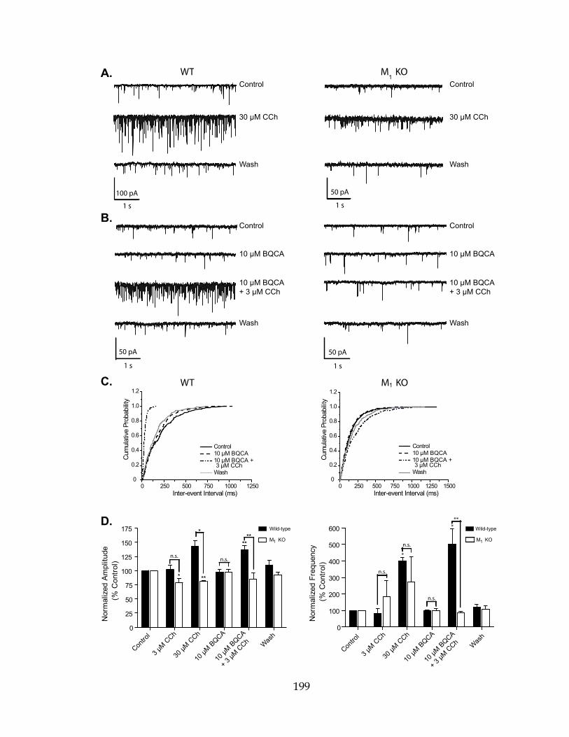
199
0 250 500 750 1000 1250 0
0.2
0.4
0.6
0.8
1.0
1.2
Cum
ulat
ivePr
obab
ility
Inter-event Interval (ms)
Control10 µM BQCA10 µM BQCA +3 µM CCh
Wash
A.Control
30 µM CCh
Wash
100 pA
1 s
Control
10 µM BQCA
10 µM BQCA+ 3 µM CCh
Wash
50 pA
1 s
WTControl
Wash
30 µM CCh
50 pA
1 s
Control
10 µM BQCA
10 µM BQCA+ 3 µM CCh
Wash
0 250 500 750 1000 1250 1500 0
0.2
0.4
0.6
0.8
1.0
1.2
Cum
ulat
ivePr
obab
ility
Inter-event Interval (ms)
Contro
l
3 µM C
Ch
30 µM
CCh
10 µM
BQCAWas
h
**
*
*
*
WT M KOC.
D.
n.s. n.s.
**
0
25
50
75
100
125
150
175
Nor
mal
ized
Am
plitu
de(%
Con
trol
)
0
100
200
300
400
500
600
Nor
mal
ized
Freq
uenc
y(%
Con
trol
)
***
n.s.
n.s.
n.s.
10 µM
BQCA
+ 3 µM
CCh
Contro
l
3 µM C
Ch
30 µM
CCh
10 µM
BQCAWas
h
10 µM
BQCA
+ 3 µM
CCh
50 pA
1 s
Wild-type
M KO
Wild-type
M KO
B.
**
1
M KO1
Control10 µM BQCA10 µM BQCA +3 µM CCh
Wash
11

200
BQCA achieves brain penetration after systemic admistration
The studies outlined above suggest that BQCA could be an excellent tool for
probing M1 receptor function. Furthermore, based on these and previous studies, it is
possible that BQCA could enhance mPFC activity and enhance PFC-dependent cognitive
function. However, before using BQCA for in vivo studies, it was critical to determine
whether this compound had a pharmacokinetic (PK) profile suitable from systemic
dosing and whether it crossed the blood brain barrier. Thus, we performed a PK analysis
of BQCA after systemic dosing. BQCA was measured at multiple time points in both
plasma and brain after i.p. injection in rats (Fig. 35 and Table 8). BQCA is slowly but
very significantly absorbed into systemic circulation with maximum concentration (~10
µg/ml) being achieved 2 h after i.p. administration. The compound is rapidly taken up
into the brain and achieves a maximal brain concentration between 30 min and 1 hr after
dosing. Furthermore the brain concentration is maintained at a stable level for
approximately 4 hr. While the brain concentrations are significantly lower when
compared to plasma concentrations (Fig. 35 and Table 8), BQCA possesss an acceptable
PK profile and brain penetration to allow use for in vivo studies of effects this compound
on CNS function.

201
0 1 2 3 4 5 6 7 8 964
128256512
1024204840968192
16384
PlasmaBrain
Time (h)C
once
ntra
tion
(ng/
mlo
rg)
Figure 35. Pharmacokinetic profiling of BQCA in rats. Concentration-time profile of BQCA in brain and plasma of male Sprague Dawley rats following 10 mg/kg ip administration. Blood and brain tissue were collected at 0.5, 1, 2, 4 and 8 h after dosing. Samples were extracted as described in Methods and analyzed by LC-MS-MS (Chapter II). BQCA is characterized by significant absorption into and slow elimination from systemic circulation after intraperitoneal administration. Although the compound is rapidly and significantly taken up into the brain, the brain concentrations are rapidly eliminated when compared to plasma concentrations. Each time point represents the mean determination ± S.D. of three rats. Experiments performed by S.B. Jadhav and U. Menon. Table 8. Pharmacokinetic Analysis of BQCA
PK Parameter Plasma Brain
Cmax (ng/mL) 10017.31 ± 1562.77 1181.65 ± 224.43
Tmax (h) 2 1
Elimination T1/2 (h) 3.33 2.04
AUC (0-8) (ng.h/mL) 52344.03± 2382.63 4797.28 ± 388.81
V/F (L/kg) 0.72 n.d.
Vss/F (L/Kg) 0.83 n.d.
Cl/F (L/h/kg) 0.149 n.d. The pharmacokinetic parameters of BQCA in exposure studies in male rats after 10.0 mg/kg intraperitoneal administration are shown. AUC (0-8) Brain/AUC (0-8) Plasma = 0.091. Values represent mean ± S.E.M. (n = 3 rats). (n.d., not determined).

202
BQCA increases the firing rate of mPFC neurons in vivo in rats
Having established the PK profile and CNS penetration of BQCA, we performed
in vivo electrophysiology studies to test the hypothesis that the electrophysiological
effects observed on mPFC neurons in vitro can lead to increases in activity of mPFC
neurons in behaving animals. To accomplish this, multiple single-unit recordings were
obtained from the mPFC of rats trained to perform an auditory detection task for food
reward. A total of 57 cells (vehicle, n = 20; BQCA, n = 37) with waveform and firing
rate characteristics consistent with those of putative pyramidal cells were obtained from 6
rats in the presence of either vehicle or drug (20 mg/kg). Figure 36 shows the average
percentage change, relative to a thirty-minute pre-injection epoch, in the spontaneous
firing rate of mPFC cells following drug or vehicle administration. Consistent with the
acute cortical slice data, BQCA caused an elevation in spontaneous firing rate
significantly different from vehicle (2-way anova: drug vs. vehicle, P < 0.005).
Significant elevations in firing rate were observed within the first thirty-minute epoch
following injection and were maintained across the entire hour and a half recording
period.

203
Frac
tiona
l Cha
nge
in F
iring
Rat
e
Baseline 0-30 min 30-60 min 60-90 min0
0.5
1.0
1.5
2.0
2.5BQCAVehicle
**
*
Figure 36. BQCA increases the firing rate of mPFC neurons in vivo in rats. Multiple single-unit recordings were obtained from the medial prefrontal cortex of conscious, freely moving rats. A total of 57 cells (vehicle, n = 20, BQCA, n = 37) with waveform and firing rate characteristics consistent with those of putative pyramidal cells were obtained from 6 rats in the presence of either vehicle or drug (20 mg/kg). The mean firing rate for each neuron within an epoch was calculated as a percent of the pre-injection baseline rate. Treatment with BQCA () resulted in an elevation in spontaneous firing rate that was significantly different from vehicle () (2-way anova; BQCA vs. vehicle; P < 0.005) within the first 30 min epoch and was maintained across the entire trial period. Experiments performed by E.P. Christian, J.J. Doherty, M.C. Quirk, and D.H. Snyder at AstraZeneca Pharmaceuticals.

204
Acute administration of BQCA restores impairment in reversal learning in Tg2576 mice Recent studies have revealed that mice over-expressing a familial AD mutant
form of the amyloid precursor protein (Tg2576 mice) are impaired on compound
discrimination reversal learning compared to littermate controls (Zhuo et al., 2007; Zhuo
et al., 2008). Interestingly, reversal learning is a PFC-dependent form of learning,
suggesting that this mouse model of AD leads to disruption of at least one form of PFC-
dependent cognition. Based on the finding that M1 receptor KO mice have deficits in
PFC function (Anagnostaras et al., 2003) and that BQCA increases PFC activity, it is
possible that this M1 receptor-selective PAM could reverse deficits in compound
discrimination reversal learning observed in Tg2576 mice. In agreement with previously
published reports, we found that Tg2576 mice exhibit impaired performance in a
compound discrimination reversal learning task (Fig. 37). Acute administration of
BQCA improved the performance of the Tg2576 mice on the compound discrimination
and the compound discrimination reversal task by reducing the odds that errors would be
committed, x2 = 23.19 and x2 = 13.03, 1, respectively (P < 0.001, Table 9, Fig. 37C-
D). On the compound discrimination, the odds that vehicle-treated Tg2576 mice made
errors were 6.89 times greater than the BQCA-treated Tg2576 mice. Similarly, on the
compound discrimination reversal, the odds of the vehicle-treated Tg2576 mice to make
errors were 3.22 times greater than the BQCA-treated Tg2576 mice. There prevalence of
errors on the simple discrimination or the simple discrimination reversal tasks did not
significantly differ across groups or treatments. Overall, the results indicate that BQCA
improves compound reversal learning which is consistent with hypothesis that M1
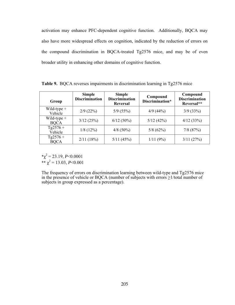
205
activation may enhance PFC-dependent cognitive function. Additionally, BQCA may
also have more widespread effects on cognition, indicated by the reduction of errors on
the compound discrimination in BQCA-treated Tg2576 mice, and may be of even
broader utility in enhancing other domains of cognitive function.
Table 9. BQCA reverses impairments in discrimination learning in Tg2576 mice
Group
Simple Discrimination
Simple Discrimination
Reversal Compound
Discrimination* Compound
Discrimination Reversal**
Wild-type + Vehicle 2/9 (22%) 5/9 (55%) 4/9 (44%) 3/9 (33%)
Wild-type + BQCA 3/12 (25%) 6/12 (50%) 5/12 (42%) 4/12 (33%)
Tg2576 + Vehicle 1/8 (12%) 4/8 (50%) 5/8 (62%) 7/8 (87%)
Tg2576 + BQCA 2/11 (18%) 5/11 (45%) 1/11 (9%) 3/11 (27%)
*χ2 = 23.19, P<0.0001 ** χ2 = 13.03, P<0.001 The frequency of errors on discrimination learning between wild-type and Tg2576 mice in the presence of vehicle or BQCA (number of subjects with errors ≥1/total number of subjects in group expressed as a percentage).
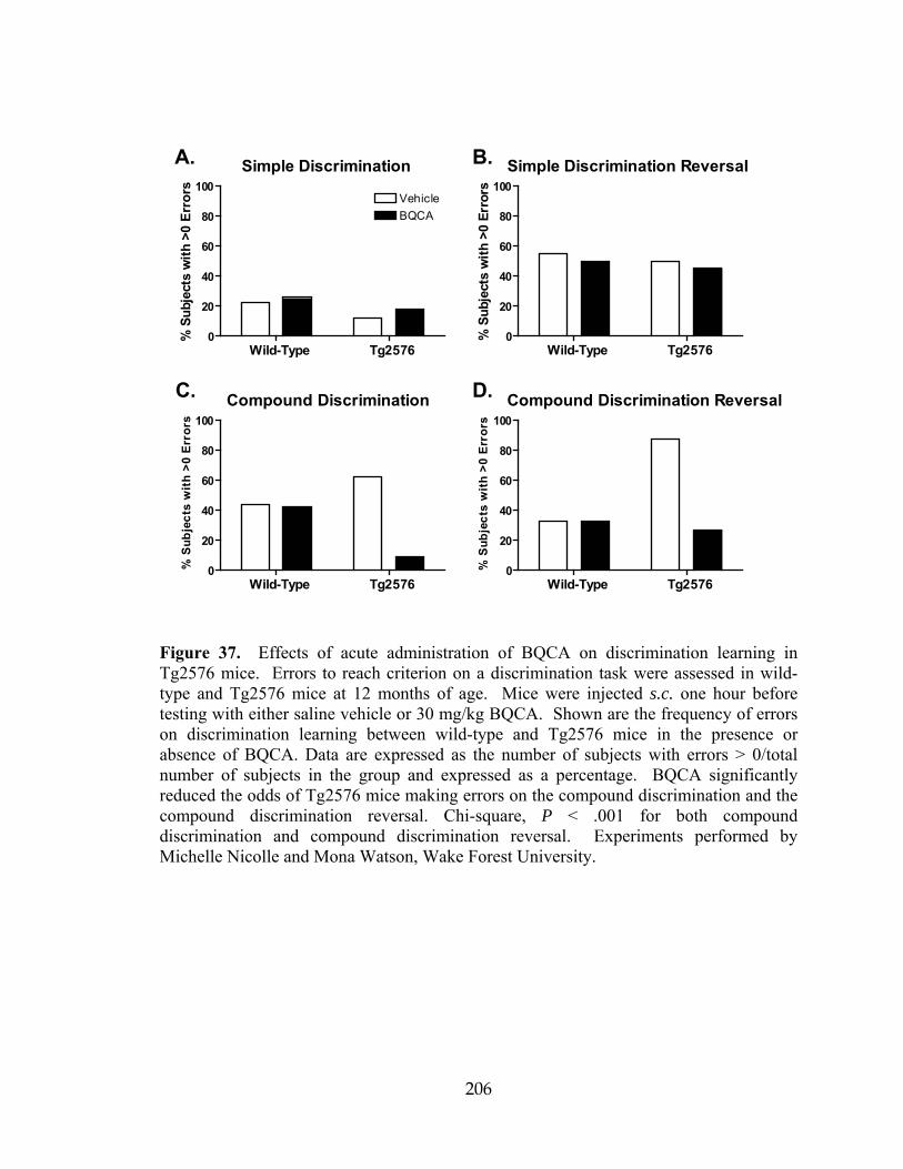
206
A. B.
C. D.
Simple Discrimination
Wild-Type Tg25760
20
40
60
80
100VehicleBQCA
%Su
bjec
tsw
ith>0
Err
ors
Simple Discrimination Reversal
Wild-Type Tg25760
20
40
60
80
100
%Su
bjec
tsw
ith>0
Erro
rs
Compound Discrimination
Wild-Type Tg25760
20
40
60
80
100
%Su
bjec
tsw
ith>0
Erro
rs
Compound Discrimination Reversal
Wild-Type Tg25760
20
40
60
80
100
%Su
bjec
tsw
ith>0
Erro
rs
Figure 37. Effects of acute administration of BQCA on discrimination learning in Tg2576 mice. Errors to reach criterion on a discrimination task were assessed in wild-type and Tg2576 mice at 12 months of age. Mice were injected s.c. one hour before testing with either saline vehicle or 30 mg/kg BQCA. Shown are the frequency of errors on discrimination learning between wild-type and Tg2576 mice in the presence or absence of BQCA. Data are expressed as the number of subjects with errors > 0/total number of subjects in the group and expressed as a percentage. BQCA significantly reduced the odds of Tg2576 mice making errors on the compound discrimination and the compound discrimination reversal. Chi-square, P < .001 for both compound discrimination and compound discrimination reversal. Experiments performed by Michelle Nicolle and Mona Watson, Wake Forest University.

207
BQCA regulates non-amyloidogenic APP processing
The data presented above suggest that BQCA has efficacy in improving at least
one form of cognitive function in an animal model of AD. In addition to providing
symptomatic relief, it has been postulated that increasing M1 receptor activity could also
have disease modifying effects in AD patients (Fisher, 2008; Caccamo et al., 2009). The
amyloid precursor protein (APP) undergoes proteolytic cleavage in two competing
pathways (Thinakaran and Koo, 2008). In the amyloidogenic pathway, sequential
cleavage by β-secretase and γ-secretase releases the Aβ peptide which forms the core of
amyloid plaques found in AD and is implicated in numerous models of neurotoxicity.
Alternatively, in the non-amyloidogenic pathway, APP is cleaved by α-secretase within
the Aβ sequence, preventing Aβ generation. Interestingly, previous studies suggest that
activation of M1 promotes APP processing through the non-amyloidogenic pathway
(Caccamo et al., 2006; Jones et al., 2008). If BQCA can promote non-amyloidogenic
processing of APP, this could provide a mechanism for slowing accumulation of Aβ and
potentially slow progression of AD.
In order to determine whether BQCA can potentiate the APP processing effect of
a low concentration of the mAChR agonist CCh, we treated PC12 cells overexpressing
human APP and the M1 receptor with an approximate EC20 concentration (50 nM) of
CCh in the presence of increasing concentrations of BQCA and measured the levels of
APP metabolites in the conditioned media and cell extracts. BQCA caused a dose-
dependent increase in the shedding of APPsα, the amino-terminal ectodomain of APP
released by α-secretase cleavage (Fig. 38A-B). The highest concentration of BQCA

208
tested (30 μM) increased APPsα levels to 244% of vehicle-treated cells (P < 0.05).
BQCA treatment also resulted in the accumulation of CTFα (C83), the corresponding
carboxy-terminal fragment generated by α-secretase (Fig. 38A, C; increased to 245% of
vehicle, P < 0.05). Finally, consistent with the observed increases in non-amyloidogenic
APP fragments, 30 μM BQCA treatment resulted in a 30% decrease (P < 0.01) in the
secretion of the β-secretase derived Aβ40 peptide (Fig. 38D). Taken together, these
results indicate that BQCA can effectively regulate non-amyloidogenic APP processing,
suggesting that M1 receptor PAMs have the potential to provide both symptomatic and
disease modifying effects in AD patients.

209
Figure 38. BQCA regulates non-amyloidogenic APP processing. A. Western blot analysis of APP metabolites from conditioned media and cell lysates demonstrates increased generation of APPsα and CTFα with increasing concentrations of BQCA as compared to the submaximal concentration of 50 nM CCh. 10 μM CCh is shown as a maximum concentration. β-actin is shown as a loading control. B. Quantitation of APPsα band intensity from conditioned media demonstrates a dose-dependent effect of BQCA on the shedding of APPsα (repeated measures ANOVA, P = 0.0271), and pairwise comparisons revealed significant differences at all concentrations of BQCA compared to 50 nM CCh alone (P-values for paired t-tests are shown). C. Quantitation of CTFα band intensity from cell lysates shows a dose-dependent effect of BQCA on the production of CTFα (repeated measures ANOVA, P = 0.0017) and a significant difference (paired t-test) between 30 μM BQCA plus 50 nM CCh as compared to 50 nM CCh alone. D. ELISA measurements from conditioned media demonstrate that BQCA decreases the secretion of Aβ40 peptide in a dose-dependent manner (repeated measures ANOVA, P = 0.0019), with significant differences between 50 nM CCh alone and the two highest concentrations of BQCA (paired t-tests). Mean values are shown from three or four independent experiments performed in duplicate. All values are normalized to vehicle-treated cells. Experiments performed by Albert Davis, Emory University.
210
Discussion
The M1 receptor has long been viewed as an exciting potential target for
increasing cognitive function in patients suffering from AD and other CNS disorders
(Wess et al., 2007; Fisher, 2008; Langmead et al., 2008a; Caccamo et al., 2009). Despite
major efforts to develop highly selective M1 agonists over the past two decades, this
receptor has proven intractable using traditional approaches, thus preventing M1 agonists
from advancing to clinical use for treatment of AD and other disorders. Also, lack of
agents that selectively activate this receptor has made it impossible to develop a full
understanding of the functional effects of selectively increasing M1 receptor activity in
the CNS. Discovery and characterization of BQCA and its structural analogs provide a
major advance in establishing the utility of M1 receptor PAMs as an alternative approach
to increasing activity of this receptor in a highly subtype-selective manner.
Studies with BQCA, along with the new M1 receptor- selective antagonist
VU0255035, provide important support for the hypothesis that the M1 receptor may
increase activation of the PFC and may enhance PFC-dependent cognitive function
(Anagnostaras et al., 2003). Non-selective mAChR agonists, such as CCh, induce an
inward current in PFC pyramidal cells, and the present data provide strong evidence that
this response is mediated by activation of the M1 receptor. In addition, activation of the
M1 receptor increases the frequency of spontaneous excitatory synaptic events in mPFC
layer V pyramidal cells. While the source of glutamatergic afferents giving rise to these
sEPSCs has not been established, this is consistent with the hypothesis that the M1
receptor plays an important role in increasing excitability and excitatory drive to mPFC
pyramidal cells.

211
One of the most important implications of these studies is that they raise the
possibility that highly selective M1 receptor PAMs may provide a novel approach for
treatment of AD and other CNS disorders that may involve impaired cholinergic
signaling. Clinical studies using both direct and indirect-acting muscarinic agonists have
reported improvements in both cognitive function and behavioral disturbances (i.e.
hallucinations, delusions, outbursts, and paranoia) observed in AD patients (Bodick et al.,
1997b; Cummings et al., 2001). If M1 receptor activation is responsible for, or plays an
important role in, these effects of nonselective cholinergic agents, M1 receptor PAMs
could provide a viable approach to symptomatic treatment of AD. Furthermore, in
addition to potential efficacy in reducing symptoms in AD patients, recent studies suggest
that mAChR activation could reduce accumulation of toxic Aβ protein, thereby also
providing disease modifying effects. For instance, the muscarinic agonist AF102B was
shown to decrease production of the amyloidogenic peptide Aβ42 in the cerebral spinal
fluid of AD patients (Nitsch et al., 2000). Furthermore, preclinical studies with a related
mAChR agonist, AF267B suggest that mAChR activation increases non-amyloidogenic
processing and prevents Aβ formation (Caccamo et al., 2006). While these earlier
mAChR agonists are not selective for the M1 receptor relative to other mAChR subtypes,
more recent studies revealed that the M1 receptor-selective agonist, TBPB, has similar
effects in PC12 cells (Jones et al., 2008).
Lastly, it is important to note that recent clinical and animal studies raise the
possibility that mAChR agonists may also provide a novel approach for treatment of
schizophrenia (Felder et al., 2001; Langmead et al., 2008a; Conn et al., 2009b). For
instance, Shekhar and colleagues (Shekhar et al., 2008) recently reported that the M1/M4
212
receptor–preferring agonist xanomeline induced a robust improvement in positive and
negative symptoms, as well as some measures of cognitive function, in schizophrenic
patients. Based on animal studies, it is likely that both M1 and M4 receptors may be
important for clinical efficacy in this patient population (Felder et al., 2001; Brady et al.,
2008; Chan et al., 2008; Jones et al., 2008; Langmead et al., 2008b; Langmead et al.,
2008a; Conn et al., 2009b). Availability of BQCA, along with the new systemically
active M4 receptor- selective PAM, VU0152100 (Brady et al., 2008) should make it
possible to evaluate the effects of selective activation of each of these mAChR subtypes
as well as co-administration of both BQCA and VU0152100 in a range of animal models
that may be relevant to the antipsychotic effects of xanomeline.

213
CHAPTER V
DISCUSSION AND FUTURE DIRECTIONS
Over the past two decades, major attention has been focused on developing highly
selective agonists of specific mAChR subtypes to use as research tools in order to achieve
a better understanding of the roles of each subtype in specific organ systems and also to
develop therapeutic agents that lack the adverse effects of AChE inhibitors and
nonselective muscarinic agents. We now report the discovery of a novel series of
compounds that act as robust positive allosteric modulators (allosteric potentiators) of the
M4 and M1 mAChR subtypes. Unlike traditional agonists, these small molecules do not
bind to the orthosteric ACh binding site, but instead act at a distinct site to potentiate
activation of the receptor by its natural ligand, ACh. This is directly analogous to the use
of benzodiazepines as selective GABA receptor PAMs, which provide an effective and
safe approach to the treatment of anxiety and sleep disorders without inducing the
potentially lethal effects of direct-acting GABAA receptor agonists (Mohler et al., 2002).
While allosteric modulators of ion channels are well established as research tools and
therapeutic agents, they have not been a traditional focus of drug discovery efforts for
GPCRs. The discovery of VU10010 and related M4 PAMS as well as M1 PAM BQCA
adds to recent major advances in developing highly selective allosteric modulators of
mAChRs (Chan et al., 2008; Ma et al., 2009; Marlo et al., 2009) and other GPCRs (May
et al., 2007; Conn et al., 2009a). These compounds are highly selective for each targeted
receptor subtype with no detectable functional activity at other mAChRs. As with

214
previously discovered allosteric potentiators of metabotropic glutamate receptors
(mGluRs), the novel PAMs of M1 and M4 receptors reported here have no detectable
affinity for the orthosteric site when examined in competition radioligand studies at
equilibrium (Brady et al., 2008; Shirey et al., 2008; Shirey et al., 2009). The action of
these compounds at a site that is distinct from the highly conserved orthosteric ACh
binding site is likely to be critical for achieving this high degree of subtype selectivity.
In addition to providing advantages in terms of achieving high selectivity for an
individual mAChR subtype, it is possible that the mechanism of action of these
compounds could provide other properties that are ultimately advantageous for
developing therapeutic agents. Positive allosteric modulators of M1 and M4 mAChRs
described here do not activate the receptor directly but induce a dramatic potentiation of
the response to activation by ACh. This could provide an advantage to traditional
agonists by amplifying endogenous cholinergic signaling for a specific mAChR subtype
and thereby maintaining a level of activity dependence of receptor activation.
Interestingly, the majority of clinically useful drugs that activate other neuromodulatory
systems exert more subtle actions rather than serving as direct-acting receptor agonists.
For instance, serotonin reuptake inhibitors have been highly successful in treatment of
depression, whereas highly effective serotonin receptor agonists have not been
successfully developed. For anxiety disorders, it is likely that direct acting GABAA
receptor agonists would have a narrow therapeutic window and would be lethal. In
contrast, benzodiazepines are selective allosteric potentiators of GABAA receptor
chloride channels and are safe and highly effective in the clinic. Dopamine receptor
agonists are used but exert more profound acute adverse effects than indirect acting

215
agents such as L-DOPA, amphetamine, and methylphenidate (Ritalin). Interestingly,
highly selective allosteric potentiators of different mGluR subtypes have robust effects in
animal models used to predict efficacy in treatment of multiple CNS disorders, including
schizophrenia, anxiety disorders and Parkinson’s disease (Marino et al., 2003; Kinney et
al., 2005; Galici et al., 2006). Furthermore, a highly selective allosteric potentiator of the
calcium-sensing receptor (a family III/C GPCR), termed Sensipar, has now received FDA
approval for use in treatment of hyperparathyroidism (Dong, 2005). As with the current
M1 and M4-selective allosteric potentiators, the key to achieving high selectivity for these
receptors has been the targeting of sites that are distinct from the highly conserved
orthosteric neurotransmitter binding site. These advances with allosteric potentiators of
family III/C GPCRs along with the present findings raise the possibility that discovery of
highly selective and robust allosteric modulators of specific mAChR subtypes and other
family I/A GPCRs will provide an exciting new approach for the discovery and
development of novel research tools and therapeutic agents that target this important class
of signaling proteins. However, discovery of these compounds also raises the question of
whether allosteric modulators may have broad activity across other GPCRs by interacting
at potentially promiscuous allosteric sites. The present finding that novel mAChR PAMs
have no major activities across multiple targets in a large panel radioligand binding
screen was encouraging, but does not address this critical question. The finding that
these compounds had no allosteric modulator activity across a panel of other family A
GPCR subtypes is exciting and suggests that they are not likely to have activity at a site
that is shared across multiple GPCRs. While it is impossible to rule out activity at other
unidentified targets, these data suggest that it may be possible to achieve higher subtype

216
selectivity across a range of receptors than has been possible with many orthosteric
ligands.
Interestingly, the M1 and M4 PAMs reported here increase affinity of ACh for the
orthosteric site. This is similar to previous reports of allosteric modulators of GPCRs
acting by altering affinity of ligands at the orthosteric site; the magnitude of binding
cooperativity or the shift in orthosteric agonist affinity is referred to as the alpha factor
(Christopoulos and Kenakin, 2002). However, while M4 PAM VU10010 apparently acts
in part by increasing ACh affinity, the magnitude of this effect was only a 14-fold
increase in ACh affinity whereas VU10010 induced an almost 50-fold shift in the ACh
concentration response curve for activating calcium mobilization, Chapter IIIA (Shirey
et al., 2008). Thus, effects on agonist binding cannot fully account for the allosteric
potentiator activity measured in the functional assay; potentiation by this compound must
also increase efficacy and coupling to downstream effectors. The present data suggest
that VU10010 acts by both increasing ACh affinity and activation of G proteins and
downstream signaling pathways. Studies of the effects of VU10010 on [35S]GTPγS
binding in cells expressing rat M4 revealed a 10-fold shift in ACh potency; this value is
close to the calculated shift in ACh affinity at the orthosteric site, suggesting that the
increase in ACh affinity could account for the shift in ACh potency by increasing G
protein activation. However, VU10010 also increased the maximal ACh-induced
[35S]GTPγS binding response, suggesting that this potentiator increases the efficiency of
M4 coupling to G proteins. These two factors together likely contribute to the robust shift
in ACh potency for inducing calcium mobilization relative to more modest shift observed
in ACh affinity. In contrast, radioligand binding studies examining the effect of BQCA
217
on M1 receptor affinity for ACh revealed that the fold-shift decrease in ACh Ki induced
by BQCA was around 30-fold, whereas the shift in the functional calcium response was
21-fold (Shirey et al., 2009). The increase in affinity for agonist could therefore fully
account for the potentiation of calcium mobilization by M1. This demonstrates the
possibility that different allosteric modulators may induce varying degrees of receptor
affinity for orthosteric agonists and downstream effector signaling.
A notion that complicates interpretation of selectivity data as discussed above is
the possibility of binding of an allosteric modulator to an off-target GPCR without a
detectable functional consequence. Because radioligands that bind to allosteric sites of
mAChRs and other GPCRs have not yet been developed (with the exception of mGluR5),
it is difficult to assess binding at these sites using traditional equilibrium experiments. It
is possible, however to determine if allosteric ligands affect the affinity of orthosteric site
ligands. For example, VU10010 and BQCA did not compete for orthosteric [3H]-NMS
binding but induced a leftward shift in the ACh competition curve (Chapters IIIA and
IV, (Shirey et al., 2008)). While VU10010 did not compete for [3H]-NMS binding in
membranes from cells expressing any mAChR subtype, its effects on ACh affinity at off-
target receptor subtypes were not determined. This will be one critical experiment that
should be performed with novel allosteric modulators to assess their binding to off-target
receptors. Another related experimental approach that may useful in screening for
compounds that only bind to a targeted mAChR or other GPCR involves examining
neutral allosteric binding to off-target receptors or receptor subtypes. With the newly
available panel of allosteric modulators (and in the case of M1, allosteric agonists such as
TBPB) of mAChRs, it is now possible to detect neutral allosteric binding to these
218
receptors. This is an important area of exploration as no neutral allosteric modulators
have been reported for mAChRs to date.
Discovery of selective allosteric modulators of mAChRs provides an
unprecedented opportunity to develop a more detailed understanding of the functional
roles of these receptors in neural circuits that are heavily modulated by cholinergic
innervation. One of the most important roles of cholinergic systems in the CNS is
modulation of transmission through the hippocampal formation, a limbic structure that
plays a critical role in learning and memory that is thought to be important for cholinergic
regulation of cognitive function. Activation of mAChRs in the hippocampus induces a
marked reduction of transmission at both excitatory glutamatergic and inhibitory
GABAergic synapses by presynaptic reduction of neurotransmitter release (Valentino and
Dingledine, 1981; Bilkey and Goddard, 1985; Fernandez de Sevilla and Buno, 2003).
Multiple mAChR subtypes are expressed in the hippocampus, and evidence suggests that
both M2 and M4 are localized in presynaptic terminals where they could regulate
excitatory and inhibitory synaptic transmission (Levey et al., 1995; Rouse et al., 1999;
Rouse et al., 2000a). In addition, evidence suggests that activation of postsynaptic
mAChRs on hippocampal pyramidal cells that express M1 and M4 could reduce synaptic
transmission in the hippocampus by inducing release of endocannabinoids that activate
presynaptic CB1 cannabinoid receptors (Ohno-Shosaku et al., 2003). Thus, the specific
mAChR subtypes involved in regulating transmission at these important synapses is not
clear. The finding that VU10010 potentiates effects of mAChR agonists on excitatory
but not inhibitory synaptic transmission provides strong evidence that M4 is one of the
mAChR subtypes responsible for regulating transmission at glutamatergic synapses in the

219
hippocampus but is not likely to be involved in regulation of transmission at inhibitory
synapses in this brain region (Chapter IIIA, (Shirey et al., 2008)). This selective
regulation of excitatory synaptic transmission is a first critical step in developing a
detailed understanding of the roles of M4 in modulating hippocampal function. Although
a growing body of literature implicates M1 receptors in many of the excitatory
postsynaptic effects of mAChR activation and in long-term plasticity (both LTP and
LTD) in the hippocampus and cortex, many of these studies have relied on compounds
that are not subtype selective or on studies employing knockout mice where
compensatory mechanisms can complicate data interpretation. In future studies, it will be
critical to take advantage of these novel selective allosteric potentiators to further
investigate the roles of M1 and M4 in hippocampus as well as other cortical and basal
ganglia structures where mAChRs have been postulated to play a critical
neuromodulatory role. For instance, if the M1 receptor subtype does mediate the
excitatory postsynaptic effects of muscarinic agonists (depolarization, blockade of spike
frequency adaptation and afterhyperpolarization currents, etc.), these effects of CCh
should be blocked by the selective M1 antagonist VU0255035 and should be mimicked
by M1 agonists. Likewise, M1 agonists and PAMs should induce or modulate
hippocampal LTP and LTD if this receptor subtype is responsible for the effects of CCh
and ACh on these forms of long-term plasticity.
The M1 receptor has long been viewed as an exciting potential target for
increasing cognitive function in patients suffering from AD and other CNS disorders
(Wess et al., 2007; Fisher, 2008; Langmead et al., 2008a; Caccamo et al., 2009). Despite
major efforts to develop highly selective M1 orthosteric agonists over the past two

220
decades, this receptor has proven intractable using traditional approaches, thus preventing
M1 agonists from advancing to clinical use for treatment of AD and other disorders.
Also, lack of agents that selectively activate this receptor has made it impossible to
develop a full understanding of the functional effects of selectively increasing M1
receptor activity in the CNS. Studies with BQCA, along with the new M1-selective
antagonist VU0255035 (Sheffler et al., 2009), provide important support for the
hypothesis that the M1 receptor may increase activation of the PFC and may enhance
PFC-dependent cognitive function (Anagnostaras et al., 2003). Non-selective mAChR
agonists, such as CCh, induce an inward current in PFC pyramidal cells, and the present
data (Chapter IV) provide strong evidence that this response is mediated by activation of
the M1 receptor. In addition, activation of the M1 receptor increases the frequency of
spontaneous excitatory synaptic events in mPFC layer V pyramidal cells. While the
source of glutamatergic afferents giving rise to these sEPSCs has not been established,
this is consistent with the hypothesis that the M1 receptor plays an important role in
increasing excitability and excitatory drive to mPFC pyramidal cells. It is also unclear
thus far whether depolarization of glutamatergic pyramidal cells in cortical circuits
underlies the increase in sEPSCs induced by CCh, or if these are two separate and
unrelated phenomena.
Interestingly, mAChR activation induces direct excitatory effects in hippocampal
CA1 pyramidal cells that are similar to those observed in mPFC pyramidal cells.
However, while CA1 pyramidal cells express high levels of the M1 receptor (Levey et al.,
1991), previous studies suggest that the M1 receptor is not the mAChR subtype
responsible for some of the excitatory effects on these cells (Rouse et al., 2000a). Thus,
221
the precise physiological roles of the M1 receptor are likely to vary in different brain
regions and neuronal populations. The finding that M1 receptor activation has excitatory
effects and increases excitatory synaptic activity in mPFC pyramidal cells is interesting in
the context of the recent finding that M1 receptor knockout mice display clear deficits in
PFC-dependent learning (Anagnostaras et al., 2003), whereas hippocampal-dependent
learning is largely unaffected in M1 receptor knockout mice (Anagnostaras et al., 2003)
and in animals treated with the M1 receptor- selective antagonist VU0255035 (Sheffler et
al., 2009).
One of the most important implications of these studies is that they raise the
possibility that highly selective M1 receptor PAMs may provide a novel approach for
treatment of AD and other CNS disorders that involve impaired cholinergic signaling.
Clinical studies using both direct and indirect-acting muscarinic agonists have reported
improvements in both cognitive function and behavioral disturbances (i.e. hallucinations,
delusions, outbursts, and paranoia) observed in AD patients (Bodick et al., 1997b;
Cummings et al., 2001). If M1 receptor activation is responsible for, or plays an
important role in, these effects of nonselective cholinergic agents, M1 receptor PAMs
could provide a viable approach to symptomatic treatment of AD. Furthermore, in
addition to potential efficacy in reducing symptoms in AD patients, recent studies suggest
that mAChR activation could reduce accumulation of toxic Aβ protein, thereby providing
additional disease modifying effects. For instance, the muscarinic agonist AF102B was
shown to decrease production of the amyloidogenic peptide Aβ42 in the cerebral spinal
fluid of AD patients (Nitsch et al., 2000). Furthermore, preclinical studies with a related
mAChR agonist, AF267B suggest that mAChR activation increases non-amyloidogenic

222
processing and prevents Aβ formation (Caccamo et al., 2006). While these earlier
mAChR agonists are not selective for the M1 receptor relative to other mAChR subtypes,
more recent studies revealed that the M1 receptor-selective agonist, TBPB, has similar
effects in PC12 cells (Jones et al., 2008).
The present finding that BQCA reverses deficits in compound discrimination
reversal learning in a transgenic mouse model of AD provides exciting support for the
hypothesis that highly selective M1 receptor PAMs may provide efficacy in treatment of
at least some domains of cognitive function in AD (Chapter IV). Furthermore, the
finding that BQCA promotes non-amyloidogenic APP processing suggests that these
agents could also reduce amyloid burden. In future studies, it will be important to fully
explore the effects of BQCA in animal models that reflect other domains of cognitive
function that are impaired in AD patients. For instance it is possible that M1 receptor-
selective PAMs will have robust efficacy in improving PFC-dependent learning, but have
lesser effects on hippocampal-dependent learning. A mouse model of AD with
impairments in hippocampal-dependent memory tasks could be used to test whether
BQCA could restore performance in the Morris water maze or in other forms of spatial
memory that require intact hippocampal functioning. Also, other types of cognitive
function may involve different mAChR subtypes and be differentially affected by
selective activators of the M1 receptor versus selective PAMs of other mAChR subtypes,
such as the recently reported M4 and M5 receptor-selective PAMs (Brady 2008; Chan
2008; Shirey 2008; Bridges 2009). Interestingly, the high subtype-selectivity of BQCA
may prove to be important for achieving maximal effects in increasing non-
amyloidogenic APP processing. Previous studies suggest that activation of M2 and/or M4
223
mAChR subtypes may have an antagonistic effect on the non-amyloidogenic APP
processing shown to be promoted by M1 receptor activation (Farber et al., 1995). Thus,
in addition to reducing the adverse effect profile, it is possible that selective activation of
the M1 receptor may provide greater efficacy in regulating APP processing.
In addition to implications for AD, the electrophysiology studies reveal
interesting findings that may provide important insights related to the potential roles of
mAChRs in regulating PFC function. For instance, when added alone, BQCA induced a
slight inward current and a slight increase in sEPSC frequency (Chapter IV, (Shirey et
al., 2009)). This suggests that there may be a low tonic level of M1 receptor activity or a
low level of endogenous acetylcholine in slices that can be potentiated by BQCA.
Furthermore, it was interesting to find that CCh induced a small reduction in sEPSC
frequency when added in the presence of a saturating concentration of the selective M1
antagonist, VU0255035. This suggests that activation of another mAChR subtype can
reduce sEPSC frequency and that this effect is unmasked when the M1 receptor is
selectively blocked. To test the hypothesis that this inhibitory effect is mediated by the
M4 receptor subtype, the selective M4 PAM, VU0152100, could be used alone and in
combination with a low concentration of CCh. If this hypothesis were correct, I would
expect to see a decrease in sEPSCs in the presence of VU0152100 and CCh.
Interestingly, while effects of CCh on sEPSC frequency were dramatically reduced in M1
receptor knockout mice, CCh did induce a small effect in slices from these animals. This
suggests that another mAChR subtype may be capable of eliciting this response and
could partially compensate for genetic deletion of the M1 receptor. Importantly, the
effect of the highly selective M1 receptor PAM, BQCA, was eliminated in M1 receptor

224
knockout mice, suggesting that the effects of this compound are fully dependent on
activation of the M1 receptor. Discovery of new mAChR subtype-selective ligands for
multiple mAChR subtypes over the last year will allow for a better understanding of the
roles of mAChR subtypes in regulating PFC function.
Recent clinical and animal studies raise the possibility that mAChR agonists may
provide a novel approach for treatment of schizophrenia (Felder et al., 2001; Langmead
et al., 2008a; Conn et al., 2009b). For decades, the prevailing theory behind the etiology
of schizophrenia has been that excessive dopaminergic neurotransmission in the central
nervous system is the major contributing factor underlying this severe psychiatric illness.
This so-called dopamine hyperfunction hypothesis is based primarily on the observation
that stimulation of the endogenous dopamine system (e.g. with amphetamine or cocaine)
often leads to transient psychotic symptoms in healthy individuals (Bymaster et al., 2002;
Raedler et al., 2007). Furthermore, all clinically relevant antipsychotic drugs, both
typical and atypical, possess significant antagonist activity at D2 dopamine receptors
(D2Rs) (Carlsson, 1988; Bymaster et al., 2002; Raedler et al., 2007). Thus, the majority
of the efforts to discover novel therapeutic agents for the treatment of schizophrenia have
been aimed at developing therapies that result in some level of D2R blockade or a
combination of blockade of D2Rs and other monoamine receptors. Nevertheless, D2R
antagonists are only partially effective in treating schizophrenia, in that they only
improve the positive symptoms associated with the disease, despite the fact that the
negative and cognitive symptoms also markedly impact the quality of life for
schizophrenic patients. In addition, these therapies also are often poorly tolerated
because of numerous side effects including sedation, weight gain, sexual dysfunction,

225
diabetes and Parkinson’s disease-like symptoms. Furthermore, greater than 25% of
schizophrenia patients do not respond to these dopamine-based therapies. Thus, while it
is evident that dopamine does play a prominent role in the pathogenesis and treatment of
schizophrenia, the dopamine hyperfunction hypothesis of schizophrenia fails to account
for all aspects of this disorder. It is increasingly evident that other neurotransmitter
systems are likely involved. Based on this, it is unlikely that exclusive focus on
discovery and development of antagonists of D2Rs and other monoamine receptors will
provide fundamental breakthroughs in the standard of treatment of schizophrenia patients
relative to current therapies.
In recent years the mAChRs, namely M1 and M4, have emerged as potential novel
targets for the treatment of schizophrenia. This is based on clinical studies demonstrating
efficacy of mAChR agonists in treatment of positive symptoms in schizophrenia patients,
as well as multiple animal studies suggesting that mAChR agonists could be useful in
treatment of cognitive dysfunction in schizophrenia patients (Bymaster et al., 2002). A
growing body of evidence from clinical and animal studies involving pharmacological
manipulations, post mortem tissue analysis, and brain imaging is consistent with this
hypothesis (Raedler et al., 2007). Furthermore, Shekhar and colleagues (Shekhar et al.,
2008) recently reported that the M1/M4 receptor–preferring agonist xanomeline induced a
robust improvement in positive and negative symptoms, as well as some measures of
cognitive function, in schizophrenic patients. While recent advances suggesting potential
utility of mAChR activators in treatment of schizophrenia have been exciting, there have
been few selective pharmacological tools available to fully explore this emerging
muscarinic hypothesis of schizophrenia. Unfortunately, previous attempts to develop

226
traditional orthosteric agonists that are highly selective for individual mAChR subtypes
have been unsuccessful.
The recent discovery and optimization of highly selective positive allosteric
modulators of M1 and M4 provides a major advance in establishing a new approach for
developing highly selective activators of these receptors. The data presented provide
further support for the ability to achieve high subtype selectivity by targeting allosteric
sites and provide exciting new data demonstrating that highly selective M4 PAMs have
robust activity in at least one animal model that is similar to effects previously described
for the non-selective orthosteric mAChR agonist, xanomeline (Stanhope et al., 2001).
The finding that VU0152099 and VU0152100 mimic effects of xanomeline in an animal
model that has been used to predict antipsychotic activity (Brady et al., 2008) is
especially promising in light of clinical studies demonstrating the clinical efficacy of
xanomeline in schizophrenia patients. This raises the exciting possibility that selective
activation of M4 may provide a novel approach for the treatment of some symptoms
associated with schizophrenia. Based on animal studies, it is likely that both M1 and M4
receptors may be important for clinical efficacy in this patient population (Felder et al.,
2001; Gerber et al., 2001; Miyakawa et al., 2001; Marino and Conn, 2002; Messer,
2002b; Anagnostaras et al., 2003; Bymaster et al., 2003b; Tzavara et al., 2004; Brady et
al., 2008; Langmead et al., 2008a; Shirey et al., 2008). Availability of BQCA along with
the systemically active M4 receptor-selective PAM, VU0152100, should make it possible
to evaluate the effects of selective activation of each of these mAChR subtypes as well as
co-administration of both BQCA and VU0152100 in a range of animal models that may
be relevant to the antipsychotic effects of xanomeline.
227
In addition to their potential relevance for schizophrenia, mAChRs also are
thought to regulate motor function by exerting effects on dopaminergic transmission in
the basal ganglia (Pisani et al., 2007; Raedler et al., 2007). For instance, multiple
electrophysiology studies with M4 knockout mice have led to the suggestion that
activation of the M4 mAChR may oppose some actions of dopaminergic neurons on
striatal motor function (Calabresi et al., 1998; Sanchez-Lemus and Arias-Montano,
2006). Again, the lack of highly selective activators of M4 has made it impossible to test
this hypothesis directly. Also, previous studies with traditional orthosteric muscarinic
agonists bypass the action of endogenous ACh in the basal ganglia and do not provide
information about the effects of endogenous acetylcholine on motor activity. The finding
that highly selective M4 potentiators reverse amphetamine-induced hyperlocomotor
activity in rats provides exciting new evidence in support of the hypothesis that
endogenous ACh plays an important role in regulating dopaminergic control of motor
function. Because these compounds do not activate M4 directly, but selectively increase
responses of M4 to endogenous ACh, this provides direct evidence that this response can
be modulated by endogenous ACh acting on M4 receptors. In addition, this raises the
possibility that selective M4 modulators could provide a novel approach to treatment of
other disorders involving altered dopaminergic function in the basal ganglia, including
Parkinson’s disease and dystonia. Ongoing studies in the Conn lab are in fact examining
the efficacy of M4 PAMS in animal models of these diseases.
In conclusion, the recent development of compounds that selectively activate or
modulate individual mAChRs provides an exciting new opportunity to begin to answer
questions about the role of these receptors in normal and pathological CNS function.

228
These tools will also allow the further validation of individual mAChR subtypes as
therapeutic targets for a variety of disorders including AD and schizophrenia. One
important aspect of research will include the elucidation of various signaling cascades
activated by different classes of orthosteric and allosteric ligands. Preliminary data from
our lab and others suggests that allosteric agonists of the M1 receptor like TBPB (Jones et
al., 2008) do not induce β-arrestin recruitment or activate mPFC neurons in the same
manner as CCh; it will be important to determine whether specific signaling pathways
like these and ERK1/2 activation are necessary for the therapeutically beneficial effects
of mAChR activation. One way to assess the involvement of β-arrestin recruitment in the
CCh-induced activation of mPFC neurons would be to perform recordings of sEPSCs in
the presence of CCh using mice lacking the β-arrestin protein. If this signaling molecule
is required for the muscarinic activation of mPFC layer V neurons, this effect would
likely be absent or altered in the β-arrestin knockout mice. An alternate strategy could
involve the use of a β-arrestin blocking peptide or antibody in the recording pipet in
experiments with wildtype mice or rats. Likewise, a selective, cell permeable MEK
inhibitor could be used in the recording bath to determine if ERK1/2 phosphorylation is
required for CCh-induced increase in sEPSCs. If these experiments yielded data that
support the involvement of these signaling proteins, this would be one possible
explanation for the lack of effect of M1 allosteric agonists like TBPB and VU0184670 on
mPFC neurons since these compounds do not appear to induce β-arrestin recruitment.
It will also be interesting to examine the efficacy of PAMs in animal models of
diseases that involve degeneration of cholinergic nuclei. Because postsynaptic mAChR
expression remains largely intact despite the loss of cholinergic input, it has been

229
hypothesized that activation of these remaining postsynaptic receptors will alleviate many
of the cognitive impairments associated with cholinergic decline (Caccamo et al., 2009).
While this may be true for direct-acting agonists, it may not be possible to achieve
therapeutic efficacy using allosteric potentiators that do not directly activate mAChRs.
Depending on the level of disease progression and cholinergic degeneration, allosteric
compounds could display a varying array of efficacies. The recent development of
selective M1 activators that are suitable for use in clinical trials with patients suffering
from AD will also allow researchers and clinicians to determine the disease-modifying
potential of these compounds. Although initial studies indicating that the M1-preferring
agonist AF102B decreased levels of Aβ in CSF of AD patients (Nitsch et al., 2000), it
remains to be seen if sustained, selective M1 activation will have any effect on disease
progression or pathology in brain tissue of this patient population.
Because the M1/M4-preferring agonist xanomeline lacked true subtype selectivity
and caused dose-limiting side effect associated with activation of peripheral M3 receptors
(Shekhar et al., 2008), clinical trial assessing the ability of this drug to significantly
improve the symptoms of schizophrenia were somewhat limited. However, a growing
body of literature including studies using knockout mice and initial studies with
VU0152100, TBPB, and BQCA support the hypothesis that M1 and M4 receptors show
great promise as therapeutic targets for diseases involving cognitive impairment such as
AD and schizophrenia. Whether drugs that selectively activate these receptors will
provide a more tolerable side-effect profile than that of AChE inhibitors or xanomeline
remains to be seen. It will also be critical to determine if selective activation of M1 or M4

230
alone will be sufficient to improve symptoms of schizophrenia in the same manner as
xanomeline.

231
REFERENCES
Abe K, Nakata A, Mizutani A, Saito H (1994) Facilitatory but nonessential role of the
muscarinic cholinergic system in the generation of long-term potentiation of population spikes in the dentate gyrus in vivo. Neuropharmacology 33:847-852.
Anagnostaras SG, Murphy GG, Hamilton SE, Mitchell SL, Rahnama NP, Nathanson NM, Silva AJ (2003) Selective cognitive dysfunction in acetylcholine M1 muscarinic receptor mutant mice. Nat Neurosci 6:51-58.
Andrade R (1991) Cell excitation enhances muscarinic cholinergic responses in rat association cortex. Brain Res 548:81-93.
Arendt T, Bigl V, Arendt A, Tennstedt A (1983) Loss of neurons in the nucleus basalis of Meynert in Alzheimer's disease, paralysis agitans and Korsakoff's Disease. Acta Neuropathol 61:101-108.
Auerbach JM, Segal M (1994) A novel cholinergic induction of long-term potentiation in rat hippocampus. J Neurophysiol 72:2034-2040.
Auerbach JM, Segal M (1996) Muscarinic receptors mediating depression and long-term potentiation in rat hippocampus. J Physiol 492 ( Pt 2):479-493.
Auld DS, Kornecook TJ, Bastianetto S, Quirion R (2002) Alzheimer's disease and the basal forebrain cholinergic system: relations to beta-amyloid peptides, cognition, and treatment strategies. Prog Neurobiol 68:209-245.
Bartus RT, Dean RL, 3rd, Beer B, Lippa AS (1982) The cholinergic hypothesis of geriatric memory dysfunction. Science 217:408-414.
Basile AS, Fedorova I, Zapata A, Liu X, Shippenberg T, Duttaroy A, Yamada M, Wess J (2002) Deletion of the M5 muscarinic acetylcholine receptor attenuates morphine reinforcement and withdrawal but not morphine analgesia. Proc Natl Acad Sci U S A 99:11452-11457.
Behrends JC, ten Bruggencate G (1993) Cholinergic modulation of synaptic inhibition in the guinea pig hippocampus in vitro: excitation of GABAergic interneurons and inhibition of GABA-release. J Neurophysiol 69:626-629.
Benardo LS, Prince DA (1982) Ionic mechanisms of cholinergic excitation in mammalian hippocampal pyramidal cells. Brain Res 249:333-344.
Berkeley JL, Gomeza J, Wess J, Hamilton SE, Nathanson NM, Levey AI (2001) M1 muscarinic acetylcholine receptors activate extracellular signal-regulated kinase in CA1 pyramidal neurons in mouse hippocampal slices. Mol Cell Neurosci 18:512-524.
Berntson GG, Sarter M, Cacioppo JT (1998) Anxiety and cardiovascular reactivity: the basal forebrain cholinergic link. Behav Brain Res 94:225-248.
Bilkey DK, Goddard GV (1985) Medial septal facilitation of hippocampal granule cell activity is mediated by inhibition of inhibitory interneurones. Brain Res 361:99-106.
Birdsall NJ, Lazareno S, Matsui H (1996) Allosteric regulation of muscarinic receptors. Prog Brain Res 109:147-151.
Bliss TV, Collingridge GL (1993) A synaptic model of memory: long-term potentiation in the hippocampus. Nature 361:31-39.
Blitzer RD, Gil O, Landau EM (1990) Cholinergic stimulation enhances long-term potentiation in the CA1 region of rat hippocampus. Neurosci Lett 119:207-210.
Bodick NC, Offen WW, Shannon HE, Satterwhite J, Lucas R, van Lier R, Paul SM (1997a) The selective muscarinic agonist xanomeline improves both the cognitive deficits and behavioral symptoms of Alzheimer disease. Alzheimer Dis Assoc Disord 11 Suppl 4:S16-22.
232
Bodick NC, Offen WW, Levey AI, Cutler NR, Gauthier SG, Satlin A, Shannon HE, Tollefson GD, Rasmussen K, Bymaster FP, Hurley DJ, Potter WZ, Paul SM (1997b) Effects of xanomeline, a selective muscarinic receptor agonist, on cognitive function and behavioral symptoms in Alzheimer disease. Arch Neurol 54:465-473.
Bonner TI, Buckley NJ, Young AC, Brann MR (1987) Identification of a family of muscarinic acetylcholine receptor genes. Science 237:527-532.
Bonner TI, Young AC, Brann MR, Buckley NJ (1988) Cloning and expression of the human and rat m5 muscarinic acetylcholine receptor genes. Neuron 1:403-410.
Bowen DM, Smith CB, White P, Davison AN (1976) Neurotransmitter-related enzymes and indices of hypoxia in senile dementia and other abiotrophies. Brain 99:459-496.
Brady AE, Jones CK, Bridges TM, Kennedy JP, Thompson AD, Heiman JU, Breininger ML, Gentry PR, Yin H, Jadhav SB, Shirey JK, Conn PJ, Lindsley CW (2008) Centrally active allosteric potentiators of the M4 muscarinic acetylcholine receptor reverse amphetamine-induced hyperlocomotor activity in rats. J Pharmacol Exp Ther 327:941-953.
Bridges TM, Marlo JE, Niswender CM, Jones CK, Jadhav SB, Gentry PR, Plumley HC, Weaver CD, Conn PJ, Lindsley CW (2009) Discovery of the first highly M5-preferring muscarinic acetylcholine receptor ligand, an M5 positive allosteric modulator derived from a series of 5-trifluoromethoxy N-benzyl isatins. J Med Chem 52:3445-3448.
Brown T, Zador A (1990) Hippocampus. In: The Synaptic Organization of the Brain (GM S, ed), pp 346-388. New York: Oxford UP.
Buckley NJ, Bonner TI, Brann MR (1988) Localization of a family of muscarinic receptor mRNAs in rat brain. J Neurosci 8:4646-4652.
Burgard EC, Sarvey JM (1990) Muscarinic receptor activation facilitates the induction of long-term potentiation (LTP) in the rat dentate gyrus. Neurosci Lett 116:34-39.
Burgard EC, Cote TE, Sarvey JM (1993) Muscarinic depression of synaptic transmission and blockade of norepinephrine-induced long-lasting potentiation in the dentate gyrus. Neuroscience 54:377-389.
Bymaster FP, Felder CC (2002) Role of the cholinergic muscarinic system in bipolar disorder and related mechanism of action of antipsychotic agents. Mol Psychiatry 7 Suppl 1:S57-63.
Bymaster FP, Felder C, Ahmed S, McKinzie D (2002) Muscarinic receptors as a target for drugs treating schizophrenia. Curr Drug Targets CNS Neurol Disord 1:163-181.
Bymaster FP, McKinzie DL, Felder CC, Wess J (2003a) Use of M1-M5 muscarinic receptor knockout mice as novel tools to delineate the physiological roles of the muscarinic cholinergic system. Neurochem Res 28:437-442.
Bymaster FP, Felder CC, Tzavara E, Nomikos GG, Calligaro DO, McKinzie DL (2003b) Muscarinic mechanisms of antipsychotic atypicality. Prog Neuropsychopharmacol Biol Psychiatry 27:1125-1143.
Bymaster FP, Carter PA, Yamada M, Gomeza J, Wess J, Hamilton SE, Nathanson NM, McKinzie DL, Felder CC (2003c) Role of specific muscarinic receptor subtypes in cholinergic parasympathomimetic responses, in vivo phosphoinositide hydrolysis, and pilocarpine-induced seizure activity. Eur J Neurosci 17:1403-1410.
Caccamo A, Fisher A, LaFerla FM (2009) M1 agonists as a potential disease-modifying therapy for Alzheimer's disease. Curr Alzheimer Res 6:112-117.
Calabresi P, Centonze D, Gubellini P, Pisani A, Bernardi G (1998) Endogenous ACh enhances striatal NMDA-responses via M1-like muscarinic receptors and PKC activation. Eur J Neurosci 10:2887-2895.
233
Callahan MJ, Kinsora JJ, Harbaugh RE, Reeder TM, Davis RE (1993) Continuous ICV infusion of scopolamine impairs sustained attention of rhesus monkeys. Neurobiol Aging 14:147-151.
Campbell UC, Lalwani K, Hernandez L, Kinney GG, Conn PJ, Bristow LJ (2004) The mGluR5 antagonist 2-methyl-6-(phenylethynyl)-pyridine (MPEP) potentiates PCP-induced cognitive deficits in rats. Psychopharmacology (Berl) 175:310-318.
Carr DB, Surmeier DJ (2007) M1 muscarinic receptor modulation of Kir2 channels enhances temporal summation of excitatory synaptic potentials in prefrontal cortex pyramidal neurons. J Neurophysiol 97:3432-3438.
Caulfield MP (1993) Muscarinic receptors--characterization, coupling and function. Pharmacol Ther 58:319-379.
Caulfield MP, Birdsall NJ (1998) International Union of Pharmacology. XVII. Classification of muscarinic acetylcholine receptors. Pharmacol Rev 50:279-290.
Chan AS, Yeung WW, Wong YH (2005) Integration of G protein signals by extracellular signal-regulated protein kinases in SK-N-MC neuroepithelioma cells. J Neurochem 94:1457-1470.
Chan WY, McKinzie DL, Bose S, Mitchell SN, Witkin JM, Thompson RC, Christopoulos A, Lazareno S, Birdsall NJ, Bymaster FP, Felder CC (2008) Allosteric modulation of the muscarinic M4 receptor as an approach to treating schizophrenia. Proc Natl Acad Sci U S A 105:10978-10983.
Christopoulos A (2002) Allosteric binding sites on cell-surface receptors: novel targets for drug discovery. Nat Rev Drug Discov 1:198-210.
Christopoulos A, El-Fakahany EE (1999) Qualitative and quantitative assessment of relative agonist efficacy. Biochem Pharmacol 58:735-748.
Christopoulos A, Kenakin T (2002) G protein-coupled receptor allosterism and complexing. Pharmacol Rev 54:323-374.
Cole AE, Nicoll RA (1984a) Characterization of a slow cholinergic post-synaptic potential recorded in vitro from rat hippocampal pyramidal cells. J Physiol 352:173-188.
Cole AE, Nicoll RA (1984b) The pharmacology of cholinergic excitatory responses in hippocampal pyramidal cells. Brain Res 305:283-290.
Colino A, Halliwell JV (1993) Carbachol potentiates Q current and activates a calcium-dependent non-specific conductance in rat hippocampus in vitro. Eur J Neurosci 5:1198-1209.
Colom LV, Nassif-Caudarella S, Dickson CT, Smythe JW, Bland BH (1991) In vivo intrahippocampal microinfusion of carbachol and bicuculline induces theta-like oscillations in the septally deafferented hippocampus. Hippocampus 1:381-390.
Conn PJ, Christopoulos A, Lindsley CW (2009a) Allosteric modulators of GPCRs: a novel approach for the treatment of CNS disorders. Nat Rev Drug Discov 8:41-54.
Conn PJ, Jones CK, Lindsley CW (2009b) Subtype-selective allosteric modulators of muscarinic receptors for the treatment of CNS disorders. Trends Pharmacol Sci 30:148-155.
Coward P, Chan SD, Wada HG, Humphries GM, Conklin BR (1999) Chimeric G proteins allow a high-throughput signaling assay of Gi-coupled receptors. Anal Biochem 270:242-248.
Coyle JT, Price DL, DeLong MR (1983) Alzheimer's disease: a disorder of cortical cholinergic innervation. Science 219:1184-1190.
Coyle JT, Tsai G, Goff DC (2002) Ionotropic glutamate receptors as therapeutic targets in schizophrenia. Curr Drug Target CNS Neurol Disord 1:183-189.
Csillik B (1975) Synaptochemistry of acetylcholine metabolism in a cholinergic neuron. Int Rev Neurobiol 18:69-140.

234
Dalley JW, Cardinal RN, Robbins TW (2004) Prefrontal executive and cognitive functions in rodents: neural and neurochemical substrates. Neurosci Biobehav Rev 28:771-784.
Damasio AR, Graff-Radford NR, Eslinger PJ, Damasio H, Kassell N (1985) Amnesia following basal forebrain lesions. Arch Neurol 42:263-271.
Davies P, Maloney AJ (1976) Selective loss of central cholinergic neurons in Alzheimer's disease. Lancet 2:1403.
Davis KL, Thal LJ, Gamzu ER, Davis CS, Woolson RF, Gracon SI, Drachman DA, Schneider LS, Whitehouse PJ, Hoover TM, et al. (1992) A double-blind, placebo-controlled multicenter study of tacrine for Alzheimer's disease. The Tacrine Collaborative Study Group. N Engl J Med 327:1253-1259.
Day J, Damsma G, Fibiger HC (1991) Cholinergic activity in the rat hippocampus, cortex and striatum correlates with locomotor activity: an in vivo microdialysis study. Pharmacol Biochem Behav 38:723-729.
Day M, Carr DB, Ulrich S, Ilijic E, Tkatch T, Surmeier DJ (2005) Dendritic excitability of mouse frontal cortex pyramidal neurons is shaped by the interaction among HCN, Kir2, and Kleak channels. J Neurosci 25:8776-8787.
Degenetais E, Thierry AM, Glowinski J, Gioanni Y (2003) Synaptic influence of hippocampus on pyramidal cells of the rat prefrontal cortex: an in vivo intracellular recording study. Cereb Cortex 13:782-792.
Dekker AJ, Connor DJ, Thal LJ (1991) The role of cholinergic projections from the nucleus basalis in memory. Neurosci Biobehav Rev 15:299-317.
DeSousa NJ, Beninger RJ, Jhamandas K, Boegman RJ (1994) Stimulation of GABAB receptors in the basal forebrain selectively impairs working memory of rats in the double Y-maze. Brain Res 641:29-38.
Drachman DA (1977) Memory and cognitive function in man: does the cholinergic system have a specific role? Neurology 27:783-790.
Drachman DA, Leavitt J (1974) Human memory and the cholinergic system. A relationship to aging? Arch Neurol 30:113-121.
Dunnett SB (1985) Comparative effects of cholinergic drugs and lesions of nucleus basalis or fimbria-fornix on delayed matching in rats. Psychopharmacology (Berl) 87:357-363.
Dutar P, Nicoll RA (1988) Classification of muscarinic responses in hippocampus in terms of receptor subtypes and second-messenger systems: electrophysiological studies in vitro. J Neurosci 8:4214-4224.
Duttaroy A, Zimliki CL, Gautam D, Cui Y, Mears D, Wess J (2004) Muscarinic stimulation of pancreatic insulin and glucagon release is abolished in m3 muscarinic acetylcholine receptor-deficient mice. Diabetes 53:1714-1720.
Eggermann E, Feldmeyer D (2009) Cholinergic filtering in the recurrent excitatory microcircuit of cortical layer 4. Proc Natl Acad Sci U S A 106:11753-11758.
Eglen RM (2005) Muscarinic receptor subtype pharmacology and physiology. Prog Med Chem 43:105-136.
Farber SA, Nitsch RM, Schulz JG, Wurtman RJ (1995) Regulated secretion of beta-amyloid precursor protein in rat brain. J Neurosci 15:7442-7451.
Farlow M, Gracon SI, Hershey LA, Lewis KW, Sadowsky CH, Dolan-Ureno J (1992) A controlled trial of tacrine in Alzheimer's disease. The Tacrine Study Group. JAMA 268:2523-2529.
Fawzi AB, Macdonald D, Benbow LL, Smith-Torhan A, Zhang H, Weig BC, Ho G, Tulshian D, Linder ME, Graziano MP (2001) SCH-202676: An allosteric modulator of both agonist and antagonist binding to G protein-coupled receptors. Mol Pharmacol 59:30-37.
Felder CC, Bymaster FP, Ward J, DeLapp N (2000) Therapeutic opportunities for muscarinic receptors in the central nervous system. J Med Chem 43:4333-4353.

235
Felder CC, Porter AC, Skillman TL, Zhang L, Bymaster FP, Nathanson NM, Hamilton SE, Gomeza J, Wess J, McKinzie DL (2001) Elucidating the role of muscarinic receptors in psychosis. Life Sci 68:2605-2613.
Fernandez de Sevilla D, Buno W (2003) Presynaptic inhibition of Schaffer collateral synapses by stimulation of hippocampal cholinergic afferent fibres. Eur J Neurosci 17:555-558.
Fernandez de Sevilla D, Cabezas C, de Prada AN, Sanchez-Jimenez A, Buno W (2002) Selective muscarinic regulation of functional glutamatergic Schaffer collateral synapses in rat CA1 pyramidal neurons. J Physiol 545:51-63.
Fernandez de Sevilla D, Nunez A, Borde M, Malinow R, Buno W (2008) Cholinergic-mediated IP3-receptor activation induces long-lasting synaptic enhancement in CA1 pyramidal neurons. J Neurosci 28:1469-1478.
Fibiger HC (1991) Cholinergic mechanisms in learning, memory and dementia: a review of recent evidence. Trends Neurosci 14:220-223.
Fibiger HC, Damsma G, Day JC (1991) Behavioral pharmacology and biochemistry of central cholinergic neurotransmission. Adv Exp Med Biol 295:399-414.
Fink-Jensen A, Fedorova I, Wortwein G, Woldbye DP, Rasmussen T, Thomsen M, Bolwig TG, Knitowski KM, McKinzie DL, Yamada M, Wess J, Basile A (2003) Role for M5 muscarinic acetylcholine receptors in cocaine addiction. J Neurosci Res 74:91-96.
Fisahn A, Yamada M, Duttaroy A, Gan JW, Deng CX, McBain CJ, Wess J (2002) Muscarinic induction of hippocampal gamma oscillations requires coupling of the M1 receptor to two mixed cation currents. Neuron 33:615-624.
Fisher A (1997) Muscarinic agonists for the treatment of Alzheimer's disease: progress and perspectives. Expert Opin Investig Drugs 6:1395-1411.
Fisher A (2008a) M1 muscarinic agonists target major hallmarks of Alzheimer's disease--the pivotal role of brain M1 receptors. Neurodegener Dis 5:237-240.
Fisher A (2008b) Cholinergic treatments with emphasis on m1 muscarinic agonists as potential disease-modifying agents for Alzheimer's disease. Neurotherapeutics 5:433-442.
Fisher A, Brandeis R, Bar-Ner RH, Kliger-Spatz M, Natan N, Sonego H, Marcovitch I, Pittel Z (2002) AF150(S) and AF267B: M1 muscarinic agonists as innovative therapies for Alzheimer's disease. J Mol Neurosci 19:145-153.
Flynn DD, Ferrari-DiLeo G, Mash DC, Levey AI (1995a) Differential regulation of molecular subtypes of muscarinic receptors in Alzheimer's disease. J Neurochem 64:1888-1891.
Flynn DD, Ferrari-DiLeo G, Levey AI, Mash DC (1995b) Differential alterations in muscarinic receptor subtypes in Alzheimer's disease: implications for cholinergic-based therapies. Life Sci 56:869-876.
Francis PT, Palmer AM, Snape M, Wilcock GK (1999) The cholinergic hypothesis of Alzheimer's disease: a review of progress. J Neurol Neurosurg Psychiatry 66:137-147.
Frotscher M, Leranth C (1985) Cholinergic innervation of the rat hippocampus as revealed by choline acetyltransferase immunocytochemistry: a combined light and electron microscopic study. J Comp Neurol 239:237-246.
Galici R, Echemendia NG, Rodriguez AL, Conn PJ (2005) A selective allosteric potentiator of metabotropic glutamate (mGlu) 2 receptors has effects similar to an orthosteric mGlu2/3 receptor agonist in mouse models predictive of antipsychotic activity. J Pharmacol Exp Ther 315:1181-1187.
Gasparini F, Kuhn R, Pin JP (2002) Allosteric modulators of group I metabotropic glutamate receptors: novel subtype-selective ligands and therapeutic perspectives. Curr Opin Pharmacol 2:43-49.
Gasparini F, Lingenhohl K, Stoehr N, Flor PJ, Heinrich M, Vranesic I, Biollaz M, Allgeier H, Heckendorn R, Urwyler S, Varney MA, Johnson EC, Hess SD, Rao

236
SP, Sacaan AI, Santori EM, Velicelebi G, Kuhn R (1999) 2-Methyl-6-(phenylethynyl)-pyridine (MPEP), a potent, selective and systemically active mGlu5 receptor antagonist. Neuropharmacology 38:1493-1503.
Gaykema RP, Luiten PG, Nyakas C, Traber J (1990) Cortical projection patterns of the medial septum-diagonal band complex. J Comp Neurol 293:103-124.
Gerber DJ, Sotnikova TD, Gainetdinov RR, Huang SY, Caron MG, Tonegawa S (2001) Hyperactivity, elevated dopaminergic transmission, and response to amphetamine in M1 muscarinic acetylcholine receptor-deficient mice. Proc Natl Acad Sci U S A 98:15312-15317.
Gether U (2000) Uncovering molecular mechanisms involved in activation of G protein-coupled receptors. Endocr Rev 21:90-113.
Gil Z, Connors BW, Amitai Y (1997) Differential regulation of neocortical synapses by neuromodulators and activity. Neuron 19:679-686.
Gill TM, Sarter M, Givens B (2000) Sustained visual attention performance-associated prefrontal neuronal activity: evidence for cholinergic modulation. J Neurosci 20:4745-4757.
Gold JM (2004) Cognitive deficits as treatment targets in schizophrenia. Schizophr Res 72:21-28.
Gomeza J, Zhang L, Kostenis E, Felder C, Bymaster F, Brodkin J, Shannon H, Xia B, Deng C, Wess J (1999) Enhancement of D1 dopamine receptor-mediated locomotor stimulation in M(4) muscarinic acetylcholine receptor knockout mice. Proc Natl Acad Sci U S A 96:10483-10488.
Gomeza J, Zhang L, Kostenis E, Felder CC, Bymaster FP, Brodkin J, Shannon H, Xia B, Duttaroy A, Deng CX, Wess J (2001) Generation and pharmacological analysis of M2 and M4 muscarinic receptor knockout mice. Life Sci 68:2457-2466.
Goto Y, O'Donnell P (2003) Altered prefrontal cortex-nucleus accumbens information processing in a developmental animal model of schizophrenia. Ann N Y Acad Sci 1003:398-401.
Greenlee W et al. (2001) Muscarinic agonists and antagonists in the treatment of Alzheimer's disease. Farmaco 56:247-250.
Groenewegen HJ, Uylings HB (2000) The prefrontal cortex and the integration of sensory, limbic and autonomic information. Prog Brain Res 126:3-28.
Gu Z, Zhong P, Yan Z (2003) Activation of muscarinic receptors inhibits beta-amyloid peptide-induced signaling in cortical slices. J Biol Chem 278:17546-17556.
Gulledge AT, Park SB, Kawaguchi Y, Stuart GJ (2007) Heterogeneity of phasic cholinergic signaling in neocortical neurons. J Neurophysiol 97:2215-2229.
Gulledge AT, Bucci DJ, Zhang SS, Matsui M, Yeh HH (2009) M1 receptors mediate cholinergic modulation of excitability in neocortical pyramidal neurons. J Neurosci 29:9888-9902.
Haj-Dahmane S, Andrade R (1996) Muscarinic activation of a voltage-dependent cation nonselective current in rat association cortex. J Neurosci 16:3848-3861.
Haj-Dahmane S, Andrade R (1997) Calcium-activated cation nonselective current contributes to the fast afterdepolarization in rat prefrontal cortex neurons. J Neurophysiol 78:1983-1989.
Haj-Dahmane S, Andrade R (1998) Ionic mechanism of the slow afterdepolarization induced by muscarinic receptor activation in rat prefrontal cortex. J Neurophysiol 80:1197-1210.
Halliwell JV (1990) Physiological mechanisms of cholinergic action in the hippocampus. Prog Brain Res 84:255-272.
Hamilton SE, Nathanson NM (2001) The M1 receptor is required for muscarinic activation of mitogen-activated protein (MAP) kinase in murine cerebral cortical neurons. J Biol Chem 276:15850-15853.

237
Harvey J, Balasubramaniam R, Collingridge GL (1993) Carbachol can potentiate N-methyl-D-aspartate responses in the rat hippocampus by a staurosporine and thapsigargin-insensitive mechanism. Neurosci Lett 162:165-168.
Hasselmo ME (1995) Neuromodulation and cortical function: modeling the physiological basis of behavior. Behav Brain Res 67:1-27.
Hasselmo ME, Bower JM (1992) Cholinergic suppression specific to intrinsic not afferent fiber synapses in rat piriform (olfactory) cortex. J Neurophysiol 67:1222-1229.
Hasselmo ME, McGaughy J (2004) High acetylcholine levels set circuit dynamics for attention and encoding and low acetylcholine levels set dynamics for consolidation. Prog Brain Res 145:207-231.
Heckers S, Rauch SL, Goff D, Savage CR, Schacter DL, Fischman AJ, Alpert NM (1998) Impaired recruitment of the hippocampus during conscious recollection in schizophrenia. Nat Neurosci 1:318-323.
Hersch SM, Levey AI (1995) Diverse pre- and post-synaptic expression of m1-m4 muscarinic receptor proteins in neurons and afferents in the rat neostriatum. Life Sci 56:931-938.
Hersch SM, Gutekunst CA, Rees HD, Heilman CJ, Levey AI (1994) Distribution of m1-m4 muscarinic receptor proteins in the rat striatum: light and electron microscopic immunocytochemistry using subtype-specific antibodies. J Neurosci 14:3351-3363.
Hirsch S, Barnes TRE (1995) Schizophrenia:pp. 440-445. Hounsgaard J (1978) Presynaptic inhibitory action of acetylcholine in area CA1 of the
hippocampus. Exp Neurol 62:787-797. Hsiao K, Chapman P, Nilsen S, Eckman C, Harigaya Y, Younkin S, Yang F, Cole G
(1996) Correlative memory deficits, Abeta elevation, and amyloid plaques in transgenic mice. Science 274:99-102.
Huang CC, Hsu KS (2009) Activation of Muscarinic Acetylcholine Receptors Induces a Nitric Oxide-Dependent Long-Term Depression in Rat Medial Prefrontal Cortex. Cereb Cortex.
Hulme (a) EC, Lu ZL, Bee MS (2003) Scanning mutagenesis studies of the M1 muscarinic acetylcholine receptor. Receptors Channels 9:215-228.
Hulme EC, Birdsall NJ, Buckley NJ (1990) Muscarinic receptor subtypes. Annu Rev Pharmacol Toxicol 30:633-673.
Jakubik J, El-Fakahany EE, Dolezal V (2006) Differences in kinetics of xanomeline binding and selectivity of activation of G proteins at M(1) and M(2) muscarinic acetylcholine receptors. Mol Pharmacol 70:656-666.
Jakubik J, Bacakova L, El-Fakahany EE, Tucek S (1997) Positive cooperativity of acetylcholine and other agonists with allosteric ligands on muscarinic acetylcholine receptors. Mol Pharmacol 52:172-179.
Jay TM, Witter MP (1991) Distribution of hippocampal CA1 and subicular efferents in the prefrontal cortex of the rat studied by means of anterograde transport of Phaseolus vulgaris-leucoagglutinin. J Comp Neurol 313:574-586.
Jay TM, Thierry AM, Wiklund L, Glowinski J (1992) Excitatory Amino Acid Pathway from the Hippocampus to the Prefrontal Cortex. Contribution of AMPA Receptors in Hippocampo-prefrontal Cortex Transmission. Eur J Neurosci 4:1285-1295.
Jones CK, Brady AE, Davis AA, Xiang Z, Bubser M, Tantawy MN, Kane AS, Bridges TM, Kennedy JP, Bradley SR, Peterson TE, Ansari MS, Baldwin RM, Kessler RM, Deutch AY, Lah JJ, Levey AI, Lindsley CW, Conn PJ (2008) Novel selective allosteric activator of the M1 muscarinic acetylcholine receptor regulates amyloid processing and produces antipsychotic-like activity in rats. J Neurosci 28:10422-10433.

238
Joseph JA, Cutler R, Roth GS (1993) Changes in G protein-mediated signal transduction in aging and Alzheimer's disease. Ann N Y Acad Sci 695:42-45.
Kapur S, Mizrahi R, Li M (2005) From dopamine to salience to psychosis--linking biology, pharmacology and phenomenology of psychosis. Schizophr Res 79:59-68.
Katzenschlager R, Sampaio C, Costa J, Lees A (2003) Anticholinergics for symptomatic management of Parkinson's disease. Cochrane Database Syst Rev:CD003735.
Kawaguchi Y (1997) Selective cholinergic modulation of cortical GABAergic cell subtypes. J Neurophysiol 78:1743-1747.
Kenakin T (2003) Ligand-selective receptor conformations revisited: the promise and the problem. Trends Pharmacol Sci 24:346-354.
Kenakin T (2008) Functional selectivity in GPCR modulator screening. Comb Chem High Throughput Screen 11:337-343.
Khurana S, Chacon I, Xie G, Yamada M, Wess J, Raufman JP, Kennedy RH (2004) Vasodilatory effects of cholinergic agonists are greatly diminished in aorta from M3R-/- mice. Eur J Pharmacol 493:127-132.
Kinney GG, O'Brien JA, Lemaire W, Burno M, Bickel DJ, Clements MK, Chen TB, Wisnoski DD, Lindsley CW, Tiller PR, Smith S, Jacobson MA, Sur C, Duggan ME, Pettibone DJ, Conn PJ, Williams DL, Jr. (2005) A novel selective positive allosteric modulator of metabotropic glutamate receptor subtype 5 has in vivo activity and antipsychotic-like effects in rat behavioral models. J Pharmacol Exp Ther 313:199-206.
Kirkwood A, Rozas C, Kirkwood J, Perez F, Bear MF (1999) Modulation of long-term synaptic depression in visual cortex by acetylcholine and norepinephrine. J Neurosci 19:1599-1609.
Klink R, Alonso A (1997) Ionic mechanisms of muscarinic depolarization in entorhinal cortex layer II neurons. J Neurophysiol 77:1829-1843.
Knapp MJ, Knopman DS, Solomon PR, Pendlebury WW, Davis CS, Gracon SI (1994) A 30-week randomized controlled trial of high-dose tacrine in patients with Alzheimer's disease. The Tacrine Study Group. JAMA 271:985-991.
Kolb B (1984) Functions of the frontal cortex of the rat: a comparative review. Brain Res 320:65-98.
Kolb B (1990) Animal models for human PFC-related disorders. Prog Brain Res 85:501-519.
Korczyn AD (2000) Muscarinic M(1) agonists in the treatment of Alzheimer's disease. Expert Opin Investig Drugs 9:2259-2267.
Krejci A, Tucek S (2002) Quantitation of mRNAs for M(1) to M(5) subtypes of muscarinic receptors in rat heart and brain cortex. Mol Pharmacol 61:1267-1272.
Kremin T, Gerber D, Giocomo LM, Huang SY, Tonegawa S, Hasselmo ME (2006) Muscarinic suppression in stratum radiatum of CA1 shows dependence on presynaptic M1 receptors and is not dependent on effects at GABA(B) receptors. Neurobiol Learn Mem 85:153-163.
Krnjevic K (1969) Central cholinergic pathways. Fed Proc 28:113-120. Krnjevic K (2004) Synaptic mechanisms modulated by acetylcholine in cerebral cortex.
Prog Brain Res 145:81-93. Krnjevic K, Pumain R, Renaud L (1971) The mechanism of excitation by acetylcholine
in the cerebral cortex. J Physiol 215:247-268. Krnjevic K, Reiffenstein RJ, Ropert N (1981) Disinhibitory action of acetylcholine in the
rat's hippocampus: extracellular observations. Neuroscience 6:2465-2474. Kruglikov I, Rudy B (2008) Perisomatic GABA release and thalamocortical integration
onto neocortical excitatory cells are regulated by neuromodulators. Neuron 58:911-924.

239
Kuczewski N, Aztiria E, Leanza G, Domenici L (2005) Selective cholinergic immunolesioning affects synaptic plasticity in developing visual cortex. Eur J Neurosci 21:1807-1814.
Langmead CJ, Watson J, Reavill C (2008a) Muscarinic acetylcholine receptors as CNS drug targets. Pharmacol Ther 117:232-243.
Langmead CJ et al. (2008b) Characterization of a CNS penetrant, selective M1 muscarinic receptor agonist, 77-LH-28-1. Br J Pharmacol 154:1104-1115.
Lanzafame A, Christopoulos A (2004) Investigation of the interaction of a putative allosteric modulator, N-(2,3-diphenyl-1,2,4-thiadiazole-5-(2H)-ylidene) methanamine hydrobromide (SCH-202676), with M1 muscarinic acetylcholine receptors. J Pharmacol Exp Ther 308:830-837.
Lanzafame AA, Sexton PM, Christopoulos A (2006) Interaction studies of multiple binding sites on m4 muscarinic acetylcholine receptors. Mol Pharmacol 70:736-746.
Laroche S, Jay TM, Thierry AM (1990) Long-term potentiation in the prefrontal cortex following stimulation of the hippocampal CA1/subicular region. Neurosci Lett 114:184-190.
Lawrence JJ, Statland JM, Grinspan ZM, McBain CJ (2006) Cell type-specific dependence of muscarinic signalling in mouse hippocampal stratum oriens interneurones. J Physiol 570:595-610.
Lazareno S (2004) Determination of allosteric interactions using radioligand-binding techniques. Methods Mol Biol 259:29-46.
Lazareno S, Dolezal V, Popham A, Birdsall NJ (2004) Thiochrome enhances acetylcholine affinity at muscarinic M4 receptors: receptor subtype selectivity via cooperativity rather than affinity. Mol Pharmacol 65:257-266.
Lazareno S, Gharagozloo P, Kuonen D, Popham A, Birdsall NJ (1998) Subtype-selective positive cooperative interactions between brucine analogues and acetylcholine at muscarinic receptors: radioligand binding studies. Mol Pharmacol 53:573-589.
Lebois EP, Bridges TM, Lewis LM, Dawson ES, Kane AS, Xiang Z, Jadhav S, Yin H, Kennedy JP, Meiler J, Niswender CM, Jones CK, Conn PJ, Weaver CD, Lindsley CW (2009) Discovery and characterization of novel subtype-selective allosteric agonists for the investigation of M1 receptor function in the central nervous system. ACS Chem Neurosci.
Lebon G, Langmead CJ, Tehan BG, Hulme EC (2009) Mutagenic mapping suggests a novel binding mode for selective agonists of M1 muscarinic acetylcholine receptors. Mol Pharmacol 75:331-341.
Lefkowitz RJ (2007) Seven transmembrane receptors: something old, something new. Acta Physiol (Oxf) 190:9-19.
Lehmann J, Nagy JI, Atmadia S, Fibiger HC (1980) The nucleus basalis magnocellularis: the origin of a cholinergic projection to the neocortex of the rat. Neuroscience 5:1161-1174.
Levey AI, Kitt CA, Simonds WF, Price DL, Brann MR (1991) Identification and localization of muscarinic acetylcholine receptor proteins in brain with subtype-specific antibodies. J Neurosci 11:3218-3226.
Levey AI, Edmunds SM, Heilman CJ, Desmond TJ, Frey KA (1994) Localization of muscarinic m3 receptor protein and M3 receptor binding in rat brain. Neuroscience 63:207-221.
Levey AI, Edmunds SM, Hersch SM, Wiley RG, Heilman CJ (1995a) Light and electron microscopic study of m2 muscarinic acetylcholine receptor in the basal forebrain of the rat. J Comp Neurol 351:339-356.
Levey AI, Edmunds SM, Koliatsos V, Wiley RG, Heilman CJ (1995b) Expression of m1-m4 muscarinic acetylcholine receptor proteins in rat hippocampus and regulation by cholinergic innervation. J Neurosci 15:4077-4092.
240
Lindsley CW, Wisnoski DD, Leister WH, O'Brien J A, Lemaire W, Williams DL, Jr., Burno M, Sur C, Kinney GG, Pettibone DJ, Tiller PR, Smith S, Duggan ME, Hartman GD, Conn PJ, Huff JR (2004) Discovery of positive allosteric modulators for the metabotropic glutamate receptor subtype 5 from a series of N-(1,3-diphenyl-1H- pyrazol-5-yl)benzamides that potentiate receptor function in vivo. J Med Chem 47:5825-5828.
Liu L, Zhao R, Bai Y, Stanish LF, Evans JE, Sanderson MJ, Bonventre JV, Rittenhouse AR (2006) M1 muscarinic receptors inhibit L-type Ca2+ current and M-current by divergent signal transduction cascades. J Neurosci 26:11588-11598.
Lu WY, Xiong ZG, Lei S, Orser BA, Dudek E, Browning MD, MacDonald JF (1999) G-protein-coupled receptors act via protein kinase C and Src to regulate NMDA receptors. Nat Neurosci 2:331-338.
Ma L et al. (2009) Selective activation of the M1 muscarinic acetylcholine receptor achieved by allosteric potentiation. Proc Natl Acad Sci U S A.
Ma XH, Zhong P, Gu Z, Feng J, Yan Z (2003) Muscarinic potentiation of GABA(A) receptor currents is gated by insulin signaling in the prefrontal cortex. J Neurosci 23:1159-1168.
Madison DV, Nicoll RA (1986) Actions of noradrenaline recorded intracellularly in rat hippocampal CA1 pyramidal neurones, in vitro. J Physiol 372:221-244.
Madison DV, Lancaster B, Nicoll RA (1987) Voltage clamp analysis of cholinergic action in the hippocampus. J Neurosci 7:733-741.
Malenka RC, Bear MF (2004) LTP and LTD: an embarrassment of riches. Neuron 44:5-21.
Mannaioni G, Marino MJ, Valenti O, Traynelis SF, Conn PJ (2001) Metabotropic glutamate receptors 1 and 5 differentially regulate CA1 pyramidal cell function. J Neurosci 21:5925-5934.
Marino MJ, Conn PJ (2002) Direct and indirect modulation of the N-methyl D-aspartate receptor. Curr Drug Targets CNS Neurol Disord 1:1-16.
Marino MJ, Rouse ST, Levey AI, Potter LT, Conn PJ (1998) Activation of the genetically defined m1 muscarinic receptor potentiates N-methyl-D-aspartate (NMDA) receptor currents in hippocampal pyramidal cells. Proc Natl Acad Sci U S A 95:11465-11470.
Marino MJ, Williams DL, Jr., O'Brien JA, Valenti O, McDonald TP, Clements MK, Wang R, DiLella AG, Hess JF, Kinney GG, Conn PJ (2003) Allosteric modulation of group III metabotropic glutamate receptor 4: a potential approach to Parkinson's disease treatment. Proc Natl Acad Sci U S A 100:13668-13673.
Markram H, Segal M (1990a) Electrophysiological characteristics of cholinergic and non-cholinergic neurons in the rat medial septum-diagonal band complex. Brain Res 513:171-174.
Markram H, Segal M (1990b) Acetylcholine potentiates responses to N-methyl-D-aspartate in the rat hippocampus. Neurosci Lett 113:62-65.
Markram H, Segal M (1992) The inositol 1,4,5-trisphosphate pathway mediates cholinergic potentiation of rat hippocampal neuronal responses to NMDA. J Physiol 447:513-533.
Marlo JE, Niswender CM, Days EL, Bridges TM, Xiang Y, Rodriguez AL, Shirey JK, Brady AE, Nalywajko T, Luo Q, Austin CA, Williams MB, Kim K, Williams R, Orton D, Brown HA, Lindsley CW, Weaver CD, Conn PJ (2009) Discovery and characterization of novel allosteric potentiators of M1 muscarinic receptors reveals multiple modes of activity. Mol Pharmacol 75:577-588.
Matsui M, Motomura D, Karasawa H, Fujikawa T, Jiang J, Komiya Y, Takahashi S, Taketo MM (2000) Multiple functional defects in peripheral autonomic organs in mice lacking muscarinic acetylcholine receptor gene for the M3 subtype. Proc Natl Acad Sci U S A 97:9579-9584.
241
May LT, Leach K, Sexton PM, Christopoulos A (2007) Allosteric modulation of G protein-coupled receptors. Annu Rev Pharmacol Toxicol 47:1-51.
McCoy PA, McMahon LL (2007) Muscarinic receptor dependent long-term depression in rat visual cortex is PKC independent but requires ERK1/2 activation and protein synthesis. J Neurophysiol 98:1862-1870.
McCutchen E, Scheiderer CL, Dobrunz LE, McMahon LL (2006) Coexistence of muscarinic long-term depression with electrically induced long-term potentiation and depression at CA3-CA1 synapses. J Neurophysiol 96:3114-3121.
McGaughy J, Dalley JW, Morrison CH, Everitt BJ, Robbins TW (2002) Selective behavioral and neurochemical effects of cholinergic lesions produced by intrabasalis infusions of 192 IgG-saporin on attentional performance in a five-choice serial reaction time task. J Neurosci 22:1905-1913.
McQuiston AR, Madison DV (1999a) Muscarinic receptor activity induces an afterdepolarization in a subpopulation of hippocampal CA1 interneurons. J Neurosci 19:5703-5710.
McQuiston AR, Madison DV (1999b) Muscarinic receptor activity has multiple effects on the resting membrane potentials of CA1 hippocampal interneurons. J Neurosci 19:5693-5702.
Messer WS, Jr. (2002a) The utility of muscarinic agonists in the treatment of Alzheimer's disease. J Mol Neurosci 19:187-193.
Messer WS, Jr. (2002b) Cholinergic agonists and the treatment of Alzheimer's disease. Current Topics in Medicinal Chemistry 2:353-358.
Messer WS, Jr., Rajeswaran WG, Cao Y, Zhang HJ, el-Assadi AA, Dockery C, Liske J, O'Brien J, Williams FE, Huang XP, Wroblewski ME, Nagy PI, Peseckis SM (2000) Design and development of selective muscarinic agonists for the treatment of Alzheimer's disease: characterization of tetrahydropyrimidine derivatives and development of new approaches for improved affinity and selectivity for M1 receptors. Pharm Acta Helv 74:135-140.
Miller EK, Cohen JD (2001) An integrative theory of prefrontal cortex function. Annu Rev Neurosci 24:167-202.
Mirza NR, Peters D, Sparks RG (2003) Xanomeline and the antipsychotic potential of muscarinic receptor subtype selective agonists. CNS Drug Rev 9:159-186.
Miyakawa T, Yamada M, Duttaroy A, Wess J (2001) Hyperactivity and intact hippocampus-dependent learning in mice lacking the M1 muscarinic acetylcholine receptor. J Neurosci 21:5239-5250.
Mohler H, Fritschy JM, Rudolph U (2002) A new benzodiazepine pharmacology. J Pharmacol Exp Ther 300:2-8.
Morton RA, Davies CH (1997) Regulation of muscarinic acetylcholine receptor-mediated synaptic responses by adenosine receptors in the rat hippocampus. J Physiol 502 ( Pt 1):75-90.
Morton RA, Manuel NA, Bulters DO, Cobb SR, Davies CH (2001) Regulation of muscarinic acetylcholine receptor-mediated synaptic responses by GABA(B) receptors in the rat hippocampus. J Physiol 535:757-766.
Mrzljak L, Levey AI, Goldman-Rakic PS (1993) Association of m1 and m2 muscarinic receptor proteins with asymmetric synapses in the primate cerebral cortex: morphological evidence for cholinergic modulation of excitatory neurotransmission. Proc Natl Acad Sci U S A 90:5194-5198.
Mufson EJ, Ma SY, Cochran EJ, Bennett DA, Beckett LA, Jaffar S, Saragovi HU, Kordower JH (2000) Loss of nucleus basalis neurons containing trkA immunoreactivity in individuals with mild cognitive impairment and early Alzheimer's disease. J Comp Neurol 427:19-30.
Muir JL, Dunnett SB, Robbins TW, Everitt BJ (1992) Attentional functions of the forebrain cholinergic systems: effects of intraventricular hemicholinium,
242
physostigmine, basal forebrain lesions and intracortical grafts on a multiple-choice serial reaction time task. Exp Brain Res 89:611-622.
Munoz-Torrero D (2008) Acetylcholinesterase inhibitors as disease-modifying therapies for Alzheimer's disease. Curr Med Chem 15:2433-2455.
Nilsson OG, Leanza G, Rosenblad C, Lappi DA, Wiley RG, Bjorklund A (1992) Spatial learning impairments in rats with selective immunolesion of the forebrain cholinergic system. Neuroreport 3:1005-1008.
Nitsch RM, Deng M, Tennis M, Schoenfeld D, Growdon JH (2000) The selective muscarinic M1 agonist AF102B decreases levels of total Abeta in cerebrospinal fluid of patients with Alzheimer's disease. Ann Neurol 48:913-918.
Noetzel MJ, Grant MK, El-Fakahany EE (2009) Mechanisms of M3 muscarinic receptor regulation by wash-resistant xanomeline binding. Pharmacology 83:301-317.
O'Brien JA, Lemaire W, Chen TB, Chang RS, Jacobson MA, Ha SN, Lindsley CW, Schaffhauser HJ, Sur C, Pettibone DJ, Conn PJ, Williams DL, Jr. (2003) A family of highly selective allosteric modulators of the metabotropic glutamate receptor subtype 5. Mol Pharmacol 64:731-740.
O'Brien JA, Lemaire W, Wittmann M, Jacobson MA, Ha SN, Wisnoski DD, Lindsley CW, Schaffhauser HJ, Rowe B, Sur C, Duggan ME, Pettibone DJ, Conn PJ, Williams DL, Jr. (2004) A novel selective allosteric modulator potentiates the activity of native metabotropic glutamate receptor subtype 5 in rat forebrain. J Pharmacol Exp Ther 309:568-577.
O'Donnell P, Grace AA (1995) Synaptic interactions among excitatory afferents to nucleus accumbens neurons: hippocampal gating of prefrontal cortical input. J Neurosci 15:3622-3639.
O'Donnell P, Lewis BL, Weinberger DR, Lipska BK (2002) Neonatal hippocampal damage alters electrophysiological properties of prefrontal cortical neurons in adult rats. Cereb Cortex 12:975-982.
Ohno-Shosaku T, Matsui M, Fukudome Y, Shosaku J, Tsubokawa H, Taketo MM, Manabe T, Kano M (2003) Postsynaptic M1 and M3 receptors are responsible for the muscarinic enhancement of retrograde endocannabinoid signalling in the hippocampus. Eur J Neurosci 18:109-116.
Origlia N, Kuczewski N, Aztiria E, Gautam D, Wess J, Domenici L (2006) Muscarinic acetylcholine receptor knockout mice show distinct synaptic plasticity impairments in the visual cortex. J Physiol 577:829-840.
Parent MA, Wang L, Su J, Netoff T, Yuan LL (2009) Identification of the Hippocampal Input to Medial Prefrontal Cortex In Vitro. Cereb Cortex.
Parikh V, Kozak R, Martinez V, Sarter M (2007) Prefrontal acetylcholine release controls cue detection on multiple timescales. Neuron 56:141-154.
Passetti F, Dalley JW, O'Connell MT, Everitt BJ, Robbins TW (2000) Increased acetylcholine release in the rat medial prefrontal cortex during performance of a visual attentional task. Eur J Neurosci 12:3051-3058.
Peralta EG, Ashkenazi A, Winslow JW, Smith DH, Ramachandran J, Capon DJ (1987) Distinct primary structures, ligand-binding properties and tissue-specific expression of four human muscarinic acetylcholine receptors. EMBO J 6:3923-3929.
Perez-Burgos A, Perez-Rosello T, Salgado H, Flores-Barrera E, Prieto GA, Figueroa A, Galarraga E, Bargas J (2008) Muscarinic M(1) modulation of N and L types of calcium channels is mediated by protein kinase C in neostriatal neurons. Neuroscience 155:1079-1097.
Perry EK, Tomlinson BE, Blessed G, Bergmann K, Gibson PH, Perry RH (1978) Correlation of cholinergic abnormalities with senile plaques and mental test scores in senile dementia. Br Med J 2:1457-1459.
Phillis JW (2005) Acetylcholine release from the central nervous system: a 50-year retrospective. Crit Rev Neurobiol 17:161-217.
243
Pitler TA, Alger BE (1990) Activation of the pharmacologically defined M3 muscarinic receptor depolarizes hippocampal pyramidal cells. Brain Res 534:257-262.
Pittel Z, Heldman E, Barg J, Haring R, Fisher A (1996) Muscarinic control of amyloid precursor protein secretion in rat cerebral cortex and cerebellum. Brain Res 742:299-304.
Potter LT (2001) Snake toxins that bind specifically to individual subtypes of muscarinic receptors. Life Sci 68:2541-2547.
Qian J, Saggau P (1997) Presynaptic inhibition of synaptic transmission in the rat hippocampus by activation of muscarinic receptors: involvement of presynaptic calcium influx. Br J Pharmacol 122:511-519.
Raedler TJ, Bymaster FP, Tandon R, Copolov D, Dean B (2007) Towards a muscarinic hypothesis of schizophrenia. Mol Psychiatry 12:232-246.
Raskind MA, Cyrus PA, Ruzicka BB, Gulanski BI (1999) The effects of metrifonate on the cognitive, behavioral, and functional performance of Alzheimer's disease patients. Metrifonate Study Group. J Clin Psychiatry 60:318-325.
Rees S, Morrow D, Kenakin T (2002) GPCR drug discovery through the exploitation of allosteric drug binding sites. Receptors Channels 8:261-268.
Reinikainen KJ, Soininen H, Riekkinen PJ (1990) Neurotransmitter changes in Alzheimer's disease: implications to diagnostics and therapy. J Neurosci Res 27:576-586.
Rogers SL, Doody RS, Mohs RC, Friedhoff LT (1998) Donepezil improves cognition and global function in Alzheimer disease: a 15-week, double-blind, placebo-controlled study. Donepezil Study Group. Arch Intern Med 158:1021-1031.
Romcy-Pereira R, Pavlides C (2004) Distinct modulatory effects of sleep on the maintenance of hippocampal and medial prefrontal cortex LTP. Eur J Neurosci 20:3453-3462.
Rouse ST, Levey AI (1996) Expression of m1-m4 muscarinic acetylcholine receptor immunoreactivity in septohippocampal neurons and other identified hippocampal afferents. J Comp Neurol 375:406-416.
Rouse ST, Gilmor ML, Levey AI (1998) Differential presynaptic and postsynaptic expression of m1-m4 muscarinic acetylcholine receptors at the perforant pathway/granule cell synapse. Neuroscience 86:221-232.
Rouse ST, Hamilton SE, Potter LT, Nathanson NM, Conn PJ (2000a) Muscarinic-induced modulation of potassium conductances is unchanged in mouse hippocampal pyramidal cells that lack functional M1 receptors. Neurosci Lett 278:61-64.
Rouse ST, Edmunds SM, Yi H, Gilmor ML, Levey AI (2000b) Localization of M(2) muscarinic acetylcholine receptor protein in cholinergic and non-cholinergic terminals in rat hippocampus. Neurosci Lett 284:182-186.
Salgado H, Bellay T, Nichols JA, Bose M, Martinolich L, Perrotti L, Atzori M (2007) Muscarinic M2 and M1 receptors reduce GABA release by Ca2+ channel modulation through activation of PI3K/Ca2+ -independent and PLC/Ca2+ -dependent PKC. J Neurophysiol 98:952-965.
Sarter M, Hasselmo ME, Bruno JP, Givens B (2005) Unraveling the attentional functions of cortical cholinergic inputs: interactions between signal-driven and cognitive modulation of signal detection. Brain Res Brain Res Rev 48:98-111.
Satoh K, Fibiger HC (1986) Cholinergic neurons of the laterodorsal tegmental nucleus: efferent and afferent connections. J Comp Neurol 253:277-302.
Scarr E, Sundram S, Keriakous D, Dean B (2007) Altered hippocampal muscarinic M4, but not M1, receptor expression from subjects with schizophrenia. Biol Psychiatry 61:1161-1170.
Scheiderer CL, Smith CC, McCutchen E, McCoy PA, Thacker EE, Kolasa K, Dobrunz LE, McMahon LL (2008) Coactivation of M(1) muscarinic and alpha1 adrenergic receptors stimulates extracellular signal-regulated protein kinase and induces
244
long-term depression at CA3-CA1 synapses in rat hippocampus. J Neurosci 28:5350-5358.
Scheiderer CL, McCutchen E, Thacker EE, Kolasa K, Ward MK, Parsons D, Harrell LE, Dobrunz LE, McMahon LL (2006) Sympathetic sprouting drives hippocampal cholinergic reinnervation that prevents loss of a muscarinic receptor-dependent long-term depression at CA3-CA1 synapses. J Neurosci 26:3745-3756.
Seeger T, Fedorova I, Zheng F, Miyakawa T, Koustova E, Gomeza J, Basile AS, Alzheimer C, Wess J (2004) M2 muscarinic acetylcholine receptor knock-out mice show deficits in behavioral flexibility, working memory, and hippocampal plasticity. J Neurosci 24:10117-10127.
Segal M (1982) Multiple action of acetylcholine at a muscarinic receptor studied in the rat hippocampal slice. Brain Res 246:77-87.
Sheffler DJ, Williams R, Bridges TM, Xiang Z, Kane AS, Byun NE, Jadhav S, Mock MM, Zheng F, Lewis LM, Jones CK, Niswender CM, Weaver CD, Lindsley CW, Conn PJ (2009) A Novel Selective Muscarinic Acetylcholine Receptor Subtype 1 (M1 mAChR) Antagonist Reduces Seizures Without Impairing Hippocampal-Dependent Learning. Mol Pharmacol.
Shekhar A, Potter WZ, Lightfoot J, Lienemann J, Dube S, Mallinckrodt C, Bymaster FP, McKinzie DL, Felder CC (2008) Selective muscarinic receptor agonist xanomeline as a novel treatment approach for schizophrenia. Am J Psychiatry 165:1033-1039.
Sheridan RD, Sutor B (1990) Presynaptic M1 muscarinic cholinoceptors mediate inhibition of excitatory synaptic transmission in the hippocampus in vitro. Neurosci Lett 108:273-278.
Shinoe T, Matsui M, Taketo MM, Manabe T (2005) Modulation of synaptic plasticity by physiological activation of M1 muscarinic acetylcholine receptors in the mouse hippocampus. J Neurosci 25:11194-11200.
Shirey JK, Xiang Z, Orton D, Brady AE, Johnson KA, Williams R, Ayala JE, Rodriguez AL, Wess J, Weaver D, Niswender CM, Conn PJ (2008) An allosteric potentiator of M4 mAChR modulates hippocampal synaptic transmission. Nat Chem Biol 4:42-50.
Shirey JK, Brady AE, Jones PJ, Davis AA, Bridges TM, Kennedy JP, Jadhav SB, Menon UN, Xiang Z, Watson ML, Christian EP, Doherty JJ, Quirk MC, Snyder DH, Lah JJ, Levey AI, Nicolle MM, Lindsley CW, Conn PJ (2009) A selective allosteric potentiator of the M1 muscarinic acetylcholine receptor increases activity of medial prefrontal cortical neurons and restores impairments in reversal learning. J Neurosci 29:14271-14286.
Soudijn W, van Wijngaarden I, AP IJ (2002) Allosteric modulation of G protein-coupled receptors. Curr Opin Drug Discov Devel 5:749-755.
Spalding TA, Trotter C, Skjaerbaek N, Messier TL, Currier EA, Burstein ES, Li D, Hacksell U, Brann MR (2002) Discovery of an ectopic activation site on the M(1) muscarinic receptor. Mol Pharmacol 61:1297-1302.
Sur C, Mallorga PJ, Wittmann M, Jacobson MA, Pascarella D, Williams JB, Brandish PE, Pettibone DJ, Scolnick EM, Conn PJ (2003) N-desmethylclozapine, an allosteric agonist at muscarinic 1 receptor, potentiates N-methyl-D-aspartate receptor activity. Proc Natl Acad Sci U S A 100:13674-13679.
Tai C, Kuzmiski JB, MacVicar BA (2006) Muscarinic enhancement of R-type calcium currents in hippocampal CA1 pyramidal neurons. J Neurosci 26:6249-6258.
Thinakaran G, Koo EH (2008) Amyloid precursor protein trafficking, processing, and function. J Biol Chem 283:29615-29619.
Thomas RL, Mistry R, Langmead CJ, Wood MD, Challiss RA (2008) G protein coupling and signaling pathway activation by m1 muscarinic acetylcholine receptor orthosteric and allosteric agonists. J Pharmacol Exp Ther 327:365-374.
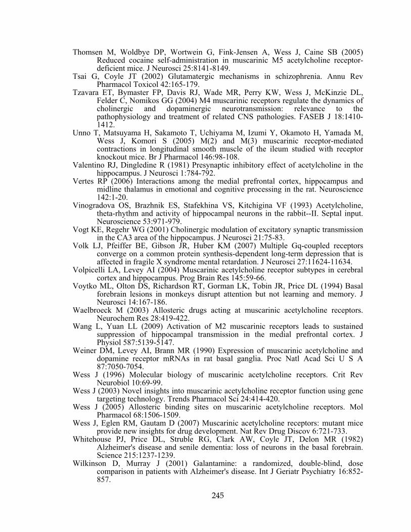
245
Thomsen M, Woldbye DP, Wortwein G, Fink-Jensen A, Wess J, Caine SB (2005) Reduced cocaine self-administration in muscarinic M5 acetylcholine receptor-deficient mice. J Neurosci 25:8141-8149.
Tsai G, Coyle JT (2002) Glutamatergic mechanisms in schizophrenia. Annu Rev Pharmacol Toxicol 42:165-179.
Tzavara ET, Bymaster FP, Davis RJ, Wade MR, Perry KW, Wess J, McKinzie DL, Felder C, Nomikos GG (2004) M4 muscarinic receptors regulate the dynamics of cholinergic and dopaminergic neurotransmission: relevance to the pathophysiology and treatment of related CNS pathologies. FASEB J 18:1410-1412.
Unno T, Matsuyama H, Sakamoto T, Uchiyama M, Izumi Y, Okamoto H, Yamada M, Wess J, Komori S (2005) M(2) and M(3) muscarinic receptor-mediated contractions in longitudinal smooth muscle of the ileum studied with receptor knockout mice. Br J Pharmacol 146:98-108.
Valentino RJ, Dingledine R (1981) Presynaptic inhibitory effect of acetylcholine in the hippocampus. J Neurosci 1:784-792.
Vertes RP (2006) Interactions among the medial prefrontal cortex, hippocampus and midline thalamus in emotional and cognitive processing in the rat. Neuroscience 142:1-20.
Vinogradova OS, Brazhnik ES, Stafekhina VS, Kitchigina VF (1993) Acetylcholine, theta-rhythm and activity of hippocampal neurons in the rabbit--II. Septal input. Neuroscience 53:971-979.
Vogt KE, Regehr WG (2001) Cholinergic modulation of excitatory synaptic transmission in the CA3 area of the hippocampus. J Neurosci 21:75-83.
Volk LJ, Pfeiffer BE, Gibson JR, Huber KM (2007) Multiple Gq-coupled receptors converge on a common protein synthesis-dependent long-term depression that is affected in fragile X syndrome mental retardation. J Neurosci 27:11624-11634.
Volpicelli LA, Levey AI (2004) Muscarinic acetylcholine receptor subtypes in cerebral cortex and hippocampus. Prog Brain Res 145:59-66.
Voytko ML, Olton DS, Richardson RT, Gorman LK, Tobin JR, Price DL (1994) Basal forebrain lesions in monkeys disrupt attention but not learning and memory. J Neurosci 14:167-186.
Waelbroeck M (2003) Allosteric drugs acting at muscarinic acetylcholine receptors. Neurochem Res 28:419-422.
Wang L, Yuan LL (2009) Activation of M2 muscarinic receptors leads to sustained suppression of hippocampal transmission in the medial prefrontal cortex. J Physiol 587:5139-5147.
Weiner DM, Levey AI, Brann MR (1990) Expression of muscarinic acetylcholine and dopamine receptor mRNAs in rat basal ganglia. Proc Natl Acad Sci U S A 87:7050-7054.
Wess J (1996) Molecular biology of muscarinic acetylcholine receptors. Crit Rev Neurobiol 10:69-99.
Wess J (2003) Novel insights into muscarinic acetylcholine receptor function using gene targeting technology. Trends Pharmacol Sci 24:414-420.
Wess J (2005) Allosteric binding sites on muscarinic acetylcholine receptors. Mol Pharmacol 68:1506-1509.
Wess J, Eglen RM, Gautam D (2007) Muscarinic acetylcholine receptors: mutant mice provide new insights for drug development. Nat Rev Drug Discov 6:721-733.
Whitehouse PJ, Price DL, Struble RG, Clark AW, Coyle JT, Delon MR (1982) Alzheimer's disease and senile dementia: loss of neurons in the basal forebrain. Science 215:1237-1239.
Wilkinson D, Murray J (2001) Galantamine: a randomized, double-blind, dose comparison in patients with Alzheimer's disease. Int J Geriatr Psychiatry 16:852-857.

246
Williams S, Johnston D (1990) Muscarinic depression of synaptic transmission at the hippocampal mossy fiber synapse. J Neurophysiol 64:1089-1097.
Yamada M, Lamping KG, Duttaroy A, Zhang W, Cui Y, Bymaster FP, McKinzie DL, Felder CC, Deng CX, Faraci FM, Wess J (2001a) Cholinergic dilation of cerebral blood vessels is abolished in M(5) muscarinic acetylcholine receptor knockout mice. Proc Natl Acad Sci U S A 98:14096-14101.
Yamada M, Miyakawa T, Duttaroy A, Yamanaka A, Moriguchi T, Makita R, Ogawa M, Chou CJ, Xia B, Crawley JN, Felder CC, Deng CX, Wess J (2001b) Mice lacking the M3 muscarinic acetylcholine receptor are hypophagic and lean. Nature 410:207-212.
Yan HD, Villalobos C, Andrade R (2009) TRPC Channels Mediate a Muscarinic Receptor-Induced Afterdepolarization in Cerebral Cortex. J Neurosci 29:10038-10046.
Zhang HM, Chen SR, Matsui M, Gautam D, Wess J, Pan HL (2006) Opposing functions of spinal M2, M3, and M4 receptor subtypes in regulation of GABAergic inputs to dorsal horn neurons revealed by muscarinic receptor knockout mice. Mol Pharmacol 69:1048-1055.
Zhang HM, Zhou HY, Chen SR, Gautam D, Wess J, Pan HL (2007) Control of glycinergic input to spinal dorsal horn neurons by distinct muscarinic receptor subtypes revealed using knockout mice. J Pharmacol Exp Ther 323:963-971.
Zhang W, Yamada M, Gomeza J, Basile AS, Wess J (2002a) Multiple muscarinic acetylcholine receptor subtypes modulate striatal dopamine release, as studied with M1-M5 muscarinic receptor knock-out mice. J Neurosci 22:6347-6352.
Zhang W, Basile AS, Gomeza J, Volpicelli LA, Levey AI, Wess J (2002b) Characterization of central inhibitory muscarinic autoreceptors by the use of muscarinic acetylcholine receptor knock-out mice. J Neurosci 22:1709-1717.
Zhong P, Gu Z, Wang X, Jiang H, Feng J, Yan Z (2003) Impaired modulation of GABAergic transmission by muscarinic receptors in a mouse transgenic model of Alzheimer's disease. J Biol Chem 278:26888-26896.