Embed Size (px)
Citation preview
Received: 11th July-2012 Revised: 13th July-2012 Accepted: 15th July-2012 Research article
PRODUCTION AND CYTOTOXICITY STUDIES OF LOVASTATIN FROM Aspergillus niger PN2 AN ENDOPHYTIC FUNGI ISOLATED FROM Taxus baccata
Raghunath R1,3, Radhakrishna A1 , Angayarkanni J2, Palaniswamy M3
1Shriram Institute for Industrial Research, Bangalore – 560 048, India2Department of Microbial Biotechnology, School of Biotechnology, Bharathiar University, Coimbatore - 641 046,
Tamil Nadu, India3Department of Microbiology, School of Life Sciences, Karpagam University, Coimbatore - 641 021, Tamil
Nadu, IndiaCorresponding author e-mail id: [email protected]
ABSTRACT: Aspergillus niger PN2 an endophytic fungus, was isolated from the healthy tissues of Taxus baccata. The fungus was screened for the production of lovastatin on a solid state fermentation with wheat bran as a substrate. The fungal species were identified by their characteristic cultural morphology and molecular analysis. The presence of lovastatin was confirmed by spectroscopic method, nuclear magnetic resonance (NMR), thin layer chromatography (TLC) and high performance liquid chromatography (HPLC) methods of analysis. The lovastatin production was quantified by ultraviolet (UV) analysis. The maximum amount of lovastatin production was recorded as 1.5 mg/g substrate. The extracted fungal lovastatin demonstrate a strong cytotoxic activity in in vitro culture of tested human cancer cells (HeLa and HepG2) by apoptotic assay. These results designate that the fungus, A. niger PN2 is an excellent candidate for lovastatin production and can serve as a potential organism for genetic engineering to enhance the production of lovastatin to a higher level.Keywords: Lovastatin, Endophytic fungus Aspergillus niger, cancer, cytotoxicity test.
INTRODUCTIONFungi are important sources for the production of several pharmaceutical compounds viz., lovastatin, mevinolin and monacolin K. Lovastatin (C24H36O5) is a potent drug for lowering blood cholesterol. Lovastatin acts by competitively inhibiting the enzyme 3-hydroxy-3-methylglutaryl coenzyme A reductase (HMG-CoA) which catalyzes the rate-limiting step of cholesterol biosynthesis (Alberts et al., 1980). Lovastatin is produced as a secondary metabolite by the fungi Penicillium sp. (Endo et al., 1976) Monascus ruber (Juzlova et al., 1996), and Aspergillus terreus (Alberts et al., 1980). A. terreus appears to be the most commonly used producer of this drug (Novak et al., 1997). Now-a-days lovastatin and its semisynthetic derivatives are very important drugs since the mortality of heart disease is becoming relatively high.A novel molecular target with strong potential for rapid application to the clinic is the rate limiting enzyme of the mevalonate pathway, HMG-CoA reductase. The end products of the mevalonate pathway are required for a number of cellular functions. These include sterols, such as cholesterol, involved in membrane integrity and steroid production, ubiquinone involved in electron transport and cellular respiration, farnesyl and geranylgeranyl isoprenoids involved in covalent binding of proteins such as the Ras family to membranes, dolichol which is required for glycoprotein synthesis and isopentenyladenine, essential for certain tRNA function and protein synthesis (Olson and Rudney., 1983; Goldstein and Brown, 1990; Russel,1992; Sinensky, 2000). Thus, HMG-CoA reductase is a unique molecular target for anticancer therapy; it holds a pivotal role in the well defined mevalonate pathway, and a specific family of inhibitors is available for immediate application in the cancer therapy. Inhibitors of this key enzyme, collectively known as statins, are well established and are effective agents used in the treatment of hypercholesterolemia (Farnier, 1998; Pedersen, 2001).
Recent evidence shows that statin not only are able to reduce cardiac disease related mortality, but cancer incidence is also reduced by 28-33 % (Blais et al., 2000). Lovastatin has shown great promise as a cholesterol lowering agent and they also suppress a variety of leukemic cell lines and a wide array of solid tumors cells in vivo, by inhibiting the synthesis of non-sterol isoprenoid compounds.
International Journal of Applied Biology and Pharmaceutical Technology Page: 342 Available online at www.ijabpt.com

Palaniswamy et al
Apart from lovastatin other statins are shown to be cytotoxic or induce apoptosis in leukemia cell lines (Dimitroulakas et al., 2000) prostate cancer cell lines (Maltese et al., 1985), colon cancer (Agarwal et al, 1999), pancreatic cancer cell line (Muller et al., 1998). In the present study lovastatin was evaluated for potential cytotoxicity, against two tumor cell lines viz. human cervical cancer cell line (HeLa cell line) and liver hepato carcinoma cell lines (HepG2). As a consequence of its importance, improving the production of lovastatin has become the focus of renewed research interest. Over the last ten years, endophytic fungi isolated from yews and other gymnosperm plant species were given sole attention for their bioactive products. In the present study, an attempt was made to screen lovastatin producing endophytic fungi from Taxus baccata.
MATERIALS AND METHODS Isolation and identification of endophytic fungiEndophytic fungi were isolated from T. baccata tissues obtained from central Himalayas, India. The plant parts (leaves, bark, stem) were surface sterilized with Tween-80 for an hour and treated with 70 % (v/v) ethanol for 15 sec. These sterilized fragments were then treated with sodium hypochlorite for 15 min and washed with sterile water. Treated tissues were cut into small pieces (0.5 × 0.5 cm), and placed on potato dextrose agar (PDA) amended with chloramphenicol 150 mg/L to decrease the amount of bacterial contamination. After several days, fungi were observed, and individual hyphal tips of the various fungi were removed and placed on a new PDA medium, and incubated at 25 °C for at least 2 weeks (Strobel et al., 1996). The fungus was identified by its morphological and conidial features in the culture growth. Genomic DNA was isolated from the selected fungal sample/culture provided using Sigma Aldrich DNA extraction Kit. Its quality was evaluated on 0.8% agarose gel a single band of high-molecular weight DNA was observed. D1/D2 region of LSU (Large subunit 28S rDNA) gene was amplified by PCR from the above isolated genomic DNA. A single discrete band was observed when resolved on agarose gel. The PCR amplicon was purified by column purification in order to remove contaminants. DNA sequencing was carried out with PCR amplicon. The 28S rDNA sequences D1/D2 region was amplified by PCR from fungal genomic DNA using PCR universal primers: DR-5'-GGTCCGTGTTTCAAGACGG-3' and DF- 5'-ACCCGCTGAACTTAAGC-3' respectively. PCR reactions were carried out as one cycle of heat treatment at 95 oC for 5 min, a total of 30 cycles of denaturation at 94o C for 30 sec, annealing at 55 oC for 30 sec, extension at 72o C for 45 sec, followed by a final extension at 72 oC for 10 min. The PCR products were stored at 4 oC, later analyzed by 0.8% agarose gel electrophoresis, and then sequenced. The D1/D2 region of LSU (Large subunit 28S rDNA) gene sequence was used to carry out BLAST with the nrdatabase of NCBI genbank database.Screening of lovastatin producing strainsThe spore suspension was prepared by adding 5 mL of suspension medium (0.9% NaCl, 0.1% Tween 80) and vigorously shaking for 1 min. The spore suspension containing 108 spores/ mL was used as the inoculum (Sathya et al., 2009). Solid state fermentation with wheat bran was performed with the isolated 57 fungal species. Solid substrate was dried at hot air oven at 50 °C, accurately weighed to 5g in Petri dishes (100 mm x 17 mm), moistened 65% with distilled water, pH maintained at 6.5 and autoclaved at 121 °C for 15 min. After cooling, the medium was inoculated with 10% (v/w) of spore suspension. Medium was thoroughly mixed and incubated at 28 °C in a humidity controlled incubator for 10 days. Extraction of lovastatinAt the end of solid state fermentation (SSF), the fermented material was dried at 50 °C for 24 h and powdered. The powdered (2g) material was extracted with ethyl acetate (pH 3.0) in 250 mL Erlenmeyer’s flasks. It was then incubated at 28 °C in rotary shaker at 200 rpm for 2 h. After 2 h, mixture was centrifuged at 10,000 rpm for 10 min and supernatant was filtered through membrane filter with a pore size of 0.45μm. This supernatant was used for further chromatographic and spectroscopic analysis.Quantitative analysis of lovastatinTo 1 mL of the supernatant, 1 mL of trifluroacetic acid (1%) was added and incubated for 10 min (Lactonization of hydroxyl acid form of lovastatin). From the above solution, 0.5 mL was taken and diluted 10 times with methanol and its absorbance was read at 238 nm by using UV-Visible Spectrophotometer (Mielcarek et al., 2009).
International Journal of Applied Biology and Pharmaceutical Technology Page: 343 Available online at www.ijabpt.com

Palaniswamy et al
Thin layer chromatographic analysisThe residue after filtration was subjected to thin layer chromatography. The standard and sample spots were spotted on the sample line and developed with cyclohexane: chloroform: isopropanol (5:2:1). High performance liquid chromatographic analysisLovastatin present in the extraction (10 mL) was lactonized with 10 mL of trifluoroacetic acid (1%) (Sayyad et al., 2007). Extracts were dried and concentrated under reduced pressure. Finally, acetonitrile was added for high performance liquid chromatography (HPLC) analysis (RPHPLC) system (Agilent 1100 series) using Novapak C18 column (150 mm length x 3.9 mm ID). Mobile phase consisted of acetonitrile and water (pH 4.4 adjusted using phosphoric acid) with a volume ratio of v/v=65:35 and with the flow rate of 1.0 mL/min and lovastatin was detected at 238 nm.Purification of lovastatinUnlactonized crude lovastatin extract was concentrated (10 mL) under reduced pressure to get viscous mass (Skoog et al., 1998). The slurry of crude lovastatin extract was then prepared by adding silica gel of 60-120-mesh size in a porcelain dish, with continuous stirring until it was adsorbed on silica gel and dried completely (Skoog et al., 1998). Elution of the column was performed in a stepwise manner starting with 70 mL of 100% dichloromethane, followed by different ratios of dichloromethane-ethyl acetate. (viz. 20:1, 10:1, 6:1, 3:1 and 1:1 v/v). The fractions with the same mobility as standard were combined and evaporated to dryness. Acetonitrile was added drop wise until it was solubilized. The solution was kept for one month at low temperature (4 ºC) for crystallization. After the crystallization process petroleum ether, in which the desired compound was insoluble, was added and then decanted to get lovastatin crystals. Washing with petroleum ether was done to remove the impurities from the crystals. Finally, the pure crystals recovered were dried in desiccators and weighed (Mendham et al, 2002). The purified lovastatin crystals was analyzed for NMR and cytotoxic studies.Structure analysisThe structures were identified by 1H NMR, 13C NMR. The purified lovastatin was dissolved in chloroform for NMR experiments, which were carried out as described by Berger and Baun, (2004). The NMR spectrum was recorded using a Varian Mercury plus 500 MHz.Cytotoxic assaysCell treatment procedureThe monolayer cells were detached with trypsin-ethylenediaminetetraacetic acid (EDTA) to make single cell suspensions and viable cells were counted using a hemocytometer and diluted with medium with 5% FBS to give final density of 105
cells/mL. One hundred microlitres per well of cell suspension were seeded into 96-well plates at plating density of 10,000 cells/well and incubated to allow for cell attachment at 37 oC, CO2 (5%) air (95%) and 100% relative humidity. After 24 h the cells were treated with serial concentrations of the extracts and fractions. They were initially dissolved in dimethylsulfoxide (DMSO) and further diluted in serum free medium to produce five concentrations. One hundred microlitres per well of each concentration was added to plates to obtain final concentrations of 100, 50, 25, 12.5 and 6.25 µM. The final volume in each well was 200 µl and the plates were incubated at 37 oC, CO2 (5%) air (95%) and 100% relative humidity for 48 h. The medium containing without samples were served as control. Triplicate was maintained for all concentrations. The percent cell inhibition was determined using the following formula. Cell inhibition (%) = 100- Abs (sample)/Abs (control) x100. Nonlinear regression graph was plotted between percent cell inhibition and Log10 concentration and IC50 was determined using GraphPad Prism software (Mosmann , 1983; Monks et al., 1991).
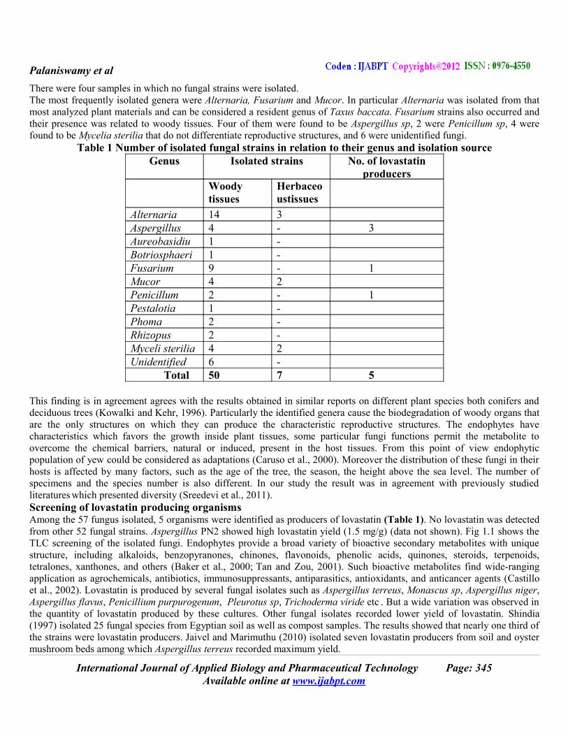
RESULTS AND DISCUSSIONIsolation of endophytic fungiFor the studies, the plant segments (bark, stems and leaves) were collected from Taxus baccata (known as Himalayan yew) trees grown in a homogenous (uniform) environment at Jageshwar, Almora district, Central Himalaya, India. Endophytic fungal strains were recovered from most of the analyzed tissues (bark, stems and leaves). Endophytes were more frequent in woody tissues (bark and stems) rather than herbaceous ones (leaves). In the first type of tissues 50 pure cultures of fungi were collected and 7 in the second one (Table 1).
International Journal of Applied Biology and Pharmaceutical Technology Page: 344 Available online at www.ijabpt.com
Palaniswamy et al
There were four samples in which no fungal strains were isolated. The most frequently isolated genera were Alternaria, Fusarium and Mucor. In particular Alternaria was isolated from that most analyzed plant materials and can be considered a resident genus of Taxus baccata. Fusarium strains also occurred and their presence was related to woody tissues. Four of them were found to be Aspergillus sp, 2 were Penicillum sp, 4 were found to be Mycelia sterilia that do not differentiate reproductive structures, and 6 were unidentified fungi.
Table 1 Number of isolated fungal strains in relation to their genus and isolation sourceGenus Isolated strains
No. of lovastatin
producersWoody tissues
Herbaceoustissues
Alternaria 14 3Aspergillus 4 - 3Aureobasidium
1 -Botriosphaeria
1 -Fusarium 9 - 1Mucor 4 2Penicillum 2 - 1Pestalotia 1 -Phoma 2 -Rhizopus 2 -Myceli sterilia 4 2Unidentified 6 -
Total 50 7 5
This finding is in agreement agrees with the results obtained in similar reports on different plant species both conifers and deciduous trees (Kowalki and Kehr, 1996). Particularly the identified genera cause the biodegradation of woody organs that are the only structures on which they can produce the characteristic reproductive structures. The endophytes have characteristics which favors the growth inside plant tissues, some particular fungi functions permit the metabolite to overcome the chemical barriers, natural or induced, present in the host tissues. From this point of view endophytic population of yew could be considered as adaptations (Caruso et al., 2000). Moreover the distribution of these fungi in their hosts is affected by many factors, such as the age of the tree, the season, the height above the sea level. The number of specimens and the species number is also different. In our study the result was in agreement with previously studied literatures which presented diversity (Sreedevi et al., 2011).Screening of lovastatin producing organismsAmong the 57 fungus isolated, 5 organisms were identified as producers of lovastatin (Table 1). No lovastatin was detected from other 52 fungal strains. Aspergillus PN2 showed high lovastatin yield (1.5 mg/g) (data not shown). Fig 1.1 shows the TLC screening of the isolated fungi. Endophytes provide a broad variety of bioactive secondary metabolites with unique structure, including alkaloids, benzopyranones, chinones, flavonoids, phenolic acids, quinones, steroids, terpenoids, tetralones, xanthones, and others (Baker et al., 2000; Tan and Zou, 2001). Such bioactive metabolites find wide-ranging application as agrochemicals, antibiotics, immunosuppressants, antiparasitics, antioxidants, and anticancer agents (Castillo et al., 2002). Lovastatin is produced by several fungal isolates such as Aspergillus terreus, Monascus sp, Aspergillus niger, Aspergillus flavus, Penicillium purpurogenum, Pleurotus sp, Trichoderma viride etc . But a wide variation was observed in the quantity of lovastatin produced by these cultures. Other fungal isolates recorded lower yield of lovastatin. Shindia (1997) isolated 25 fungal species from Egyptian soil as well as compost samples. The results showed that nearly one third of the strains were lovastatin producers. Jaivel and Marimuthu (2010) isolated seven lovastatin producers from soil and oyster mushroom beds among which Aspergillus terreus recorded maximum yield.
International Journal of Applied Biology and Pharmaceutical Technology Page: 345 Available online at www.ijabpt.com

Palaniswamy et al
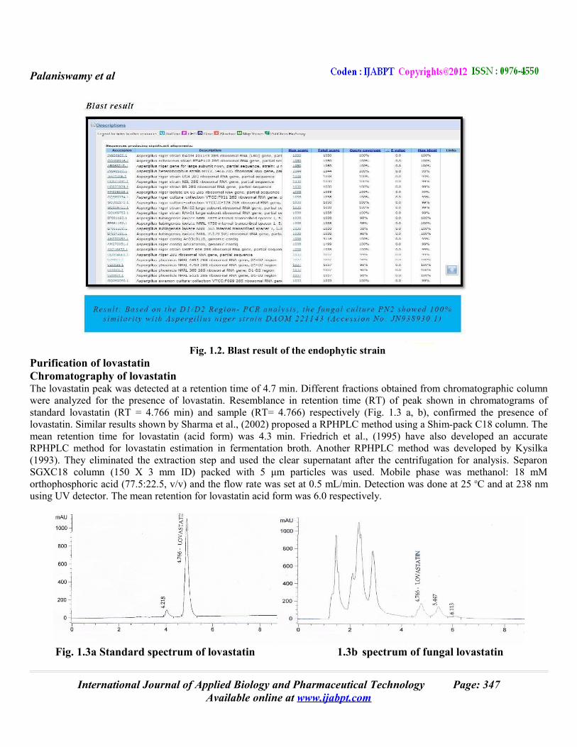
Similarly 79 organisms were screened for lovastatin producing potential by Neurospora crassa bioassay (Sreedevi et al., 2011). Twenty three fungal species of ten genera isolated from soil were screened for lovastatin production on oat meal production medium. The results showed that 56% of the isolates were positive for lovastatin production Osman et al., 2011). From the results obtained during the present study 5 organisms were identified by TLC screening (Fig 1) and quantitative analysis by UV spectrophotometer (data not shown) as producers of lovastatin by solid state fermentation of wheat bran. The fungi A. niger strain PN2 producing the highest lovastatin content was selected for further studies.The spore-producing filamentous fungi were detected and identified to the genus and species levels based on morphological characteristics (Raper and Fennel, 1965; Qi and Kong, 1997; Klich, 2002). Culture and collection of mycelium were carried out as previously described (Zhao et al., 2004). For sequence analysis, DNA was extracted from lovastatin-producing fungus PN2 and identified using methods previously described. The sequences obtained were submitted to GenBank for homology search with Blast (http:// rdp.cme.msu.edu and http ⁄ ⁄ ncbi.nim.nih.gov). The sequences of the 28S rDNA were aligned with those of related fungal strains retrieved from the GenBank databases using ClustalX. Based on the D1/D2 Region- PCR analysis , the fungal culture PN2 showed 100% similarity with Aspergillus niger strain DAOM 221143 (Accession No: JN938930.1). The endophytic fungus Aspergillus niger from Taxus baccata has never been reported to be able to produce lovastatin. In addition, Aspergillus niger var. taxi, a new species variant of taxol-producing fungus isolated from Taxus cuspidata in China was reported by Zhao et al (2009) which confirmed that Aspergillus niger was an endophytic fungi whose genome has been recently sequenced. Annotation of genomic sequences suggested that it also has lovastatin biosynthetic gene cluster. Our present study is the first report for the isolation, characterization and identification of a new variant of the Aspergillus niger from Taxus baccata in India that is able to produce lovastatin at a high amount in SSF.
Fig. 1.1 TLC screening of the isolated fungi.Identification and characterization of lovastatin producing strain A. niger PN2Consensus sequence data of culture Aspergillus niger PN2: (568 bp)GGATTGCCTCAGTAACGGCGAGTGAAGCGGCAAGAGCTCAAATTTGAAAGCTGGCTCCTTCGGAGTCCGCATTGTAATTTGCAGAGGATGCTTTGGGTGCGGCCCCCGTCTAAGTGCCCTGGAACGGGCCGTCAGAGAGGGTGAGAATCCCGTCTTGGGCGGGGTGTCCGTGCCCGTGTAAAGCTCCTTCGACGAGTCGAGTTGTTTGGGAATGCAGCTCTAAATGGGTGGTAAATTTCATCTAAAGCTAAATACTGGCCGGAGACCGATAGCGCACAAGTAGAGTGATCGAAAGATGAAAAGCACTTTGAAAAGAGAGTTAAACAGCACGTGAAATTGTTGAAAGGGAAGCGCTTGCGACCAGACTCGCCCGCGGGGTTCAGCCGGCATTCGTGCCGGTGTACTTCCCCGTGGGCGGGCCAGCGTCGGTTTGGGCGGCCGGTCAAAGGCCCCTGGAATGTAGTACCCTCCGGGGTACCTTATAGCCAGGGGTGCAATGCGGCCAGCCTGGACCGAGGAACGCGCTTCGGCACGGACGCTGGCATAATGGTCGTAAACGACCCGTCTT
International Journal of Applied Biology and Pharmaceutical Technology Page: 346 Available online at www.ijabpt.com
Palaniswamy et al
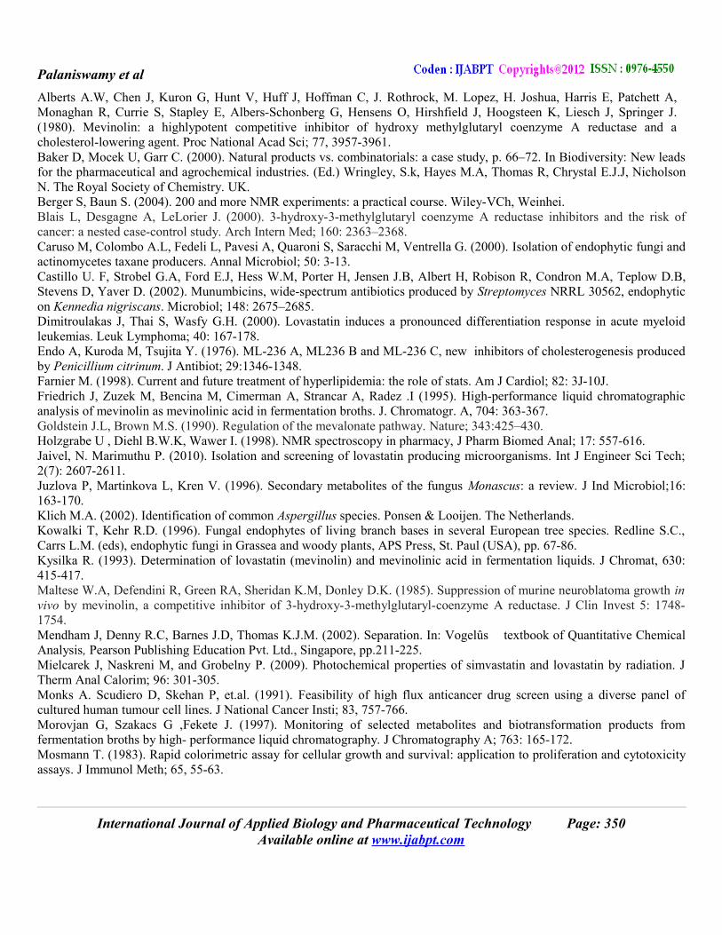
Fig. 1.2. Blast result of the endophytic strain Purification of lovastatinChromatography of lovastatinThe lovastatin peak was detected at a retention time of 4.7 min. Different fractions obtained from chromatographic column were analyzed for the presence of lovastatin. Resemblance in retention time (RT) of peak shown in chromatograms of standard lovastatin (RT = 4.766 min) and sample (RT= 4.766) respectively (Fig. 1.3 a, b), confirmed the presence of lovastatin. Similar results shown by Sharma et al., (2002) proposed a RPHPLC method using a Shim-pack C18 column. The mean retention time for lovastatin (acid form) was 4.3 min. Friedrich et al., (1995) have also developed an accurate RPHPLC method for lovastatin estimation in fermentation broth. Another RPHPLC method was developed by Kysilka (1993). They eliminated the extraction step and used the clear supernatant after the centrifugation for analysis. Separon SGXC18 column (150 X 3 mm ID) packed with 5 μm particles was used. Mobile phase was methanol: 18 mM orthophosphoric acid (77.5:22.5, v/v) and the flow rate was set at 0.5 mL/min. Detection was done at 25 oC and at 238 nm using UV detector. The mean retention for lovastatin acid form was 6.0 respectively.
Fig. 1.3a Standard spectrum of lovastatin 1.3b spectrum of fungal lovastatin
International Journal of Applied Biology and Pharmaceutical Technology Page: 347 Available online at www.ijabpt.com

Palaniswamy et al
Fig. 1.4a. 1H NMR spectrum of fungal lovastatin 1.4b. 13C NMR spectrum of fungal lovastatin
Morovjan et al, (1997) developed a method in which fermentation broth was mixed directly with acetonitrile and H3PO4 was added to maintain the acidic condition. A Novapak C18 column (150 X 3.9 mm ID) with packed particles of 4 μm size was used. A 1:1 mixture of acetonitrile and 0.1 % phosphoric acid was used as a mobile phase and the flow rate was set at 1.5 mL/min. Detection was done at 25 oC at 235 nm. The injection volume was 10 μL and the detection limit was 50ng/mL. The mean retention time for lovastatin acid form was 4.7 min .NMR determination of lovastatin showed resonances at δ 6.05-6.01 ppm, δ 5.90-5.84 ppm, δ 5.61-5.56 ppm, δ 5.37-5.32 ppm, δ 4.39-4.34 ppm (mid-field region) and δ 2.85-2.76 ppm, δ 2.63-2.56 ppm, δ 2.44-2.33 ppm, δ 1.98-1.93 ppm, δ 1.64-1.57 ppm, δ 0.91-0.85 ppm (aliphatic range) (Holzgrabe et al., 1998). However, the resonances in the aliphatic range were unsuitable for quantification because they showed strong overlap with matrix compounds considering signals in the mid- field region (Figure 1.4a, b), we used the multiplet at δ 5.37-5.32 ppm for quantification because this led to the best sensitivity and this signal was not interfered in any case in our samples. Thus, more advanced techniques, such as multivariate regression or curve deconvolution, were not required. While the conditions were not directly comparable to any of these studies, the literature data along with the spectral prediction showed that the multiplet at δ 5.37-5.32 ppm clearly belonged to an H-atom at the hexahydronaphthalene moiety, most probably H6, H4 or an overlap of the signals of both atoms (Fig. 1.4 a, b). The 1H NMR spectral data of lovastatin was found to be in concurrence with reported spectral data (Alarcon et al., 2003; Patel and Patel, 2007). (A) (B) (C) (D)
(E)
(E) (F) (G)
Fig. 1.5. HeLa cell line sample concentration (A) Control, (B) 6.25 µM, (C) 12.5 µM, (D) 25 µM, (E) 50 µM and (F) 100 µM (G) Growth inhibition (%)
International Journal of Applied Biology and Pharmaceutical Technology Page: 348 Available online at www.ijabpt.com
0.0 0.5 1.0 1.5 2.0 2.50
20
40
60
80
100
Log10 concentration (uM)
% G
row
th In
hib
itio
n

Palaniswamy et al
(A) (B) (C) (D)
(E)
(E) (F) (G)
Fig 1.6. HepG2 cell line sample concentration (A) Control, (B) 6.25 µM, (C) 12.5 µM, (D) 25 µM, (E) 50 µM and (F) 100 µM. (G) Growth inhibition (%)
Cytotoxic assayCytotoxicity is one of the properties of antitumor agents (Suffness and Pezzuto, 1991). The effective concentration of 50% cell death (EC50) was found to be 23.58µM for the human cervical cancer cell line (HeLa cell line) and 39.77µM for liver hepatocarcinoma cell lines (HepG2) respectively (Fig 1.5 and 1.6). These results indicated that lovastatin is a strong cytotoxic agent which partially explains its antitumor activity and can be suggested for therapeutic use as a cancer chemo preventive agent. Recent evidence shows that statin is not only capable of reducing cardiac disease related mortality, but cancer incidence is also reduced by 28-33 % (Blais et al., 2000). Apart from lovastatin other statins are shown to be cytotoxic or induce apoptosis in leukemia cell lines (Dimitroulakas et al., 2000), prostate cancer cell lines (Maltese et al., 1985), colon cancer (Agarwal et al, 1999), pancreatic cancer cell line (Muller et al., 1998).
CONCLUSIONThus far, lovastatin production has only been reported from soil fungi A. terreus. Whereas, in this investigation we documented the isolation and production of lovastatin by an endophytic fungi A. niger for the first time. This investigation evidently confirmed through spectroscopic and chromatographic analysis that the lovastatin purified was identical to the authentic standard lovastatin. Moreover, the amount of lovastatin production from endophytic fungus A. niger PN2 reported was found to be higher than those of already the reported fungi A. terreus. This fungus can serve as a potential species for genetic engineering and biotransformations of lovastatin in order to enhance the production of lovastatin.
REFERENCESAgarwal B, Bhendwal S, Halmos B. (1999). Lovastatin augments apoptosis induced by hemotherapeutic agents in colon cancer cells. Clinical Cancer Res; 5:2223-2229.Alarcon J. Aguila S, Avila P.A, Fuentes O, Ponce E. Z and Hernandez M. (2003). Production and purification of statins from Pleurotus ostreatus (basidiomycetes) strains, Z. Naturforsch; 58c: 62-64.
International Journal of Applied Biology and Pharmaceutical Technology Page: 349 Available online at www.ijabpt.com
0.0 0.5 1.0 1.5 2.0 2.50
20
40
60
80
100
Log10 concentration (uM)
% G
row
th In
hib
itio
n
Palaniswamy et al
Alberts A.W, Chen J, Kuron G, Hunt V, Huff J, Hoffman C, J. Rothrock, M. Lopez, H. Joshua, Harris E, Patchett A, Monaghan R, Currie S, Stapley E, Albers-Schonberg G, Hensens O, Hirshfield J, Hoogsteen K, Liesch J, Springer J. (1980). Mevinolin: a highlypotent competitive inhibitor of hydroxy methylglutaryl coenzyme A reductase and a cholesterol-lowering agent. Proc National Acad Sci; 77, 3957-3961.Baker D, Mocek U, Garr C. (2000). Natural products vs. combinatorials: a case study, p. 66–72. In Biodiversity: New leads for the pharmaceutical and agrochemical industries. (Ed.) Wringley, S.k, Hayes M.A, Thomas R, Chrystal E.J.J, Nicholson N. The Royal Society of Chemistry. UK.Berger S, Baun S. (2004). 200 and more NMR experiments: a practical course. Wiley-VCh, Weinhei.Blais L, Desgagne A, LeLorier J. (2000). 3-hydroxy-3-methylglutaryl coenzyme A reductase inhibitors and the risk of cancer: a nested case-control study. Arch Intern Med; 160: 2363–2368. Caruso M, Colombo A.L, Fedeli L, Pavesi A, Quaroni S, Saracchi M, Ventrella G. (2000). Isolation of endophytic fungi and actinomycetes taxane producers. Annal Microbiol; 50: 3-13.Castillo U. F, Strobel G.A, Ford E.J, Hess W.M, Porter H, Jensen J.B, Albert H, Robison R, Condron M.A, Teplow D.B, Stevens D, Yaver D. (2002). Munumbicins, wide-spectrum antibiotics produced by Streptomyces NRRL 30562, endophytic on Kennedia nigriscans. Microbiol; 148: 2675–2685.Dimitroulakas J, Thai S, Wasfy G.H. (2000). Lovastatin induces a pronounced differentiation response in acute myeloid leukemias. Leuk Lymphoma; 40: 167-178. Endo A, Kuroda M, Tsujita Y. (1976). ML-236 A, ML236 B and ML-236 C, new inhibitors of cholesterogenesis produced by Penicillium citrinum. J Antibiot; 29:1346-1348.Farnier M. (1998). Current and future treatment of hyperlipidemia: the role of stats. Am J Cardiol; 82: 3J-10J.Friedrich J, Zuzek M, Bencina M, Cimerman A, Strancar A, Radez .I (1995). High-performance liquid chromatographic analysis of mevinolin as mevinolinic acid in fermentation broths. J. Chromatogr. A, 704: 363-367.Goldstein J.L, Brown M.S. (1990). Regulation of the mevalonate pathway. Nature; 343:425–430.Holzgrabe U , Diehl B.W.K, Wawer I. (1998). NMR spectroscopy in pharmacy, J Pharm Biomed Anal; 17: 557-616. Jaivel, N. Marimuthu P. (2010). Isolation and screening of lovastatin producing microorganisms. Int J Engineer Sci Tech; 2(7): 2607-2611. Juzlova P, Martinkova L, Kren V. (1996). Secondary metabolites of the fungus Monascus: a review. J Ind Microbiol;16: 163-170.Klich M.A. (2002). Identification of common Aspergillus species. Ponsen & Looijen. The Netherlands. Kowalki T, Kehr R.D. (1996). Fungal endophytes of living branch bases in several European tree species. Redline S.C., Carrs L.M. (eds), endophytic fungi in Grassea and woody plants, APS Press, St. Paul (USA), pp. 67-86. Kysilka R. (1993). Determination of lovastatin (mevinolin) and mevinolinic acid in fermentation liquids. J Chromat, 630: 415-417. Maltese W.A, Defendini R, Green RA, Sheridan K.M, Donley D.K. (1985). Suppression of murine neuroblatoma growth in vivo by mevinolin, a competitive inhibitor of 3-hydroxy-3-methylglutaryl-coenzyme A reductase. J Clin Invest 5: 1748-1754. Mendham J, Denny R.C, Barnes J.D, Thomas K.J.M. (2002). Separation. In: Vogelûs textbook of Quantitative Chemical Analysis, Pearson Publishing Education Pvt. Ltd., Singapore, pp.211-225. Mielcarek J, Naskreni M, and Grobelny P. (2009). Photochemical properties of simvastatin and lovastatin by radiation. J Therm Anal Calorim; 96: 301-305. Monks A. Scudiero D, Skehan P, et.al. (1991). Feasibility of high flux anticancer drug screen using a diverse panel of cultured human tumour cell lines. J National Cancer Insti; 83, 757-766. Morovjan G, Szakacs G ,Fekete J. (1997). Monitoring of selected metabolites and biotransformation products from fermentation broths by high- performance liquid chromatography. J Chromatography A; 763: 165-172. Mosmann T. (1983). Rapid colorimetric assay for cellular growth and survival: application to proliferation and cytotoxicity assays. J Immunol Meth; 65, 55-63.
International Journal of Applied Biology and Pharmaceutical Technology Page: 350 Available online at www.ijabpt.com
Palaniswamy et al
Muller C, Bockhorn A.G, Klusmeier S, Kiehl M, Roeder C, Kalthoff H, Koch O.M. (1998). Lovastatin inhibits proliferation of pancreatic cancer cell lines with mutant as well as with wild type K-ras oncogene but has different effects o protein phosphorylation and induction of apoptosis. Int J Oncol 12: 717-723. Novak N. Gerdin S, Berovic M. (1997). Increased lovastatin formation by Aspergillus terreus using repeated fed-batch process. Biotechnol Lett; 19: 947-948.Olson RE, Rudney H. (1983). Biosynthesis of ubiquinone. Vitam Horm 40: 1-43.Osman M.E, Khattab O.H , Zaghlol G.M , Abd El-Hameed, R.M .(2011). Screening for the production of cholesterol lowering drugs lovastatin by some fungi. Aust J Basic Appl Sci; 5(6): 698-703. Patel R.P, Patel M.M. (2007). Preparation and evaluation of inclusion complex of the lipid lowering drug lovastatin with β-cyclodextrin, Dhaka Univ. J. Pharm. Sci. 6: 25-36. Pedersen TR (2001). Pro and con: low density lipoprotein cholesterol lowering is and will be key to the future of lipid management. Am J Cardiol; 87: 8B-12B. Qi Z.T, Kong H.Z. (1997). Flora Fungorum Sinicorum –Aspergillus et Teleomorphi Cognati. Beijing: Science Press. Raper K.B. and Fennell D.I (1965). The Genus Aspergillus. Baltimore: Williams & Wilkins. Russel D.W.(1992). Cholesterol biosynthesis and metabolism. Cardiovasc Drugs Ther; 6: 103-110. Sathya R, Pradeep, B.V, Angayarkanni J, Palaniswamy M. (2009). Production of milk clotting protease by a local isolate of Mucor circinelloides unde SSF using agro-industrail wates. Biotech Bioprocess Eng; 14: 788-794. Sharma P, Chawla H.P.S, Panchagnula R. (2002). Analytical method for monitoring concentrations of cyclosporin and lovastatin In vitro in an everted rat intestinal sac absorption model. J Chroma B; 768: 349-359.Shindia, A A. (1997). Mevinolin production by some fungi. Folia Microbiol; 42: 477-480. Sinensky M. (2000). Recent advances in the study of prenylated proteins. Biochem Biophys Acta; 1484: 93-106. Skoog D.A, HollerF.J, Nieman T.A. (1998). Separation methods. In: Principles of Instrumental Analysis, Saunders College Publishing, Philadelphia, USA, pp.674-700. Sreedevi K , VenkateswaraRao J, Lakshmi N, Fareedullah M. (2011). Strain improvement of Aspergillus terreus for the enhanced production of lovastatin, a HMG-COA reductase inhibitor. J Microbiol Biotech Res; 1 (2):96-100. Strobel G, Yang X.S, Sears J, Kramer R, Sidhu R.S, Hess W.M (1996). Taxol from Pestalotiopsis microspora, an endophytic fungus of Taxus wallachiana. Microbiol; 142: 435–440. Suffness M, Pezzuto J.M. (1991). Assays related to cancer drug discovery. In: suffness M, Pezzuto JM, editors. Methods in plant biochemistry New York’ Academic Press; P. 71-110. Tan, R. X., Zou W.X. (2001). Endophytes: a rich source of functional metabolites. Nat. Prod. Rep. 18:448–459. Wei Pei-lian, Xu Zhi-nan, Cen Pei-lin ( 2007). Lovastatin production by A. terreus in solid state fermentation. Zhejiang University Sci A; 8(9) 1521-1526. Zhao K, Ping W, Li Q, Hao S, Zhao L, Gao T .D. (2009). Zhou Aspergillus niger var. taxi, a new species variant of taxol-producing fungus isolated from Taxus cuspidata in China. J App Microbiol; 1364-5072. Zhao K, Zhou D.P, Ping W.X, Ge J.P. (2004). Study on preparation and regeneration of protoplast from taxol producing fungus Nodulisporum sylviforme. Nature Sci 2: 52–59.
International Journal of Applied Biology and Pharmaceutical Technology Page: 351 Available online at www.ijabpt.com