Embed Size (px)
Citation preview
Loop-mediated Isothermal gene Amplification (LAMP):
Towards Field Molecular Diagnosis of African Trypanosomosis
Oriel M. M. Thekisoe
National Research Center for Protozoan DiseasesObihiro University of Agriculture and Veterinary Medicine
Obihiro, Hokkaido, Japan
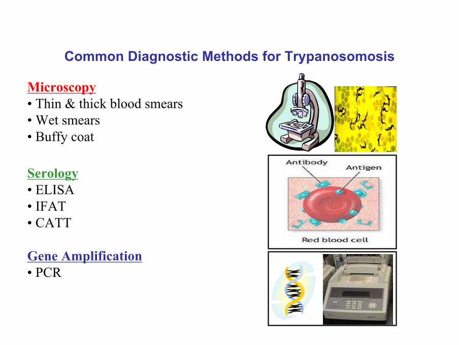
Common Diagnostic Methods for Trypanosomosis
Gene Amplification• PCR
Serology• ELISA• IFAT• CATT
Microscopy• Thin & thick blood smears• Wet smears• Buffy coat
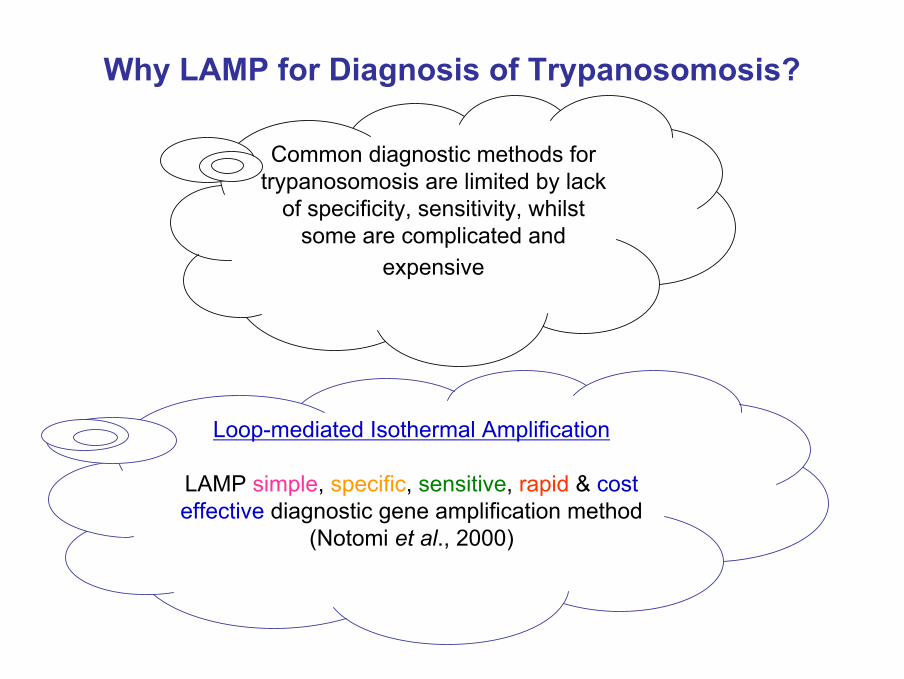
Why LAMP for Diagnosis of Trypanosomosis?
Loop-mediated Isothermal Amplification
LAMP simple, specific, sensitive, rapid & cost effective diagnostic gene amplification method
(Notomi et al., 2000)
Common diagnostic methods for trypanosomosis are limited by lack
of specificity, sensitivity, whilst some are complicated and
expensive
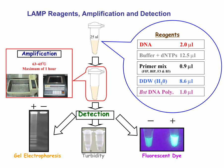
LAMP Reagents, Amplification and Detection
Detection
Turbidity
ー +
Fluorescent DyeGel Electrophoresis
Reagents
Buffer + dNTPs 12.5 µl
Primer mix 0.9 µl(FIP, BIP, F3 & B3)
DNA 2.0 µl
Bst DNA Poly. 1.0 µl
DDW (H20) 8.6 µl
Amplification
63~65℃Maximum of 1 hour
25 ul
+ ー
Loop-mediated Isothermal Amplification for Detection of African Trypanosomes
N. Kuboki, N. Inoue, T. Sakurai, F. Di Cello, D. J. Grab, H. Suzuki, C. Sugimoto, I. Igarashi
J. Clin. Microbiol. 41:5517 - 5524 (2003)
Objectives
To develop a specific LAMP for diagnosis of T. bruceigroup and T. congolese
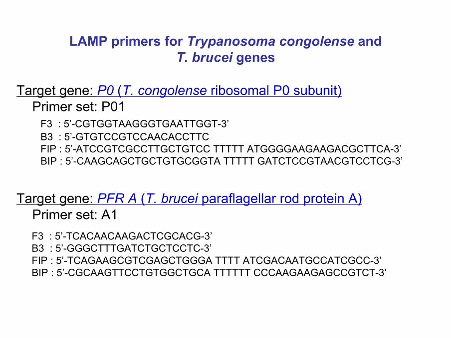
LAMP primers for Trypanosoma congolense andT. brucei genes
Target gene: P0 (T. congolense ribosomal P0 subunit)Primer set: P01
F3 : 5’-CGTGGTAAGGGTGAATTGGT-3’B3 : 5’-GTGTCCGTCCAACACCTTCFIP : 5’-ATCCGTCGCCTTGCTGTCC TTTTT ATGGGGAAGAAGACGCTTCA-3’BIP : 5’-CAAGCAGCTGCTGTGCGGTA TTTTT GATCTCCGTAACGTCCTCG-3’
Target gene: PFR A (T. brucei paraflagellar rod protein A)Primer set: A1F3 : 5’-TCACAACAAGACTCGCACG-3’B3 : 5’-GGGCTTTGATCTGCTCCTC-3’FIP : 5’-TCAGAAGCGTCGAGCTGGGA TTTT ATCGACAATGCCATCGCC-3’BIP : 5’-CGCAAGTTCCTGTGGCTGCA TTTTTT CCCAAGAAGAGCCGTCT-3’
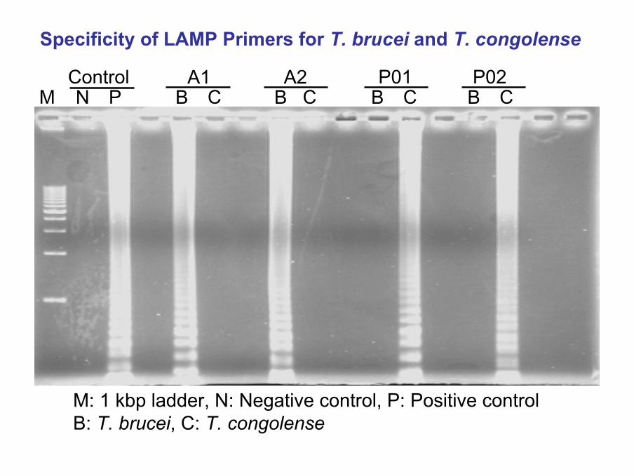
Specificity of LAMP Primers for T. brucei and T. congolense
M N P B C B C B C B CControl A1 A2 P01 P02
M: 1 kbp ladder, N: Negative control, P: Positive controlB: T. brucei, C: T. congolense
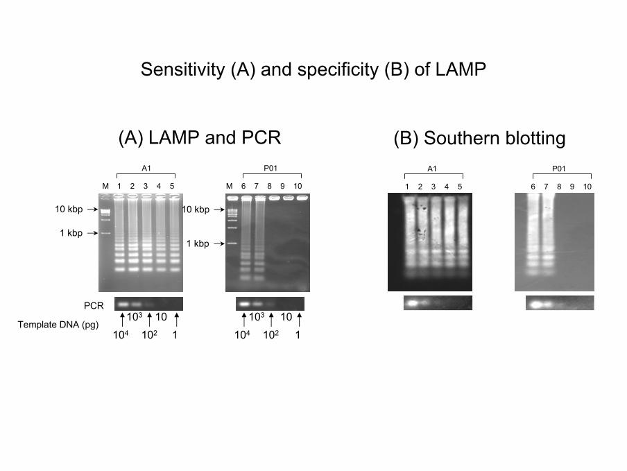
Template DNA (pg)
PCR
(A) LAMP and PCR
10 kbp
1 kbp
104
103
102 110
A1
M 1 2 3 4 5
104
103
102 110
P01
M 6 7 8 9 10
10 kbp
1 kbp
A1
1 2 3 4 5
P01
6 7 8 9 10
(B) Southern blotting
Sensitivity (A) and specificity (B) of LAMP
M 1 2 3 4 5 6 7 8 9 10 11 12
1 kbp
5 kbp10 kbp
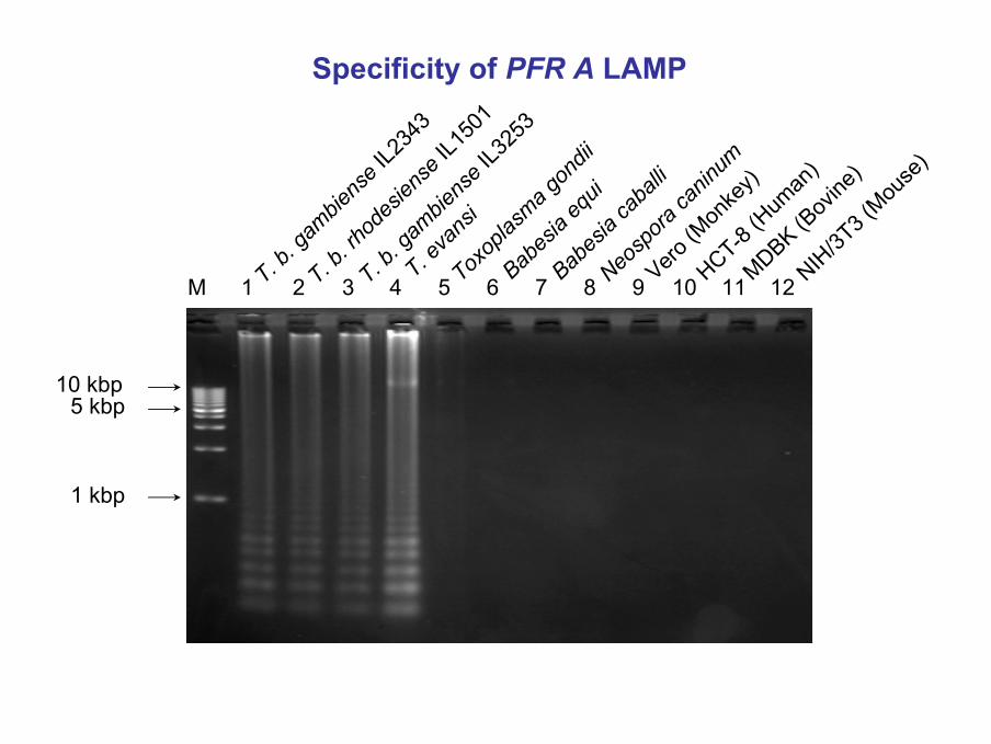
Specificity of PFR A LAMP
T. b. g
ambie
nse IL2
343
T. b. rh
odes
iense
IL150
1
T. b. g
ambie
nse IL3
253
T. eva
nsi
Toxop
lasma g
ondii
Babes
ia eq
ui
Babes
ia ca
balli
Neosp
ora ca
ninum
Vero (M
onke
y)
HCT-8 (H
uman
)
MDBK (Bov
ine)
NIH/3T
3 (Mou
se)
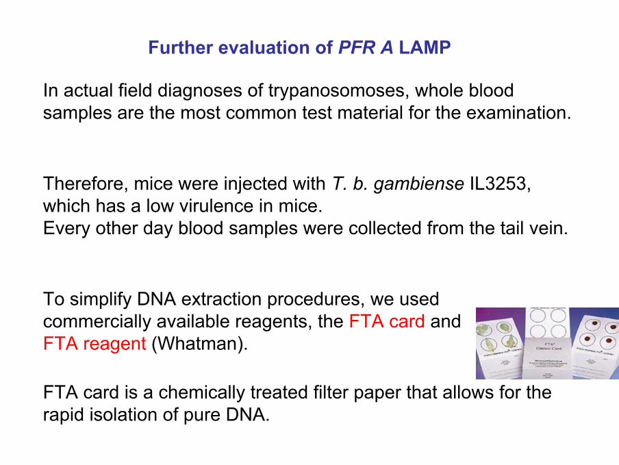
In actual field diagnoses of trypanosomoses, whole blood samples are the most common test material for the examination.
FTA card is a chemically treated filter paper that allows for the rapid isolation of pure DNA.
To simplify DNA extraction procedures, we used commercially available reagents, the FTA card and FTA reagent (Whatman).
Therefore, mice were injected with T. b. gambiense IL3253, which has a low virulence in mice. Every other day blood samples were collected from the tail vein.
Further evaluation of PFR A LAMP
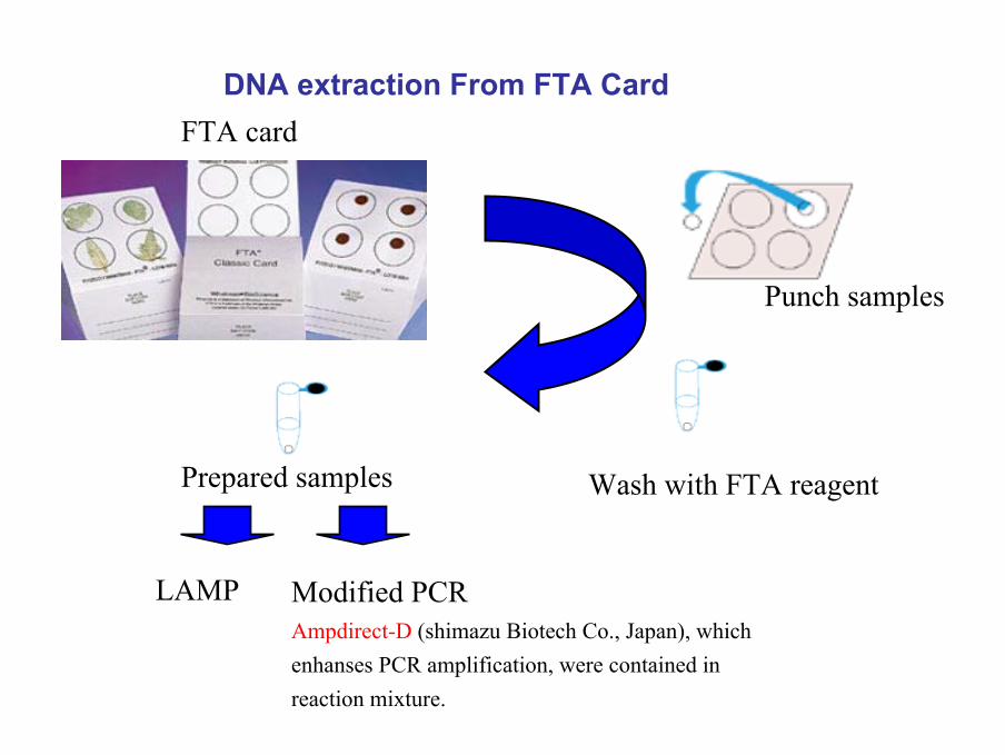
FTA cardDNA extraction From FTA Card
Punch samples
Wash with FTA reagentPrepared samples
LAMP Modified PCRAmpdirect-D (shimazu Biotech Co., Japan), which enhanses PCR amplification, were contained in reaction mixture.
4 12 142 6 8 10 16 18 20 22 24 26 28 30M
+ + + +
5 kbp10 kbp
1 kbp
PCR
Buffy coat
DPI 0
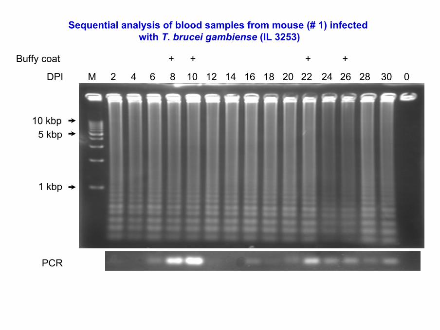
Sequential analysis of blood samples from mouse (# 1) infectedwith T. brucei gambiense (IL 3253)
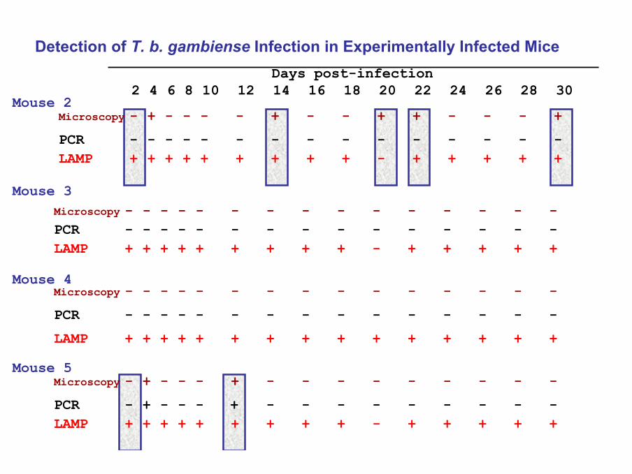
Mouse 2
Mouse 5
Mouse 4
Mouse 3
LAMP + + + + + + + + + - + + + + +
LAMP + + + + + + + + + + + + + + +
LAMP + + + + + + + + + - + + + + +
LAMP + + + + + + + + + - + + + + +
PCR - + - - - + - - - - - - - - -
PCR - - - - - - - - - - - - - - -
PCR - - - - - - - - - - - - - - -
PCR - - - - - - - - - - - - - - -
Microscopy - + - - - + - - - - - - - - -
Microscopy - - - - - - - - - - - - - - -
Microscopy - - - - - - - - - - - - - - -
Microscopy - + - - - - + - - + + - - - +
Days post-infection2 4 6 8 10 12 14 16 18 20 22 24 26 28 30
Detection of T. b. gambiense Infection in Experimentally Infected Mice
Conclusions
• T. brucei (PFR A) and T. congolense (P0) genes are specifically and rapidly amplified by LAMP.
• LAMP reaction is specific for the T. brucei group and can detect a T. brucei gambiense infection in mice with greater sensitivity than microscopic tests or PCR.
• However, PFRA LAMP primers also amplify T. evansiDNA, because the gene is conserved amongst species and sub-species of Trypanozoon.
Evaluation of LAMP, PCR and parasitological tests for detection of T. evansi in experimentally
infected pigs
O. M. M. Thekisoe, N. Inoue, N. Kuboki, D. Tuntasuvan, W. Bunnoy, S. Borisutsuwan, I. Igarashi, C. Sugimoto.
Vet. Parasitol. 130: 327- 330 (2005)
Objectives
• To determine efficiency of LAMP as a diagnostic method for T. evansi infections.
• To conduct comparative evaluation of LAMP, PCR and parasitological methods for detection of T. evansiinfections.
Background
• Trypanosoma evansi causes epidemics of disease commonly called surra and is mechanically transmitted by the tabanid flies.
• It is distributed in Asia, Africa & South America and it infects buffaloes, cattle, horses, donkeys, camels, goats, sheep and pigs.
• Microscopic and serological diagnostic methods are limited by lack of sensitivity, specificity and by the presence of cross reactivity.
• PCR has been reported as sensitive and specific but is not commonly used in surra endemic countries as it requires an expensive automated thermal cycler.
• Recent reports of T. evansi infection in humans in India (WHO, 2005), highlighted the significance of accurate diagnosis of this protozoan parasite.
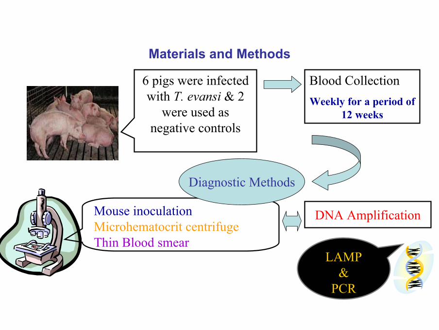
Materials and Methods
6 pigs were infected with T. evansi & 2
were used as negative controls
Blood CollectionWeekly for a period of
12 weeks
DNA Amplification
LAMP&
PCR
Mouse inoculationMicrohematocrit centrifugeThin Blood smear
Diagnostic Methods
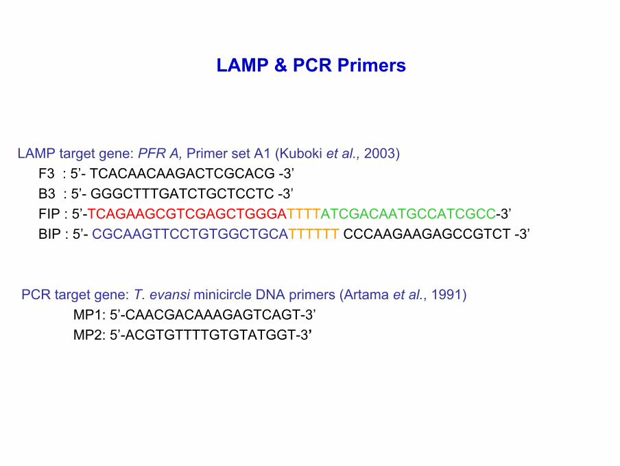
LAMP & PCR Primers
LAMP target gene: PFR A, Primer set A1 (Kuboki et al., 2003) F3 : 5’- TCACAACAAGACTCGCACG -3’B3 : 5’- GGGCTTTGATCTGCTCCTC -3’FIP : 5’-TCAGAAGCGTCGAGCTGGGATTTTATCGACAATGCCATCGCC-3’BIP : 5’- CGCAAGTTCCTGTGGCTGCATTTTTT CCCAAGAAGAGCCGTCT -3’
PCR target gene: T. evansi minicircle DNA primers (Artama et al., 1991)MP1: 5’-CAACGACAAAGAGTCAGT-3’MP2: 5’-ACGTGTTTTGTGTATGGT-3’
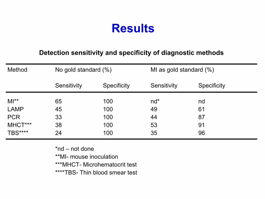
Results
Detection sensitivity and specificity of diagnostic methods
Method No gold standard (%) MI as gold standard (%)
Sensitivity Specificity Sensitivity Specificity
MI** 65 100 nd* ndLAMP 45 100 49 61PCR 33 100 44 87MHCT*** 38 100 53 91TBS**** 24 100 35 96
*nd – not done**MI- mouse inoculation***MHCT- Microhematocrit test****TBS- Thin blood smear test
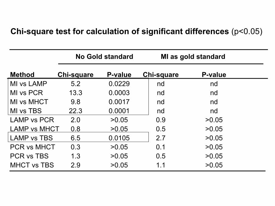
Chi-square test for calculation of significant differences (p<0.05)
No Gold standard MI as gold standard
Method Chi-square P-value Chi-square P-valueMI vs LAMP 5.2 0.0229 nd ndMI vs PCR 13.3 0.0003 nd ndMI vs MHCT 9.8 0.0017 nd ndMI vs TBS 22.3 0.0001 nd ndLAMP vs PCR 2.0 >0.05 0.9 >0.05LAMP vs MHCT 0.8 >0.05 0.5 >0.05LAMP vs TBS 6.5 0.0105 2.7 >0.05PCR vs MHCT 0.3 >0.05 0.1 >0.05PCR vs TBS 1.3 >0.05 0.5 >0.05MHCT vs TBS 2.9 >0.05 1.1 >0.05
Conclusion
• MI (mouse inoculation test) was the most sensitive method but is not practically applicable for diagnosis as its time consuming.
• LAMP showed relatively higher detection sensitivity than PCR as similarly reported by Kuboki et al. (2003).
• This study validates LAMP as an alternative gene amplification method for the diagnosis T. evansi infections.

Oriel M.M. Thekisoe, Andrew Nambota, Jun Yasuda, Noritaka Kuboki, Kozo Fujisaki, Ikuo Igarashi, Chihiro Sugimoto, Noboru Inoue
Manuscript Submitted - under review
Species-Specific Loop-Mediated Isothermal Amplification (LAMP) for Diagnosis of Trypanosomiasis and Its Application
to Epidemiological Studies
Objectives of the Study
• Specific LAMP for detection of T. b. gambiense, T. b. rhodesiense, T. evansi, T. congolense, and T. cruzi.
• Application of LAMP in epidemiological studies
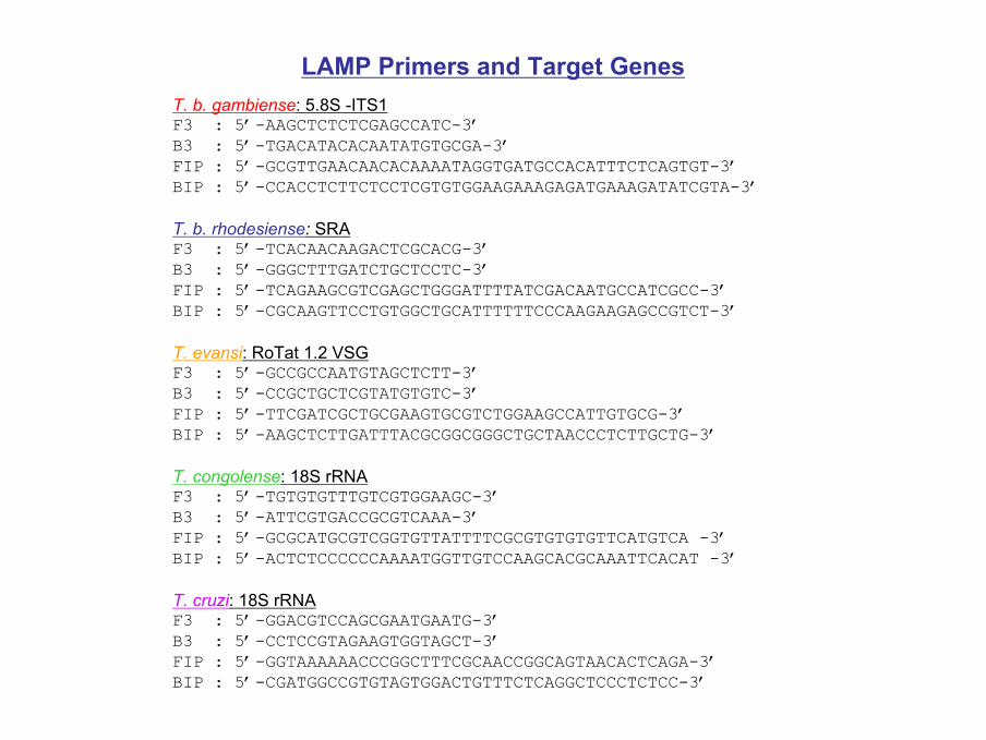
LAMP Primers and Target GenesT. b. gambiense: 5.8S -ITS1F3 : 5’-AAGCTCTCTCGAGCCATC-3’B3 : 5’-TGACATACACAATATGTGCGA-3’FIP : 5’-GCGTTGAACAACACAAAATAGGTGATGCCACATTTCTCAGTGT-3’BIP : 5’-CCACCTCTTCTCCTCGTGTGGAAGAAAGAGATGAAAGATATCGTA-3’
T. b. rhodesiense: SRAF3 : 5’-TCACAACAAGACTCGCACG-3’B3 : 5’-GGGCTTTGATCTGCTCCTC-3’FIP : 5’-TCAGAAGCGTCGAGCTGGGATTTTATCGACAATGCCATCGCC-3’BIP : 5’-CGCAAGTTCCTGTGGCTGCATTTTTTCCCAAGAAGAGCCGTCT-3’
T. evansi: RoTat 1.2 VSGF3 : 5’-GCCGCCAATGTAGCTCTT-3’B3 : 5’-CCGCTGCTCGTATGTGTC-3’FIP : 5’-TTCGATCGCTGCGAAGTGCGTCTGGAAGCCATTGTGCG-3’BIP : 5’-AAGCTCTTGATTTACGCGGCGGGCTGCTAACCCTCTTGCTG-3’
T. congolense: 18S rRNAF3 : 5’-TGTGTGTTTGTCGTGGAAGC-3’B3 : 5’-ATTCGTGACCGCGTCAAA-3’FIP : 5’-GCGCATGCGTCGGTGTTATTTTCGCGTGTGTGTTCATGTCA -3’BIP : 5’-ACTCTCCCCCCAAAATGGTTGTCCAAGCACGCAAATTCACAT -3’
T. cruzi: 18S rRNAF3 : 5’-GGACGTCCAGCGAATGAATG-3’B3 : 5’-CCTCCGTAGAAGTGGTAGCT-3’FIP : 5’-GGTAAAAAACCCGGCTTTCGCAACCGGCAGTAACACTCAGA-3’BIP : 5’-CGATGGCCGTGTAGTGGACTGTTTCTCAGGCTCCCTCTCC-3’
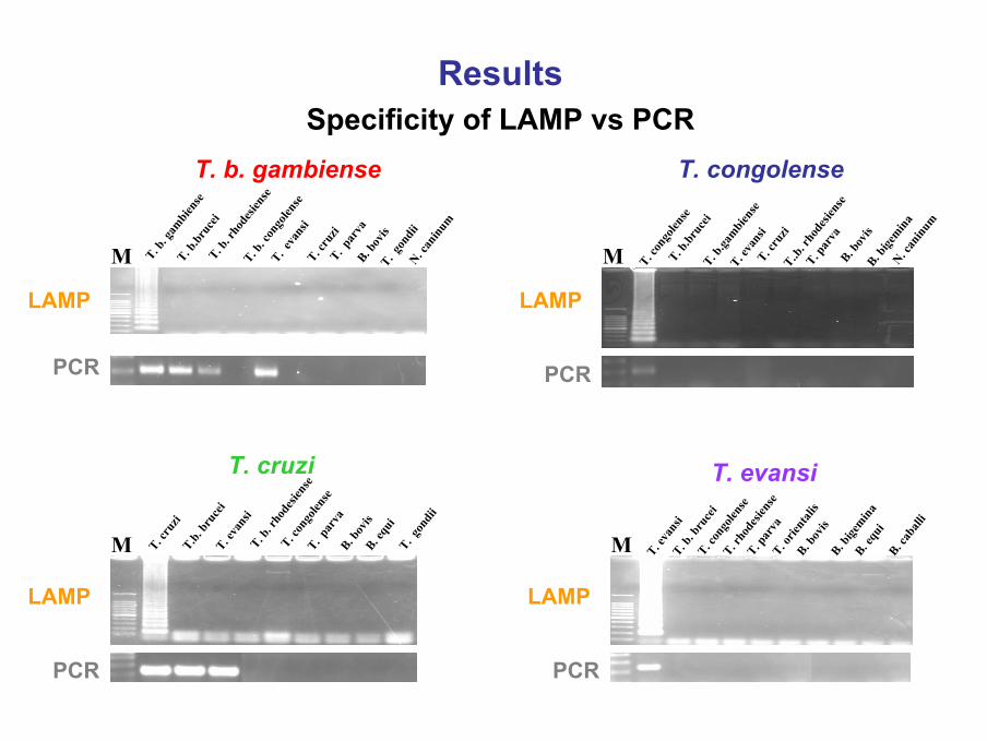
Specificity of LAMP vs PCRT. b. gambiense T. congolense
T. cruzi T. evansi
Results
T. b. g
ambie
nse
M T. b.br
ucei
T. b. r
hode
siens
eT. b
. con
golen
se
T. eva
nsi
T. cru
ziT. p
arva
B. bov
isT. g
ondii
N. can
inum
M T. con
golen
seT. b
.bruc
eiT. b
.gambie
nse
T. eva
nsi
T. cru
ziT..b
. rho
desie
nse
T. par
vaB. b
ovis
B. bige
minaN. c
aninu
m
M T. cru
ziT.b.
bruc
eiT. e
vans
iT. b
. rho
desie
nse
T. con
golen
seT. p
arva
B. bov
isB. e
qui
T. gon
dii
M T. eva
nsi
T. b. b
ruce
iT. c
ongo
lense
T. rho
desie
nse
T. par
vaT. o
rient
alis
B. bov
isB. b
igemina
B. equ
iB. c
aball
i
LAMP LAMP
PCR
LAMP LAMP
PCR
PCR PCR
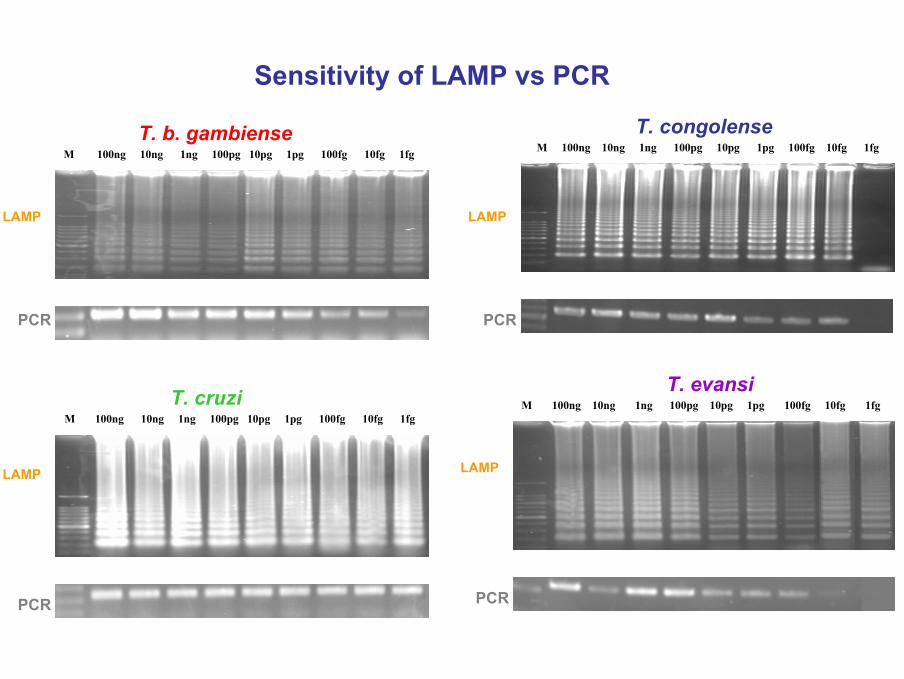
M 100ng 10ng 1ng 100pg 10pg 1pg 100fg 10fg 1fg
PCR
Sensitivity of LAMP vs PCR
M 100ng 10ng 1ng 100pg 10pg 1pg 100fg 10fg 1fg
PCR
T. b. gambiense
PCR
M 100ng 10ng 1ng 100pg 10pg 1pg 100fg 10fg 1fgT. congolense
M 100ng 10ng 1ng 100pg 10pg 1pg 100fg 10fg 1fg
PCR
T. cruzi T. evansi
LAMP LAMP
LAMP LAMP
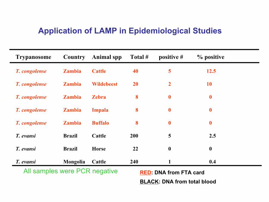
Trypanosome Country Animal spp Total # positive # % positive
T. congolense Zambia Cattle 40 5 12.5
T. congolense Zambia Wildebeest 20 2 10
T. congolense Zambia Zebra 8 0 0
T. congolense Zambia Impala 8 0 0
T. congolense Zambia Buffalo 8 0 0
T. evansi Brazil Cattle 200 5 2.5
T. evansi Brazil Horse 22 0 0
T. evansi Mongolia Cattle 240 1 0.4
Application of LAMP in Epidemiological Studies
RED: DNA from FTA cardBLACK: DNA from total blood
All samples were PCR negative
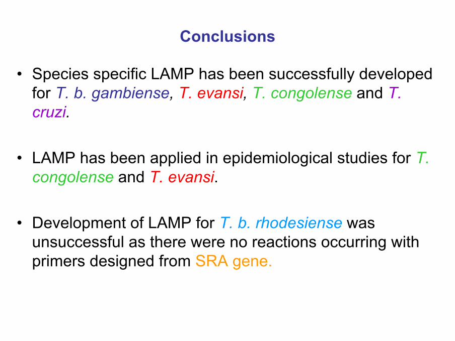
• Species specific LAMP has been successfully developed for T. b. gambiense, T. evansi, T. congolense and T. cruzi.
• LAMP has been applied in epidemiological studies for T. congolense and T. evansi.
• Development of LAMP for T. b. rhodesiense was unsuccessful as there were no reactions occurring with primers designed from SRA gene.
Conclusions
Stability of LAMP Reagents Stored at Different temperatures, itsAmplification Efficiency on Different DNA Templates and its
Tolerance to Inhibitory Substances
Oriel M.M. Thekisoe, Raoul B. S. Bazie, Andrea Coronel-Servian, Chihiro Sugimoto, Kozo Fujisaki, Noboru Inoue
Manuscript Submitted – under review

Background
• Due to its simplicity, LAMP is a strong candidate for application of molecular diagnosis in the field where cost and environmentalconstraints prohibit the use of PCR.
• However, conditions in the field differ significantly to laboratory conditions.
• For example, temperature of the surroundings, availability of equipment for reagent storage and DNA extraction.
• There is a need to conduct experiments under conditions similar to field, clinics or hospitals, with simplified DNA template preparation methods.
Objectives
• To evaluate stability of LAMP reagents when stored at 25ºC and 37ºC.
• To assess detection efficiency of LAMP on different DNA templatepreparations.
• To determine the tolerence of LAMP on PCR inhibitory substances.
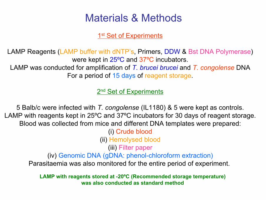
Materials & Methods1st Set of Experiments
LAMP Reagents (LAMP buffer with dNTP’s, Primers, DDW & Bst DNA Polymerase)were kept in 25ºC and 37ºC incubators.
LAMP was conducted for amplification of T. brucei brucei and T. congolense DNAFor a period of 15 days of reagent storage.
2nd Set of Experiments
5 Balb/c were infected with T. congolense (IL1180) & 5 were kept as controls.LAMP with reagents kept in 25ºC and 37ºC incubators for 30 days of reagent storage.
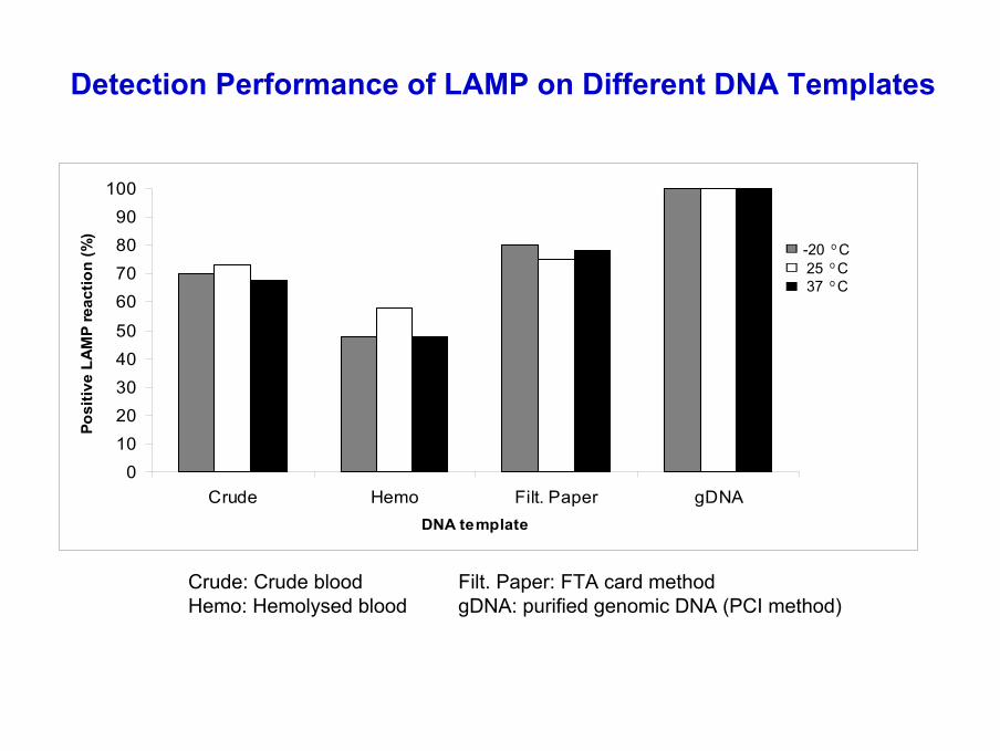
Blood was collected from mice and different DNA templates were prepared:(i) Crude blood
(ii) Hemolysed blood(iii) Filter paper
(iv) Genomic DNA (gDNA: phenol-chloroform extraction)Parasitaemia was also monitored for the entire period of experiment.
LAMP with reagents stored at -20ºC (Recommended storage temperature) was also conducted as standard method
3rd set of experiments
PCR Inhibitory substances
• EDTA• Hemoglobin from bovine blood• Heparin• IgG• IgM
Were prepared in concentrations of 1 mg/ml, then diluted serially and added into the LAMP and PCR reaction.
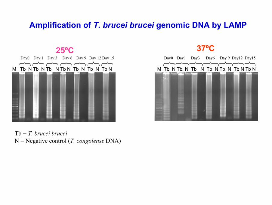
Day0 Day 1 Day 3 Day 6 Day 9 Day 12 Day 15
M Tb N Tb N Tb N Tb N Tb N Tb N Tb N
Day0 Day1 Day3 Day6 Day 9 Day12 Day15
M Tb N Tb N Tb N Tb N Tb N Tb N Tb N
25ºC 37ºC
Amplification of T. brucei brucei genomic DNA by LAMP
Tb – T. brucei bruceiN – Negative control (T. congolense DNA)
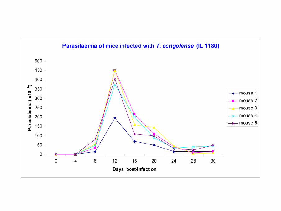
Parasitaemia of mice infected with T. congolense (IL 1180)
0
50
100
150
200
250
300
350
400
450
500
0 4 8 12 16 20 24 28 30
Days post-infection
Para
siat
emia
( x1
06 )
mouse 1mouse 2mouse 3mouse 4mouse 5
01020304050
60708090
100
Crude Hemo Filt. Paper gDNADNA template
Posi
tive
LAM
P re
actio
n (%
)
-20 оC25 оC37 оC
Detection Performance of LAMP on Different DNA Templates
Crude: Crude blood Filt. Paper: FTA card methodHemo: Hemolysed blood gDNA: purified genomic DNA (PCI method)
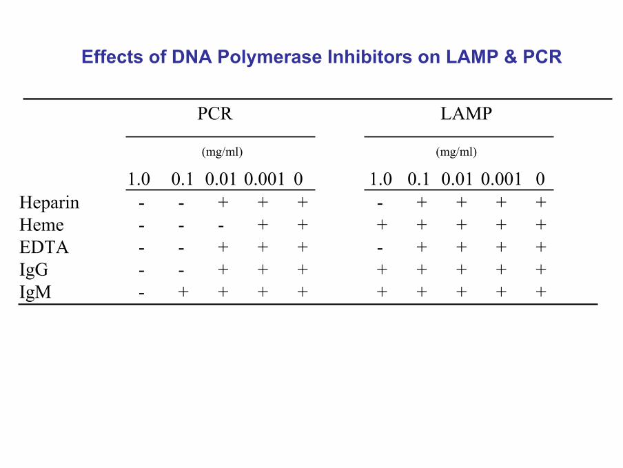
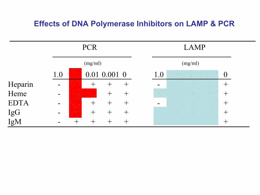
Effects of DNA Polymerase Inhibitors on LAMP & PCR
PCR LAMP
1.0 0.1 0.01 0.001 0 1.0 0.1 0.01 0.001 0Heparin - - + + + - + + + +Heme - - - + + + + + + +EDTA - - + + + - + + + +IgG - - + + + + + + + +IgM - + + + + + + + + +
(mg/ml) (mg/ml)
Effects of DNA Polymerase Inhibitors on LAMP & PCR
PCR LAMP
1.0 0.1 0.01 0.001 0 1.0 0.1 0.01 0.001 0Heparin - - + + + - + + + +Heme - - - + + + + + + +EDTA - - + + + - + + + +IgG - - + + + + + + + +IgM - + + + + + + + + +
(mg/ml) (mg/ml)
Conclusions
• LAMP with reagents stored at 25ºC and 37ºC can amplify trypanosome DNA which are possible ambient temperatures in the field.
• No significant difference of detection sensitivity between LAMP reagents stored at -20ºC, 25ºC and 37ºC.
• Best LAMP performance was achieved with gDNA template (100 %), followed by filter paper (78 %), crude (72 %) and hemolysed blood (51 %).
• LAMP reactions were only inhibited by high concentrations (1 mg/ml) of EDTA and Heparin.
• Further improvement of simple DNA extraction method would enhance chances of application of LAMP in the field.
Development of LAMP for Detection of Bovine Theileriosis
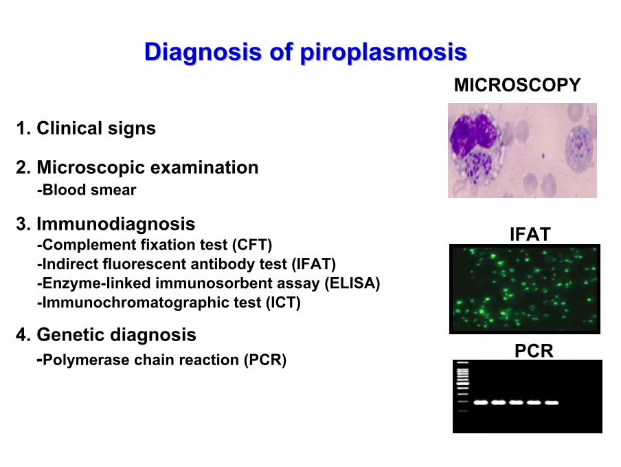
1. Clinical signs
2. Microscopic examination-Blood smear
3. Immunodiagnosis-Complement fixation test (CFT) -Indirect fluorescent antibody test (IFAT)-Enzyme-linked immunosorbent assay (ELISA)-Immunochromatographic test (ICT)
4. Genetic diagnosis-Polymerase chain reaction (PCR)
Diagnosis of piroplasmosisDiagnosis of piroplasmosis
IFAT
PCR
MICROSCOPY
Objective
To develop LAMP for specific detection of Theileria parva
Target genes
p104
p67
HSP 70
18S rRNA
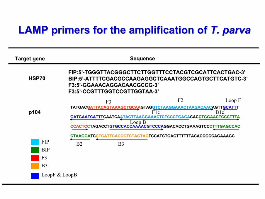
LAMP primers for the amplification of LAMP primers for the amplification of T. parvaT. parva
Target geneTarget gene SequenceSequence
HSPHSP7070FIP:5FIP:5’’--TGGGTTACGGGCTTCTTGGTTTCCTACGTCGCATTCACTGACTGGGTTACGGGCTTCTTGGTTTCCTACGTCGCATTCACTGAC--33’’BIP:5BIP:5’’--ATTTTCGACGCCAAGAGGCTCAAATGGCCAGTGCTTCATGTCATTTTCGACGCCAAGAGGCTCAAATGGCCAGTGCTTCATGTC--33’’F3:5F3:5’’--GGAAACAGGACAACGCCGGGAAACAGGACAACGCCG--33’’F3:5F3:5’’--CCGTTTGGTCCGTTGGTAACCGTTTGGTCCGTTGGTAA--33’’
p104p104TATGACTATGACGATTACAGTAAAGCTGCAAGATTACAGTAAAGCTGCAAGTAGGTAGGTCTAAGGAAACTAAGACAACGTCTAAGGAAACTAAGACAACAGTTAGTTGCATTTGCATTT
GATGAATCATTTGGATGAATCATTTGAATCAAATCAATACTTAAGGAAACTCTCCCTGAGAATACTTAAGGAAACTCTCCCTGAGACACCACCTGGAACTCCCTTTACTGGAACTCCCTTTA
CCACTCCCCACTCCTAGACCTTAGACCTGTGCCACCAAAACGTCCCAGTGCCACCAAAACGTCCCAGGACACCTGAAAGTCCGGACACCTGAAAGTCCCTTTGAGCCACCTTTGAGCCAC
CTAAGGACTAAGGATCTCCTGATTCACCGTCTAGTAGCTGATTCACCGTCTAGTAGTCCATCTGAGTTTTTTACACCGCCAGAAAGCTCCATCTGAGTTTTTTACACCGCCAGAAAGC
B3
F3 F2 Loop F
F1c
B2
B1c
Loop B
F3
FIPBIP
B3
LoopF & LoopB
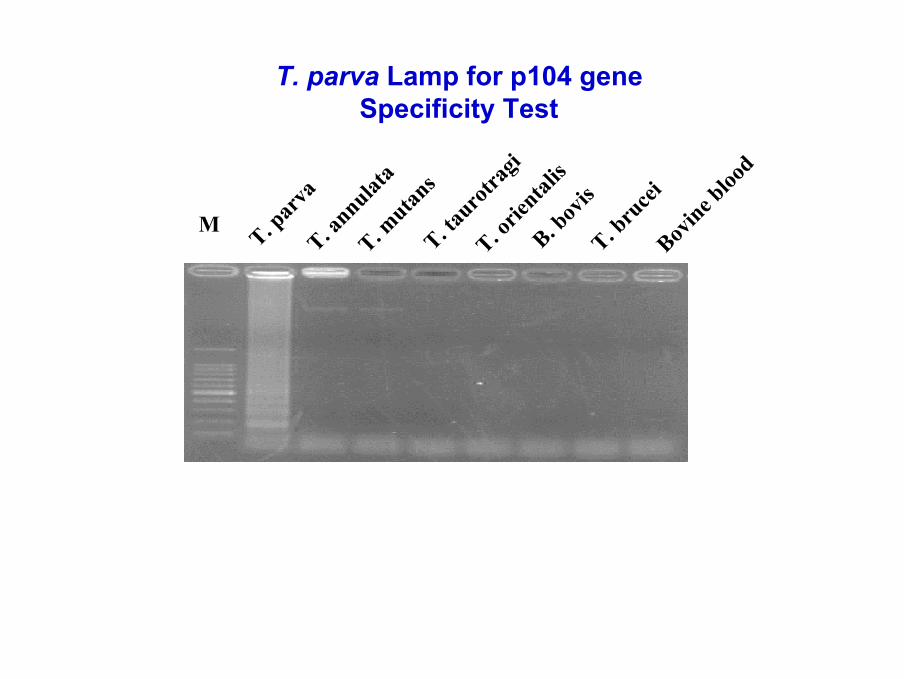
T. parva Lamp for p104 geneSpecificity Test
M
T. par
vaT. a
nnulata
T. mutan
sT. ta
urotra
giT. o
riental
isB. b
ovis
T. bru
ceiBov
ine bloo
d
T. parva Lamp for p104 geneSensitivity
M 100ng 10ng 1ng 100pg 10pg 1pg 100fg 10fg 1fg DDW
PCR 181bp
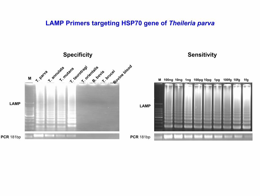
LAMP Primers targeting HSP70 gene of Theileria parva
LAMP
Specificity
M 100ng 10ng 1ng 100pg 10pg 1pg 100fg 10fg 1fg
LAMP
PCR 181bp
Sensitivity
M
T. parv
aT. a
nnulata
T. mutan
sT. ta
urotra
giT. o
riental
isB. b
ovisT. b
ruce
iBovin
e blood
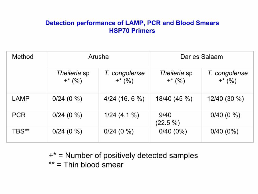
Method Arusha Dar es Salaam
Theileria sp +* (%)
T. congolense+* (%)
Theileria sp +* (%)
T. congolense+* (%)
LAMP 0/24 (0 %) 4/24 (16. 6 %) 18/40 (45 %) 12/40 (30 %)
PCR 0/24 (0 %) 1/24 (4.1 %) 9/40 (22.5 %)
0/40 (0 %)
TBS** 0/24 (0 %) 0/24 (0 %) 0/40 (0%) 0/40 (0%)
Detection performance of LAMP, PCR and Blood SmearsHSP70 Primers
+* = Number of positively detected samples** = Thin blood smear
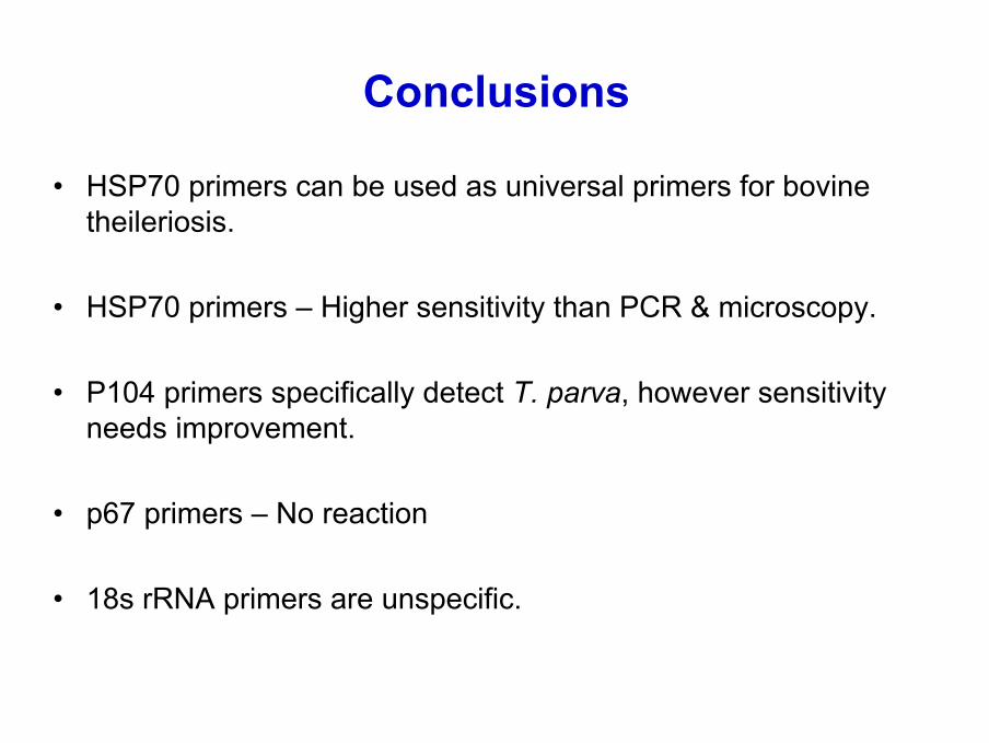
Conclusions
• HSP70 primers can be used as universal primers for bovine theileriosis.
• HSP70 primers – Higher sensitivity than PCR & microscopy.
• P104 primers specifically detect T. parva, however sensitivity needs improvement.
• p67 primers – No reaction
• 18s rRNA primers are unspecific.
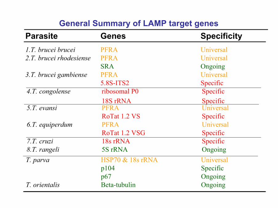
General Summary of LAMP target genes
1.T. brucei brucei PFRA Universal2.T. brucei rhodesiense PFRA Universal
SRA Ongoing3.T. brucei gambiense PFRA Universal
5.8S-ITS2 Specific4.T. congolense ribosomal P0 Specific
18S rRNA Specific
Parasite Genes Specificity
T. parva HSP70 & 18s rRNA Universalp104 Specificp67 Ongoing
T. orientalis Beta-tubulin Ongoing
7.T. cruzi 18s rRNA Specific8.T. rangeli 5S rRNA Ongoing
5.T. evansi PFRA UniversalRoTat 1.2 VS Specific
6.T. equiperdum PFRA UniversalRoTat 1.2 VSG Specific
Andy Alhassan, Oriel M. M. Thekisoe, Naoaki Yokoyama, Noboru Inoue, Makhosazana Y. Motloang, Peter A. Mbati, Hong Yin, Chihiro Sugimoto, Ikuo Igarashi
Vet. Parasitol. In press (2006)
Development of LoopDevelopment of Loop--Mediated Isothermal Mediated Isothermal Amplification (LAMP) MethodAmplification (LAMP) Method for Dfor Diagnosis iagnosis
of Equineof Equine piroplasmosispiroplasmosis
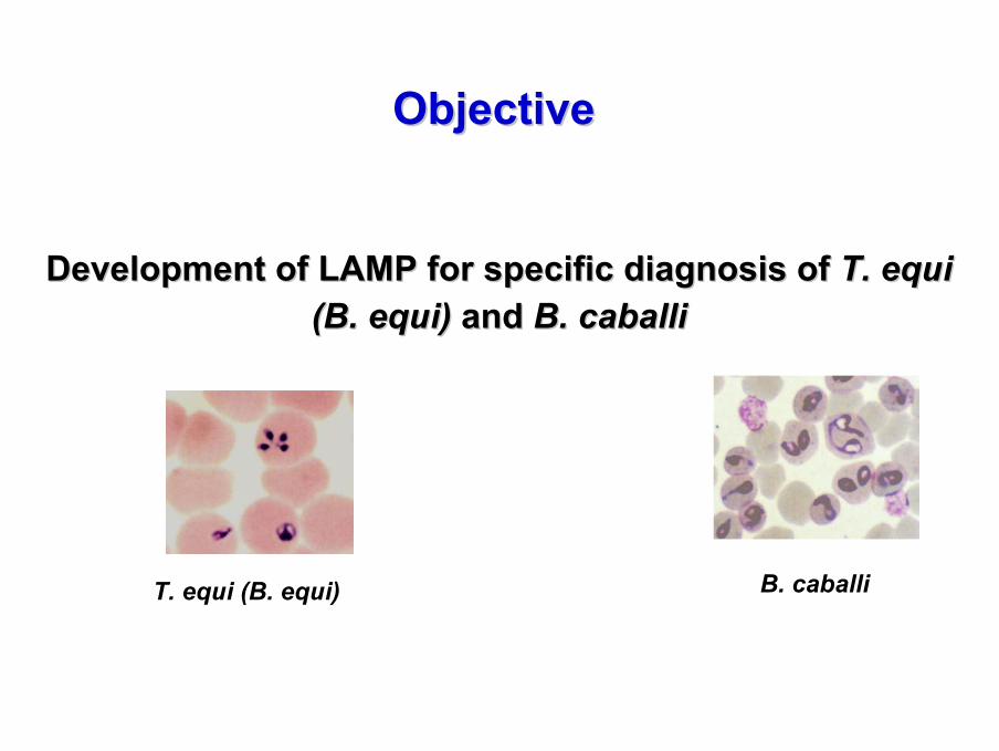
Development of LAMP for specific diagnosisDevelopment of LAMP for specific diagnosis of of T. equi T. equi (B. equi)(B. equi) and and B. caballiB. caballi
ObjectiveObjective
T. equi (B. equi) B. caballi
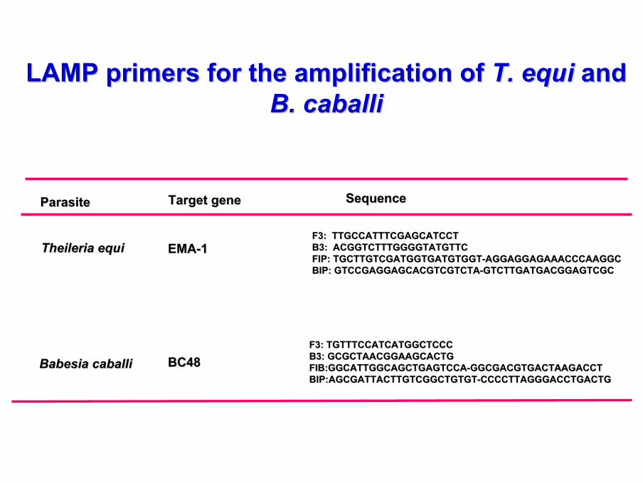
LAMP primers for the amplification of LAMP primers for the amplification of T. equiT. equi and and B. caballiB. caballi
ParasiteParasite Target geneTarget gene SequenceSequence
Theileria equiTheileria equi EMAEMA--11F3: TTGCCATTTCGAGCATCCTF3: TTGCCATTTCGAGCATCCTB3: ACGGTCTTTGGGGTATGTTCB3: ACGGTCTTTGGGGTATGTTCFIP: TGCTTGTCGATGGTGATGTGGTFIP: TGCTTGTCGATGGTGATGTGGT--AGGAGGAGAAACCCAAGGCAGGAGGAGAAACCCAAGGCBIP: GTCCGAGGAGCACGTCGTCTABIP: GTCCGAGGAGCACGTCGTCTA--GTCTTGATGACGGAGTCGCGTCTTGATGACGGAGTCGC
BC48BC48F3: TGTTTCCATCATGGCTCCCF3: TGTTTCCATCATGGCTCCCB3: GCGCTAACGGAAGCACTGB3: GCGCTAACGGAAGCACTGFIB:GGCATTGGCAGCTGAGTCCAFIB:GGCATTGGCAGCTGAGTCCA--GGCGACGTGACTAAGACCTGGCGACGTGACTAAGACCTBIP:AGCGATTACTTGTCGGCTGTGTBIP:AGCGATTACTTGTCGGCTGTGT--CCCCTTAGGGACCTGACTGCCCCTTAGGGACCTGACTG
Babesia caballiBabesia caballi
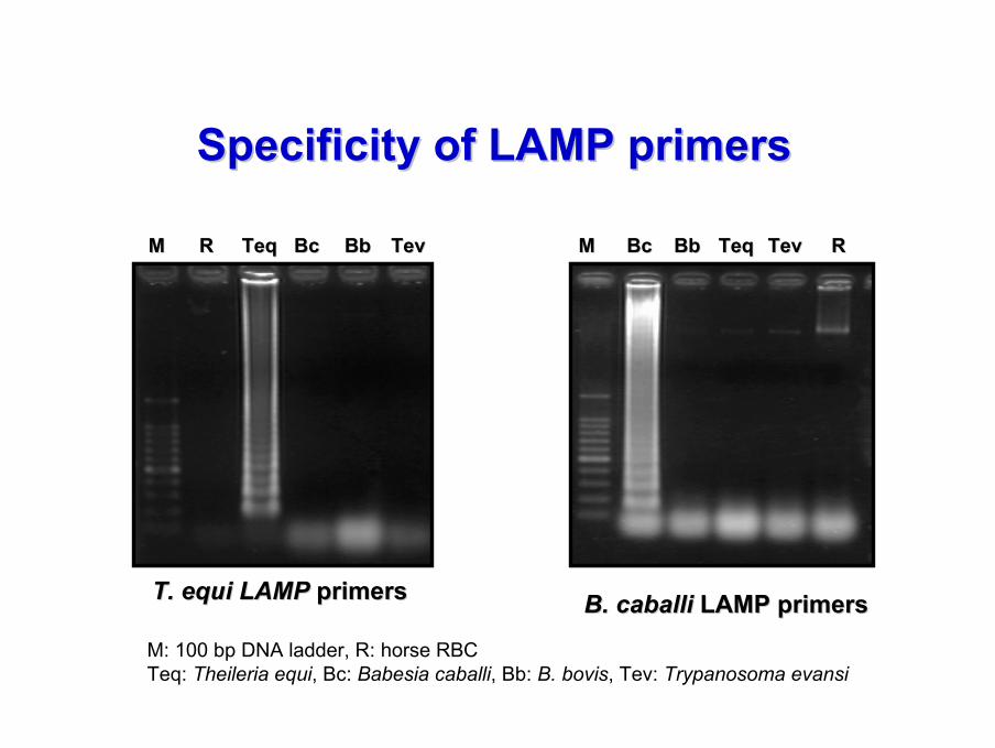
Specificity of LAMP primersSpecificity of LAMP primers
MM RR TeqTeq BcBc BbBb TTevev MM TeqTeqBcBc BbBb TTevev RR
B. caballiB. caballi LAMP primersLAMP primersT. equi LAMP T. equi LAMP primers primers
M: 100 bp DNA ladder, R: horse RBCTeq: Theileria equi, Bc: Babesia caballi, Bb: B. bovis, Tev: Trypanosoma evansi
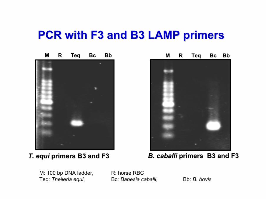
PCR with F3 and B3 LAMP primers PCR with F3 and B3 LAMP primers
T. equiT. equi primers B3 and F3primers B3 and F3 B. caballiB. caballi primers B3 and F3primers B3 and F3
MM RR TeqTeq BcBc BbBb
M: 100 bp DNA ladder, R: horse RBCTeq: Theileria equi, Bc: Babesia caballi, Bb: B. bovis
MM RR TeqTeq BcBc BbBb
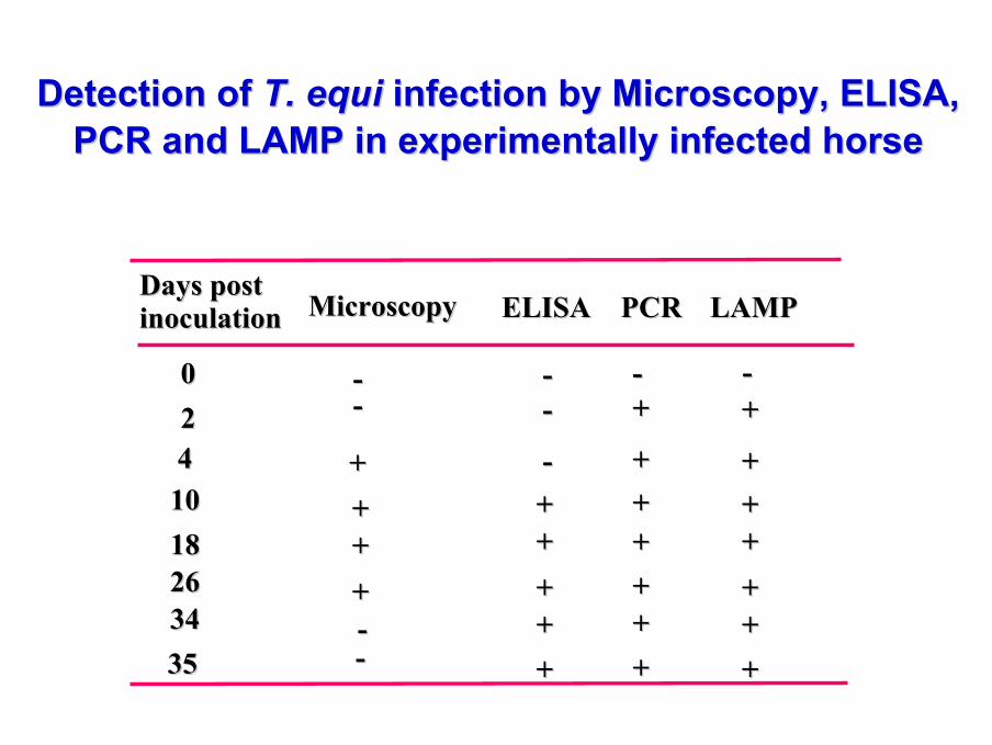
002244
10101818262634343535
Days post Days post inoculationinoculation ELISAELISA LAMP LAMP
------ ++
-- ++++ ++++ ++++ ++++ ++++ ++
MicroscopyMicroscopy
----
++++++++----
PCRPCR
--++
++++++++++++
Detection of Detection of T. equiT. equi infection by Microscopy, ELISA, infection by Microscopy, ELISA, PCR and LAMP in experimentally infected horsePCR and LAMP in experimentally infected horse
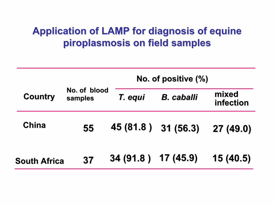
T. equiT. equi B. caballiB. caballiNo. of blood No. of blood samplessamples
No. of positive (%)No. of positive (%)
mixed mixed infectioninfection
ChinaChina
South AfricaSouth Africa 3737
5555 45 (81.8 )45 (81.8 ) 31 (56.3)31 (56.3) 27 (49.0)27 (49.0)
34 (91.8 )34 (91.8 ) 17 (45.9)17 (45.9) 15 (40.5)15 (40.5)
CountryCountry
Application of LAMP for diagnosis of equine Application of LAMP for diagnosis of equine piroplasmosis on field samplespiroplasmosis on field samples
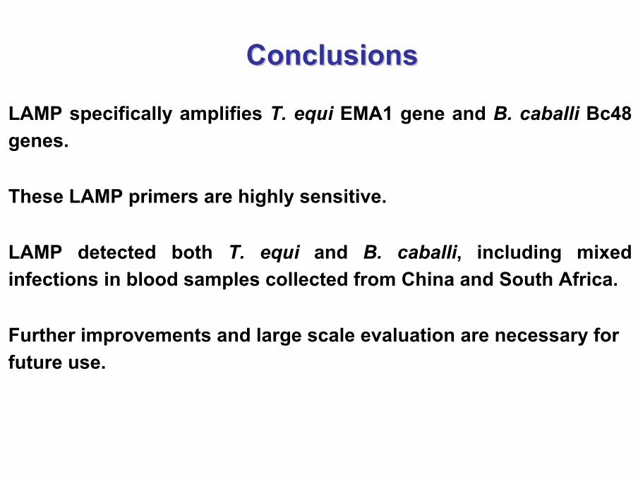
ConclusionsConclusions
LAMP specifically amplifies T. equi EMA1 gene and B. caballi Bc48 genes.
These LAMP primers are highly sensitive.
LAMP detected both T. equi and B. caballi, including mixed infections in blood samples collected from China and South Africa.
Further improvements and large scale evaluation are necessary for future use.

National Research Center for Protozoan Diseases
Research Unit for Advanced Preventive Medicine


![Lab on chip world congress poster - Technology Networks · instruments for amplification or elaborative method, due to low specificity [3]. Loop-mediated isothermal amplification](https://img.dokumen.tips/doc/110x75/5fbb5c3d508f9702cb1b6e66/lab-on-chip-world-congress-poster-technology-networks-instruments-for-amplification.jpg)