Embed Size (px)
Citation preview

i
LONG TERM RESULTS OF OPEN ABDOMINAL
AORTIC ANEURYSM REPAIR
Thesis
From 1st January 2011 to 31st December 2013
Under the guidance of
Prof. M. Unnikrishnan MCh
Head, Division of Vascular Surgery
Department of CTVS
By
Shashidhar K P, M.S
SREE CHITRA TIRUNAL INSTITUTE FORMEDICAL SCIENCES
AND TECHNOLOGY
Thiruvananthapuram – 695011, India

ii
Travancore, an erstwhile province of pre-independent India, was ruled by
Sree Chitra Tirunal Maharajah until the country became independent in 1947. The
Government of India took over the province after the independence.
Known for their munificence, the Maharajah and members of the royal family
of Travancore considered themselves 'dasas' (servants) of Lord Padmanabha, the
reigning deity of Travancore. Interestingly, they wore a turban instead of a crown as
a mark of respect to Lord Padmanabha. Their philanthropy finds expression in their
countless contributions to the country, then and now.

iii
On a visit to a super-specialty hospital in Europe, Sree Chitra Tirunal
Maharajah was seized with a deep desire to establish a similar institution in Kerala.
Those were the times when tertiary cares in cardiovascular and neurological diseases
were not available in the State.
In the summer of 1974, the Maharajah's dream was fulfilled when the royal
family made a gift that carried in its womb the beginnings of what later turned out to
be the Sree Chitra Tirunal Institute for Medical Sciences & Technology. About this
time, Dr. M.S. Valiathan, trained abroad in surgery and biomedical science, returned
to India to guide the destiny of the Institute. Supported magnificently by Shri. C.
Achutha Menon, the then chief minister of Kerala, the Government of Kerala took
the unusual step of placing the centre under the Department of Science and
Technology in the State.
The Sree Chitra Tirunal Institute for Medical Sciences & Technology
(SCTIMST), Thiruvananthapuram is an Institute of National Importance established
by an Act of the Indian Parliament. It is an autonomous Institute under the
administrative control of the Department of Science and Technology, Government of
India.
The Institute signifies the convergence of medical sciences and technology
and its mission is to enable the indigenous growth of biomedical technology, besides
demonstrating high standards of patient care in medical specialties and evolving
postgraduate training programs in advanced medical specialties, biomedical
engineering and technology, as well as in public health.

iv
BIO – MEDICAL TECHNOLOGY WING
SCTIMST

v
ACKNOWLEDGEMENT
I have great pleasure to place on record my debt of gratitude to
Prof M Unnikrishnan, MCh, Senior Professor and Head of the Division of Vascular
Surgery, Dept of CVTS, SCTIMST, my esteemed teacher, who introduced me to the field
of vascular surgery, provided updated information, suggested improvisations and guided
me to imbibe vascular surgical skills during the course.
I am very much grateful to Prof Jayakumar K, Senior Professor and Head,
Department of CVTS, SCTIMST for his wholehearted support during my course.
I would like to express my sincere gratitude to Dr.Shivananda S,
Dr Balasubramaniom KR, & Dr Nedounsejiane M, Consultants, Division of Vascular
Surgery for their support and guidance.
I also appreciate the help and the company of my colleagues
Dr. Vikram Patra, Dr Srujal Shah, Dr.Vivek Agrawal, Dr S. Vishwanathan and
Dr Ajay Savlania. Last but not the least, I would like to thank the nursing staff & the
patients for their cooperation.
/10/2013 Dr Shashidhar K P Thiruvananthapuram

vi
DECLARATION
I, Shashidhar K P, hereby declare that the project in this book was
undertaken by me under the supervision of Prof M Unnikrishnan, MCh, Professor
and Head of the Division of Vascular Surgery, Department of CVTS, Sree Chitra
Tirunal Institute for Medical Sciences and Technology, Thiruvananthapuram.
Shashidhar K P
Date: Senior resident, Vascular Surgery
Forwarded
The candidate, Shashidhar K P, has carried out the minimum
required work in this project.
Prof. M. Unnikrishnan Prof. Jayakumar K
Head, Division of Vascular Surgery Head of the Department
Department of CVTS, SCTIMST Department of CVTS, SCTIMST
Thiruvananthapuram Thiruvananthapuram

vii
CERTIFICATE
Certified that this is bonafide record of Shashidhar K P, the work done at
Vascular Surgery division, Department of CVTS, as part of MCh programme, at
Sree Chitra Tirunal Institute for Medical Sciences and Technology,
Thiruvananthapuram, for a period of three year from 1ST January 2011 to 31ST
December 2013.
Prof. M. Unnikrishnan Prof. Jayakumar K
Head, Division of Vascular Surgery Head of the Department
Department of CVTS, SCTIMST Department of CVTS, SCTIMST
Thiruvananthapuram Thiruvananthapuram

viii
TITLE
LONG TERM RESULTS OF OPEN ABDOMINAL
AORTIC ANEURYSM REPAIR

ix
INDEX
Sl. No Particulars Page no.
General contents
Thesis
i Introduction 1
ii Aims of the study 2
iii Review of literature 3
iv Materials and Methods of the study 5
v Results 8
vi Discussion 16
vii Conclusions 20
References 21

1
INTRODUCTION
Of all the diverse atherosclerotic expressions in human body, abdominal
aortic aneurysm (AAA) with its concerted refinements in diagnosis and therapeutic
paradigm over time is considered the flagship of glorious modern vascular surgical
practice. Of all the arterial aneurysms in the body, abdominal aortic aneurysms the
most common aneurysm and its prevalence is approximately 75%. Abdominal aortic
aneurysm is the 13th
cause of death in developed countries and prevalent across the
globe, eventually leads to fatal rupture if not intervened in or on time. Similarly
elective repair, be it open surgical or state of the art endovascular has a low mortality
risk in contradistinction to emergent for ruptured aneurysm. SVS and ICVS have
formulated 5.5 cms size as the threshold for intervention/repair. There is paradigm
shift towards endovascular repair of abdominal aortic aneurysm ever since Juan C
parodi introduced the revolutionary minimally invasive technique in the early 1990s.
But open repair of is still the gold standard and most durable.

2
AIMS OF THE STUDY
1) To determine the predictors of mortality in open abdominal aortic aneurysm
repair.
2) To determine the long-term survival of open abdominal aortic aneurysm
repair.

3
REVIEW OF LITERATURE
Arterial aneurysms have been known to physicians for a long time. The Ebers
papyrus (2000 BC) mentions about aneurysms. Galen (131-200 AD) also described
aneurysms.1 The ancient Indian surgeon Sushruta in his book Sushruta Samhita
describes vascular malformations including aneurysms (Chapter 11, Nidana Stana,
Slokas 8 and 9)2. The earliest report of surgery for an aneurysm is from the 3rd
century, and numerous surgeons reported performing ligation of feeder and runoff
vessels from aneurysms in the 17th
and 18th
centuries.
With John Hunter (1728-1793), surgery began to emerge as a scientific
discipline on the basis of anatomy and physiology. On December 12, 1785, he ligated
the superficial femoral artery high in the thigh in the area now known as Hunter's
canal to treat a popliteal aneurysm. The patient did well, the aneurysm shrunk to a
hard knot, and the limb survived. Astley Cooper (1768-1841) made contributions in
many fields of surgery, but his name is linked permanently to advances in vascular
surgery. His operation on a leaking iliac aneurysm was the first recorded case of
ligation of the aorta for aneurysm. Matas, in 1888, performed the first definitive
repair, endoaneurysmorrhaphy, by ligating the braches of a brachial artery aneurysm
from inside the aneurysm sac.3 However it was not until suture repair of arteries
became feasible that aneurysms preservation of blood flow. The first such operation
was performed by Dubost in 1951 when he replaced an abdominal aortic aneurysm
(AAA) with a thoracic aortic homograft harvested from a recently deceased 20-year-
old. This patient lived for 8 years postoperatively.4 In 1954, Blakemore and Voorhees
published their series of 17 aortic and 1 popliteal aneurysms repaired with Vinyon
"N" cloth prostheses (with materials provided by the Union Carbide and Carbon
Corporation).5 This work launched the modern age of synthetic replacement of
aneurysms. The following year, Cooley and DeBakey presented their work on repair
of thoracic aneurysms with homografts.6
Thoracic and thoracoabdominal aneurysms have presented a challenge for
many years. DeBakey and Cooley reported the first case of a successful resection and

4
graft, of a fusiform, thoracic aneurysm that was performed January 5th
1953, since
that time, all sections of the thoracic aorta from arch to the diaphragm have been
resected successfully and replaced by grafts of various sorts.7 In 1974, Stanley
Crawford reported his experience. In the later cases, the graft was inserted inside the
aneurysm with reattachment of visceral branch origins directly to an opening in the
graft wall, which is the inclusion technique that we use today.8 Open repair refined
over the next 4 decades, and in present day it is one of the safest procedure with
minimal morbidity and mortality.
Since its introduction minimal invasive endovascular aneurysm repair (EVAR) by
parodi et al.9
in 1991, EVAR has become a safe and increasingly common alternative
to traditional open aneurysm repair. Numerous studies have demonstrated a high rate
of technical success and relatively low morbidity rate with EVAR.10
EVAR is not
currently applicable to all patients, due to difficult anatomy.11
As a result, open AAA
repair is still the gold standard.

5
MATERIAL AND METHODS
From June 1998 to August 2013, 274 consecutive open AAA repair were
included in the study. Patients with both infrarenal and juxtarenal aneurysms were
included. Those patients with suprarenal, type IV thoracoabdominal aneurysms and
those who underwent endovascular aneurysm repair were excluded. Among these
274, elective procedure for intact aneurysms was performed in 214, urgent
procedures for contained/threatened rupture in 39 and emergent repair in 21 for free
rupture.
Demographic, preoperative, perioperative, postoperative data and follow up
were recorded from review of patient records and institutional electronic medical
records. Patients undergoing elective repair underwent diagnostic workup with
ultrasound abdomen, computed tomography(CT) angiogram/magnetic resonance
angiogram, and work up for fitness included, complete haemogram, liver function
tests, renal function tests, coagulation profile, chest X-ray, pulmonary function test,
electrocardiogram, echocardiogram and coronary arteriogram. Renal insufficiency
was defined as a creatinine level of 1.5 mg/dL or more, reverse staging in the form of
274
Elective
Semi-emergency
Emergency
214
39
21

6
stenting or coronary artery bypass was mandated for significant/symptomatic
coronary artery disease prior to elective AAA repair in 21 (9.8%) of patients.
Procedure: Supine position with parts prepared from nipple to midthigh, ryle’s tube,
urinary catheter, central & peripheral venous access taken. Abdomen accessed
through midline xipho-pubic laparotomy. Then transverse colon was put upwards and
small bowel to the right covered in wet towel. Posterior peritoneum overlying
aneurysm incised. Then dissection is started between duodenum and inferior
mesenteric vein on top and continued onto right common iliac artery avoiding injury
to duodenum and ureter. Left common iliac artery is dissected and clamp space
crested avoiding injury to sympathetic fibers. Left renal vein retracted above to
decide clamp space. Test clamp applied to confirm on clamp space and graft
material, type & length selected. Protective medicines – Mannitol, Fluids, and two
suctions kept ready.
Then heparinised at 1 mg/kg, Clamp aorta at predetermined space and clamp
both common iliac arteries with bulldogs. Aortotomy is done from centre of
aneurysm extending proximally to neck, distally up to bifurcation. Evacuate
thrombus, send for culture sensitivity & gram stain. Then decide suture
ligation/reimplantation of inferior mesenteric artery if open. Suture ligate all
lumbar’s/median sacral arteries with ticron 3-0. Proximal T cut and stay sutures taken
with ticron. Proximal anastomosis done with Prolene 3-0/4-0 (Felt optional).
Complete anastomosis and release clamps ratchet by ratchet & checks proximal
suture line. Distal T-cut and stay sutures taken. Anastomosis using prolene 4-0
double stranded technique. De-air and complete the suture line. Perfuse each limb
one after the other, cauterize aneurysm cut margins and wrap wall around graft with
ticron stitches. Peritoneum closure with chromic catgut and abdomen closed in
layers.
Perioperative mortality, early and long-term events were identified through
review of patient records and the hospital’s electronic medical records. Additional
information beyond 10 years regarding patient survival and death was obtained from
telephonic and letter communications. Aneurysm-related mortality included death

7
from any cause within 30 days of the primary AAA procedure or any secondary
procedure related to aneurysm. Finally, secondary graft-related interventions and
non-graft related interventions were determined, as well as time, method, and success
of such reinterventions. Overall survival (Kaplan-Meier test), factors predictive of
these end points were determined by univariate and multivariate analysis.
Statistical analysis.
Long term survival was assessed by Kaplan-Meier analysis. Predictors of
long term mortality were analysed with univariate (Chi-square) and
multivariate (Cox regression) analysis.
Variables used in the multivariate analysis included
1) Age
2) Sex
3) Smoking
4) Hyperlipidemia
5) Hypertension
6) Diabetes
7) Chronic obstructive pulmonary disease
8) Coronary artery disease
9) Renal insufficiency
10) Clamp placement (Infrarenal or Juxtarenal)
11) Type of graft (tube or bifurcated)
12) Reinterventions
P value of <0.05 was considered significant for all statistical analysis.

8
RESULTS
During the study period, 274 patients underwent open AAA repair, among
whom 214 patients underwent elective for intact aneurysms, 39 urgent repairs for
contained/threatened rupture and 21 emergent for free rupture. Further analysis
pertained to elective cohort only. Demographic and clinical factors are summarized
in Table I.
Table I . Demographic and clinical data
No. of patients n=214
Mean age in yrs (range) 63.70 (21-85)
Male: Female 14.3:1
Hypertension 115(53.7%)
Hyperlipidaemia 68(31.7)
CAD 102(47.6 %)
COPD 56(26.1%)
Smoking 169(78.9 %)
Renal insufficiency 39(22.3 %)
History of stroke 10(4.6 %)
Diabetes mellitus 30(14.0 %)
CAD, Coronary artery disease; COPD, chronic obstructive pulmonary disease
The mean abdominal aortic diameter was 69 mm (40 -147mm). The proximal
cross-clamp position was infrarenal in 178(83.2%) and interrenal/suprarenal in
36(16.8%). All patients had prosthetic polyester grafts implanted. Tube prosthesis
was placed in 167(78%) and the remaining 47(22%) had bifurcated reconstructions.
The distal target for the bifurcated grafts was iliac arteries in 20(9.3%) and femoral
arteries in 27(12.6%).

9
0
20
40
60
80
100
120
140
160
180
Infrarenal Juxtarenal
Aneurysm configuration
Aneurysm configuration17883.2%
3616.8%

10
The perioperative mortality rate was 3.73% (n=8), and major complications occurred
in 41(19.1 %) patients (Table II).
Table II. Thirty-day outcomes
Variable Number (%)
Mortality 8(3.73% )
Major morbidity 41(19.1%)
Renal dysfunction
Increase in S.Cr >0.5mg/dl 30(14%)
Renal failure * 4 (1.8%)
Respiratory complications
Respiratory failure # 11 (5.1%)
Re-intubation 7 (3.2%)
Pneumonia 10 (4.6%)
Thromboembolic complications
Deep vein thrombosis 0 (0)
Pulmonary embolism 0 (0)
Cardiac complications
Myocardial infarction 4 (1.8%)
Arrhythmia(atrial) 20 (9.3%)
Neurological complications
TIA/stroke 0 (0)
Spinal cord ischemia 0 (0)
Wound dehiscence 5(2.3%)
* Requiring haemodialysis
# Prolonged ventilator support > 48hrs

11
Long term complications shown in Table III.
Table III Long term complications
Pseudo aneurysms 6(2.8%)
Graft limb occlusions 4(1.8%)
Graft related reinterventions 8(3.7%)
Non graft related reinterventions. 6(2.8%)

12
Factors predictive of long term survival with univariate analysis were
1) Age (p=<.001)
2) Coronary artery disease (p=<.001),
3) Renal dysfunction (p=<.001),
4) Size of aneurysm more than 7 cms (p=.027)
5) Reinterventions (p=.005).

13
Factors predictive of long term survivability with multivariate analysis were
1) Age (p=<.001, HR=5.37, CI for HR-2.74-10.51, HR-hazard ratio, CI-
confidence interval),
2) Coronary artery disease (p=.002, HR=3.51, CI for HR-1.60-7.71),
3) Renal dysfunction (p=.002 HR=2.98, CI for HR-1.49-5.93),
4) Reinterventions (p=.045, HR=2.58, CI for HR-1.02-6.53).
Size of aneurysm was not significant in multivariate analysis.

14
The follow-up period ranged from 1 to 185 months and 172(80.4%) patients
remain alive. Negative predictors of long-term survival identified by multivariate
model included age, coronary artery disease, renal dysfunction and reinterventions.
2 late aneurysm-related deaths occurred and one patient died from
complications related to removal of an infected graft and creating thoraco-bifemoral
bypass 6 months after the original operation and the other due to bleeding from left
femoral anastomosis elsewhere.
There were 10 graft-related complications which included 6 anastomotic
pseudo aneurysms (1 proximal and 5 distal). The proximal pseudo aneurysm
underwent Blaisdell procedure and 5 distal ones underwent open operative repair. Of
the four graft limb occlusions in the bifurcated grafts, 2 were treated with open
thrombectomy and revision of the distal (femoral) anastomosis and 2 were
maintained on anticoagulation. Graft-related reintervention was identified by
multivariate analysis as significant variable factor for long term survival. 5 and 10
year survival rates were 84.6% and 66.6% as shown in Kaplan–Meier life table
analysis.
Histopathology of aneurismal wall was reported as degenerative/atherosclerotic
mostly; however in 3 patients below 40 years Takayasu’s arteritis was underlying
etiopathogenesis.

15
Kaplan-Meier Life Table analysis
172(80.4%) patients remain alive
5 and 10 year survival rates were 84.6% and 66.6%

16
DISCUSSION
Abdominal aortic aneurysm is a ubiquitous vascular clinical entity
encountered by all populations across the globe. Among all the arterial aneurysms in
the body, AAA is commonest and its prevalence is approximately 75%.12
SVS and ISCVS, 13
through benchmark studies and trials, have formulated 5.5
cm size as threshold for repair of AAA. Aneurysm measuring 4─5.5 cms is called
small AAA that calls for risk factor control and serial follow up for progress in size.
The natural history of small aneurysms (4─5.4cm) and the fate of such subjects who
harbour these, often silently, are very well elucidated in studies namely UK Small
Aneurysm study, 14
Aneurysm Detection and Management Study, 15
natural history
studies by Peter Brown from Ontario, Canada and reports by Jack Cronenwett et al.16,
17 Typically Cronenwett et al mentioned that a third of these patients die from
myocardial infarction, stroke, cancer or accident. The remaining two thirds of
survivors would require attention to their aneurysm in 1-2 years, meaning thereby
small aneurysms would continue to grow in size, and faster as the size increases.
Ruptured aortic aneurysm leads to immediate and inevitable death of the patient, if
not treated expeditiously. In contradistinction, elective repair of intact aneurysm
carries a very high success rate and centres across the globe have achieved 2-3%
mortality for open conventional, and less than 1% for endovascular aneurysm repair,
ever since Juan C Parodi18
introduced the revolutionary minimally invasive technique
in the early 1990’s. Principal determinant for rupture is its size compounded by
factors like age, smoking, chronic obstructive pulmonary disease and hypertension.
AAA, even today is by and large incidentally detected and hence rupture
chance, operative risk and life expectancy have to be carefully considered before
planning and advising treatment. Estimates of the risk for AAA rupture are imprecise
because large numbers of patients with AAAs have not been observed
without intervention. Studies conducted before the widespread application of surgical
repair documented the likelihood of large AAAs to rupture.19
Data are insufficient to

17
develop an accurate prediction rule for AAA rupture in individual patients, which
makes surgical decision making difficult. The estimated risk of rupture is
summarized in the following table below.20
AAA Diameter Rupture Risk
(cm) (%/yr)
<4 0
4-5 0.5-5
5-6 3-15
6-7 10-20
7-8 20-40
>8 30-50.
Operative mortality is the most widely reported of all aneurysm-related
statistics in the literature. A review by Blankensteijn and co-workers found that
population-based studies reported mortality rates as high as 8% after open AAA
repair (prospective) and as a whole are significantly higher than single-centre
reports, which averaged 3.8%.12
A review by Hallin and colleagues found a
weighted operative mortality for elective open AAA repair of 5%, 22
consistent with
the UKSAT (United Kingdom Small Aneurysm Trial) figure of 5.6%, 23
U.S.
hospital discharge data (5.6% in a review of 360,000 repairs), and the Canadian
Aneurysm Study (4.8 %).24
Our study has a 30 day mortality of 3.73 %.
As expected, age is the strongest predictor of life expectancy, which ranges
from 18 years for a 60-year-old man to 5 years for an 85-year-old.25
Survival in
patients with AAA, is lower than that in sex and age matched populations without
18
AAA. In a report from 28 medical centres on 794 patients who survived elective
AAA repair, survival rates were 94% at 1 year, 90% at 2 years, 84% at 3 years, 78%
at 4 years, and 67% at 5 years.26
Essentially every subsequent study has confirmed
Crawford and colleagues report that cardiac disease and cancer, 27
followed by stroke,
28 pulmonary disease, and renal failure,
29 are, in decreasing order, the principal
causes of death in AAA patients. In Batt and associates’ series, AAA repair was
associated with a reduction in 5-year survival rates from 91% to 72% versus matched
controls, 30
Johnston reported that 6-year survival rates decreased from 79.2% to
60.2%.28
5 and 10 year survival rates in our study were 84.6% and 66.6% as shown in
Kaplan–Meier life table analysis. The cause of death in decreasing order in our study
was Myocardial infarction, stroke, malignancy and renal failure.
The principal factors determining long-term survival after identification or
initially successful repair of AAAs are not aneurysm related. As would be
anticipated, predictors of diminished long-term survival include age at the time of
surgery, abnormal renal function, and the presence of cardiovascular disease.26, 27, and
28
Factors predictive of long tern survivability with multivariate analysis in our
study were, Age (p=<.001, HR=5.37, CI for HR-2.74-10.51, HR-hazard ratio, CI-
confidence interval), Coronary artery disease (p=.002, HR=3.51, CI for HR-1.60-
7.71), Renal dysfunction (p=.002 HR=2.98, CI for HR-1.49-5.93) and
Reinterventions (p=.045, HR=2.58, CI for HR-1.02-6.53).
The perceived excellent durability of open AAA is indeed substantiated in the
literature. Hertzer et al31
reported rare graft-related complications (0.4%) with 5-year
follow-up, although only clinically evident (as opposed to computed tomography
scan–detected) events were considered. Adam et al32
reported a low (3.4%)
reintervention rate with an average follow-up of 3.4 years, with most being either
anastomotic pseudo aneurysms or graft limb thromboses.

19
The reintervention rate after EVAR has become an increasing problem as
results with longer follow-up have become available, thus leading some to question
the long-term effectiveness of stent-graft repair.33
In contrast to the long-term data associated with open repair, durability
studies of EVAR have typically had relatively short surveillance intervals, with most
reports averaging less than 24 months of follow-up. 34
Despite this, the reintervention
rates have ranged from 8.7% at 12 months (from the EUROSTAR study34
) to 27% at
18 months. Thus, as intuitively expected, graft-related reintervention is higher in
patients undergoing EVAR than in those with open repair.
However, most events leading to secondary intervention after EVAR
involved endoleaks, and most of these were managed percutaneously or
endovascularly via a groin incision. Despite the minimally invasive nature of
reinterventions, frequent admissions for treatment-related complications essentially
negate the shorter hospital length of stay enjoyed by EVAR over open repair in the
perioperative period.35
Graft related reinterventions was 3.73% in our study. There were 10 graft-
related complications which included 6 anastomotic pseudo aneurysms (1 proximal
and 5 distal). All pseudoaneurysms underwent open operative repair and of the four
graft limb occlusions in the bifurcated grafts, 2 were treated with open thrombectomy
and revision of the distal (femoral) anastomosis and 2 were maintained on
anticoagulation.

20
CONCLUSIONS
Open repair of AAAs remains a safe and durable option for the management
of abdominal aortic aneurysms, with an excellent survival in patients who undergo
operation at 70 years of age or younger. Factors predictive of long term survivability
with multivariate analysis were age, coronary artery disease, renal dysfunction, and
reinterventions. Freedom from graft-related reintervention is superior to that of
EVAR with easy surveillance protocol.
21
REFERENCES
1. Thompson JE: Early history of aortic surgery. J Vasc Surg 1998;28:746-52.
2. Ailawadi G, Nagji AS, Jones AR: The Legends behind cardiothoracic
surgical instrument. The Annals of Thoracic surgery 2010;89:1693-1700.
3. Cordell AR: A lasting legacy: the life and work of Rudolph Matas.
JVascSurg 1985; 2:613-619.
4. Fiieciman SG: The 50th anniversary of abdominal aortic
reconstruction. J Vase Surg 2001; 33:895-898.
5. Blakemore AH, Voorhees AB: The use of tubes constructed from
Vinyon "N" cloth in bridging arterial defects—experimental and
clinical. AnnSurx 1954; 140:324-333.
6. Cooley DA, DeBakey ME: Resection of the thoracic aorta with
replacement by homograft for aneurysms and constrictive lesions.
J Thorac Surg 1955; 29:66-100.
7. DeBakey ME, Cooley DA: Successful resection of aneurysm of
thoracic aorta and replacement by graft. JAMA 1953; 152: 673-676.
8. Crawford ES. Thoracoabdominal aneurysms involving the renaL,
superior mesenteric and celiac arteries. Ann Surg 1974; 17'9:
793-794.
9. Parodi JC, Palmaz JC, Barone HD. Transfemoral intraluminal graft
implantation for abdominal aortic aneurysms. Ann Vasc Surg 1991;5:491-9.

22
10. Biebl M, Lau LL, Hakaim AG, Oldenburg WA, Klocker J, Neuhauser B, et
al. Midterm outcome of endovascular abdominal aortic aneurysm repair in
octogenarians: a single institution’s experience. J Vasc Surg 2004;40:435-
42.
11. Ballotta E, Da Giau G, Bridda A, Gruppo M, Pauletto A, Martella B. Open
abdominal aortic aneurysm repair in octogenarians before and after the
adoption of endovascular grafting procedures. J Vasc Surg 2008;47:23-30.
12. Lawrence PF, Gazak C, Bhirangi L, et al. The epidemiology of surgically
repaired aneurysms in the United States. J Vasc Surg 1999; 30:632-40.
13. Johnston KW, Rutherford RB, Tilson MD, Shah DM, Hollier L, Stanley
JC.Suggested standards for reporting on arterial aneurysms. Subcommittee o
n ReportingStandards for Arterial Aneurysms, Ad Hoc Committee on Report
ing Standards, SocietyforVascular Surgery and North American Chapter, Int
ernational Society for Cardiovascular Surgery. J Vasc Surg 1991; 13:452-8.
14. Mortality results for randomised controlled trial of early elective surgery or
ultrasonographic surveillance of small aneurysms. The UK small Aneurysm
Trial Participants. Lancet 1998; 352:1649-1655
15. Lederle FA, Johnson GR, Wilson SE, et al. Relationship of age, gender, race,
and body size to infrarenal aortic diameter. The Aneurysm Detection and
Management (ADAM) Veterans Affairs Cooperative Study Investigators. J
Vasc Surg 1997; 26:595-601.
16. Cronenwett JL, Sargent SK, Wall MH, et al. Variables that affect the
expansion rate and outcome of small abdominal aortic aneurysms. J Vasc
Surg 1990; 11:260-8; discussion 268-9.

23
17. Cronenwett JL, Murphy TF, Zelenock GB, et al. Actuarial analysis of
variables associated with rupture of small abdominal aortic aneurysms.
Surgery 1985; 98:472-83.
18. Parodi JC, Palmaz JC, Barone HD, Transfemoral intraluminal graft
implantation for abdominal aortic aneurysm. Ann Vasc Surg 1991; 5:491-
499.
19. Schatz 1J, Fairbairn 2nd .IF, Juergens JL: Abdominal aortic
aneurysms. A reappraisal. Circulation 1962; 26:200-205.
20. Brewster DC, Cronenwett JL, Hallett JW Jr, et al. Guidelines for the
treatment of abdominal aortic aneurysms. Report of a subcommittee
of the Joint Council of the American Association for Vascular
Surgery and Society for Vascular Surgery. J Vase Surg. 2003;
37:1106-1117.
21. Olsen PS, Schroeder T, Agerskov K, et. al: Surgery for abdominal aortic
aneurysms. A survey of 656 patients. J Cardiovasc Surg (Torino) 1991;
32:636-642. Gillum RF: Epidemiology of aortic aneurysm in the United
States. J Clin Epidemiol 1995; 48:1289-1298.
22. Gillum RF:-Epidemiology of aortic aneurysm in the United States. J Clin
Epidemiol 1995; 48:1289-1298
23. Bickcrstaff LK, Pairolero PC, Hollicr LH, el al: Thoracic aortic ancurysms:
a population-based study. Surgery 1982; 92:1 103-1 108.
24. Johnston KW: Multicenter prospective study of nonruptured abdominal
aortic aneurysm. Part II. Variables predicting morbidity and mortality. J
Vasc Surg 1989; 9:437-447.

24
25. Brewster DC, Cronenwett JL, Hallett Jr JW, et al: Guidelines for the
treatment of abdominal aortic aneurysms. Report of a subcommittee of the
Joint Council of the American Association for Vascular Surgery and Society
for Vascular Surgery. J Vasc Surg 2003; 37:1106-1117.
26. Koskas F, Kieffer E: Long-term survival after elective repair of infrarenal
abdominal aortic aneurysm: results of a prospective multicentric study.
Association for Academic Research in Vascular Surgery (AURC). Ann
Vasc Surg 1997; 11:473-481.
27. Crawford ES, Saleh SA, Babb 3rd JW, et al: Infrarenal abdominal aortic
aneurysm: factors influencing survival after operation performed over a 25-
year period. Ann Surg 1981; 193:699-709.
28. Johnston KW: Nonruptured abdominal aortic aneurysm: six-year follow-up
results from the multicenter prospective Canadian aneurysm study. Canadian
Society for Vascular Surgery Aneurysm Study Group. J Vasc
Surg 1994; 20:163-170.
29. Hertzer NR, Mascha EJ, Karafa MT, et al: Open infrarenal abdominal aortic
aneurysm repair: the Cleveland Clinic experience from 1989 to 1998. J
Vasc Surg 2002; 35:1145-1154.
30. Batt M, Staccini P, Pittaluga P, et al: Late survival after abdominal aortic
aneurysm repair. Eur J Vasc Endovasc Surg 1999; 17:338-342.
31. Hertzer NR, Mascha EJ, Karafa MT, et al. Open infrarenal abdominal aortic
aneurysm repair: the Cleveland Clinic experience from 1989 to1998. J Vasc
Surg 2002;35:1145-54.

25
32. Adam DJ, Fitridge RA, Raptis S. Late reintervention for aortic graftrelated
events and new aortoiliac disease after open abdominal aortic aneurysm
repair in an Australian population. J Vasc Surg 2006;43:701-5; discussion
705-6.
33. Ohki T, Veith FJ, Shaw P, et al. Increasing incidence of midterm and long-
term complications after endovascular graft repair of abdominal aortic
aneurysms: a note of caution based on a 9-year experience. Ann Surg
2001;234:323-34; discussion 334-5.
34. Hobo R, Buth J. Secondary interventions following endovascular abdominal
aortic aneurysm repair using current endografts. A EUROSTAR report. J
Vasc Surg 2006;43:896-902.
35. Carpenter JP, Baum RA, Barker CF, et al. Durability of benefits of
endovascular versus conventional abdominal aortic aneurysm repair. J Vasc
Surg 2002;35:222-8.

26
CLASSICAL CASES
Case of Giant Juxta Renal aortic aneurysm with Bi-iliac extension
Figure 1a: CT angiogram Volume rendered 3D image showing juxtarenal aortic
aneurysm with Bi-iliac extension.
Figure 1b: Coronal section of CT Angiogram of abdomen showing a huge fusi-
saccular aneurismal dilatation of infrarenal aorta in flush with left renal artery
involving whole of infrarenal aorta with bi-iliac extensions with a maximum
diameter of 14.7 cms.

27
Figure 2a: Per operative image showing aorto biiliac 16X8mm collagen Impregnated
Dacron graft. Inferior mesenteric artery carreled to left limb of the Graft.
Figure 2b: Follow up CT angiogram with intact repair status
Figure 3a: Low power view with vessel wall showing intense mononuclear
inflammation in media and intima and haemorrhage on the thick fibrotic adventia
(HE x 100).
Figure 3b: Verhoeff van gieson staining showing the patchy disruption of medial
elastic lamina (HE x200)

28
Case of degenerative Infrarenal aortic aneurysm with Bi-iliac extension.
CT angiogram 3D Volume rendered image showing Infrarenal aortic aneurysm with
Bi-iliac extension.
Peroperative picture showing repair of aneurysm with bifurcated Dacron graft, aorto
right femoral and left iliac bypass
29
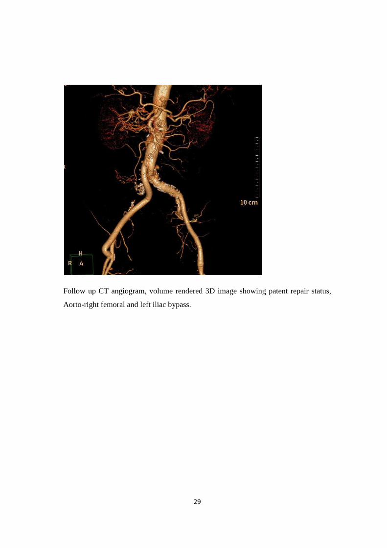
Follow up CT angiogram, volume rendered 3D image showing patent repair status,
Aorto-right femoral and left iliac bypass.