Embed Size (px)
Citation preview
Date of origin: 1998 Last review date: 2016
ACR Appropriateness Criteria® 1 Osteoporosis and Bone Mineral Density
American College of Radiology ACR Appropriateness Criteria®
Clinical Condition: Osteoporosis and Bone Mineral Density
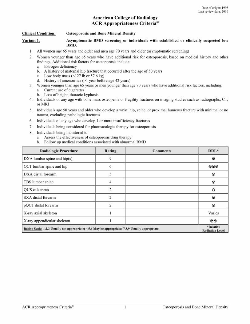
Variant 1: Asymptomatic BMD screening or individuals with established or clinically suspected low BMD.
1. All women age 65 years and older and men age 70 years and older (asymptomatic screening) 2. Women younger than age 65 years who have additional risk for osteoporosis, based on medical history and other
findings. Additional risk factors for osteoporosis include: a. Estrogen deficiency b. A history of maternal hip fracture that occurred after the age of 50 years c. Low body mass (<127 lb or 57.6 kg) d. History of amenorrhea (>1 year before age 42 years)
3. Women younger than age 65 years or men younger than age 70 years who have additional risk factors, including: a. Current use of cigarettes b. Loss of height, thoracic kyphosis
4. Individuals of any age with bone mass osteopenia or fragility fractures on imaging studies such as radiographs, CT, or MRI
5. Individuals age 50 years and older who develop a wrist, hip, spine, or proximal humerus fracture with minimal or no trauma, excluding pathologic fractures
6. Individuals of any age who develop 1 or more insufficiency fractures 7. Individuals being considered for pharmacologic therapy for osteoporosis 8. Individuals being monitored to:
a. Assess the effectiveness of osteoporosis drug therapy b. Follow up medical conditions associated with abnormal BMD
Radiologic Procedure Rating Comments RRL*
DXA lumbar spine and hip(s) 9 ☢
QCT lumbar spine and hip 6 ☢☢☢
DXA distal forearm 5 ☢
TBS lumbar spine 4 ☢
QUS calcaneus 2 O
SXA distal forearm 2 ☢
pQCT distal forearm 2 ☢
X-ray axial skeleton 1 Varies
X-ray appendicular skeleton 1 ☢☢
Rating Scale: 1,2,3 Usually not appropriate; 4,5,6 May be appropriate; 7,8,9 Usually appropriate *Relative Radiation Level
ACR Appropriateness Criteria® 2 Osteoporosis and Bone Mineral Density
Clinical Condition: Osteoporosis and Bone Mineral Density
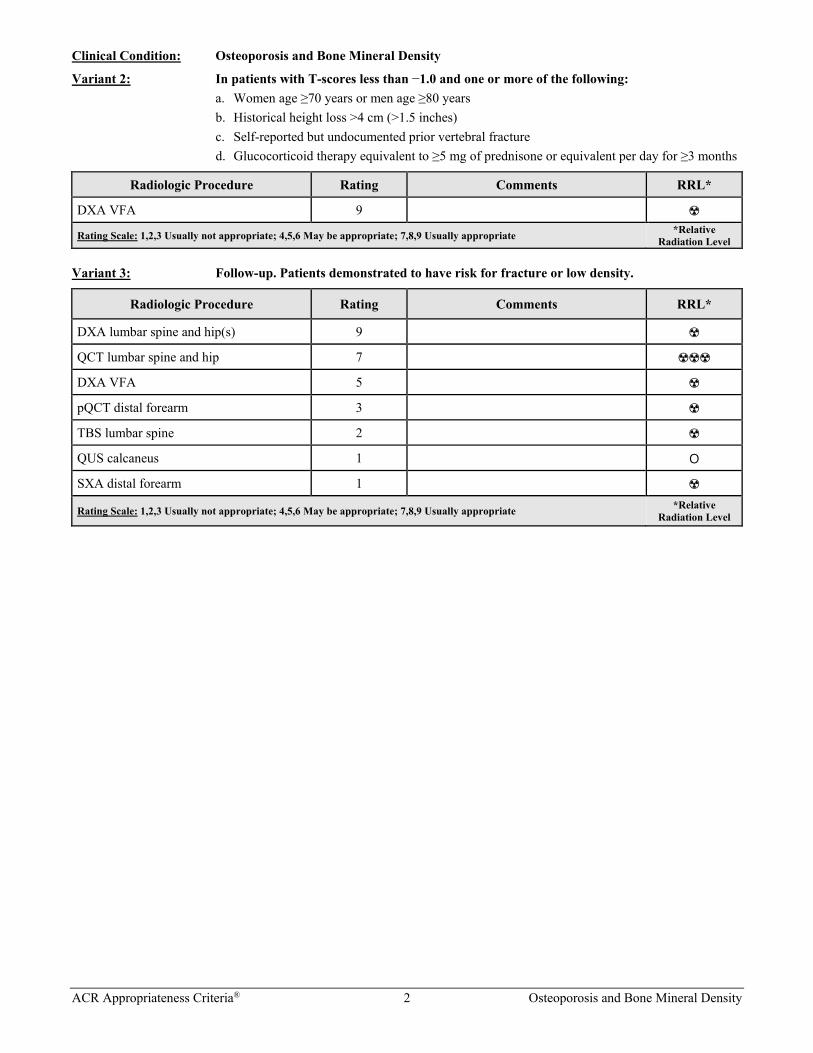
Variant 2: In patients with T-scores less than −1.0 and one or more of the following: a. Women age ≥70 years or men age ≥80 years b. Historical height loss >4 cm (>1.5 inches) c. Self-reported but undocumented prior vertebral fracture d. Glucocorticoid therapy equivalent to ≥5 mg of prednisone or equivalent per day for ≥3 months
Radiologic Procedure Rating Comments RRL*
DXA VFA 9 ☢
Rating Scale: 1,2,3 Usually not appropriate; 4,5,6 May be appropriate; 7,8,9 Usually appropriate *Relative Radiation Level
Variant 3: Follow-up. Patients demonstrated to have risk for fracture or low density.
Radiologic Procedure Rating Comments RRL*
DXA lumbar spine and hip(s) 9 ☢
QCT lumbar spine and hip 7 ☢☢☢
DXA VFA 5 ☢
pQCT distal forearm 3 ☢
TBS lumbar spine 2 ☢
QUS calcaneus 1 O
SXA distal forearm 1 ☢
Rating Scale: 1,2,3 Usually not appropriate; 4,5,6 May be appropriate; 7,8,9 Usually appropriate *Relative Radiation Level
ACR Appropriateness Criteria® 3 Osteoporosis and Bone Mineral Density
Clinical Condition: Osteoporosis and Bone Mineral Density
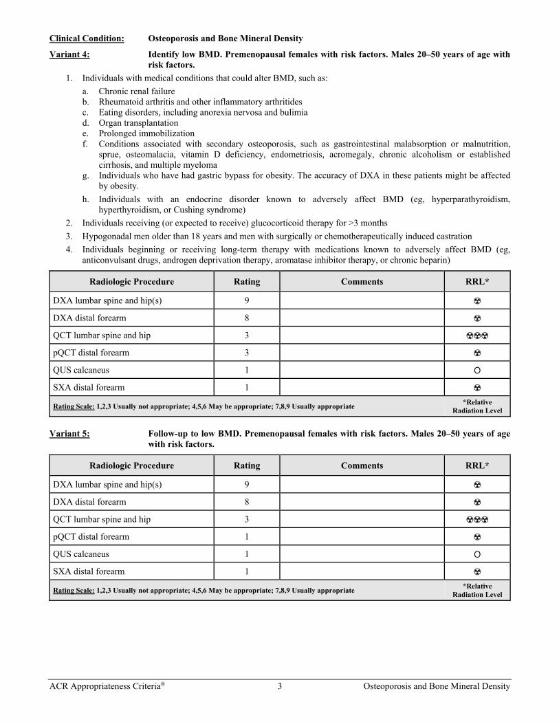
Variant 4: Identify low BMD. Premenopausal females with risk factors. Males 20–50 years of age with risk factors.
1. Individuals with medical conditions that could alter BMD, such as: a. Chronic renal failure b. Rheumatoid arthritis and other inflammatory arthritides c. Eating disorders, including anorexia nervosa and bulimia d. Organ transplantation e. Prolonged immobilization f. Conditions associated with secondary osteoporosis, such as gastrointestinal malabsorption or malnutrition,
sprue, osteomalacia, vitamin D deficiency, endometriosis, acromegaly, chronic alcoholism or established cirrhosis, and multiple myeloma
g. Individuals who have had gastric bypass for obesity. The accuracy of DXA in these patients might be affected by obesity.
h. Individuals with an endocrine disorder known to adversely affect BMD (eg, hyperparathyroidism, hyperthyroidism, or Cushing syndrome)
2. Individuals receiving (or expected to receive) glucocorticoid therapy for >3 months 3. Hypogonadal men older than 18 years and men with surgically or chemotherapeutically induced castration 4. Individuals beginning or receiving long-term therapy with medications known to adversely affect BMD (eg,
anticonvulsant drugs, androgen deprivation therapy, aromatase inhibitor therapy, or chronic heparin)
Radiologic Procedure Rating Comments RRL*
DXA lumbar spine and hip(s) 9 ☢
DXA distal forearm 8 ☢
QCT lumbar spine and hip 3 ☢☢☢
pQCT distal forearm 3 ☢
QUS calcaneus 1 O
SXA distal forearm 1 ☢
Rating Scale: 1,2,3 Usually not appropriate; 4,5,6 May be appropriate; 7,8,9 Usually appropriate *Relative Radiation Level
Variant 5: Follow-up to low BMD. Premenopausal females with risk factors. Males 20–50 years of age with risk factors.
Radiologic Procedure Rating Comments RRL*
DXA lumbar spine and hip(s) 9 ☢
DXA distal forearm 8 ☢
QCT lumbar spine and hip 3 ☢☢☢
pQCT distal forearm 1 ☢
QUS calcaneus 1 O
SXA distal forearm 1 ☢
Rating Scale: 1,2,3 Usually not appropriate; 4,5,6 May be appropriate; 7,8,9 Usually appropriate *Relative Radiation Level
ACR Appropriateness Criteria® 4 Osteoporosis and Bone Mineral Density
Clinical Condition: Osteoporosis and Bone Mineral Density
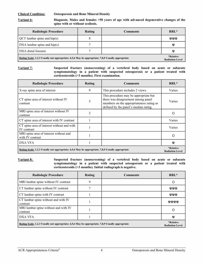
Variant 6: Diagnosis. Males and females >50 years of age with advanced degenerative changes of the spine with or without scoliosis.
Radiologic Procedure Rating Comments RRL*
QCT lumbar spine and hip(s) 8 ☢☢☢
DXA lumbar spine and hip(s) 7 ☢
DXA distal forearm 7 ☢
Rating Scale: 1,2,3 Usually not appropriate; 4,5,6 May be appropriate; 7,8,9 Usually appropriate *Relative Radiation Level
Variant 7: Suspected fracture (nonscreening) of a vertebral body based on acute or subacute symptomatology in a patient with suspected osteoporosis or a patient treated with corticosteroids (>3 months). First examination.
Radiologic Procedure Rating Comments RRL*
X-ray spine area of interest 9 This procedure includes 2 views. Varies
CT spine area of interest without IV contrast 5
This procedure may be appropriate but there was disagreement among panel members on the appropriateness rating as defined by the panel’s median rating.
Varies
MRI spine area of interest without IV contrast 2 O
CT spine area of interest with IV contrast 1 Varies CT spine area of interest without and with IV contrast 1 Varies
MRI spine area of interest without and with IV contrast 1 O
DXA VFA 1 ☢
Rating Scale: 1,2,3 Usually not appropriate; 4,5,6 May be appropriate; 7,8,9 Usually appropriate *Relative Radiation Level
Variant 8: Suspected fracture (nonscreening) of a vertebral body based on acute or subacute symptomatology in a patient with suspected osteoporosis or a patient treated with corticosteroids (>3 months). Initial radiograph is negative.
Radiologic Procedure Rating Comments RRL*
MRI lumbar spine without IV contrast 9 O
CT lumbar spine without IV contrast 7 ☢☢☢
CT lumbar spine with IV contrast 1 ☢☢☢ CT lumbar spine without and with IV contrast 1 ☢☢☢☢
MRI lumbar spine without and with IV contrast 1 O
DXA VFA 1 ☢
Rating Scale: 1,2,3 Usually not appropriate; 4,5,6 May be appropriate; 7,8,9 Usually appropriate *Relative Radiation Level
ACR Appropriateness Criteria® 5 Osteoporosis and Bone Mineral Density
Clinical Condition: Osteoporosis and Bone Mineral Density
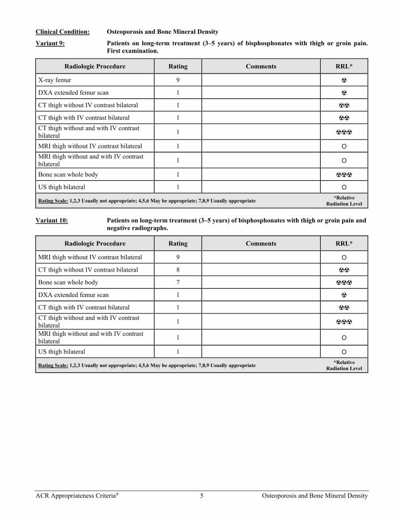
Variant 9: Patients on long-term treatment (3–5 years) of bisphosphonates with thigh or groin pain. First examination.
Radiologic Procedure Rating Comments RRL*
X-ray femur 9 ☢
DXA extended femur scan 1 ☢
CT thigh without IV contrast bilateral 1 ☢☢
CT thigh with IV contrast bilateral 1 ☢☢ CT thigh without and with IV contrast bilateral 1 ☢☢☢
MRI thigh without IV contrast bilateral 1 O MRI thigh without and with IV contrast bilateral 1 O
Bone scan whole body 1 ☢☢☢
US thigh bilateral 1 O
Rating Scale: 1,2,3 Usually not appropriate; 4,5,6 May be appropriate; 7,8,9 Usually appropriate *Relative Radiation Level
Variant 10: Patients on long-term treatment (3–5 years) of bisphosphonates with thigh or groin pain and negative radiographs.
Radiologic Procedure Rating Comments RRL*
MRI thigh without IV contrast bilateral 9 O
CT thigh without IV contrast bilateral 8 ☢☢
Bone scan whole body 7 ☢☢☢
DXA extended femur scan 1 ☢
CT thigh with IV contrast bilateral 1 ☢☢ CT thigh without and with IV contrast bilateral 1 ☢☢☢
MRI thigh without and with IV contrast bilateral 1 O
US thigh bilateral 1 O
Rating Scale: 1,2,3 Usually not appropriate; 4,5,6 May be appropriate; 7,8,9 Usually appropriate *Relative Radiation Level
ACR Appropriateness Criteria® 6 Osteoporosis and Bone Mineral Density
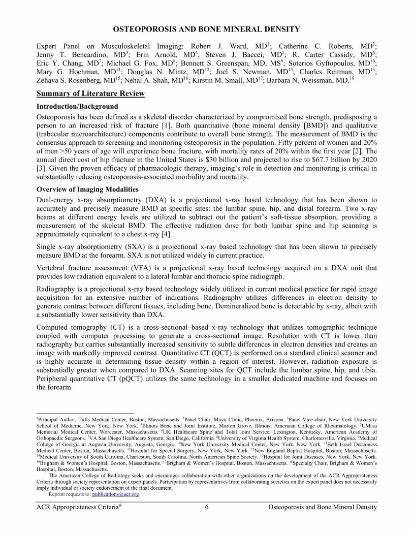
OSTEOPOROSIS AND BONE MINERAL DENSITY
Expert Panel on Musculoskeletal Imaging: Robert J. Ward, MD1; Catherine C. Roberts, MD2; Jenny T. Bencardino, MD3; Erin Arnold, MD4; Steven J. Baccei, MD5; R. Carter Cassidy, MD6; Eric Y. Chang, MD7; Michael G. Fox, MD8; Bennett S. Greenspan, MD, MS9; Soterios Gyftopoulos, MD10; Mary G. Hochman, MD11; Douglas N. Mintz, MD12; Joel S. Newman, MD13; Charles Reitman, MD14; Zehava S. Rosenberg, MD15; Nehal A. Shah, MD16; Kirstin M. Small, MD17; Barbara N. Weissman, MD.18
Summary of Literature Review Introduction/Background Osteoporosis has been defined as a skeletal disorder characterized by compromised bone strength, predisposing a person to an increased risk of fracture [1]. Both quantitative (bone mineral density [BMD]) and qualitative (trabecular microarchitecture) components contribute to overall bone strength. The measurement of BMD is the consensus approach to screening and monitoring osteoporosis in the population. Fifty percent of women and 20% of men >50 years of age will experience bone fracture, with mortality rates of 20% within the first year [2]. The annual direct cost of hip fracture in the United States is $30 billion and projected to rise to $67.7 billion by 2020 [3]. Given the proven efficacy of pharmacologic therapy, imaging’s role in detection and monitoring is critical in substantially reducing osteoporosis-associated morbidity and mortality.
Overview of Imaging Modalities Dual-energy x-ray absorptiometry (DXA) is a projectional x-ray based technology that has been shown to accurately and precisely measure BMD at specific sites: the lumbar spine, hip, and distal forearm. Two x-ray beams at different energy levels are utilized to subtract out the patient’s soft-tissue absorption, providing a measurement of the skeletal BMD. The effective radiation dose for both lumbar spine and hip scanning is approximately equivalent to a chest x-ray [4].
Single x-ray absorptiometry (SXA) is a projectional x-ray based technology that has been shown to precisely measure BMD at the forearm. SXA is not utilized widely in current practice.
Vertebral fracture assessment (VFA) is a projectional x-ray based technology acquired on a DXA unit that provides low radiation equivalent to a lateral lumbar and thoracic spine radiograph.
Radiography is a projectional x-ray based technology widely utilized in current medical practice for rapid image acquisition for an extensive number of indications. Radiography utilizes differences in electron density to generate contrast between different tissues, including bone. Demineralized bone is detectable by x-ray, albeit with a substantially lower sensitivity than DXA.
Computed tomography (CT) is a cross-sectional–based x-ray technology that utilizes tomographic technique coupled with computer processing to generate a cross-sectional image. Resolution with CT is lower than radiography but carries substantially increased sensitivity to subtle differences in electron densities and creates an image with markedly improved contrast. Quantitative CT (QCT) is performed on a standard clinical scanner and is highly accurate in determining tissue density within a region of interest. However, radiation exposure is substantially greater when compared to DXA. Scanning sites for QCT include the lumbar spine, hip, and tibia. Peripheral quantitative CT (pQCT) utilizes the same technology in a smaller dedicated machine and focuses on the forearm.
1Principal Author, Tufts Medical Center, Boston, Massachusetts. 2Panel Chair, Mayo Clinic, Phoenix, Arizona. 3Panel Vice-chair, New York University School of Medicine, New York, New York. 4Illinois Bone and Joint Institute, Morton Grove, Illinois, American College of Rheumatology. 5UMass Memorial Medical Center, Worcester, Massachusetts. 6UK Healthcare Spine and Total Joint Service, Lexington, Kentucky, American Academy of Orthopaedic Surgeons. 7VA San Diego Healthcare System, San Diego, California. 8University of Virginia Health System, Charlottesville, Virginia. 9Medical College of Georgia at Augusta University, Augusta, Georgia. 10New York University Medical Center, New York, New York. 11Beth Israel Deaconess Medical Center, Boston, Massachusetts. 12Hospital for Special Surgery, New York, New York. 13New England Baptist Hospital, Boston, Massachusetts. 14Medical University of South Carolina, Charleston, South Carolina, North American Spine Society. 15Hospital for Joint Diseases, New York, New York. 16Brigham & Women’s Hospital, Boston, Massachusetts. 17Brigham & Women’s Hospital, Boston, Massachusetts. 18Specialty Chair, Brigham & Women’s Hospital, Boston, Massachusetts. The American College of Radiology seeks and encourages collaboration with other organizations on the development of the ACR Appropriateness Criteria through society representation on expert panels. Participation by representatives from collaborating societies on the expert panel does not necessarily imply individual or society endorsement of the final document. Reprint requests to: [email protected]
ACR Appropriateness Criteria® 7 Osteoporosis and Bone Mineral Density
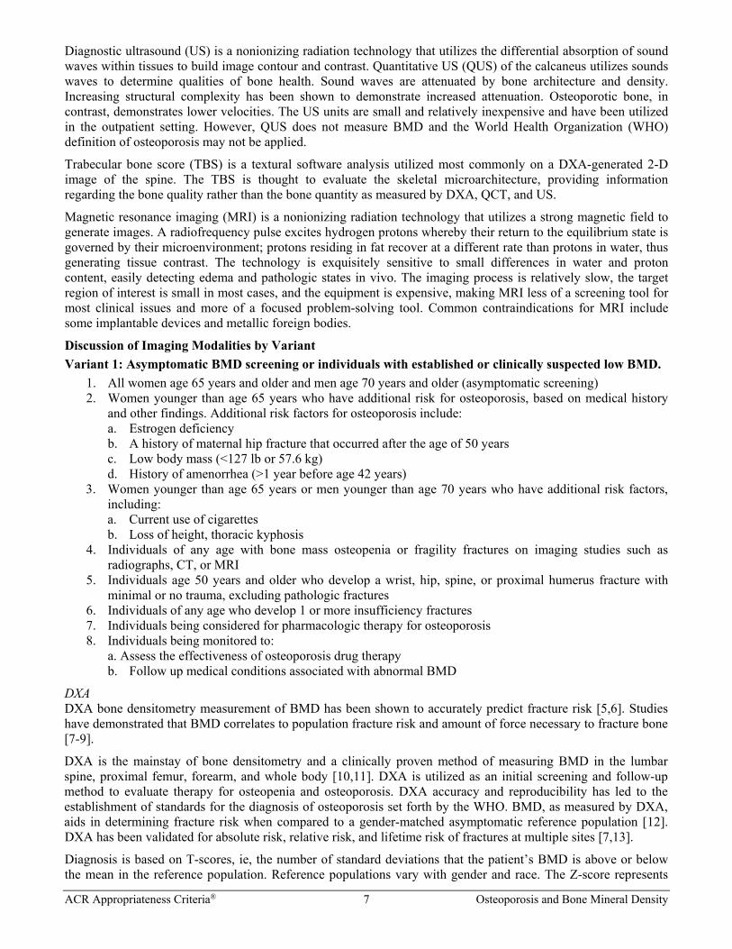
Diagnostic ultrasound (US) is a nonionizing radiation technology that utilizes the differential absorption of sound waves within tissues to build image contour and contrast. Quantitative US (QUS) of the calcaneus utilizes sounds waves to determine qualities of bone health. Sound waves are attenuated by bone architecture and density. Increasing structural complexity has been shown to demonstrate increased attenuation. Osteoporotic bone, in contrast, demonstrates lower velocities. The US units are small and relatively inexpensive and have been utilized in the outpatient setting. However, QUS does not measure BMD and the World Health Organization (WHO) definition of osteoporosis may not be applied.
Trabecular bone score (TBS) is a textural software analysis utilized most commonly on a DXA-generated 2-D image of the spine. The TBS is thought to evaluate the skeletal microarchitecture, providing information regarding the bone quality rather than the bone quantity as measured by DXA, QCT, and US.
Magnetic resonance imaging (MRI) is a nonionizing radiation technology that utilizes a strong magnetic field to generate images. A radiofrequency pulse excites hydrogen protons whereby their return to the equilibrium state is governed by their microenvironment; protons residing in fat recover at a different rate than protons in water, thus generating tissue contrast. The technology is exquisitely sensitive to small differences in water and proton content, easily detecting edema and pathologic states in vivo. The imaging process is relatively slow, the target region of interest is small in most cases, and the equipment is expensive, making MRI less of a screening tool for most clinical issues and more of a focused problem-solving tool. Common contraindications for MRI include some implantable devices and metallic foreign bodies.
Discussion of Imaging Modalities by Variant Variant 1: Asymptomatic BMD screening or individuals with established or clinically suspected low BMD.
1. All women age 65 years and older and men age 70 years and older (asymptomatic screening) 2. Women younger than age 65 years who have additional risk for osteoporosis, based on medical history
and other findings. Additional risk factors for osteoporosis include: a. Estrogen deficiency b. A history of maternal hip fracture that occurred after the age of 50 years c. Low body mass (<127 lb or 57.6 kg) d. History of amenorrhea (>1 year before age 42 years)
3. Women younger than age 65 years or men younger than age 70 years who have additional risk factors, including: a. Current use of cigarettes b. Loss of height, thoracic kyphosis
4. Individuals of any age with bone mass osteopenia or fragility fractures on imaging studies such as radiographs, CT, or MRI
5. Individuals age 50 years and older who develop a wrist, hip, spine, or proximal humerus fracture with minimal or no trauma, excluding pathologic fractures
6. Individuals of any age who develop 1 or more insufficiency fractures 7. Individuals being considered for pharmacologic therapy for osteoporosis 8. Individuals being monitored to:
a. Assess the effectiveness of osteoporosis drug therapy b. Follow up medical conditions associated with abnormal BMD
DXA DXA bone densitometry measurement of BMD has been shown to accurately predict fracture risk [5,6]. Studies have demonstrated that BMD correlates to population fracture risk and amount of force necessary to fracture bone [7-9].
DXA is the mainstay of bone densitometry and a clinically proven method of measuring BMD in the lumbar spine, proximal femur, forearm, and whole body [10,11]. DXA is utilized as an initial screening and follow-up method to evaluate therapy for osteopenia and osteoporosis. DXA accuracy and reproducibility has led to the establishment of standards for the diagnosis of osteoporosis set forth by the WHO. BMD, as measured by DXA, aids in determining fracture risk when compared to a gender-matched asymptomatic reference population [12]. DXA has been validated for absolute risk, relative risk, and lifetime risk of fractures at multiple sites [7,13].
Diagnosis is based on T-scores, ie, the number of standard deviations that the patient’s BMD is above or below the mean in the reference population. Reference populations vary with gender and race. The Z-score represents

ACR Appropriateness Criteria® 8 Osteoporosis and Bone Mineral Density
the number of standard deviations above or below the mean of age-matched controls. Z-scores are utilized to detect secondary causes of osteoporosis.
The WHO defines normal BMD as a T-score greater than −1.0. Low bone mass or osteopenia is defined as −1.0 to −2.4, whereas T-scores equal to or less than −2.5 indicate osteoporosis [14]. The National Osteoporosis Foundation (NOF) recommends pharmacologic treatment for all postmenopausal women and men over age 50 years with a T-score equal to or less than −2.5. In patients with low bone mass but T-scores greater than −2.5 (−1.0 to −2.4), a fracture risk assessment tool, FRAX, is utilized. The FRAX tool factors include hip BMD, age, sex, height, weight, family history of hip fracture, smoking, steroid use >3 months, rheumatoid arthritis, and alcohol use [15]. The FRAX algorithm is country specific and intended for the use of previously untreated postmenopausal women and men aged 40–90 years. The NOF recommends treatment in patients with a 10-year probability of a hip fracture ≥3% or a 10-year probability of a major osteoporosis-related fracture ≥20% based on FRAX [16].
Two sites are routinely evaluated with DXA: the lumbar spine and hip. Up to 4 vertebral levels, L1-L4, are measured and reported in addition to 2 regions of the hip: the total and the neck [12]. In the event of artifactually elevated lumbar spine BMD due to fracture, facet joint osteoarthritis, or spondylosis, up to 2 vertebral levels can be excluded. If >2 levels are to be excluded, scans of the second hip can be obtained as a substitute for the spine [12]. Routinely, 1 hip is obtained as part of the standard DXA scan. If only 1 hip is available for scanning, a third site, the distal forearm, is scanned.
The third site, the forearm, is primarily utilized for patients with hyperparathyroidism. Primary hyperparathyroidism preferentially decreases mineralization at cortical-rich sites such as the hip and mid radius, in contrast to the predominantly cancellous lumbar spine. The addition of the one-third radius as the third DXA site is indicated in these patients [12]. The Third International Workshop on Hyperparathyroidism recommendations include DXA scanning every 1–2 years as well as a parathyroidectomy in patients with T-scores equal to or less than −2.5 at any of the 3 sites [17].
Quantitative Computed Tomography QCT provides a volumetric BMD, in contrast to the areal BMD of the DXA, which is based on a 2-D projectional area measurement. QCT can be performed on the vast majority of commercially available CT scanners, provided they include densitometry analysis software and a calibration phantom.
The WHO’s spine T-scores that define osteoporosis were derived from DXA measurements and do not apply to QCT. One significant difference in technologies relates to monitoring. Spine BMD values measured by QCT demonstrate relatively increased rates of bone loss with advanced age when compared to DXA values due to the exclusively cancellous bone measurements of QCT—the rate of change in cancellous bone is greater than that of cortical bone. By contrast, the projectional properties of DXA summate the cortically predominant end plates and posterior elements with the cancellous vertebral body measurements, thereby decreasing their rate of change. The American College of Radiology (ACR) parameters indicate that BMD values from 120–80 mg/mL are defined as low bone mass/osteopenic, whereas values <80 mg/mL are deemed compatible with osteoporosis [18].
Projectional QCT of the hip, by contrast, provides a calculated postprocessed areal BMD that is comparable to DXA, thus enabling the use of the WHO classification system. The definition of osteoporosis as a T-score of greater than or equal to −2.5 is applicable to QCT.
Indications for QCT include the same indications as DXA; however, DXA is recommended as the first-line screening and follow-up test for bone density. If DXA is not available, QCT may be used as a secondary technique. Specific cases in which QCT is considered superior to DXA include: 1. Extremes in body height (ie, very large and very small patients) 2. Patients with extensive degenerative disease of the spine 3. Severely obese patients (BMI >35 kg/m2) 4. A clinical scenario that requires increased sensitivity to small changes in trabecular bone density (parathyroid
hormone and glucocorticoid treatment monitoring) [19]
Radiographs Although radiographs may detect fragility fractures, their use as a primary screening tool is limited due to their low sensitivity to bone loss [20]. Alternatively, patients with radiographs interpreted as demonstrating radiographic osteopenia and/or fragility fractures should be referred to DXA for further characterization.
ACR Appropriateness Criteria® 9 Osteoporosis and Bone Mineral Density
Peripheral Ultrasound Peripheral ultrasound (QUS) represents a low-cost alternative easily accessible to primary care providers. The heel represents the only validated site for the clinical use of QUS [21-24]. QUS does not measure BMD and therefore the WHO classification system cannot be utilized and a diagnosis of osteoporosis cannot be made. Additionally, discordance between QUS and central DXA is not infrequent [22]. However, QUS in conjunction with clinical risk factors can predict an increased risk for fractures as well as identifying populations that demonstrate no increased risk [22].
Single X-ray Absorptiometry SXA is utilized at the forearm and is less expensive than central DXA. BMD evaluation is comparable at the forearm with DXA. However, the tests are substantially less predictive of hip and spine fractures relative to central DXA [25].
Peripheral Quantitative Computed Tomography pQCT utilizes scans of the forearm. pQCT radiation is low relative to central QCT and its accuracy is superior to DXA [26]. However, correlation with central DXA is poor and the high variability of positioning limits the use of pQCT as a screening tool [27]. The WHO classification cannot be utilized. pQCT of the forearm has been shown to predict hip, but not spine, fractures in postmenopausal women [26]. There is a lack of evidence to support utilization for men.
Trabecular Bone Score TBS is a textural method derived from DXA spine images to describe skeletal microarchitecture. TBS measures relative pixel amplitude variations summing the squared gray-level differences. Porous osteoporotic bone has been shown to have lower values than normal bone [28]. Unlike BMD measurements, TBS quantifies the bone microarchitecture, yielding a metric of bone quality. Cross-sectional and prospective studies have demonstrated associations with fracture in postmenopausal women [29-35]. When TBS is utilized in conjunction with BMD, marginal improvements in fracture risk predication are achieved. TBS has been found to show incremental improvement in fracture prediction when utilized with FRAX [31,36]. TBS may be of use in stratifying risk in patients with relatively normal or marginally osteopenic BMD values as most fractures are in this nonosteoporotic subset of patients [30].
Significantly reduced TBSs are associated with fragility fractures in secondary osteoporosis. In these patients, TBS has been found to have a substantially higher association with fracture risk than BMD [37-39]. TBSs in diabetic, glucocorticoid-receiving, and hyperparathyroid patients demonstrated increased fracture risk even in the setting of normal BMD [34].
TBS may have a role in evaluating patients with secondary causes of osteoporosis as well as patients with relatively high BMD but increased fracture risk.
Variant 2: Vertebral fracture assessment. VFA is a feature of DXA scanners in which lateral thoracic and lumbar spine images are obtained and screened for fracture. The detection of fractures in some patients with low bone mineralization is a predictor of future fractures and allows for their risk restratification and potential initiation of pharmacotherapy [40].
Indications for VFA include patients with T-scores less than −1.0 and 1 or more of the following: a. Women age ≥70 years or men age ≥80 years b. Historical height loss >4 cm (>1.5 inches) c. Self-reported but undocumented prior vertebral fracture d. Glucocorticoid therapy equivalent to ≥5 mg of prednisone or equivalent per day for ≥3 months [41]
A semiquantitative method is utilized for evaluation based on morphometry. Vertebrae are characterized by the shape (wedge, concave, or crush) and by location of the defect (anterior, posterior, and/or middle). The semiquantitative scoring of fractures relies on a comparison to an atlas as well as experience in image interpretation [42]. The number and severity of fractures are associated with increased risk of fracture independent of the BMD. Fracture severity is graded on a scale from 1 to 3 [43].
The utility of VFA is in the identification of patients who would otherwise not qualify for treatment under the guidelines of the NOF, which are based solely on BMD measurements. Several studies have demonstrated such populations of patients who were reclassified due to vertebral fracture [44-46]. The study in the Netherlands
ACR Appropriateness Criteria® 10 Osteoporosis and Bone Mineral Density
demonstrated that 60% of the patients found to have a fracture on VFA were in the nonosteoporotic range, and of these, 74% were previously unknown to have fractures [44]. Three older studies have demonstrated that 10%–17% of patients with osteopenia as measured by DXA had grade 2 or 3 vertebral fractures detected by VFA [47-49].
As 50% of fragility fractures occur in postmenopausal women who have T-scores greater than −2.5, identification of this population’s increased risk is essential for potential medical treatment that has been shown to be beneficial in multiple studies [50-53].
Variant 3: Follow-up. Patients demonstrated to have risk for fracture or low density. DXA Follow-up DXA scanning is important for both untreated and treated patients. Patients receiving treatment who demonstrate decreasing BMD on follow-up scans may require an adjustment in their pharmacotherapy regimen [54]. Alternatively, nontreated patients with statistically significant decreasing BMD may require consideration for therapy initiation in the setting of primary osteoporosis or clinical correlation for identifying potential secondary causes of osteoporosis [55].
It is essential for patients to be scanned on the same DXA scanner as vendor differences in technologies prohibit a direct comparison unless cross calibration has been performed. BMD values, not T-scores, are compared between the previous and current scans. The monitoring time interval is based upon the expected rate of change of bone mineralization and is typically 2 years [55]. In patients initiating therapy or in patients that are thought to be at risk for substantial short-term decreases in mineralization, such as patients receiving glucocorticoid therapy, a 1-year follow-up is recommended [55]. Scan intervals <1 year are discouraged [55].
QCT and pQCT Both QCT and pQCT demonstrate excellent precision and reproducibility and can be used for the monitoring of BMD in untreated and treated patients [19]. QCT is more sensitive to change in comparison to DXA due to its isolated trabecular measurement—the portion of bone most sensitive to rapid mineralization changes [56].
QUS US monitoring is considered inadequate for follow-up in either treated or untreated patients [55]. Central DXA is the recommended method.
SXA Although the precision of SXA is comparable to DXA, the correlation with proximal femoral fracture risk is low and central DXA is preferred [57].
TBS Data indicate that smaller changes on follow-up are seen in TBS on follow-up in contrast to DXA-derived BMD. No TBS data exist supporting follow-up of either treated or untreated patients [58-60]. Interestingly, different classes of therapeutic medications have demonstrated variable degrees of change, with teriparatide yielding greater increases than bisphosphonates [60,61].
VFA If VFA was performed in the initial DXA study or if the patient now meets the criteria for inclusion, a follow-up VFA scan is recommended concurrently with DXA scanning intervals.
Variant 4: Identify low BMD. Premenopausal females with risk factors. Males 20–50 years of age with risk factors.
1. Individuals with medical conditions that could alter BMD, such as: a. Chronic renal failure b. Rheumatoid arthritis and other inflammatory arthritides c. Eating disorders, including anorexia nervosa and bulimia d. Organ transplantation e. Prolonged immobilization f. Conditions associated with secondary osteoporosis, such as gastrointestinal malabsorption or
malnutrition, sprue, osteomalacia, vitamin D deficiency, endometriosis, acromegaly, chronic alcoholism or established cirrhosis, and multiple myeloma
ACR Appropriateness Criteria® 11 Osteoporosis and Bone Mineral Density
g. Individuals who have had gastric bypass for obesity. The accuracy of DXA in these patients might be affected by obesity.
h. Individuals with an endocrine disorder known to adversely affect BMD (eg, hyperparathyroidism, hyperthyroidism, or Cushing syndrome)
2. Individuals receiving (or expected to receive) glucocorticoid therapy for >3 months 3. Hypogonadal men older than 18 years and men with surgically or chemotherapeutically induced
castration 4. Individuals beginning or receiving long-term therapy with medications known to adversely affect BMD
(eg, anticonvulsant drugs, androgen deprivation therapy, aromatase inhibitor therapy, or chronic heparin)
DXA DXA represents the primary modality for the evaluation of bone mineralization in this patient population. The WHO criteria do not apply and only Z-scores (not T-scores) should be reported [55]. The Z-score represents gender- and age-matched controls for the evaluation of secondary osteoporosis. Z-scores of −2.0 or less are considered to be below the expected age range [55]. Additionally, a diagnosis of osteoporosis cannot be made in men under the age of 50 on the basis of BMD alone [55].
QCT and pQCT A study utilizing QCT in premenopausal women demonstrated a good correlation between QCT-generated BMD and DXA-generated BMD. However, the study was based on 32 subjects and was inconclusive [62]. A variant of pQCT, high-resolution pQCT (HR-pQCT), has demonstrated correlation in premenopausal women between peripheral stiffness, measured at the forearm and distal tibia, and mineralization, measured by DXA at the spine and hip [63]. An alternative study demonstrated a weak relationship between peripheral and central mechanical competence [64].
QUS The benefits of QUS in the premenopausal population are unclear. One study from Italy demonstrated that risk factors associated with low BMD measured by DXA in elderly women are also associated with calcaneal bone stiffness, as measured by QUS in premenopausal women [65].
SXA The role of SXA is limited in the premenopausal population.
Variant 5: Follow-up to low BMD. Premenopausal females with risk factors. Males 20–50 years of age with risk factors. Follow-up for premenopausal women and men aged 20–50 years is based on the underlying clinical conditions. It is unlikely that changes greater than the computed least significant change divided by the expected mean change in BMD would require a monitoring time interval <1 year.
DXA DXA is the primary modality by which to monitor bone density in the premenopausal population. The need for follow-up DXA is dictated by the clinical circumstance of the patients.
QCT and pQCT QCT is an accurate means by which to follow premenopausal women and men between the ages of 20–50 years. pQCT and HR-pQCT have utility in research, but no validated follow-up data exist in premenopausal women demonstrating a link between distal architecture and central fracture risk.
QUS QUS is considered inappropriate for follow-up monitoring in both the post- and premenopausal populations. The same applies for men of all ages.
SXA No SXA follow-up data exist in premenopausal women utilizing this modality.
Variant 6: Diagnosis. Males and females >50 years of age with advanced degenerative changes of the spine with or without scoliosis. DXA Given the projectional nature of DXA, spuriously elevated values of the lumbar spine may be encountered in patients with spondylosis and degenerative facet osteoarthritis. Such findings may involve 1 or more of the L1
ACR Appropriateness Criteria® 12 Osteoporosis and Bone Mineral Density
through L4 levels scanned. The International Society of Clinical Densitometry recommends that no more than 2 levels may be excluded from the overall calculation of lumbar spine BMD [55]. If more than 2 levels are involved, then the entire spine should be excluded from evaluation [55]. In this circumstance the recommendation is to scan the contralateral hip in the affected patient [55]. It must be taken into consideration, however, that the predominance of cortical bone in the hip is less sensitive to change than the cancellous rich bone within the vertebrae. If the contralateral hip is unsuitable for scanning due to arthroplasty or advanced degeneration, for instance, then the distal forearm can be substituted.
QCT QCT is ideally suited for the evaluation of the vertebral body in the setting of advanced degeneration as it selectively samples only the cancellous portion of the vertebral body, excluding the end plates, cortices, and posterior elements. Sensitivity to change is greater in QCT than in DXA in degenerative spines [66].
Variant 7: Suspected fracture (nonscreening) of a vertebral body based on acute or subacute symptomatology in a patient with suspected osteoporosis or a patient treated with corticosteroids (>3 months). First examination.
Variant 8: Suspected fracture (nonscreening) of a vertebral body based on acute or subacute symptomatology in a patient with suspected osteoporosis or a patient treated with corticosteroids (>3 months). Initial radiograph is negative. Radiography Radiography has been considered the first line of imaging in the assessment of the spine. However, in a large study evaluating 2452 lateral lumbar and thoracic spine radiographs, community radiologists missed approximately 32% of vertebral fractures when compared to expert readers utilizing the semiquantitative morphometry method described by Genant [67]. In a similar study, community radiologists demonstrated a false-positive rate of 26% versus experts utilizing the method of Genant [68]. The routine utilization of a semiquantitative method of radiographic evaluation of the spine may improve fracture detection and ought to be considered by practicing radiologists [69].
CT Studies comparing radiography versus CT in the setting of spine trauma have indicated the substantially higher sensitivity and specificity of CT [70]. However, no current evidence exists comparing the 2 modalities in the setting of atraumatic fracture in suspected osteoporosis. Substantial differences exist in the mechanism of injury, trabecular architecture, bone mineralization, patterns of fracture, and pretest probability, which raises doubt that the trauma data apply. However, the relative cost of radiographs and comparatively favorable radiation profile in contrast to CT suggest that radiographs likely remain the best first test with respect to the detection of vertebral fracture in the setting of osteoporosis. The use of intravenous contrast has not been shown to demonstrate utility in evaluation for spine fracture.
MRI MRI studies have described the appearance of osteoporotic vertebral fractures [71-73] and have demonstrated ≥99% sensitivity [74,75]. Given the cost, speed, and availability of MRI, it is not recommended as a primary diagnostic tool. MRI is useful as a secondary choice to either exclude a potential underlying malignancy or in the setting of a negative radiograph evaluate for the presence of a radio-occult fracture. The use of intravenous contrast has not been shown to demonstrate utility in evaluation for spine fracture.
VFA Studies have compared the diagnostic accuracy of VFA with spine radiography. These studies were performed in patients with osteoporosis and demonstrated a VFA sensitivity and specificity ranging from 50% to 95% and 82% to 98% when compared to radiography [76,77]. Two studies examined VFA accuracy in the nonosteoporotic population [78,79]. Results demonstrated lower sensitivity and specificity in the subset of patients with normal bone mineralization. However, in one study, radiograph evaluation was performed by nonradiologists. As grade 1 fractures are considered to pose less risk of incident fracture, decreased sensitivity and specificity at the lower end of the semiquantitative scale are thought to be less clinically relevant [42].
ACR Appropriateness Criteria® 13 Osteoporosis and Bone Mineral Density
Variant 9: Patients on long-term treatment (3–5 years) of bisphosphonates with thigh or groin pain. First examination.
Variant 10: Patients on long-term treatment (3–5 years) of bisphosphonates with thigh or groin pain and negative radiographs. Atypical subtrochanteric fractures in patients receiving long-term (for approximately 5 years) bisphosphonate therapy have been reported in the literature. This paradoxical relationship is associated with unique radiographic imaging features that differ from typical subtrochanteric fractures related to trauma. Atypical fracture recognition is critical to patient treatment. A task force of the American Society for Bone and Mineral Research (ASBMR) published a table describing major and minor features related to atypical femoral fractures in 2010 and then later modified in the table in 2013. A summary of the table is presented here.
Major features: a. The fracture is associated with minimal or no trauma, as in a fall from a standing height or less. b. The fracture line originates at the lateral cortex and is substantially transverse in its orientation, although it
may become oblique as it progresses medially across the femur. c. Complete fractures extend through both cortices and may be associated with a medial spike; incomplete
fractures involve only the lateral cortex. d. The fracture is noncomminuted or minimally comminuted. e. Localized periosteal or endosteal thickening of the lateral cortex is present at the fracture site (“beaking” or
“flaring”).
Minor features: a. Generalized increase in cortical thickness of the femoral diaphysis b. Unilateral or bilateral prodromal symptoms such as dull or aching pain in the groin or thigh c. Bilateral incomplete or complete femoral diaphysis fractures d. Delayed fracture healing [80]
No causal relationship between atypical fractures and bisphosphonates has been proven, but theories suggesting diminished remodeling due to long-term osteoclast arrest have been suggested [80-82].
Radiography Radiographs have been shown to be reliable in evaluating bisphosphonate-related fractures in one study, with sensitivity and specificity ranging from 90%–95% and 68%–100% [83]. A number of studies have described bilateral involvement, up to 30%, and consideration for imaging the contralateral femur has been suggested by the ASBMR task force [80,84].
DXA Although studies have shown that atypical femoral fractures may be detected by DXA, the use of DXA over radiography in patients with prodromal symptoms to evaluate for atypical femoral fracture is not recommended [85,86]. There may be a screening role for extended-field DXA in patients on long-term therapy in the asymptomatic patient as a recently presented abstract by Cheung et al [87] demonstrated 66%–75% sensitivity for a single energy-extended femur scan in the detection of an atypical fracture. Radiography was utilized as the gold standard in this study.
CT CT has a problem-solving role in evaluating atypical fractures. In patients with contraindications to MR, CT is useful for discerning the presence of lucency in incomplete fractures [88,89]. Such lucency has been suggested as an indication for intramedullary nailing [80]. CT is also useful in excluding the presence of a neoplastic-related pathologic fracture. The use of intravenous contrast has not been shown to demonstrate utility in the evaluation for proximal femoral fracture.
MRI The primary utility of MRI is to determine both the presence of radio-occult stress-related changes common in atypical femoral fractures and to define the extent of involvement of the cortex; the degree of cortical involvement determines whether patients receive conservative or surgical treatment [80]. MRI has been shown to demonstrate subtle marrow signal abnormality and cortical thickening [89]. Contralateral MR scanning in the setting of a
ACR Appropriateness Criteria® 14 Osteoporosis and Bone Mineral Density
negative contralateral but positive ipsilateral radiograph has been advocated by the ASBMR task force [80]. The use of intravenous contrast has not been shown to demonstrate utility in evaluation for proximal femoral fracture.
Bone Scintigraphy Technetium-labeled bone scan has been shown to demonstrate atypical femoral fractures. [89] It is considered a second-line diagnostic test following negative radiography in those patients with prodromal symptoms/suspicion for fracture who cannot undergo MRI or CT evaluation [89].
US No data exist regarding the utility of US and atypical femoral fracture.
Summary of Recommendations • DXA is the primary diagnostic choice by which to screen women >65 years of age and men >70 years of age
for osteoporosis. • DXA is indicated in postmenopausal women <65 years of age with additional risk factors for fracture. • DXA is the primary diagnostic choice by which to follow patients’ BMD. • VFA represents a useful screening study to identify patients at risk whose BMD may be above treatment
thresholds. • QCT can be utilized to evaluate baseline and follow-up BMD. • Patients on long-term bisphosphonate therapy who present with thigh or groin pain should be imaged
bilaterally with radiography followed by MRI. • Extended-femoral-view DXA is not a substitute for femoral radiography in the setting of thigh or groin pain
in long-term bisphosphonate patients.
Summary of Evidence Of the 89 references cited in the ACR Appropriateness Criteria® Osteoporosis and Bone Mineral Density document, 80 are categorized as diagnostic references including 1 well designed study, 6 good quality studies, and 17 quality studies that may have design limitations. Additionally, 6 references are categorized as therapeutic references including 4 well designed studies and 2 good quality studies. There are 56 references that may not be useful as primary evidence. There are 3 references that are meta-analysis studies.
The 89 references cited in the ACR Appropriateness Criteria® Osteoporosis and Bone Mineral Density document were published from 1967-2015.
While there are references that report on studies with design limitations, 13 well designed or good quality studies provide good evidence.
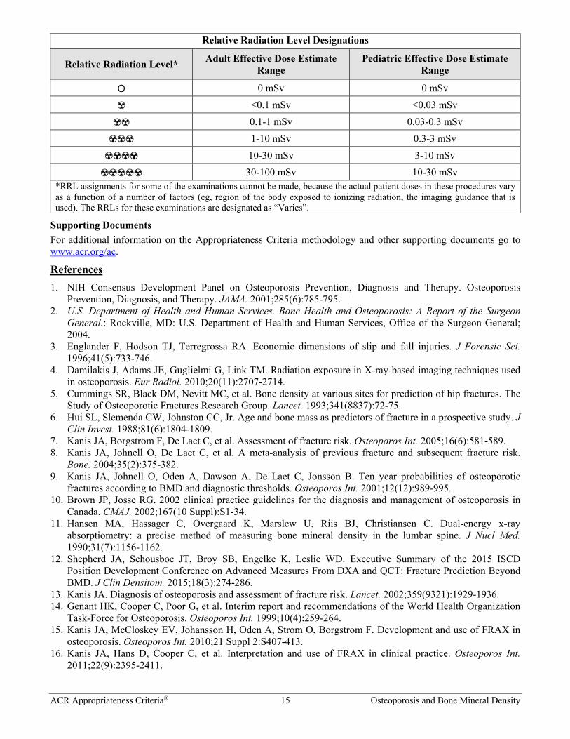
Relative Radiation Level Information Potential adverse health effects associated with radiation exposure are an important factor to consider when selecting the appropriate imaging procedure. Because there is a wide range of radiation exposures associated with different diagnostic procedures, a relative radiation level (RRL) indication has been included for each imaging examination. The RRLs are based on effective dose, which is a radiation dose quantity that is used to estimate population total radiation risk associated with an imaging procedure. Patients in the pediatric age group are at inherently higher risk from exposure, both because of organ sensitivity and longer life expectancy (relevant to the long latency that appears to accompany radiation exposure). For these reasons, the RRL dose estimate ranges for pediatric examinations are lower as compared to those specified for adults (see Table below). Additional information regarding radiation dose assessment for imaging examinations can be found in the ACR Appropriateness Criteria® Radiation Dose Assessment Introduction document.
ACR Appropriateness Criteria® 15 Osteoporosis and Bone Mineral Density
Relative Radiation Level Designations
Relative Radiation Level* Adult Effective Dose Estimate Range
Pediatric Effective Dose Estimate Range
O 0 mSv 0 mSv
☢ <0.1 mSv <0.03 mSv
☢☢ 0.1-1 mSv 0.03-0.3 mSv
☢☢☢ 1-10 mSv 0.3-3 mSv
☢☢☢☢ 10-30 mSv 3-10 mSv
☢☢☢☢☢ 30-100 mSv 10-30 mSv *RRL assignments for some of the examinations cannot be made, because the actual patient doses in these procedures vary as a function of a number of factors (eg, region of the body exposed to ionizing radiation, the imaging guidance that is used). The RRLs for these examinations are designated as “Varies”.
Supporting Documents For additional information on the Appropriateness Criteria methodology and other supporting documents go to www.acr.org/ac.
References 1. NIH Consensus Development Panel on Osteoporosis Prevention, Diagnosis and Therapy. Osteoporosis
Prevention, Diagnosis, and Therapy. JAMA. 2001;285(6):785-795. 2. U.S. Department of Health and Human Services. Bone Health and Osteoporosis: A Report of the Surgeon
General.: Rockville, MD: U.S. Department of Health and Human Services, Office of the Surgeon General; 2004.
3. Englander F, Hodson TJ, Terregrossa RA. Economic dimensions of slip and fall injuries. J Forensic Sci. 1996;41(5):733-746.
4. Damilakis J, Adams JE, Guglielmi G, Link TM. Radiation exposure in X-ray-based imaging techniques used in osteoporosis. Eur Radiol. 2010;20(11):2707-2714.
5. Cummings SR, Black DM, Nevitt MC, et al. Bone density at various sites for prediction of hip fractures. The Study of Osteoporotic Fractures Research Group. Lancet. 1993;341(8837):72-75.
6. Hui SL, Slemenda CW, Johnston CC, Jr. Age and bone mass as predictors of fracture in a prospective study. J Clin Invest. 1988;81(6):1804-1809.
7. Kanis JA, Borgstrom F, De Laet C, et al. Assessment of fracture risk. Osteoporos Int. 2005;16(6):581-589. 8. Kanis JA, Johnell O, De Laet C, et al. A meta-analysis of previous fracture and subsequent fracture risk.
Bone. 2004;35(2):375-382. 9. Kanis JA, Johnell O, Oden A, Dawson A, De Laet C, Jonsson B. Ten year probabilities of osteoporotic
fractures according to BMD and diagnostic thresholds. Osteoporos Int. 2001;12(12):989-995. 10. Brown JP, Josse RG. 2002 clinical practice guidelines for the diagnosis and management of osteoporosis in
Canada. CMAJ. 2002;167(10 Suppl):S1-34. 11. Hansen MA, Hassager C, Overgaard K, Marslew U, Riis BJ, Christiansen C. Dual-energy x-ray
absorptiometry: a precise method of measuring bone mineral density in the lumbar spine. J Nucl Med. 1990;31(7):1156-1162.
12. Shepherd JA, Schousboe JT, Broy SB, Engelke K, Leslie WD. Executive Summary of the 2015 ISCD Position Development Conference on Advanced Measures From DXA and QCT: Fracture Prediction Beyond BMD. J Clin Densitom. 2015;18(3):274-286.
13. Kanis JA. Diagnosis of osteoporosis and assessment of fracture risk. Lancet. 2002;359(9321):1929-1936. 14. Genant HK, Cooper C, Poor G, et al. Interim report and recommendations of the World Health Organization
Task-Force for Osteoporosis. Osteoporos Int. 1999;10(4):259-264. 15. Kanis JA, McCloskey EV, Johansson H, Oden A, Strom O, Borgstrom F. Development and use of FRAX in
osteoporosis. Osteoporos Int. 2010;21 Suppl 2:S407-413. 16. Kanis JA, Hans D, Cooper C, et al. Interpretation and use of FRAX in clinical practice. Osteoporos Int.
2011;22(9):2395-2411.
ACR Appropriateness Criteria® 16 Osteoporosis and Bone Mineral Density
17. Bilezikian JP, Khan AA, Potts JT, Jr. Guidelines for the management of asymptomatic primary hyperparathyroidism: summary statement from the third international workshop. J Clin Endocrinol Metab. 2009;94(2):335-339.
18. American College of Radiology. ACR–SPR–SSR Practice Parameter for the Performance of Dual-Energy X-Ray Absorptiometry (DXA) . Available at: http://www.acr.org/~/media/ACR/Documents/PGTS/guidelines/DXA.pdf. 2014.
19. American College of Radiology. ACR–SPR–SSR Practice Parameter for the Performance of Quantitative Computed Tomography (QCT) Bone Densitometry. Available at: http://www.acr.org/~/media/ACR/Documents/PGTS/guidelines/QCT.pdf. 2014.
20. Edelstyn GA, Gillespie PJ, Grebbell FS. The radiological demonstration of osseous metastases. Experimental observations. Clin Radiol. 1967;18(2):158-162.
21. Durosier C, Hans D, Krieg MA, Schott AM. Prediction and discrimination of osteoporotic hip fracture in postmenopausal women. J Clin Densitom. 2006;9(4):475-495.
22. Krieg MA, Barkmann R, Gonnelli S, et al. Quantitative ultrasound in the management of osteoporosis: the 2007 ISCD Official Positions. J Clin Densitom. 2008;11(1):163-187.
23. Marin F, Gonzalez-Macias J, Diez-Perez A, Palma S, Delgado-Rodriguez M. Relationship between bone quantitative ultrasound and fractures: a meta-analysis. J Bone Miner Res. 2006;21(7):1126-1135.
24. Marshall D, Johnell O, Wedel H. Meta-analysis of how well measures of bone mineral density predict occurrence of osteoporotic fractures. Bmj. 1996;312(7041):1254-1259.
25. Forsen L, Berntsen GK, Meyer HE, Tell GS, Fonnebo V. Differences in precision in bone mineral density measured by SXA and DXA: the NOREPOS study. Eur J Epidemiol. 2008;23(9):615-624.
26. Engelke K, Adams JE, Armbrecht G, et al. Clinical use of quantitative computed tomography and peripheral quantitative computed tomography in the management of osteoporosis in adults: the 2007 ISCD Official Positions. J Clin Densitom. 2008;11(1):123-162.
27. Lochmuller EM, Muller R, Kuhn V, Lill CA, Eckstein F. Can novel clinical densitometric techniques replace or improve DXA in predicting bone strength in osteoporosis at the hip and other skeletal sites? J Bone Miner Res. 2003;18(5):906-912.
28. Silva BC, Leslie WD, Resch H, et al. Trabecular bone score: a noninvasive analytical method based upon the DXA image. J Bone Miner Res. 2014;29(3):518-530.
29. Del Rio LM, Winzenrieth R, Cormier C, Di Gregorio S. Is bone microarchitecture status of the lumbar spine assessed by TBS related to femoral neck fracture? A Spanish case-control study. Osteoporos Int. 2013;24(3):991-998.
30. Krueger D, Fidler E, Libber J, Aubry-Rozier B, Hans D, Binkley N. Spine trabecular bone score subsequent to bone mineral density improves fracture discrimination in women. J Clin Densitom. 2014;17(1):60-65.
31. Lamy O, Krieg MA, Stoll D, Aubry-Rozier B, Metzger M, Hans D. The OsteoLaus Cohort Study. Bone mineral density, micro-architecture score and vertebral fracture assessment extracted from a single DXA device in combination with clinical risk factors improve significantly the identification of women at high risk of fracture. Osteologie. 2012;21(2):77-82.
32. Pothuaud L, Barthe N, Krieg MA, Mehsen N, Carceller P, Hans D. Evaluation of the potential use of trabecular bone score to complement bone mineral density in the diagnosis of osteoporosis: a preliminary spine BMD-matched, case-control study. J Clin Densitom. 2009;12(2):170-176.
33. Rabier B, Heraud A, Grand-Lenoir C, Winzenrieth R, Hans D. A multicentre, retrospective case-control study assessing the role of trabecular bone score (TBS) in menopausal Caucasian women with low areal bone mineral density (BMDa): Analysing the odds of vertebral fracture. Bone. 2010;46(1):176-181.
34. Silva BC, Boutroy S, Zhang C, et al. Trabecular bone score (TBS)--a novel method to evaluate bone microarchitectural texture in patients with primary hyperparathyroidism. J Clin Endocrinol Metab. 2013;98(5):1963-1970.
35. Winzenrieth R, Dufour R, Pothuaud L, Hans D. A retrospective case-control study assessing the role of trabecular bone score in postmenopausal Caucasian women with osteopenia: analyzing the odds of vertebral fracture. Calcif Tissue Int. 2010;86(2):104-109.
36. Leslie W, Kanis JA, Lamy O, Johansson H, McCloskey EV, Hans D. Adjustment of FRAX probability according to lumbar spine Trabecular Bone Score (TBS): The Manitoba BMD Cohort. Journal of Clinical Densitometry. 2013;16(3):267-268.
37. Eller-Vainicher C, Filopanti M, Palmieri S, et al. Bone quality, as measured by trabecular bone score, in patients with primary hyperparathyroidism. Eur J Endocrinol. 2013;169(2):155-162.
ACR Appropriateness Criteria® 17 Osteoporosis and Bone Mineral Density
38. Leslie WD, Aubry-Rozier B, Lamy O, Hans D. TBS (trabecular bone score) and diabetes-related fracture risk. J Clin Endocrinol Metab. 2013;98(2):602-609.
39. Romagnoli E, Cipriani C, Nofroni I, et al. "Trabecular Bone Score" (TBS): an indirect measure of bone micro-architecture in postmenopausal patients with primary hyperparathyroidism. Bone. 2013;53(1):154-159.
40. Black DM, Arden NK, Palermo L, Pearson J, Cummings SR. Prevalent vertebral deformities predict hip fractures and new vertebral deformities but not wrist fractures. Study of Osteoporotic Fractures Research Group. J Bone Miner Res. 1999;14(5):821-828.
41. Rosen HN, Vokes TJ, Malabanan AO, et al. The Official Positions of the International Society for Clinical Densitometry: vertebral fracture assessment. J Clin Densitom. 2013;16(4):482-488.
42. Genant HK, Wu CY, van Kuijk C, Nevitt MC. Vertebral fracture assessment using a semiquantitative technique. J Bone Miner Res. 1993;8(9):1137-1148.
43. Siris ES, Genant HK, Laster AJ, Chen P, Misurski DA, Krege JH. Enhanced prediction of fracture risk combining vertebral fracture status and BMD. Osteoporos Int. 2007;18(6):761-770.
44. Jager PL, Jonkman S, Koolhaas W, Stiekema A, Wolffenbuttel BH, Slart RH. Combined vertebral fracture assessment and bone mineral density measurement: a new standard in the diagnosis of osteoporosis in academic populations. Osteoporos Int. 2011;22(4):1059-1068.
45. Kanterewicz E, Puigoriol E, Garcia-Barrionuevo J, del Rio L, Casellas M, Peris P. Prevalence of vertebral fractures and minor vertebral deformities evaluated by DXA-assisted vertebral fracture assessment (VFA) in a population-based study of postmenopausal women: the FRODOS study. Osteoporos Int. 2014;25(5):1455-1464.
46. Mrgan M, Mohammed A, Gram J. Combined vertebral assessment and bone densitometry increases the prevalence and severity of osteoporosis in patients referred to DXA scanning. J Clin Densitom. 2013;16(4):549-553.
47. Greenblatt D, Jones LA, Wilson KE. Use of instant vertebral assessment in an internal medicine practice. J Bone Miner Res. 2002;17:S640.
48. Nattrass SM, Jones LA, Kelly TL, von Stetten E, Wilson KE. Vertebral fractures identified by IVA in postmenopausal women. J Bone Miner Res. 2001;16(S1):S515.
49. Vokes TJ, Dixon LB, Favus MJ. Clinical utility of dual-energy vertebral assessment (DVA). Osteoporos Int. 2003;14(11):871-878.
50. Chesnut CH, 3rd, Skag A, Christiansen C, et al. Effects of oral ibandronate administered daily or intermittently on fracture risk in postmenopausal osteoporosis. J Bone Miner Res. 2004;19(8):1241-1249.
51. Kanis JA, Barton IP, Johnell O. Risedronate decreases fracture risk in patients selected solely on the basis of prior vertebral fracture. Osteoporos Int. 2005;16(5):475-482.
52. Neer RM, Arnaud CD, Zanchetta JR, et al. Effect of parathyroid hormone (1-34) on fractures and bone mineral density in postmenopausal women with osteoporosis. N Engl J Med. 2001;344(19):1434-1441.
53. Quandt SA, Thompson DE, Schneider DL, Nevitt MC, Black DM. Effect of alendronate on vertebral fracture risk in women with bone mineral density T scores of-1.6 to -2.5 at the femoral neck: the Fracture Intervention Trial. Mayo Clin Proc. 2005;80(3):343-349.
54. Cosman F, de Beur SJ, LeBoff MS, et al. Clinician's Guide to Prevention and Treatment of Osteoporosis. Osteoporos Int. 2014;25(10):2359-2381.
55. Lewiecki EM, Watts NB, McClung MR, et al. Official positions of the international society for clinical densitometry. J Clin Endocrinol Metab. 2004;89(8):3651-3655.
56. Adams JE. Quantitative computed tomography. Eur J Radiol. 2009;71(3):415-424. 57. Baran DT, Faulkner KG, Genant HK, Miller PD, Pacifici R. Diagnosis and management of osteoporosis:
guidelines for the utilization of bone densitometry. Calcif Tissue Int. 1997;61(6):433-440. 58. Hans D, Krieg MA, Lamy O, Felsenberg D. Beneficial Effects Of Strontium Ranelate Compared To
Alendronate On Trabecular Bone Score In Post Menopausal Osteoporotic Women: A 2-Year Study. Paper presented at: Osteoporosis International. 2012.
59. Kalder M, Hans D, Kyvernitakis I, Lamy O, Bauer M, Hadji P. Effects of Exemestane and Tamoxifen treatment on bone texture analysis assessed by TBS in comparison with bone mineral density assessed by DXA in women with breast cancer. J Clin Densitom. 2014;17(1):66-71.
60. Krieg MA, Aubry-Rozier B, Hans D, Leslie WD. Effects of anti-resorptive agents on trabecular bone score (TBS) in older women. Osteoporos Int. 2013;24(3):1073-1078.
ACR Appropriateness Criteria® 18 Osteoporosis and Bone Mineral Density
61. Popp AW, Guler S, Lamy O, et al. Effects of zoledronate versus placebo on spine bone mineral density and microarchitecture assessed by the trabecular bone score in postmenopausal women with osteoporosis: a three-year study. J Bone Miner Res. 2013;28(3):449-454.
62. Cohen A, Lang TF, McMahon DJ, et al. Central QCT reveals lower volumetric BMD and stiffness in premenopausal women with idiopathic osteoporosis, regardless of fracture history. J Clin Endocrinol Metab. 2012;97(11):4244-4252.
63. Liu XS, Cohen A, Shane E, et al. Bone density, geometry, microstructure, and stiffness: Relationships between peripheral and central skeletal sites assessed by DXA, HR-pQCT, and cQCT in premenopausal women. J Bone Miner Res. 2010;25(10):2229-2238.
64. Cohen A, Dempster DW, Muller R, et al. Assessment of trabecular and cortical architecture and mechanical competence of bone by high-resolution peripheral computed tomography: comparison with transiliac bone biopsy. Osteoporos Int. 2010;21(2):263-273.
65. Adami S, Giannini S, Giorgino R, et al. Effect of age, weight and lifestyle factors on calcaneal quantitative ultrasound in premenopausal women: the ESOPO study. Calcif Tissue Int. 2004;74(4):317-321.
66. Link TM, Lang TF. Axial QCT: clinical applications and new developments. J Clin Densitom. 2014;17(4):438-448.
67. Delmas PD, van de Langerijt L, Watts NB, et al. Underdiagnosis of vertebral fractures is a worldwide problem: the IMPACT study. J Bone Miner Res. 2005;20(4):557-563.
68. Fechtenbaum J, Cropet C, Kolta S, Verdoncq B, Orcel P, Roux C. Reporting of vertebral fractures on spine X-rays. Osteoporos Int. 2005;16(12):1823-1826.
69. Lenchik L, Rogers LF, Delmas PD, Genant HK. Diagnosis of osteoporotic vertebral fractures: importance of recognition and description by radiologists. AJR Am J Roentgenol. 2004;183(4):949-958.
70. Nunez DB, Jr., Zuluaga A, Fuentes-Bernardo DA, Rivas LA, Becerra JL. Cervical spine trauma: how much more do we learn by routinely using helical CT? Radiographics. 1996;16(6):1307-1318; discussion 1318-1321.
71. Chan JH, Peh WC, Tsui EY, et al. Acute vertebral body compression fractures: discrimination between benign and malignant causes using apparent diffusion coefficients. Br J Radiol. 2002;75(891):207-214.
72. Fu TS, Chen LH, Liao JC, Lai PL, Niu CC, Chen WJ. Magnetic resonance imaging characteristics of benign and malignant vertebral fractures. Chang Gung Med J. 2004;27(11):808-815.
73. Uetani M, Hashmi R, Hayashi K. Malignant and benign compression fractures: differentiation and diagnostic pitfalls on MRI. Clin Radiol. 2004;59(2):124-131.
74. Abdel-Wanis ME, Solyman MT, Hasan NM. Sensitivity, specificity and accuracy of magnetic resonance imaging for differentiating vertebral compression fractures caused by malignancy, osteoporosis, and infections. J Orthop Surg (Hong Kong). 2011;19(2):145-150.
75. Choi WH, Oh SH, Lee CJ, Rhim JK, Chung BS, Hong HJ. Usefulness of SPAIR Image, Fracture Line and the Adjacent Discs Change on Magnetic Resonance Image in the Acute Osteoporotic Compression Fracture. Korean J Spine. 2012;9(3):227-231.
76. Ferrar L, Jiang G, Eastell R, Peel NF. Visual identification of vertebral fractures in osteoporosis using morphometric X-ray absorptiometry. J Bone Miner Res. 2003;18(5):933-938.
77. Rea JA, Chen MB, Li J, et al. Morphometric X-ray absorptiometry and morphometric radiography of the spine: a comparison of prevalent vertebral deformity identification. J Bone Miner Res. 2000;15(3):564-574.
78. Binkley N, Krueger D, Gangnon R, Genant HK, Drezner MK. Lateral vertebral assessment: a valuable technique to detect clinically significant vertebral fractures. Osteoporos Int. 2005;16(12):1513-1518.
79. Ferrar L, Jiang G, Clowes JA, Peel NF, Eastell R. Comparison of densitometric and radiographic vertebral fracture assessment using the algorithm-based qualitative (ABQ) method in postmenopausal women at low and high risk of fracture. J Bone Miner Res. 2008;23(1):103-111.
80. Shane E, Burr D, Ebeling PR, et al. Atypical subtrochanteric and diaphyseal femoral fractures: report of a task force of the American Society for Bone and Mineral Research. J Bone Miner Res. 2010;25(11):2267-2294.
81. Compston J. Pathophysiology of atypical femoral fractures and osteonecrosis of the jaw. Osteoporos Int. 2011;22(12):2951-2961.
82. van der Meulen MC, Boskey AL. Atypical subtrochanteric femoral shaft fractures: role for mechanics and bone quality. Arthritis Res Ther. 2012;14(4):220.
83. Rosenberg ZS, La Rocca Vieira R, Chan SS, et al. Bisphosphonate-related complete atypical subtrochanteric femoral fractures: diagnostic utility of radiography. AJR Am J Roentgenol. 2011;197(4):954-960.
ACR Appropriateness Criteria® 19 Osteoporosis and Bone Mineral Density
84. Capeci CM, Tejwani NC. Bilateral low-energy simultaneous or sequential femoral fractures in patients on long-term alendronate therapy. J Bone Joint Surg Am. 2009;91(11):2556-2561.
85. Kim S, Yang KH, Lim H, et al. Detection of prefracture hip lesions in atypical subtrochanteric fracture with dual-energy x-ray absorptiometry images. Radiology. 2014;270(2):487-495.
86. McKenna MJ, van der Kamp S, Heffernan E, Hurson C. Incomplete atypical femoral fractures: assessing the diagnostic utility of DXA by extending femur length. J Clin Densitom. 2013;16(4):579-583.
87. Cheung AM, Bleakney R, Ridout R, et al. Detection of Incomplete Non-Displaced Atypical Femur Fractures by Densitometer. Journal of Clinical Densitometry. 2014;17(3):418.
88. Ahlman MA, Rissing MS, Gordon L. Evolution of bisphosphonate-related atypical fracture retrospectively observed with DXA scanning. J Bone Miner Res. 2012;27(2):496-498.
89. Chan SS, Rosenberg ZS, Chan K, Capeci C. Subtrochanteric femoral fractures in patients receiving long-term alendronate therapy: imaging features. AJR Am J Roentgenol. 2010;194(6):1581-1586.
The ACR Committee on Appropriateness Criteria and its expert panels have developed criteria for determining appropriate imaging examinations for diagnosis and treatment of specified medical condition(s). These criteria are intended to guide radiologists, radiation oncologists and referring physicians in making decisions regarding radiologic imaging and treatment. Generally, the complexity and severity of a patient’s clinical condition should dictate the selection of appropriate imaging procedures or treatments. Only those examinations generally used for evaluation of the patient’s condition are ranked. Other imaging studies necessary to evaluate other co-existent diseases or other medical consequences of this condition are not considered in this document. The availability of equipment or personnel may influence the selection of appropriate imaging procedures or treatments. Imaging techniques classified as investigational by the FDA have not been considered in developing these criteria; however, study of new equipment and applications should be encouraged. The ultimate decision regarding the appropriateness of any specific radiologic examination or treatment must be made by the referring physician and radiologist in light of all the circumstances presented in an individual examination.