Embed Size (px)
Citation preview
Page 1 of 20
KNEE SURGERY – ARTHROSCOPIC AND OPEN PROCEDURES
Version 18.0; Effective 07-15-2016
This version incorporates accepted revisions prior to 12/31/15
CPT® (Current Procedural Terminology) is a registered trademark of the American Medical Association (AMA). CPT® five digit codes, nomenclature and other data are copyright 2016 American Medical Association. All Rights Reserved. No fee schedules, basic units, relative values or related listings are included in the CPT® book. AMA does not directly or indirectly practice medicine or dispense medical services. AMA assumes no liability for the data contained herein or not contained herein.
eviCore healthcare. This tool addresses common symptoms and symptom complexes. Imaging requests for patients with atypical Clinical Decision Support Tool symptoms or clinical presentations that are not specifically addressed will require physician review. Diagnostic Strategies Consultation with the referring physician, specialist and/or patient’s Primary Care Physician (PCP) may provide additional insight.

Page 2 of 20
CMM-312~Knee Surgery-Arthroscopic and Open Procedures
CMM 312 Knee Surgery-Arthroscopic and Open Procedures 312.1 Definitions 3 312.2 Indications and Non-Indications 4
Diagnostic Arthroscopy 4 Arthroscopic Lavage 5
Meniscectomy 6 Autologous Chondrocyte Implantation 6
Meniscal Allograft Transplantation 7 Osteochondral Allograft/Autograft Transplantation
System (OATS)/Mosaicplasty 8
Anterior Cruciate Ligament Reconstruction 9 Posterior Cruciate Ligament Reconstruction 10
Medial Collateral/Lateral Collateral Ligament Repair/Reconstruction
10
Patella Tendon Re-Alignment 11 Subchondral Drilling or Microfracturing 12
High Tibial Osteotomy 12 312.3 CPT® Codes 13 312.4 References 17

Page 3 of 20
CMM-312~Knee Surgery-Arthroscopic and Open Procedures
CMM-312.1 Definition
Modified Outerbridge Classification is a system that has been developed for judging
articular cartilage injury to the knee. This system allows delineation of varying areas of
chondral pathology, based on the qualitative appearance of the cartilage surface and can
assist in identifying those injuries that are suitable for repair techniques. The
characterization of cartilage in this system is as follows:
o Grade I – softening with swelling
o Grade II – fragmentation and fissuring less than one square centimeter (1 cm2)
o Grade III – fragmentation and fissuring greater than one square centimeter (1 cm2)
o Grade IV – subchondral bone exposed.
Autologous Chondrocyte Implantation (ACI) (a.k.a. Autologous Chondrocyte
Transplantation (ACT)) is a surgical technique which utilizes an individual’s own cells
in an effort to repair damage to articular cartilage with the goal of improving joint
function and reducing pain. The procedure involves the collection and culture of
articular cartilage cells (i.e., chondrocytes) that are then implanted into the cartilage
defect with the intent that the cultured cells will contribute to the regeneration and
repair of the articular surface.
Mosaicplasty (or osteochondral cylinder transplantation) is a surgical technique which
consists of harvesting cylindrical bone-cartilage grafts and transplanting them into focal
chondral or osteochondral defects in the knee. After excision of the chondral lesion, an
abrasion arthroplasty is performed to refresh the base of the defect. The grafting procedure
involves collecting grafts from the posterior aspect of the distal femoral articular surfaces
(medial condyle, lateral condyle or trochlea) and implanting the grafts in a mosaic-like
pattern that will contribute to regeneration and repair the articular surface. A recipient
tunnel is created and sized with a drill bit slightly larger than the length of the graft. The
harvested graft is placed in the tunnel by a press-fit method. All subsequent grafts are
inserted in a similar pattern.
The Osteochondral Allograft Transplantation (OATS Procedure) is similar to
mosaicplasty, involving the use of a larger, single plug that usually fills an entire defect.
It is often performed to graft chondral defects that are also associated with anterior
cruciate ligament (ACL) tears. This method allows arthroscopic access to both the ACL
and the chondral defect for the performance of a repair and the grafting procedure.

Page 4 of 20
Subchondral Drilling or Microfracturing is a surgical procedure which is performed
after the calcified cartilage is debrided and the surgeon creates tiny fractures in the
adjacent bones (through the use of an awl). Blood and bone marrow (which contains stem
cells) seep out of the fractures, creating a blood clot that releases cartilage-building cells.
The microfractures are treated as an injury by the body, which is why the surgery results
in new, replacement cartilage. Studies have shown that microfracturing techniques don’t
fill the chondral defect fully and the repair material they form is fibrocartilage.
Fibrocartilage is not as good mechanically as the original hyaline cartilage; it is much
denser and isn’t able to withstand the demands of everyday activities as well as hyaline
cartilage and is; therefore, a higher risk of breaking down. The procedure is less effective
in treating older individuals, overweight individuals, or in larger cartilage lesions.
Furthermore, chances are high that after only one or two years, symptoms start to return as
the fibrocartilage wears away, forcing the individual to reengage in articular cartilage
repair. This is not always the case and microfracture surgery is; therefore, considered to
be an intermediate step.
Non-surgical care with regard to the treatment of the knee is defined as any non-
surgical treatment which has been demonstrated in the scientific literature as efficacious
and/or is considered a standard of care in the treatment of knee pain. The types of
treatment involved can include, but are not limited to: ice, relative rest/activity
modification, acupuncture, manual therapy, physiotherapy modalities, supervised
therapeutic exercises, oral medications, bracing, and/or injections (steroid and/or
viscosupplementation).
KT 1000 Arthrometer (used as an option to the Lachman test) was developed to provide
objective measurement of the sagittal plane motions of the tibia relative to the femur.
This motion, sometimes referred to as drawer motion, occurs when an examiner applies
force to the lower limb or when the muscles of the quadriceps are contracted. The
accuracy of the Lachman test is as good as the instrument evaluation if the end point is
taken into consideration. Both measurements can help to improve the quality of the
clinical examination if the examiners are inexperienced. Nevertheless, instrument
measurements of anterior knee laxity are not necessary if a thorough clinical examination
is performed, taking the end point of the Lachman test into considerations.
CMM-312.2 Indications and Non-Indications
A knee arthroscopic or open procedure is considered medically necessary in an
individual in whom surgery is being performed for fracture, tumor, infection or foreign
body that has led to or will likely lead to progressive destruction.
Diagnostic Arthroscopy
Diagnostic Arthroscopy is considered medically necessary when all of the following

Page 5 of 20
criteria have been met:
Severe, disabling mechanical pain
Loss of knee function which interferes with the ability to carry out age appropriate
activities of daily living and/or demands of employment for at least (six) 6 months
in duration
All of the following criteria:
o Failure of non-surgical management for at least three (3) months in duration
o MRI is inconclusive for internal derangement/pathology
o ANY one of the following:
Limited range of motion
Evidence of joint swelling/effusion
Joint line tenderness
Diagnostic Arthroscopy is considered not medically necessary when physical
examination fails to document ALL of the following:
Limited range of motion
Evidence of ligamentous instability
Evidence of meniscal involvement
Evidence of joint swelling/effusion
Joint tenderness
MRI evaluation fails to demonstrate internal derangement/pathology.
Arthroscopic Lavage
Arthroscopic lavage, with and without chondroplasty, (debridement) is considered medically necessary when all of the following criteria have been met:
Individual has severe, disabling pain
Loss of knee function which interferes with the ability to carry out age appropriate
activities of daily living and/or demands of employment
MRI demonstrates articular cartilage degeneration and ANY one of the following
conditions:
o Loose bodies within the joint
o Unstable flaps of articular cartilage
o Frank meniscal tear in conjunction with articular cartilage degeneration
o Impinging osteophytes, which would be reasonably expected to result in
mechanical symptoms and loss of knee joint function
Individual reports pain and ANY one of the following subjective complaints:
o Knee range of motion is “blocked” due to pain
o Giving way weakness/buckling of the knee
o Painful locking, clicking or popping during weight bearing activities
Failure of non-surgical management for at least three (3) months in duration.

Page 6 of 20
Arthroscopic lavage with or without chondroplasty is considered not medically necessary for osteoarthritis of the knee unless the above listed criteria are met.
Meniscectomy
Meniscectomy (partial or total) or meniscal repair is considered medically necessary
when ALL of the following criteria have been met:
Severe, disabling pain and a documented loss of knee function which interferes
with the ability to carry out age appropriate activities of daily living and/or
demands of employment
MRI demonstrates a frank meniscal tear (not simply degenerative changes,
i.e., fraying) that correlates with the individual’s reported symptoms and
physical exam findings
Pain and at least one (1) of the following subjective complaints:
o Knee range of motion is “blocked” due to pain
o Giving way weakness/buckling of the knee
o Painful locking, clicking or popping during weight bearing activities
Two (2) or more of the following on physical examination:
o Limited range of motion
o Evidence of joint swelling/effusion
o Joint line tenderness
o Positive McMurray test (or other equivalent tests for meniscal pathology)
With the exception of the individual who experiences an acute meniscal tear
with associated disabling pain and loss of function, failure of non-surgical
management for at least three (3) months in duration. Meniscal debridement is considered medically necessary when performed in
conjunction with other medically necessary arthroscopic procedures on the knee (e.g.,
anterior cruciate reconstruction).
Meniscectomy (partial or total) or meniscal repair is considered not medically
necessary for any other indication.
Autologous Chondrocyte Implantation (Transplantation cartilage restoration procedures) Autologous chondrocyte implantation is considered medically necessary for the
treatment of symptomatic cartilaginous defects of the distal femoral articular surface
(i.e., medial condyle, lateral condyle or trochlea) caused by acute or repetitive trauma
when ALL of the following criteria have been met:
Severe, disabling pain and a loss of knee function which interferes with the ability to
carry out age appropriate activities of daily living and/or demands of employment
A distal femoral articular surface (i.e., medial condyle, lateral condyle or trochlea)

Page 7 of 20
defect of 1-10 cm 2 in size has been identified during arthroscopy or during an MRI
which is classified by the Modified Outerbridge Scale as Grade III or Grade IV or
symptomatic, full- thickness articular cartilage lesions of the trochlea
Failure of non-surgical management for at least three (3) months in duration
Presence of ALL of the following on physical examination:
o A stable knee with intact or reconstructed ligaments (ACL or PCL)
o Normal joint alignment
o Normal joint space
Absence of osteoarthritis or generalized tibial chondromalacia
Normal articular cartilage at the lesion border (contained lesion)
Absence of a corresponding tibial or patellar lesion (“kissing lesion”) with a
Modified Outerbridge Scale of Grade III or Grade IV
Body Mass Index (BMI) 35 or less
Age 15 - 55 years
Individual must be capable and willing to participate in a supervised post-operative
physical rehabilitation program.
Meniscal Allograft Transplantation
Meniscal allograft transplantation is considered medically necessary when ALL of the
following criteria have been met:
Severe, disabling pain and a loss of knee function which interferes with the ability
to carry out age appropriate activities of daily living and/or demands or
employment
Prior significant trauma resulting in a irreparable meniscal tear or has undergone
a meniscectomy where at least one-half of the meniscus has been removed
MRI demonstrates articular cartilage degeneration in the affected compartment
classified by the Modified Outerbridge Scale as Grade I or Grade II
Failure of non-surgical management for at least three (3) months in duration
Presence of ALL of the following on physical examination:
o A stable knee with intact or reconstructed ligaments (ACL or PCL)
o Normal joint alignment
o Normal joint space
Two (2) or more of the following:
o Individual is not considered an appropriate candidate for total knee
arthroplasty
o Body Mass Index (BMI) 35 or less
o Age 49 years or younger
o Individual must be capable and willing to participate in a post-
operative supervised physical rehabilitation program.
o ANY one of the following:
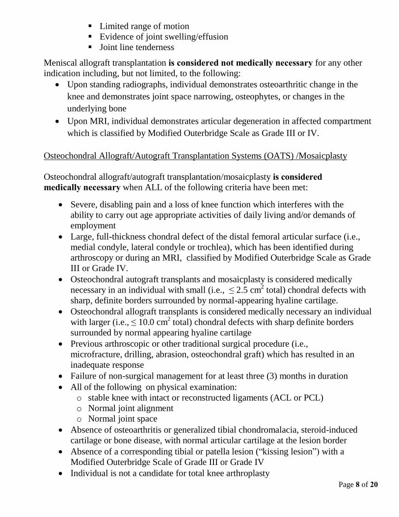
Page 8 of 20
Limited range of motion
Evidence of joint swelling/effusion
Joint line tenderness
Meniscal allograft transplantation is considered not medically necessary for any other
indication including, but not limited, to the following:
Upon standing radiographs, individual demonstrates osteoarthritic change in the
knee and demonstrates joint space narrowing, osteophytes, or changes in the
underlying bone
Upon MRI, individual demonstrates articular degeneration in affected compartment
which is classified by Modified Outerbridge Scale as Grade III or IV.
Osteochondral Allograft/Autograft Transplantation Systems (OATS) /Mosaicplasty
Osteochondral allograft/autograft transplantation/mosaicplasty is considered medically necessary when ALL of the following criteria have been met:
Severe, disabling pain and a loss of knee function which interferes with the
ability to carry out age appropriate activities of daily living and/or demands of
employment
Large, full-thickness chondral defect of the distal femoral articular surface (i.e.,
medial condyle, lateral condyle or trochlea), which has been identified during
arthroscopy or during an MRI, classified by Modified Outerbridge Scale as Grade
III or Grade IV.
Osteochondral autograft transplants and mosaicplasty is considered medically
necessary in an individual with small (i.e., ≤ 2.5 cm2 total) chondral defects with
sharp, definite borders surrounded by normal-appearing hyaline cartilage.
Osteochondral allograft transplants is considered medically necessary an individual
with larger (i.e., ≤ 10.0 cm2 total) chondral defects with sharp definite borders
surrounded by normal appearing hyaline cartilage
Previous arthroscopic or other traditional surgical procedure (i.e.,
microfracture, drilling, abrasion, osteochondral graft) which has resulted in an
inadequate response
Failure of non-surgical management for at least three (3) months in duration
All of the following on physical examination:
o stable knee with intact or reconstructed ligaments (ACL or PCL)
o Normal joint alignment
o Normal joint space
Absence of osteoarthritis or generalized tibial chondromalacia, steroid-induced
cartilage or bone disease, with normal articular cartilage at the lesion border
Absence of a corresponding tibial or patella lesion (“kissing lesion”) with a
Modified Outerbridge Scale of Grade III or Grade IV
Individual is not a candidate for total knee arthroplasty

Page 9 of 20
Body Mass Index (BMI) of less than 35
Age 49 years or younger
Individual must be capable and willing to participate in an extensive period of
non-weight bearing and supervised post-operative physical rehabilitation
program.
Anterior Cruciate Ligament Reconstruction
Allograft Knee Ligament Reconstruction - Knee ligament reconstruction (i.e., anterior
cruciate) using allograft tissue is considered medically necessary for the treatment of
ligament injury (e.g., rupture, laxity) when ANY of the following conditions is met:
Previous reconstruction has failed and requires revision
Surgical reconstruction requires the use of multiple ligament transfers
Individual has a medical condition (e.g., anatomic anomaly, prior knee injury or
prior knee surgery) that precludes the use of autograft tissue.
Knee ligament reconstruction (i.e., anterior cruciate) using allograft tissue for ANY other
indication not listed above is considered not medically necessary.
Anterior cruciate ligament reconstruction with allograft (see above for allograft
specific criteria) or autograft is considered medically necessary when ALL the
following criteria have been met:
Severe, disabling pain and a documented loss of knee function which interferes
with the ability to carry out age appropriate activities of daily living and/or
demands of employment
Knee instability which is noted as “giving way weakness”, or “buckling”
MRI, Arthroscopy, or Arthrogram demonstrates a tear/disruption or significant
laxity of the anterior cruciate ligament
Positive Lachman’s Test
ANY of the following abnormal physical examination findings:
o Positive Anterior Drawer Test
o Positive Pivot Shift Test
o Positive KT arthrometer (>3.5 mm =+1, >5-7 mm = +2, >7 mm =+3)
Failure of non-surgical management for at least three (3) months in duration.
Anterior cruciate ligament reconstruction with allograft (see above for allograft specific
criteria) or autograft is considered medically necessary in an acute injury setting where
hemathrosis, effusion, and joint instability have been documented. This may include
ANY of the following:
A confirmed ACL tear and a repairable meniscus tear
Need to return to high demand activities that require cutting, pivoting, and/or
agility activities in which ACL insufficiency may predispose to further
instability episodes, that may result in new articular or meniscal cartilage
injuries

Page 10 of 20
Concomitant ligament injuries (i.e., multiligamentous knee injury) that require
reconstruction to provide stability.
Posterior Cruciate Ligament Reconstruction
Allograft Knee Ligament Reconstruction - Knee ligament reconstruction (i.e., posterior
cruciate) using allograft tissue is considered medically necessary for the treatment of
ligament injury (e.g., rupture, laxity) when ANY of the following conditions is met:
Previous reconstruction has failed and requires revision
Surgical reconstruction requires the use of multiple ligament transfers
Individual has a medical condition (e.g., anatomic anomaly, prior knee injury or
prior knee surgery) that precludes the use of autograft tissue.
Knee ligament reconstruction (i.e., posterior cruciate) using allograft tissue for ANY other
indication not listed above is considered not medically necessary.
Posterior cruciate ligament reconstruction with allograft (see above for allograft specific
criteria) or autograft is considered medically necessary when ALL the following criteria
have been met:
Severe, disabling pain and a documented loss of knee function to an extent which
interferes with the ability to carry out the age appropriate activities of daily living
and/or demands of employment
Individual has undergone an MRI or Arthroscopy or Arthrogram which
demonstrates a tear/disruption or significant laxity of the posterior cruciate
ligament;
Individual demonstrates Positive Posterior Drawer Sign and/or positive Tibial
Drop Back Test and/or Quadriceps Active Test either of the following abnormal
physical examination findings:
o Eight (8) millimeters or more of increased posterior translation on stress
radiographs
o Positive KT-1000 arthrometer (>7.6 mm of increased posterior translation)
Failure of non-surgical care for at least three (3) months in duration
Posterior cruciate ligament reconstruction with allograft (see above for allograft
specific criteria) or autograft is considered medically necessary in an acute injury
setting where hemathrosis, effusion and joint instability have been documented.
This may include instances where there are concomitant ligament injuries (i.e.,
multiligamentous knee) that require reconstruction.
Medial Collateral/Lateral Collateral Ligament Repair/Reconstruction
Allograft Knee Ligament Reconstruction - Knee ligament reconstruction (i.e., medial
collateral, lateral collateral) using allograft tissue is considered medically necessary for

Page 11 of 20
the treatment of ligament injury (e.g., rupture, laxity) when ANY of the following
conditions is met:
Previous reconstruction has failed and requires revision
Surgical reconstruction requires the use of multiple ligament transfers
Individual has a medical condition (e.g., anatomic anomaly, prior knee injury or
prior knee surgery) that precludes the use of autograft tissue.
Knee ligament reconstruction (i.e., medial collateral, lateral collateral) using allograft
tissue for ANY other indication not listed above is considered not medically necessary.
Medial collateral/lateral collateral ligament repair with allograft (see above for allograft
specific criteria) or autograft is considered medically necessary when ALL of the
following criteria have been met:
Severe, disabling pain
Loss of knee function which interferes with the ability to carry out age appropriate
activities of daily living and/or demands of employment
Individual reports knee instability which is noted as “giving way
weakness” or “buckling”
MRI or other diagnostic study demonstrates a tear/disruption of the medial or
lateral collateral ligament
Positive Valgus Stress Test (Medial), or Varus Stress Test (Lateral)
Failure of non-surgical management for at least six (6) weeks duration.
Medial collateral or lateral collateral ligament repair/reconstruction with allograft or
autograft is considered medically necessary in an acute injury setting where total
disruption of the ligament (i.e., multi-ligamentous knee injury) is documented on MRI
examination and effusion and joint instability have been documented on physical
examination. Patella Tendon Re-Alignment (Lateral Retinacular Release, Elmslie-Trillat-Maquet,
Fulkerson Procedures)
Patella Tendon re-Alignment procedure(s) is considered medically necessary when
ALL of the following criteria have been met:
Severe anterior knee pain
Loss of knee function which interferes with the ability to carry out age appropriate
activities of daily living and/or demands of employment
Confirmed osteochondral defect of the patellofemoral joint (X-ray, CT scan, MRI
or previous arthroscopic procedure)
Failure of non-surgical management for at least three (3) months.
Patella Tendon re-Alignment procedure(s) as a treatment of recurrent patellar

Page 12 of 20
instability is considered medically necessary when ALL of the following criteria have
been met:
Recurrent patellar instability interferes with the ability to carry out age appropriate
activities of daily living and/or demands of employment
Positive Patellar Apprehension Test on examination
Increased Q angle of >15 degrees or elevated TT-TG (tibial tubercle trochlear
groove) distance
Failure of non-surgical management for at least three (3) months.
Lateral retinacular release is considered medically necessary when the individual
presents with an acute patellar dislocation with associated intra- articular fracture.
Subchondral Drilling or Microfracturing
Subchondral drilling or microfracturing is considered medically necessary when ALL of
the following criteria have been met:
Severe, disabling pain and a loss of knee function interferes with the ability to
carry out age appropriate activities of daily living and/or demands of
employment
Large, full-thickness distal femoral articular (medial condyle, lateral condyle or
trochlea) cartilage defect on the weight-bearing surface which has been identified
during arthroscopy or during an MRI which is classified by the Modified
Outerbridge Scale as Grade III or IV provided the lesion is ≤ 2.5 cm2 total
All of the following physical examination findings:
o Stable knee with intact ligaments and menisci
o Normal joint alignment
o Normal joint space
Failure of non-surgical management for at least three (3) months
High Tibial Osteotomy
High tibial osteotomy is considered medically necessary when ALL of the following
criteria have been met:
Severe, disabling pain and a loss of knee function interferes with the ability to
carry out age appropriate activities of daily living and/or demands of
employment
Unicompartmental osteoarthritis of the knee
All of the following on physical examination:
o Less than 15 degrees of fixed varus deformity
o The individual must be capable of at least 90 degrees of flexion
o Joint stability in full extension
o Intact anterior cruciate ligament (ACL)
Failure of non-surgical management for at least three (3) months in duration
Page 13 of 20
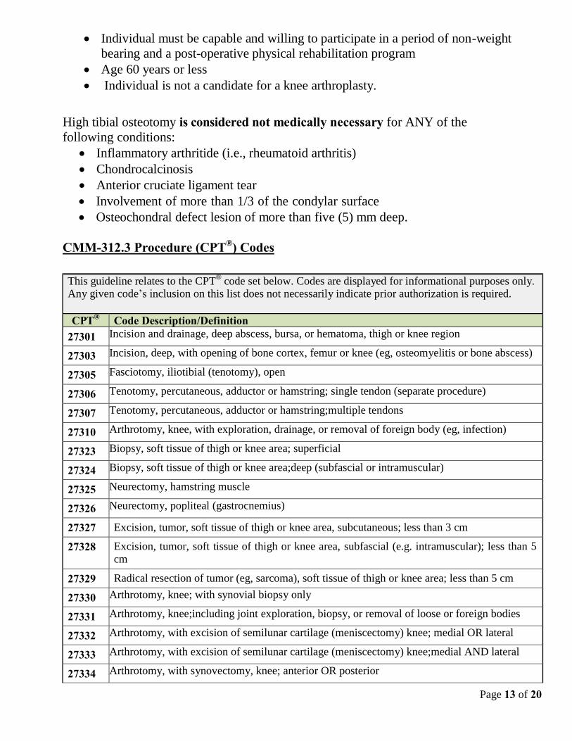
Individual must be capable and willing to participate in a period of non-weight
bearing and a post-operative physical rehabilitation program
Age 60 years or less
Individual is not a candidate for a knee arthroplasty.
High tibial osteotomy is considered not medically necessary for ANY of the
following conditions:
Inflammatory arthritide (i.e., rheumatoid arthritis)
Chondrocalcinosis
Anterior cruciate ligament tear
Involvement of more than 1/3 of the condylar surface
Osteochondral defect lesion of more than five (5) mm deep.
CMM-312.3 Procedure (CPT®) Codes This guideline relates to the CPT
® code set below. Codes are displayed for informational purposes only.
Any given code’s inclusion on this list does not necessarily indicate prior authorization is required.
CPT® Code Description/Definition
27301 Incision and drainage, deep abscess, bursa, or hematoma, thigh or knee region
27303 Incision, deep, with opening of bone cortex, femur or knee (eg, osteomyelitis or bone abscess)
27305 Fasciotomy, iliotibial (tenotomy), open
27306 Tenotomy, percutaneous, adductor or hamstring; single tendon (separate procedure)
27307 Tenotomy, percutaneous, adductor or hamstring;multiple tendons
27310 Arthrotomy, knee, with exploration, drainage, or removal of foreign body (eg, infection)
27323 Biopsy, soft tissue of thigh or knee area; superficial
27324 Biopsy, soft tissue of thigh or knee area;deep (subfascial or intramuscular)
27325 Neurectomy, hamstring muscle
27326 Neurectomy, popliteal (gastrocnemius)
27327 Excision, tumor, soft tissue of thigh or knee area, subcutaneous; less than 3 cm
27328 Excision, tumor, soft tissue of thigh or knee area, subfascial (e.g. intramuscular); less than 5
cm
27329 Radical resection of tumor (eg, sarcoma), soft tissue of thigh or knee area; less than 5 cm
27330 Arthrotomy, knee; with synovial biopsy only
27331 Arthrotomy, knee;including joint exploration, biopsy, or removal of loose or foreign bodies
27332 Arthrotomy, with excision of semilunar cartilage (meniscectomy) knee; medial OR lateral
27333 Arthrotomy, with excision of semilunar cartilage (meniscectomy) knee;medial AND lateral
27334 Arthrotomy, with synovectomy, knee; anterior OR posterior

Page 14 of 20
27335 Arthrotomy, with synovectomy, knee;anterior AND posterior including popliteal area
27337 Excision, tumor, soft tissue of thigh or knee area, subcutaneous; 3 cm or greater
27339 Excision, tumor, soft tissue of thigh or knee area, subfascial (eg, intramuscular); 5 cm or greater
27340 Excision, prepatellar bursa
27345 Excision of synovial cyst of popliteal space (e.g. Baker’s cyst)
27347 Excision of lesion of meniscus or capsule (e.g. cyst, ganglion), knee
27350 Patellectomy or hemipatellectomy
27355 Excision or curettage of bone cyst or benign tumor of femur
27356 Excision or curettage of bone cyst or benign tumor of femur; with allograft
27357 Excision or curettage of bone cyst or benign tumor of femur; with autograft (includes
obtaining graft)
27358 Excision or curettage of bone cyst or benign tumor of femur; with internal fixation (List in
addition to code for primary procedure)
27360 Partial excision (craterization, saucerization, or diaphysectomy) bone, femur, proximal tibia
and/or fibula (eg, osteomyelitis or bone abscess)
27364 Radical resection of tumor (e.g. sarcoma), soft tissue of thigh or knee area; 5 cm or greater
27365 Radical resection of tumor, femur or knee
27372 Removal of foreign body, deep, thigh region or knee area
27380 Suture of infrapatellar tendon; primary
27381 Suture of infrapatellar tendon;secondary reconstruction, including fascial or tendon graft
27385 Suture of quadriceps or hamstring muscle rupture; primary
27386 Suture of quadriceps or hamstring muscle rupture;secondary reconstruction, including fascial or
tendon graft 27390 Tenotomy, open, hamstring, knee to hip; single tendon
27391 Tenotomy, open, hamstring, knee to hip;multiple tendons, one leg
27392 Tenotomy, open, hamstring, knee to hip;multiple tendons, bilateral
27393 Lengthening of hamstring tendon; single tendon
27394 Lengthening of hamstring tendon;multiple tendons, one leg
27395 Lengthening of hamstring tendon;multiple tendons, bilateral
27396 Transplant, hamstring tendon to patella; single tendon
27397 Transplant, hamstring tendon to patella;multiple tendons
27400 Transfer, tendon or muscle, hamstrings to femur (eg, Egger's type procedure)
27403 Arthrotomy with meniscus repair, knee
27405 Repair, primary, torn ligament and/or capsule, knee; collateral
27407 Repair, primary, torn ligament and/or capsule, knee; cruciate
27409 Repair, primary, torn ligament and/or capsule, knee; collateral and cruciate ligaments

Page 15 of 20
27412 Autologous chondrocyte implantation, knee
27415 Osteochondral allograft, knee, open
27416 Osteochondral autograft(s), knee, open (e.g. mosaicplasty) (includes harvesting of
autograph[s])
27418 Anterior tibial tubercleplasty (e.g. Maquet type procedure)
27420 Reconstruction of dislocating patella; (e.g. Hauser type procedure)
27422 Reconstruction of dislocating patella; with extensor realignment and/or muscle advancement
or release (e.g. Campbell, Goldwaite type procedure)
27424 Reconstruction of dislocating patella; with patellectomy
27425 Lateral retinacular release, open
27427 Ligamentous reconstruction (augmentation), knee; extra-articular
27428 Ligamentous reconstruction (augmentation), knee; intra-articular (open)
27429 Ligamentous reconstruction (augmentation), knee; intra-articular (open) and extra-articular.
27430 Quadricepsplasty (eg, Bennett or Thompson type)
27435 Capsulotomy, posterior capsular release, knee
27448 Osteotomy, femur, shaft or supracondylar; without fixation
27450 Osteotomy, femur, shaft or supracondylar;with fixation
27454 Osteotomy, multiple, with realignment on intramedullary rod, femoral shaft (eg, Sofield type
procedure)
27455 Osteotomy, proximal tibia, including fibular excision or osteotomy (includes correction of
genu varus [bowleg] or genu valgus [knock-knee]); before epiphyseal closure
27457 Osteotomy, proximal tibia, including fibular excision or osteotomy (includes correction of
genu varus [bowleg] or genu valgus [knock-knee])l after epiphyseal closure
27465 Osteoplasty, femur; shortening (excluding 64876)
27466 Osteoplasty, femur;lengthening
27468 Osteoplasty, femur;combined, lengthening and shortening with femoral segment transfer
27470 Repair, nonunion or malunion, femur, distal to head and neck; without graft (eg, compression
technique)
27472 Repair, nonunion or malunion, femur, distal to head and neck;with iliac or other autogenous
bone graft (includes obtaining graft)
27475 Arrest, epiphyseal, any method (eg, epiphysiodesis); distal femur
27477 Arrest, epiphyseal, any method (eg, epiphysiodesis);tibia and fibula, proximal
27479 Arrest, epiphyseal, any method (eg, epiphysiodesis);combined distal femur, proximal tibia and
fibula
27485 Arrest, hemiepiphyseal, distal femur or proximal tibia or fibula (eg, genu varus or valgus)
27495 Prophylactic treatment (nailing, pinning, plating, or wiring) with or without methylmethacrylate,
femur
27496 Decompression fasciotomy, thigh and/or knee, one compartment (flexor or extensor or
adductor);

Page 16 of 20
27497 Decompression fasciotomy, thigh and/or knee, one compartment (flexor or extensor or
adductor);with debridement of nonviable muscle and/or nerve
27498 Decompression fasciotomy, thigh and/or knee, multiple compartments;
27499 Decompression fasciotomy, thigh and/or knee, multiple compartments;with debridement of
nonviable muscle and/or nerve
29850 Arthroscopically aided treatment of intercondylar spine(s) and/or tuberosity fracture(s) of the
knee, with or without manipulation; without internal or external fixation (includes
arthroscopy)
29851 Arthroscopically aided treatment of intercondylar spine(s) and/or tuberosity fracture(s) of the
knee, with or without manipulation; with internal or external fixation (includes arthroscopy)
29855 Arthroscopically aided treatment of tibial fracture, proximal (plateau); unicondylar, includes
internal fixation, when performed (includes arthroscopy)
29856 Arthroscopically aided treatment of tibial fracture, proximal (plateau); bicondylar, includes
internal fixation, when performed (includes arthroscopy)
29866 Arthroscopy, knee, surgical; osteochondral autograft(s) (e.g. mosaicplasty) (includes
harvesting of the autograft[s])
29867 Arthroscopy, knee, surgical; osteochondral allograft (e.g. mosaicplasty)
29868 Arthroscopy, knee, surgical; meniscal transplantation (includes arthrotomy for meniscal
insertion), medial or lateral
29870 Arthroscopy, knee, diagnostic; with or without synovial biopsy (separate procedure)
29871 Arthroscopy, knee, surgical; for infection, lavage and drainage
29873 Arthroscopy, knee, surgical; with lateral release
29874 Arthroscopy, knee, surgical; for removal of loose body or foreign body (e.g. osteochondritis
dissecans fragmentation, chondral fragmentation)
29875 Arthroscopy, knee, surgical;synovectomy, limited (eg, plica or shelf resection) (separate
procedure)
29876 Arthroscopy, knee, surgical;synovectomy, major, two or more compartments (eg, medial or
lateral)
29877 Arthroscopy, knee, surgical; debridement/shaving of articular cartilage (chondroplasty)
29879 Arthroscopy, knee, surgical; abrasion Arthroplasty (includes chondroplasty where necessary)
or multiple drilling or microfracture
29880 Arthroscopy, knee, surgical; with meniscectomy (medial AND lateral, including any meniscal
shaving) including debridement/shaving of articular cartilage (chondroplasty), same or
separate compartment(s), when performed
29881 Arthroscopy, knee, surgical; with meniscectomy (medial OR lateral, including any meniscal
shaving) including debridement/shaving of articular cartilage (chondroplasty), same or
separate compartment(s), when performed
29882 Arthroscopy, knee, surgical; with meniscus repair (medial OR lateral)
29883 Arthroscopy, knee, surgical; with meniscus repair (medial AND lateral)

Page 17 of 20
29884
Arthroscopy, knee, surgical;with lysis of adhesions, with or without manipulation (separate
procedure)
29885 Arthroscopy, knee, surgical; drilling for osteochondritis dissecans with bone grafting, with or
without internal fixation (including debridement of base of lesion)
29886 Arthroscopy, knee, surgical; drilling for intact osteochondritis dissecans lesion
29887 Arthroscopy, knee, surgical; drilling for intact osteochondritis dissecans lesion with internal
fixation
29888 Arthroscopically aided anterior cruciate ligament repair/augmentation or reconstruction
29889 Arthroscopically aided posterior cruciate ligament repair/augmentation or reconstruction
This list may not be all inclusive and is not intended to be used for coding/billing purposes. The final determination of reimbursement for services is the decision of the health plan and is based on the individual’s policy or benefit entitlement structure as well as claims processing rules.
CMM-312.4 References
1. Aaron R, Skolnick A, Reinert S, Ciombor D. Arthroscopic debridement for osteoarthritis of
the knee. J Bone Joint Surg Am. 2006;88(5):936-943.
2. Adler V, Pa L, Ko J, et al. Autologous chondrocyte transplantation for the treatment of
articular defects of the knee. Scr Med. 2003;76(3):241-250.
3. Alleyne K, Galloway M. Management of osteochondral injuries of the knee. Clin Sports Med.
2001;20(2):343-364.
4. Altman R, Hochberg M, Moskowics, R, et al.; Subcommittee on Osteoarthritis Guidelines.
Recommendations for the medical management of osteoarthrits of the hip and knee. American
College of Rheumatology Subcommittee on Osteoarthritis Guidelines. Arthritis Rheum.
2000;43(9):1905-1915.
5. Bartha L, Vajda A, Duska Z, et al. Autologous osteochondral mosaicplasty grafting. J Orthop
Sports Phys Ther. 2006;36(10):739-750.
6. Bentley G, Biant L, Carrington R, et al. A prospective, randomised comparison of autologous
chondrocyte implantation versus mosaicplasty for osteochondral defects in the knee. J Bone
Joint Surg Br. 2003;85(2):223-230.
7. Bernstein J, Quach T. A perspective on the study of Moseley et al: Questioning the value of
arthroscopic knee surgery for osteoarthritis. Cleve Clin J Med. 2003;70(5):401, 405-406, 408-410.
8. Biau D, Tournoux C, Katsahian Set al. Bone-patellar tendon-bone autografts versus
hamstring autografts for reconstruction of anterior cruciate ligament: meta-analysis. BMJ.
2006;332(7548):995-1001.
9. Bradley J, Heilman D, Katz B, et al. Tidal irrigation as treatment for knee osteoarthritis: A
sham- controlled, randomized, double-blinded evaluation. Arthritis Rheum. 2002;46(1):100-108.
10. Briggs T, Mahroof S, David L, et al. Histological evaluation of chondral defects after
autologous chondrocyte implantation of the knee. J Bone Joint Surg Br. 2003;85(7):1077-1083.
11. Brouwer, Reinoud W, Huizinga, Maarten R, Duivenvoorden, Tijs, van Raaij, Tom M, Verhagen,
Arianne P, Bierma-Zeinstra, Sita MA, Verhaar, Jan AN. Osteotomy for treating knee
osteoarthritis. Cochrane Database of Systematic Reviews, 2014, Issue 12. Art. No.: CD004019.
DOI: 10.1002/14651858.CD004019.pub4.
12. Calvert G, Wright R. The use of arthroscopy in the athlete with knee osteoarthritis. Clin Sports Med.

Page 18 of 20
2005;24(1):133-152.
13. Chambers K, Schulzer M. Arthroscopic surgery for osteoarthritis of the knee. N Engl J
Med. 2002;347:1718.
14. Chatain F, Adeleine P, Chambat P, Neyret P; Society Francaise d'Arthroscopie. A comparative
study of medial versus lateral arthroscopic partial meniscectomy on stable knees: 10-year minimum
follow-up. Arthroscopy. 2003; 19(8):842-849.
15. Crawford DC, Safran MR. Osteochondritis Dissecans of the knee. J Am Acad Orthop Surg. 2006;
14: 90-100.
16. Dervin G, Stiell I, Rody K, Grabowski J. Effect of arthroscopic debridement for osteoarthritis of
the knee on health-related quality of life. J Bone Joint Surg Am. 2003;85-A(1):10-19.
17. Dozin B, Malpeli M, Cancedda R, et al. Comparative evaluation of autologous chondrocyte
implantation and mosaicplasty: A multicentered randomized clinical trial. Clin J Sport
Med. 2005;15(4):220-226.
18. Englund M, Guermazi A, Roemer FW, et al. Meniscal tear in knees without surgery and the
development of radiographic osteoarthritis among middle-aged and elderly persons: The multicenter
osteoarthritis study. Arthritis Rheum. 2009;60(3):831-9.
19. Englund M, Roos E, Lohmander L. Impact of type of meniscal tear on radiographic and
symptomatic knee osteoarthritis: a sixteen-year follow-up of meniscectomy with matched controls.
Arthritis Rheum. 2003;48(8):2178-87.
20. Felson D, Buckwalter J. Debridement and lavage for osteoarthritis of the knee. N Engl J
Med. 2002;347:132-3.
21. Felson D. Osteoarthritis of the knee. N Engl J Med. 2006;354:841-8.
22. Fond J, Rodin D, Ahmad S, Nirschl R. Arthroscopic debridement for the treatment of osteoarthritis
of the knee: 2- and 5-year results. Arthroscopy. 2002;18(8):829-834.
23. Forster M, Straw R. A prospective randomised trial comparing intra-articular Hyalgan injection and
arthroscopic washout for knee osteoarthritis. Knee. 2003;10(3):291-293.
24. Graf K, Sekiya J, Wojitys E. Long-term results after combined medial meniscal allograft
transplantation and anterior cruciate ligament reconstruction: Minimum 8.5-year follow-up
study. Arthroscopy. 2004:20(2):129-140.
25. Haasper C, Zelle B, Knobloch K, et al. No mid-term difference in mosaicplasty in previously
treated versus previously untreated individuals with osteochondral lesions of the talus. Arch Orthop
Trauma Surg. 2008;128(5):499-504.
26. Hangody L, Vásárhelyi G, Hangody L, et al. Autologous osteochondral grafting--technique
and long-term results. Injury. 2008;39 Suppl 1:S32-S39.
27. Harner C, Waltrip R, Bennett C, et al. Surgical management of knee dislocations. J Bone Joint
Surg Am. 2004;86-A(2):262-73.
28. Henderson I, Tuy B, Connell D, et al. Prospective clinical study of autologous
chondrocyte implantation and correlation with MRI at three and 12 months. J Bone Joint
Surg Br. 2003;85(7):1060-1066.
29. Hunt S, Jazrawi L, Sherman O, Arthroscopic management of osteoarthritis of the knee. J Am
Acad Orthop Surg. 2002;10(5):356-63.
30. Jackson R, Dieterichs C. The results of arthroscopic lavage and debridement of osteoarthritic
knees based on the severity of degeneration: A 4- to 6-year symptomatic follow-up. Arthroscopy.
2003;19(1):13-20.
31. Jakob R, Franz T, Gautier E, Mainil-Varlet P. Autologous osteochondral grafting in the
knee: Indication, results, and reflections. Clin Orthop. 2002;(401):170-184.
32. Karataglis D, Green M, Learmonth D. Autologous osteochondral transplantation for the treatment

Page 19 of 20
of chondral defects of the knee. Knee. 2006;13(1):32-35.
33. Karataglis D, Learmonth D. Management of big osteochondral defects of the knee
using osteochondral allografts with the MEGA-OATS technique. Knee.
2005;12(5):389-393.
34. Kelly M. Role of arthroscopic debridement in the arthritic knee. J Arthroplasty. 2006;21:Suppl
1:9- 10.
35. Kirkley A, Birmingham T, Litchfield R, et al. A Randomized Trial of Arthroscopic Surgery
for Osteoarthritis of the Knee. N Engl J Med. 2008; 59:1097-1107,1169-1170.
36. Kreuz P, Steinwachs M, Erggelet C, et al. Mosaicplasty with autogenous talar autograft for
osteochondral lesions of the talus after failed primary arthroscopic management: A prospective
study with a 4-year follow-up. Am J Sports Med. 2006;34(1):55-63.
37. Lahav A, Burks R, Greis P, et al. Clinical outcomes following osteochondral
autologous transplantation (OATS). J Knee Surg. 2006;19(3):169-173.
38. Laupattarakasem W, Laopaiboon M, Laupattarakasem P, Sumananont C. Arthroscopic
debridement for knee osteoarthritis. Cochrane Database Syst Rev. 2008;(1):CD005118.
39. Linko E, Harilainen A, Malmivaara A, Seitsalo S. Surgical versus conservative interventions
for anterior cruciate ligament ruptures in adults. Cochrane Database Syst Rev. 2005 Apr
18;(2):CD001356.
40. Ma H, Hung S, Wang S, et al. Osteochondral autografts transfer for post-traumatic
osteochondral defect of the knee -- 2 to 5 years follow-up. Injury. 2004;35(12):1286-1292.
41. Marcacci M, Kon E, Zaffagnini S, et al. Multiple osteochondral arthroscopic grafting
(mosaicplasty) for cartilage defects of the knee: Prospective study results at 2-year follow-
up. Arthroscopy. 2005;21(4):462-470.
42. Marx R. Arthroscopic surgery for osteoarthritis of the knee? N Engl J Med. 2008;359(11):1169-
1170.
43. Moseley J, O’Malley K, Petersen N, et al. A controlled trial of arthroscopic surgery for
osteoarthritis of the knee. N Engl J Med. 2002;347:81-88.
44. Noyes F, Barber-Westin S, Rankin M. Meniscal transplantation in symptomatic individuals less
than fifty years old. J Bone Joint Surg Am. 2005;87 Suppl 1(Pt.2):149-165.
45. Pearse E, Craig D. Partial meniscectomy in the presence of severe osteoarthritis does not hasten the
symptomatic progression of osteoarthritis. Arthroscopy. 2003;19(9):963-968.
46. Peterson L, Minas T, Brittberg M, Lindahl A. Treatment of osteochondritis dissecans of the knee
with autologous chondrocyte transplantation. J Bone Joint Surg Am. 2003;85(Suppl 2):17-24.
47. Peterson R, Shelton W, Bomboy A. Allograft versus autograft patellar tendon anterior cruciate
ligament reconstruction: A 5-year follow-up. Arthroscopy. 2001;17(1):9-13.
48. Roos E, Ostenberg A, Roos H, et al. Long-term outcome of meniscectomy: symptoms, function,
and performance tests in individuals with or without radiographic osteoarthritis compared to
matched controls. Osteoarthritis Cartilage. 2001;9(4):316-24.
49. Roos E, Roos H, Ryd L, Lohmander L. Substantial disability 3 months after arthroscopic partial
meniscectomy: A prospective study of individual-relevant outcomes. Arthroscopy. 2000;16(6):619-
26.
50. Ruano-Ravina A, Jato Diaz M. Autologous chondrocyte implantation: a systematic review.
Osteoarthritis Cartilage. 2006;14(1):47-51.
51. Ryu R, Dunbar V, Morse G, Meniscal allograft replacement: a 1-year to 6-year experience,
Arthroscopy. 2002;18(9):989-994.
52. Sekiya J, Giffin J, Irrgang J, et al. Clinical outcomes after combined meniscal allograft

Page 20 of 20
transplantation and anterior cruciate ligament reconstruction. Am J Sports Med.
2003;31(6):896-906.
53. Sharpe J, Ahmed S, Fleetcroft J, Martin R. The treatment of osteochondral lesions using a
combination of autologous chondrocyte implantation and autograft: Three-year follow-up. J
Bone Joint Surg Br. 2005;87(5):730-735.
54. Sherman SL, Garrity J, Bauer K, Cook J et al. Fresh osteochondral allograft transplantation for
the knee: current concepts. J Am Acad Orthop Surg. 2014;22(2):121-33.
55. Solomon D, Avorn J, Warsi Aet al. Which individuals with knee problems are likely to benefit
from nonarthroplasty surgery? Development of a clinical prediction rule. Arch Intern Med.
2004;164(5):509-513.
56. Stuart M, Lubowitz J. What, if any, are the indications for arthroscopic debridement of the
osteoarthritic knee? Arthroscopy. 2006;22(3):238-239.
57. Zhang W, Moskowitz R, Nuki Get al. OARSI recommendations for the management of hip and
knee osteoarthritis, Part II: OARSI evidence-based, expert consensus guidelines. Osteoarthritis
Cartilage. 2008;16(2):137-162.


![A Virtual Reality Training System for Knee Arthroscopic Surgeryttwong/papers/arthro/arthro2.pdf · 2004-01-28 · The knee arthroscopic surgery systems presented in [5] [6] mostly](https://img.dokumen.tips/doc/110x75/5f1d2df84d161c32a3381233/a-virtual-reality-training-system-for-knee-arthroscopic-ttwongpapersarthroarthro2pdf.jpg)