Embed Size (px)
Citation preview

69:1 (2014) 9–15 | www.jurnalteknologi.utm.my | eISSN 2180–3722 |
Full paper Jurnal
Teknologi
Chromobacterium Violaceum for Rapid Measurement of Biochemical Oxygen Demand Beng Hooi Khora, Abd. Khamim Ismailc, Rahmalan Ahamadb, Shafinaz Shahira* aDepartment of Biosciences and Health Sciences, Faculty of Biosciences and Medical Engineering, Universiti Teknologi Malaysia, 81310 UTM Johor Bahru, Johor, Malaysia bDepartment of Chemistry, Faculty of Science, Universiti Teknologi Malaysia, 81310 UTM Johor Bahru, Johor, Malaysia cDepartment of Physics, Faculty of Science, Universiti Teknologi Malaysia, 81310 UTM Johor Bahru, Johor, Malaysia
*Corresponding author: [email protected]
Article history
Received :26 November 2013 Received in revised form :
16 April 2014
Accepted :15 June 2014
Graphical abstract
Abstract
Abstract: Biochemical Oxygen demand (BOD) is an important parameter indicating the total biodegradable organic pollutants in waters. Fast BOD determination could be achieved using BOD biosensor. However,
most of the developed BOD biosensors are dependent on dissolved oxygen concentrations. Low solubility
of oxygen in water decreases the reliability of oxygen-dependent BOD biosensor. However, replacement of oxygen with a mediator solution solves this problem. In the present study, an effective ferricyanide-
mediated approach was modified from ferricyanide-mediated BOD assay and used for BOD determination
in a water system. Several different types of microorganisms were isolated from different organic-rich environmental sources and their ability to use ferricyanide during organic (standard glucose-glutamic acid
solution) degradation were effectively assessed using ferricyanide-mediated BOD assay. Around 90% of
the GGA was degraded by Chromobacterium violaceum after 1 hour of incubation period. Therefore, C. violaceum has been found to be a potential microorganism to be used as a biosensing element in the BOD
biosensor. This assay is not only effective in selecting the suitable microorganisms for ferricyanide-
mediated BOD detection; it also could be applied to select the suitable microorganisms for other mediated microbial biosensor and bioremediation by changing the substrate and conditions.
Keywords: Biochemical Oxygen Demand (BOD); Ferricyanide; Ultramicroelectrode (UME);
Chromobacterium violaceum, redox-mediated biosensor; substrate specificity
Abstrak
Keperluan Oksigen Biokimia (BOD) merupakan satu parameter penting yang menunjukkan jumlah bahan pencemar organik biodegradasi dalam air. Penentuan BOD yang pantas boleh dilaksanakan menggunakan
biopenderiaan BOD. Walau bagaimanapun, kebanyakan biopenderiaan BOD yang dibangunkan
bergantung kepada kepekatan oksigen terlarut. Kelarutan rendah oksigen dalam air mengurangkan kebolehpercayaan oksigen yang bergantung kepada biopenderiaan BOD. Penggantian oksigen dengan satu
pengantara penyelesaian dapat mengatasi masalah ini. Dalam kajian ini, pendekatan pengantara ferisianida
yang berkesan telah diubah suai daripada analisis pengantara BOD ferisianida dan digunakan untuk penentuan BOD dalam sistem air. Beberapa jenis mikroorganisma telah diasingkan daripada pelbagai
sumber alam sekitar yang kaya dengan bahan organik dan keupayaan mereka untuk menggunakan
ferisianida semasa degradasi organik (larutan standard glukosa- asid glutamik) telah dinilai dengan berkesan menggunakan analisa pengantara BOD ferisianida. Sekitar 90% daripada GGA telah didegradasi
oleh Chromobacterium violaceum selepas tempoh 1 jam pengeraman. Oleh itu, C. violaceum adalah
mikroorganisma yang berpotensi untuk digunakan sebagai satu elemen dalam biopenderiaan BOD. Analisa ini tidak hanya berkesan dalam memilih mikroorganisma yang sesuai untuk mengesan pengantara BOD
ferisianida, tetapi ia juga boleh digunakan untuk memilih mikroorganisma lain yang sesuai sebagai
pengantara biopenderiaan yang mikrob dan biopemulihan dengan menukar substrat dan keadaan-keadaan tertentu.
Kata kunci: Keperluan Oksigen Biokimia (BOD); Ferisianida; Elektrodultramikro (UME); Chromobacterium violaceum, biopenderiaan dengan pengantara redoks; pengkhususan substrat
© 2014 Penerbit UTM Press. All rights reserved.

10 Khor et al. / Jurnal Teknologi (Sciences & Engineering) 69:1 (2014), 9–15
1.0 INTRODUCTION
In the last few decades, intense industrialization has led to rapid
development of urban population. As a result of urbanization,
environmental pollution has become a serious problem due to
manifold wastes discharged from the agriculture, domestic, and
industries into the environment [1]. BOD is an important and
widely used parameter to access the degree of pollution in the water
system; it represents the amount of oxygen required for microbial
aerobic decomposition processes of biodegradable organic
pollutants in the water system. Thus, BOD indicates the quantity of
biodegradable organic pollutants and the oxygen used to oxidise
inorganic material in the water systems [2]. Although some
portions of organic pollutants can be degraded easily by the
indigenous microorganisms, the level of dissolved oxygen (DO) in
the water after decomposition of the organic pollutants gives a
significant impact to the health of the aquatic ecosystem. The
reduction of DO levels was reported to cause lethal and sublethal
effects (physiological and behavioural) in aquatic organisms,
especially in fish [3].
The standard method for BOD analysis is the conventional 5-
day biochemical oxygen demand (BOD5) method described by the
American Public Health Association (APHA) Standard Methods
Committee [4] and the Japanese Industrial Standard (JIS)
Committee [5]. Although BOD5 is a universal method of measuring
most wastewater samples and the required equipment is
inexpensive, it is time-consuming and requires complicated
procedure and skilled analysts to obtain reproducible results.
Therefore, the BOD5 is not suitable for in situ determination or on-
line process monitoring of wastewater treatment systems where fast
feedback is required [6].
Hence, in order to overcome the weakness of BOD5, several
alternative methods to BOD5 have been widely developed, such as
BOD assay and BOD biosensor. In the past decade, alternative
BOD assays assimilated the use of ferricyanide mediator were
introduced [7; 8; 9; 10]. These assays were proved to give a great
benefit on the perspective of time constraint and the detection range
of BOD [9]. However, the preparation and complexity of the assays
were the limitation to the assays.
Fast determination of BOD could also be achieved by the
biosensor-based methods. A biosensor is a self-contained
integrated device, which is capable of providing specific analytical
information using a biological sensing element, which is
immobilized onto a transducer [12]. Nowadays, microbial
biosensor has been widely used in toxicity detection, heavy metal
detection, specific carbohydrate detection, and organic pollution
detection [13]. However, a large portion of the developed microbial
biosensors for BOD detection has focused on measuring the
remaining dissolved oxygen levels or concentration after the
immobilized microbes utilized DO to degrade the organic
pollutants in the sample over a certain period. Due to poor
solubility of oxygen (O2) in water (8.7 mg/l at 25oC), DO rapidly
becomes the limiting reactant in the biodegradation process.
Subsequently, the amount of organic compounds biodegraded
within the short time is small, which represents only a small
fraction of the total organic content [10]. This results in narrow
response ranges and poor reliability of microbial BOD biosensor.
Recently, ferricyanide-mediated amperometric approach has
been developed to overcome the oxygen limitation problems [4; 5;
14; 15]. Ferricyanide can serve as a redox mediator, which can be
reduced by certain microorganisms, thereby, by-passes the oxygen
reduction step during microbial metabolic process. In this
approach, O2 is replaced by the ferricyanide ion, which serves as
an alternative electron acceptor which is preferentially reduced to
ferrocyanide during the metabolic oxidation of organic pollutants
(Figure 1). The ferrocyanide is then reoxidized at a working
electrode which is held at a sufficiently high electric potential. As
a result, a current is generated and detected using the electrode
system [14]. The high solubility of ferricyanide enables the use of
higher microbial population without rapid depletion of
ferricyanide. Since the metabolic rate of the microorganisms is not
affected in the presence of the large amount of ferricyanide, the
upper detection limit of the sensor is increased and the excessive
dilution of samples might not be necessary as required for the
oxygen-dependent sensor. Moreover, the reduced form of the
mediator has an initial background concentration of zero, thereby,
increases the accuracy when quantifying the amount of oxidised
mediator converted to its reduced form [10]. Hence, mediated
microbial sensors have received much attention for rapid BOD
measurement [5; 8; 9; 10; 11; 14; 16; 17; 18].
Figure 1 Scheme of ferricyanide-mediated amperometric approach and unmediated amperometric approach (modified from Yoshida et al., 2000).

11 Khor et al. / Jurnal Teknologi (Sciences & Engineering) 69:1 (2014), 9–15
Furthermore, most of the reported BOD biosensors focus on the
use of conventional electrode as working electrode in the BOD
detection. However, the use of ultramicroelectrode (UME) is
rather limited in BOD biosensor. UME is defined as an electrode
that has at least one dimension (the critical dimension) smaller
than 25 μm. The small size of UME gives them relatively large
diffusion layers and small overall currents. These features allow
the UME to achieve useful steady-state conditions rapidly and
very high scan rates (V/s) with limited distortion. Therefore,
highly accurate measurements are possible to be made with two-
electrode system even in non-polar solvent or resistive solution
without deliberately adding supporting electrolytes. Thus, it
allows electrochemical measurements to be carried out with or
without low amount of supporting electrolyte. Hence, UME
permits in situ characterization and avoids disturbance of the
equilibria which can be the source of impurities and can limit the
electro activity range [10]. Therefore, it can be used as a chip-
based microelectronic device for natural in situ water monitoring.
In the present study, the possibility of FM-BOD assay is
explored to screen for potential microorganisms, which could be
used for FM-BOD detection.
2.0 MATERIALS AND METHODS
2.1 Chemicals
All chemicals used were of analytical reagent grade, and all
solutions were prepared in deionised water. The standard GGA
solution (150 mg glucose/L and 150 mg glutamic acid/L) was
prepared according to the APHA standard (1992) methods [19].
The BOD5 values of the standard GGA solution were 198 ± 30
mg O2/L. Potassium ferricyanide solutions were prepared in
phosphate buffered saline solution (PBS, 10.8872 g/L K2HPO4/
20.9016 g/L K2HPO4/ 7.456 g/L KCl, pH 7).
2.2 Isolation of Microorganisms
Microorganisms were isolated from different organic-rich
environmental sources, such as petroleum refinery liquid effluent
(P), sludge from textile industry (TS), raw pineapple liquid waste
(PA), river water (R), and raw palm oil mill effluent wastewater
(POME). Samples from different environmental sources were
streaked onto Nutrient Agar (20 g/L Nutrient Agar, MERCK) and
Potato Dextrose Agar (39 g/L Potato Dextrose Agar, DifcoTM /10
g/L tartaric acid) plates using dilution streak plate technique, and
were incubated at 37oC for 24 hours. Pure cultures were obtained
by restreaking single colonies with different morphologies onto
new respective agar plates.
2.3 Screening of Potential Microorganisms
Ferricyanide-mediated BOD assay [8] was slightly modified and
was used to screen the potential microorganisms using
ferricyanide as an electron acceptor during the degradation
according to the standard GGA.
2.3.1 Preparation of Inoculum
Overnight culture (10% v/v) was aseptically inoculated into
Tryptic Soy Broth (TSB) and incubated aerobically at 37°C with
shaking at 200 rpm. Cultures were grown until the end of the
exponential phase (as determined by growth curves) and
harvested by centrifugation at 4000 rpm for 15 min at room
temperature. Cell pellets were washed twice with PBS and
subsequently were re-suspended in PBS. The optical density of
the suspension at 600 nm wavelength of light (OD600) was
adjusted to 10 using 100 VIS Spectrophotometer (Jenway).
2.3.2 Preparation of the Samples
Three solutions were prepared. The sample solution consisted of
potassium ferricyanide (1.5 mL, 131.696 g/L), standard GGA
solution (3.5 mL, BOD5: 198 mg O2/L), and cells suspension (5.0
mL, OD600 10). Endogenous solutions were prepared by replacing
the standard GGA solution with deionized water. Blank solutions
were prepared by replacing ferricyanide solution with PBS. All
the solutions were prepared in a closed serum bottle, and the
solutions were sparged with oxygen free nitrogen gas for 15
minutes.
2.3.3 Incubation of the Samples
The samples were incubated at 37°C with shaking at 100 rpm for
5 hours in a shaking water bath. After one hour interval, 1.0 mL
aliquots were withdrawn and centrifuged at 4000 rpm for 15
minutes. Subsequently, 1.0 mL of supernatant was diluted to 30x
dilution using PBS solution (pH 7.0) and sparged with oxygen
free nitrogen gas for 10 minutes. The production of ferrocyanide
in supernatant solution was then analysed using voltammetry.
2.3.4 Voltammetric Detection
The concentration of ferrocyanide was determined
chronoamperometrically. Chronoamperometric measurements
were conducted in a single potential time base mode at +450 mV.
eDAQ e-corder 410 (Model ED410) and eDAQ picostat (Model
EA162) were used in the measurement, together with a three
electrode system which consisted of a 10 μm Platinum (Pt)
microelectrode (working electrode), 1.0 mm diameter Pt gauze
auxiliary electrode, and Ag/AgCl reference electrode. The
amperometric current value, obtained one minute after the
imposition of the applied potential, was taken as the limiting
current value and used as the analytical signal.
2.3.5 Calculation of the FM-BOD5 Equivalent Values:
The FM-BOD5 equivalent value (Eq 1) was obtained by dividing
the limiting current values obtained from the sample with the
limiting current values obtained from the GGA standard solution
and multiplied by BOD5 value of the standard GGA solution
(BOD5: 198 mg O2/L). The ratio of the limiting current values
obtained from the sample to the limiting current values obtained
from the GGA standard solution indicates the amount of
biodegradation that can occur in the standard GGA solution for a
given batch culture of microorganisms [9].
𝐅𝐌 − 𝐁𝐎𝐃5 𝐞𝐪𝐮𝐢𝐯𝐚𝐥𝐞𝐧𝐭 𝐯𝐚𝐥𝐮𝐞 = {𝒊𝒍𝒊𝒎(𝒔𝒂𝒎𝒑𝒍𝒆) −𝒊𝒍𝒊𝒎(𝒆𝒏𝒅𝒐𝒈𝒆𝒏𝒐𝒖𝒔)
𝒊𝒍𝒊𝒎(𝑮𝑮𝑨) −𝒊𝒍𝒊𝒎(𝒆𝒏𝒅𝒐𝒈𝒆𝒏𝒐𝒖𝒔)} × 𝟏𝟗𝟖 𝐦𝐠 ∕ 𝐦𝐠 𝐎𝟐/𝐋 (Equation 1)

12 Khor et al. / Jurnal Teknologi (Sciences & Engineering) 69:1 (2014), 9–15
2.4 Identification of Isolate
Promega DNA Purification kit was used to extract the genomic
DNA. After the extraction, the bacterial 16s rRNA (1354 bp) was
amplified by polymerase chain reaction (PCR) with 2 universal
primer (pH and pA). The amplified PCR products were sent to
First BASE Laboratories Sdn Bhd for purification and
sequencing.
3.0 RESULTS AND DISCUSSION
3.1 Isolation of Microorganisms
A total of 14 types of bacteria (coded P1, P2, P3, PA1, PA2, PA3,
TS1, TS2, TS3, R1, R5, MC5, POME22, and S2) and 3 types of
yeasts (Y1, Y2 and Y3) were isolated from different
environmental sources in Malaysia. The colonies of the isolates
were characterized on the basis of colour, shape, texture,
elevation, and margin (Table 1). The purpose of this isolation was
to select the microbes that were capable of degrading a wide
range of organic compounds. Thus, all the microbes were isolated
from the sources that were rich in organic compounds. For
example, raw pineapple liquid waste consists of sugars and
lignocellulosic components [20], while textile wastewater
contains various complex organic compounds made from dyes,
detergents, solvents, grease, and oils [21]. POME consists of a
spectrum of carbohydrates, nitrogenous compounds, free organic
acids, lipids, and mineral constituents [22], and petroleum
refinery liquid effluent mainly consists of oil products such as
saturated hydrocarbon and polycyclic aromatic hydrocarbons
[23].
Table 1 Colonies of microorganisms isolated from different environmental sources with their morphologies
No Isolate Colony Morphologies Cellular Shape
1 PA1 White, slightly gummy, translucent, circular, convex with an entire margin cocci 2 PA2 White, translucent, circular, convex with an entire margin bacillus
3 PA3 White, irregular, umbonate with undulate margin bacillus 4 P1 White, translucent, circular, convex and curled margin bacillus
5 P2 White, circular, convex with an entire margin bacillus
6 P3 White, slightly gummy, translucent, circular, convex with an entire margin cocci 7 TS1 White, irregular, slightly convex and curled margin bacillus
8 TS2 White, slightly gummy, translucent, circular, convex with an entire margin cocci
9 TS3 White, slightly gummy, translucent, circular, convex with an entire margin coccibacillus 10 R1 Purple, slightly gummy, circular, convex with an entire margin cocci
11 R5 White, slightly gummy, translucent, circular, convex with an entire margin coccibacillus
12 POME22 Large, white, irregular and flat with an undulate margin bacillus 13 MC5 Large, white, circular, flat with undulate margin cocci
14 S2 White, slightly gummy, translucent, circular, convex with an entire margin cocci
15 Y1 White, waxy, circular, umbonate with filamentous margin Oval with budding 16 Y2 White, waxy, circular, umbonate with entire margin Elongated with budding
17 Y3 White, waxy, circular, umbonate with curled margin Oval with budding
3.2 Screening of Potential Microorganisms
Figure 2 shows the limiting current values generated over 5 hours
of incubation period in the GGA for each of the isolated microbes
after the limiting current values of the endogenous control
solution were subtracted from the sample. Based on the figure,
R1 attained the highest limiting current after 60 minutes of
incubation and this was followed by P3, TS2, PA1, PA3, R5, TS3,
P1, TS1, POME22, MC5, PA2, and P2. Zero limiting current was
attained by yeasts (Y1, Y2, Y3). A rapid linear increase in the
limiting current value was observed for the sample solution after
60 minutes of incubation (Figure 2). This was because the GGA
substrates were degraded by the microorganisms using
ferricyanide as an alternative electron acceptor to oxygen. As a
result, ferricyanide was reduced to ferrocyanide by the electron
transport chain systems of the microorganisms. When the
potential of working electrode was shifted to +450 mV, the
potential of the working electrode was high enough and sufficient
to reoxidize the reduced ferrocyanide to ferricyanide. Therefore,
the reduced ferrocyanide was reooxidized to ferricyanide by
donating an electron to the working electrode. As a result, the
limiting current obtained indicated the amount of ferrocyanide
produced by the bacterial cells during GGA degradation, which
is directly proportional to the amount of the degraded substrates.
As shown in Figure 2, the limiting current of the sample solution
reached an equilibrium after 60 minutes of incubation. This can
be explained by the high rate of microbial metabolism when a
large amount of substrate was present in the initial stage, and
subsequently, the metabolic rate decreased with the unavailability
of the substrates in the solution.

13 Khor et al. / Jurnal Teknologi (Sciences & Engineering) 69:1 (2014), 9–15
Figure 2 Limiting current values were obtained at various incubation times for single cultures in GGA standard solution and endogenous control values have
been subtracted from the sample. Microbial consortium final absorbance = 5. Ferricyanide final concentration = 60 mM. Limiting currents determined by
voltammetry at Eapp = + 450 mV (vs Ag/AgCl)
To give measurable analytical signal, the limiting current of
the endogenous control was subtracted from the sample. The
difference in the values indicates the amount of degraded
substrates in the sample solution. The limiting current values of
endogenous control arise from the metabolism of carbon storage
polymer present within the cell during limitation of nutrients [7].
Thus, it is necessary to subtract the limiting current values of the
endogenous control solution from the sample.
By referring to the generated limiting current (Figure 2) and
the known amount of substrate in the sample solution, the
percentage of GGA that was degraded could be calculated (Figure
3). R1 was able to degrade around 90% of the GGA in the first
hour of the incubation period. In addition, PA1, PA3, TS2, R1,
P3, and S2 were capable of degrading more than 60% of the GGA
after 60 minutes of incubation. Similar work was reported by the
Morris’ group, whereby only 40% of the GGA was degraded after
60 minutes of incubation [11]. The results obtained in the present
study were also compared with the more favourable conventional
aerobic BOD5 assay, in which approximately 60% conversion of
the standard GGA solution was achieved in 5 days [7; 8].
Therefore, from the present study, the R1 can be used as a
biocatalyst for further studies.
Figure 3 Percentages of GGA degradation by the microorganism after 60 minutes incubation period
0.00%
10.00%
20.00%
30.00%
40.00%
50.00%
60.00%
70.00%
80.00%
90.00%
100.00%
PA
1
PA
2
PA
3
TS1
TS2
TS3
R1
R5
P1
P2
P3
po
me
22 S2
MC
5 Y1 Y2 Y3
GG
A D
egr
adat
ion
Microorganisms
14 Khor et al. / Jurnal Teknologi (Sciences & Engineering) 69:1 (2014), 9–15
In contrast, GGA degradation was not found in yeasts.
Approximately, zero current output was detected from yeasts
over the 5-hour incubation. Ferrocyanide was not produced
during the incubation. Yeast is classified as a eukaryotic
microorganism, where the electron transport chain system is
located in the mitochondria. The hydrophilic characteristic of
ferricyanide prevents it from penetrating the yeast’s outer cell
wall [15]. Thus, during the biodegradation, ferricyanide was
unable to facilitate the electron transfer from electron transport
chain system of yeast to the working electrode. Therefore, double
mediator system is required for eukaryotic microorganisms where
a lipophilic mediator is required to transfer electron from inner
mitochondria to the outer cell, and a hydrophilic mediator is
required to transfer electrons from the outer cell to the working
electrode [15].
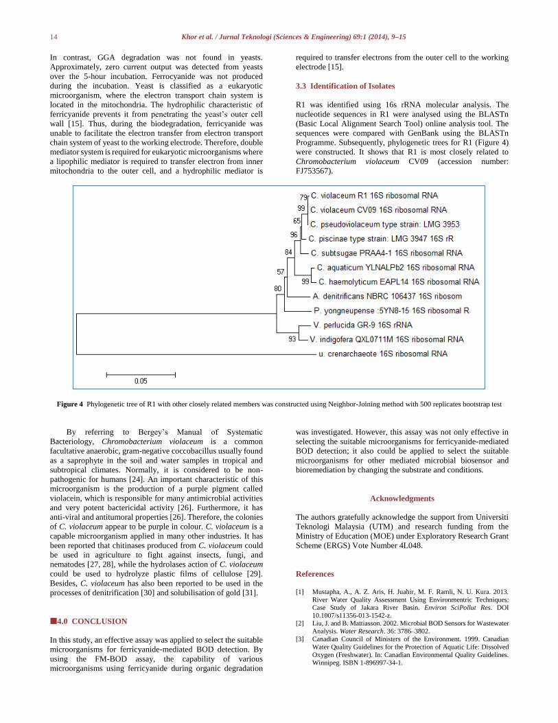
3.3 Identification of Isolates
R1 was identified using 16s rRNA molecular analysis. The
nucleotide sequences in R1 were analysed using the BLASTn
(Basic Local Alignment Search Tool) online analysis tool. The
sequences were compared with GenBank using the BLASTn
Programme. Subsequently, phylogenetic trees for R1 (Figure 4)
were constructed. It shows that R1 is most closely related to
Chromobacterium violaceum CV09 (accession number:
FJ753567).
Figure 4 Phylogenetic tree of R1 with other closely related members was constructed using Neighbor-Joining method with 500 replicates bootstrap test
By referring to Bergey’s Manual of Systematic
Bacteriology, Chromobacterium violaceum is a common
facultative anaerobic, gram-negative coccobacillus usually found
as a saprophyte in the soil and water samples in tropical and
subtropical climates. Normally, it is considered to be non-
pathogenic for humans [24]. An important characteristic of this
microorganism is the production of a purple pigment called
violacein, which is responsible for many antimicrobial activities
and very potent bactericidal activity [26]. Furthermore, it has
anti-viral and antitumoral properties [26]. Therefore, the colonies
of C. violaceum appear to be purple in colour. C. violaceum is a
capable microorganism applied in many other industries. It has
been reported that chitinases produced from C. violaceum could
be used in agriculture to fight against insects, fungi, and
nematodes [27, 28], while the hydrolases action of C. violaceum
could be used to hydrolyze plastic films of cellulose [29].
Besides, C. violaceum has also been reported to be used in the
processes of denitrification [30] and solubilisation of gold [31].
4.0 CONCLUSION
In this study, an effective assay was applied to select the suitable
microorganisms for ferricyanide-mediated BOD detection. By
using the FM-BOD assay, the capability of various
microorganisms using ferricyanide during organic degradation
was investigated. However, this assay was not only effective in
selecting the suitable microorganisms for ferricyanide-mediated
BOD detection; it also could be applied to select the suitable
microorganisms for other mediated microbial biosensor and
bioremediation by changing the substrate and conditions.
Acknowledgments
The authors gratefully acknowledge the support from Universiti
Teknologi Malaysia (UTM) and research funding from the
Ministry of Education (MOE) under Exploratory Research Grant
Scheme (ERGS) Vote Number 4L048.
References
[1] Mustapha, A., A. Z. Aris, H. Juahir, M. F. Ramli, N. U. Kura. 2013.
River Water Quality Assessment Using Environmentric Techniques:
Case Study of Jakara River Basin. Environ SciPollut Res. DOI
10.1007/s11356-013-1542-z. [2] Liu, J. and B. Mattiasson. 2002. Microbial BOD Sensors for Wastewater
Analysis. Water Research. 36: 3786–3802.
[3] Canadian Council of Ministers of the Environment. 1999. Canadian
Water Quality Guidelines for the Protection of Aquatic Life: Dissolved
Oxygen (Freshwater). In: Canadian Environmental Quality Guidelines.
Winnipeg. ISBN 1-896997-34-1.

15 Khor et al. / Jurnal Teknologi (Sciences & Engineering) 69:1 (2014), 9–15
[4] Chen, H., T. Ye, B. Qiu, G. Chen, and X. Chen. 2008. A Novel Approach
Based on Ferricyanide-mediator Immobilized in an Ion-exchangeable
Biosensing Film for the Determination of Biochemical Oxygen Demand.
Analytica Chimica Acta. 612(1): 75–82.
[5] Yoshida, N., J. Hoashi, T. Morita, S. J. McNiven, H. Nakamura, and I. Karube. 2001. Improvement of a Mediator-type Biochemical Oxygen
Demand Sensor for On-site Measurement. Journal of Biotechnology. 88:
269–275.
[6] Raud, M., M. Tutt, E. Jõgi,, and T. Kikas. 2012. BOD Biosensors for
Pulp and Paper Industry Wastewater Analysis. Environ SciPollut Res.
19: 3039–3045.
[7] Pasco, N., K. Baronian, C. Jeffries, and J. Hay. 2000. Biochemical Mediator Demand-A Novel Rapid Alternative for Measuring
Biochemical Oxygen Demand. Appl Microbiol Biotechnol. 53: 613–618.
[8] Morris, K., K. Catterall, H. Zhao, N. Pasco, and R. John. 2001.
Ferricyanide Mediated Biochemical Oxygen Demand–Development of
a Rapid Biochemical Oxygen Demand Assay. Analytical Chimica Acta.
442: 129–139.
[9] Catterall, K., H. Zhao, N. Pasco, and R. John. 2003. Development of a
Rapid Ferricyanide-mediated assay for Biochemical Oxygen Demand Using a Mixed Microbial Consortium. Anal. Chem. 75: 2584–2590.
[10] Pasco, N., K. Baronian, C. Jeffries, and J. Hay. 2004. MICREDOX®—
Development of a Ferricyanide-Mediated Rapid Biochemical Oxygen
Demand Method Using an Immobilized Proteus vulgaris
Biocomponent. Biosensors and Bioelectronics. 20: 524–532.
[11] Morris, K. 2005. Optimization of the Biocatlytic Component in a
Ferricyanide Mediated Approach to Rapid Biochemical Oxygen
Demand Analysis. Griffith University: Australia. 90. [12] Thevenot, D. R., K. Toth, R. A. Durst, and G. S. Wilson. 2001.
Electrochemical Biosensors: Recommended Definitions and
Classification. Biosens Bioelectron. 16(1–2): 121–31.
[13] Su, L., W. Jia, C. Houb, and Y. Lei. 2011. Microbial Biosensors: A
Review. Biosensors and Bioelectronics. 26: 1788–1799.
[14] Yoshida, N., K. Yano, T. Morita, S. J. McNiven, H. Nakamura, and I.
Karube. 2000. A Mediator-type Biosensor as a New Approach to Biochemical Oxygen Demand Estimation. Analyst. 125: 2280–2284.
[15] Nakamura, H., K. Suzuki, H. Ishikuro, S. Kinoshita, R. Koizumi, S.
Okumaa, M. Gotoh, and I. Karube. 2007. A New BOD Estimation
Method Employing a Double-mediator System by Ferricyanide and
Menadione Using the Eukaryote Saccharomyces cerevisiae. Talanta. 72:
210–216.
[16] Catterall, K., K. Morris, C. Gladman, H. Zhao, N. Pasco, and R. John.
2001. The Use of Microorganisms with Broad Range Substrate Utilisation for the Ferricyanide-Mediated Rapid Determination of
Biochemical Oxygen Demand. Talanta. 55: 1187–1194.
[17] Trosok, S. P., B. T. Driscoll, and J. H. T. Luong. 2001. Mediated
Microbial Biosensor Using a Novel Yeast Strain for Wastewater BOD
Measurement. Appl Microbiol Biotechnol. 56: 550–554.
[18] Shahir, S., S. L. Chun, and R. Ahamad. 2011. Application of an
Acclimated Microbial Consortium as Bio Catalyst for Rapid
Determination of Biochemical Oxygen Demand. Asian Journal of
Biotechnology. 3(5): 519–529.
[19] American Public Health Association (APHA). 1992. Standard Methods for the Examination of Water and Wastewater. 18th ed. Washington DC,
USA.
[20] Prados, M. D., L. Seguí, and P. Fito. 2010. Industrial Pineapple Waste
as a Feasible Source to Produce Bioethanol. International Conference on
Food Innovation: Food Innova 2010.
[21] Ong, S., E. Toorisaka, M. Hirata, and T. Hano. 2008. Combination of
Absorption and Biodegradation Processes for Textile Effluent Treatment Using a Granular Activated Carbon-Biofilm Configured Packed Column
System. Journal of Environmental Sciences. 20: 952–956.
[22] Badiei, M., J. M. Jahim, N. Anuar, and S. Abdullah. 2011. Effect of
Hydraulic Retention Time on Biohydrogen Production from Palm Oil
Mill Effluent in Anaerobic Sequencing Batch Reactor. International
Journal of Hydrogen Energy. 36: 5912–5919.
[23] Wang, J., Y. Zhang, Y. Wang, R. Xu, Z. Sun, and Z. Jie. 2010. An
Innovative Reactor-type Biosensor for BOD Rapid Measurement. Biosens Bioelectron. 25: 1705–1709.
[24] Durán, N. and Menck, C. F. M. 2001. Chromobacterium violaceum: A
Review of Pharmacological and Industiral Perspectives. Critical
Reviews in Microbiology. 27(3): 201–222.
[25] Brenner, D. J., Krieg, N. R., Garrity, G. M., Staley, J. T., Boone, D. R.,
Vos, P., Goodfellow, M., Rainey, F. A., Schleifer, K.-H., Gillis, M. and
Logan, N. 2005. Chromobacterium Bergonzini 1881: 153AL. Bergey’s
Manual of Systematic Bacteriology. Springer: US. [26] Durán, N. and Menck, C.F.M. 2001. Chromobacterium Violaceum: A
Review of Pharmacological and Industrial Perspectives. Crit. Rev.
Microbiol. 3: 201–222.
[27] Cronin, D., Moenne-Loccoz, Y., Dunne, C. and O´Gara, F. 1997.
Inhibition of Egg Hatch of the Potato Cyst Nematode Globodera
Rostochiensis by Chitinase-Producing Bacteria. Eur. J. Plant Pathol.
103: 433–440. [28] Patil, R.S., Ghormade, V. and Despande, M.V. 2000. Chitinolytic
Enzymes: An Exploration. Enzyme Microb. Technol. 26: 473–483.
[29] Gourson, C., Benhaddou, R., Granet, R., Krausz, P., Verneuil, B.,
Branland, P., Chauvelon, G., Thibault, J.F., and Saulnier, L. 1999.
Valorization of maize bran to obtain biodegradable plastic films, J. Appl.
Poly. Sci. 74: 3040–3045.
[30] Bazylinski, D. A., Palome, E., Blakemore, N. A., and Blakemore, R. P.
1986. Denitrification by Chromobacterium violaceum. Appl. Environ. Microbiol. 52: 696–699.
[31] Smith, A. D. and Hunt, R. J. 1985. Solubilization of Gold by
Chromobacterium violaceum, J. Chem. Technol. Biotechnol. B.
Biotechnol. 35: 110–116.