Embed Size (px)
Citation preview
ORIGINAL ARTICLE
B-Scan ultrasonography before surgery in eyes with advanced cataracts- A useful prognostic tool
ABSTRACTBACKGROUND: Ocular ultrasonography is an important tool for evaluating the posterior segment in eyes with opaque media. In cases with dense cataract, where posterior segment evaluation by ophthalmoscopy is not possible, B-scan ultrasonography before surgery can help in surgical planning and guiding the expectations of patients.
PURPOSE: To determine the relevance and prevalence of posterior segment abnormalities in patients with dense cataracts prior to surgery by ultrasonography.
DESIGN: Prospective diagnostic study.
METHODS: Diagnostic B-scan ultrasound was performed on 158 eyes of 132 patients with dense cataract precluding visualization of fundus on ophthalmoscopy from January 2013 to December 2013. Patients were divided in two groups, traumatic (22) and non traumatic (136). Patients in the age range of 1 to 79 years of both sexes were included. Detailed history and some basic eye examination techniques, like slit lamp and tonometry were done. Patients having already posterior segment lesions and those who had previous history of ocular surgery were excluded from the study.
RESULTS:, 26 (16.4%) patients, out of total 158 patients, had posterior segment lesions. Among traumatic group of 22 patients, 15 (68.1%) had positive posterior segment lesions, while only 11(8%) patients in the non-traumatic group of 136 patients had positive posterior segment lesions. Out of the 26 positive cases, retinal detachment was found in 8(5%) patients, 7(4.4%) had posterior vitreous detachment, 7(4.4%) had vitreous hemorrhage, 2(1.26%) had retinal detachment with vitreous hemorrhage, 1(0.63%) had asteroid hyalosis, 1(0.63%) had intra-ocular foreign body.

ORIGINAL ARTICLE
CONCLUSION: We concluded that B-scan ultrasound has significant importance in the preoperative evaluation of patients with dense cataracts to detect pathologies that may influence the surgical strategy and the postoperative visual prognosis.
Key words: Ultrasonic B-Scan, Cataract, Posterior Segment lesion, Trauma
ORIGINAL ARTICLE
INTRODUCTION
Ultrasound is an acoustic wave that consists of an oscillation of particles within a
medium. In 1880, French Scientist Curie brothers described the “Piezoelectric
Phenomenon” upon which the current diagnostic ultrasound is based. [1]
Ultrasound technology, also known as sonar, echography or acoustic imaging, was
developed during world war-I as a method of detecting under water objects,
including submarines.[2] Mundt and Hughes first reported the use of ultrasound in
ophthalmic diagnosis in 1956.[3] They utilized A-scan mode. Two years later, Baum
and Greenwood described B-scan ophthalmic ultrasonography.[4] The first
commercially available B-scan was developed by Coleman in 1970’s.
In ophthalmic ultrasonic examination, frequencies used lie mostly in the range
from 5 MHz to 20 MHz .[5]
B-Scan Ultrasonography is an important tool for evaluating the posterior
segment in eyes with opaque media and provides a method of assessing the
structural changes in the posterior segment of the eye in such patients.6 The
ability to examine the posterior segment of the eye accurately in patient in dense
cataracts, is essential before surgery to aid surgical planning and guide the
expectations of patients.
The purpose of the study was to evaluate the usefulness of diagnostic B-
scan ultrasound and to know the prevalence of posterior segment abnormalities,
in patients with dense cataracts prior to surgery by ultrasonography.
ORIGINAL ARTICLE
PATIENTS AND METHODS
Present study was a prospective diagnostic study for assessment of
posterior segment lesion at pre-operative stage, carried out in Department of
Ophthalmology, S.S. Medical College, Rewa for a period of twelve months from
January 2013 to December 2013. Diagnostic B-scan ultrasound was performed on
158 patients with dense cataract, precluding visualization of fundus on
ophthalmoscopy. On the basis of previous history of ocular trauma, patients were
divided into two groups- traumatic & non traumatic. Detailed history, slit lamp
examination and tonometry were done in both groups of patients. Patients in the
age range of 1 to 79 years of both sexes were included and those already having
posterior segment lesions and previous history of ocular surgery were excluded
from the study. B-scan ultrasonography was done using Sonomed E-Z scan AB
5500 machine. The examination was performed with patient in supine position,
using a coupling jelly and the transducer head held over a closed eyelid. Both eyes
were scanned serially in transverse and horizontal axial scans. High gain (90-100
dB) and low gain (60 to 70dB) sensitivity were selected during ultrasonography.
OBSERVATIONS AND RESULTS:
Out of a total of 158 patients, 22 (13.93%) patients were of post-traumatic
cataract and 136 (86.07%) were of non-traumatic cataract. Patients of both sexes
in age range 1 to 79 years were included (Table-1).

ORIGINAL ARTICLE
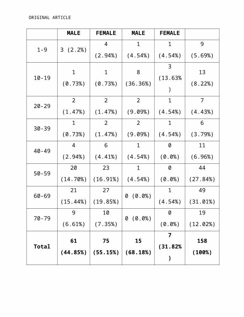
TABLE-1. AGE AND SEX DISTRIBUTION
Age(Years)Non Traumatic Group
(136 Patients)
Traumatic Group
(22 Patients)
Total
Patients
(158
Patients)
MALE FEMALE MALE FEMALE
1-9 3 (2.2%) 4 (2.94%) 1 (4.54%) 1 (4.54%) 9 (5.69%)
10-19 1 (0.73%) 1 (0.73%) 8 (36.36%) 3 (13.63%) 13 (8.22%)
20-29 2 (1.47%) 2 (1.47%) 2 (9.09%) 1 (4.54%) 7 (4.43%)
30-39 1 (0.73%) 2 (1.47%) 2 (9.09%) 1 (4.54%) 6 (3.79%)
40-49 4 (2.94%) 6 (4.41%) 1 (4.54%) 0 (0.0%) 11 (6.96%)
50-59 20 (14.70%) 23 (16.91%) 1 (4.54%) 0 (0.0%) 44 (27.84%)
60-69 21 (15.44%) 27 (19.85%) 0 (0.0%) 1 (4.54%) 49 (31.01%)
70-79 9 (6.61%) 10 (7.35%) 0 (0.0%) 0 (0.0%) 19 (12.02%)
Total 61 (44.85%) 75 (55.15%) 15 (68.18%) 7 (31.82%) 158 (100%)
In the non-traumatic cataract group of patients, most of the patients
(66.91%) were in the range of 50 to 69 years of age as senile cataract is common
in this age group. In traumatic cataract group, most of the patients (50%) were in
the range of 10 to 19 years, the age group where trauma is more common. Males
(68.18%) outnumbered females (31.82%) in traumatic group as males are more
involved in outdoor activities and thus more prone to ocular trauma (Table-1).

ORIGINAL ARTICLE
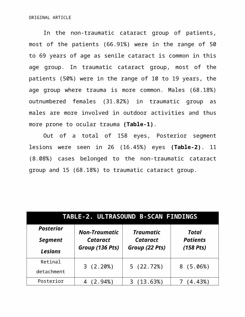
Out of a total of 158 eyes, Posterior segment lesions were seen in 26
(16.45%) eyes (Table-2). 11 (8.08%) cases belonged to the non-traumatic cataract
group and 15 (68.18%) to traumatic cataract group.
TABLE-2. ULTRASOUND B-SCAN FINDINGS
Posterior Segment
Lesions
Non-TraumaticCataract
Group (136 Pts)
TraumaticCataract
Group (22 Pts)
TotalPatients(158 Pts)
Retinal detachment 3 (2.20%) 5 (22.72%) 8 (5.06%)
Posterior vitreousdetachment 4 (2.94%) 3 (13.63%) 7 (4.43%)
Vitreous hemorrhage 2 (1.47%) 5 (22.72%) 7 (4.43%)
Retinal detachment +
Vitreous Hemorrhage1 (0.73%) 1 (4.54%) 2 (1.26%)
Asteroid Hyalosis 1 (0.73%) 0 (0.0%) 1 (0.63%)
Intra-ocular foreignbody 0 (0.0%) 1(4.54%) 1 (0.63%)
Total 11 (8.08%) 15 (68.18%) 26 (16.45%)
Out of the 26 positive cases, 8 (5.06%) had retinal detachment (Fig 1), 7
(4.43%) had posterior vitreous detachment (Fig-2), 7 (4.43%) had vitreous
hemorrhage (Fig-3), 2 (1.26%) were retinal detachment with vitreous
ORIGINAL ARTICLE
haemogrrhage (Fig 4) while asteroid hyalosis (Fig-5) and intra-ocular foreign body
(Fig 6) were found in 1 (0.63%) eye each (Table-2).
DISCUSSION
In developing countries like India, Cataract is an important cause of
blindness and due to lack of proper awareness, many patients presents with
advanced cataracts that precludes visualization of fundus prior to cataract
surgery. Such visualization is considered important to provide accurate prognosis
for vision after cataract surgery. Under such circumstances ultrasonographic
examination can provide information regarding such abnormalities. Over the last
30 years, ultrasonography has greatly advanced which has enabled us to study
posterior segment of the eye even in the presence of opaque media like dense
cataract.
Posterior segment of a total of 158 eyes in 1-79 years old patients, divided
into two groups, were examined under B-scan ultrasound. Non traumatic group
constitutes majority of the patients (86.07%) and a small number (13.93%)
belongs to traumatic cataract group. Similar groups in different age ranges have
also been discussed in other studies.[7-10] In traumatic cataract group, 50% of the
patients were 10-19 years old, correlating with the age range when children are
more active and involved in outdoor games and other activities.
Findings of posterior segment lesions (16.4%) in this study were similar to
other published studies and very much less than that in the study by Haile and
Mengistu who found a 66% incidence of detectable abnormalities.[8,10,11,12]
However, the latter study included cases with orbital pathology and clear media
ORIGINAL ARTICLE
(10%) and it was not clear whether ultrasonography was being performed
routinely on all eyes prior to cataract surgery or only on eyes where intraocular
pathology was suspected. In our study 8.08% patients of non-traumatic cataract
group and 68.18% patients in traumatic cataract group had posterior segment
lesions. Qureshi et al[10] reported posterior segment lesions in 8.64 % non
traumatic cataract patients and in 54.93 % patients with traumatic cataract. Ali
and Rehman[7] reported posterior segment lesions in 11% non traumatic cataract
patients and in 65.85% patients with traumatic cataract.
Retinal detachment (22.72%) in traumatic cataract patients and (2.2%) in
non traumatic cataract patients was similar to that reported by Qureshi et al [10]
who found retinal detachment 21.12% in traumatic cataract and 1.47% in non
traumatic cataract patients; and other study[7] found retinal detachment in
29.26% of traumatic cataract and 3.3% of non traumatic cataract patients.
13.63% patients in traumatic group and 2.94% patients in non-traumatic group
were found to have posterior vitreous detachment (PVD), which is closer to an
earlier report[10] of 9.86% in traumatic cataract and 1% in non-traumatic cataract
patients. Vitreous hemorrhages were present in 22.72% traumatic cataract
patients and 1.47% in non traumatic cataract patients. Other investigators
reported vitreous hemorrhage in traumatic cataract group as 15.49% and
18.3%[7,10], whereas in non traumatic cases vitreous hemorrhage was present in
1.91% cases[10]. Retinal Detachment along with vitreous hemorrhage is found in
4.54% of traumatic cataract and 0.73% of non traumatic cataract. A lower
incidence of asteroid hyalosis (0.73%) was noted in non-traumatic cataract
patients as compared to that reported by Qureshi et al[10] (1.77%) and Ali and
Rehman[7] (2.93%) in their study but higher incidence was noted as compared to

ORIGINAL ARTICLE
other study[9](0.4%). Intraocular foreign body was found in 4.54% traumatic
cataract patients which is less than earlier reports (8.45%)[4].
CONCLUSION
We concluded that two dimensional B-scan ultrasound is simple, safe, non-
invasive, cost-effective, easily available, reproducible and quick investigative
technique which proves accurate and beneficial in opaque ocular media to detect
posterior segment pathologies. B-Scan ultrasonography should be performed
routinely in pre-operative assessment of cataract patients to diagnose pathologies
of posterior segment that may influence the surgical strategy and visual prognosis
of patients after cataract surgery
REFERENCES
1. Athey PA, McClendon L. Diagnostic Ultrasound for Radiographers. Ist ed.Multimedia Publishing Inc: Denver, 1983.
2. Richard L, Hart LJ. Ultrasound Diagnosis of the eye and orbit; principle and practice of ophthalmology. 1997; 5: 98.
3. Mundt GH Jr, Hughes WF Jr. Ultrasonics in ocular diagnosis. Am J Ophthalmol. 1956; 41:488-98.
4. Baum G, Greenwood I. The application of ultrasonic locating technique to Ophthalmology. Arch Ophthalmol. 1958; 60: 263-79.
5.Newell FW. Ophthalmology: principles and concept. 7th ed. St Louis: CV Mosby Company. 1992
6. Bello TO, Adeoti CO. Ultrasonic assessment in pre-operative cataract patients. Niger Postgrad Med J. 2006; 13(4):326-8.
ORIGINAL ARTICLE
7. Ali SI, Rehman H. Role of B-scan in preoperative detection of posterior segment pathologies in cataract patients. Pak J Ophthalmol 1997;13(4):108-112.
8. Anteby II, Blumenthal EZ, Zamir E, Waindim P. The role of preoperative ultrasonography for patients with dense cataract: a retrospective study of 509 cases. Ophthalmic Surg Lasers 1998; 29 (2):114-8.
9. Correa. Ultrasound findings in patients with dense cataracts. Arq Bras Ophthalmol 2002; 65: 609-13.
10. Qureshi MA, Laghari K. Role of B-Scan Ultrasonography in pre-operative cataract patients. Int J Health Sci (Qassim) 2010 January; 4 (1): 31–7.
11. Haile M, Mengistu Z. B-scan ultrasonography in ophthalmic diseases. East Afr Med J. 1996; 73(11):703-7.
12. Salman A, Parmar P, Vanila CG, Thomas PA, Nelson Jesudasan CA. Is ultrasonography essential before surgery in eyes with advanced cataracts?. J Postgrad Med. 2006; 52: 19-22.
ORIGINAL ARTICLE
Fig. 1: B-Scan ultrasonography of the globe shows a retinal detachment
ORIGINAL ARTICLE
Fig. 2: B-Scan ultrasonography of the globe shows posterior vitreous detachment

ORIGINAL ARTICLE
Fig. 3: B-Scan ultrasonography of the globe shows Vitreous Hemorrhage

ORIGINAL ARTICLE
Fig. 4: B-Scan ultrasonography of the globe shows Retinal Detachment with Vitreous Hemorrhage

ORIGINAL ARTICLE
Fig. 5: B-Scan ultrasonography of the globe shows asteroid hyalosis

ORIGINAL ARTICLE
Fig. 6: B-Scan ultrasonography of the globe shows intra ocular foreign body