Embed Size (px)
Citation preview
6754 J . Am. Chem. SOC. 1982, 104, 6754-6764
torsional rigidity than linear DNA. It is not clear whether a direct relevance to the NMR experiment exists because the fluorescence depolarization results are quite insensitive to bending motions and also because the intercalated ethidium bromide might significantly alter the dynamic properties of the supercoiled DNA compared to its natural form. In another study, conducted by the perturbed y y angular correlation (PAC) method (Martin, P. W.; El-Kuteb, S.; Kuhnlein, U. J . Chem. Phys. 1982, 76, 3819-3822) on su- percoiled PM2 DNA, and apparent isotropic correlation time of 8.2 f 0.6 ns was measured, compared to 62 (+12/-10 ns) for presumably much more flexible single-stranded DNA (Kolfas, C. A.; Sideris, E. G.; El-Kuteb, S.; Martin, P. W.; Kuhnlein, U. Chem. Phys. Lett. 1980, 73, 311-314). Although the agreement with the N M R results, both in the trend and the absolute value
of T, is intriguing, it is not clear how much of the correlation time observed by PAC is due to internal mobility of the “lIn probe at the binding site.
In any case, it appears that some of the unique properties of superhelical DNA are accessible to N M R experiments and that these might help to elucidate the importance of this DNA form in biological processes.
Acknowledgment. Financial support for this research was provided by research grants GM25018 and CA27343 from the National Institutes of Health. T.L.J. also wishes to acknowledge receipt of a Research Career Development Award (AM00291) from the National Institutes of Health.
Isolation and Characterization of Pyrimidine-Psoralen-Pyrimidine Photodiadducts from DNA
David Kanne, Kenneth Straub, John E. Hearst,* and Henry Rapoport
Contribution from the Department of Chemistry and Lawrence Berkeley Laboratory, University of California, Berkeley, California 94720. Received February 16, 1982
Abstract: We report the isolation and characterization for the first time of pyrimidine-psoralen-pyrimidine photodiadducts from DNA. For each of the four psoralens studied, a single pair of diastereomeric thymidine-psoralen-thymidine photcdiadducts, each with cis-syn stereochemistry, was found to account for >90% of the diadducts formed. In addition, we have carried out pulse-chase experiments that establish that these photo cross-links are formed by cycloaddition of a second thymidine residue to the 3,4 double bond (pyrone side) of an initially formed 4’,5’ (furan-side) psoralen-thymidine photomonoadduct.
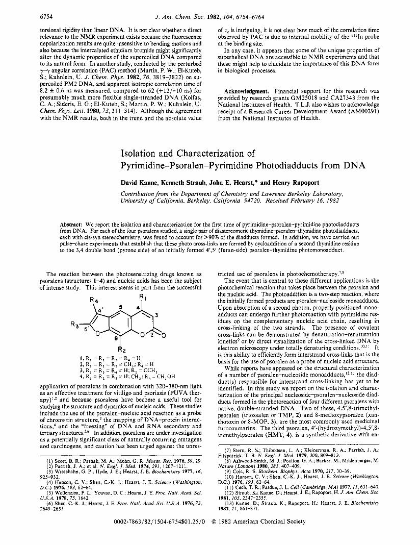
The reaction between the photosensitizing drugs known as psoralens (structures 1-4) and nucleic acids has been the subject of intense study. This interest stems in part from the successful
R 2 1 , R, = R, = R, = R, = H 2, R, = R, = R, = CH,; R, = H 3, R, = R, = R, = H; R, = OCH, 4, R, = R, = R, = H; CH,; R, = CH,OH
application of psoralens in combination with 320-380-nm light as an effective treatment for vitiligo and psoriasis (PUVA ther- apy) lv2 and because psoralens have become a useful tool for studying the structure and dynamics of nucleic acids. These studies include the use of the psoralen-nucleic acid reaction as a probe of chromatin ~ t r u c t u r e , ~ the mapping of DNA-protein interac- t i o n ~ , ~ and the “freezing” of DNA and RNA secondary and tertiary ~tructures .~.~ In addition, psoralens are under investigation as a potentially significant class of naturally occurring mutagens and carcinogens, and caution has been urged against the unres-
(1) Scott, B. R.; Pathak, M. A,; Mohn, G. R. Mutat. Res. 1976, 39, 29. (2) Parrish, J. A.; et al. N . Engl. J . Med. 1974, 291, 1207-1211. (3) Wiesehahn, G. P.; Hyde, J. E.; Hearst, J. E. Biochemistry 1977, 16,
925-932. (4) Hanson, C. V.; Shen, C.-K. J.; Hearst, J. E. Science (Washington,
D.C.I 1976. 193. 62-64. ~.
(g)-Wolienzien, P. L.; Youvan, D. C.; Hearst, J. E. Proc. Natl. Acad. Sci.
(6) Shen, C.-K. J.; Hearst, J. E. Proc. Natl. Acad. Sci. U.S .A. 1976, 73, U.S.A. 1978, 75, 1642.
2649-2653.
tricted use of psoralens in photochem~therapy .~~~ The event that is central to these different applications is the
photochemical reaction that takes place between the psoralen and the nucleic acid. The photoaddition is a two-step reaction, where the initially formed products are psoralen-nucleoside monoadducts. Upon absorption of a second photon, properly positioned mono- adducts can undergo further photoreaction with pyrimidine res- idues on the complementary nucleic acid chain, resulting in cross-linking of the two strands. The presence of covalent cross-links can be demonstrated by denaturation-renaturation kinetics9 or by direct visualization of the cross-linked DNA by electron microscopy under totally denaturing conditions.lOsll It is this ability to efficiently form interstrand cross-links that is the basis for the use of psoralen as a probe of nucleic acid structure.
While reports have appeared on the structural characterization of a number of psoralen-nucleoside mono ad duct^,'^*'^ the diad- duct(s) responsible for interstrand cross-linking has yet to be identified. In this study we report on the isolation and charac- terization of the principal nucleoside-psoralen-nucleoside diad- ducts formed in the photoreaction of four different psoralens with native, double-stranded DNA. Two of these, 4,5’&trimethyl- psoralen (trioxsalen or TMP, 2) and 8-methoxypsoralen (xan- thotoxin or %MOP, 3), are the most commonly used medicinal furocoumarins. The third psoralen, 4’-(hydroxymethyl)-4,5’,8- trimethylpsoralen (HMT, 4), is a synthetic derivative with en-
(7) Stern, R. s.; Thibodeau, L. A,; Kleinerman, R. A,; Parrish, J. A.;
(8) Ashwood-Smith, M. J.; Poulton, G. A,; Barker, M.; Mildenberger, M.
(9) Cole, R. S. Biochem. Biophys. Acta 1970, 217, 30-39. (10) Hanson, C. V.; Shen, C.-K. J.; Hearst, J. E. Science (Washington,
(1 1) Cech, T. R.; Pardue, J. L. Cell (Cambridge, M A ) 1977,II, 631-640. (12) Straub, K.; Kanne, D.; Hearst, J. E.; Rapoport, H. J . Am. Chem. SOC.
(13) Kanne, D.; Straub, K.; Rapoport, H.; Hearst, J. E. Biochemistry
Fitzpatrick, T. B. N . Engl. J . Med. 1979, 300, 809-813.
Nature (London) 1980, 285, 407-409.
D.C.) 1976, 193, 62-64.
1981, 103, 2347-2355.
1982, 21, 861-871.
0002-7863/82/l504-6754$01.25/0 0 1982 American Chemical Society
Pyrimidine-Psoralen-Pyrimidine Photodiadducts
Table I. 3H Recoveries and Modification Levels for Psoralen-DNA Reactions
J . Am. Chem. SOC., Vol, 104, No. 24, 1982 6155
8-MOP’ T M P ~ HMTC Psod
psoralen added per base pair 0.28 0.50 psoralen bound per base pair 0.065 0.110 HPLC fractions, enzyme-hydrolyzed
psoralen-DNA, %bound 3H in
fraction % fraction %
dT-Pso-dT diadduct F33 21 F35 39 (f)dU-Pso monoadducte F36 2 F4 I <2 (f)dT-Pso monoadduct F31 17 F49 53
(p)dT-Pso monoadduct F42 19 F52 2.5
72 of total counts accounted for 93 96.5
F38 28
- -
0.14 0.05
0.28 0.013
fraction 72 fraction %
F31 25 F32 28 F4 3 8 F40A 61 F38A 46 F40B F39
3.0 F38B 2o F4 1 -
F48
9 1 94 -
a Single addition of psoralen, 2-h irradiation. Multiple additions of psoralen. Single addition of psoralen, 10-min irradiation. Single addition of psoralen, 20-min irradiation. e (p) designates that the deoxyribose moiety is bound on the pyrone side. (f) designates that the deoxyribose-moiety is bound on the furan side.
hanced solubility and photobinding ~r0per t ies . l~ We have also characterized the photoadducts derived from psoralen (Pso, 1) itself in order to provide information on the effects of substituents on the psoralen-DNA photoreaction. For each psoralen, a single pair of diastereomeric thymidine-psoralen-thymidine adducts, each with cis-syn15 stereochemistry, was found to account for >90% of all the cross-links. In addition, we have carried out pulse-chase experiments that show that these cross-links are formed by cycloaddition of a second thymidine residue to the 3,4 (pyrone-side) double bond of an initially formed 4,’5’ (furan-side) psoralen-thymidine monoadduct.
Experimental Section Materials. Calf thymus DNA and hydrolytic enzymes were obtained
from Sigma (St. Louis, MO). Poly(dA-dT).poly(dA-dT) was obtained from PL Biochemicals (Milwaukee, WI). [3H]Psoralens were prepared as previously described.I6
Photobinding. To a solution of 43.2 mg of calf thymus DNA in 60 mL of Tris buffer (10 mM of Tris, pH 7.2) was added 0.019 mmol of the psoralen dissolved in a minimum volume of EtOH. The solution was irradiated at 20 OC for 2.5 h for 8-MOP, 20 min for Pso, and 10 min for HMT. Photobinding of [3H]TMP (0.1-0.5 Ci mmol-I) to DNA was carried out by adding five 300-pL aliquots of a stock solution (1.25 mg of T M P in 1.5 mL of ethanol) to the DNA solution (7.2 mg of DNA at a concentration of 0.12 mg mL-’ in Tris buffer). The solution was irradiated for 10 min after each of the psoralen additions.
Two 400-W GE mercury vapor lamps were used for the irradiation, and cooling of the DNA solution was achieved by circulation of a solution of cobaltous nitrate (40% w/w) through an outer jacket; this solution also served as a 365-nm transmission filter. The light intensity delivered to the sample in this device was 100 mW cm-2. After the appropriate irradiation time, the psoralen-DNA solution was extracted with four volumes of chloroform to remove unreacted psoralen and its photo- degradation products. The remaining solution was made 0.2 M in sodium chloride and precipitated by addition of ethanol. The isolated DNA pellet was then dried under vacuum and redissolved in hydrolysis buffer ( I5 mM of sodium acetate, pH 5.00). Alternatively, the DNA was subjected to acid hydrolysis as described below.
In the pulse-chase experiment, 5 mg of poly(dA-dT).poly(dA-dT) was dissolved in 5 mL of Tris buffer (10 mM, pH 7.2). To this solution was added 0.640 mg of 8-MOP dissolved in 300 pL of ethanol. After a 45-s irradiation, the solution was extracted four times with chloroform, made 0.4 M in NaCI, cooled to -20 OC, and precipitated with two volumes of cold ethanol. After a second precipitation, the pellet was redissolved in 5 mL of buffer, and 833 pL was removed. The remaining volume was irradiated further, and aliquots were removed after 15 s, 30 s, 1 min, 2.5
(14) Isaacs, S. T.; Shen, C. J.; Hearst, J. E.: Rapoport, H. Biochemistry 1977, 16, 1058.
(15) Syn for the furan side is defined as shown in 5 or 5’ in which the 01’ of the furan and N1 of the pyrimidine are bonded to adjacent corners of the cyclobutane. Syn for the pyrone side is defined as shown also in 5 or 5’ in which C2 of the pyrone and N1 of the pyrimidine are bonded to adjacent corners of the cyclobutane. Anti stereochemistry is applied to bonding in which these atoms are bonded to diagonal corners. Cis and trans refer to the position of the psoralen and pyrimidine moieties relative to the place of the cyclobutane.
(16) Isaacs, S. T.; Rapoport, H.; Hearst, J. E. J . Labelled Compd. Rn- diopharm. 1982, 19, 345-356.
min, and 5 min. Enzymatic hydrolysis and HPLC analysis were carried out as described below.
Adduct Isolation. Enzyme hydrolysis of the psoralen-modified poly- (dA-dT).poly(dA-dT) or DNA was accomplished by dissolving the nu- cleic acid in hydrolysis buffer (sodium acetate, pH 5.00) and adding 60 units of DNAase-I1 (EC 3.1.22.1) per mg of material. After 12 h, the pH of the solution was adjusted to 7.0, and 0.2 unit of phosphodiesterase I1 (EC 3.1.3.1) per mg of nucleic acid was added. A second addition of phosphodiesterase was made after 12 h, and after an additional 12-24 h, the mixture was adjusted to pH 8.0 and treated with 0.2 unit of alkaline phosphatase (EC 3.1.3.1) per mg of nucleic acid. After 6 h, the hydrolysis mixture was concentrated to a minimal volume and applied directly to a 10 mm X 25 cm reverse-phase (CIS) HPLC column (Ul- trasphere ODS, Altex-Beckman, Berkeley, CA). The column was eluted with either water-methanol or 10 mM KH2P04 (pH 2.2)-methanol, at a flow rate of 4 mL min-l. Fractions were collected and assayed for the presence of 3H by scintillation counting.
Acid hydrolysis was accomplished by dissolving the TMP-modified nucleic acid in 0.4 N HCI and heating at 70 OC for 2 h. The hydrolysis mixture was then neutralized with sodium hydroxide and applied to a C-18 Sep Pak cartridge (Waters, Milford, MA). The cartridge was first eluted with water and then with methanol. The methanol fraction was then concentrated and further analyzed by HPLC.
Mass Spectrometry. The HPLC fractions of interest were collected and analyzed by field-desorption (FD) mass spectrometry. In addition, adducts were converted to per(trimethy1)silyl) (Me&) derivatives by heating 0.5-2.0 pg of adduct with 50 pL of pyridine-BSTFA (N,O-bis- (trimethylsilyl)trifluoroacetamide, 1:4, v/v) for 40 min at 60 OC. These TMS derivatives were analyzed by electron-impact MS at a resolving power of 10000.
Mass spectra were recorded on either a Kratos MS5OS or a modified MS902 mass spectrometer interfaced to a Xerox-Sigma 7/Logos I1 data system.17 Samples analyzed by electron-impact MS were admitted to the ion source (source temperature, 250 “C) via the direct-insertion probe. Field-desorption mass spectra were obtained by using benzo- nitrile-activated emitters.
IH N M R spectra were recorded on either a Nicolet Technologies NT360 or on a Bruker HX-360 spectrometer at 26 O C ; 256-2048 transients were collected for each spectrum using a spectral width of 1500 Hz. Spectra used for detecting NOE enhancements were obtained with the decoupling field gated off during data acquisition and with a delay time of 3-4 s inserted between the end of data acquisition and the following pulse. Spectral assignments were made with the aid of extensive homonuclear decoupling experiments.
Samples were exchanged a total of three times from 99.996% D20. All exchange and loading operations were carried out under a dry ni- trogen atmosphere. Chemical shifts are referenced to Me,Si (6 HDO = 4.75 ppm).
Photoreversion. Adducts were photoreverted by irradiating at 254 nm in methanol-water for various times (30 s-30 min). Reaction products were then analyzed by HPLC.
Results HPLC/Hydrolysis. The HPLC elution profile of [3H]TMP-
modified DNA that has been hydrolyzed enzymatically is shown
’H NMR.
(17) Burlingame, A. L.; Smith, D. H.; Meran, T. 0.; Olsen, R. W. “Progress in Analytical Chemistry”: Orr, C. H., Norris, J . A,, Eds.: Plenum: New York, 1970; Vol. 4, pp 17-38.
6756 J . Am. Chem. SOC., Vol. 104, No. 24, 1982
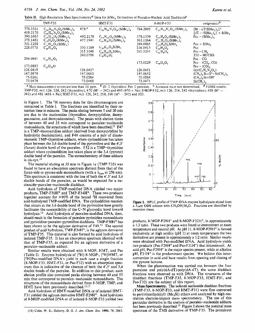
Table 11. High-Resolution Mass Spectrometryd Data for SiMe, Derivatives of Psoralen-Nucleic Acid DiadductsD
Kanne et al.
TMP-F35 HMT-F3 1 8-MOP-F33 assignmentb 776.3321 459.2170 390.1663 375.146 1 301.125 8 228.0775
200.0841
172.0892 126.0419 147.0670
75.0261 73.0478
C,,H,,N,09(SiMe,), 878.c C,, H,, N,O, (SiMe,), C,, H, , 0, (SiMe, ), 492.2 170 C,,H,O,(SiMe,), 477.1 94 1 C,,H,, O,(SiMe,)
14 3 330.1 289 31 5.1 049 240.0796
C13H1202
CI,Hl,O 126.0427 147.0663 75.0264 7 3.0469
C,,H,,N,O,,(SiMe,), 764.2995
C, 5HI3O5 (SiMe, ), 378.1339 C,,H,, 0, (SiMe, 1, 363.1064
2 8 9.0 8 8 5 C H,, O,SiMe, 216.0413 C,, H,,O, SiMe, 201.0201 '15 HIZ '3
173.0229
126.0431 147.0652
75.0268 73.0471
C,,H,,N,O,(SiMe,), [ M - dT(SiMe,),]+.
ClzH805 (SiMe,), Pso - (SiMe,), C,, H,O, (SiMe,), C, , H,O, SiMe, Cl,H*O, Pso
[dT - (SiMe,),] + SiMe,
Pso + SiMe,
CllHSO, PSO - CH, 330 - HOTMS Pso - co PSO - (CH,, CO) Pso - (CO), thy (C5H6 N,O,)
',OH 5'3
(CH,), Si=O+--Si(CH,), (CH , ) , Si= OH+ SiKH, ),
Mass measurement errors are less than 10 ppm. dT E thymidine; Pso psoralen. Accurate mass not determined. FDMS results: TMP-F35, m / z 126, 228, 242 (thymidine), 470 (M'. - 242) and 493 (470 + Na); 8-MOP-F32, m/z 126, 216, 242 (thymidine), 458 (M'.- 242) and 481 (458 + Na); HMT-F31, m/z 126 ,242 ,258 ,500 (M+. - 242) and 523.
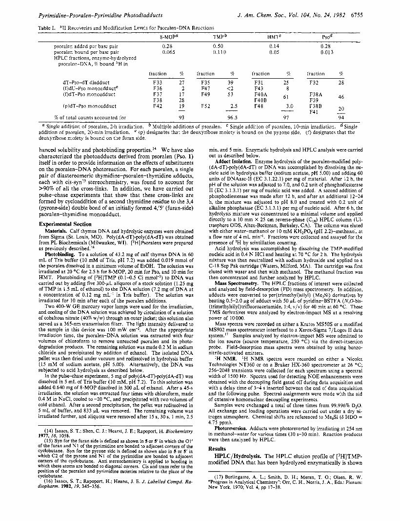
in Figure 1. The 3H recovery data for this chromatogram are contained in Table I. The fractions are identified by their re- tention time in minutes. The peaks eluting between 5 and 20 min are due to the nucleosides (thymidine, deoxycytidine, deoxy- guanosine, and deoxyadenosine). The peaks with elution times of between 40 and 5 5 min correspond to psoralen-nucleoside monoadducts, the structures of which have been described.13 F47 is a TMP-deoxyuridine adduct (derived from deoxycytidine by hydrolytic deamination), and F49 consists of a pair of diaste- reomeric TMP-thymidine adducts, where cycloaddition has taken place between the 5,6 double bond of the pyrimidine and the 4'3' (furan) double bond of the psoralen. F52 is a TMP-thymidine adduct where cycloaddition has taken place at the 3,4 (pyrone) double bond of the psoralen. The stereochemistry of these adducts is cis-syn.15
The material eluting at 35 min in Figure l a (TMP F35) was found to have an absorption spectrum distinct from that of the furan-side or pyrone-side monoadducts (with a A,, at 256 nm). This spectrum is consistent with the loss of both the 4'3' and 3,4 double bonds of the psoralen, as would be expected for a nu- cleoside-psoralen-nucleoside diadduct.
Acid hydrolysis of TMP-modified DNA yielded two major products, TMP-F5OH+ and TMP-F34H+. These two products together account for >90% of the bound 3H recovered from acid-hydrolyzed TMP-modified DNA. The cycloaddition reaction that occurs at the 5,6 double bond of the pyrimidine base greatly facilitates the susceptibility of the C-N glycosidic bond toward hydrolysis.ls Acid hydrolysis of psoralen-modified DNA, then, should result in the formation of psoralen-pyrimidine monoadducts and pyrimidine-psoralen-pyrimidine diadducts. TMP-F5OH' has been shown to be the aglycon derivative of F49.I3 The second product of acid hydrolysis, TMP-F34H+, is the aglycon derivative of TMP-F35. This material is also formed by acid hydrolysis of isolated TMP-F35. It has an absorption spectrum identical with that of TMP-F35, as expected for an aglycon derivative of a psoralen-nucleoside adduct.
Similar results were obtained with 8-MOP, HMT, and Pso (Table I). Enzyme hydrolysis of [3H]-8-MOP-, [3H]HMT, or [3H]Pso-modified DNA's yield in each case a single fraction (8-MOP-F33, HMT-F31, or Pso-F32) with an absorption spec- trum that is indicative of the absence of both the 4'3' and 3,4 double bonds of the psoralen. In addition to this product, each elution profile also contained peaks eluting between 40 and 55 min that correspond to psoralen-nucleoside monoadducts. The structures of the monoadducts derived from 8-MOP, TMP, and HMT have been previously de~c r ibed . '~ J~
Acid hydrolysis of HMT-modified DNA or of isolated HMT- F3 1 yielded the aglycon derivative HMT-F29H+. Acid hydrolysis of 8-MOP-modified DNA or of isolated 8-MOP-F33 yielded two
(18) Cohn, W. E.; Doherty, D. G . J . Am. Chem. SOC. 1956, 78, 2863.
1 I I I I I
0 IO 20 30 40 50 60
T ime (min)
Figure 1. HPLC profile of TMP-DNA enzyme hydrolysate eluted from a 5-pm ODS column with C H 3 0 H / H 2 0 . Fractions are identified by retention times.
products, 8-MOP-F29H+ and 8-MOP-F32HC, in approximately a 1:3 ratio. These two products were found to interconvert at room temperature and neutral pH. At pH 11,8-MOP-F29H+ is formed exclusively; at high acidity (pH 1) at room temperature the two derivatives are present in approximately a 1:2 ratio. Similar results were obtained with Pso-modified DNA. Acid hydrolysis yields two products (Pso-F29H+ and Pso-F31H+) that interconvert. At acid pH, Pso-F29H+ is the major species present, while at alkaline pH, F31H' is the predominant species. We believe this inter- conversion in acid and base results from opening and closing of the pyrone lactone.
When the photoreaction was carried out between the four psoralens and poly(dA-dT)-poly(dA-dT), the same diadduct fractions were observed as with DNA. The structures of the diadduct fractions (TMP-F35, 8-MOP-F33, HMT-F31, and Pso-F32) are the subject of this report.
Mass Spectrometry. The isolated nucleoside diadduct fractions (TMP-F35, 8-MOP-F33, and HMT-F3 1) were first converted to per(trimethylsily1) (Me3Si) ethers and analyzed by high-res- olution electron-impact mass spectrometry. The use of this particular derivative in the analysis of psoralen-nucleoside adducts has been previously de~cribed.'~ Figure 2 shows the nominal mass spectrum of the TMS derivative of TMP-F35. The prominent
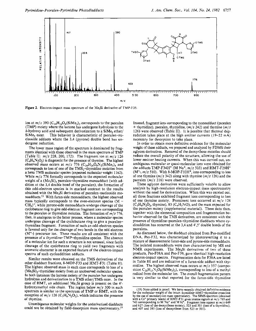
Pyrimidine-Psoralen-Pyrimidine Photodiadducts J . Am. Chem. SOC., Vol. 104, No. 24, 1982 6757
c I Q
147
0 22 8
I90
O T M S OTMS S
45 9
776
I
' I 330 400 5 00 600 700
Figure 2. Electron-impact mass spectrum of the MelSi derivative of TMP-F35.
ion at m / z 390 (C14H1204(SiMe3)2 corresponds to the psoralen (TMP) moiety where the lactone has undergone hydrolysis to the &hydroxy acid and subsequent derivatization to a SiMe, ether/ SiMe3 ester. This behavior is characteristic of psoralen-nu- cleoside adducts where the 3,4 (pyrone) double bond has un- dergone reduction.
The lower mass region of the spectrum is dominated by frag- ments identical with those observed in the mass spectrum of TMP (Table 11: m / z 228, 200, 172). The fragment ion at m / z 126 (C6H6N,0,) is diagnostic for the presence of thymine. The highest observed mass occurs a t m / z 776 (C24H2409Nt(SiMe3)4 and corresponds to loss of one of the TMS,-thymidine moieties from a hexa TMS molecular species (expected molecular weight 1162). While m / z 776 formally corresponds to the expected molecular weight of a psoralen-thymidine monoadduct (with ad- dition at the 3,4 double bond of the psoralen), the formation of this odd-electron species is in marked contrast to the results obtained with the Me3Si derivatives of psoralen-nucleoside mo- n0add~cts . l~ With furan-side monoadducts, the highest observed mass typically corresponds to the even-electron species (M - CH3)+, while pyrone-side monoadducts undergo cleavage of the cyclobutane ring to give odd-electron fragment ions corresponding to the psoralen or thymidine moieties. The formation of m / z 776, then, is analogous to the latter process, where a molecular species undergoes cleavage of the cyclobutane ring to give a psoralen- thymidine fragment. Formation of such an odd-electron species is favored only for the cleavage of two bonds in the odd electron (M'.) precursor ion. These results are all consistent with the presence of a thymidine-TMP-thymidine species. The absence of a molecular ion for such a structure is not unusual, since facile cleavage of the cyclobutane ring to yield two fragments with aromatic character is an expected feature of the electron-impact spectra of such cycloaddition adducts.
Similar results were obtained on the T M S derivatives of the other diadduct fractions, 8-MOP-F33 and HMT-F3 1 (Table 11). The highest observed mass in each case corresponds to loss of a (Me$),-thymidine moiety from an unobserved molecular species. In both instances the lactone moiety of the psoralen has undergone hydrolysis and derivatization to a TMS ether/TMS ester. In the case of HMT, an additional Me3Si group is present on the 4'- hydroxymethyl side chain. The region below m / z 300 in each spectrum is similar to the spectrum of T M P or HMT, with the exception of m / z 126 (CSH6N2O2), which indicates the presence of thymine.
Unambiguous molecular weights for the underivatized diadducts could not be obtained by field-desorption mass spe~trometry. '~
Instead, fragment ions corresponding to the monoadduct (psoralen + thymidine), psoralen, thymidine, ( m / z 242) and thymine ( m / z 126) were observed (Table 11). It is possible that thermal deg- radation takes place at the high emitter currents (19-22 mA) necessary for desorption to take place.
In order to obtain more definitive evidence for the molecular weight of these adducts, we prepared and analyzed by FDMS their aglycon derivatives. Removal of the deoxyribose moieties should reduce the overall polarity of the structure, allowing the use of lower emitter heating currents. When this was carried out, un- ambiguous molecular or quasi-molecular ions were obtained for the adducts TMP-F34H+ (M Na+, m / z 503) and HMT-F29Hf (M+., m / z 510). With 8-MOP-F32H+, ions corresponding to loss of one thymine ( m / z 342) along with thymine ( m / z 126) and the psoralen ( m / z 216) were observed.
These aglycon derivatives were sufficiently volatile to allow analysis by high-resolution electron-impact mass spectrometry without the need for derivatization. When this was carried out, all three fractions exhibited fragment ions corresponding to loss of one thymine moiety. Prominent ions occurred at m / z 126 (C5H6N,0z, thymine), 83 (C4H,NO), and the mass expected for the psoralen moiety (supplemental material). These data then, together with the elemental composition and fragmentation be- havior observed for the TMS derivatives, are consistent with the presence of thymidine-psoralen-thymidine diadducts where cy- cloaddition has occurred at the 3,4 and 4',5'double bonds of the psoralens.
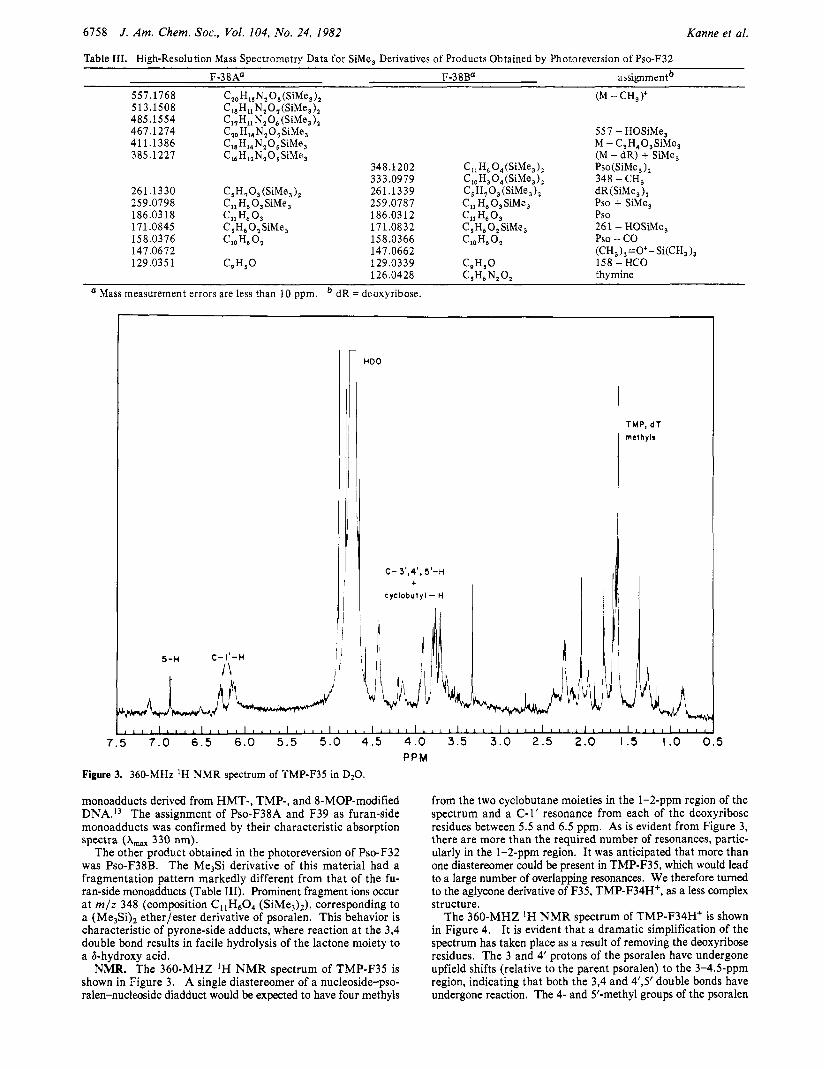
As discussed below, the diadduct obtained from Pso-modified DNA, Pso-F32, was characterized by photoreverting it to a mixture of diastereomeric furan-side and pyrone-side monoadducts. The isolated monoadducts were then characterized by MS and N M R experiments. The Me$% derivatives of two of these products, Pso-F38A and Pso-F39, gave identical high-resolution electron-impact spectra. Fragmentation data for F38A are listed in Table I11 and are indicative of a furan-side adduct with thy- midine. The highest observed mass occurs at m / z 557 (compo- sition CZOH15N20s(SiMe3)2), corresponding to loss of a methyl radical from the molecular ion. The overall fragmentation pattern is very similar to that reported for the furan-side thymidine
~~~~
(19) Note added in proof We have recently obtained definitive evidence for the molecular weight of the intact thymidine-HMT-thymidine cross-link by the use of secondary-ion mass spectrometry. The SIMS spectrum (obtained with a Cs' primary beam) of HMT-F31 gives intense signals at m/z 765 and 743 corresponding to M Na' and M H*. Fragment ions appear at m/z 649 and 627 (loss of the deoxyribose moiety), 523 and 501 (loss of a thymidine), and 407 and 385 (loss of deoxyribose from 523 or 501).
6158 J . Am. Chem. SOC., Vol. 104, No. 24, 1982 Kanne et al.
Table 111. High-Resolution Mass Spectrometry Data for SiMe, Derivatives of Products Obtained by Photoreversion of Pso-F32
F-38Aa F-38Ba assignmen?
557.1768 Cz0 H,, N, 0, (SiMe, 1, (M - CH,)+ 513.1508 C18H11 N,O,(SiMe,), 485.1554 C17Hi1 N,O, (SiMe,), 467.1274 C,, H14N,0, SiMe, 557 - HOSiMe, 411.1386 C18Hl,N,05SiMe, M - C,H,O,SiMe, 385.1227 C,, H,,N,O SiMe, (M - dR) + SiMe,
348.1202 C I I H ~ O,(SiMe,), Pso(SiMe,),
261.1330 C5H703 (SiMe,), 261.13 39 C,H,O,(SiMe,), dR (SiMe, ), 259.0798 CllH,O,SiMe, 259.0787 C,, H6 0, SiMe, Pso + SiMe,
171.0845 C,H,O,SiMe, 17 1.08 32 C ,H,O,SiMe, 261 - HOSiMe,
147.0672 147.0662 (CH,),=O+- Si(CH, ),
333.0979 C,J, 0, (SiMe, 1, 348 - CH,
186.03 18 Cll H, 0, 186.0312 Cll H6 0 3 Pso
158.0376 '10 H6 '2 158.0366 ClOH,O, Pso - co 129.035 1 C9H5O 129.0339 C,H,O 158 - HCO
thymine 126.0428 C J L N, 0, Mass measurement errors are less than 10 ppm. dR = deoxyribose.
.ID0
C- 3',4', 5'-H +
Cyclobutyl - H
TMP, dT methyls
monoadducts derived from HMT-, TMP-, and 8-MOP-modified DNA.I3 The assignment of Pso-F38A and F39 as furan-side manoadducts was confirmed by their characteristic absorption spectra (A,,, 330 nm).
The other product obtained in the photoreversion of Pso-F32 was Pso-F38B. The Me3Si derivative of this material had a fragmentation pattern markedly different from that of the fu- ran-side monoadducts (Table 111). Prominent fragment ions occur a t m / z 348 (composition C11H60, (SiMe3)?), corresponding to a (Me3Si), ether/ester derivative of psoralen. This behavior is characteristic of pyrone-side adducts, where reaction at the 3,4 double bond results in facile hydrolysis of the lactone moiety to a &hydroxy acid.
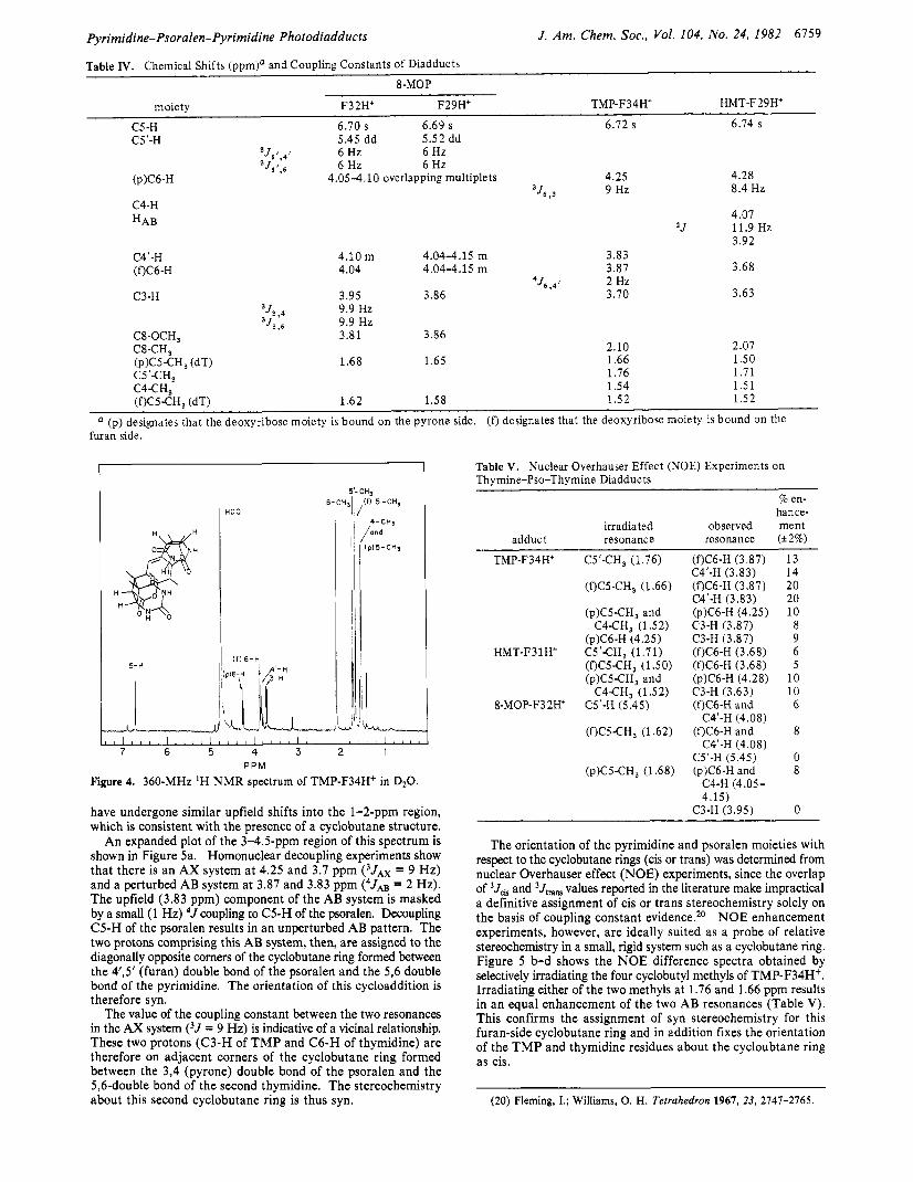
NMR. The 360-MHZ 'H N M R spectrum of TMP-F35 is shown in Figure 3. A single diastereomer of a nucleoside-pso- ralen-nucleoside diadduct would be expected to have four methyls
from the two cyclobutane moieties in the 1-2-ppm region of the spectrum and a C-1' resonance from each of the deoxyribose residues between 5.5 and 6.5 ppm. As is evident from Figure 3, there are more than the required number of resonances, partic- ularly in the 1-Zppm region. It was anticipated that more than one diastereomer could be present in TMP-F35, which would lead to a large number of overlapping resonances. We therefore turned to the aglycone derivative of F35, TMP-F34H+, as a less complex structure.
The 360-MHZ *H N M R spectrum of TMP-F34H+ is shown in Figure 4. It is evident that a dramatic simplification of the spectrum has taken place as a result of removing the deoxyribose residues. The 3 and 4' protons of the psoralen have undergone upfield shifts (relative to the parent psoralen) to the 3-4.5-ppm region, indicating that both the 3,4 and 4',5' double bonds have undergone reaction. The 4- and 5'-methyl groups of the psoralen
Pyrimidine-Psoralen-Pyrimidine Photodiadducts J. Am. Chem. SOC., Vol. 104, No. 24, 1982 6759
Table IV. Chemical Shifts (ppm)' and Coupling Constants of Diadducts 8-MOP
moiety F3 2H+ F29H+ TMP- F 3 4 H+ HMT-F29H+
C5-H 6.70 s 6.69 s 6.12 s 6.74 s C5'-H 5.45 dd 5.52 dd
Js1.41 6 Hz 6 Hz 6 Hz 6 Hz
4.28 'J51.6
4.05-4.10 overlapping multiplets 4.25 9 Hz 8.4 Hz
(pX6-H ,J6 . 3
C4-H HAB
C4'-H (f)C6-H
C3-H
4.10 m 4.04-4.15 m 3.83 4.04 4.04-4.15 m 3.87
4J6,41 2 Hz 3.95 3.86 3.70
3J3,4 9.9 Hz 3J3,6 9.9 Hz
C8-OCH, 3.81 3.86
1.68 1.65 2.10 1.66 1.76
4.07
3.92 1 J 11.9 Hz
3.68
3.63
2.07 1.50 1.71
C4CH3- 1.54 1.51 (f)C5-CH3 (dT) 1.62 1.58 1.52 1.52
a (p) designates that the deoxyribose moiety is bound on the pyrone side. ( f ) designates that the deoxyribose moiety is bound on the furan side.
5'-CH3 8-CH31 ,If) 5 - C H 3
I HDo
1 1 1 1 1 1 1 1 ~ I l l l l l l l i r ~ l ~ ~ ~ ~ ~ ~ ~ r l l i ~ ~ L
7 6 5 4 3 2 I PPM
Figure 4. 360-MHz 'H NMR spectrum of TMP-F34H+ in D20.
have undergone similar upfield shifts into the 1-2-ppm region, which is consistent with the presence of a cyclobutane structure.
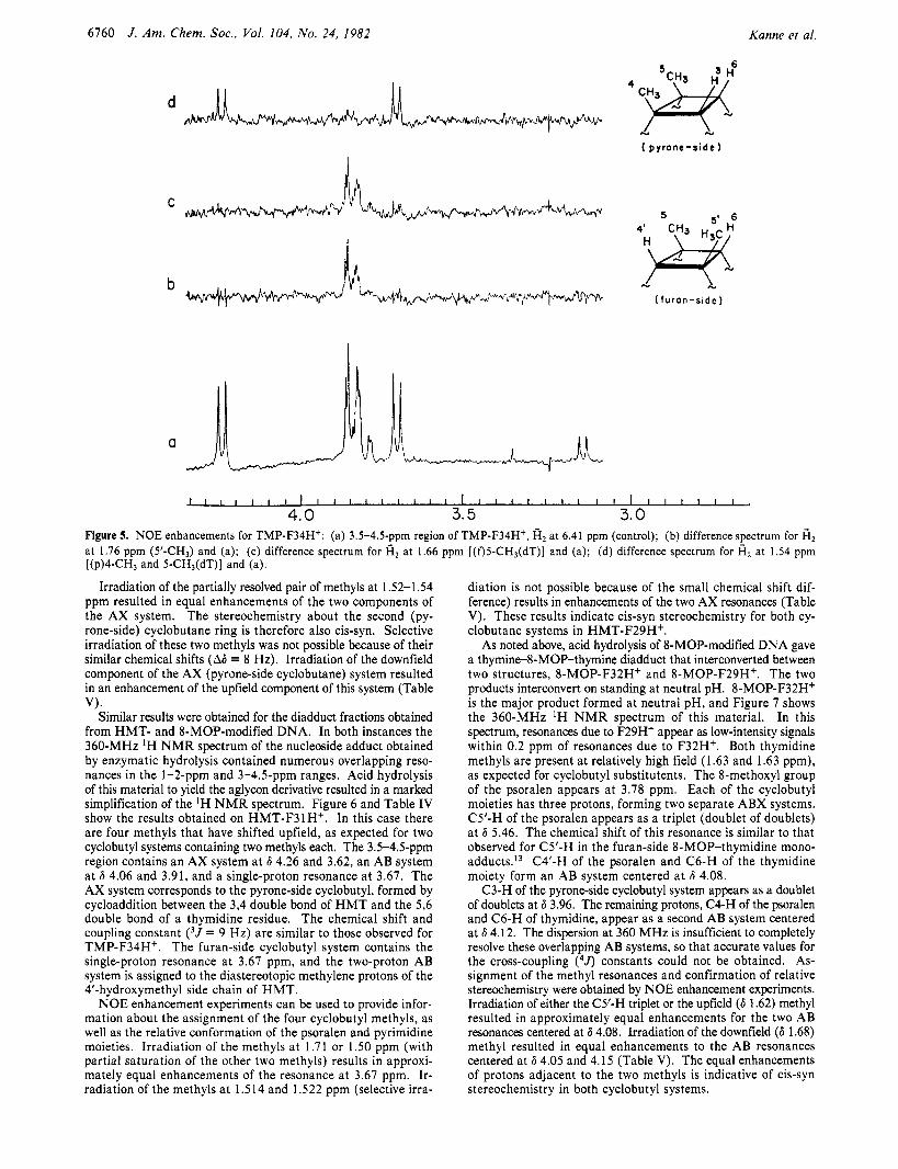
An expanded plot of the 3-4.5-ppm region of this spectrum is shown in Figure 5a. Homonuclear decoupling experiments show that there is an AX system at 4.25 and 3.7 ppm (3JAx = 9 Hz) and a perturbed AB system at 3.87 and 3.83 ppm (4JAB = 2 Hz). The upfield (3.83 ppm) component of the AB system is masked by a small (1 Hz) 4J coupling to C5-H of the psoralen. Decoupling C5-H of the psoralen results in an unperturbed AB pattern. The two protons comprising this AB system, then, are assigned to the diagonally opposite comers of the cyclobutane ring formed between the 4',5' (furan) double bond of the psoralen and the 5,6 double bond of the pyrimidine. The orientation of this cycloaddition is therefore syn.
The value of the coupling constant between the two resonances in the AX system (3J = 9 Hz) is indicative of a vicinal relationship. These two protons (C3-H of T M P and C6-H of thymidine) are therefore on adjacent corners of the cyclobutane ring formed between the 3,4 (pyrone) double bond of the psoralen and the 5,6-double bond of the second thymidine. The stereochemistry about this second cyclobutane ring is thus syn.
Table V. Nuclear Overhauser Effect (NOE) Experiments on Thymine-Pso-Thymine Diadducts
% en- hance-
irradiated observed ment adduct resonance resonance (*2%)
TMP-F34H+ C5'-CH, (1.76) (f)C6-H (3.87) 13 C4'-H (3.83) 14
(f)C5-CH3 (1.66) (f)C6-H (3.87) 20 C4'-H (3.83) 20
(p)CS-CH, and (p)C6-H (4.25) 10 C4-CH3 (1.52) C3-H (3.87) 8
(p)C6-H (4.25) C3-H (3.87) 9 HMT-F31H+ CS'CH, (1.71) (f)C6-H (3.68) 6
(QCS-CH, (1.50) (f)C6-H (3.68) 5
C4CH, (1.52) C3-H (3.63) 10 (p)C5-CH3 and (p)C6-H (4.28) 1 0
8-MOP-F32Ht C5'-H (5.45) (f)C6-H and 6
(f)CS-CH, (1.62) (f)C6-H and 8
C5'-H (5.45) 0
C4'-H (4.08)
C4'-H (4.08)
(p)C5-CH3 (1.68) (pK6-H and 8 C4-H (4.05- 4.15)
C3-H (3.95) 0
The orientation of the pyrimidine and psoralen moieties with respect to the cyclobutane rings (cis or trans) was determined from nuclear Overhauser effect (NOE) experiments, since the overlap of 3Jh and 3Jm values reported in the literature make impractical a definitive assignment of cis or trans stereochemistry solely on the basis of coupling constant evidence.*O NOE enhancement experiments, however, are ideally suited as a probe of relative stereochemistry in a small, rigid system such as a cyclobutane ring. Figure 5 b-d shows the NOE difference spectra obtained by selectively irradiating the four cyclobutyl methyls of TMP-F34H+. Irradiating either of the two methyls at 1.76 and 1.66 ppm results in an equal enhancement of the two AB resonances (Table V). This confirms the assignment of syn stereochemistry for this furan-side cyclobutane ring and in addition fixes the orientation of the T M P and thymidine residues about the cycloubtane ring as cis.
(20) Fleming, I.; Williams, 0. H. Tetrahedron 1967. 23, 2747-2765.
6760 J . Am. Chem. Soc., Vol. 104, No. 24, 1982 Kanne et al.
d
S
u N
( p y r o n e - s i d e )
a
Figure 5. NOE enhancements for TMP-F34H*: (a) 3.5-4.5-ppm region of TMP-F34H+, fi2 at 6.41 ppm (control); (b) difference spectrum for fi2 at 1.76 ppm (5'-CH3) and (a); (c) difference spectrum for fi, at 1.66 ppm [(f)5-CH3(dT)] and (a); (d) difference spectrum for fi2 at 1.54 ppm [(p)4-CH3 and 5-CH3(dT)] and (a).
Irradiation of the partially resolved pair of methyls at 1 S2-1.54 ppm resulted in equal enhancements of the two components of the AX system. The stereochemistry about the second (py- rone-side) cyclobutane ring is therefore also cis-syn. Selective irradiation of these two methyls was not possible because of their similar chemical shifts (A6 = 8 Hz). Irradiation of the downfield component of the AX (pyrone-side cyclobutane) system resulted in an enhancement of the upfield component of this system (Table
Similar results were obtained for the diadduct fractions obtained from HMT- and 8-MOP-modified DNA. In both instances the 360-MHz 'H N M R spectrum of the nucleoside adduct obtained by enzymatic hydrolysis contained numerous overlapping reso- nances in the 1-2-ppm and 3-4.5-ppm ranges. Acid hydrolysis of this material to yield the aglycon derivative resulted in a marked simplification of the 'H N M R spectrum. Figure 6 and Table IV show the results obtained on HMT-F31H+. In this case there are four methyls that have shifted upfield, as expected for two cyclobutyl systems containing two methyls each. The 3.5-4.5-ppm region contains an AX system at 6 4.26 and 3.62, an AB system at 6 4.06 and 3.91, and a single-proton resonance at 3.67. The AX system corresponds to the pyrone-side cyclobutyl, formed by cycloaddition between the 3,4 double bond of H M T and the 5,6 double bond of a thymidine residue. The chemical shift and coupling constant ( 3 J = 9 Hz) are similar to those observed for TMP-F34Hf. The furan-side cyclobutyl system contains the single-proton resonance at 3.67 ppm, and the two-proton AB system is assigned to the diastereotopic methylene protons of the 4'-hydroxymethyl side chain of HMT.
NOE enhancement experiments can be used to provide infor- mation about the assignment of the four cyclobutyl methyls, as well as the relative conformation of the psoralen and pyrimidine moieties. Irradiation of the methyls at 1.7 1 or 1 .SO ppm (with partial saturation of the other two methyls) results in approxi- mately equal enhancements of the resonance at 3.67 ppm. Ir- radiation of the methyls a t 1.514 and 1.522 ppm (selective irra-
VI.
diation is not possible because of the small chemical shift dif- ference) results in enhancements of the two AX resonances (Table V). These results indicate cis-syn stereochemistry for both cy- clobutane systems in HMT-F29H+.
As noted above, acid hydrolysis of 8-MOP-modified DNA gave a thymine-8-MOP-thymine diadduct that interconverted between two structures, 8-MOP-F32H+ and 8-MOP-F29H+. The two products interconvert on standing at neutral pH. 8-MOP-F32H+ is the major product formed at neutral pH, and Figure 7 shows the 360-MHz 'H N M R spectrum of this material. In this spectrum, resonances due to F29H' appear as low-intensity signals within 0.2 ppm of resonances due to F32H+. Both thymidine methyls are present at relatively high field (1.63 and 1.63 ppm), as expected for cyclobutyl substitutents. The 8-methoxyl group of the psoralen appears a t 3.78 ppm. Each of the cyclobutyl moieties has three protons, forming two separate ABX systems. C5'-H of the psoralen appears as a triplet (doublet of doublets) at 6 5.46. The chemical shift of this resonance is similar to that observed for C5'-H in the furan-side 8-MOP-thymidine mono- adducts.I3 C4'-H of the psoralen and C6-H of the thymidine moiety form an AB system centered at 6 4.08.
C3-H of the pyrone-side cyclobutyl system appears as a doublet of doublets at 6 3.96. The remaining protons, C4-H of the psoralen and C6-H of thymidine, appear as a second AB system centered at 6 4.12. The dispersion at 360 MHz is insufficient to completely resolve these overlapping AB systems, so that accurate values for the cross-coupling (4J) constants could not be obtained. As- signment of the methyl resonances and confirmation of relative stereochemistry were obtained by NOE enhancement experiments. Irradiation of either the C5'-H triplet or the upfield (6 1.62) methyl resulted in approximately equal enhancements for the two AB resonances centered at 6 4.08. Irradiation of the downfield (6 1.68) methyl resulted in equal enhancements to the AB resonances centered at 6 4.05 and 4.15 (Table V). The equal enhancements of protons adjacent to the two methyls is indicative of cis-syn stereochemistry in both cyclobutyl systems.
Pyrimidine-Psoralen-Pyrimidine Photodiadducts J. Am. Chem. SOC., Vol. 104, No. 24, 1982 6761
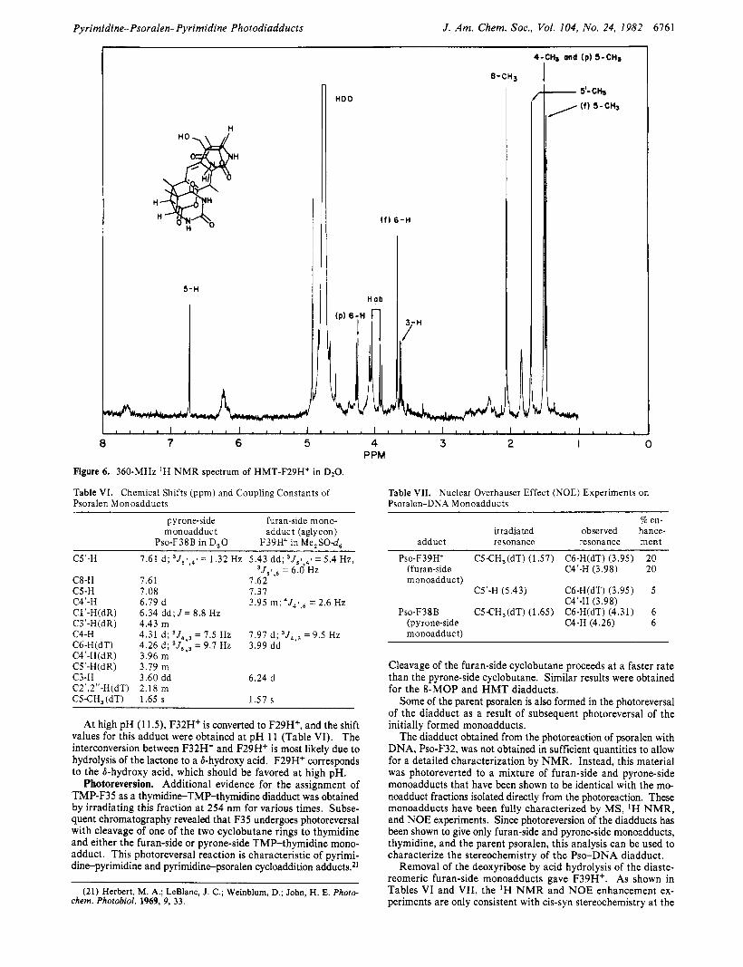
Figure 6. 360-MHz 'H N M R spectrum of HMT-F29Ht in D20.
Table VI. Chemical Shifts (ppm) and Coupling Constants of Psoralen Monoadducts
pyrone-side furan-side mono- monoadduc t adduct (aglycon)
Pso-F38B in D,O F39H+ in Me,SO-d, C5'-H
C8-H 7.61 7.62 C5-H 7.08 7.37 C4'-H 6.79 d 3.95 m ; 4J41,6 = 2.6 Hz C1'-H(dR) 6.34 d d ; J = 8.8 HZ C3'-H(dR) 4.43 m C4-H
C4'-H(dR) 3.96 m C5'-H(dR) 3.79 m C3-H 3.60 dd 6.24 d C2',2"-H(dT) 2.18 m CS€H,(dT) 1.65 s 1.57 s
7.61 d; 3J51,41 = 1.32 Hz 5.43 dd; 3J51,4' = 5.4 Hz, ' J , ! , , = 6.0 HZ
4.31 d ; 3J4,3 = 7.5 Hz 7.91 d ; ,J4,, = 9.5 Hz C6-H(dT) 4.26 d ; 3 J 6 , 3 = 9.7 HZ 3.99 dd
At high pH (1 1 3 , F32H' is converted to F29H+, and the shift values for this adduct were obtained at pH 11 (Table VI). The interconversion between F32H' and F29H+ is most likely due to hydrolysis of the lactone to a &hydroxy acid. F29H' corresponds to the &hydroxy acid, which should be favored at high pH.
Photoreversion. Additional evidence for the assignment of TMP-F35 as a thymidine-TMP-thymidine diadduct was obtained by irradiating this fraction at 254 nm for various times. Subse- quent chromatography revealed that F35 undergoes photoreversal with cleavage of one of the two cyclobutane rings to thymidine and either the furan-side or pyrone-side TMP-thymidine mono- adduct. This photoreversal reaction is characteristic of pyrimi- dine-pyrimidine and pyrimidine-psoralen cycloaddition adducts.*'
(21) Herbert, M. A.; LeBlanc, J. C.; Weinblum, D.; John, H. E. Phoro- chem. Photobiol. 1969, 9, 3 3 .
Table VII. Nuclear Overhauser Effect (NOE) Experiments on Psoralen-DNA Monoadducts
Q en- irradiated observed hance-
adduct resonance resonance ment
Pso-F39H+ CS€H,(dT) (1.57) C6-H(dT) (3.95) 20 (furan-side C4'-H (3.98) 20
C5'-H (5.43) C6-H(dT) (3.95) 5
P s o - F ~ ~ B CS€H,(dT) (1.65) C6-H(dT) (4.31) 6
monoadduct)
C4'-H (3.98)
(pyrone-side C4-H (4.26) 6 monoadduc t)
Cleavage of the furan-side cyclobutane proceeds at a faster rate than the pyrone-side cyclobutane. Similar results were obtained for the 8-MOP and H M T diadducts.
Some of the parent psoralen is also formed in the photoreversal of the diadduct as a result of subsequent photoreversal of the initially formed monoadducts.
The diadduct obtained from the photoreaction of psoralen with DNA, Pso-F32, was not obtained in sufficient quantities to allow for a detailed characterization by NMR. Instead, this material was photoreverted to a mixture of furan-side and pyrone-side monoadducts that have been shown to be identical with the mo- noadduct fractions isolated directly from the photoreaction. These monoadducts have been fully characterized by MS, 'H NMR, and NOE experiments. Since photoreversion of the diadducts has been shown to give only furan-side and pyrone-side monoadducts, thymidine, and the parent psoralen, this analysis can be used to characterize the stereochemistry of the Pso-DNA diadduct.
Removal of the deoxyribose by acid hydrolysis of the diaste- reomeric furan-side monoadducts gave F39H'. As shown in Tables VI and VII, the 'H N M R and NOE enhancement ex- periments are only consistent with cis-syn stereochemistry at the
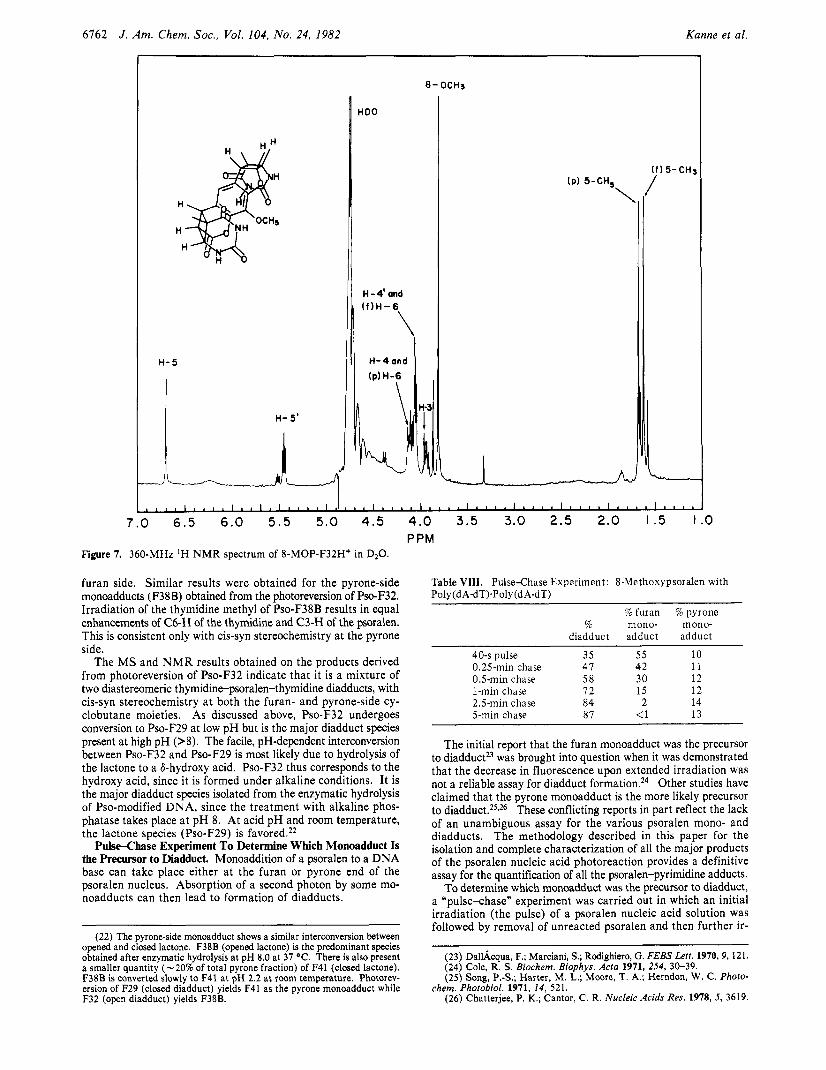
6162 J . Am. Chem. SOC., Vol. 104, No. 24, 1982 Kanne et al.
H- 5
HDO
,, H-4'and
IP) 5-CH, \
Y5-CH3
7.0 6 . 5 6.0 5 . 5 5.0 4.5 4.0 3.5 3.0 2.5 2.0 I .5 I PPM
Figure 7. 360-MHz 'H N M R spectrum of 8-MOP-F32H* in D20.
furan side. Similar results were obtained for the pyrone-side monoadducts (F38B) obtained from the photoreversion of Pso-F32. Irradiation of the thymidine methyl of Pso-F38B results in equal enhancements of C6-H of the thymidine and C3-H of the psoralen. This is consistent only with cis-syn stereochemistry at the pyrone side.
The M S and N M R results obtained on the products derived from photoreversion of Pso-F32 indicate that it is a mixture of two diastereomeric thymidine-psoralen-thymidine diadducts, with cis-syn stereochemistry at both the furan- and pyrone-side cy- clobutane moieties. As discussed above, Pso-F32 undergoes conversion to Pso-F29 at low pH but is the major diadduct species present at high pH (>8). The facile, pH-dependent interconversion between Pso-F32 and Pso-F29 is most likely due to hydrolysis of the lactone to a &hydroxy acid. Pso-F32 thus corresponds to the hydroxy acid, since it is formed under alkaline conditions. It is the major diadduct species isolated from the enzymatic hydrolysis of Pso-modified DNA, since the treatment with alkaline phos- phatase takes place at pH 8. At acid pH and room temperature, the lactone species (Pso-F29) is favored.22
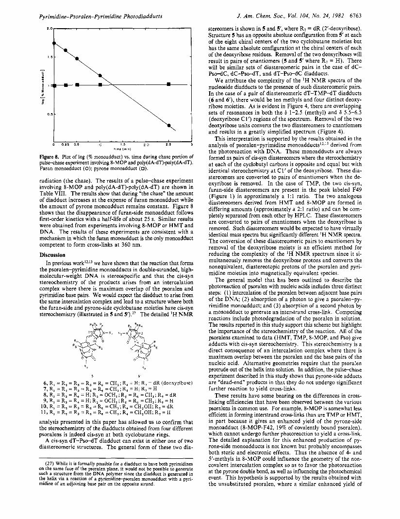
Pulse-Chase Experiment To Determine Which Monoadduct Is the Precursor to Diadduct. Monoaddition of a psoralen to a DNA base can take place either a t the furan or pyrone end of the psoralen nucleus. Absorption of a second photon by some mo- noadducts can then lead to formation of diadducts.
(22) The pyrone-side monoadduct shows a similar interconversion between opened and closed lactone. F38B (opened lactone) is the predominant species obtained after enzymatic hydrolysis at pH 8.0 at 37 "C. There is also present a smaller quantity (-20% of total pyrone fraction) of F41 (closed lactone). F38B is converted slowly to F41 at pH 2.2 at room temperature. Photorev- ersion of F29 (closed diadduct) yields F41 as the pyrone monoadduct while F32 (open diadduct) yields F38B.
Table VIII. PulseChase Experiment: 8-Methoxypsoralen with Poly(dA-dT)'Poly(dA-dT)
% furan % pyrone % mono- mono-
diadduct adduct adduct
40-s pulse 35 55 10 0.25-min chase 41 42 11 0.5-min chase 5 8 30 12 1-min chase 12 15 12 2.5-min chase 84 2 14 5-min chase 87 <1 13
~
The initial report that the furan monoadduct was the precursor to diadduct2) was brought into question when it was demonstrated that the decrease in fluorescence upon extended irradiation was not a reliable assay for diadduct formation.24 Other studies have claimed that the pyrone monoadduct is the more likely precursor to d i a d d u ~ t . ~ ~ , ~ ~ These conflicting reports in part reflect the lack of an unambiguous assay for the various psoralen mono- and diadducts. The methodology described in this paper for the isolation and complete characterization of all the major products of the psoralen nucleic acid photoreaction provides a definitive assay for the quantification of all the psoralen-pyrimidine adducts.
To determine which monoadduct was the precursor to diadduct, a "pulse-chase" experiment was carried out in which an initial irradiation (the pulse) of a psoralen nucleic acid solution was followed by removal of unreacted psoralen and then further ir-
(23) DallAqua, F.; Marciani, S.; Rodighiero, G. FEBS Lett. 1970,9, 121. (24) Cole, R. S . Biochem. Biophys. Acta 1971, 254, 30-39. (25) Song, P.-S.; Harter, M. L.; Moore, T. A,; Herndon, W. C. Photo-
(26) Chatterjee, P. K.; Cantor, C. R. Nucleic Acids Res. 1978, 5, 3619. chem. Photobiol. 1971, 14, 521.
Pyrimidine-Psoralen-Pyrimidine Photodiadducts J. Am. Chem. SOC.. Vol. 104, No. 24, 1982 6163
t 1 rr - D -
0 . 5 -
0 0 .25 0.5 10 I .s 2 0 2.5 1 ” ) (mn)
Figure 8. Plot of log (% monoadduct) vs. time during chase portion of pulsechase experiment involving 8-MOP and poly(dA-dT).poly(dA-dT). Furan monoadduct (0); pyrone monoadduct (0).
radiation (the chase). The results of a pulse-chase experiment involving 8-MOP and poly(dA-dT).poly(dA-dT) are shown in Table VIII. The results show that during “the chase” the amount of diadduct increases at the expense of furan monoadduct while the amount of pyrone monoadduct remains constant. Figure 8 shows that the disappearance of furan-side monoadduct follows first-order kinetics with a half-life of about 25 s. Similar results were obtained from experiments involving 8-MOP or H M T and DNA. The results of these experiments are consistent with a mechanism in which the furan monoadduct is the only monoadduct competent to form cross-links at 360 nm.
Discussion In previous ~ o r k l L ’ ~ we have shown that the reaction that forms
the psoralen-pyrimidine monoadducts in double-stranded, high- molecular-weight DNA is stereospecific and that the cis-syn stereochemistry of the products arises from an intercalation complex where there is maximum overlap of the psoralen and pyrimidine base pairs. We would expect the diadduct to arise from the same intercalation complex and lead to a structure where both the furan-side and pyrone-side cyclobutane moieties have cis-syn stereochemistry (illustrated in 5 and 5’).27 The detailed ‘H NMR
5 5 ’
6 , R, = R, = R, = R, = R, = CH,; R, = H; R, = dR (deoxyribose) 7, R, = R, = R, = R, = R, = CH,; R, = H; R, = H 8, R, = R, = R, = H; R, = OCH, ; R, = R, = CH,; R, = dR 9, R, = R, = R, = H; R, = OCH, ; R, = R, = CH,; R, = H
10, R, = R, = R, = R, = R, = CH,; R, = CH,OH; R, = dR 1 1 , R, = R , = R, = R , = R, =CH,;R, =CH,OH; R, = H
analysis presented in this paper has allowed us to confirm that the stereochemistry of the diadducts obtained from four different psoralens is indeed cis-syn at both cyclobutane rings.
A cis-syn dT-Pso-dT diadduct can exist in either one of two diastereomeric structures. The general form of these two dia-
(27) While it is formally possible for a diadduct to have both pyrimidines on the same face of the psoralen plane, it would not be possible to generate such a structure from the DNA polymer since the diadduct is generated in the helix via a reaction of a pyrimidine-psoralen monoadduct with a pyri- midine of an adjoining base pair on the opposite strand.
stereomers is shown in 5 and 5’, where R7 = dR (2’-deoxyribose). Structure 5 has an opposite absolute configuration from 5’ at each of the eight chiral centers of the two cyclobutane moieties but has the same absolute configuration at the chiral centers of each of the deoxyribose residues. Removal of the two deoxyriboses will result in pairs of enantiomers (5 and 5‘ where R7 = H). There will be similar sets of diastereomeric pairs in the case of dC- Pso-dC, dC-Pso-dT, and dT-Pso-dC diadducts.
We attribute the complexity of the ‘H N M R spectra of the nucleoside diadducts to the presence of such diastereomeric pairs. In the case of a pair of diastereomeric dT-TMP-dT diadducts (6 and 6 9 , there would be ten methyls and four distinct deoxy- ribose moieties. As is evident in Figure 4, there are overlapping sets of resonances in both the 6 1-2.5 (methyl) and 6 5.5-6.5 (deoxyribose Cl’) regions of the spectrum. Removal of the two deoxyribose units converts the two diastereomers to enantiomers and results in a greatly simplified spectrum (Figure 4).
This interpretation is supported by the results obtained in the analysis of psoralen-pyrimidine monoadducts12J3 derived from the photoreaction with DNA. These monoadducts are always formed as pairs of cis-syn diastereomers where the stereochemistry at each of the cyclobutyl carbons is opposite and equal but with identical stereochemistry at C1’ of the deoxyribose. These dia- stereomers are converted to pairs of enantiomers when the de- oxyribose is removed. In the case of TMP, the two cis-syn, furan-side diastereomers are present in the peak labeled F49 (Figure 1) in approximately a 1:l ratio. The two analogous diastereomers derived from H M T and 8-MOP are formed in differing amounts (approximately a 2:l ratio) and can be com- pletely separated from each other by HPLC. These diastereomers are converted to pairs of enantiomers when the deoxyribose is removed. Such diastereomers would be expected to have virtually identical mass spectra but significantly different IH NMR spectra. The conversion of these diastereomeric pairs to enantiomers by removal of the deoxyribose moiety is an efficient method for reducing the complexity of the ‘H N M R spectrum since it si- multaneously removes the deoxyribose protons and converts the nonequivalent, diastereotopic protons of the psoralen and pyri- midine moieties into magnetically equivalent species.
The general model that has been outlined to describe the photoreaction of psoralen with nucleic acids includes three distinct steps: (1) intercalation of the psoralen between adjacent base pairs of the DNA; (2) absorption of a photon to give a psoralen-py- rimidine monoadduct; and ( 3 ) absorption of a second photon by a monoadduct to generate an interstrand cross-link. Competing reactions include photodegradation of the psoralen in solution. The results reported in this study support this scheme but highlight the importance of the stereochemistry of the reaction. All of the psoralens examined to data (HMT, TMP, &MOP, and Pso) give adducts with cis-syn stereochemistry. This stereochemistry is a direct consequence of an intercalation complex where there is maximum overlap between the psoralen and the base pairs of the nucleic acid. Alternative geometries require that the psoralen protrude out of the helix into solution. In addition, the pulse-chase experiment described in this study shows that pyrone-side adducts are “dead-end” products in that they do not undergo significant further reaction to yield cross-links.
These results have some bearing on the differences in cross- linking efficiencies that have been observed between the various psoralens in common use. For example, 8-MOP is somewhat less efficient in forming interstrand cross-links than are TMP or HMT, in part because it gives an enhanced yield of the pyrone-side monoadduct (8-MOP-F42, 19% of covalently bound psoralen), which cannot undergo further photoreaction to yield a cross-link. The detailed explanation for this enhanced production of py- rone-side monoadducts is not known but probably encompasses both steric and electronic effects. Thus the absence of 4- and 5’-methyls in 8-MOP could influence the geometry of the non- covalent intercalation complex so as to favor the photoreaction at the pyrone double bond, as well as influencing the photochemical event. This hypothesis is supported by the results obtained with the unsubstituted psoralen, where a similar enhanced yield of
6164 J . Am. Chem. SOC. 1982, 104, 6764-6769
pyrone-side monoadduct was found. Prior to the present work, it has been reported that irradiation
of a solution of native DNA and psoralen resulted in a higher yield of pyrone adducts than furan adducts.28 These conclusions were based on a nonquantitative, indirect assay in which only some of the adducts were isolated, and those that were isolated did not receive complete structural characterization. This interpretation agreed with theoretical predictions of the relative reactivities of the furan and pyrone double bonds of p s o r a l e n ~ . ~ ~ ~ ~ ~ Our results with four different psoralens (TMP, HMT, 8-MOP, and Pso), however, reveal that the predominant event is photoaddition at the 4‘,5‘ furan double bond. Our conclusion is based on quan- titative assays that involve isolation and complete structural characterization of all mono- and diadducts.
In previous work we have shown that the two diastereomeric furan-side psoralen-thymidine monoadducts are formed in unequal amounts. This is most pronounced in the case of 8-MOP, Pso, and HMT, where approximately a 2:l preference was found. With TMP, the two diasteromers are formed in roughly equal amounts. If one assumes equal conversion rates for the two diastereomeric furan-side precursor monoadducts to the diastereomeric diadducts, then it would follow that a short-range sequence specificity exists for the interaction of the psoralen with double-stranded DNA. This specificity would result from a preference by the psoralen for intercalation on top of or underneath a given base pair (5’-TpX or 5’-XpT sequence).
The overall efficiency of the psoralen-nucleic acid photoreaction is dependent upon a number of interrelated factors such as drug solubility, efficiency of noncovalent binding to the nucleic acid, and quantum yields for the photocycloaddition and photodegra- dation reactions. A fuller understanding of these various pa- rameters is necessary if a program of rational drug design is to be successfully applied to the development of psoralens with en- hanced nucleic acid binding and cross-linking efficiencies. The methodology described in this study allows for the quantification of the mono- and diadducts formed in the psoralen-nucleic acid photoreaction. An analysis of the adducts obtained with a series of psoralens having systematic structural variations could provide
(28) DallAcqua, F.; Magno, S. M.; Zambon, F.; Rodighiero, G. Photo-
(29) Mantulin, M. W.; Song, P. S. J . Am. Chem. SOC. 1973, 95, 5122. chem. Photobiol. 1979, 29, 489.
structure-activity information useful in designing functionally specific psoralens. Such a study is now in progress.
Acknowledgment. This work was supported in part by the Division of Biomedical and Environmental Research (DOE) and the National Institute of General Medical Sciences, DHHW GM 25 15 1. K.S. was supported by the Biomedical Mass Spectrometry Resource, University of California, San Francisco, CA. NMR studies were carried out a t the University of California, Davis, N M R facility, under the auspices of NSF Grant CHE 79-04832 and at Stanford University under the auspices of NIH RR00711. Mass spectrometry studies were carried out a t the University of California, San Francisco, Biomedical Mass Spectrometry Re- source, supported by N I H Grant R R 00719, Dr. A. L. Burlin- game, Director.
Registry No. 1, 66-97-7; 2, 3902-71-4; 3, 298-81-7; 4, 62442-59-5; 6, 83380-30-7; 6’, 83435-1 1-4; 7,83380-31-8; 8,83380-32-9; 8’, 83435-12-5; 9, 83380-33-0; 10, 83380-34-1; 1W, 83435-13-6; 11, 83380-35-2; Pso-F32 (isomer l), 83435-14-7; Pso-F32 (isomer 2), 83380-36-3; Pso-F38A (isomer l), 83380-37-4; Pso-F38A (isomer 2), 83435-15-8; Pso-F38A di(Me,Si) (isomer l), 83380-38-5; Pso-F38A di(Me,Si) (isomer 2), 83435-16-9; Pso-F38B (isomer l), 83380-39-6; Pso-F38B (isomer 2), 83435-17-0; Pso-F38B tetra(Me,Si) (isomer l), 83398-66-7; Pso-F38B tetra(Me,Si) (isomer 2), 83461-44-3; Pso-F41 (isomer l ) , 83380-40-9; Pso-F41 (isomer 2), 83435-18-1; 8-MOP-F33 hexa(Me2Si) (isomer I ) , 83380-41-0; 8-MOP-F33 hexa(Me3Si) (isomer 2), 83435-1 9-2; 8-MOP- F36 (isomer l), 83380-42-1; 8-MOP-F36 (isomer 2), 83435-20-5; 8- MOP-F37 (isomer l), 80603-65-2; 8-MOP-F37 (isomer 2), 80656-97-9; 8-MOP-F42 (isomer l), 83435-21-6; 8-MOP-F42 (isomer 2), 80603- 69-6; TMP-F35 hexa(Me3Si) (isomer l), 83380-43-2; TMP-F35 hexa- (Me&) (isomer 2), 83461-45-4; TMP-F47 (isomer l ) , 83380-44-3; TMP-F47 (isomer 2), 83435-22-7; TMP-F49 (isomer l), 80603-66-3; TMP-F49 (isomer 2), 80657-61-0; TMP-F52 (isomer l), 83435-23-8; TMP-F52 (isomer 2), 80659-95-6; HMT-F31 hepta(MelSi) (isomer l) , 83380-45-4; HMT-F3 1 hepta(Me3Si) (isomer 2), 83435-24-9; HMT-F43 (isomer l), 77340-93-3; HMT-F43 (isomer 2), 77268-07-6; HMT-F40A (isomer l), 77268-05-4; HMT-F40A (isomer 2), 77340-92-2; HMT-F48 (isomer l), 83380-46-5; HMT-F48 (isomer 2), 83435-25-0; poly(dA-
Supplementary Material Available: A listing of high-resolution mass spectral data for aglycon derivatives of psoralen diadduct (1 page). Ordering information is given on any current masthead page.
dT)*poly(dA-dT), 26966-61-0.
Epimerization of Aldoses by Molybdate Involving a Novel Rearrangement of the Carbon Skeleton
Michael L. Hayes, Nicholas J. Pennings, Anthony S. Serianni, and Robert Barker*
Contribution from the Section of Biochemistry, Molecular and Cell Biology, Division of Biological Sciences, Cornell University, Ithaca, New York 14853. Received March 19, 1982
Abstract The molybdate-catalyzed C-2 epimerization of aldoses has been investigated by using 13C- and 2H-enriched compounds and 75-MHz ”C NMR spectroscopy. The epimerization product of D-[ l-”C]mannose was exclusively ~-[2-~~C]glucose, demonstrating that the reaction involves a 1,2 shift of the carbon skeleton resulting in inversion of configuration at C-2. All of the aldotetroses, aldopentoses, and aldohexoses examined reacted similarly, producing equilibrium mixtures of the starting [ l-13C]aldose and the 2-epimeric 2-13C product. Reaction of D-[ I-13C,2H]mannose and D-[ 1,3-”C,3-2H]mannose in H 2 0 and D-mannose in ’H20 demonstrated that the epimerization occurs without C-3 transposition or C-H bond breaking. Studies with aldose analogues including mannitol, 3deoxy-~-urubirt~~hexose, 4-deoxy-~-lyxehexose, lactose, and 4,6-Gethylidene~glucose suggest that the reactive molybdate complex involves the carbonyl oxygen and three hydroxylic oxygens of the aldehyde form of aldoses. The rates of reaction are influenced by the ability of the ring forms of the starting aldose to form stable unreactive molybdate complexes. Slower secondary reactions involving the simultaneous inversion of configuration of C-2 and C-3 occur without carbon skeletal rearrangement.
In an extensive series of reports, Bilik and his co-workers have shown that in mildly acidic solutions of molybdate, aldotetroses,’
a l d o p e n t ~ s e s , ~ ~ ~ and a l d o h e x o ~ e s ~ ~ ~ epimerize at C-2 with the formation of a thermodynamic equilibrium mixture of the two
0002-7863/82/1504-6764$01.25/0 0 1982 American Chemical Society