Embed Size (px)
Citation preview
Isolation and Characterization of Anti-malarial Compounds
From a Natural Product Library
By
Holly Marie Carrell
Thesis
Submitted to the faculty of the
Graduate school of Vanderbilt University
In partial fulfillment of the requirements
For the degree of
MASTER OF SCIENCE
In
Chemistry
August, 2013
Nashville, Tennessee
Approved:
Professor David W. Wright
Professor Brian O. Bachmann
ii
TABLE OF CONTENTS
Page
List of Tables ....................................................................................................................... iv
List of Figures ………………………………………………..…………………………………………………………….. . v
Chapter
I. Hemozoin: Crystal Engineering Survivability …………………………………………………….. 1
Introduction………………………………………………………………………………………………………..1
Homeostasis of Heme ..……………………………………………………………….. ...................... 2
Detoxification of Heme in Humans…………………………………………………………………… . 3
Heme Detoxification in the Malaria Parasite……………………………………………………... 4
The Structure of Hemozoin …………………………………………………….. ........................... 9
Hemozoin and B-hematin are equivalent ………………………………………………………….11
Mechanism of Hemozoin Formation ................................................................... 12
Biological Formation of Hemozoin........................................................................ 13
Implication of Neutral Lipids in Hemozoin Formation .......................................... 17
Localization of Free Heme in the Neutral Lipid Body ........................................... 19
Kinetically competent Site for BH Formation ....................................................... 21
Significance of the Blend of Neutral lipids ............................................................ 22
Molecular Dynamics Simulations .......................................................................... 24
Antimalarials ......................................................................................................... 26
Evaluating Small Molecule Interactions with Heme ............................................. 27
Vacuolar Accumulation ......................................................................................... 28
Antimalarials and Small Molecule Inhibitors of BH Formation ............................ 30
Quinoline Derivatives ............................................................................................ 32
Arylmethanol Derivatives ..................................................................................... 33
Xanthones ............................................................................................................. 34
Artemisinin ............................................................................................................ 35
High-Throughput Screening and Drug Discovery ................................................. 36
Conclusion ............................................................................................................. 41
II. Isolation of Anti-malarial Compounds from a Natural Product Library……………….42
Introduction……………………………………………………………………………………………………….42

iii
Page
Actinomycetes ...................................................................................................... 43
Experimental ......................................................................................................... 44
Results ................................................................................................................... 46
Conclusion ............................................................................................................. 50
III. Anti-malarial effects of Genistein ......................................................................... 51
Introduction .......................................................................................................... 51
Experimental ......................................................................................................... 51
Results ................................................................................................................... 53
Conclusion ............................................................................................................. 55
Appendix
A. B-HEMATIN ASSAYS OF ACTINOMYCETE STRAINS................................................ 60
B. CHROMATOGRAPHY DATA OF PURE ACTIVE COMPOUNDS ................................ 69
C. MASS ANALYSIS DATA OF BBHARD25 AND BBHARD23 ........................................ 70
D. NMR DATA OF BBHARD25 .................................................................................... 72
E. CURRICULUM VITAE .............................................................................................. 74
References ........................................................................................................................ 76
iv
LIST OF TABLES
Table ............................................................................................................................... Page
1. 13C, 1H, and HMBC data for Compound A (600 MHz in CD3O3) ........................................ 49

v
LIST OF FIGURES
Figure ............................................................................................................................. Page Page
1. Pathway of free heme detoxification by Heme Oxygenase ................................... 4
2. Life cycle of the malaria parasite ............................................................................ 6
3. Structure of hemozoin .......................................................................................... 11
4. Crystal morphology of hemozoin .......................................................................... 11
5. Electron micrographs of B-hematin and hemozoin crystals ................................. 12
6. Hemozoin formation within lipid droplets ........................................................... 19
7. Confocal micrographs of heme partitioning into SNLDs ...................................... 21
8. Nile Red quenching as a function of time ............................................................. 22
9. Activation energy of neutral lipid droplets ........................................................... 24
10. Molecular Dynamics of hemozoin formation ....................................................... 26
11. Speciation of heme ............................................................................................... 29
12. Structures quinoline and arylmethanol antimalarials .......................................... 32
13. Structures of small molecule antimalarials .......................................................... 32
14. Structures of xanthone antimlarials ..................................................................... 36
15. Structures of antimalarial compounds active in the parasite .............................. 41
16. Structures of Streptomycin and Erythromycin ..................................................... 44
17. Overview of hit discovery and identification ........................................................ 46
18. NELI data of BBHARD25 ........................................................................................ 50

vi
Figure ............................................................................................................................. Page
19. Structure of Genistin with NMR assignments ...................................................... 52
20. NELI data of BBHARD23 ........................................................................................ 53
21. IC50 curves of active compounds from BBHARD23 and BBHARD25…………………..58
22. IC50 curve of synthetic genistein…………………………………………………………………….. 58
23. NELI data of BBHARD25 showing presence of second compound ....................... 59
1
Chapter I
Hemozoin: Crystal Engineering Survivability
(Chapter I taken from Hemozoin: Crystal Engineering Survivability, published in the
Encyclopedia of inorganic and Bioinorganic Chemistry, 2012)
Introduction
In nature, biomineralization is observed across the entire biosphere. These biologically
synthesized materials can consist of carbonate, phosphate, oxalate, silica, iron, go2ld
and many other diverse compositions of organic or inorganic materials. The most well-
known examples of biomineralization are those that lend structural support to a species,
including the formation of bones in vertebrates and the shells of eggs. Magnetotactic
bacteria also rely on the formation of magnetic nanometer-size Fe3O2 crystallites in
order to locate oxygen-rich environments by sensing the Earth’s geomagnetic field. The
formation of these biominerals generally occurs under physiological conditions in a
highly controlled and organized sequence of events. Another biomineral, hemozoin, is a
heme crystal synthesized by the malaria parasite as a survival mechanism to escape
heme related toxicity. The discovery of this detoxification biomineral actually precedes
the discovery of the malaria parasite itself. Although hemozoin has been studied since
the 18th century, scientists have only begun to obtain a detailed understanding of the
structure and formation of this biomineral in the last twenty five years. Hemozoin

2
formation is an important drug target in antimalarial drug discovery and development.
In fact, the most successful antimalarial developed to date, chloroquine, acts by
disrupting formation of hemozoin. Unfortunately, chloroquine is now ineffective as an
antimalarial due to the emergence 2of multidrug-resistant strains of the parasite,
though hemozoin remains a viable drug target in antimalarial drug discovery. In order
to develop new antimalarial compounds that target the formation of hemozoin, it is
important to have a sound understanding of the formation of this biocrystal.
Homeostasis of Heme
The internal environment of all biological systems must be carefully regulated.
The heme molecule is a perfect example of the importance of this homeostasis. Heme is
essential to aerobic organisms, playing important roles in many biological reactions such
as oxygen transport, respiration, drug detoxification and signal transduction (1-4).
However, when heme is released in an uncontrolled fashion into a living organism it can
become extremely toxic. One example demonstrating the toxic effects of heme is seen
in rhabdomylosis. Here, trauma or injury results in the rapid breakdown of muscle
tissue, releasing myoglobin into the bloodstream. This harmful heme-containing protein
is not normally found in blood. In the kidney, heme exposure promotes oxidative stress
in renal cells by increasing lipid peroxidation, and can cause local inflammatory
reactions ultimately leading to renal failure (5, 6). The ability of heme to promote
oxidative stress can be attributed to the presence of the redox-active iron center that
can generate reactive oxygen species (ROS), leading to damage to lipids, proteins, and

3
DNA. Free heme has also been shown to interact with proteins, catalyzing the
degradation of proteins into small peptide fragments and causing covalent cross-linking
of Apolipoprotein B, triggering atherosclerosis (7). Studies have also shown that
exposure to heme results in nicking and degradation of DNA. The mtDNA of liver cells
shows that the large region that codes for cytochrome c oxidase and NADH
dehydrogenase is deleted in cells exposed to hemin. This mtDNA damage leads to the
altered expression of mitochondrial cell death proteins, suggesting a role of hemin in
influencing apoptosis (8).
Detoxification of Heme in Humans
As protection against the drastic toxic effects of free heme, humans are
equipped with several detoxification mechanisms. Primarily, heme detoxification is
carried out by the heme oxygenase (HO) systems (HO-1, HO-2 and HO-3), and by extra-
HO systems less frequently, including hemopexin and albumin (9). These HO enzymes
play a large role in protecting cells from the oxidative stress caused by free heme by
working with NADPH–cytochrome P450 to break down the porphyrin ring into
equimolar amounts of free iron, biliverdin, and carbon monoxide (10). In this process,
HO transfers reducing equivalents to the α-methene bridge of heme from NADPH-
cytochrome P-450 reductase to open the tetra-pyrrolic ring, freeing CO and biliverdin.
Biliverdin is then converted to bilirubin by biliverdin reductase, conjugated to glucuronic
acid, and excreted from the body (Figure 1) (11, 12). The HO systems are very efficient
at detoxifying free heme and restoring homeostasis in the organisms that have them,

4
but organisms that lack the HO systems have developed alternative mechanisms to
protect themselves from free heme toxicity.
Figure 1. Pathway of free heme detoxification by Heme
Oxygenase.
5
Heme Detoxification in the Malaria Parasite
One organism lacking a HO system is the parasite responsible for malaria.
Malaria is caused by several species of intracellular parasites of the Plasmodium genus.
Of this genus, there have been at least 200 species identified, with P. falciparum being
the primary causative agent of human malaria. The lifecycle of the malaria parasite is
quite complex (Figure 2). P. falciparum sporozoites are transmitted to humans through
the saliva of a female Anopheles mosquito during a blood meal. Once in the host’s
bloodstream, P. falciparum sporozites invade the hepatocytes and undergo a phase of
growth and differentiation followed by the release of merozoites into the bloodstream.
The merozoites then enter host red blood cells, referred to as the the intraerythrocytic
stage of infection. This stage is characterized by the onset of the symptoms of a malaria
infection. Inside the erythrocyte, the parasite goes through three distinct growth phases
that can be distinguished under a light microscope. The ring stage is first, lasting about
24 hours. The second stage is the very active trophozoite stage. It is during this stage
that most of the erythrocyte cytoplasm is consumed. Third , the parasite undergoes 4-5
cycles of binary divisions, producing merozoites that eventually rupture the red blood
cell membrane, and enter the bloodstream to infect new red blood cells (13). During
this intraerythrocytic cycle, the host cytoplasm is consumed, and an estimated 60-80%
of available hemoglobin is degraded for use as a nutrient source (14). During this
degradation process, the amino acids obtained from hemoglobin catabolism are
incorporated into P. falciparum proteins and used for energy metabolism (15) (16-18).

6
The amino acids P. falciparum cannot obtain from hemoglobin are readily scavenged
from the environment (19, 20).
The malaria parasite obtains hemoglobin from the cytosol through invagination
of the parasitophorous vacuolar and plasma membrane, creating a double membrane
vesicle. These vesicles fuse together, creating a single membrane digestive food vacuole.
Figure 2. Life-cycle of Plasmodium falciparum
in the human host.
7
Once formed, the digestive food vacuole becomes the primary site of hemoglobin
digestion (21, 22). This specialized organelle maintains an acidic environment, estimated
pH of 4.8 (19, 23). Inside this organelle, aspartic and cysteine protease activities have
been detected that are the primary enzymes responsible for globin proteolysis. Aspartic
proteases make up about 60-80% of enzyme activity, while cysteine proteases make up
20-40% (24, 25). The process of hemoglobin degradation has been found to occur in a
specific order, requiring an initial aspartic protease cleavage, followed by secondary
aspartic protease and cysteine protease cleavages (26). Vacuolar degradation produces
small polypeptides, but no free amino acids. This suggests that cleavage of the small
peptide fragments occurs in the cytoplasm, outside of the digestive vacuole (26-28).
Sequencing of the P. falciparum genome has found at least ten genes encoding
aspartic proteases, plasmepsins I, II, IV-X, and histo-aspartic protease (HAP) (26, 28).
HAP is 60% homologous to plasmepsins I and II, but has several substitutions, one of
which is the replacement of the catalytic aspartate with a histidine (29). Of the ten
genes that encode plasmepsins in P. falciparum, three are not expressed in
intraerythrocytic growth stages (plasmepsins VI, VII, and VIII), and three are expressed,
but do not seem to be active in the digestive food vacuole (plasmepsins V, IX, and X).
Plasmepsins I, II, IV, and HAP are the only aspartic proteases that are found in the
digestive food vacuole, and participate in hemoglobin degradation. Plasmepsin I has
been shown to cleave hemoglobin between α33Phe-34Leu with exceptional specificity
(30). This is significant because the α33-34 bond is in the hinge region of the hemoglobin
molecule. This region is responsible for holding the protein together when oxygen is
8
bound. Cleaving this domain unravels the heme molecule, exposing other amino acid
residues for cleavage by the secondary proteases (31). Plasmepsins II and IV have been
shown to cleave native hemoglobin, but prefer to cleave already denatured hemoglobin
indicating that they function primarily as secondary proteases (28, 32, 33). HAP is a
secondary protease, showing less efficiency in cleaving the α33-34 bond of native
hemoglobin, although it is a full order of magnitude faster than the other plasmepsins at
cleaving denatured globin at other sites (28, 32, 34).
The P. falciparum genome encodes four falcipains; falcipain I, II, II’ and falcipain
III (28, 33, 35). Falcipain I has not been shown to be involved in hemoglobin
degradation, but falcipains II and III have acidic optimal pH, and are localized in the
digestive food vacuole, indicating their involvement in hemoglobin digestion (33). Both
falcipain II and III are capable of hydrolyzing native and denatured hemoglobin, but
falcipain II has been shown to be more active against peptidyl substrates compared to
falcipain III (36-38).
Finally, a metalloprotease, falcilysin, also participates in the hemoglobin
degradation pathway. Falcilysin is a member of the M16 family of the clan ME zinc
metalloproteases. This family of metalloproteases is characterized by an inverted active
site motif of HXXEH. The two histidine residues and glutamate residues C-terminal to
this motif are involved in coordinating a catalytic zinc ion (37, 38). Falcilysin is unable to
cleave native hemoglobin or denatured globin, instead preferring to cleave small globin
peptides up to 20 amino acids in length (39). It appears that falcilysin acts as a
complement to the plasmepsins and falcipains, acting downstream, and cleaving

9
preferentially at polar residues, while plasmepsins and falcipains cleave at hydrophobic
residues (40). The cleavage of globin peptides by facilysin is essential for production of
peptides small enough to be transported to the cytosol of the parasite for further
cleavage to amino acids, signifying the end of hemoglobin degradation in the digestive
food vacuole.
During this process of hemoglobin degradation, large quantities of free heme are
released and can reach concentrations of 400 mM if heme detoxification is prevented
(40). The presence of free heme within the digestive food vacuole has several
undesirable consequences. Heme concentrations as low as 10-20 µM have been shown
to inhibit several proteases present within the digestive food vacuole including the
plasmepsins and falcipains (40). In addition to inhibiting enzymatic activity, the
presence of free heme is detrimental to the stability and deformability of the parasite
vacuolar membrane (15). The resulting hemolysis is likely caused by perturbations to
the membrane structure from the insertion of lipophilic heme into the phospholipid
bilayer. A further consequence of free heme accumulation is the increase in oxidative
stress on the parasite (41). Since the malaria parasite does not have HO like vertebrates,
the parasite detoxifies free heme by converting soluble, toxic free heme into an
insoluble, nontoxic crystal called hemozoin. This process is essential to parasite survival,
as inhibition of hemozoin formation results in parasite death (42). Indeed, the most
successful antimalarial ever developed, chloroquine, has been shown to inhibit this
detoxification pathway supposedly by binding free heme and preventing its insertion
into the growing hemozoin crystal. Unfortunately, due to resistance mechanisms

10
developed by the parasite, chloroquine no longer has clinical efficacy. However,
hemozoin remains a valid drug target as resistance is a result of efflux, and resistant
strains of the parasite still produce hemozoin normally. Therefore, it is essential that
the formation of this biocrystal is better understood in order to promote the discovery
of new antimalarials that target hemozoin formation.
The Structure of Hemozoin
Lancisi reported the discovery of hemozoin, the dark brown-black malarial
pigment, in 1717 (43). As these deposits are quite pronounced in the brains, spleens and
livers of malaria victims, the discovery of hemozoin actually precedes the discovery of
the malaria parasite itself by over 150 years. For many years, it was hotly contested as
to whether or not pigment was the actual cause of malaria, but in 1890, Golgi presented
a photograph of a pigmented parasite in the blood of a malaria patient, forging the
connection between hemozoin and malaria (15, 44). The composition and structure of
hemozoin remained a subject of intense debate until 2000 when X-ray powder
diffraction methods were used to identify the structure of β-hematin, the synthetic
equivalent of hemozoin (45). Once thought to consist of polymeric strands of O-Fe(III)
linked heme units, XRD revealed a centrosymmetric triclinic unit cell comprised of
reciprocal head-to-tail dimeric units of heme bound through propionate O-Fe(III) with a
bond distance of 1.866(2) Å (Figure 3). The propionic acid groups of the heme dimer
then hydrogen bond with other dimers to form the extended crystal. Morphologically,
β-hematin and hemozoin crystals are needle-like and exhibit dominant, slow growing

11
{100} and {010} side faces with minor, less pronounced fast growing {011} and (46) end
faces (Figure 4). This morphology is conserved among various species of Plasmodium
(Figure 5).
Figure 3. Structure of Hemozoin
Figure 4. Crystal morphology of hemozoin showing
slow-growing {100} and {010} side faces, and fast-
growing {011} and {46} faces.
12
Hemozoin and β-hematin are equivalent
Understanding the mechanism by which the parasite converts soluble, toxic free
heme into an insoluble and non-toxic crystal is valuable for antimalarial drug-discovery,
as perturbations to this crystallization process can result in parasite death. However,
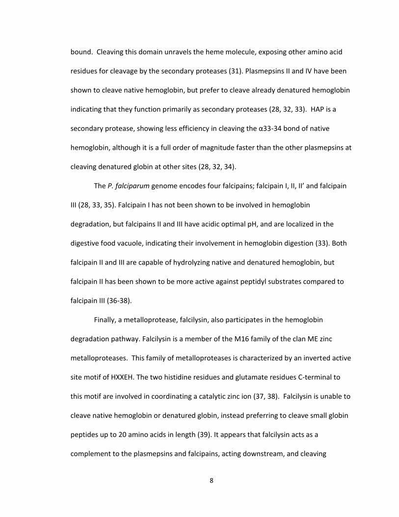
Figure 5. Electron micrographs show the heterogeneity of shape and size
for B-hematin and hemozoin. Images of B-hematin (a) compared to that
of hemozoin from P. falciparum clones Dd2 (b1) and 3B-D5 (b2), and
hemozoin from P. Vivax (c), P. ovale (d), and P. malariae (e). The size bar
is 200 nm (50).

13
studying the formation of hemozoin in vivo has proven challenging to researchers. β-
hematin (BH), the abiological version, has been utilized as a tool to study hemozoin
formation in vitro. For such comparisons to be meaningful, it had to be unequivocally
established that these entities are isostructural. Chemical, spectroscopic, and
crystallographic evidence have been used to conclude the similarity of these two
materials.
Chemically, both hemozoin and BH are composed of insoluble, head-to-tail
dimers of Fe(III)PPIX (47). Spectroscopically, the similarity of hemozoin and BH dimeric
units can be confirmed from the infrared spectrum, that exhibits fingerprint vibrations
around 1664 and 1211 cm-1 corresponding to the C=O and C-O stretching from
coordination of the propionate O atom to the Fe(III) metal center of the neighboring
heme molecule (48). Furthermore, the Fe(III) spin state of each species is identical,
confirmed by variable temperature EPR that has shown that both BH and hemozoin
exist in the high spin S=5/2 state, a paramagnetic complex. This observation is in
agreement with Mӧssbauer data where the isomer shift and quadrupolar splitting
values suggest that the Fe(III) exists in a high-spin state (47, 49). Morphologically, SEM
images reveal that both BH and hemozoin are composed of needle-like crystals with
tapered habits (Figure 5A and 5B). The crystal structure of each species is nearly
identical. Synchrotron x-ray powder diffraction patterns have demonstrated that the
crystal structure of hemozoin is identical to its synthetic counterpart (50). However, a
more recent study suggests hemozoin may contain slightly more disorder in the Fe-O
bonds than BH formed under nonaqueous equilibrium conditions (51).
14
Mechanism of Hemozoin Formation
The conversion of free heme to BH does not occur at a physiologically relevant
rate in aqueous solution, suggesting that the in vivo formation of hemozoin within the
digestive food vacuole of the parasite requires the presence of a biological mediator.
While the precise mechanism by which hemozoin formation occurs in vivo is not fully
elucidated, recent studies strongly support the hypothesis that neutral lipid bodies are
the biological template for hemozoin crystallization. In support of this hypothesis,
several synthetic routes for BH formation have been described demonstrating that
neutral lipid bodies serve as both a protective reservoir for free heme and a kinetically
competent site for crystallization.
Biological formation of Hemozoin
Until recently, identification of the biological mediator responsible for hemozoin
formation was a controversial topic, as proteins, membrane lipids and neutral lipids
extracted from the Plasmodium parasite were each shown to mediate BH formation in
vitro. Indeed, many biological components within parasite lysates are quite capable of
promoting the formation of BH in vitro. One of the earliest studies of these lysates was
conducted by Slater and Cerami who examined trophozoite extracts (52). The formation
of BH in the presence of these extracts was dependent on pH, time and concentration.
Furthermore, several quinoline-antimalarials (known to inhibit the formation of
hemozoin in vivo) were found to inhibit the process of BH formation in the presence of
these lysates. The authors concluded that the presence of an enzyme, a heme
15
polymerase, was responsible for mediating the formation of BH, as activity of these
lysates was sensitive to both heat and treatment with 1% SDS. However, subsequent
work contradicted this hypothesis, demonstrating that heat treated parasite lysates
retained activity and therefore are not dependent on the presence of an enzyme (49). It
was suggested that BH formation in these experiments was actually due to the presence
of hemozoin crystals within the lysates. In fact, the authors showed that preformed BH
crystals were sufficient to seed the process of crystallization in vitro. While these
studies demonstrated that an enzyme is not necessary for hemozoin formation, the
mechanism of hemozoin crystallization remained unanswered.
Polar membrane lipids were also implicated as a possible mediator of hemozoin
formation. Bendrat et al. first implicated these polar lipids when acetonitrile extracts of
authentic hemozoin were found to promote the formation of BH (53). MS analysis of
these extracts identified the presence of the methyl esters of oleic, palmitic and stearic
acids and low yields of phospholipids within the active fractions. Further, it seemed that
these studies were in agreement with a previously published TEM image of an intact
parasite showing aligned parallelepiped crystals of hemozoin present within the DV of
the parasite (54). These observations supported the hypothesis that crystallization
occurs via epitaxial nucleation of hemozoin at the lipid layer of the vacuolar membrane
(55). Despite the strength of this argument, it was later revealed that axenic parasite
cultures lacking a parasitophorus vacuolar membrane still produce hemozoin, ruling out
any role for these membrane lipids in the parasite’s mechanism of heme detoxification
16
(56). While these polar membrane lipids are capable of promoting BH formation in vitro,
they are not responsible for the in vivo formation of hemozoin.
Though polar membrane lipids were ruled out as the mediator of hemozoin
formation, additional studies by Dorn et al. showed that extracts from uninfected
erythrocytes as well as several types of phospholipids are also capable of promoting the
formation of BH (56, 57). These observations suggest that a hydrophobic environment is
ideal for promoting formation of BH. Several in vitro studies substantiate this
hypothesis by probing both the rate and activation barrier of BH formation in the
presence of several types of solvents, alcohols and acids with varying degrees of
hydrophobicity (58). Several acids, including benzoic and acetic acid have been shown to
mediate BH formation (59). The ability of the acid to promote BH formation increases
as a function of concentration. It has been suggested that this concentration
dependence is due to an increase in heme solubility, the limiting step in BH formation.
This observation would explain the abundance of active BH mediators obtained from
lipophilic biological extracts. As heme itself is quite hydrophobic and insoluble in
aqueous solution, it would be expected that heme solubility would increase of a
function of increasing hydrophobicity of the surrounding environment.
Following this line of thinking, aromatic and aliphatic alcohol-water mixtures of
varying hydrophobicity have been studied for their ability to promote BH crystallization
(60-62). In one such study, the ability of several small normal and structurally similar
alcohols with varying degrees of hydrophobicity were investigated (61). The results
indicate that activity follows the trend of butanol > propanol > ethanol > methanol.
17
These data suggest that the hydrophobicity of the alcohol is directly related to its
potency as a mediator of BH formation. The authors further investigated the
solubilization of heme in the presence of the alcohols and found an exponential increase
of heme solubility is observed as a function of increasing alcohol concentration.
Further, the ability of an alcohol to solubilize free heme followed the trend of proponal
> ethanol > methanol indicating that the hydrophobicity of the alcohol increases the
solubility of heme. These data would support the concept of an in vivo mediator of
hemozoin formation that is hydrophobic in nature and acts to solubilize free heme.
In vitro experiments have shown that BH formation occurs efficiently at the
surface of a hydrophilic/hydrophobic interfacial region. These observations were made
using aqueous/alcohol mixtures in the presence or absence of an interfacial region (63,
64). To study the effects of this interfacial region, a cosolvent system was implemented
where a 9:1 solution of methanol:alcohol was used. In the case of 1-octanol, 1-hexanol
and 1-pentanol, the first two solvents are biphasic in aqueous medium, while the latter
exhibits a single phase in aqueous solution. The rates of formation of BH at
physiological temperature and digestive food vacuole pH for the solvent systems are 7-9
minutes for the biphasic solutions, but 110 minutes for the 1-pentanol solution, which
could suggest that the presence of an interfacial region is important. To confirm the
importance of this lipophilic interface, a solution of 6:4 methanol:pentanol or octanol
was used, so as to induce an interfacial region in the pentanol solution. The results
show that the 1-octanol solution has a similar half-life (12 min) as observed in the 9:1
solution (7.1 min). However, the 1-pentanol solution with an interfacial region shows a
18
half-life of 10 min, as compared to the kinetics in the absence of an interface where the
half-life is 110 min. Another study with resonance raman spectroscopy using an
immersion probe confirmed that the formation of BH occurs within 1 µm of a
pentanol/water interface. Due to the dependence of BH formation on the presence of
an interfacial region, perhaps formation of hemozoin at a lipid/aqueous interface
explains the alignment of crystals on neutral lipid bodies observed in TEM images.
These in vitro experiments demonstrate that hydrophobic interactions are
important in free heme detoxification, suggesting that a lipophilic source present within
the digestive food vacuole is responsible for mediating hemozoin formation. Since polar
membrane lipids have been previously ruled out, is there another possible lipophilic
source present at the site of hemozoin formation? With the identification of neutral
lipids bodies present within the digestive food vacuole, the focus shifted to these
neutral lipids as the possible site of hemozoin formation.
Implication of Neutral Lipids in Hemozoin Formation
During infection with P. falciparum, there is a six-fold increase of lipid content
within the erythrocyte for the support of growth and differentiation of the parasite.
While the parasite does support limited synthesis of short chain fatty acids, the lipid
content of the growing parasite is mostly scavenged from the host. During this process,
there is a significant increase in triacylglycerol content, a neutral lipid that is only
detected in small amounts within an uninfected erythrocyte. Within the digestive food
vacuole of the parasite, aggregates of these neutral lipids are observed. Confocal
19
images reveal these lipid bodies are a few hundred nm in diameter and determined to
consist of a mixture of di- and triacylglycerols (neutral lipids) (64). Based on previous
knowledge that mono- di- and triacylglycerols are capable of promoting BH formation, it
was proposed that the neutral lipid bodies serve as a mediating template for the
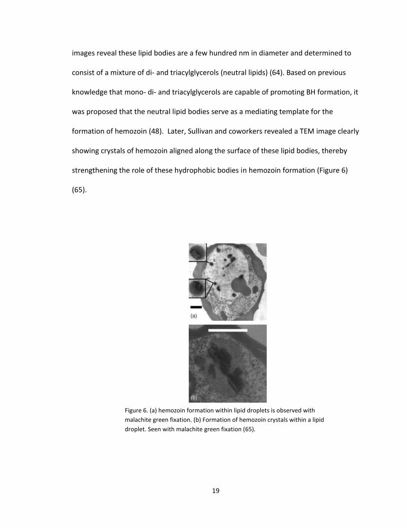
formation of hemozoin (48). Later, Sullivan and coworkers revealed a TEM image clearly
showing crystals of hemozoin aligned along the surface of these lipid bodies, thereby
strengthening the role of these hydrophobic bodies in hemozoin formation (Figure 6)
(65).
Figure 6. (a) hemozoin formation within lipid droplets is observed with
malachite green fixation. (b) Formation of hemozoin crystals within a lipid
droplet. Seen with malachite green fixation (65).
20
In an attempt to identify the specific composition of these neutral lipid bodies,
the authors extracted the digestive food vacuole of the parasite and performed lipid
analysis using ESI-MS/MS (electrospray ionization tandem MS) on the hemozoin
extracts. The results showed that the lipid composition surrounding the hemozoin
crystals are composed of a specific ratio of neutral lipids in an approximate ratio of
4:2:1:1:1 of MSG (monostearic glycerol), MPG (monopalmitic glycerol), DPG (dipalmitic
glycerol), DOG (dioleic glycerol) and DLG (dilinoleic glycerol). The authors investigated
the kinetic formation of BH in the presence of this blend of neutral lipids and revealed
that BH crystallization occurred within ten minutes of incubation with linear growth for
2 h before reaching 80% product conversion. The authors also provided evidence that
the neutral lipid bodies help protect heme from peroxide degradation, which would lead
to parasite toxicity. In the absence of these lipids, a 50 mM H2O2 solution degraded 50%
of dilipidated hemozoin, while a 90 mM concentration of peroxide was necessary to
reduce encapsulated hemozoin by 50%, suggesting that the lipids may also serve as a
protective reservoir for free heme preceding its incorporation into the nontoxic
hemozoin crystal.
Localization of free heme in the neutral lipid body
This interpretation of the lipid body serving as a reservoir for free heme is
supported by in vitro confocal evidence (66). Fluorescently labeled synthetic neutral
lipid bodies (SNLDs) were generated by dissolving the biological blend of neutral lipids in
1:9 acetone:methanol in the presence of Nile Red. When added to a pH 4.8 citric buffer,
21
the hydrophobic lipids minimize interactions with water by forming spherical,
continuous hydrophobic bodies, very similar to those observed within the native
environment of the parasitic digestive food vacuole. As these SNLDs are formed, Nile
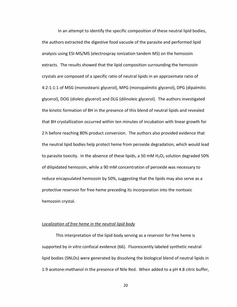
Red becomes trapped within the hydrophobic environment, and any Nile Red not
enveloped within the SNLDs will be quenched by water. The resultant fluorescent
SNLDs can be visualized using confocal microscopy (Figure 7). This system was then
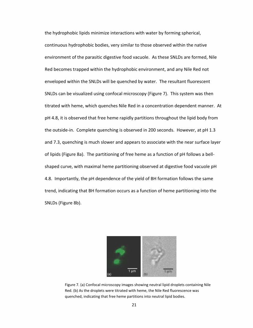
titrated with heme, which quenches Nile Red in a concentration dependent manner. At
pH 4.8, it is observed that free heme rapidly partitions throughout the lipid body from
the outside-in. Complete quenching is observed in 200 seconds. However, at pH 1.3
and 7.3, quenching is much slower and appears to associate with the near surface layer
of lipids (Figure 8a). The partitioning of free heme as a function of pH follows a bell-
shaped curve, with maximal heme partitioning observed at digestive food vacuole pH
4.8. Importantly, the pH dependence of the yield of BH formation follows the same
trend, indicating that BH formation occurs as a function of heme partitioning into the
SNLDs (Figure 8b).
Figure 7. (a) Confocal microscopy images showing neutral lipid droplets containing Nile
Red. (b) As the droplets were titrated with heme, the Nile Red fluorescence was
quenched, indicating that free heme partitions into neutral lipid bodies.
22
Kinetically competent site for BH formation
It has been estimated that the maximum half-life required for hemozoin
conversion must be no more than 40 minutes in order for the parasite to escape free
heme toxicity (67). If the neutral lipid body is imagined to be a reservoir for toxic heme,
then it must be capable of promoting the formation of hemozoin at a kinetically
Figure 8. (a) Comparison of Nile Red quenching as a function
of time at different pH. (b) Partitioning of free heme as a
function of pH follows a bell-shaped curve, with maximum
partitioning occurring at 4.8.

23
competent rate. Recently, an in vitro study that mimics digestive food vacuole
conditions has established the ability of these SNLDs to mediate the formation of BH
(68, 69). SNLDs were created in a pH 4.8 citric acid buffer at 37⁰C. An aliquot of
Fe(III)PPIX was then added to the SNLDs in order to determine the half-life of BH
formation. The product obtained was then crystallographically verified to be equivalent
to hemozoin. In the biologically relevant 4:2:1:1:1 ratio SNLDs, the half-life of BH
formation was 1.9 ± 0.01 min, demonstrating that the neutral lipid blend is capable of
successfully detoxifying free heme at a physiologically realistic rate that would protect
the parasite from toxicity.
Significance of the blend of neutral lipids
Based on the wealth of data that supports BH formation in a lipophilic
environment, it is not surprising that these neutral lipid bodies would be the in vivo
location of hemozoin formation. It is interesting, however, that the parasite uses the
specific blend of mono- and di-acylglycerols in the 4:2:1:1:1 ratio. Does this unique
blend of neutral lipids offer an advantage over utilizing individual lipid components? To
probe this question, temperature dependent kinetic measurements have been
undertaken to determine whether the neutral lipid blend offers a lower activation
barrier for BH formation over the individual lipid components (70, 71). For these
experiments, SNLDs of each individual lipid component and the biological mixture (100
μM) were synthesized followed by the addition of Fe(III)PPIX. Kinetic observations were
made to determine the half-life of BH formation as a function of time (Figure 9b). The
24
resulting data was fit to the Arrhenius equation and the activation energy was
determined (Figure 9c). The activation barriers for MSG, MPG, DOG, DLG, and DPG were
74.8 ± 5.3, 60.4 ± 7.1, 37.7 ± 3.3, 44.5 ± 15.4 and 35.2 ± 9.4 kJ/mol, respectively. These
results show that the monoglycerides exhibit an energy barrier that is higher than
observed for the diglycerides. However, in the neutral lipid blend (final concentration
100 µM) the activation barrier is 27.8 ± 3.4 kJ/mol (Figure 9a), lower than that of any of
the individual lipid components.
Figure 9. (a) Activation energy of neutral lipid blend, and separate mono-, di-
glycerides. (b) half-life of BH formation as a function of time. (c) Activation energy
was determined by the Arrhenius Equation.

25
This lower activation barrier suggests that the lipid blend offers an advantage compared
to using an individual lipid component. Recognizing this difference, Hoang et al. further
examined these lipids to identify whether or not the lipid blend exhibited properties
that are unique from the individual lipid components. Using differential scanning
calorimetry (DSC), a thermoanalytical technique that detects phase transitions by
measuring the heat flux of a sample, it was discovered that new intermolecular
interactions are observed in the lipid blend that are not present within the individual
components, demonstrating that the blend has a different molecular organization than
the individual components.
Molecular Dynamics Simulations
In addition to experimental data that suggests hemozoin formation occurs in the
presence of neutral lipid bodies, molecular dynamics (MD) simulations have further
shown that crystallization would be supported in this lipophilic environment (67-69). In
MD in vacuum, the interaction of two H2O-Fe(III)PPIX molecules form a precursor to the
head to tail dimer (Figure 10) in which the propionate groups interact with the
corresponding Fe(III) center. A ligand exchange then occurs with the displacement of
the water ligand from the axial position of Fe(III) and formation of the Fe-O bond. Rapid
hydrogen bonding between the propionic groups of dimers then occur. In these
simulations, if water molecules are present with the precursor dimer, the propionate
groups move away from the Fe(III) center to interact with the water molecules. These
simulations would suggest that the neutral lipid body serves as a hydrophobic

26
environment that minimizes the interactions with water molecules and drives formation
of the hemozoin crystal.
No single piece of evidence presented here alone could unequivocally implicate
neutral lipids in heme detoxification in vivo. However, collectively, a role for neutral
lipid bodies in hemozoin formation is undeniable. Not only has confocal evidence
shown that heme spontaneously localizes within these neutral lipid bodies, but they also
serve as a kinetically competent site for hemozoin formation. Previous evidence
showing hemozoin encapsulated within these neutral lipid bodies provides convincing
evidence that this is indeed the site for hemozoin formation within the digestive food
vacuole. MD studies further substantiate the likelihood that hemozoin formation occurs
at this site. It is clear that free heme accumulates within the neutral lipid body, and that
the lipid bodies are sufficient to quickly detoxify free heme on a physiologically relevant
time scale. The rapid formation of the head-to-tail dimer is favored in the lipophilic
Figure 10. Molecular Dynamics (MD) simulation of
hemozoin formation

27
environment and would not be expected to occur rapidly in aqueous solution due to
competitive hydrogen bonding.
Antimalarials
The malaria parasite contains several unique metabolic pathways which may be
exploited for the discovery of new antimalarials. These include targeting the synthesis
of DNA precursors (69), fatty acid synthesis (71), glycolysis (72) , and de novo heme
biosynthesis (73), among others. Of relevance here, however, is inhibition of the
formation of hemozoin. One of the earliest effective treatments for malaria dating from
the 17th century (74) was prepared from bark of Cinchona spp., The most active
component of “Jesuit’s bark” was later identified as quinine, a potent inhibitor of BH
formation (75). Chloroquine, arguably the most successful antimalarial developed to
this day, is an analogue of quinine and is thought to share a similar mechanism of
action: inhibition of hemozoin formation. Unfortunately, the efficacy of chloroquine has
widely diminished in endemic areas due to rampant drug resistance. These resistant
strains of P. falciparum exhibit several mutations in the Plasmodium falciparum
chloroquine resistance transporter (PfCRT), a transmembrane vacuolar protein. In
resistant strains the accumulation of CQ within the digestive food vacuole is significantly
less than in wild-type strains, as it is actively transported out of the DV (76).
Nevertheless, hemozoin formation remains a valid drug target, as resistance is the result
of diminished vacuolar accumulation of CQ, rather than changes to the hemozoin
formation pathway itself. Importantly, the interactions of hemozoin and antimalarial
28
inhibitors of this pathway have been studied in great detail, lending a wealth of
information that can facilitate the drug discovery process.
Evaluating Small Molecule Interactions with Heme
In order to understand the mode of action of hemozoin inhibitors, it is critical to
understand possible interactions between these drugs and heme and or hemozoin.
These efforts have been hampered by the insoluble nature of heme in a physiologically
relevant environment. In vivo, the speciation of free heme preceding the formation of
hemozoin is not fully understood, however, in vitro experiments reveal that free heme
can exist as a monomer, or several different dimeric species depending on
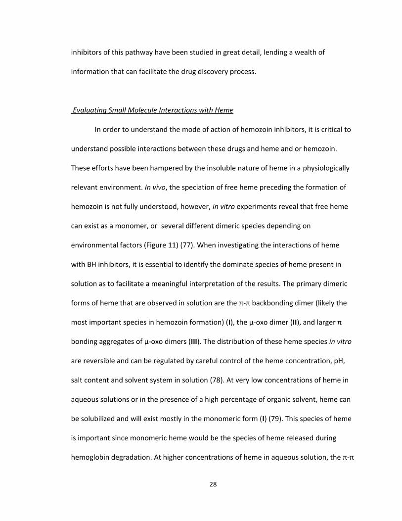
environmental factors (Figure 11) (77). When investigating the interactions of heme
with BH inhibitors, it is essential to identify the dominate species of heme present in
solution as to facilitate a meaningful interpretation of the results. The primary dimeric
forms of heme that are observed in solution are the π-π backbonding dimer (likely the
most important species in hemozoin formation) (I), the µ-oxo dimer (II), and larger π
bonding aggregates of µ-oxo dimers (III). The distribution of these heme species in vitro
are reversible and can be regulated by careful control of the heme concentration, pH,
salt content and solvent system in solution (78). At very low concentrations of heme in
aqueous solutions or in the presence of a high percentage of organic solvent, heme can
be solubilized and will exist mostly in the monomeric form (I) (79). This species of heme
is important since monomeric heme would be the species of heme released during
hemoglobin degradation. At higher concentrations of heme in aqueous solution, the π-π
29
dimer (II) will dominate (79). This is also true for protic solvent mixtures, since the
conversion between µ-oxo dimer and the π-π dimer depend on the protonation of the
oxygen bridging atom. This π-π species is likely the dominant form of heme present in
the digestive food vacuole, so is perhaps the most important species of heme that
should be studied in drug interactions.
Figure 11.Speciation of heme in different environments. Heme can exist as a
monomer (I), a π-π dimer (II), a µ-oxo dimer (III), or as π bonding aggregates of
the µ-oxo dimer (IV).
30
Vacuolar Accumulation
One of the confounding aspects of hemozoin inhibitors is that a compound’s
ability to inhibit the formation of β-hematin in vitro will not necessarily have a direct
correlation to the in vivo inhibition of hemozoin unless the drug can successfully
accumulate within the digestive food vacuole (DV) of the parasite at the sight of
nucleation and crystallization. Consequently, targeting vacuolar accumulation is an
important aspect of hemozoin inhibitor design. The primary mode that results in the
accumulation of a drug within the DV is by ‘pH trapping’ as is observed in the
mechanism of action of CQ, amodiaquine and mefloquine, among others. Here,
lipophlic species that are neutral at physiological pH are capable of permeating cell
membranes to gain entry into the malaria parasite. Upon entry into the digestive food
vacuole, however, the species has entered an acidic compartment (pH 4.8). If
protonatable functional groups are present (such as weakly basic functional groups), the
species will become protonated and will no longer freely permeate the cell membrane,
becoming trapped. This pH trapping drives the accumulation of antimalarials within the
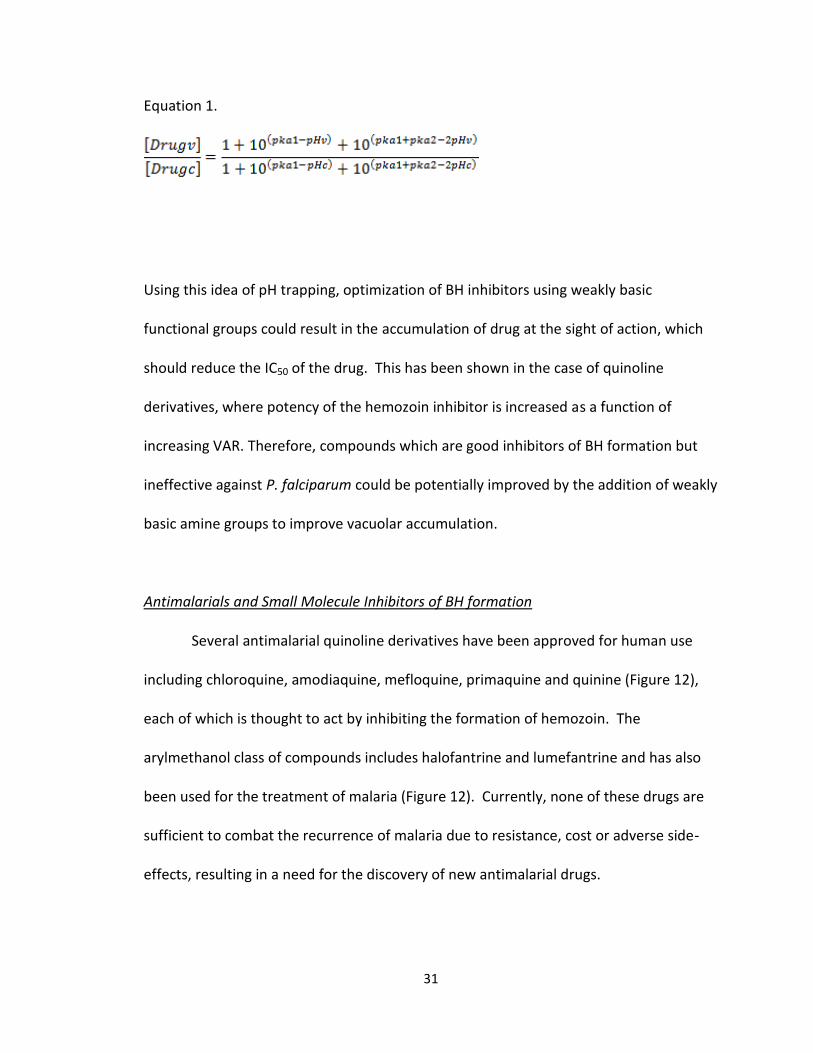
digestive food vacuole. In fact, using the known or calculated pKa values for BH
inhibitors, the vacuolar accumulation ratio (VAR) can be easily determined using a
modified Henderson-Hasselbach equation (Equation 1) (79, 80).
31
Equation 1.
Using this idea of pH trapping, optimization of BH inhibitors using weakly basic
functional groups could result in the accumulation of drug at the sight of action, which
should reduce the IC50 of the drug. This has been shown in the case of quinoline
derivatives, where potency of the hemozoin inhibitor is increased as a function of
increasing VAR. Therefore, compounds which are good inhibitors of BH formation but
ineffective against P. falciparum could be potentially improved by the addition of weakly
basic amine groups to improve vacuolar accumulation.
Antimalarials and Small Molecule Inhibitors of BH formation
Several antimalarial quinoline derivatives have been approved for human use
including chloroquine, amodiaquine, mefloquine, primaquine and quinine (Figure 12),
each of which is thought to act by inhibiting the formation of hemozoin. The
arylmethanol class of compounds includes halofantrine and lumefantrine and has also
been used for the treatment of malaria (Figure 12). Currently, none of these drugs are
sufficient to combat the recurrence of malaria due to resistance, cost or adverse side-
effects, resulting in a need for the discovery of new antimalarial drugs.

32
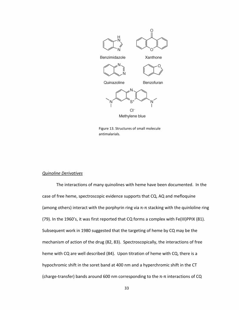
Aside from the potent quinoline derivatives, many other small molecules are
known to inhibit the formation of BH, including derivatives of benzimidazoles,
xanthones, quinazolines, phenanthrenes, methylene blue and benzofurans, to name a
few (Figure 13). The reason for the potency of these chemical scaffolds varies, but
generally occurs via π-π interactions of the compound with the aromatic porphyrin ring
and/or direct coordination to the Fe(III) center of heme. Small molecule inhibitors of BH
formation can interact not only with free heme, but also with the hemozoin crystal, or
both, to inhibit formation of product. It is important to understand the interactions of
an active compound with heme to facilitate the design and optimization of new
antimalarials targeting the hemozoin formation pathway.
Figure 12. Structures of anti-malarial compounds of the quinoline and
arylmethanol classes.
33
Quinoline Derivatives
The interactions of many quinolines with heme have been documented. In the
case of free heme, spectroscopic evidence supports that CQ, AQ and mefloquine
(among others) interact with the porphyrin ring via π-π stacking with the quinloline ring
(79). In the 1960’s, it was first reported that CQ forms a complex with Fe(III)PPIX (81).
Subsequent work in 1980 suggested that the targeting of heme by CQ may be the
mechanism of action of the drug (82, 83). Spectroscopically, the interactions of free
heme with CQ are well described (84). Upon titration of heme with CQ, there is a
hypochromic shift in the soret band at 400 nm and a hyperchromic shift in the CT
(charge-transfer) bands around 600 nm corresponding to the π-π interactions of CQ
Figure 13. Structures of small molecule
antimalarials.
34
with heme (85). Further support for the formation of a π-π complex is shown in the
NMR spectrum of CQ following the addition of Fe(III)PPIX. The paramagnetic Fe(III) has
a strong effect on the spectrum of CQ, where paramagnetic shifts are observed for both
1H and 13C (86, 87). Paramagnetic shifts and relaxation data indicate the presence of a
π-π complex rather than a coordination complex.
It has also been proposed that some antimalarials such as the quinolines can
absorb onto the surface of the hemozoin crystal itself, resulting in growth inhibition
(88). When subinhibitory concentrations of {3H}CQ were incubated with parasite
cultures, electron autoradiographs of infected red blood cells revealed that the majority
of the {3H}CQ was colocalized with the hemozoin crystal (88). Later, a model of
quinoline binding was proposed that suggested incorporation of CQ into the fast
growing (89) corrugated faces of the crystal where grooves expose propionic acid
groups, vinyl and methyl groups (90). CQ can intercalate the aromatic groups of the BH
crystal and cap the (48) surface by forming a salt bridge between the quinoline amine
and the propionic acid of the porphyrin, thereby inhibiting further growth of the crystal
along this face. The CQ molecule would be further stabilized by interactions with the
surrounding methyl and vinyl functional groups.
The ability of many of these quinoline derivatives to accumulate within the
digestive food vacuole explains the potency of these drugs against wild-type strains of P.
falciparum. For CQ, a percentage of this weakly basic, lipophilic quinoline derivative is
neutral at physiological pH. The protonatable tertiary amine and quinoline nitrogen
atoms of CQ have respective pKa’s of 10.2 and 8.4. Consequently, as CQ passes into the
35
acidic (pH 4.8) DV, a portion of the drug becomes doubly protonated. The pH trapping
effect can be quite large. In CQ treatment, nanomolar concentrations of the drug in
plasma results in millimolar concentrations within the DV (89). This pH trapping effect
can be observed for other quinoline antimalarials including mefloquine and
amodiaquine.
Arylmethanol Derivatives
The arylmethanol family of compounds is another important class of quinoline
derived antimalarials that have been shown to interact with Fe(III)PPIX (48). This class
of compounds includes quinine, mefloquine, lumefantrine and halofantrine, among
others (Figure 13). Recently, the halofantrine-Fe(III)PPIX crystal structure was solved,
revealing that interactions occur with monomeric heme, but not dimeric heme (90).
The hydroxyl group of halofantrine coordinates directly to the Fe(III) center of heme
resulting in a five coordinate porphyrin complex. The hydroxide functionality of
halofantrine has a pKa of ~14 and would be expected to exist exclusively in the
protonated state in the acidic DV. However, the Fe(III)-O bond length (1.840(4) Å)
suggests the formation of an alkoxide bond (1.816 – 1.851 Å) and not an Fe(III)-alcohol
bond (2.113 – 2.160 Å). The depression of the hydroxyl pKa may be due to the strong
coordination to the Fe(III) atom which induces a pKa shift. Additionally, π-stacking of the
phenanthrene and porphyrin was confirmed. The unprotonated propionate of
Fe(III)PPIX interacts with both the protonated N atom of halofantrine and the propionic
acid group of a neighboring heme.

36
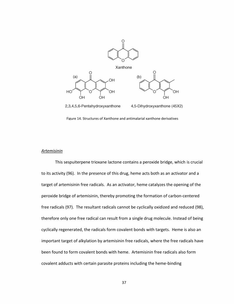
Xanthones
Several other classes of BH inhibitors have been identified for potential use as
antimalarial drugs including the xanthones. Xanthones are known to exist naturally as
secondary metabolites primarily of the Guttiferae and Gentianaceae family of plants,
and were first identified for antimalarial activity by Riscoe and Winter (85, 91). Soon
after the discovery of the antiplasmodial activity of the xanthones, Ignatushchenko et al.
observed that these compounds exhibited stage specifity, noting that trophozoites were
more sensitive than were ring-stage parasites (92). This observation followed studies
that hypothesized heme as the potential drug target of these compounds (93, 94). It was
also shown that the xanthone derivative 2,3,4,5,6-pentahydroxyxanthone (Figure 14a)
formed a soluble complex with free heme. Later, the interactions between 45X2 (Figure
14b) and heme were examined in detail using NMR spectroscopy (93). These studies
demonstrated that the xanthone carbonyl moiety coordinates to the Fe(III) center of
heme. Further, hydrogen bonding between the hydroxyl groups of 45X2 and the
propionate side chains of heme were observed in addition to π-π stacking of the
aromatic regions of both species. The ratio of xanthone:heme was found to be 1:2 for
45X2. Since the discovery of xanthone activity against the parasite, studies have sought
to increase the potency of these compounds. The 3,6-bis-ω-
diethylaminoalkoxyxanthone class of xanthone derivatives developed by Kelly et al.
boasted an increased affinity for heme binding that resulted in an increased potency
against the parasite (95).
37
Artemisinin
This sespuiterpene trioxane lactone contains a peroxide bridge, which is crucial
to its activity (96). In the presence of this drug, heme acts both as an activator and a
target of artemisinin free radicals. As an activator, heme catalyzes the opening of the
peroxide bridge of artemisinin, thereby promoting the formation of carbon-centered
free radicals (97). The resultant radicals cannot be cyclically oxidized and reduced (98),
therefore only one free radical can result from a single drug molecule. Instead of being
cyclically regenerated, the radicals form covalent bonds with targets. Heme is also an
important target of alkylation by artemisinin free radicals, where the free radicals have
been found to form covalent bonds with heme. Artemisinin free radicals also form
covalent adducts with certain parasite proteins including the heme-binding
Figure 14. Structures of Xanthone and antimalarial xanthone derivatives

38
translationally controlled tumor protein (TCTP), and in membrane containing structures
including the plasma membrane, endoplasmic reticulum, nuclear envelope, digestive
vacuole membrane, and mitochondria (99-102). Because artemisinin and its derivatives
are activated by free heme, it represents a very parasite specific malaria treatment.
High-Throughput Screening and Drug Discovery
With parasite antimalarial resistance on the rise, there is a pressing need for the
discovery of novel chemical scaffolds that are active against Plasmodium spp. One
discovery technique is high-throughput screening (HTS), where thousands to millions of
small molecules are rapidly tested for activity in assay. An HTS suitable assay is
generally formatted for use in 384- or 1536-well microtiter plates in order to test a large
quantity of compounds in a small amount of time. Furthermore, an HTS amenable assay
must involve minimal processing steps, be highly robust and should be comprised of
easily accessible reagents at a minimal cost. Assays can be either phenotypic or target-
based in nature. In antimalarial HTS, phenotypic assays are those that seek to identify
small molecules that are capable of killing the parasite itself in P.falciparum infected
erythrocytes. The disadvantage to this approach is that the mechanism of action of
inhibitors is unknown, and can be quite difficult to elucidate. In target-based assays, a
specific cellular process, such as hemozoin formation, is used to identify potential
antimalarials. Inhibitor/target interactions can be studied immediately after identifying
hits, an advantage over phenotypic screening. An additional advantage is the option to
39
select pathways that are specific to the parasite. The disadvantage to target-based
screening is that activity does not necessarily confer to the whole-cell assay.
The use of HTS in antimalarial drug discovery has been utilized in recent years for
both target-based and phenotypic assays (103). Of the target based assays, several have
been developed for the purpose of identifying inhibitors of BH formation (104-108).
One of the first successful assays developed for use in HTS utilized radioactive 14C-
labeled hematin to quantitate BH formation using scintillation counting (109-114).
Crystallization was mediated by the addition of lipid-rich extracts from parasite lysates.
The semi-automated assay tested over 100,000 compounds in 96-well plates and
identified 45 nonquinoline hits. Of these 45 compounds, the structural classes of
compounds identified included triarylcarbinols, piperazines, benzophenones, imides,
hydrazides, indoles and isoxazoles. The non-quinoline hits were then tested in a
secondary whole-cell assay consisting of cultures of CQ-sensitive and CQ-resistant P.
falciparum. Four compounds were identified to have activity in both strains at
concentrations of <5 µM (the IC50 of CQ in sensitive strains of P. falciparum is ~25 nM).
Though few of the compounds identified in the BH inhibition screen were active against
parasite cultures, the ability of HTS to identify novel pharmacophores supported the
utility of this approach. However, this assay was not utilized to its full potential due to
deficiencies in design. The semi-automated use of 96-well plates would be considered a
medium-throughput method compared to the more often used 384- and 1536- well
plates. Further, the need for trophozoite lysates and radioactive hematin limits the use
of this assay to laboratories capable of maintaining parasite cultures and open to the

40
restrictions imposed by utilizing radioactive substrates. Fortunately, subsequent BH
formation assays have improved upon the original protocol by increasing the
throughput of compound screening, incorporating more readily available substrates,
and using improved methods of quantification that do not require the use of radio-
labeled heme.
In 2009, Clardy and coworkers developed a true HTS assay that utilizes acetate
buffer for the synthesis of BH in 384-well microtiter plates (113, 115, 116). In this assay,
a high concentration (9.7 M, pH 4.8) of sodium acetate buffer is utilized to mediate the
formation of BH by solubilizing Fe(III)PPIX, the rate limiting step in BH formation. The
assay is incubated at 60⁰C for 120 min, then analyzed using the pyridine ferrichrome
method of quantification developed by Egan and coworkers (114). Using this assay to
screen 16,000 compounds at a 220 µM concentration of test compound, 644 hits (3.96%
hit-rate) capable of inhibiting >50% BH formation were identified. Of these 644 hits, 17
were identified as active against in vitro cultures of P. falciparum (hits were identified as
having activity of >80% against the parasite). As a direct result of this screening effort,
the authors identified two novel classes of compounds that show activity in both the BH
formation assay, as well as the whole-cell parasite assay. Two classes of compounds,
the pyrimidine and 1,3-benzoxathiol-2-one compounds were identified in this screen.
While this assay was a major improvement upon previously published medium-
throughput assays, the conditions for BH formation were not physiologically relevant.
This is perhaps the reason that so few of the BH inhibitors show activity against cultures
of P. falciparum.

41
Improved understanding of the in vivo formation of hemozoin has facilitated
advances in HTS assay design for the discovery of novel BH inhibitors. Of vital
importance is the identification of neutral lipids as playing a role in hemozoin formation
(117). The composition of these lipids has been found to mediate the formation of BH
and is inhibited upon co-incubation with the quinoline antimalarials. Due to the
relatively high cost of these neutral lipids, they are not practical for use in HTS. An
alternative is the use of commercially available, low cost detergents, several of which
have been shown to mediate formation of BH in 96- and 384-well plate format in vitro
and in HTS formats (116) (65). The assay uses the lipophilic detergent, NP-40, to
mediate BH crystallization at a pH of 4.9 (116, 118). Further, when co-incubated with
CQ or AQ, NP-40 exhibits similar IC50’s to those obtained when using the neutral lipid
blend. Using this assay, a pilot screen of over 38,000 compounds resulted in the
identification of 161 hits, a 0.42% hit-rate. IC50 values were obtained for each of these
hits and included the identification of 113 compounds with lower IC50 values than that
of AQ in this assay. These compounds were then tested for efficacy against P.falciparum
infected erythrocytes and 48 compounds were identified as capable of inhibiting >90%
of parasitemia, including the identification of eight compounds with activities at
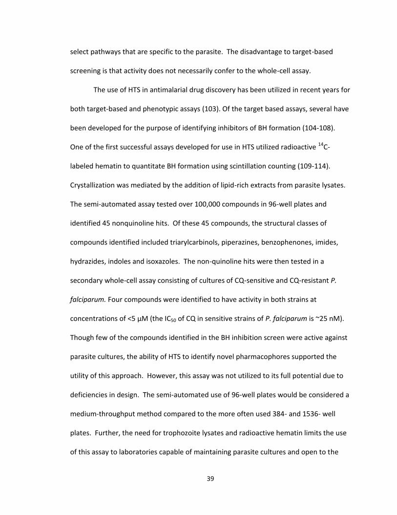
nanomolar concentrations (Figure 15a). Four distinct chemical scaffolds were identified
(Figure 15b).
42
Conclusion
In order to survive, the malaria parasite relies on a unique detoxification
pathway in which toxic free heme is converted to the nontoxic biomineral hemozoin.
Disruption of this crystallization process results in parasite death, and therefore serves
as an important strategy for drug design. Hemozoin formation does not occur
Figure 15. (a) Structures of antimalarial compounds found to be
active in the malaria parasite, and (b) four distinct chemical
scaffolds.
43
spontaneously at a physiologically relevant rate, and identification of the biological
mediator of this crystal remained elusive for many years. Recently, neutral lipid bodies
present within the digestive food vacuole of the parasite have been replicated in vitro
(SNLDs). It has been found that Fe(III)PPIX rapidly accumulates within these SNLDs.
Furthermore, the SNLDs have been shown to serve as a kinetically competent site for BH
formation. These data demonstrate that the neutral lipid bodies are the biological
mediator of hemozoin formation. Using this information, an assay that mimics the
physiological environment in which hemozoin formation takes place has been
developed and used in high-throughput screening to identify novel inhibitors of this
target. Furthermore, the interactions of hemozoin inhibitors with free heme have been
studied in detail. These studies have revealed that inhibitors can interact through π-π
interactions and/or direct coordination to the Fe(III) center, inhibiting the incorporation
of the heme into the growing hemozoin crystal. The information gained from these
interactions can now be used to optimize compounds that inhibit BH formation. All of
these advances have only been made possible by first understanding the formation of
the biomineral, hemozoin.
44
Chapter II
Isolation of Anti-malarial Compounds from a Natural Product Library
Introduction
Natural products have long been the single most productive source of leads for
drug development (119). Between 1981 and 2002, 49% of all small-molecule New
Chemical Entities (NCEs) were natural products, natural product analogues, or natural
product derivatives (120). In fact, the anti-malarial drug chloroquine is derived from
quinine, a natural product found in the bark of the Cinchona tree (See figure 12).
Despite the success of natural products in the drug discovery field, the emphasis on
natural product drug discovery has waned in the past decade for several reasons.
First, the emergence of combinatorial libraries and their promise of many
potential biologically active compounds, short timelines, and high-throughput
capabilities took the focus away from the slow process of natural product purification,
screening, structure elucidation, and subsequent scale-up (119). Secondly, the increase
in molecular biology, genomics, and cellular biology techniques made the discovery of
new drug targets possible, increasing the demand for shorter drug discovery timelines.
Finally, the reduction of emphasis on infectious disease research - a typical strong point
of many biologically active natural products - encouraged scientists to move from
natural product screening to screening much larger, synthetic libraries (120).
45
Utilizing the new technologies born from the combinatorial chemistry drug
discovery era, science is once again turning to natural product drug discovery to
produce useful biologically active compounds. Many bioactive compounds discovered
previously have come from a class of soil bacteria called actinomycetes. This class of
bacteria has been the main producer of approved antibiotics and other biologically
active metabolites to date such as the well-known antibiotics streptomycin and
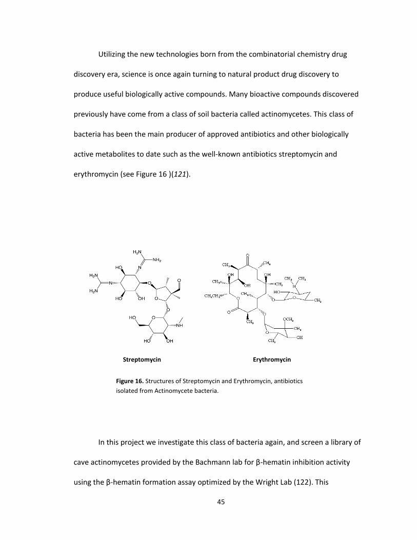
erythromycin (see Figure 16 )(121).
In this project we investigate this class of bacteria again, and screen a library of
cave actinomycetes provided by the Bachmann lab for β-hematin inhibition activity
using the β-hematin formation assay optimized by the Wright Lab (122). This
Figure 16. Structures of Streptomycin and Erythromycin, antibiotics
isolated from Actinomycete bacteria.
Streptomycin Erythromycin
46
actinomycete library has already gone through a pre-screen in the Wright lab, producing
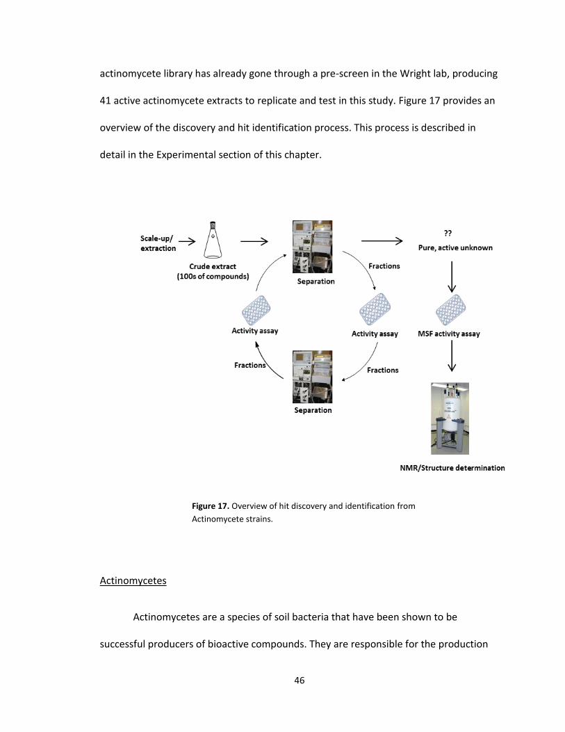
41 active actinomycete extracts to replicate and test in this study. Figure 17 provides an
overview of the discovery and hit identification process. This process is described in
detail in the Experimental section of this chapter.
Actinomycetes
Actinomycetes are a species of soil bacteria that have been shown to be
successful producers of bioactive compounds. They are responsible for the production
Figure 17. Overview of hit discovery and identification from
Actinomycete strains.
47
of nearly half of all discovered bioactive secondary metabolites (123). This species has
been the source of antibiotics, antitumor agents, and immunosuppressive agents and
the subject of intensive study (123-125). Recently, success of isolation of novel
compounds from terrestrial actinomycetes has declined, and the re-isolation of
previously discovered compounds has increased (126). Therefore, a need for novel
bioactive compounds from actinomycetes is presented. Screening of new actinomycete
strains from unexplored or unexploited environments, such as caves, promises to
uncover an array of novel bioactive compounds.
Experimental
Growth of Actinomycete Culture
30 µL of an Actinomycete strain were grown on individual solid agar plates for one week
at 30°C. Individual colonies were then seeded into six separate flasks containing 50 mL
of liquid media, and allowed to incubate at 30°C with shaking for one week. 25mL of 5 of
the seed cultures was inoculated into separate 500mL production cultures, and allowed
to incubate at 30°C with shaking for one week. This produced 5L of total bacteria
culture.
Purification of actinomycete extracts
The 5L of bacterial culture was extracted with an equal amount of solvent, incubated
with shaking for 30 minutes, and the resulting mixture spun down at 3500 rpm for 5
minutes. The solvent was removed from the aqueous media, and dried by Rotovap,
48
producing a crude extract. The crude extract was then purified using both a Sephadex
LH-20 column (100% MeOH) and LC-ESI-MS (A: 95% Acetonitrile: 5% H2O, 1mM
ammonium Accetate, B: 95% H2O: 5% Acetonitrile, 1mM Ammonium Acetate) to
produce two sets of extract fractions. These resulting fractions were transferred to 96-
well plates, dried, and screened using the β-hematin formation assay (122).
Detergent mediated β-hematin formation assay
The assay conditions were as follows: All liquid delivery was automated using a Thermo
Scientific Multidrop Combi robot. To suspend each natural product well fraction, 5-10 μL
of acetonitrile was added followed by ten minutes of shaking and 65 μL of water. A 20
μL volume of the hemozoin formation promoter NP-40 (30.55 μM) and 5 μL acetone
were added. Finally, a 90 μL volume of hematin (222.2 μM) suspended in 2.0 M sodium
acetate solution (pH 4.8) was added to give a final volume of 100 μM hematin. Each
plate was incubated at 3 C and 55 rpm for at least 5 hours. Upon completion, the assay
wells were treated with 25 μL of 50% pyridine solution (pH .5) and the plates were
shaken for 10 minutes. The absorbance was read at 405 nm using a SpetraMax M5
microplate reader and data was plotted using GraphPad Prism 4. A plate of water and
amodiaquine at the IC50 was run as negative and positive controls for inhibition.
Fractions which showed activity were combined, and put through another round of
purification and screening. Rounds of purification and screening were performed until
pure fractions containing only active compounds were obtained. The pure compounds
were then analyzed by 2D NMR.
49
2D NMR structure elucidation of Pure Compounds
Active compounds were dissolved in CH3OD (Sigma Aldrich), and added to a 3mm or
5mm NMR tube. Samples were run using a Bruker AV-II 600 MHz NMR spectrometer.
Samples were used for 1D 1H-NMR experiments, as well as 2D NMR suite experiments
(COSY, HMBC, HSQC). Data obtained from these experiments will be analyzed, and
structures elucidated.
MSF Assay
10mM samples of each active compound will then be tested in an MSF assay following a
modified version of the protocol outlined by (113). Briefly, active compounds dissolved
in DMSO will be prescreened at 23 μM at a 0.3% starting parasitemia (at 2% hematocrit)
in 384-well optical-bottom plates. To establish dose-response curves, active compounds
will be delivered in a range of concentrations from 0 to 23 µM with a final DMSO
concentration of 0.23% per well. To ensure that DMSO does not interfere with parasite
growth, a control plate will be used containing wells with 0.23% DMSO, and wells
containing no DMSO will also be used. Concentration-response curves will be generated
using GraphPad Prism v5.0
Results
Of the 41 active actinomycete extracts discovered in the preliminary screening, So far,
we have been able to reproduce screening data for 5 Actinomycete strains in the
Bachmann library (See figure 6). See Appendix A for graphs of all fractions for these

50
strains. Of the 5 strains for which we were able to reproduce screening data, we found
two compounds that were active both in vitro and in vivo. The first compound
(Compound A) was found in an actinomycete strain identified as BBHARD25. Compound
A was found to elute at 19.19 minutes using HPLC (A: 95% Acetonitrile: 5% H2O, 1mM
Ammonium Acetate; B: 95% H2O: 5% Acetonitrile, 1mM Ammonium Acetate). Using the
NELI program, we were able to compare HPLC trace with mass and activity data (see
figure 18).
The NELI data shows an active (highlighted) peak at 19.19 minutes. The LC-MS Mass
data of the peak shows a relatively pure Compound A with an m/z of 270.55. Accurate
mass data of Compound A showed an m/z of 271.06 the difference in m/z
measurements for Compound A are too small to be fragments, and so are most likely
due to differences in MS instrumentation. 13C NMR analysis of Compound A showed the
presence of 1 carbonyl carbon, 12 aromatic carbons, 1 tertiary, and 1 secondary carbon.
m/z 270.55
Figure 18. NELI data of BBHARD25 overlapping HPLC data with activity. LC-MS data is
shown on the right.
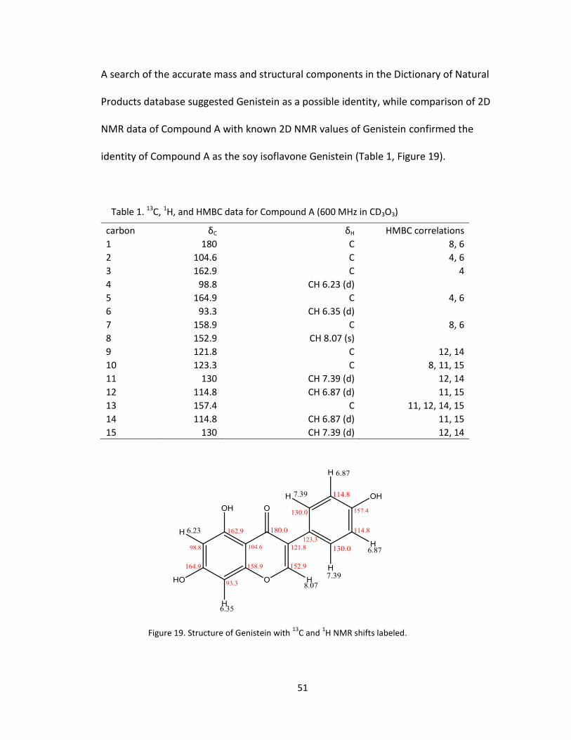
51
A search of the accurate mass and structural components in the Dictionary of Natural
Products database suggested Genistein as a possible identity, while comparison of 2D
NMR data of Compound A with known 2D NMR values of Genistein confirmed the
identity of Compound A as the soy isoflavone Genistein (Table 1, Figure 19).
carbon δC δH HMBC correlations
1 180 C 8, 6
2 104.6 C 4, 6
3 162.9 C 4
4 98.8 CH 6.23 (d) 5 164.9 C 4, 6
6 93.3 CH 6.35 (d) 7 158.9 C 8, 6
8 152.9 CH 8.07 (s) 9 121.8 C 12, 14
10 123.3 C 8, 11, 15
11 130 CH 7.39 (d) 12, 14
12 114.8 CH 6.87 (d) 11, 15
13 157.4 C 11, 12, 14, 15
14 114.8 CH 6.87 (d) 11, 15
15 130 CH 7.39 (d) 12, 14
Table 1. 13C, 1H, and HMBC data for Compound A (600 MHz in CD3O3)
Figure 19. Structure of Genistein with 13
C and 1H NMR shifts labeled.

52
The second compound (Compound B) that was found to be active in vitro and in vivo
was isolated from an actinomycete strain identified as BBHARD23. Compound B was
found to elute from HPLC at 22.20 minutes HPLC (A: 95% Acetonitrile: 5% H2O, 1mM
Ammonium Acetate; B: 95% H2O: 5% Acetonitrile, 1mM Ammonium Acetate). NELI data
of Compound B shows an active peak at 22.20 minutes. The LC-MS mass data of the
peak shows a major compound with an m/z of 375.75 (See Figure 20), and accurate
mass data of Compound B showed an m/z of 376.13. The small difference in m/z
measurements can be explained by differences in MS instrumentation. Preliminary 13C
NMR data of Compound B suggests the presence of 2 carbonyl carbons, 5 methylene
carbons, 8 methnine carbons, and 10 quaternary carbons. A full 2D NMR analysis of
Carbon B is in progress.
m/z 375.71
Figure 20. NELI data of BBHARD23 overlapping HPLC data with activity. LC-MS data is
shown on the right.
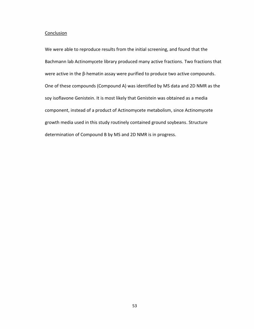
53
Conclusion
We were able to reproduce results from the initial screening, and found that the
Bachmann lab Actinomycete library produced many active fractions. Two fractions that
were active in the β-hematin assay were purified to produce two active compounds.
One of these compounds (Compound A) was identified by MS data and 2D NMR as the
soy isoflavone Genistein. It is most likely that Genistein was obtained as a media
component, instead of a product of Actinomycete metabolism, since Actinomycete
growth media used in this study routinely contained ground soybeans. Structure
determination of Compound B by MS and 2D NMR is in progress.
54
Chapter III
Anti-malarial Effects of Genistein
Introduction
Genistein is an isoflavonoid commonly found in soybeans. It is the simplest of the
isoflavanoid compounds, and an important precursor to more complex isoflavanoids
that have antimicrobial action (127). Studies on Genistein show it to have tyrosine
kinase activity, and action against cancer and cardiovascular disease (127). Genistein has
also been found to be active against liver and blood stage malaria parasite, reducing
liver infections by 64% in mice given Genistein 6hrs prior to infection, and blood
infections by 33% when administered at 200uM (128, 129).The antimalarial activity of
genistein in these studies was attributed to its function as a tyrosine kinase inhibitor,
but no link to any interaction with hemozoin has been found. In this study, we found the
active component of an actinomycete extract to be genistein.
Experimental
Growth of Actinomycete Culture
30 µL of an Actinomycete strain were grown on individual solid agar plates for one week
at 30°C. Individual colonies were then seeded into six separate flasks containing 50 mL

55
of liquid media, and allowed to incubate at 30°C with shaking for one week. 25mL of 5 of
the seed cultures was inoculated into separate 500mL production cultures, and allowed
to incubate at 30°C with shaking for one week. This produced 5L of total bacteria
culture.
Purification of actinomycete extracts
The 5L of bacterial culture was extracted with an equal amount of ethylacetate,,
incubated with shaking for 30 minutes, and the resulting mixture spun down at 3500
rpm for 5 minutes. The solvent was removed from the aqueous media, and dried by
Rotovap, producing a crude extract. The crude extract was then purified using both a
Sephadex LH-20 column (100% MeOH) and LC-ESI-MS (A: 95% Acetonitrile:5% H2O, B:
5% Acetonitrile: 95% H2O, 1mM Ammonium Acetate) to produce two sets of extract
fractions. These resulting fractions were transferred to 96-well plates, dried, and
screened using the β-hematin formation assay (46).
Detergent mediated Β-hematin formation assay
The assay conditions were as follows: All liquid delivery was automated using a Thermo
Scientific Multidrop Combi robot. To suspend each natural product well fraction, 5-10 μL
of acetonitrile was added followed by ten minutes of shaking and 65 μL of water. A 20
μL volume of the hemozoin formation promoter NP-40 (30.55 μM) and 5 μL acetone
were added. Finally, a 90 μL volume of hematin (222.2 μM) suspended in 2.0 M sodium
acetate solution (pH 4.8) was added to give a final volume of 100 μM hematin. Each
plate was incubated at 3 C and 55 rpm for at least 5 hours. Upon completion, the assay
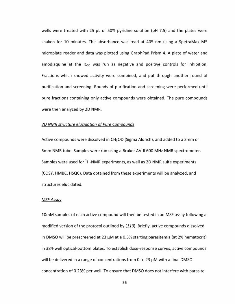
56
wells were treated with 25 μL of 50% pyridine solution (pH .5) and the plates were
shaken for 10 minutes. The absorbance was read at 405 nm using a SpetraMax M5
microplate reader and data was plotted using GraphPad Prism 4. A plate of water and
amodiaquine at the IC50 was run as negative and positive controls for inhibition.
Fractions which showed activity were combined, and put through another round of
purification and screening. Rounds of purification and screening were performed until
pure fractions containing only active compounds were obtained. The pure compounds
were then analyzed by 2D NMR.
2D NMR structure elucidation of Pure Compounds
Active compounds were dissolved in CH3OD (Sigma Aldrich), and added to a 3mm or
5mm NMR tube. Samples were run using a Bruker AV-II 600 MHz NMR spectrometer.
Samples were used for 1H-NMR experiments, as well as 2D NMR suite experiments
(COSY, HMBC, HSQC). Data obtained from these experiments will be analyzed, and
structures elucidated.
MSF Assay
10mM samples of each active compound will then be tested in an MSF assay following a
modified version of the protocol outlined by (113). Briefly, active compounds dissolved
in DMSO will be prescreened at 23 μM at a 0.3% starting parasitemia (at 2% hematocrit)
in 384-well optical-bottom plates. To establish dose-response curves, active compounds
will be delivered in a range of concentrations from 0 to 23 μM with a final DMSO
concentration of 0.23% per well. To ensure that DMSO does not interfere with parasite

57
growth, a control plate will be used containing wells with 0.23% DMSO, and wells
containing no DMSO will also be used. Concentration-response curves will be generated
using GraphPad Prism v5.0
Results
MSF assays of the Genistein isolated from BBHARD25 and the isolated active fraction
from BBHARD23 showed very little activity. The IC50 values of these fractions were
18µM, an order of magnitude less active than chloroquine (See figure 21). To confirm
that Genistein is the active compound, a β-hematin assay was performed on synthetic
Genistein. The IC50 curves of the β-hematin assay performed on synthetic Genistein
showed no activity (See figure 22). Therefore, it is likely that Genistein was not the
major contributor of activity in the β-hematin assay from BBHARD25. Further analysis of
NELI data shows the presence of another compound at very low levels (See Figure 23).
LC-MS data shows that the second compound has an m/z of 412.72. Efforts to obtain
NMR and accurate mass data from this compound were unsuccessful due to the limited
amount of sample that was left, and the low concentration of the compound.

58
0
1
2
3
4
5
1 2 3 4 5 6 7 8
Ab
s (4
05
)
Genistein IC50 curve
5 ul acetonitrile
10 ul acetonitrile
Positive
positive + SD
positive - SD
Negative
Figure 22: IC50 curve of synthetic genistein performed under normal assay conditions.
Figure 21: MSF assay IC50 curves of active fractions from BBHARD23 and BBHARD25
0.00 0.25 0.50 0.75 1.00 1.25 1.50 1.750
10000
20000
30000
log (uM inhibitor)
RF
U
BBHARD23
0.00 0.25 0.50 0.75 1.00 1.25 1.50 1.750
10000
20000
30000
log (uM inhibitor)
RF
U
BBHARD25
m/z 412.72
Figure 23. NELI data of BBHARD25 overlapping HPLC data with activity. LC-MS data is shown on the right, revealing the presence of a second compound with m/z of 412.72.

59
Conclusion
Although the isolated genistein did not show high levels of activity in vivo, it is promising
that activity can be seen in the malaria parasite even at low levels. Using SAR studies, it
may be possible to modify the active scaffolds to increase antimalarial activity, thereby
producing a novel antimalarial treatment. Since synthetic Genistein showed no activity
in vitro, this suggests that another compound was responsible for that activity. The
second compound found in the active fraction at low levels it is also possible that
another compound present at very low levels is causing the anti-malarial activity in the
β-hematin assay. This is interesting because any compound producing anti-malarial
activity at such low levels is probably very potent. Efforts to obtain NMR and accurate
mass data from this unknown compound were unsuccessful due to the limited amount
and low concentration of the sample. A more concentrated sample may be obtained by
growing another culture of the BBHARD25 actinomycete strain, and re-isolating the
unknown compound.
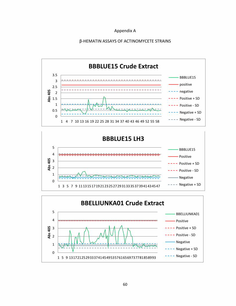
60
Appendix A
β-HEMATIN ASSAYS OF ACTINOMYCETE STRAINS
0
0.5
1
1.5
2
2.5
3
3.5
1 4 7 10 13 16 19 22 25 28 31 34 37 40 43 46 49 52 55 58
Ab
s 4
05
BBBLUE15 Crude Extract
BBBLUE15
positive
negative
Positive + SD
Positive - SD
Negative + SD
Negative - SD
0
1
2
3
4
5
1 3 5 7 9 11131517192123252729313335373941434547
Ab
s 4
05
BBBLUE15 LH3
BBBLUE15
Positive
Positive + SD
Positive - SD
Negative
Negative + SD
0
1
2
3
4
5
1 5 9 131721252933374145495357616569737781858993
Ab
s 4
05
BBELLIUNKA01 Crude Extract
BBELLIUNKA01
Positive
Positive + SD
Positive - SD
Negative
Negative + SD
Negative - SD
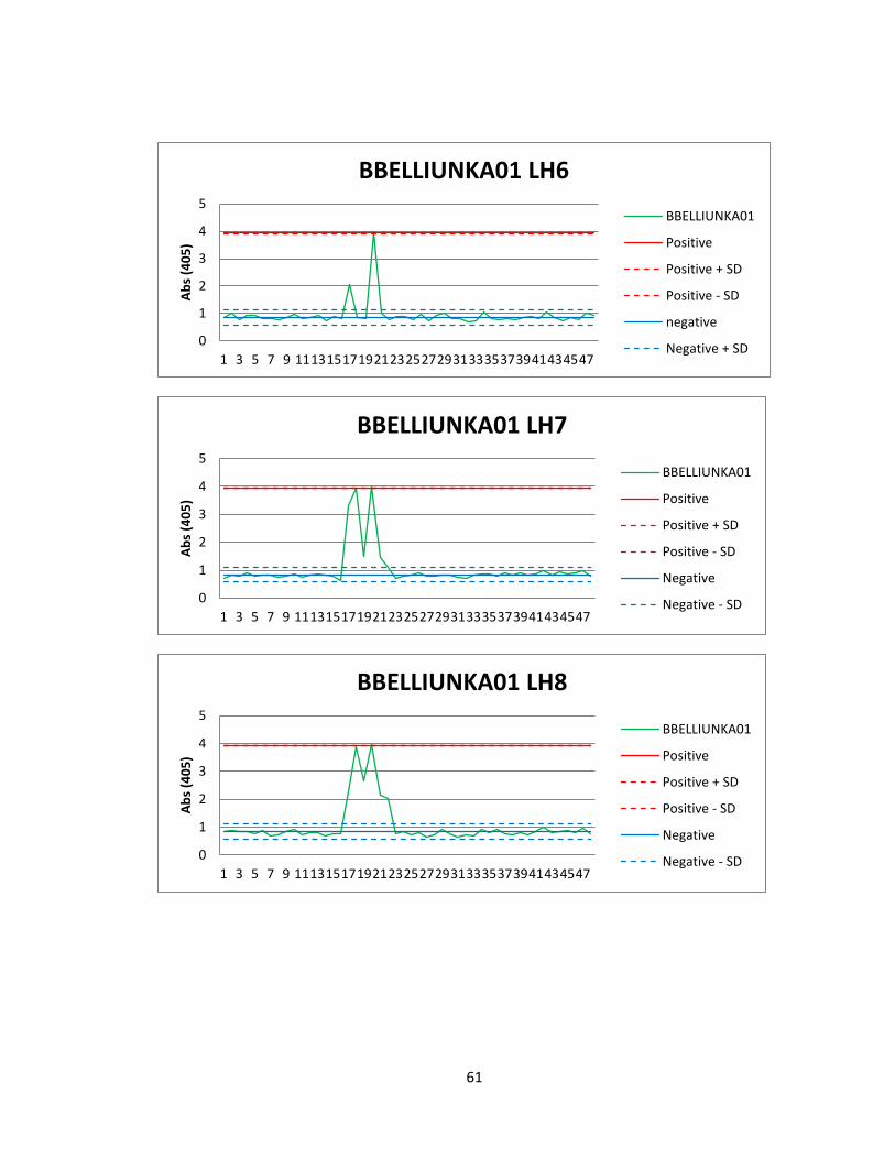
61
0
1
2
3
4
5
1 3 5 7 9 11131517192123252729313335373941434547
Ab
s (4
05
) BBELLIUNKA01 LH6
BBELLIUNKA01
Positive
Positive + SD
Positive - SD
negative
Negative + SD
0
1
2
3
4
5
1 3 5 7 9 11131517192123252729313335373941434547
Ab
s (4
05
)
BBELLIUNKA01 LH7
BBELLIUNKA01
Positive
Positive + SD
Positive - SD
Negative
Negative - SD
0
1
2
3
4
5
1 3 5 7 9 11131517192123252729313335373941434547
Ab
s (4
05
)
BBELLIUNKA01 LH8
BBELLIUNKA01
Positive
Positive + SD
Positive - SD
Negative
Negative - SD

62
0
1
2
3
4
1 5 9 131721252933374145495357616569737781858993
Ab
s (4
05
)
BBHARD19 Crude Extract
BBHARD19
Positive
Positive + SD
Positive - SD
Negative
Negative + SD
0
1
2
3
4
1 3 5 7 9 11131517192123252729313335373941434547
Ab
s (4
05
)
BBHARD19 LH2
BBHARD19
Positive
Positive + SD
Positive - SD
Negative
Negative + SD
0
1
2
3
4
1 3 5 7 9 11131517192123252729313335373941434547
Ab
s (4
05
)
BBHARD19 LH4
BBHARD19
Positive
Positive + SD
Positive - SD
Negative
Negative + SD
63
0
1
2
3
4
1 3 5 7 9 11 13 15 17 19 21 23 25 27 29 31 33 35 37 39 41 43 45 47
Ab
s (4
05
)
BBHARD19 LH6
BBHARD19
Positive
Positive + SD
Positive - SD
Negative
0
1
2
3
4
1 3 5 7 9 11131517192123252729313335373941434547
Ab
s (4
05
)
BBHARD19 LH8
BBHARD19
Positive
Positive + SD
Positive - SD
Negative
Negative + SD
0
1
2
3
4
1 3 5 7 9 11131517192123252729313335373941434547
Ab
s (4
05
)
BBHARD23 Crude Extract
BBHARD23
Positive
Positive + SD
Positive - SD
Negative
Negative - SD
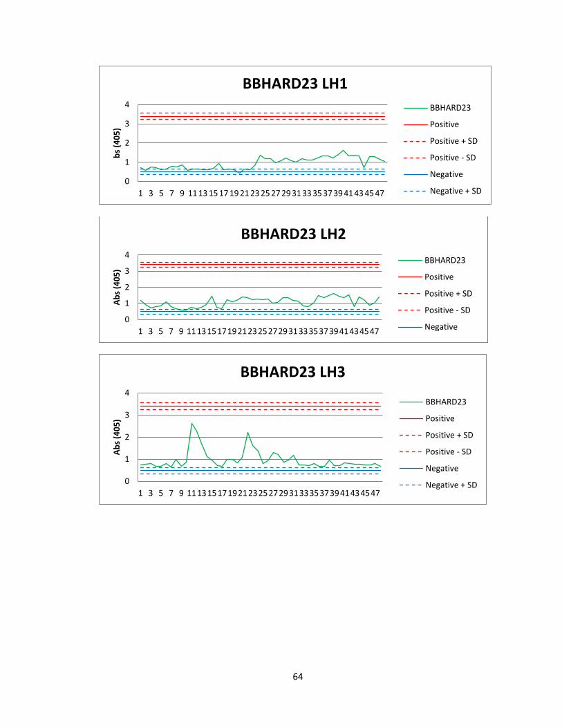
64
0
1
2
3
4
1 3 5 7 9 11 13 15 17 19 21 23 25 27 29 31 33 35 37 39 41 43 45 47
bs
(40
5)
BBHARD23 LH1
BBHARD23
Positive
Positive + SD
Positive - SD
Negative
Negative + SD
0
1
2
3
4
1 3 5 7 9 11131517192123252729313335373941434547
Ab
s (4
05
)
BBHARD23 LH2
BBHARD23
Positive
Positive + SD
Positive - SD
Negative
0
1
2
3
4
1 3 5 7 9 11131517192123252729313335373941434547
Ab
s (4
05
)
BBHARD23 LH3
BBHARD23
Positive
Positive + SD
Positive - SD
Negative
Negative + SD

65
0
1
2
3
4
1 3 5 7 9 11 13 15 17 19 21 23 25 27 29 31 33 35 37 39 41 43 45 47
Ab
s (4
05
)
BBHARD23 LH4
BBHARD23
Positive
Positive + SD
Positive - SD
Negative
Negative + SD
0
1
2
3
4
1 3 5 7 9 11131517192123252729313335373941434547
Ab
s (4
05
)
BBHARD23 LH5
BBHARD23
Positive
Positive + SD
Positive - SD
Negative
Negative + SD
0
1
2
3
4
1 5 9 131721252933374145495357616569737781858993
Ab
s (4
05
)
BBHARD28 Crude Extract
BBHARD28
Positive
Positive + SD
Positive - SD
Negative
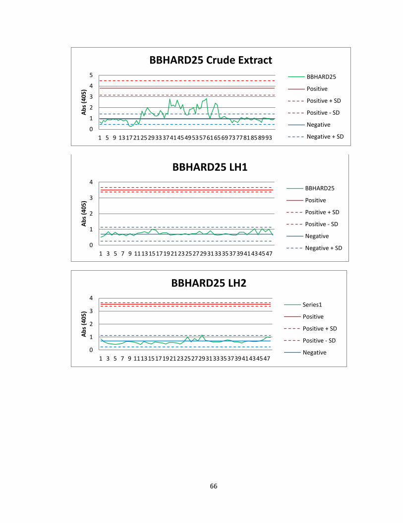
66
0
1
2
3
4
5
1 5 9 131721252933374145495357616569737781858993
Ab
s (4
05
)
BBHARD25 Crude Extract
BBHARD25
Positive
Positive + SD
Positive - SD
Negative
Negative + SD
0
1
2
3
4
1 3 5 7 9 11131517192123252729313335373941434547
Ab
s (4
05
)
BBHARD25 LH1
BBHARD25
Positive
Positive + SD
Positive - SD
Negative
Negative + SD
0
1
2
3
4
1 3 5 7 9 11131517192123252729313335373941434547
Ab
s (4
05
)
BBHARD25 LH2
Series1
Positive
Positive + SD
Positive - SD
Negative

67
0
1
2
3
4
1 3 5 7 9 11131517192123252729313335373941434547
Ab
s (4
05
)
BBHARD25 LH3
BBHARD25
Positive
Positive + SD
Positive - SD
Negative
0
1
2
3
4
1 3 5 7 9 11131517192123252729313335373941434547
Ab
s (4
05
)
BBHARD25 LH4
BBHARD25
Positive
Positive + SD
Positive - SD
Negative
Negative + SD
0
1
2
3
4
5
1 3 5 7 9 11131517192123252729313335373941434547
Ab
s (4
05
)
BBHARD25 LH5
BBHARD25
Positive
Positive + SD
Positive - SD
Negative
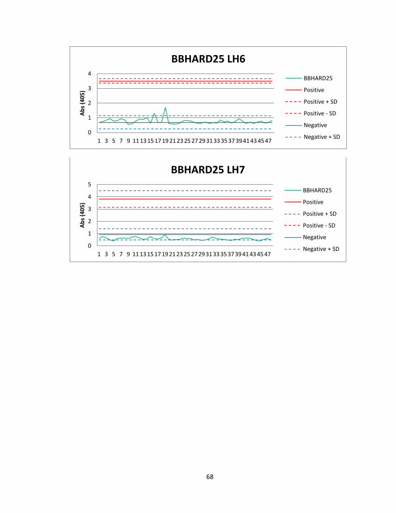
68
0
1
2
3
4
1 3 5 7 9 11 13 15 17 19 21 23 25 27 29 31 33 35 37 39 41 43 45 47
Ab
s (4
05
)
BBHARD25 LH6
BBHARD25
Positive
Positive + SD
Positive - SD
Negative
Negative + SD
0
1
2
3
4
5
1 3 5 7 9 11 13 15 17 19 21 23 25 27 29 31 33 35 37 39 41 43 45 47
Ab
s (4
05
)
BBHARD25 LH7
BBHARD25
Positive
Positive + SD
Positive - SD
Negative
Negative + SD
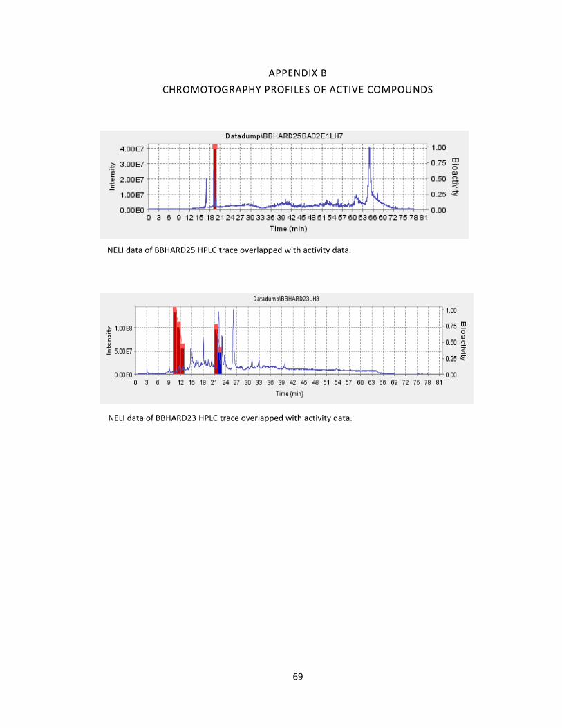
69
APPENDIX B
CHROMOTOGRAPHY PROFILES OF ACTIVE COMPOUNDS
NELI data of BBHARD25 HPLC trace overlapped with activity data.
NELI data of BBHARD23 HPLC trace overlapped with activity data.
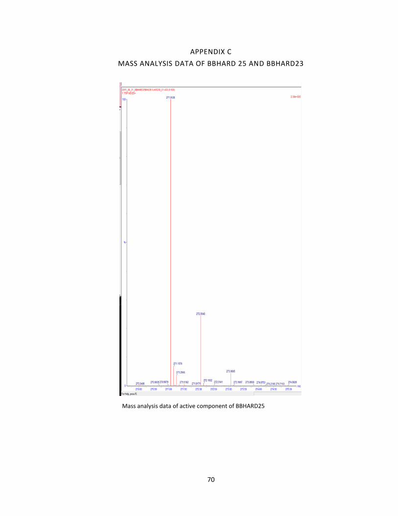
70
APPENDIX C
MASS ANALYSIS DATA OF BBHARD 25 AND BBHARD23
Mass analysis data of active component of BBHARD25

71
APPENDIX C CONT.
MASS SPECTROMETRY DATA OF BBHARD25 AND BBHARD23
Mass analysis data of active fraction from BBHARD23
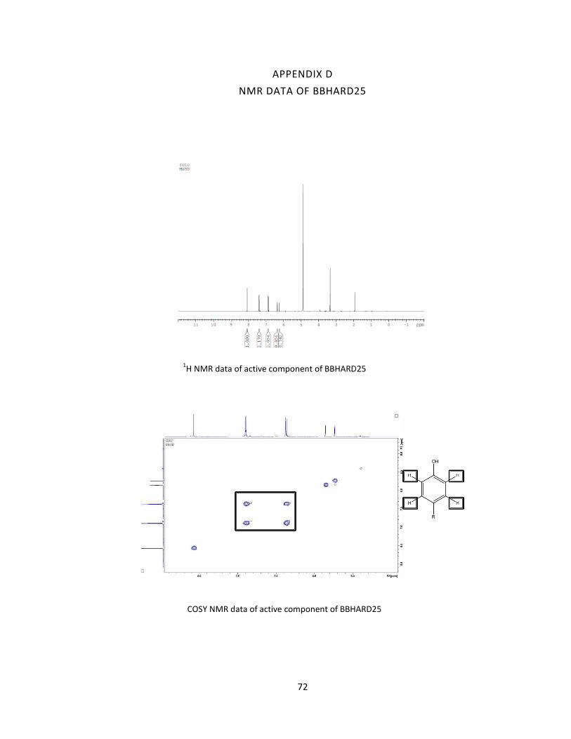
72
APPENDIX D
NMR DATA OF BBHARD25
COSY NMR data of active component of BBHARD25
1H NMR data of active component of BBHARD25
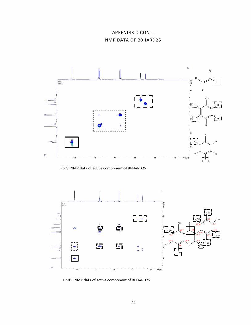
73
APPENDIX D CONT.
NMR DATA OF BBHARD25
HSQC NMR data of active component of BBHARD25
HMBC NMR data of active component of BBHARD25
74
APPENDIX E
CURRICULUM VITAE
HOLLY MARIE CARRELL
EDUCATION
Vanderbilt University, Nashville, TN
M.S. in Chemistry 2013
Thesis: “Isolation and Characterization of Anti-malarial Compounds From a Natural
Product Library”
Oakwood University, Huntsville, AL
B.S. in Biochemistry 2009
AWARDS
Provost’s Graduate Fellowship, Vanderbilt University 2009 – Present Premier Scholarship, Oakwood University 2005 – 2009
TEACHING EXPERIENCE
Vanderbilt University, Nashville, TN
Teaching Assistant – to Professor Adam List in “Organic Chemistry Lab” 2010-2012
Supervised lab experiments, held office hours, administered quizzes and lab final,
documented all grades.
Oakwood University, Huntsville, AL
Teaching Assistant – to Professor Kenneth LaiHing in “Organic Chemistry Lab” 2008
Supervised lab experiments, administered quizzes and lab final, documented all grades.
Teaching Assistant – to Professor Kenneth LaiHing in "General Chemistry Lab” 2006-
2007
Supervised lab experiments, administered quizzes and lab final, documented all grades.
RELATED EXPERIENCE
Vanderbilt University, Nashville, TN
Graduate Research assistant – to Professor David Wright 2009 – 2012

75
Experiment design and execution, data collection and analysis on isolating and
identifying antimalarial probes from a natural product library.
Oakwood University, Huntsville, AL
Research Assistant – to Professor Alexander Volkov 2007 – 2009
Design and execute experiments on the electrophysiology of the Venus Flytrap, data
collection and analysis.
Oakwood University, Huntsville, AL
Research Assistant – to Professor Christopher Perry 2006-2008
Responsible for data collection by Atomic Force Microscopy on various projects.
Donald Danforth Plant Science Center, St. Louis, MO
Research Intern 2007
Conducted research on the mode of action of plant defensins in the inhibition of
Fusarium graminearum.
PUBLICATIONS AND PAPERS
Sandlin, Rebecca D., Holly M. Carrell, and David W. Wright. "Hemozoin: Crystal
Engineering Survivability." Encyclopedia of Inorganic and Bioinorganic Chemistry.
Volkov, Alexander G., Holly Carrell, and Vladislav S. Markin. "Biologically closed electrical
circuits in Venus flytrap." Plant Physiology 149.4 (2009): 1661-1667.
Volkov, Alexander G., et al. "Electrical memory in Venus
flytrap."Bioelectrochemistry 75.2 (2009): 142-147.
Volkov, Alexander G., Holly Carrell, and Vladislav S. Markin. "Molecular electronics of
the Dionaea muscipula trap." Plant signaling & behavior 4.4 (2009): 353-354.
Volkov, Alexander G., et al. "Plant electrical memory." Plant signaling & behavior 3.7
(2008): 490-492.
Waddell, Emanuel A., et al. "Surface modification of Sylgard 184 polydimethylsiloxane
by 254nm excimer radiation and characterization by contact angle goniometry, infrared
spectroscopy, atomic force and scanning electron microscopy." Applied Surface
Science 254.17 (2008): 5314-5318.
76
REFERENCES
1. Perutz, M. F., Sci. Am. 1978, 239 (6), 92. 2. Wittenberg, B.; Wittenberg, J., Annu. Rev. Physiol. 1989, 51 (1), 857. 3. Mason, H., Science. 1957, 125 (3259), 1185. 4. Gong, W.; Hao, B.; Mansy, S. S.; Gonzalez, G.; Gilles-Gonzalez, M. A., Chan, M. K., Proc. Natl. Acad. Sci. 1988, 95 (26), 15177. 5. Nath, K. A.; Balla, J.; Croatt, A. J.; Vercellotti, G. M., Kidney Int. 1995, 47 (2), 592. 6. Nath, K.A., Vercellotti, G.M., Grande, J.P., Miyoshi, H., Paya, C.V., Manivel, J.C., Haggard, J.J., Croatt, A.J., Payne W.D., Alam, J., Kidney Int. 2001, 59 (1), 106. 7. Aft, R., Mueller, G., J. Biol. Chem. 1984, 259 (1), 301. 8. Miller, Y. I., Shaklai, N., Biochem. Mol. Biol. Int. 1994, 34 (6), 1121. Epub 1994/12/01. 9. Suliman, H. B., Carraway, M. S., Velsor, L. W., Day, B. J., Ghio, A. J., Piantadosi, C. A., Free Radical Biol. Med. 2002, 32 (3), 246. 10. Castellani, R., Harris, P., Lecroisey, A., Izadi-Pruneyre, N., Wandersman, C., Perry, G., Smith, M. A., Antioxid. Redox Signaling. 2000, 2 (1), 137. 11. Kutty, R., Daniel, R. F., Ryan, D. E., Levin W., Maines, M. D., Arch. Biochem. Biophys. 1988, 260 (2), 638. 12. Docherty, J.C., Firneisz, G.D., Schacter, B.A., Arch. Biochem. Biophys. 1984, 235 (2), 657. 13. Ryter, S.W., Tyrrell, R.M., Free Radical Biol. Med. 2000, 28 (2), 289. 14. Tenhunen, R., Marver, H.S., Schmid, R., J. Biol. Chem. 1969, 244, 6388. 15. Francis, S.E., Sullivan, D.J., Goldberg, D.E., Annu. Rev. Microbiol. 1997, 51 (1), 97. 16. Morrison, D.B., Jeskey, H.A., J. Natl. Malar. Soc. 1948, 7 (4), 259. Epub 1948/12/01.
77
17. Ball, E., McKee, R., Anfinsen, C., Cruz, W., Geiman, Q., J. Biol. Chem. 1948, 175 (2), 547. 18. Orjih, A.U., Fitch, C.D., Biochim. Biophys. Acta. 1993, 1157 (2), 270. 19. Sherman, I., Bull. World Health Organiz.1977, 55 (2–3), 265. 20. Ting, I.P., Sherman, I.W., Comp. Biochem. Physiol. 1966, 19 (4), 855. 21. Alan, F. C., Brendan, S. C., Cell. 2006, 124, 755. 22. Sherman I.W., Tanigoshi, L., Int. J. Biochem. 1970, 1 (5), 635. 23. Theakston, R.D., Fletcher, K.A., Maegraith, B.G., Ann. Trop. Med. Parasitol. 1970, 64 (1), 63. Epub 1970/03/01. 24. McCormick, G.J., Exp. Parasitol. 1970, 27 (1), 143. 25. Slomianny, C., Blood Cells. 1990, 16 (2–3), 369. Epub 1990/01/01. 26. Olliaro, P., Goldberg, D., Parasitol. Today. 1995, 11 (8), 294. 27. Goldberg, D., Slater, A., Cerami, A., Henderson, G., Proc. Natl. Acad. Sci. U. S. A., 1990, 87 (8), 2931. 28. Francis, S.E., Gluzman, I.Y., Oksman, A., Banerjee, D., Goldberg, D.E., Mol. Biochem. Parasitol. 1996, 83 (2), 189. 29. Gluzman, I.Y., Francis, S.E., Oksman, A., Smith, C.E., Duffin, K.L., Goldberg, D.E., J. Clin. Invest. 1994, 93 (4), 1602. 30. Kolakovich, K.A., Gluzman, I.Y., Duffin K. L., Goldberg, D. E., Mol. Biochem. Parasitol. 1997, 87 (2), 123. 31. Coombs, G., Goldberg, D., Klemba, M., Berry, C., Kay, J., Mottram, J., Trends Parasitol. 2001, 17 (11), 532. 32. Berry, C., Humphreys, M., Matharu, P., Granger, R., Horrocks. P., Moon, R., Certa, U., Ridley, R.G., Bur, D., Kay, J., FEBS Lett. 1999, 447 (2–3), 149. 33. Goldberg, D., Slater, A., Beavis, R., Chait, B., Cerami, A., Henderson, G., J. Exp. Med. 1991, 173 (4), 961.

78
34. Banerjee, R., Liu, J., Beatty, W., Pelosof, L., Klemba, M., Goldberg, D. E., Proc. Natl. Acad. Sci. 2002, 99 (2), 990. 35. M. F. Perutz, M.F., Edited by Stamatoyannopoulos, G., Nienhuis. A.W., Saunders, W.B., Philadelphia, 1987, 127. 36. Wyatt, D.M., Berry, C., FEBS Lett. 2002, 513 (2–3), 159. 37. Rosenthal, P.J., Nelson, R.G., Mol. Biochem. Parasitol. 1992, 51 (1), 143. 38. Sijwali, P.S., Shenai, B.R., Gut, J., Singh, A., Rosenthal, P.J., Biochem. J. 2001, 360 (2), 481. 39. Shenai, B.R., Sijwali, P.S., Singh, A., Rosenthal, P.J., J. Biol. Chem. 2000, 275 29000. 40. Shenai, B.R., Rosenthal, P.J., Mol. Biochem. Parasitol. 2002, 122 (1), 99. 41. Eggleson, K.K., Duffin, K.L., Goldberg, D.E., J. Biol. Chem. 1999, 274, 32411. 42. Gluzman, I.Y., Francis, S.E., Oksman, A., Smith, C.E., Duffin, K.L., Goldberg, D.E., J. Clin. Invest. 1994, 93 (4), 1602. 43. Fitch, C., Chevli, R., Kanjananggulpan, P., Dutta, P., Chevli, K., Chou, A., Blood. 1983, 62 (6), 1165. 44. Becker, K., Tilley, L., Vennerstrom, J.L., Roberts, D., Rogerson, S., Ginsburg, H., Int. J. Parasitol. 2004, 34 (2), 163. 45. Har-el, R., Marva, M., Chevion, M., Golenser, J., Free Radical Res. 1993, 18 (5), 279. 46. Lancisi, G.M., in ‘De Noxiis Paludum Effluvius Eorumque Remediis’, ed. Salvioni, J. M., Rome, 1717. 47. Golgi, C., Representation photographique du development des parasites de l’infection paldeene, Verhandldes X Internat. Med. Cong. Berlin Bd II abth III.200, 1890. 48. Weissbuch, I., Leiserowitz, L., Chem. Rev. 2008, 108, 4899. 49. Chen, M.M., Shi, L., Sullivan Jr, D.J., Mol. Biochem. Parasitol. 2001, 113, 1. 50. Noland, G.S., Briones, N., Sullivan Jr, D.J., Mol. Biochem. Parasitol. 2003, 130, 91.

79
51. Pagola, S., Stephens, P.W., Bohle, D.S., Kosar, A.D., Madsen, S.K., Nature 2000, 404 (6775), 307. 52. Pasternack, R.F., Munda, B., Bickford, A., Gibbs, E.J., Scolaro, L.M., J. Inorg. Biochem. 2010, 104 (10), 1119. Epub 2010/07/30. 53. Klonis, N., Dilanian, R., Hanssen, E., Darmanin, C., Streltsov, V., Deed, S., Quiney, H., Tilley, L., Biochemistry 2010, 49 (31), 6804. 54. Slater, A.F., Swiggard, W.J., Orton, B.R., Flitter, W.D., Goldberg, D.E., Cerami, A., Henderson, G.B., Proc. Natl. Acad. Sci. U. S. A. 1991, 88 (2), 325. 55. Bohle, D.S., Debrunner, P., Jordan, P.A., Madsen, S.K., Schulz, C.E., J. Am. Chem. Soc. 1998, 120 (32), 8255. 56. Bohle, D.S., Dinnebier, R.E., Madsen, S.K., Stephens, P.W., J. Biol. Chem. 1997, 272, 713. 57. Slater, A. F. G., Cerami, A., Nature 1992, 355, 167. 58. Dorn, A., Stoffel, R., Matile, H., Bubendorf, A., Ridley, R.G., Nature 1995, 374 (6519), 269. 59. Bendrat, K., Berger, B.J., Cerami, A., Nature 1995, 378 (6553), 138. 60. Goldberg, D.E., Slater, A.F., Cerami, A., Henderson, G.B., Proc. Natl. Acad. Sci. 1990, 87 (8), 2931. 61. Solomonov, I., Osipova, M., Feldman, Y., Baehtz, C., Kjaer, K., Robinson, I.K., Webster, G.T., McNaughton, D., Wood, B.R., Weissbuch, E., Leiserowitz, L., J. Am. Chem. Soc. 2007, 129, (9), 2615. 62. Trager, W., Trends Parasitol. 2003, 19 (9), 388. 63. Dorn, A., Vippagunta, S.R., Matile, H., Bubendorf, A., Vennerstron, J.L., Ridley, R. G., Biochem. Pharmacol. 1998, 55 (6), 737. 64. Egan, T.J., Mavuso, W. W., Ncokazi, K. K., Biochemistry 2001, 40 (1), 204. Epub 2001/01/05. 65. Egan, T. F., Tshivhase, M. G., Dalton Trans. 2006 42, 5024.
80
66. Stiebler, R., Hoang, A. N., Egan, T. J., Wright, D. W., Oliveira, M. F., PLoS One 2010, 5 (9), e12694. 67. Blauer, G., Akkawi, M., Arch. Biochem. Biophys. 2002, 398 (1), 7. 68. Huy, N. T., Maeda, A., Uyen, D. T., Trang, D. T. X., Sasai, M., Shiono, T., Oida, T., Harada, S., Kamei, K., Acta Trop. 2007, 101 (2), 130. 69. Jackson, K. E., Klonis, N., Ferguson, D. J. P., Adisa, A., Dogovski, C., Tilley, L., Mol. Microbiol. 2004, 54 (1), 109. 70. Pisciotta, J., Coppens, I., Tripathi, A., Scholl, P., Shuman, J., Bajad, S., Shulaev, V., Sullivan Jr, D. J., Biochem. J. 2007, 402 (Pt 1), 197. 71. Papalexis, V., Siomos, M. -A., Campanale, N., Guo, X. –G., Kocak, G., Foley, M., Tilley, L., Mol. Biochem. Parasitol. 2001, 115 (1), 77. 72. Hoang, A. N., Sandlin, R. D., Omar, A., Egan, T. J., Wright, D. W., Biochemistry 2010, 49 (47), 10107. 73. Hoang, A. N., Ncokazi, K. K., de Villiers, K. A., Wright, D. W., Egan, T. J., Dalton Trans. 2010, 39 (5), 1235. 74. Egan, T. J., Combrinck, J. M., Egan, J., Hearne, G. R., Marques, H. M., Ntenteni, S., Sewell, B. T., Smith, P. J., Taylor, D., van Schalkwyk, D. A., Walden, J. C., Biochem J. 2002, 365 (Pt 2), 343. 75. Egan, T. J., Chen, J. Y. –J., de Villiers, K. A., Mabotha, T. E., Naidoo, K. J., Ncokazi, K. K., et al. FEBS Lett. 2006, 580, 5105. 76. Downie, M. J., Kirk, K., Mamoun, C. B., Eukaryot. Cell. 2008, 7 (8), 1231. 77. Ben Mamoun, C., Prigge, S. T., Vial, H., Drug Dev. Res. 2010, 71 (1), 44. 78. Woodrow, C. J., Krishna, S., eds, ‘Molecular Approaches to Malaria: Glycolysis in Asexual-Stage Parasites’, 1st edition, American Society for Microbiology, Washington D.C., 2005. 79. Sato, S., Clough, B., Coates, L., Wilson, R. J. M., Protist 2004, 155 (1), 117. 80. Bergman, G. J., Sci. Edu. 1948, 32 (2), 93. 81. Warhurst, D., Craig, J., Adagu, I., Meyer, D., Lee, S., Malar. J. 2003, 2 (1), 26.
81
82. Krogstad, D., Gluzman, I., Kyle, D., Oduola, A., Martin, S., Milhous, W., Schlesinger, P. H., Science 1987, 238 (4831), 1283. 83. Asher, C., de Villiers, K. A., Egan, T. J., Inorg. Chem. 2009, 48 (16), 7994. 84. de Villiers, K. A., Kaschula, C. H., Egan, T. J., Marques, H. M., J. Biol. Inorg. Chem. 2007, 12 (1), 101. 85. Krogstad, D. J., Schlesinger, P. H., Biochem. Pharmacol. 1986, 35, 547. 86. Moreau, S., Perly, B., Biguet, J., Biochimie 1982, 64 (11–12), 1015. Epub 1982/11/01. 8 . O’Neill, P. M., Willock, D. J., Hawley, S. R., Bray, P. G., Storr, R. C., Ward, S. A., Park, B. K., J. Med. Chem. 1997, 40 (4), 437. 88. Cohen, S. N., Phifer, K. O., Yielding, K. L., Nature 1964, 202, 805. 89. Chou, A. D., Chevli, R., Fitch, C. D., Biochemistry 1980, 19 (8), 1543. 90. Egan, T. J., Ncokazia, K. K., J. Inorg. Biochem. 2004, 98 (1), 144. 91. Egan, T. J., Mavuso, W. W., Ross, D. C., Marques, H. M., J. Inorg. Biochem. 1997, 68 (2), 137. 92. Egan, T. J., J. Inorg. Biochem. 2006, 100 (5–6), 916. 93. Buller, R., Peterson, M. L., Almarsson, O., Leiserwitz, L., Cryst. Growth Des. 2002, 2 (6), 553. 94. Sullivan, D. J., Gluzman, I. Y., Russell, D. G., Goldberg, D. E., Proc. Natl. Acad. Sci. U. S. A. 1996, 93 (21), 11865. 95. Dorn, A., Vippagunta, S. R., Matile, H., Jaquet, C., Vennerstrom, J. L., Ridley, R. G., Biochem. Pharmacol. 1998, 55 (6), 727. Epub 1998/05/20. 96. de Villiers, K. A., Marques, H. M., Egan, T. J., J. Inorg. Biochem. 2008, 102 (8), 1660. 97. Ignatushchenko, M. A., Winter, R. W., Riscoe, M., Am. J. Trop. Med. Hyg. 2000, 62 (1), 77. 98. Winter, R. W., Cornell, K. A., Johnson, L. L., Ignatushchenko, M., Hinrichs, D. J., Riscoe, M. K., Antimicrob. Agents Chemother. 1996, 40 (6), 1408.

82
99. Ignatushchenko, M. V., Winter, R. W., Bachinger, H. P., Hinrichs, D. J., Riscoe, M. K., FEBS Lett. 1997, 409 (1), 67. 100. Kelly, J. X., Winter, R., Riscoe, M., Peyton, D. H., J. Inorg. Biochem. 2001, 86 (2 -3), 617. 101. Kelly, J. X., Winter, R., Peyton, D. H., Hinrichs, D. J., Riscoe, M., Antimicrob. Agents Chemother. 2002, 46 (1), 144. 102. Group, C. C. R., J. Tradit. Chin. Med. 1982, 2, 3. 103. Meshnick, S. R., Taylor, T. E., Kamchonwongpaisan, S., Microbiol. Rev. 1996, 60, 301. 104. Posner, G. H., Oh, C. H., Wang, D., Gerena, L., Milhous, W. K., Meshnick, S. R., Asawamahasadka, W., J. Med. Chem., 1994, 37, 1256. 105. Kamchonwongpaisan, S., Meshnick, S. R., Gen. Pharmacol. 1996, 27, 587. 106. Meshnick, S., Yang, Y., Lima, V., Kuypers, F., Kamchonwongpaisan, S., Yuthavong, Y., Antimicrob. Agents Chemother. 1993, 37 (5), 1108. 107. Zhang, F., Gosser Jr, D. K., Meshnick, R., Biochem. Pharmacol. 1992, 43, 1805. 108. Bhisutthibhan, J., Pan, X. Q., Hossler, P. A., Walker, D. J., Yowell, C. A., Carlton, J., Dame, J. B., Meshnick, S. R., J. Biol. Chem. 1998, 273, 16192. 109. Bhisutthibhan, J., Meshnick, S. R., Antimicrob. Agents Chemother. 2001, 45, 2397. 110. Ellis, D. S., Li, Z. L., Gu, H. M., Peters, W., Robinson, B. L., Tovey, G., Warhurst, D. C., Ann. Trop. Med. Parasitol. 1985, 79, 367. 111. Jiang, J. B., Jacobs, G., Liang, D. S., Aikawa, M., Am. J. Trop. Med. Hyg. 1985, 34, 424. 112. Maeno, Y. T., Toyoshima, T., Fujioka, H., Ito, Y., Meshnick, S. R., Benakis, A., Milhous, W. K., Aikawa, M., Am. J. Trop. Med. Hyg. 1993, 49, 485. 113. Johnson, J. D., Dennull, R. A., Gerena, L., Lopez-Sanchez, M., Roncal, N. E., Waters, N. C., Antimicrobial. Agents Chemother. 2007, 51 (6), 1926. 114. Lucumi, E., Darling, C., Jo, H., Napper, A. D., Chandramohanadas, R., Fisher, N., Shone, A. E., Jing, J., Ward, S. A., Biagini, G. A., DeGrado, W. F., Diamond, S. L., Greenbaum, D. C., Antimicrobial. Agents Chemother. 2010, 54 (9), 3597.

83
115. Baniecki, M. L., Wirth, D. F., Clardy, J., Antimicrobial. Agents Chemother. 2007, 51 (2), 716. 116. Corbett, Y., Herrera, L., Gonzalez, J., Cubilla, L., Capson, T. L., Coley, P. D., Kursar, T. A., Romero, L. I., Ortega-Barria, E., Am. J. Trop. Med. Hyg. 2004, 70, 119. 117. Rush, M. A., Baniecki, M. L., Mazitschek, R., Cortese, J. F., Wiegand, R., Clardy, J., Wirth, D. F., Antimicrobial. Agents Chemother. 2009, 53 (6), 2564. 118. Kurosawa, Y., Dorn, A., Kitsuji-Shirane, M., Shimada, H., Satoh, T., Matile, H., Hofheinz, W., Masciadri, R., Kansy, M., Ridley, R. G., Antimicrobial. Agents Chemother. 2000, 44 (10), 2638. 119. Koehn, F. E., Carter, G. T., Nature Reviews Drug Discovery 2005, 4, 206-220. 120. Bérdy, J., J. Antibiot. 2005, 58, 1-26. 121. Fenical, W., Jensen, P. R., Palladino, M. A., Lam, K. S., Lloyd, G. K., Potts, B. C., Bioorganic and Medicinal Chemistry 2009, 17, 2175-2180. 122. Carter, M., Phelan, V., Sandlin, R., Bachmann, B., Wright, D., Combinatorial Chemistry & High Throughput Screening 2010, 13, 285-292. 123. Mann, J., Natural Product Reports 2001, 18, 417-430. 124. Lam, K. S., Current Opinion In Microbiology 2006, 9, 245-251. 125. Chabala, J. C., Mrozik, H., Tolman, R. L., Eskola, P., Lusi, A., Peterson, L. H., Woods, M. F., Fisher, M. H., Campbell, W. C., Journal of Medicinal Chemistry 1980, 23, 1134-1136. 126. Fowler, A., Swift, D., Longman, E., Acornley, A., Hemsley, P., Murray, D., Unitt, J., Dale, I., Sullivan, E., Coldwell, M., Analytical Chemistry 2002, 308, 223-231.
127. Dixon, R.A, Ferreira, D., Phytochemistry 2002, 60, 205-211.
128. Cunha-Rodrigues, M., Portugal, S., Prudencio, M., Goncalves, L. A., Casalou, C., Buger, D., Sauerwein, R., Haas, W., Mota, M. M., PLoS ONE 2008, 3 (7), e2732.
129. Gazarini, M. L., Garcia, C. R. S., Braz. J. Med. Biol. Res. 2003, 36, 1465-1469.

84