Embed Size (px)
Citation preview
47
CHAPTER 4:
Isolation of compounds from Elaeodendron
transvaalense extract
4.1 Introduction
Extracts from Elaeodendron transvaalense (Burtt Davy) (Celastraceae)
have been used in traditional medicine by the Vhavenda people of South
Africa (Limpopo province) to treat coughs, diarrhoea, stomach ailments,
herpes and sexually associated diseases. Stem bark is mostly used to
prepare infusions and decoctions (Mabogo, 1990). Other medicinal uses of E.
transvaalense are shown in Table 4.1. Traditional healers prescribe it
presently to people who are suffering from HIV/AIDS (Bessong et al., 2005).
Dimethyl–1,3,8,10–tetrahydroxy–9- methoxypeltogynan and three pentacyclic
triterpenes have been isolated from its bark which is also reported to contain
13.4 % catechol tannin (Hutchings,1996).
Other species belonging to the same family (Celastraceae) have been
used in the Amazonian region against cancer, rheumatism and inflammation
(Nakagawa et al., 2004). Previous reports have shown that species from the
Celastraceae family contain biologically active metabolites with antimicrobial
and cytotoxic activities (Sansores-Peraza et al., 2000). The aim of this part of
study was to isolate compounds from E. transvaalense .

48
4.1.1 Plant description
E. transvaalense is a shrub or small tree (about 5 m high) occurring in forests
and quite often on rocky outcrops in mountainous regions. The bark is
generally smooth and has a grey colour. Leaves are often clustered on
reduced lateral shoots, oblong in shape, about 50 mm long and 20 mm wide.
The leaf margin is sometimes toothed. The flowers are greenish in colour and
produce oblong, yellow to dark orange, berry-like fruits, which are edible
(Figure. 4.1).
a b
Figure 4.1 (a) Bark and (b) branches of E. transvaalense (Van Wyk et al.,
1997).
The species is widely distributed in the north-eastern parts of South Africa.
It also occurs along the coastal parts of KwaZulu-Natal and in Mpumalanga,
Gauteng and the Limpopo province (Van Wyk, 2000).

49
4.1.2 Medicinal uses
E. transvaalense is used as remedy for several diseases (Table 4.11). The
bark is extensively used for cleaning of the stomach and used as an enema
for stomach ache, diarrhoea and fever (Mabberley, 1981).
Table 4.1: Other medicinal uses of E. transvaalense
Disease Place Preparation References
Menorrhagia Zimbabwe Infusion Gelfand, 1985
Haemorrhoids South Africa Unspecified Hutchings , 1996
Van Wyk & Gericke, 2000
Stomach cramps
Diarrhoea
South Africa
South Africa
Decoction
Decoction
Pujol, 1988
Mabberley, 1981 Van Wyk, 1997
Herpes simplex South Africa Decoction Felhabert, 1997 Mabogo, 1990
Herpes zoster South Africa Decoction Felhabert, 1997
Stroke South Africa Decoction Felhabert, 1997
Gout South Africa Decoction Felhabert, 1997
Rash
HIV/AIDS
Southern Africa
South Africa
Infusion
Infusion
Van Wyk, 2000
Bessong et al., 2005

50
4.1.3 Chemistry
Elaeodendron species are rich in gallotannins, proanthocyanidins and a
few other phenolic compounds like elaeocyanidin (Figure 4.2) have been
isolated from this species (Van Wyk et al., 1997).
a b
O
O
OH
OH
OH
OH
OMe
OH
CH2OH
O
HO H
c d e
Figure 4.2 Compounds isolated from E. transvaalense: (a) cassinine, (b)
elaeocyanidin, (c) 6R, 13R-11, 11-dimethyl-1, 3, 10-tetra-hydroxy-9-
methoxypeltogynan, (d) canophyllol and (e) 6-�-hydroxy-lup-20(30)-en-3-one
(Drewes & Mashimbye, 1993, Drewes et al., 1991).
O H
O
O O
O H
O H
O H
O
O O O
O O
N
O
O
O
O O
O
O
O
O O
O

51
4.2 Materials and methods
4.2.1 Plant material
Stem bark of E. transvaalense was collected in Venda (Northern
Limpopo). A voucher specimen is preserved in the HGWJ Schweickerdt
herbarium at the University of Pretoria (Tshikalange 092524).
4.2.2 Preparation of extracts
In unreported results, different fresh extracts (acetone, chloroform, ethyl
acetate and ethanol) of E. transvaalense were tested for their ability to inhibit
NF-κB and Tat proteins. The ethanol extract exhibited the best activity and led
us to isolate compounds from this extract.
Stem bark of E. transvaalense was collected and left to dry at room
temperature for two weeks. The dried powder stem bark was placed in a
container and soaked in ethanol. The container was closed and left in a dark
cupboard for three days at room temperature before the extract was filtered
and concentrated to dryness under reduced pressure (40 0C). The residue
was soaked again in ethanol and filtered. The filtrates were dried with a rotary
evaporator to give a total mass of 150 g (extract).
4.2.3 Isolation and identification of compounds
A 10 cm diameter glass column (Figures 4.3 and 4.4) was filled with 1.5 kg
silica gel. The extract (120 g) was dissolved in a minimal amount of solvent

52
and mixed with 200 g silical gel. The column was eluted with a solvent
gradient of hexane: ethyl acetate in 100:0 to 0:100 ratios. The column was
then washed with ethyl acetate:methanol (9:1) and 100 % methanol. 45
fractions of 50 ml each were collected; fractions containing the same
compounds as determined by TLC plates were combined and concentrated to
dryness under reduced pressure. TLC plates of 11 pooled fractions (A-K)
were developed with hexane: ethyl acetate 9:1, 7:3 and 3:7. Fraction I yielded
a pure compound (1). Other fractions were crystallized and yielded 5 pure
compounds (2, 3, 5, 6 & 7). TLC plates were examined under UV light (254
and 366 nm) after development and also dipped in vanillin (15 g vanillin, 500
ml ethanol and 10 ml concentrated 98 % sulphuric acid) and heated to detect
compounds not absorbing UV. Fractions G and H were combined and
subjected to a second column eluted with a solvent gradient of hexane: ethyl
acetate in 100:0 to 0:100 ratios which resulted in isolation of compound 4.
4.3 Results and discussion
The column chromatography (Figure 4.3) yielded 45 fractions (Figure
4.5) which were pooled together according to their TLC profile and resulted in
11 pooled fractions (Figure 4.6). From these pooled fractions seven
compounds were isolated (Figure 4.7).

53
Figure 4.3 Column chromatography

54
Figure 4.4: Schematic presentation of isolation steps followed.
1.2 kg of E. transvaalense stem bark powder
Extraction in ethanol (150 g)
Silica column 1 collected 45 fractions ( 11 pooled fractions) fractions)fractions)
Fraction B (48 mg) Fraction C (27 mg)
Fraction D (40 mg)
Fraction F (47 mg)
Fraction I (50 mg) Compound 5
Fraction G &H (40 mg) Compound 7
Preparative TLC
Compound 6
Crystallized
Compound 3
Crystallized
Compound 2
Compound 1
Precipitated
Silica column 2
Compound 4
HO
HO
1O
HO
2
O
OMeOH
OH
OH
HO
5
OH
OC O
CHO
OHCOOH
HO
CH3
OH
COOCH3
CH3
7
O OCH3
CH3HO
6
H3COH O
3
OH
HO
4

55
Figure 4.5 Fractions from the first silica column on TLC plates sprayed with Vanillin reagent. Plate A and B developed with hexane:
ethyl acetate (9:1 and 7:3), Plate C and D fractions developed with hexane: ethyl acetate (1:9).
Plate : A Plate: B
Plate: D
Plate: C
144
25 1
45 25 35

56
Figure 4.6 The 11 pooled fractions (silica column 1) TLC plates sprayed with
Vanillin reagent.
Figure 4.7 Isolated compounds as seen on TLC plates sprayed with Vanillin
reagent.
1 4 3 2
5 7
6

57
4.3.1 Triterpenoids isolated
Fraction F contained a white precipitate that was washed with ethyl
acetate to give one pure compound 1 (Figure 4.7) as determined by TLC and
other spectroscopic methods (Table. 4.2).
lup-20(30)-ene-3,29-diol, (3�)-(9Cl) lup-20(29)-ene-30-hydroxy-(9Cl)
� – taraxastanonol �-sitosterol
Figure 4.8 Structures of isolated triterpenes.
HO
HO
1
O
HO
2
HO
4
O
3
OH

58
Compound 1 showed in its 1H-NMR (Figure 4.8) spectrum a triterpenoid
pattern with six methyl singlets at δH 0.76 , 0.78 , 0.83 , 0.95, 0.97 and 1.03,
two olefinic protons at δH 4.95 (d 1.4 H), 4.88 (s) attached to carbon at δC
106.9, both protons correlated in HMBC (Figure 4.10) with the carbon at δC
65.1, another two protons attached to a oxygen bearing carbon at δH 4.11 (d,
J=14.8 Hz), 406 (d, J=14.8 Hz), and a proton δH 3.16 (dd, J=10.4, 5.7 Hz) on
carbon at δC 79.0. The 13C – NMR spectrum of compound 1 is shown in
Figure 4.9. The data obtained with 2D NMR experiments HMBC, HMQC,
COSY and NOESY (Figures 4.10 & 4.11) supported the structure for
compound 1 (Abdel-Mogib, 1999). This compound has been previously
isolated from the whole plant extract of Daphne oleoides, which is used as a
purgative and the infusion of the leaves is used to treat gonorrhoea and
applied to abscesses (Ullah et al., 1999).
Fractions C and D were crystallized to give compounds 2 and 3 as shown
previously in Figure 4.3. Both compounds were obtained as white crystals, but
showed different colours on TLC plates. Compound 2 showed similar signals
in 1H-NMR (Figure 4.12) with the previous compound except for the
disappearance of the C-3 proton and the appearance of a carbonyl carbon in
13 C-NMR (Figure 4.13) at δC 218.2, which indicate the enzymatic oxidation of
the hydroxyl group into the corresponding ketone group in compound 2. This
was supported by the HMBC correlation between C-3 and the H-23 and H-24
methyls and previously reported data (Fang et al., 1984). Compound 3’s
structure was supported by data published by Anjaneyulu et al. (1999) and
Hinge et al. (1966). This compound was also isolated from resin of Protium

59
heptaphyllum and has shown analgesic effects (Susunaga et al., 2001 &
Rudiger et al., 2007).
Fractions G and H were combined and subjected to a second silica gel
column 2 and one pure compound (4) was obtained. This compound was
identified as β - sitosterol when spectra were compared to published data
(Prozesky, 2004). 13C – NMR data (Table 4.2) of all the triterpenes isolated
form E. transvaalense was also supported by other published data (Scleich et
al., 2006, Sasunaga et al., 2001, Mahato & Kundu, 1994, Ullah et al., 1999 &
Burns et al., 2000).

60
Table 4.2: 13C – NMR data of tritepernoids isolated compounds (1-3).
C 1 δC
2 δC
3 δC
1 38.7 t 39.6 39.6
2 27.4 t 34.1 33.8
3 79.0 d 218.3 218.0
4 38.9 s 47.3 47.4
5 55.3 d 54.9 54.8
6 18.3 t 19.7 19.8
7 34.3 t 33.6 34.1
8 40.9 s 40.8 41.4
9 50.4 d 49.7 49.1
10 37.1 s 36.9 36.7
11 21.0 t 21.6 22.2
12 26.7 t 26.7 26.7
13 38.0 d 31.8 39.0
14 43.0 s 42.9 43.2
15 27.4 27.4 26.8
16 35.5 t 35.4 38.1
17 43.0 s 43.0 35.6
18 49.0 d 48.8 47.3
19 43.8 d 43.8 38.7
20 154.8 s 154.7 73.5
21 31.8 t 31.8 35.4
22 39.9 t 39.8 37.7
23 28.0 q 26.7 26.8
24 15.4 q 21.0 21.1
25 16.1 q 16.0 16.2
26 16.0 q 15.8 16.1
27 14.6 q 14.5 14.8
28 17.7 q 17.7 17.8
29 106.9 t 106.8 17.8
30 65.1 t 65.0 30.3

61
Figure 4.9 1H – NMR spectrum of Compound 1
HO
HO
1

62
Figure 4.10 13C – NMR spectrum of Compound 1
63
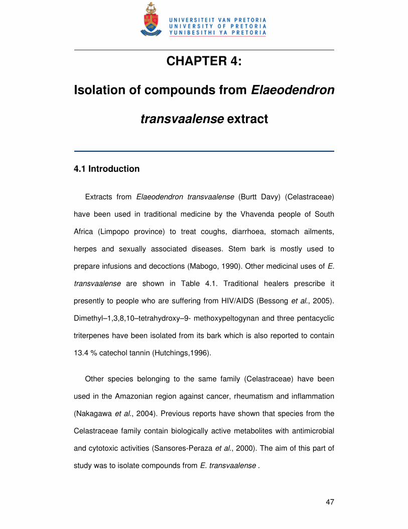
Figure 4.11 HMBC spectrum of Compound 1

64
Figure 4.12 NOESY spectrum of Compound 1

65
Figure 4.13 1H – NMR spectrum of Compound 2
O
HO
2

66
Figure 4.14 13C – NMR spectrum of Compound 2

67
Figure 4.15 HMBC spectrum of Compound 3

68
Figure 4.16 HMQC spectrum of compound 3
O
3
OH

69
4.3.2 Methylepigallocatechin
Fraction I contained one pure compound 5, (Figure 4.16) as determined by
TLC and other spectroscopic methods. It was obtained as a brown powder
(50 mg). The 1H-NMR (Table 4.3 and Figure 4.17) spectrum showed four
aromatic protons at δH 5.71 (d, J=2.3), 5.89 (d, J=2.3), 6.40 (2H, d, J= 0.6),
two methane protons at δH 4.68 (d, J=0.8) and 3.45 (s br), methylene protons
at δH 2.45 (J=4.4 ) and 2.70 (J=3.2 ), and one methoxyl group at δH 3.65 (s).
The 13C-NMR spectra (Figure 4.18) indicated the presence of two methine
carbons attached to an oxygen function (δC 78.6, 65.6), a methylene carbon
(δC 28.8 t), 12 aromatic carbons δC 156.3 (s), 95.8 (d), 157.2 (s), 94.8 (d),
156.9 (s), 99.2 (s), 135.6 (s), 106.8 (x2C, d each), 150.7 (x2c, s each), 135.2
(s), and a methoxyl carbon (δC 60.3 q). The coupling constant between
protons at δH 4.68 and 3.45 is 2.3=Hz which indicated ββ relative
configuration. The above spectroscopic data indicated that compound 5 is (-
)4’-O-methylepigallocatechin which have been isolated from the same genus
previously (Drewes & Mashimbywe, 1993). Hussein et al. (1999) reported
anti-HIV -1-protease activity for this compound. .
O
OMeOH
OH
OH
HO
OH
(-)4’-O-methylepigallocatechin (5)
Figure 4.17 Structure of compound 5

70
Table 4.3 1H – NMR and 13C – NMR data of compound 5.
Position δH δC
2 4.68 d (J= 0.8) 78.6 d
3 3.45 (J= 3.9) 65.6 d
4 a
4 b
2.73 dd (J=3.2)
2.87 dd (J= 4.4
28.8 t
5 156.3 s
6 5.71 d (J=2.3) 95.8 d
7 157.2 s
8 5.89 d (J= 2.3) 94.8 d
9 156.9 s
10 99.2 s
1′ 135.6 s
2’, 6’ 6.40 d (J= 0.6) 106.8 d
3’,5’ 150.7 s
4’ 3.65 135.2 s
OMe 60.3 q

71
Figure 4.18 1H – NMR spectrum of Compound 5

72
Figure 4.19 13 C – NMR spectrum of Compound 5

73
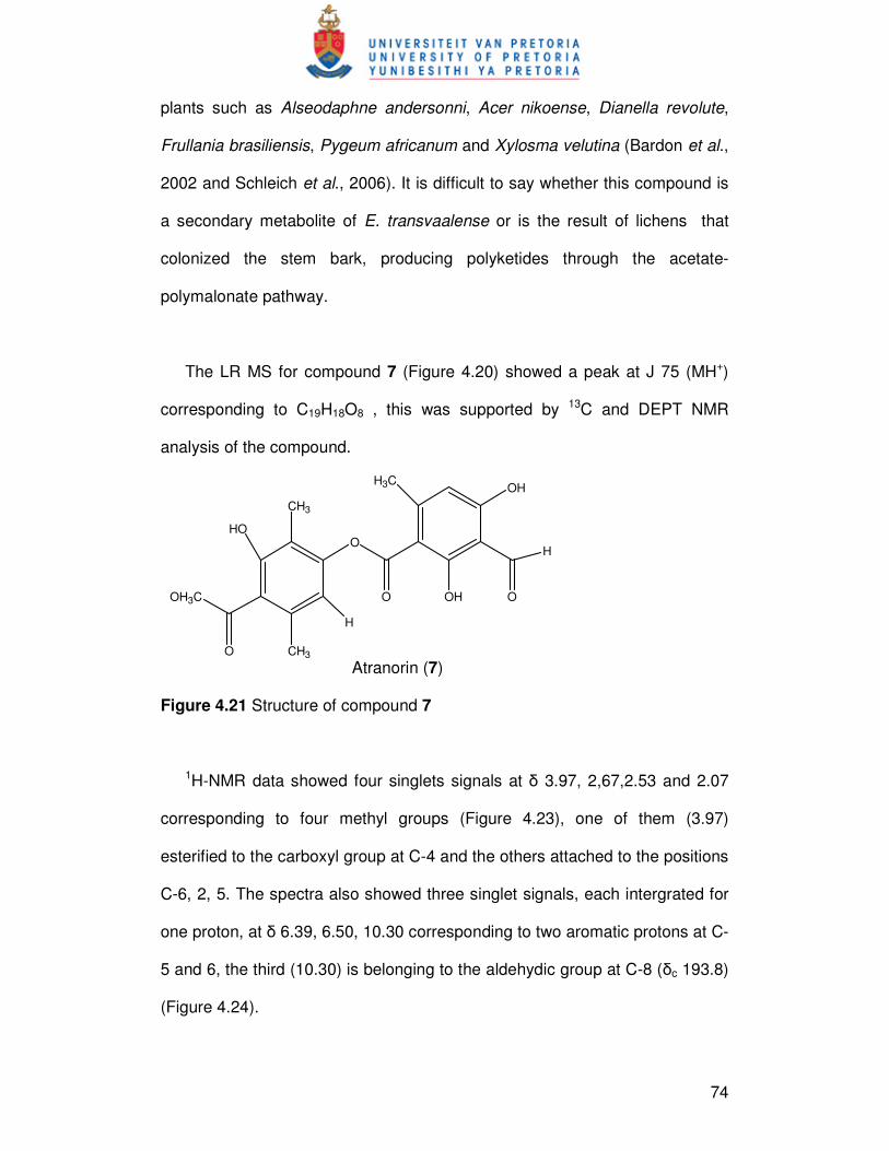
4.3.3 Phenolic derivative and depside
Compounds 6 and 7 (Figures 4.19 and 4.20) were obtained through
preparative TLC of fraction B which was developed in
hexane:chloroform:water (10:4:1). The plate was half developed, dried and
redeveloped fully.
Atraric acid (6) Figure 4.20 Structure of compound 6
Compound 6 was formed as crystals. The 13C-NMR (Figure 4.22) revealed
the presence of 10 carbons, including six of which five where substituted on a
benzene ring, including two hydroxyls (δC 163.1, 158.2) and one methylated
(δC 51.8) carboxyl group (δC
172.6), in addition to two aromatic methyl groups
(δC 24.1, 7.6) 1H-NMR (Figure 4.21) spectral data showed two aromatic
methyls δH 2.43, 2.07 singlets and an aromatic proton at δH 6.19.
The foregoing data indicated that the isolated compound is methyl 2,4-
dihydroxy-3,6-dimethylbenzoate a phenolic derivative (atraric acid). This was
confirmed by 2D-NMR spectra e.g. HMBC, HMQC, COSY. The NMR spectral
data are in agreement with those previously reported (Gormann et al., 2003;
Lee et al., 2001).The compound was isolated for the first time from lichens
(Cooke & Down,1971), however it was reported to be a constituent in higher
O OCH3
CH3HO
H3COH
74
plants such as Alseodaphne andersonni, Acer nikoense, Dianella revolute,
Frullania brasiliensis, Pygeum africanum and Xylosma velutina (Bardon et al.,
2002 and Schleich et al., 2006). It is difficult to say whether this compound is
a secondary metabolite of E. transvaalense or is the result of lichens that
colonized the stem bark, producing polyketides through the acetate-
polymalonate pathway.
The LR MS for compound 7 (Figure 4.20) showed a peak at J 75 (MH+)
corresponding to C19H18O8 , this was supported by 13C and DEPT NMR
analysis of the compound.
CH3
HO
O CH3
H
O
O
H3C
OH
OH
H
OOH3C
Atranorin (7) Figure 4.21 Structure of compound 7
1H-NMR data showed four singlets signals at � 3.97, 2,67,2.53 and 2.07
corresponding to four methyl groups (Figure 4.23), one of them (3.97)
esterified to the carboxyl group at C-4 and the others attached to the positions
C-6, 2, 5. The spectra also showed three singlet signals, each intergrated for
one proton, at � 6.39, 6.50, 10.30 corresponding to two aromatic protons at C-
5 and 6, the third (10.30) is belonging to the aldehydic group at C-8 (�c 193.8)
(Figure 4.24).

75
This data indicated the compound to be the depside, atranorin as depicted
in Figure 4.20, and is in agreement with those data reported for the same
compound. Also this was supported by 2D NMR data, COSY, HSPC, HMBC
and NOESY (Figures 4. 23 and 4.24). Compound 7 was also isolated
originally from lichens (Santos et al., 2004) but recent literature reported it’s
presence in higher plants as well (Athukoralage et al., 2001).

76
Figure 4.22 1H – NMR spectrum of Compound 6

77
Figure 4.23 13 C – NMR spectrum of Compound 6

78
Figure 4.24 1H – NMR spectrum of Compound 7

79
Figure 4.25 13 C – NMR spectrum of Compound 7

80
4.4 References
ABDEL-MOGIB, A. 1999. A lupine triterpenoid from Maerua oblingifolia.
Phytochemistry 51: 445-448.
ANJANEYULU, V., SATYANARAYANA, P., VISWANADHAM, K.N., JYOTHI,
V.G., RAO, K.N. & RADHIKA, P. 1999. Triterpenoids from Mangifera
indica. Phytochemistry 50: 1229-1236.
ATHUKORALAGE, P.S., HERATH, H.M.T.B., DERANIYAGALA, S.A. &
WEERASINGHE., P.A. 2001. Antifungal constituents from Gordonia
dassanayakei. Fitoterapia 72: 565-567.
BARDON, A., MITRE, G.B., KAMIYA, N., TOYOTA, M. & ASAKAWA , Y.
2002. Eremophilanolides and other constituents from the Argentine
liverwort Frullania brasiliensis. Phytochemistry 59: 205-213.
BESSONG, P.O., OBI, C.L., ANDRÉOLA, M., ROJAS, L.B., POUYSÉGU, L.,
IGUMBOR, E.,MEYER, J.J.M., QUIDEAU, S & LITVAK, S. 2005. Evaluation
of selected South African medicinal plants for inhibitory properties against
human immunodeficiency virus type 1 reverse transcriptase and
integrase. Journal of Ethnopharmacology 99: 83-91.
BURNS, D., REYNOLDS, W.F., BUCHANAN, G., REESE, P.B. & ENRIQUEZ,
R.G. 2000. Assignment of 1H and 13C spectra and investigation of hindered

81
side-chain rotation in lupeol derivatives. Magnetic resonance in chemistry.
38:488-493.
COOKE, R.G, & DOWN, J.G. 1971. Colouring matters of Australian plants.
XVI. Minor constituents of Dianella revolute and Stypandra grandis.
Australian Journal of Chemistry 24: 1257-1265.
DREWES, S.E. & MASHIMBYE, M.J. 1993. Flavonoids and triterpenoids from
Cassine papillosa and the absolute configuration of 11,11-Dimethyl-
1,3,8,10-Tetra-Hydroxy-9-Methoxypeltogynan. Phytochemistry 32: 1041-
1044.
DREWES, S.E., MASHIMBYE, M.J., FIELD, J.S. & RAMESAR, N. 1991.
11,11-Dimethyl-1,3,8,10-Tetrahydroxy-9-Methoxypeltogynan and three
pentacyclic triterpenes from Cassine transvaalensis. Phytochemistry 30:
3490-3493.
FANG, S., BERRY, D.E., LYNN, D.G., HECHT, S.M., CAMPBELL, J. & LYNN,
W.S. 1984. The chemistry of toxic principles from Maytenus memerosa.
Phytochemistry. 23: 631-633.
FELHABERT, T. 1997. South African traditional healers' primary health care
handbook Cape Town : Kagiso.

82
GELFAND, M. 1985. The traditional medical practitioner in Zimbabwe. Gweru:
Mambo press.
GORMANN, R., KALOGA., LI, X.,. FERREIRA , D., BERGENTHAL, D. &
KOLODZIEJ, H. 2003. Furanonaphthoquinones, atraric acid and a
benzofuran from the stem barks of Newbouldia laevis. Phytochemistry 64:
583-587.
HINGE, V.K., PAKNIKAR, S.K., DAS, K.G., BOSE, A.K. &
BHATTACHARYYA, S.C. 1966. Terpenoids – LXXXVI structure of Epi - ψ
- Taraxastanonol and epi-ψ-Taraxastanediol. Tetrahedron 22: 2861-2868.
HUTCHINGS, A. 1996. Zulu medicinal plants-an inventory. University of Natal
press, Pietermaritzburg.
HUSSEIN, G., MIYASHIRO, H., NAKAMURA,N., HATTORI, M., KAWAHATA,
T., OTAKE, T., KAKICHU, N & SHIMOTOHNO, K. 1999. Inhibitory effects
of Sudanese plant extracts on HIV-1 replication and HIV-1 protease.
Phytotherapy Research 13: 31-36.
LEE, S., CHANG, S. & CHEN, C. 2001. Chemical constituents from
Alseodaphne andersonii. Journal of Natural products 64: 1548-1551.
MABOGO, D.E.N. 1990. The ethnobotany of the Vhavenda. M.Sc. thesis,
University of Pretoria.

83
MABBERLEY, D.J. 1981. The plant-book: a portable dictionary of the higher
plants. Cambridge University Press. New york
MAHATO, S.B. & KUNDU, A.P. 1994. 13 C NMR spectra of pentacyclic
triterpenoids – a compilation and some salient features. Phytochemistry 6:
1517-1575.
NAKAGAWA, C., TAKAISHI, Y., FUJIMOTO, Y., DUQUE, C., GARZON, C.,
SATO, M., OKAMOTO, M., OSHIKAWA, T. & AHMED, S.U. 2004.
Chemical constituents from the Colombian medicinal plant Maytenus
laevis. Journal of Natural Products 67: 1919-1924.
PALGRAVE, K.C. 1977. Trees of Southern Africa. Struik Publishers. Cape
Town. South Africa.
PUJOL, J. 1988. The herbalist handbook African flora medicinal plants.
Durban.
PROZESKY, E.A. 2004. Antiplasmodial-and chloroquine resistance reversal
properties of a new diterpene from Croton steenkampianus. PhD thesis,
University of Pretoria, Pretoria, South Africa.
RUDIGER, A.L., SIAN, A.C. & VEIGER JUNIOR, V.F. 2007. The chemistry
and Pharmacology of the South America genus Protium Burm. F.
(Burseraceae). Pharmacognosy Reviews 1: 93-104.

84
SANTOS, L.C., HONDA, N.K., CARLOS, I.Z. & VILEGAS, W. 2004.
Intermediate reactive oxygen and nitrogen from macrophages induced by
Brazilian lichens. Fitoterapia 75: 473-479.
SANSORES-PERAZA, P., ROSADO-VALLADO, M., BRITO-LOEZA, W.,
MENA-REJON, G.H. & QUIJANO, L. 2000. Cassine, an antimicrobial
alkaloid from Senna racemosa, Fitoterapia 71: 690-692.
SCHLEICH, S., PAPAIOANNOU, M., BANIAHMAD, A. & MATUSCH, R. 2006.
Activity-guided isolation of an Antiandrogenic compound of Pygeum
africanum. Planta Medica 72: 547-551.
SUSUNAGA, G.S., SIANI, A.C., PIZZOLATTI, M.G., YUNES, R.A. &
MONACHE, F.D. 2001. Triterpenes from the resin of Protium
heptaphyllum. Fitoterapia 72: 709-711.
ULLAH, N., AHMED, Z., AHMED, S., MUHAMMAD, P. & MALIK, A. 1999. A
pentacyclic triterpene from Daphne oleoides. Phytochemistry. 50: 839-841.
VAN WYK, B-E., VAN OUDTSHOORN, B & GERICKE, N. 1997. Medicinal
plants of South Africa. Pretoria: Briza publications.
VAN WYK, B-E & GERICKE, N. 2000. People’s plants A guide to useful plants
of Southern Afica. Pretoria: Briza publications.